Contributors to Volume X X X V l Article numbers
are in parentheses following, the names Affiliations listed are curren...
14 downloads
820 Views
37MB Size
Report
This content was uploaded by our users and we assume good faith they have the permission to share this book. If you own the copyright to this book and it is wrongfully on our website, we offer a simple DMCA procedure to remove your content from our site. Start by pressing the button below!
Report copyright / DMCA form
Contributors to Volume X X X V l Article numbers
are in parentheses following, the names Affiliations listed are current.
of contributors.
A. M. AKBAR (2), Department o] Endo-
FRANCESCO BRESCIANI (30), Institute o]
crinology, Cook County General Hospital, Chicago, Illinois PIERRE ALLOUGH (11), Unitd de Recherches sur le Mgtabolisme Moldculaire de l'Institut National de la Santd et de la Recherche Mgdicale, D~partment de Chimie Biologique, Universit$ Paris-Sud, Bic$tre, France J. N. ANDERSON (25), Department o] Biological Sciences, Stan]ord University, Stan]ord, Calilornia FERNANDO ARIAS (33), Departments o] Obstetrics, Gynecology, and Biological Chemistry, Washington University School o] Medicine, St. Louis, Missouri MICHEL ATGER (11). Unit$ de Recherches sur le M~tabolisme Mol~culaire de l'Institut National de la Santg et de la Recherche M$dicale, Dgpartement de Chimie Biologique, Universit~ Paris-Sud, Bic~tre, France C. WAYNE BARDIN (6), Departments o] Medicine, Physiology and Comparative Medicine, Pennsylvania State University, Hershey, Pennsylvania E. E. BAULIEU (29), Unit~ de Recherches sur le Mdtabolisme Molgculaire et la Physio-Pathologie des Stgroides de l'Institut National de la Santg et de la Recherche Mddicale, Dgpartement de Chimie Biologique, Universitg ParisSud, Bic~tre, France JOHN D. BAXTER (19, 20), Metabolic Research Unit and Department o] Medicine, University o] CaIi]ornia, San Francisco, Cali]ornia M. M. BOUTON (29), Roussel-UCLAF, Romainville, France H. LEON BRADLOW (9, 44), The Institute ]or Steroid Research, Montefiore Hospital and Medical Center, Bronx, New York P. I. BRECHER (23), Department o] Medicine, Boston University School o] Medicine, Boston, Massachusetts
General Pathology, 1st Medical Faculty, University o] Naples, Naples, Italy RICHARD E. BULLER (27), Department o] Cell Biology, Baylor College o] Medicine, Houston, Texas LESLIE P. BULLOCK (6), Department o] Medicine, Division o] Endocrinology, Milton S. Hershey Medical Center, Hershey, Pennsylvania ELIZABETH P. BURROWS (34), Department o] Chemistry, Vanderbilt University, Nashville, Tennessee ROBERT M. BURTON (8), Department o] Obstetrics and Gynecology, University o] Louisville School o] Medicine, Louisville, Kentucky EVANGELINA CASTA~EDA (4a), The Ben May Laboratory ]or Cancer Research, The University o] Chicago, Chicago, Illinois M. G. CATELLI (29), Unit~ de Recherches sur le Mdtabolisme MolSculaire et la Physio-Pathologie des Stgroides de l'Institut National de la Santd et de la Recherche M~dicale, Ddpartement de Chimie Biologique, Universitd Paris-Sud, Bic~tre, France RALPH R. CAVALIERI (12), Department o] Medicine, University o] Cali]ornia and Veterans Administration Hospital, San Francisco, Cali]ornia GARY C. CHAMNESS (16), Department o] Medicine, University o] Texas Health Science Center, San Antonio, Texas J. H. CLARK (25), Department o] Cell Biology, Baylor College o] Medicine, Houston, Texas H. F. DELucA (46), Department o] Biochemistry, University o] Wisconsin at Madison, Madison, Wisconsin EUGENE R. DESOMBRE (23, 31), The Ben May Laboratory ]or Cancer Research, The University o] Chicago, Chicago, Illinois ix
X
CONTRIBUTORS
TO V O L U M E
XXXVI
I. S. EDELMAN (26), Cardiovascular Re-
E. V. JENSEN (23), The Ben May Lab-
search Institute and the Departments o] Medicine and o] Biochemistry and Biophysics, University o] Cali/ornia School o] Medicine, San Francisco, Cali]ornia H. A. FELD~AN (1), Division o/ Engineering and Applied Physics, Harvard University, Cambridge, Massachusetts DAVID K. FUKUSHIMA (44), Institute /or Steroid Research, Montefiore Hospital and Medical Center, Bronx, New York STANLEY R. GLASSER (39, 41), Department o] Cell Biology, Baylor College o] Medicine, Houston, Texas THOMAS A. GORELL (31), The Ben May Laboratory /or Cancer Research, The University o] Chicago, Chicago, Illinois JACK GORSKI (15, 24, 38), Departments o] Biochemistry and Meat and Animal Science, University o] Wisconsin at Madison, Madison, Wisconsin ERLIO GURPIDE (7), Departments o] Biochemistry and Obstetrics and Gynecology, Mount Sinai School o/ Medicine, New York, New York GEORGE B. HARDING (8), Department o] Biochemistry,. University o] Louisville School o] Medicine, Louisville, Kentucky Ross G. HARDISON (34), Department o] Biochemistry, University o] Iowa, Iowa City, Iowa STEPHEN J, I~IGGINS (20), An&'ogen Physiology Department, Imperial Cancer Research Fund Laboratories, London, England MICHAEL F. HOLICK (46), Department o] Biochemistry, University o/ Wisconsin at Madison, Madison, Wisconsin SIDNZY H. INGBAR (12), Department o/ Medicine, University o] Cali]ornia and Veterans Administration Hospital, San Francisco, Cali]ornia R. IRVING (32), Androgen Physiology Department, Imperial Cancer Research Fund Laboratories, London, England
oratory ]or Cancer Research, The University o/ Chicago, Chicago, Illinois PETER LETH J~RGENSEN (36), Institute o] Physiology, University o/ Aarhus, Denmark BENITA S. KATZENELLENBOGEN(38), Department o] Physiology and Biophysics, and School o] Basic Medical Scierwes, University o] Illinois, Urbana, Illinois MARVIN A. KIRSCHNER (5), Newark Beth Israel Medical Center, New Jersey Medical School, Newark, New Jersey S. KORENMAN (4), Departments o] Internal Medicine and Biochemistry, College o] Medicine, University o] Iowa, Iowa City, Iowa TEHMING LIANG (28), The Ben May Laboratory ]or Cancer Research, The University o] Chicago, Chicago, Illinois SHUTSUNG LIAO (4a, 28), The Ben May Laboratory ]or Cancer Research and the Department o] Biochemistry, The University o] Chicago, Chicago, IUinois WILLIAM L. McGUIRE (16, 21), Department o/Medicine, University o] Texas Health Science Center, San Antonio, Texas W. I. P. MAINWARINa (32), Androgen Physiology Department, Imperial Cancer Research Fund Laboratories, London, England D. MARVER (26), Cardiovascular Research Institute, University o] Cali]ornia School o] Medicine, San Francisco, Cali]ornia EDWIN MILOROM (11), Unitd de Recherches sur le MgtaboIisme Moldculaire de l'Institut National de la Santd et de la Recherche Mddicale, Ddpartemeat de Chimie Biologique, Universitd Paris-Sud, Bic$tre, France RONALD J. MOORE (40), Department o1 Internal Medicine, University o] Texas Southwestern Medical School, Dallas, Texas ALLAN MUNCK (22, 35), Department o] Physiology, Dartmouth Medical School, Hanover, New Hampshire
CONTRIBUTORS TO VOLUME KXXVI
xi
GuY G. R o v s s ~ c (19, 20), D@artment de Physiologie, Universit~ de Louvain, Chemistry, Vanderbilt University, Louvain, Belgium Nashville, Tennessee T. M. NETT (2), Department o] Physi- DANIEL V. SANTI (19), Department o] Biochemistry and Biophysics, Univerology and Biophysics, Colorado State sity o] Cali]ornia, San Francisco, University, Fort Collins, Colorado Cali]ornia G. D. NISWENDER (2), Department o] Physiology and Biophysics, Colorado MADHA~A~ANDA SA~ (13), Laboratories ]or Reproductive Biology, University State University, Fort Collins, o] North Carolina, Chapel Hill, North Colorado Carolina ERNESTO NOLA (30), Institute of General Pathology, 1st Medical Faculty, Uni- WILLIAM T. SCHRADER (17, 27), Departmerit o] Cell Biology, Baylor College versity o] Naples, Naples, Italy o] Medicine, Houston, Texas M. NUMATA(23), Department o] Obstetrics and Gynecology, Tokyo Medical GEOFFREY W. G. SI~ARP (37), Massachusetts General Hospital and Harvard and Dental University, Tokyo, Japan Medical School, Boston, Massachusetts JACK H. OPPENHEIMER (47), Division o] Endocrinology, Albert Einstein Col- MERRY R. SHERMAN (14, 18), Memorial Sloan-Kettering Cancer Center, New lege o] Medicine, Montefiore Hospital York, New York. and Medical Center, Bronx, New York E. J. PECK, JR. (25), Department o] Cell VINCENZO SICA (30), Institute of General Pathology, 1st Medical Faculty, UniBiology, BayIor College o] Medicine, versity o] Naples, Naples, Italy Houston, Texas DONALD PROVGH (6), Milton S. Hershey PENTTI K. SIITERI (42), Department o] Obstetrics and Gynecology, University Medical Center, Hershey, Pennsylvania o] Cali]ornia, San Francisco, San FranGmVANNI ALFREDO PUCA (30), Institute cisco, Cali]ornia o] General Pathology, 1st Medical Faculty, University o] Naples, Naples, HOWARD E. SMITH (34), Department o] Chemistry, and Center ]or Population Italy Research and Studies in Reproductive LEYLA C. RAMmEZ (45), Veterans AdBiology, Vanderbilt University, Nashministration Hospital, Bronx, New ville, Tennessee York C. RAYNAUD-JAMMET (29), Unitd de REx SMITI~ (10), Department o] Medicine, Roosevelt Hospital, and ColRecherches sur le Mgtabolisme MoIgclege o] Physicians and Surgeons, ulaire et la Physio-Pathologie des Columbia University, New York, New St$roides de l'Institut National de la York Santg et de la Recherche Mgdicale, D$partement de Chimie Biologique, RoY G. SMITH (34), Department o] Cell Biology, Baylor College o] Medicine, Universitd Paris-Sud, Bic$tre, France Houston, Texas D. RODBARD (1), Reproduction Research Branch, National Institute o] S. SMITH (23), The Ben May Laboratory ]or Cancer Research, The UniverChild Health and Human Developsity o] Chicago, Chicago, Illinois ment, National Institutes o] Health, SUSAN H. SOCHER (27), Department o] Bethesda, Maryland Cell Biology, Baylor College o] MediWILLIAM ROSNER (9, 10), Department o] cine, Houston, Texas Medicine, Roosevelt Hospital, and GEORGE M. STA~CEL (15), Program in the College o] Physicians and SurPharmacology, University o] Texas geons, Columbia University, New Medical School, Texas Medical Center, York, New York Houston, Texas
JoY R. NEERGAARD(34), Department o]
xii
CONTRIBUTORS
TO V O L U M E XXXVI
CHARLES A. STROTT (3, 43), Reproduction
Research Branch, National Institute o] Child Health and Human Development, National Institutes o] Health, Bethesda, Maryland WALTER:n. STUMPF (13), Departments o] Anatomy and Pharmacology, Laboratories ]or Reproductive Biology, University o] North Carolina, Chapel Hill, North Carolina MARTIN I. SVRKS (47), Division o] Endocrinology, Albert Einstein College o] Medicine, Montefiore Hospital and Medical Center, Bronx, New York FREDERICK SWEET (33), Departments o] Obstetrics, Gynecology, and Biological Sciences, Washington University School o] Medicine, St. Louis, Missouri DAVID O. TOFT (14), Department o] Endocrine Research, Mayo Clinic, Rochester, Minnesota STANLEY ULICK (45), Veterans Administration Hospital and Department o]
Medicine, Mount Sinai School o] Medicine, Bronx, New York JAMES C. WARREN (33), Departments o] Obstetrics, Gynecology, and Biological Chemistry, Washington University School o] Medicine, St. Louis, Missouri ULRICH WESTPHAL (8), Department o] Biochemistry, University o] Louisville School o] Medicine, Louisville, Kentucky DAVID WILLIAMS (24), Department oJ Biochemistry, University o] Cali]ornia Medical Center, San Francisco, Cali]ornia JEAN D. WILSON (40), Department o] Internal Medicine, The University o] Texas, Southwestern Medical School, Dallas, Texas CHARLES WIRA (22), Department o] Physiology, Dartmouth Medical School, Hanover, New Hampshire LAWRENCE ZYSKOWSKI (35), Department o] Physiology, Dartmouth Medical School, Hanover, New Hampshire
Preface The creation of a series of volumes dealing with methodological aspects of hormone action has been a substantial undertaking. The large investment of time required for this project on the part of both the editors and the contributors appears to be justified because of the rapidly increasing number of investigators in the field of hormone action. A rough estimation gleaned from journal articles and programs of national meetings leads us to the striking conclusion that an approximate sixfold expansion of this field has occurred over the past eight years. For this reason we have attempted to select a representative sample of the basic methods employed in this field and present them in this series of volumes for "Methods in Enzymology." The volumes have been arbitrarily subdivided into five major categories, the first three of which deal with techniques employed primarily in studies on steroid hormones, peptide hormones, and cyclic nucleotides. An additional volume deals with isolated cell, tissue, and organ systems used for studies of all hormones, and a final volume contains contributions in the areas of nuclear structure and function in addition to various other techniques covering a wide variety of topics. The progress made by the investigators working on steroid hormone action in the early 1960's appears to have formed much of the basis for development of the "hormone action" field. Preparation of radioactive tracers which could be used as probes for intracellular hormone action and the realization that steroid hormones control nucleic acid and protein synthesis in target cells provided the foundation for the growth of interest in this area. This volume contains a compilation of techniques, and as such is intended to provide a methodological reference for studies of steroid hormone action. Thyroid hormones are also included in this volume because of their apparent similarities in regard to molecular mechanism of action. Considerations are provided concerning the manner in which hormones circulate in the blood stream and are eventually sequestered in target cells. This leads to description of their interactions with cytoplasmic and nuclear receptors as well as presentation of techniques for purification of the steroid hormone receptor molecules. Methods dealing with the biochemical and biological processes stimulated by steroid hormones are included in addition to methods for assessing hormone metabolism. Although a good deal of work and interest in this field has been at the level of gene transcription and nucleic acid synthesis, such methods have been and will continue to be dealt with in other volumes of this series. Although some overlap exists between contributions to this volume, it is our opinion xiii
xiv
PREFACE
that investigators should have a choice of laboratory approaches in cases where some controversy exists. Omissions have inevitably occurred--some because potential authors were overcommitted, some because of editorial oversight, some because of the timing of new developments relative to the publication deadline. Some apparent omissions have been covered in previous volumes of "Methods in Enzymology." We thank Drs. S. P. Colowick and N. O. Kaplan who originated the idea for and encouraged the compilation of this volume. We thank the staff of Academic Press for their help and advice. We especially thank the contributing authors for their patience and full cooperation and for carrying out the research that made this volume possible. BERT W. O'MALLEY JOEL G. HARDMAN
METHODS IN ENZYMOLOGY E D I T E D BY
Sidney P. Colowick and Nathan 0. Kapl~n DEPARTMENT OF C H E M I S T R Y
VANDERBILT UNIVERSITY
UNIVERSITY OF CALIFORNIA
SCHOOL OF MEDICINE
AT SAN DIEGO
NASHVILLE~ T E N N E S S E E
LA JOLLA~ CALIFORNIA
I. II. III. IV. V. VI.
Preparation and Assay of Enzymes Preparation and Assay of Enzymes Preparation and Assay of Substrates Special Techniques for the Enzymologist Preparation and Assay of Enzymes Preparation and Assay of Enzymes (Continued) Preparation and Assay of Substrates Special Techniques VII. Cumulative Subject Index
XV
METHODS IN ENZYMOLOGY EDITORS-IN-CHIEF Sidney P. Colowick
Nathan O. Kaplan
VOLUME VIII. Complex Carbohydrates
Edited by ELIZABETHF. NEUFELDANDVICTORGINSBURG VOLUME IX. Carbohydrate Metabolism
Edited by WILLIS A. WOOD VOLUMEX. Oxidation and Phosphorylation
Edited by RONALDW. ESTABROOKAND MAYNARDE. PULLMAN VOLUME XI. Enzyme Structure
Edited by C. H. W. Hms VOLUME XII. Nucleic Acids (Part A and B)
Edited by LAWRENCEGROSSMANAND KIVIE MOLDAVE VOLUME XIII. Citric Acid Cycle Edited by J. M. LOWENSTEIN VOLUME XIV. Lipids Edited by J. M. LOWENSTEIN VOLUME XV. Steroids and Terpenoids
Edited by RAYMONDB. CLAYTON VOLUME XVI. Fast Reactions Edited by KENNETH KUSTIN VOLUME XVII. Metabolism of Amino Acids and Amines (Parts A and B) Edited by HERBERTTABORAND CELIAWHITE TABOR VOLUMEXVIII. Vitamins and Coenzymes (Parts A, B, and C)
Edited by DONALDB. MCCORMICKAND LEMUELD. WRIGHT VOLUME XIX. Proteolytic Enzymes
Edited by
GERTRUDEE. PERLMANN AND LASZLO LORAND xvi
METHODS IN ENZYMOLOGY
VOLUME XX. Nucleic Acids and Protein Synthesis (Part C) Edited by KIVIE MOLDAVEAND LAWRENCEGROSSMAN VOLUME XXI, Nucleic Acids (Part D) Edited by LAWRENCEGROSSMANAND KIVIE MOLDAVE VOLUMEXXI~. Enzyme Purification and Related Techniques Edited by WILLIAMB. JAKOBY VOLUME XXIII. Photosynthesis (Part A) Edited by ANTHONY SAN PIETRO VOLUME XXIV. Photosynthesis and Nitrogen Fixation (Part B) Edited by ANTHONY SAN PIETRO VOLUME XXV. Enzyme Structure (Part B) Edited by C. H. W. HIRS AND SERGEN. TIMASHEFF VOLUME XXVI. Enzyme Structure (Part C) Edited by C. H. W. HIRS AND SERGEN. TIMASHEFF VOLUME XXVII. Enzyme Structure (Part D) Edited by C. H. W. HIRS AND SERGEN. TIMASHEFF VOLUME XXVIII. Complex Carbohydrates (Part B) Edited by VICTOR GINSBURG VOLUMEXXIX. Nucleic Acids and Protein Synthesis (Part E) Edited by LAWRENCEGROSSMANANDKIVIE MOLDAVE VOLUMEXXX. Nucleic Acids and Protein Synthesis (Part F) Edited by KIVIE MOLDAVEAND LAWRENCEGROSSMAN VOLUME XXXI. Biomembranes (Part A) Edited by SIDNEY FLEISCHERAND LESTERPACKER VOLUME XXXII. Biomembranes (Part B)
Edited by SIDNEY FLEISCHERAND LESTERPACKER VOLUME XXXIII. Cumulative Subiect Index Volumes I - X X X Edited by MARTHAG. DENNIS AND EDWARDA. DENNIS
xvii
xviii
METHODS I N ENZYMOLOGY
VOLUM~ XXXIV. Affinity Techniques (Enzyme Purification: Part B)
Edited by WILLIAMB. JAKOBYAND MEre WILCHEK VOLUME XXXV. Lipids (Part B) Edited by JOHN M. LOWENSTEIN VOLUMEXXXVI. Hormone Action (Part A: Steroid Hormones)
Edited by BERT W. O'MALLEYAND JOEL G. HARDMAN VOLUMEXXXVII. Hormone Action (Part B : Peptide Hormones)
Edited by BERT W. O'MALLEYAND JOEL G. HARDMAN VOLUMEXXXVIII. Hormone Action (Part C: Cyclic Nucleotides) Edited by JOEL G. HARDMANANDBERT W. O'MALLEY VOLUME XXXIX. Hormone Action (Part D: Isolated Cells, Tissues, and Organ Systems) Edited by JOEL G. HARDMANAND BERT W. O'MALLEY VOLUME XL. Hormone Action (Part E: Nuclear Structure and Function) Edited by BERT W. O'MALLEYAND JOEL G. HARDMAN VOLUME 41. Carbohydrate Metabolism (Part B) Edited by W. A. WOOD VOLUME 42. Carbohydrate Metabolism (Part C)
Edited by W. A. WOOD VOLUME 43. Antibiotics
Edited by JOHN H. HASH
[1]
PROTEIN--LIGAND INTERACTION
3
[1] T h e o r y o f P r o t e i n - L i g a n d I n t e r a c t i o n B y D. RODBARDand H. A. FELDMA•
The concentration of "free" (unbound) hormones (e.g., steroids, thyroxine) and drugs in plasma is "buffered" by their interactions with plasma proteins. The initial step in the action of both steroid and protein hormones is binding to a receptor. The hormone-receptor complex may, in turn, bind to other proteins and/or nucleic acids. Thus, the theory of protein-ligand interaction is central to evaluation of the mechanism of hormone action. It appears that the theory of protein-ligand interaction previously developed in connection with the enzyme-substrate, antigenantibody, drug-receptor, D N A - R N A hybridization, and oxygen-hemoglobin reactions will suffice for description of most hormone-protein interactions (see Rodbard I for recent review). The basic problem is to describe quantitatively the ligand-protein interaction in terms of a biochemical (and the corresponding mathematical) model. We wish to estimate the affinity constant (or constants), molar concentration of binding sites (binding capacities), or, alternatively, the number of binding sites per molecule of protein and the reaction rate constants. Further, we seek to develop criteria to select the best from among a series of closely related models. We shall now present a review of the more important, popular, simple models and the corresponding equations which are most frequently needed. We begin with very simple models and progress to consider relatively more complex (and realistic) ones. Equilibrium Models Two Parameter. The most elementary model of protein-ligand interaction is to assume that both species are homogeneous and univalent. P + Q ~--PQ
(1)
This reaction is described by an association rate constant (k), a dissociation rate constant (k' or k_l), and the equilibrium constant of association. K = k/k' -
[PQ]
(2a)
[P][Q] D. Rodbard, in "Receptors for Reproductive Hormones" (B. W. O'Malley and A. R. Mean~, eds.), p. 289-326. Plenum, New York, 1973.
4
HORMONE-BINDING PROTEINS
[1]
Combining the expression for K with the expressions for conservation of mass for P and Q, P = [P] -~ [PQ] q = [Q] ~- [PQ]
(25)
and the definition of the bound-to-free ratio, (2c)
R - B / F =---[PQ]/[P]
one obtains the Scatchard plot 2 which is analogous to the Eadie 3 or the Hofstee 4 plot of enzymology. (3)
R =- B / F = K[Q] = K ( q - B)
In this simple, ideal case, a plot of R = B / F vs B = [PQ] produces a straight line with slope - - K and an intercept on the bound-axis equal to q. [Our use of the term Scatchard plot is analogous but not identical with the original method of Scatchard, which involves dividing both sides of Eq. (3) by the molar concentration of the binding protein. We adapt this approach, since in m a n y circumstances the molarity of the binding protein is unknown. Thus, our expression for "bound," B, has dimensions of concentration, rather than being a dimensionless number (of binding sites filled per molecule). Note that the ~ of Scatchard corresponds to [ P Q ] / q in our nomenclature, for the case of the univalent binding protein. The c of Scatchard corresponds to our Free = F = [P]. In the case of a multivalent binding protein, our q represents the total concentration of sites and not the molar concentration of the binding protein.] However, when interested in the relationship between B / F (or F I B or B / T ) and the ]ree ligand concentration, F = [P], one uses 5 R = B/F -
Kq
1 + KF
(4)
The relations between B / F , F / B , or B I T and total ligand concentration, T o t a l = T = p, are given b y 6 R 2TR(1-kKp-Kq)
-Kq
=0
(5a)
G. Scatchard, Ann. N.Y. Acad. Sci. 51, 660 (1949). 3G. S. Eadie, J. Biol. Chem. 146, 85 (1942). B. H. J. Hofstee, Science 116, 329 (1952). ~J. T. Edsall and J. Wyman, "Biophysical Chemistry," Vol. 1, p. 610ff. Academic Press, New York, 1958. 6R. P. Ekins, G. B. Newman, and J. L. It. O'Riordan, in "Radioisotopes in Medicine: In Vitro Studies" (R. L. Hayes, F. A. Goswitz, and B. E. P. Murphy, eds.), p. 59. U.S. At. Energy Comm., Oak Ridge, Tennessee, 1968.
[1]
PROTEIN--LIGAND INTERACTION
(F/B) ~ + (F/B) ( 1 (B/T)~-(B/T)(1
pq
I~lq) - K~q =
+ q -{-~) P
+ q- = 0 P
5
(5b) (5c)
Usually, Eq. (5a) is the best form to use, since it is well behaved as either p or q approach zero. After calculating B/F, one can then calculate F / B , B / T , etc., using the convenient forms 1 1 B/F R [PQ] B/F B/T p 1 + B/F [PO] = p[B/T]
F I B = [P]/[PQ] -
[P]
=
p
-
[PO]
[Q]
=
q -
[PO]
=
P
I~-R
(6a) (6b) (6c)
(6d) (6e)
Thus, it is possible to calculate the concentrations of all three species present ([P], [Q], [PQ]) when given K, p, and q. "One on Two" or Four Parameter Model. When dealing with two distinct and independent, noninteracting species of binding sites, e.g.,
P1 -t- Q1 ~ P1Q, PI + Q2 ~ P~Q2
(7)
with two distinct affinity constants, K n , K ~ , the Scatchard plot of B / F vs. bound, where [PIQx] + [PxQ2] [PI] Bound = B -- [P~Q1] + [PIQ2]
B/F=
(8)
is now a hyperbola, given by 7 R -- 1/~(Kn(ql - B) + K12(q~ - B) + {[Kl~(q~ - B) - K12(q2 - B)] ~ -t- 4Ki1K12qlq2} 112) (9) Graphical methods for fitting this hyperbola are discussed by Berson and Yalow s and Feldman. ~ H. A. Feldman, Anal. Biochem. 48, 317 (1972). s S. A. Berson and R. S. Yalow, J. Clin. Invest. 38, 1996 (1959).
6
HORMONE-BINDING PgOTEINS
[1]
The B / F ratio is related to the free ligand concentration as 5 Kllql Kleq2 B / F = 1 -~ KitE -~- 1 -~ KIsF
(10)
It is possible to calculate B / F "parametrically" as a function of total ligand concentration, using either Eq. (9) or Eq. (10). Procedure: Specify amount bound; calculate B/F; then calculate total from bound and B/F. Alternatively, specify free, calculate B/F, and then calculate total: [Eq. (9)]
B / F = fn(B) or
[Eq. (10)]
B / F = fn(F) (I~R) p =B - -
p =F(I+R).
(11)
Three Parameter Model. In many cases, the second class of sites may appear to be present with unlimited, nonsaturable, or "infinite" binding capacity but "zero" affinity, thus producing a horizontal asymptote on the Scatchard plot, so that K~2q~ remains finite (designated KaN8 by Baulieu and Raynaud 9 or K3 by Rodbard 1) whereas terms involving K12 or 1/q2 vanish. Then B/F -
Knql + K3 1 + KnF
(12)
and the corresponding Scatchard plot is described by 1° R = 1/~(Kn(ql -- B) -- Ka + {(KnB -- Kltqi
--
Ka) 2 - ' [ - 4 K n K s B } 1/2)
(13) The "dose response curve" analogous to Eq. (5a) becomes ~°
R ~q-R(1 + K I ~ p - - K l l q ~ - K 3 )
- Kl~ql-
K3(1 + K ~ p ) = 0
(14)
Five Parameter Model. If we are dealing with two distinct classes or orders of saturable sites, and, in addition, there is a third class of sites with infinite capacity but zero affinity (as in the three parameter case), then 9 R -
Kllql
"b
1 + KllF
Kl~q2
"k K3
(15)
1 + KI~F
when all three classes of sites are noninteracting. The importance of this model [especially Eq. (15)] and the importance of the Ka term was pointed E. E. Baulieu and J.-P. Raynaud, E u r . J. ~0R. E. Bertino, personal communication.
Biochem.
13, 293 (1970).
[1]
PROTEIN--LIGAND INTERACTION
7
out by Baulieu and R a y n a u d 2 Without this term (i.e., in the two and especially the four parameter models), m a n y parameter-fitting programs will frequently fail to converge, as K~2--* 0 and q2-~ ~ . In this case, the Scatchard plot is given by a cubic of R in terms of B or a quadratic of B in terms of R. n Ra+
Re[Kll(B
-
ql) +
K12(B
-
Ka] +
q2) -
R[KnK~2B(B
- K a B ( K n + KI=)I -KllKI~K~aB 2 = 0
-
qlq2) (16)
The above expressions are useful in plotting and curve-fitting for the two, three, four, and five parameter cases. One L i g a n d - m Classes of Binding Sites. The next obvious generalization is to consider any arbitrary number of classes or orders of binding sites. ~,6 Here, the expressions defining the affinity constants and the statements for conservation of mass may be combined into one equation, similar to Eqs. (4), (10), and (15):
•
[PIQJl B / F - j= 1 [P1]
-
-
~
K,jqj 1 + KI~F
(17)
(again, assuming no interaction between sites). These equations may be used to calculate B / F and, in turn, the complete chemical composition of a reaction system of equilibrium, when p, all m qj values, and all m K~i values are known. Two Ligands -- One Bindir~g Site. When dealing with two different ligands (e.g., labeled and unlabeled species, drug and antagonist, two closely related congeners) reacting with the same binding site, then the bound-to-free ratio for one of the ligands (e.g., P1) is related to the amount of t h a t ligand bound to one of the binding proteins (B1) as Eq. (18) of Feldman. 7 Kll R 1 = 2K2--~ ( -
1 -
K~I(B1 +
p2 -
ql)
+ {[1 - K:I(B1 + p2 - q~)]2 + 4K21p2}1/2)
(18)
The amount bound, B1 = [P1Q1], does not represent the total concentration of all binding sites filled. This plot differs from the " a p p a r e n t " Scatchard plot which one would erroneously obtain by plotting R1 versus 11G. R. Frazier, personal communication.
8
HORMONE-BINDING PROTEINS
[1]
the amount presumed bound if P1 and P2 were assumed to have the same bound-to-free ratio. Apparent bound = (pl + p~)(RI--~R~)
(19)
R~ m a y be expressed in terms of total ligand concentration as [Eq. (31) of Ekins 6 in slightly modified nomenclature] R12 + R~(1 +
Kllpl
-
K~ql)
-
K11q1 -[-
p2K21(R1 + 1)R1 = 0 (K2~/KI~)R1 + 1
(20)
The "n X m" Case. When dealing with n ligands, each reacting simultaneously with m classes of binding sites, we have n X m reactions of the t y p e Pi + Qi ~ P,Qi (21) This set of reactions is described by the set of (nm + n + m) simultaneous equations ~ [PiQj] Ko"- p , (22a) [ ~][Q~-] p~ = [P~] + ~ [P~Qi],
(225)
j=l T~
qi = [Qi] + ~
[PiQi]
(22c)
i=l
These m a y be condensed to n equations, 1~ which are quite similar to Eqs. (5), (10), (15), and (17).
Ri = p~ - 1 [Pi]
j=l 1 + ~ Kas[Pa]
(23)
a=l
By virtue of symmetry of P and Q in Eq. (21), we can use either n or m simultaneous equations, whichever is smaller (either P or Q may be regarded as the ligand). This is quite important computationally: e.g., for a 6 X 2 system, we need to invert a 2 X 2 matrix instead of a 20 X 20 matrix ! ~ H. Feldman, D. Rodbard, and D. Levine, Anal. Biochem. 45, 530 (1972).
[1]
PROTEIN--LIGAND INTERACTION
9
This family of equations can be solved to any desired precision by numerical methods (e.g., the Newton-Raphson method) to provide a complete description of all (nra + n + m) chemical moieties present when the n X m K values and the total concentrations (p's, q's) are given. TM Conversely, given an appropriate set of data, one can calculate the parameters (Ki/s, qs's) which best describe the system for any given model (n and m).7 Computer programs for this purpose are available on request from the authors. ~,l~a
Multivalency and Stepwise Binding Reactions at Equilibrium Until now, we have assumed that all "classes" or "orders" of sites are. available and presumably reacting simultaneously, and that all species were univalent. Another general class of models, involving a single type of ligand but n classes of binding sites, is the series of sequential or stepwise reactions 5,~3:
P ~ QP .~- Q2P ~
....
~ Q~-IP ~ QnP
(24a)
(The free or unbound Q's are not shown for simplicity.) Here we change our nomenclature: P represents the multi- (n-) valent binding protein; Q represents the ligand, and corresponds to L of Edsall and Wyman ~ or A of Fletcher et al.13; the subscript indicates the stoichiometry and class of binding site and not the type of ligand. Then
K1K2
--
Kn -
[QP] [Q][P] [Q2P] _ _ [QP][P]
[QnP] [Qn-IP][P]
(24b)
represent the macroscopic, stepwise equilibrium constants. This model includes the "1 X m" model [Eq. (17) above] at equilibrium, but the two models lead to different predictions in terms of the kinetics of the 12, D. Rodbard and G. R. Frazier, "Radioimmunoassay Data Processing, 2nd ed.," PB217367. Nat. Tech. Infor. Serv., U.S. Dept. of Commerce, Springfield, Virginia, 1973. l'J. E. Fletcher, A. A. Spector, and J. D. Ashbrook, Biochemistry 9, 4580 (1970).
10
HORMONE-BINDINGPROTEINS
[1]
transient state of the reaction. This model provides one of the simplest though still a completely general model of cooperative binding phenomena a n d / o r "allosteric" effects. No assumptions are made concerning the changes in conformational state of the protein due to binding of ligand. The properties of this model and its interconvertibility with the model of Scatchard and others have been discussed. ~3 ~5 The relationship of this model to those of Monod-Changeau-Wyman ~6 and of Koshland '~ and Frieden is considered in Magar e t al. T M The ratio of bound ligand to total macromolecule, i.e., the average number of ligand molecules bound to each molecule of binding protein, is given by '~,~:~ K ~ [ Q ] -t- 2 K ~ K 2 [ Q ] 2 -t- • • • + n K ~ K 2
(1 -t- KI[Q] ~-
=
2
i[Q]' I ]
~-1 n
K 1 K 2 [ Q ] 2 ~-
• • • + KIK2
• • • K~[Q] ~ " " "
Kn[Q] n)
Kj
iffil i
(25)
1 + ~ [Q]' [I g j iffil
jffil
The bound-to-free ratio for
ligand Q
is
Rq = [q7 "p
(26)
where p is the molarity of the binding protein in this case and not the total number of binding sites. For discussion of particular cooperative models, one should see Fletcher e t a l . , 1~ Monod e t aI., ~6 and Koshland e t al. '7 However, the stepwise model should be adequate for most purposes23,14, TM
Sips and Hill Plot The preceding models are based on the concept of the presence of discrete classes or orders of sites. Another approach is to regard the 14j. E. Fletcher and J. D. Ashbrook, A n n . N . Y . Acad. Sci. 226, 69 (1973). ~ R. I. Shrager, "MODELAIDE: A Computer Graphics Program for the Evaluation of Mathematical Models," Tech. Rep. No. 5. Div. Comput. Res. Technol., National Institutes of Health, U.S. Dept. of Health, Education and Welfare, Washington, D.C., 1970. 1~j. Monod, J. Wyman, and J.-P. Changeux, J. Mol. Biol. 12, 88 (1965). 1~D. E. Koshland, G. Ndmdthy, and D. Filmer, B i o c h e m i s t r y 5, 365 (1966). ~7~M. E. Magar, "Data Analysis in Biochemistry and Biophysics," Chapter 13, p. 411ft. Academic Press, New York, 1972; K. Kirschner, E. Gallego, I. Schuster, and D. Goodall, J. M o l . Biol. 58, 29 (1971); K. Kirschner, Curt. Top. Cell. R e g u l . 4, 167 (1971).
[1]
PROTEIN--LIGAND INTERACTION
11
affinity of the ligand for the receptor (s) or the protein as a continuously distributed variable, e.g., with a Gaussian distribution for log(K), i.e., a log-normal distribution for K. The behavior of these systems may be calculated directly, as by use of the above models [e.g., Eq. (17) for the "1 X m " case] to approximate any desired frequency distribution. When log (K) is normally distributed, then aeog(K) becomes a measure of "heterogeneity" and Ko represents the median and modal K value, is In lieu of the use of the log-normal distribution, which leads to some unwieldly results, one can utilize the Sips distribution. 19 Then the binding equations may be put in the form 1
B
1 -
Kaq[P] a
1 q- -
(27)
q
(We return to use of Q as the binding protein, P as the ligand.) If a = 1, this is equivalent to the Lineweaver-Burk equation. 2° In the original application of the Sips distribution to hapten-antibody reactions, Nisonoff and Pressman 21 recommended the use of a plot of 1 - -
B
1 V S .
- -
Fa
for various values of a. (When a = 1, there is no apparent heterogeneity. The lower the value of a, the greater the apparent degree of heterogeneity of the K values of the binding sites.) However, most workers prefer to rearrange this equation in the form ~ logit(qB--) = log (q _B B ) = a log ( K ) + a l o g
[P]
(28a)
or
B-
q
Thus, there is a linear relationship between the log of the bound-tofree ratio for the antibody (or receptors or binding protein), which corresponds to the logit of the fraction of antibody sites filled, and the log of the concentration of free (not total) ligand. A value of a ~ 1 here corresponds to a Scatchard plot which is concave (multiple of orders of binding sites, and/or "negative cooperativity"). 1~L. F. 19R. 2o H. 2, A.
Pauling, D. Pressman, and A. L. Grossberg, J. Amer. Chem. Soc. 66, 784 (1944) ; Karush, ibid. 72, 2705 (1950). Sips, J. Chem. Phys. 16, 490 (1948). Lineweaver and D. Burk, J. Amer. Chem. Soc. 56, 65 (1934). Nishonoff and D. Pressman, J. Immunol. 81, 126 (1958).
12
HORMONE-BINDING
PROTEINS
[1]
A value of a ~ 1 (usually in the presence of nonlinearity of the Hill or Sips plot) is suggestive of "positive" cooperativity. Note that the Sips plot of immunology, as described here, and the Hill plot of enzymology are directly analogous aside from the change in nomenclature, if we assume that "velocity" is analogous to concentration of bound complex B, and maximal velocity is analogous to binding capacity q. Kinetic
Models
The time course of the reaction shown in Eq. (1) can be described by
d[PQ] - k[P][Q] - k'[PQ] dt dIP] d[Q] d[PQ] dt dt dt
(29)
The analytical solution to these equations is given by Vassent and Jard 22 and by Rodbard and Weiss. 2s Further, if a fraction of the ligand is "labeled" (e.g., with radioisotope) without affecting the rate constants k or k', the analytical solution for the concentrations of all species, viz., [P], [ P ' ] , [PQ], [P~Q], and [Q], as a function of time is available? 2,23 Note: when multiple crossreacting or mul~ivalent species are present, then we can use numerical integration to describe the time course of the reactions. 24,25 Methods for estimation of the association and dissociation rate constants are discussed elsewhere (e.g., see Rodbardl). When there are multiple classes of sites, but only a single species of ligand, one can estimate the various k' values by permitting the ligand and binding protein(s) to react (nearly) to equilibrium, and then remove all the free ligand. The amount of ligand bound as a function of time is a linear combination of negative exponential terms. The multiple orders of k' values can be estimated as the parameters of the equation Bound = ~ Aie-k'i t
(30)
j=l
where A i corresponds to [P1Qj]0, and m itself is unknown. 22G. Vassent and S. Jard, C. R. Acad. Sci. 272, 880 (1971). 23D. Rodbard and G. H. Weiss, Anal. Biochem. 52, 10 (1973). 54D. Rodbard, H. J. Ruder, J. Vaitukaitis, and H. S. Jacobs, J. Clin. Endocrinol. Metab. 33, 343 (1971) 25K. Brown-Grant, R. D. Brennan, and F. E. Yates, J. Clin. Endocrinol. Metab. 30, 733 (1970).
[1]
PROTEIN--LIGAND INTERACTION
13
As with the equilibrium models, it is essential to try the "next-mostcomplicated" and the "next-most-simple" models, involving one or two additional or fewer parameters. Then one should use statistical methods to evaluate which model appears to give the best "fit." The estimation of k and k' (together with q) for even a single reaction is a challenging problem in terms of statistical curve-fitting. For homogeneous systems, one can perform the transient-state and the equilibrium curve-fitting simultaneously. 1 In the presence of multiple classes of sites, precise estimation of the various k and k' values often becomes formidable. Then it becomes necessary to purify the protein (receptors, etc.) to "homogeneity" (by binding criteria) to obtain accurate estimates of rate constant. The value of the ratio k / k ' obtained by kinetic analysis should be compared with the K value from analysis of equilibrium models. Only by analysis of the time course of the reaction(s) can we hope to distinguish between the simultaneous reaction model and the sequential or stepwise model, particularly in the case of negative cooperativity. Likewise, kinetic data will be needed to discriminate between the various models proposed for allosteric effects. However, the models are quite similar, both algebraically and conceptually. Thus, it is almost impossible for data of the highest quality to make this discrimination. Also, we must remember that kinetic data alone (including both equilibrium or steady state and non-steady state) cannot be used as "proof" of reaction schemes. However, it can provide proof of a negative variety in the ability to exclude certain modeis.
Notes on P a r a m e t e r - F i t t i n g Recent reviews of the problems and pitfalls or parameter-fitting for various models are available. 1,13,14,26 Here we only co/nment as follows. (1) Parameter-fitting can and should be performed by general nonlinear curve-fitting routines and packages now available at nearly every major computer center (e.g., see Shrager 15 and others27-29). Alternatively, special purpose programs and packages are available (e.g., Rodbard and 26j. G. Reich, G. Wangerman, M. Falck, and K. Rohde, Eur. J. Biochem. 26, 368 (1972) 5, G. D. Knott and D. K. Reece, "Proceedings, On-Line 72 Symposium." Vol. 1, p. 497. Brunel University, Uxbridge, Middlesex, England, 1972. 2sM. Berman and M. P. Weiss, "Users Manual for SAAM (Simulation, Analysis and Modeling)," Version SAAH25. Public Health Service, National Institutes of Health, U.S. Dept. of Health, Education and Welfare, NIAMD, 1971; M. Berman, E. Shahn, and M. F. Weiss, Biophys. J. 2, 275 (1962). ~ P. F. Sampson, "BMDX85." UCLA Computer Center, Los Angeles, California, 1970; R. I. Jennrich and P. F. Sampson, Technometrics 10, 63 (1968).
14
HORMONE-BINDING PROTEINS
[1]
Frazier 12a and Reich e t al.~6). This is distinctly preferable to the use of simple unweighted linear regression, which is commonly incorrectly applied to the linearized forms of the binding equations, such as the Scatchard, Lineweaver-Burk, Hill, Sips plots, etc. 1 This is to avoid (or reduce) the problems of nonuniformity of variance and of correlation of errors in the dependent and independent variables, which are usually introduced by these linearizing transforms. Whenever possible, the curve-fitting should be performed in terms of the variables which were actually measured directly. The subtraction of "blanks" and nonspecific counts prior to curve-fitting may introduce systematic errors, which may lead to spurious, fallacious interpretation in terms of "cooperativity" and change the overall behavior of the system. [The Newton-Raphson, conjugategradient, and Marquardt-Levenberg methods have all been used successfully, although the latter is probably best in general in terms of stability and speed of convergence. Also, the Nelder-Mead method (and minor modifications thereof) have been used successfully.] (2) After the curve-fitting procedure, the goodness-of-fit should be evaluated both graphically and by appropriate statistical methods (e.g., examination of number of residual sign changes, a plot of the residuals versus the position on the curve). The curves should be plotted in terms of the variables which were actually measured. In addition, they may be plotted in one or more of the coordinate systems which provide linearization (e.g., the Scatchard plot), together with a plot of the individual "components. ''1 (3) Almost without exception, it is necessary to use a weighted regression or curve-fitting procedure. If an unweighted procedure is used, the onus of proof is on the investigator to show that there is uniformity of variance. The weighting function is the reciprocal of the variance of the dependent variable. 1 W
~-
-2 fly
In general, one should measure ay~ at several points on the curve, and then describe av 2 as a smooth (e.g., quadratic) function of y (or of x). (4) It is necessary that the independent variable be error-free, or that errors in the dependent and independent variables are independent. (5) Confidence limits should be stated for all parameters (K's, etc.), even when these are approximate. Monte-Carlo techniques can be used to estimate the errors and the correlation between the errors of the parameters. An empirical estimate of between-experiment error for the parameters should also be obtained. (6) Before selecting any model, it is necessary to compare its perfor-
[I]
PROTEIN--LIGAND INTERACTION
15
mance with the "next-most-simple" and the "next-most-complicated" models, involving one (or two) less or one (or two) more parameters. A Practical Guide
(1) Study time course of binding of ligand to protein at several widely spaced concentrations of ligand and receptor. Verify results by use of at least two different methods to separate bound and free ligand? ° Estimate time necessary to achieve equilibrium. (2) Perform dissociation studies: estimate k' (or several k' values). (3) Perform "equilibrium" binding studies (measure B / F as fraction of total). Construct a Scatchard plot. Estimate K and q values and compare various models. (4) Using "q" value(s) from the equilibrium studies, recalculate the association and dissociation rate constant(s) using data of steps 1 and 2. (5) If the Scatchard plot appears linear (on testing by formal statistical analysis), one can use the data of step 1 and Eq. (29) to estimate k, k', and q simultaneously. (6) Construct joint confidence limits for parameters, e.g., Kll and q~, by Monte-Carlo methods or by studying the magnitude of the residual variance for various combinations of parameters. (7) The Sips or Hill plot should be constructed. This is quite useful for detecting cooperativity and heterogeneity. If cooperativity is present by this criterion (or if the Scatchard plot is convex), then proceed with the stepwise model or one of the allosteric models. (8) Repeat the binding studies at different temperatures. Relate k values to temperature by the Arrhenius equation. (9) Test stability of reagents; metabolic degradation of either ligand or receptor (as by proteolytic enzymes) can have disastrous effects on the apparent K and k values. Further, no satisfactory mathematical model is available to describe these effects. Summary and Conclusions
The theory of ligand-protein interaction has been extensively developed. This permits description of both the equilibrium and the nonequilibrium behavior of reaction systems involving any number of ligands and any number of orders or classes of "proteins" or receptor sites), with or without "interaction" between these sites. However, in practice, it is difficult to obtain data with sufficient precision, freedom from system3oD. Rodbard and K. J. Catt, J. Steroid Biochem. 3, 255 (1972).
16
HORMONE-BINDING PROTEINS
[2]
atic errors, and number of observations to permit (a) calculation of parameters and testing goodness-of-fit for a given model and (b) selection among competing models. Appropriate methods of statistical analysis must be applied to extract maximal information from the data of binding studies.
Acknowledgments R. E. Bertino provided Eqs. (13) and (14). R. C. Rodgers provided Eq. (5c). G. R. Frazier provided Eq. (16). J. E. Fletcher, J. D. Ashbrook, and H. A. Saroff provided stimulating discussions and valuable critical reviews of the manuscript.
[2] U s e o f S p e c i f i c A n t i b o d i e s f o r Q u a n t i f i c a t i o n
of
Steroid Hormones B y G. D.
NISWENDER,A.
M.
AKBAR,and
T. M. N~rT
Since the first reports in 1969 of the use of antibodies for the quantification of testosterone 1 and estradiol/,3 radioimmunoassays have been developed for nearly every major steroid hormone, and the procedures have been described in detail. 4,5 The broad application of this procedure is based on the unique potential for specificity with antibodies and the lack of specific binding proteins for a number of physiologically important steroid hormones. The ability of nonradioactive antigen to compete with radioactive antigen for the available binding sites on the antibody molecule is the theoretical basis of radioimmunoassay. If the number of antibody binding sites and the amount of radioactive antigen are kept constant, then the amount of radioactivity associated with the antibody is a quantitative function of the mass of nonradioactive antigen present in the assay tube. The theoretical aspects of radioimmunoassays have been discussed in detail in several publications. 4-7 1G. D. Niswender and A. R. Midgley, Jr., Proc. Endocrine Soc., 51st Abstract No. 22 (1969). G. E. Abraham, J. Clin. Endocrinol. Metab. 29, 866 (1969). 3A. R. Midgley, Jr., G. D. Niswender, and S. Ram, Steriods 13, 731 (1969). 4F. G. P~ron and B. V. Caldwell, eds., "Immunologic Methods in Steroid Determination." Appleton, New York, 1970. 5E. Diczfalusy, Acta Endocrinol. (Copenhagen), Suppl. 147 (1970). 6E. Diczfalusy, Acta Endocrinol. (Copenhagen), Suppl. 142 (1969). W. D. Odell and W. H. Daughaday, eds., "Principles of Competitive Protein Binding Assays." Lippincott, Philadelphia, Pennsylvania, 1971.
[2]
RADIOIMMUNOASSAY OF STEROID HORMONES
17
D e v e l o p m e n t of Antisera M e t h o d s for C o n j u g a t i o n of Steroid Molecules to Protein I n general, steroid hormones, by themselves, are nonantigenic but are effective in eliciting antibody formation when used as haptens covalently linked to large protein molecules. The same general approach has been used to develop antibodies to a variety of small molecules including nucleotides, ~,9 vasopressin, 1° digitoxin, ~ penicillin, v-' morphine, '3 angiotensin, ~4 thyrotropin-releasing hormone, ~ and gonadotropin-releasing hormone.' ~ D e r i v a t i v e F o r m a t i o n . The most widely used procedures for covalently linking steroid molecules to proteins have been those originally described by Erlanger et al. ~ These procedures involve the formation of a derivative of the steroid which terminates in a reactive group such as carboxylic acid. When the steroid molecule is to be conjugated through a ketone substituent an oxime derivative is usually prepared, whereas if conjugation is to be done through a hydroxyl group either a hemisuccinate or a chlorocarbonate derivative is made. Although the exact procedures for the formation of steroid derivatives depend on the individual steroid molecule of interest, the following general discussion should be useful to those who wish to develop specific antisera to steroid hormones. There are several considerations for making oxime derivatives of steroids. The usual procedure is to reflux O - ( c a r b o x y m e t h y l ) hydroxylamine with the steroid in alkalinized ethanol for 3 hours. The reaction product is collected by acidification with HC1 to p H 2.0 and extracted with dis B. F. Erlanger, D. Senitzer, O. J. Miller, and S. M. Beiser, Acta Endocrinol. (Copenhagen), Suppl. 168 (1972). A. L. Steiner, C. W. Parker, and D. M. Kipnis, Biochem. Psychopharmacol. 3, 89 (1970). ~0M. Miller and A. M. Moses, E~docrinology 84, 557 (1969). ~1G. C. Oliver, Jr., B. M. Parker, D. L. Brasfield, and C. W. Parker, J. Clin. Invest. 47, 1035 (1968). 12C. W. Parker, in "Methods in Immunology and Immunochemistry" (C. A. Williams and M. W. Chase, eds.), p. 133. Academic Press, New York, 1967. 13B. Berkowitz and S. Spector, Science 178, 1290 (1972). 14T. L. Goodfriend, in "Methods in Investigative and Diagnostic Endocrinology" (S. A. Berson and R. S. Yalow, eds.). In press. ~5R. M. Bassiri and R. D. Utiger, Endocrinology 90, 722 (1972). 1~T. M. Nett, A. M. Akbar, G. D. Niswender, M. T. Hedlund, and W. F. White, J. Clin. Endocrinol. Metab. 36, 880 (1973). ~7B. F. Erlanger, F. Borek, S. M. Beiser, and S. Lieberman, J. Biol. Chem. 228, 713 (1958).
18
HORMONE-BINDING PROTEINS 12
17
11 ~ '
lo93.
[2]
16 ``
!
~"
t
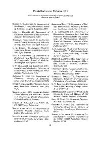
4 8 Fla. 1. The numbering sequence for the 17 carbon atoms and the letter designations for the four rings in the eyelopentano-perhydro-phenanthrenenucleus which is the basic structure of all steroid hormones.
ethylether for crystallization. If the steroid contains a single ketone substituent (i.e., testosterone) then conditions should be used which optimize the yield of derivative. Ratios of 4 to 6 moles of O-(carboxymethyl) hydroxylamine per mole of steroid optimize yield. However, other considerations become important if there is more than one ketone substituent present on the steroid molecule (i.e., progesterone). One can derivatize a related steroid which has a single ketone group (i.e., 20fl-hydroxypregn4-en-3-one or pregnenolone) using conditions which optimize yields and then chemically convert this molecule to the desired steroid derivative. It is also possible to alter the reaction conditions to favor formation of the oxime, at the desired position and then chemically isolate the desired steroid derivative. For example, again in the case of progesterone, a ratio of 0.5 mmoles O-(carboxymethyl) hydroxylamine per millimole of progesterone favors oxime formation at the C-3 (Fig. 1) and this product can be isolated chromatographically. Hemisuccinate derivatives can be made at hydroxyl substituents by reacting 3 to 5 mmoles of succinic anhydride per millimole steroid refluxed with pyridine for 4 to 5 hours. The solution is taken to dryness under reduced pressure, the residue dissolved in ethyl acetate, washed with water, and extracted with 5% NaOH. This alkaline extract is washed with ethyl acetate and acidified with HC1 to pH 2.0. The derivative precipitates and should be washed and recrystallized from appropriate solvents. In the case where more than one hydroxyl substituent is present on the steroid molecule it is usually possible to adjust the reaction conditions to take advantage of the varying reactivities of different hydroxyl groups. For example, the phenolic hydroxyl at C-3 in estradiol-17fl is more reactive than the secondary alcohol at C-17. To make estradiol3-monosuccinate a ratio of 1 mmole estradiol to 3 mmole succinic anhydride in pyridine should be reacted for 5 minutes at 25 °. Under these very mild conditions the yield will be 55 to 80% of the estradiol-3-mono-
[2l
RADIOIMMUNOASSAY OF STEROID HORMONES
19
succinate. 18 The reaction product can be purified by thin-layer chromatography. If the reaction is continued for 3 days, only the 3,17-disuccinate is formed and selective hydrolysis of the C-3 ester may be performed by adding 0.5 g of sodium carbonate to 10 ml of a methanol solution containing 500 mg of the estradiol-3,17-disuccinate. The reaction should be carried out at room temperature for 18 to 24 hours. This reaction product should also be purified by thin-layer chromatography. To prepare a chlorocarbonate derivative at a hydroxyl position on a steroid, the method of Miescher e t al. 19 is used. One millimole of steroid is dissolved in 10 ml anhydrous chloroform, cooled to 0 °, and treated with excess (5 to 10 mmoles) of liquid phosgene for 4 hours with cooling. At room temperature, the excess methanol and phosgene are evaporated under reduced pressure, the oily residue is azeotroped with dry benzene, and the product is crystallized from acetone-hexane. It is very important that all steroid derivatives be extensively characterized by melting point, nuclear magnetic resonance, infrared spectroscopy, ultraviolet spectrophotometry, or combinations of these procedures to determine if the steroid derivative has the intended structure and to assess its purity. It seems likely that in many cases the failure to produce antibodies to steroid hormones is due to inadequate chemical procedures rather than to a failure of the immunized animal to respond. The structure of steroid hormones differs only in the type and location of functional groups attached to the basic cyclopentanophenanthrene nucleus (Fig. 1). Hence, in order to obtain antibodies with maximal specificity one must take full advantage of the structural differences which exist between the steroid of interest and related compounds. This may be done by conjugating the steroid molecule to protein through a site distant to the functional groups of interest. For example, if one wishes to discriminate between testosterone and androstenedione the testosterone molecule should be conjugated to protein at the 3, 6 or 11 position (Fig. 1) but not at the 17 position since this is the site at which the only difference between these molecules exists. To illustrate this point more clearly we have included the data in Table I depicting the relative activities of 17 representative steroids when antibodies were developed to progesterone conjugated to protein in the A (C--3), B (C--6), C (C--11), or D (C--20) ring of the molecule. Immunization with progesterone conjugated at C--20 resulted in the forma1~G. E. Abraham and P. K. Grover, in "Principles of Competitive Protein-Binding Assays" (W. D. Odell and W. H. Daughaday, eds.), p. 134. Lippincott, Philadelphia, Pennsylvania, 1971. 1~K. Miescher, H. Kagi, C. Scholz, A. Wettstein, and E. Tschzep, Biochem. Z. 294, 39 (1937).
20
HORMONE-BINDING PROTEINS
[2]
tion of antibodies which could not distinguish between progesterone and other 3-keto, A4 steroids whose structures are similar in the A, B, C, and D rings (i.e., 17a-hydroxyprogesterone, testosterone, deoxycorticosterone, etc.). Immunization with progesterone-3-BSA resulted in formation of antibodies which easily distinguished between progesterone and steroids with different functional groups attached to the C and D rings. However, this antiserum was less able to distinguish between progesterone and steroids where the only differences in structure occur in the A or B rings (i.e., pregnenolone, 5a-pregnane-3,20-dione). Antibodies developed against progesterone-lla-BSA were quite specific for progesterone and easily detected differences in the A, B, or D ring of the molecule. In fact, this antibody could also distinguish between lla-hydroxyprogesterone and llfl-hydroxyprogesterone. Antibodies developed against progesterone-6fl-BSA easily detected differences in the A, C, or D ring of the molecule but, as expected, were less able to distinguish changes in the B ring (i.e., 5a-pregnane-3,20-dione). Similar data suggests that antibodies produced against testosterone and estradiol-17fl conjugated through the 6 or 11 positions are much more specific than antibodies obtained with these steroids conjugated at the 3 or 17 positions (Table I). From the preceding discussion it should be obvious that it is possible to develop an antibody to perform a particular analytical task. For example, if it is important to distinguish between progesterone and 17a-hydroxyprogesterone the antibody should be developed against progesterone conjugated to protein at the 3, 6 or 11 positions but not at the 20 position. However, if one wishes to quantify progesterone, testosterone, deoxycorticosterone, 17a-hydroxyprogesterone or pregn-4-en-20fl-ol-3-one following chromatographic purification of the sample, then the antibody against progesterone-20-BSA has the advantage because it will quantify all these steroids with equal sensitivity. The trend has been to develop antisera which are highly specific for a given steroid so that many of the expensive and tedious procedures for preparation of the sample can be eliminated. For example, it is now possible to quantify testosterone, s° progesterone, 2j and estradiol-17f~ 22 without chromatographic purification of the sample. Similar procedures have been or are being developed for most of the other major steroid hormones. The complexity of the chemistry involved in making the appropriate derivative is increased considerably when the steroid is to be conjugated 2oA. A. A. Ismail, G. D. Niswender, and A. R. Midgley, Jr., J. Clin. Endocrinol. Metab. 34, 177 (1972). 21G. D. Niswender, Steroids (in press). 2~B. G. England, G. D. Niswender, and A. R. Midgley, Jr., J. Clin. Endocrinol. Metab. 38, 42 (1974).
[2]
RADIOIMMUNOASSAY OF STEROID HORMONES
21
TABLE I RELATIVE ACTIVITY a OF SELECTED STEROIDS IN FOUR DIFFERENT PROGESTERONE RADIOIMMUNOASSAYS
Progesterone20
Progesteron~3
Progesteron~lla
Progesteron~6~ 1.00 .034 .0092 .015 .0032 .30 .0012 .0016 .0004 .0001
1.00 .011 .071 .02 .981 .46 .965 .336 .003
1 O0 133 134 08 046 61 003 001 < 0001
1.00 .005 .345 .08 .0122 .13 .0011 .0014
cpm
,
z s 8, s
~'~
•
,
~
I:10 Dilution
~, ~
4O
i
*e /
i
o,O
.~.w
'
o
O.o.,o
-
'
' 7,S
o~
6oo
!
]!I ~'~
f 450
I I
• r
150
~ S "I
~ #k
'
'
Undiluted
Cytosol
4oo
I
I
~
I
300
o I
I
I00
-
/ v
I0
YW
FRACTFON Fia. 1. Effect of dilution on sedimentation coefficient of estrogen receptor in T E gradients. Cytosol from i m m a t u r e r a t uterus (5 u t e r i / m l in T E buffer) was prepared a~ described in " P r e p a r a t i o n of Cytoplasmic Receptor In Vitro" and incubated (0 °) for 3 hours with 5 X 10-~ M tritiated estradiol. One aliquot was used without further t r e a t m e n t (B), and one aliquot was diluted 1:10 with T E buffer before use (A). Centrifugation at 1 ° was for 12 hours at 50,000 rpm in a Beckman SW 56 Rotor. The dotted line represents the 1'C-markers, ovalbumin and ~/-globulin, and the solid line the tritium counts. See " I n t e r p r e t a t i o n of Results" for explanation of dilution effect.
In some cases aggregation of uterine receptor proteins may lead to the formation of very high molecular weight species which "pellet" to the bottom of the centrifuge tube. Therefore, if the recovery of proteinbound counts in the peak area is low, it is advisable to determine whether any protein-bound counts are present in the bottom portion of the centrifuge tube. Denaturing Agents. Low concentrations of urea (less than 4 M) completely prevent the aggregation phenomena described above for the cytoplasmic receptor. 15 Furthermore, in the presence of 4 M urea, both nuclear and cytoplasmic receptors have identical sedimentation coefficients of approximately 4 S. 15
[15]
ANALYSIS OF ESTROGEN-RECEPTOR PROTEINS
175
Degree o] Purification. After cytoplasmic estrogen receptors have been partially purified, the sedimentation coefficients show much less variability. ~7 In most experiments performed in our laboratory, the variability we observe seems to stem mainly from the tendency of cytoplasmic estrogen receptors to aggregate (see above). Therefore, in order to reproducibly measure the sedimentation coefficient of uterine cytoplasmic estrogen receptors we have found it necessary to always use internal 14C-labeled marker proteins, rather than the more commonly used BSA standard in a separate gradient, and to carefully control experimental conditions, especially protein concentration. 4 We strongly recommend that future experiments using this technique adhere to these precautions. Even with these precautions there is some variability in values for different cytosol preparations and, therefore, we further suggest t h a t conclusions concerning structural features of estrogen-receptor proteins be based only on comparison of samples derived from the same original cytosol preparation.
Comparative Studies Gradient centrifugation has been used to examine estrogen receptors from a number of tissues. In the rat, for example, cytoplasmic receptors from the uterus, 4 pituitary, 9 and breast TM sediment at approximately 8 S in low ionic strength media. Similar values have also been reported for receptors from calf uteri. 26,27 Other literature reports, however, are conflicting. Thus, values of 5 S have been reported for cytoplasmic receptors from human uterus 28,~9 and monkey uterus, 2s,29 while other laboratories have reported values of 8 S for receptors from human uteri2 ° Because numerous factors described in the preceding section can affect the sedimentation of receptors, comparative studies between different tissue and different species should involve mixing experiments as illustrated below (Fig. 2). Aliquots of A-2 ~ and B-2 ~': containing labeled receptors from the different cytosols may not be run on different gradients. Aliquot A-2 ~ m a y now be mixed with B-l, and B-2 ~ mixed with A-l, and the resulting mixtures centrifuged. The mixing will then allow one to determine if any ..8R. H. Wyss, R. Karsznia, W. L. Heinrichs, and W. L. Herrmann, J. Clin. Endocrinol. Metab. 28, 1824 (1968). 59R. H. Wyss, W. L. Heinrichs, and W. L. Herrmann, J. Cli~. Endocrinol. Metab. 28, 1227 (1968). 3°A. C. Notides, D. E. Hamilton, and J. H. Rudolph, Biochim. Biophys. Acta 271, 214 (1972).
176
CYTOPLASMIC RECEPTORS FOR STEROID HORMONES
Cytosols prepared from different tissues
~ Cytosol A
in vitro, Cell free incubation
[16]
Cytosol B
A-1 +
A-2* +
B-I +
B-2* +
E
E*
E
E*
~-
RPA
-A
RPA
-E *
RPB
-E
RPB
-E *
FIG. 2. Mixing experiments. RP, receptor protein; E, unlabeled estrogen; E*, labeled estrogen.
differences observed between receptors in cytosols A (aliquot A-2 ~) and B (aliquot B-2 ~) represent structural differences in receptors, or the presence of interfering substances in either tissue preparation.
[16] E x t r a c t i o n A r t i f a c t s in t h e P r e p a r a t i o n o f Estrogen Receptor
By GARY C. CHAMNESS and WILLIAM L. McGumE The estrogen receptor found in the cytoplasm of estrogen target tissues assumes a variety of forms on extraction, depending on the conditions of extraction and examination. After binding estrogen in the cytoplasm the receptor can enter the nucleus, either in vivo or in cell-free incubation, and this recovered nuclear receptor assumes another variety of forms. Sucrose gradient sedimentation of receptor, introduced in 1966 by Toft and Gorski, 1 has been generally employed to reveal these distinctions. Here we present those procedures currently used in our own laboratory which have consistently produced certain receptor forms.
Cytoplasmic Receptor Tissues. Either mature uteri from 150-200 g intact Sprague-Dawley rats or immature uteri from Sprague-Dawley rats 21-23 days old are used immediately. Pituitary tumor MtTW5, originally obtained from Dr. 1D. Toft and J. Gorski, Proc. Nat. Acad. Sci. U.S. 55, 1574 (1966).
[16l
MULTIPLE FORMS OF ESTROGEN RECEPTOR
177
Vincent Hollander, is transplanted serially by subcutaneous implantation into mature intact female Wistar-Furth rats. These rats are ovariectomized at least 2 days prior to removal of 5-10 g tumors, which are frozen in liquid nitrogen and maintained at --56 ° until use. Although only results from these two tissues will be described in detail, similar procedures are employed for receptor studies in DMBA-induced or transplantable rat mammary tumors ~ and in human breast tumors. 3 Bullets. TE 7.4 (0.01 M Tris-HC1, pH 7.4, with 1.5 mM Na2EDTA) is used throughout. Other solutes are added or EDTA omitted as indicated. Preparation o] Cytosol. All procedures are carried out at 2-4% Tissues are minced with scissors and homogenized in 2.5 volumes (uterus) or 4 volumes (pituitary tumor) of TE 7.4 in a Duall conical glass-glass homogenizer. The homogenate is centrifuged 40 minutes at 140,000 g and the supernatant is recovered with a transfer pipet, leaving behind any floating fat layer. The supernatant is immediately charged with [3H]estradiol (48 or 95 Ci/mmole) at 1 X 10-" M or 4.5 X l0 -' M as needed. Radiochemical purity of the estradiol is periodically checked by thin-layer chromatography.4 After at least 60 minutes the cytosol containing charged, labeled receptor is ready for use, though for some applications free estradiol is removed by passage over Sephadex G-25 or by treatment with dextran-coated charcoal. The charcoal procedure, adapted from Korenman, ~ uses a suspension of 2.5 g/liter of Norit A and 25 mg/liter of dextran in 0.01 M Tris-HC1 (pH 8.0) with 1.5 mM Na.~EDTA. A volume equal to that of the cytosol preparation to be treated is centrifuged 10 minutes at 2000 g and the supernatant discarded; the pellet is resuspended directly in the cytosol and, after 15 minutes, centrifuged 10 minutes at 2000 g, leaving the supernatant free of unbound estradiol. Sucrose Gradients. All sucrose solutions contain TE 7.4, and some also include 0.15 M or 0.4 M KC1. Linear gradients are prepared with an ISCO gradient former in cellulose nitrate tubes for the Beckman SW 56 rotor. (An earlier procedure 6 in which sucrose solutions of decreasing density were layered in the tube and allowed to diffuse overnight to reach a continuous gradient produced equally linear gradients but required more time.) Generally 5-20% sucrose is used for those gradients containing salt and 10-30% sucrose for the others in order to emphasize the respec2 W. L. McGuire, J. A. Julian, and G. C. Chamness, Endocrinology 89, 969 (1971). 3 W. L. McGuire, ]. Clin. Invest. 52, 73 (1973). R. H. Bishara and I. M. Jakovljevic, J. Chromatogr. 41, 136 (1969). 5 S. G. Korenman, J. Clin. Endocrinol, Metab. 28, 127 (1968). G. C. Chamness and W. L. McGuire, Biochemistry 11, 2466 (1972).
178
CYTOPLASMIC RECEPTORS FOR STEROID HORMONES
[15]
rive regions of interest after the standard 15.5-hour centrifugation at 56,000 rpm (308,000 gay). The gradients are chilled after preparation, and the upper 300 ~l are gently removed before 200-~1 samples are layered. Each sample contains about 1000 cpm of l~C-labeled bovine serum albumin 7 as an internal marker; the 14C-peak provides a check of the quality of each gradient as well as a more precise sedimentation standard than achieved with the usual separate albumin gradient. Neither the presence or absence of 0.4 M KC1 in the gradient nor any other variable used appears to affect the sedimentation of the 14C-labeled albumin. After eentrifugation, 4-drop fractions are collected and suspended in modified Bray's solution (125 g of naphthalene, 7.4 g of 2,5-diphenyloxazole, 0.38 g of 1,4-bis [2- (5-phenyloxazolyl) ] benzene, 1 liter of p-dioxane) for liquid scintillation counting in a Beckman LS 233 ; 80-90% of the applied counts are recovered on the gradients. Sedimentation values are determined according to Martin and Ames s from the [14C]BSA standard. E)~ects o / I o n i c Strength. When freshly prepared cytoplasmic receptor from either rat uterus (Fig. 1), pituitary tumor, 9 or hormone-dependent m a m m a r y tumor TM is sedimented in T E 7.4 alone, it forms a peak at 8-9 S, independent of the original salt concentration of the cytosol preparation. Erdos ll first showed that receptor sediments at 4-5 S if the sucrose gradient contains 0.3 or 0.4 M KC1; since the transition is rapid, the sample need not be adjusted to the final salt concentration before layering. More recently it has been shown 6,~,~3 that extraction and centrifugation at physiological ionic strength (0.15 M KC1) produces a distinct receptor form at approximately 6 S (Fig. 1). If the cytosol is originally prepared in T E 7.4 and then brought to 0.15 M KC1, the transition from 8 S to 6 S requires many hours at 4°; bringing the sample to 0.4 M KC1 a few minutes before layering on a gradient containing 0.15 M salt essentially eliminates this period. Ef]ect o / C a l c i u m . In calf uterus cytosol, Puca et al. TM have been able to manipulate the addition of 4 m M CaC12, KC1, and ammonium sulfate to produce either stable 8 S or stable 4 S receptors as desired. We find these procedures to have no effect with either rat uterus or rat pituitary tumor cytoplasmic receptor. However, the addition of 4 m M CaCl~ and ' R. H. Rice and G. E. Means, J. Biol. Chem. 246, 831 (1971). R. G. Martin and B. N. Ames, J. Biol. Chem. 236, 1372 (1961). W. L. McGuire, M. DeLaGarza, and G. C. Chamness, Endocrinol. 93, 810 (1973). ~oW. L. McGuire and J. A. Julian, Cancer Res. 31, 1440 (1971). " T. Erdos, Biochem. Biophys. Res. Commun. 32, 338 (1968). ,2 I. Reti and T. Erdos, Biochimie 53, 435 (1971). 1~It. Rochefort, Proc. Int. Congr. Horm. Steroids, 3rd, 1970 p. 23 (1971). 14G. A. Puca, E. Nola, V. Sica, and F. Bresciani, Biochemistry 10, 3769 (1971).
[16]
MULTIPLE
FORMS
OF E S T R O G E N
RECEPTOR
179
5.8S 4.9S|
1500-
9.
g S
(3)
thus, ka can be obtained as the slope of a plot of 1
T~
T--X In ~
vs. t
(4)
The dissociation of RH* is studied by preventing the reassociation of receptor and labeled hormone if a dissociation of the complex occurs. This is done by adding a large excess of unlabeled progesterone to the preparation. Thus the reaction studied is: kd R H * -~- H ~ - R H -~ H* k~
(5)
When H >> RH*, the reaction is pseudo first order and the rate law is dX - k~(x) dt
(6)
lnX = -k~t+lnXo
(7)
In integrated form, where kd is the dissociation constant in time -1 and X0 is the concentration of RH* at time = 0. Thus, --kd is obtained as the slope of a plot of In X vs. t. The equilibrium constant for reaction (1) is the ratio of the rate constants: K~ = k~/ka (8) It must be stressed that the derived Kd obtained in this way will agree with that obtained by equilibrium methods (i.e., Scatchard plots) only if the reaction proceeds by a pathway adequately defined by the
198
CYTOPLASMIC
RECEPTORS
FOR
STEROID
HORMONES
[17]
rate equations. This question is treated in greater detail elsewhere. ~s'~z More complex behavior will be detected as differences between the two values. This is in fact observed with the case of the progesterone receptor, where the two values of Ka differ by two orders of magnitude, as shown in Table I. 1. Association R a t e Constant
To start the binding reaction, a 1.0-ml sample of an unlabeled receptor fraction is mixed with 10 t~l of various dilutions of [all]progesterone in H20. The binding reaction is stopped by removing 100-ttl aliquots at specific times and adding each to a tube containing 10 gl of unlabeled progesterone (1 t~g) in 10% ethanol. The concentrations of bound and free hormone are then determined immediately by the ammonium sulfate assay. The results are plotted as described above, yielding a rate constant from the slope of the line23 2. Dissociation Rate Constant
This is measured in a receptor sample which has been saturated with [SH]progesterone to fill virtually all the receptor sites. To a 1.0-ml sample of receptor, 10 tL1 of unlabeled progesterone (1 gg) in 10% ethanol is added to start the dissociation experiment. At various times after the addition of unlabeled progesterone, 100-~l aliquots are removed and assayed for bound and free [3H]progesterone by the ammonium sulfate assay. Semilogarithmic plots of bound ~H vs. time give straight lines for the pseudo first-order dissociation reaction. The dissociation rate constant is determined from the slope of the curve. V. Partial Purification The initial steps of purification are outlined in Table II. Steps after the DEAE-cellulose procedure are not included, since the protein concentrations were too low to measure. These steps were done using an ammonium sulfate precipitate as starting material. A. Ammonium Sulfate Precipitation The crude eytosol receptor preparation can be purified 20-30-fold in about 70-80% yield by precipitation of the receptors at 30% saturation = I. M. Klotz and D. L. Hunston, Biochemistry 10, 3065 (1971). M. Best-Belpomme,J. Fries, and T. Erdos, Eur. J. Biochem. 17, 425 (1970).
[17]
CHICK
OVIDUCT
,-~
PROGESTERONE
RECEPTOR
199
¢.D
./
¢C
z2 t'~ C
•F R cq
d
,-4 N
@
o
©
cq
~
5
Z 0
v ~ a ©
x
m~ N
6
d
d
,--t
°N
~
O
Z
~
©
100,000 g supernatant fraction) or LSS (800 g supernatant fraction (0.05-2.0 ml) is added to the HAP slurry (0.50 ml) and allowed to stand on ice for 10-20 minutes with several mixings. Washing buffer (0.05 M Tris-HC1, pH 7.3, 25 °) " G. Giannopoulosand J. Gorski, I. Biol. Chem. 246, 2524 (1971). ~8D. Williams and J. Gorski, Acta Endocrinol. (Copenhagen), Suppl. 168, 420 (1972). ,8 W. E. Stumpf, Endocrinology 83, 777 (1968). u G. A. Puca and F. Bresciani, Nature (London) 218, 967 (1968). D. Tort and J. Gorski, Proc. Nat. Aead. Sci. U.S. 55, 1574 (1966). ~6T. Erdos, M. Best-Belpomme, and R. Bessada, Anal. Bioehem. 37, 244 (1970). 8,D. Williams, Ph.D. Thesis, University of Illinois, Urbana (1972).
[24]
E S T R O G E N B I N D I N G IN C Y T O P L A S M A N D N U C L E U S
281
(4 ml) is added, the sample mixed, and centrifuged at 800 g for 2 minutes. The wash is discarded, the HAP pellet resuspended in washing buffer (5 ml), mixed vigorously with a vortex mixer, and centrifuged as above. This washing procedure is repeated three more times. The washed HAP pellet is extracted with 100% ethanol (4 ml) for 15 minutes at room temperature, the sample centrifuged, and the ethanol extract assayed for radioactivity after addition to a toluene-based scintillation fluid. This extraction procedure transfers greater than 97% of the radioactivity to the scintillation vial. Comments. Adsorption of EB to HAP is maximal by 5 minutes and stable for at least 90 minutes. Adsorption of EB to HAP is linear with increasing quantities of HSS or LSS up to at least 1.2 uterine equivalents of EB per 0.3 ml packed HAP. Identical results are obtained with HSS and LSS. Assay of EB in HSS by HAP or gel filtration on Sephadex G-25 gives identical results. With 0.3 ml packed HAP the adsorption of one uterine equivalent of HSS or nuclear extract (see below) is independent of volume between total assay volumes of 0.55-2.5 ml, independent of Tris concentrations of 0.01-0.05 M, and independent of KC1 concentrations up to and including 0.4 M. Unbound E does not significantly adsorb to HAP under the conditions of the assay. Estradiol bound to nonspecific sites also adsorbs to HAP, necessitating the use of competitive methods to correct for contributions from this component (see section Distinguishing EB from EP).
Assay o] Nuclear EB The preparation of the washed nuclear fraction separates unbound E from nuclear bound hormone present as EB and EP. Consequently, nuclear bound E can be determined with an ethanol extraction of the washed nuclear pellet as described above for the HAP pellet. EB can then be distinguished from EP as described below. Alternatively, EB can be extracted from the washed pellet or a purified nuclear preparation with 0.01 M Tris-HCl, 0.001 M EDTA, 0.4 M KC1, pH 8.5, for 1 hour at 0°. 14 Subsequent centrifugation at 226,000 g for 45 minutes yields the high speed nuclear extract which is suitable for further examination of EB. EB in this extract can be assayed with HAP as described above without altering the KC1 concentration. Distinguishing E B from E P
Principles With the possible exception of sucrose gradient centrifugation under conditions where the 8 S binding protein is separated from the bulk of
282
NVCLEAR RECEPTORS FOR STEROID HORMONES
[24]
uterine proteins, 15 all the methods noted above measure bound E irrespective of whether it is EB or EP. Although the use of nonequilibrium assay methods minimizes the contribution of EP to the total bound E, EP is never zero and it may be as large as EB at high E concentrations. The quantity of E present as EB can be distinguished from that present as EP by taking advantage of the fact that the nonspecific P binding sites are a low affinity binding system present in very large numbers while the specific B binding sites are a high affinity binding system present in very limited numbers. TM Hence, the specific B binding system saturates at E concentrations that are still in the linear range of the nonspecific P binding system. The quantity of [~H] EP present at any [3H] E concentration can be determined with a parallel sample which contains a 100fold excess of unlabeled E in addition to the [3HIE. In this parallel sample the limited number of specific B sites contain E and [3H]E in a 100:1 ratio, such that EB complexes contribute only a few percent of the radioactivity they have in the sample containing only [~H]E. On the other hand, the unlimited number of P sites contain 100 times as much E at a specific radioactivity equal to 1% of that in the sample containing only [~H]E; hence the EP complexes contain the same quantity of radioactivity in both samples. The difference between the samples gives the quantity of [3H]EB present. Procedures For any sample at a particular [SH]E concentration, a parallel sample containing [~H]E and a 100-fold excess of unlabeled E is also incubated. The samples are then treated by the methods described above. The values of EP determined for cytoplasmic and. nuclear fractions are subtracted from the total bound E in each fraction to yield the quantities of EB present. Comments For this procedure to be valid, EP must behave as a linear function of the E concentration within the employed concentration range. When working with immature rat uteri in vitro, EP present in cytoplasmic and washed nuclear fractions behaves linearly up to E concentrations of at least 2 X 10.6 M.I~" It should be noted that assayable EP is an operational quantity which may differ in magnitude depending on the method used for assay. In conI*D. J. Ellis and H. J. Ringold, in "The Sex Steroids" (K. W. McKerns, ed.), p. 73. Appleton, New York, 1971.
[251
[3H]ESTRADIOL EXCHANGE ASSAY
283
trast to the situation with EB, the quantity of E P remaining after a nonequilibrium assay may be only a small fraction of that present during the tissue incubation. Since the nonspecific P sites are present in great excess over the specific B sites, the equilibrium quantity of EP will be attained much faster than the equilibrium quantity of EB. During brief incubations, therefore, EP will constitute a greater part of the total bound hormone than it will at equilibrium. The use of E in a 100-fold excess is chosen simply for flexibility over a large part of the saturation range. When working in the lower 10% of the saturation curve, a higher ratio of E to [3H]E can be employed to ensure that EB complexes contribute little to the value obtained for EP. Similarly, when working at [3H]E concentrations appreciably greater than 10.8 M, a lower ratio of E to [3H]E can be employed to ensure that the hormone stays in solution.
[25] [ 3 H ] E s t r a d i o l E x c h a n g e A s s a y f o r the D e t e r m i n a t i o n o f Nuclear Receptor-Estrogen Complex 1
By J. H.
CLARK,J.
N.
ANDERSON,and
E. J.
PECK, JR.
Introduction The association of the receptor-estrogen complex (RE) with nuclear binding sites is considered to be of fundamental importance in the mechanism of action of estrogen. We have developed a method for the determination of the concentration of RE in the nuclear fraction of estrogen target tissues which relies on the exchange of [3H]estradiol with estradiol which is bound to the nuclear fraction. TM This method permits an evaluation of the concentration of nuclear RE under in vivo conditions and as a function of nonlabeled estrogenic compounds.
Assay Method The [3H]estradiol exchange assay can be used for the measurement of nuclear RE in the uterus, pituitary, and hypothalamus. The method as used for the analysis of uterine tissue will be described initially and This work was supported by grants from The National Institutes of Health (HD 04985) and The Research Corporation, Atlanta, Ga., USA. 1~j. N. Anderson, J. H. Clark, and E. J. Peck, Jr., Biochem. J. 126, 561 (1972).
284
(a)
fl.
[25]
NUCLEAR RECEPTORS FOR STEROID HORMONES
(b)
2.5
$
i 2.0 1.5 1.0
1.00
/C /
y"
/
i!
0.75 o.~
U) laJ
/
r - ~ 0.5
I0 I
20 I
[[3H]ESTRADIOL](nM)
/
/
t
/
//
/ 0.25
40 80 I
0
-~7~ (M-I)
FIG. 1. The determination of the q u a n t i t y of nuclear R E by the [3H]estradiol exchange assay. (a) This experiment is presented as an example of the use of the [3H]estradiol exchange assay for the determination of the q u a n t i t y of nuclear R E following an injection of estradiol. I m m a t u r e rats, age 21-23 days, were injected subcutaneously with 2.5 tzg of estradiol. One hour after the administration of hormone the rats were sacrificed and the uterine nuclear fraction was prepared as described in Methods. This fraction was incubated for 1 hour at 37 ° with either [*H]estradiol alone (A) or [3H]estradiol plus DES at 100 times the concentration of [3H]estradiol (B). The dashed line (C) was obtained by subtracting B from A. Each point represents the m e a n of 6 determinations with 3 rats per determination. (b) A double-reciprocal plot of the data represented b y (C) of P a r t la. The apparent Kd R is 1.3 X l i f o M and the n u m b e r of binding sites per uterus is 1.8 pmoles.
the necessary modifications for the analysis of the hypothalamus or pituitary will be noted. The uterus is stripped of adhering fat and mesentery and placed in cold 0.9% NaC1. All subsequent procedures should be performed at 4 ° unless otherwise stated. The tissue is washed in 10 mM Tris buffer, pH 7.4, that contains 1.5 mM EDTA (TE buffer) and subsequently homogenized in 1.5 ml of TE (wet weight/volume, 20 mg/ml) in an all glass Kontes homogenizer using a motor-driven pestle. The nuclear fraction is obtained by centrifugation of the homogenate at 800 g for 10 minutes. This fraction is washed three times with 3 ml of TE buffer and the nuclear pellet is mixed thoroughly each time with a vortex mixer. Each wash is followed by centrifugation at 800 g for 10 minutes. The washed nuclear pellet is rehomogenized and suspended in TE buffer to a concentration
[25]
[3H]
ESTRADIOL EXCHANGE ASSAY
285
of 0.5 uterus/ml (immature rat uterus). Portions (0.5 ml) of this suspension are dispensed into two series of tubes, A and B, containing 0.2 ml of buffer. Series A should contain various concentrations of [3H]estradiol (1-25 nM) and is used to determine the total amount of [SH]estradiol exchange. Series B should contain the same concentration of [aH]estradiol as in A plus a 100-fold excess of diethylstilbestrol (DES) and is used to determine the nonspecific [3H]estradiol exchange. The nuclear fractions are incubated with shaking at 37 ° for 1 hour. Following incubation, the nuclear fraction is washed three times with 3 ml of TE buffer as described above. The nuclear fraction is extracted with 3 ml of 100% ethanol and the extract is added to 10 ml of scintillation fluid [95.5% toluene, 0.45% 2-5-diphenyloxazole, 0.05% 1,4-bis-(5-phenyloxazol-2-yl) benzene] for the determination of radioactivity. The amount of [3H]estradiol that is bound in the nuclear fraction in the absence (series A) and presence (series B) of DES represents total and nonspecific binding, respectively (Fig. 1). The [3H]estradiol that is bound in a specific, DES-competable manner (Fig. la, C) may be determined by subtracting the nonspecific binding (Fig. la, B) from the total binding (Fig. la, A). The maximum quantity of nuclear RE and the dissociation constant for RE, KdR, are obtained from a double reciprocal plot of the data for the specifically bound [3H]estradiol (Fig. la, C). A representative plot is shown in Fig. lb. The procedure for the measurement of nuclear RE in the hypothalamus or pituitary is essentially the same as that used for the analysis of uterine tissue with these modifications: The tissue is homogenized in a glass homogenizer, with a Teflon pestle (0.11-0.15 mm clearance). The nuclear fraction is obtained and washed as above. The washed nuclear pellets are resuspended in TE buffer to a concentration of 3 hypothalami/ml (wet weight, 26 mg/immature hypothalamus) or 3 pituitaries/ml (wet weight, 2.2 mg/immature pituitary). One-milliliter portions of this suspension are dispensed into two sets of tubes, series A and B as above. The concentrations of [3HI estradiol should range from 0.46-3.66 nM. The remainder of the procedure is identical to that used for the uterus. To determine the number of picomoles of receptor-estrogen complex present in the nuclear fraction under any experimental situation, either a saturation curve such as Fig. 1 or a concentration of estradiol equivalent to K~a X 103 must be employed. Since either experiment is expensive in experimental animals and/or labeled estradiol, the assay may be performed at a concentration of 13 nM [3H]estradiol for uterine tissue and 3.6 nM [3H]estradiol for hypothalamic and pituitary tissue. These concentrations are l0 fold greater than the apparent K¢1R for the receptor of these tissues (uterus, 1.3 -----0.16 nM; hypothalamus, 0.39---+ 0.19 nM;
286
NUCLE&R RECEPTORS FOR STEROID HORMONES
[26]
pituitary, 0.35 ± 0.06 nM) and should be sufficient to saturate approximately 91% of the specific binding sites. Under our conditions, approxim a t e l y 95% of the specific binding sites are measured at these concentrations. The difference between observed and predicted degrees of saturation are reflected in the means obtained for maximal binding sites ± the standard error of the mean for each tissue (uterus, 60.0 ± 5.9; pituitary, 14.3 ± 0.23; hypothalamus, 0.47 ± 0.06 f m o l e / m g wet weight). Conclusion The [~H]estradiol exchange assay can be used to determine the number of specific nuclear binding sites in estrogen-sensitive tissues and has permitted us to examine the influence of endogenous and exogenous estrogen on the concentration of nuclear R E and the concentration-time-response parameters of estrogen action. 2,8 This assay is of special significance in view of the recent demonstration t h a t approximately 90-95% of the estrogen which is specifically bound in the uterus is t h a t which is present in the nuclear fraction. 4 25. H. Clark, J. N. Anderson, and E. J.. Peck, Jr., Science 176, 528 (1972). J. N. Anderson, J. H. Clark, and E. J. Peck, Jr.,, Biochem. Biophys. Res. Commun. 48, 1460 (1972). 4D. Williams and J. Gorski, Biochem. Biophys. Res. Commun. 45, 258 (1971).
[26] A T e c h n i q u e f o r D i f f e r e n t i a l E x t r a c t i o n Nuclear Receptors 1
of
B y D. ~VIARVERand I. S. EDELMAN
Introduction Mineralocorticoid-specific binding proteins have been isolated and characterized in target tissues of adrenalectomized rats injected with [~H]aldosterone. 2-7 I n vivo, the binding of aldosterone to these receptors s 1 Supported by USPHS, National Heart and Lung Institute, Grant Nos. HL-06285 and HL-05725. D. Marver, D. Goodman, and I. S. Edelman, Kidney Int. 1, 210 (1972). 8j. W. Funder, D. Feldman, and I. S. Edelman, d. Sleroid Biochem. 3, 209 (1972). 4T. S. Herman, G, M. Fimognari, and I. S. Edelman, J. Biol. Chem. 243, 3849 (1968). 5 G. E. Swaneck, L. L. H. Chu, and I. S. Edelman, J. Biol. Chem. 245, 5382 (1970). D. D. Fanestil and I. S. Edelman, Proc. Nat. Acad. Sci. U.S. 56, 872 (1966). G. E. Swaneck, E. Highland, and I. S. Edelman, Nephron 6, 297 (1969). 8The term "receptor" is used only to imply high-affinity, specific binding sites.
[26]
EXTRACTION OF NUCLEAR RECEPTORS
287
is an early event, occurring well before the physiological response. Maximum nuclear labeling of [~H]aldosterone in the rat kidney occurs 10 minutes after intravenous injection of steroid, although the physiological change in urinary Na+/K + levels is not demonstrable for about an hour and reaches a peak response in 3-4 hours.:' In this chapter, we describe the methods for labeling of the aldosterone receptors and differential extraction of aldosterone-receptor complexes from the nucleus.
Preparation of Labeled Complexes In Vivo. Aldosterone-binding proteins can be labeled in vivo by injecting adrenalectomized rats intravenously or subcutaneously with physiological doses of [3H]aldosterone (0.1-0.5 ~g/100 g body wt., 50 C i / m M ) . To correct for nonspecific binding, control animals receive the same dose of [aH]aldosterone plus a 100-fold excess of unlabeled aldosterone. With intravenous administration, animals are sacrificed in 2 to 10 minutes, depending on whether maximal cytosol labeling (2 minutes) or maximal nuclear labeling (10 minutes) is desired. Subcutaneous injections result in a somewhat lengthened time course, with maximal cytoplasmic and nuclear labeling occurring at about 15 to 30 minutes, respectively. G In Vitro: Slices. Kidney slices from adrenalectomized rats are labeled with [3H]aldosterone by incubation in a complete salt media at 25 ° with 10-1° to 10.2 M [3HI aldosterone. The kidneys are removed, decapsulated, and transferred immediately to a solution composed of: NaC1 = 135, KHoPO~ = 5, Tris-HC1 = 5, MgCI.,, = 0.5, CaCI._, = 1, glucose = 5 (all in raM), pH 7.4, at 0 °. The kidneys are sliced (275 t~m) in a McIlwain tissue slicer, 1° collected on Nytex, H and briefly rinsed with cold media. Erlenmeyer flasks containing 20 ml incubating media and 10 -1° to 10-s M [3H]aldosterone-+- a 100-fold excess of unlabeled steroid are placed in a r o t a r y shaking water bath. At zero time, slices from one to two kidneys are transferred to each flask and incubated for 5 to 60 minutes. Within this time period, cytoplasmic labeling reaches a plateau in 15 minutes, while nuclear labeling rises steadily for an hour or more. At the end of the incubation period, the slices are recollected on Nytex, rinsed with ice-cold buffer, and immediately transferred to homogenizing media (0.25 M sucrose, 3 m M CaCI~) at 0% In Vitro: Cell-Free Recombination Studies. Cell-free mixtures of nuclei and cytosol labeled with [aHIaldosterone can be used to generate G. M. Fimognari, D. D. Fanestil, and I. S. Edelman, Amer. J. Physiol. 213, 954 (1967). 1oBrinkmann Instrument Co., Burlingame, Calif. 1~Nylon cloth, 132 mesh, John Stanier & Co., Manchester, England.
288
NUCLEAR RECEPTORS FOR STEROID HORMONES
[25]
both low salt- and high salt-extractable nuclear complexes. For cell-free incubations, cytosol obtained from the 19,000 g isolation step or the 105,000 g isolation step (details in next section) is diluted with spectroquality glycerol to a final concentration of 25% (v/v). [~H]Aldosterone (10-~° to 10-s M) _ a 100-fold excess of unlabeled aldosterone is added and allowed to stand at 0 ° for 30 minutes. The crude nuclear 600 g pellet (details in next section) is washed twice with 0.25 M sucrose, 3 mM CaC12 using volumes at least equivalent to the decanted crude cytosol volume, by resuspending and recentrifuging at 600 g for 10 minutes. After washing, the pellet is suspended in a small amount of 0.25 M sucrose, 3 mM CaC12 and divided between a number of 25-ml Erlenmeyer flasks so that each flask contains the nuclei from 1-2 kidneys. Labeled cytosol is added to the nuclear suspension at 0 °. For optimal binding, the cytosol added per flask should be equivalent to the amount obtained from 1-2 kidneys and the proportion of cytosol volume to nuclear suspension volume in the incubating flask should be 2 4 : 1 . At zero time, the mixtures are placed in a 25 ° shaking water bath, incubated 15 to 30 minutes, and then poured into iced centrifuge tubes and spun at 600 g for 10 minutes (0 °) to separate nuclei and cytosol.
Preparation of Purified Cytosol and Nuclear Fractions All procedures are at 0-4 ° . Kidneys or kidney slices are homogenized in 0.25 M sucrose, 3 mM CaC12 ( ~ 1 g tissue/4 ml sucrose) using a Teflon-glass homogenizer. A crude nuclear and cytoplasmic fraction is obtained by centrifugation of the homogenate at 600 g for 10 minutes. The supernatant is decanted and recentrifuged at 19,000 g for 15 minutes and then 105,000 g for 1 hour. A separation of bound and free aldosterone in the final 105,000 g supernatant is made by filtering 1-ml aliquots over 0.28 X 15 cm G-50 fine Sephadex ~: columns equilibrated with 0.1 M TrisHCI, 3 mM CaC12, pH 7.2 (the pH of this Tris buffer can be 7.2-8.0 for both cytosol and nuclear complexes). Following filtration, 0.5-1.0 ml fractions of the void volume of each G±50 column is added to 15 ml of dioxane counting solution 13 and assayed for radioactivity in a liquid scintillation counter. The protein content of the void volume is estimated from the ratio of the O.D. at 260 and 280 nm ~4 and the final results expressed as moles of aldosterone bound per milligram protein. "Specific" 1~Pharmacia, Piscataway, :New Jersey. 1~10 g 2,5-diphenyloxazole (PPO), 0.5 g 1,4-bis-2-(4-methyl-5-phenyloxazolyl)benzene (dimethyl-POPOP), and 80 g napthalene in 498 ml ethylene glycol monomethyl ether, 428 ml p-dioxane, and 143 ml xylene. 1~O. Warburg and W. Christian, Biochem. Z. 310, 384 (1942).
[26]
EXTRACTION OF NUCLEAR RECEPTORS
289
binding is then calculated as the difference between total binding and binding in the presence of a 100-fold excess of unlabeled aldosterone. To purify nuclei obtained from in vivo, slice, or recombination studies, the 600 g pellet is resuspended twice in 0.25 M sucrose, 3 mM CaC12 and recentrifuged each time at 600 g for 10 minutes. The pellet from 2-4 kidneys is then mixed with 20 ml of 2.2 M sucrose, 3 mM CaC12 and layered over 10 ml of this same solution. This mixture is centrifuged at 56,000 g in a Spinco 30 rotor 1'~ for 1 hour, using polycarbonate 30-ml tubes. The 2.2 M sucrose and cellular debris are decanted and excess sucrose removed from the walls of the centrifuge tube with cotton-tipped applicator sticks. The sedimented nuclei are gently resuspended in 1.0 ml of 0.1 M Tris-HC1 3 mM CaCl=,, pH 7.2, and transferred by Pasteur pipet to 3.0 ml of the same Tris buffer. The suspension is allowed to stand 10 minutes at 0 ° and then spun at 10,000 g for 10 minutes. The aldosterone-bound protein released into the supernatant of this fraction is referred to as the "nuclear Tris-soluble complex." The supernatant Tris extract is decanted and the pellet resuspended in 2 ml of 0.1 M Tris-HC1 buffer, recentrifuged at 10,000 g for 10 minutes, and the supernatant discarded. The pellet is then extracted with 1-2 ml of 0.4 M KC1, 0.01 M Tris, pH 7.2. This mixture is incubated at 0 ° for 10 minutes and then centrifuged at 10,000 g for 10 minutes. The resultant supernatant contains the majority of the residual [3H] aldosterone nuclear counts, and the protein:steroid conjugate obtained in this fraction is referred to as the "chromatin-bound aldosterone complex. ''5 For concentration of both fractions, an equal volume of saturated ammonium sulfate is added to the fraction at 0 °. After 30 minutes the mixture is spun at 19,000 g for 10 minutes and the supernatant discarded. The precipitate is resuspended in 0.5/1.5 ml of either 0.1 M Tris-HC1 or 0.4 M KC1, 0.01 M Tris-HC1, pH 7.2, and any insoluble material removed by centrifugation at 19,000 g for 10 minutes. To assay binding, aliquots of the Tris-soluble and chromatin-bound nuclear fractions are added to dioxane counting solution and protein estimated spectrophotometrically by the O.D. at 260 and 280 nm. Results are expressed as moles of aldosterone bound per milligram protein minus nonspecific binding determined in the presence of a 100-fold excess of unlabeled aldosterone. Characterization o f N u c l e a r A l d o s t e r o n e C o m p l e x e s on Glycerol D e n s i t y Gradients
The nuclear binding proteins can be further characterized by their sedimentation properties on glycerol density gradients. Aliquots of ~ Spinco Corp., Palo Alto, Calif.
290
NUCLEAR
RECEPTORS
FOR
STEROID
HORMONES
[26]
n u c l e a r low a n d high s a l t e x t r a c t s can be l a y e r e d d i r e c t l y on g r a d i e n t s or c o n c e n t r a t e d first w i t h a m m o n i u m s u l f a t e as d e s c r i b e d above. L i n e a r g r a d i e n t s m a d e f r o m s t a r t i n g c o n c e n t r a t i o n s of 10 a n d 3 4 % s p e c t r o q u a l i t y g l y c e r o l ( v / v ) which c o n t a i n in a d d i t i o n , e i t h e r 0.1 M T r i s , p H 7.2, or 0.4 M KC1 + 0.01 M T r i s , p H 7.2. [ 3 H ] A l d o s t e r o n e , a t a final c o n c e n t r a t i o n of 10 -9 M , m a y also be a d d e d as a c o n s t a n t b a c k g r o u n d to s t a b i l i z e b o u n d complexes d u r i n g c e n t r i f u g a t i o n . E i t h e r 12.5 or 4 ml g r a d i e n t s can be used, t h e l a r g e r g r a d i e n t s p i n n i n g a t 283,000 g for 40 hours ( r o t o r SB 283) a n d t h e s m a l l e r a t 405,000 g for 16 hours ( r o t o r 5S A, NO ADDITIONS I00
I 2
B,
3 5
7
d- ALDOSTERONE
9
II 13
C. 17E- ISOALDOSTERONE
E 150 IO0 0
n-,-
I.-0 ~ ...J
50
0
I D
:3 5 7
9 II 13
I
3
5
7
9 II 13
9 ~ - FLUOROCORTISOL E. ESTRADIOL- 178
150 I00 50
__~.-~_~_ I
3
_~.~..~.-,-",
n
5 7" 9 II 1:3 I 3 5 7' 9 VOLUME FROM BOTTOM ( m l )
II 13
Fro. 1. Glycerol density gradients of Tris-soluble nuclear extracts labeled in vivo. Adrenalectomized 150 g rats were injected with 2.6 X 10-~° moles [~H]aldosterone and either diluent (Panel A), or 2.6 X 10⁻⁹ moles d-aldosterone (Panel B), or 2.6 X 10-° moles 17a-isoaldosterone (Panel C), or 2 X 10-s moles 9,-fluorocortisol (Panel D), or 2 X 10-s moles estradiol-17~ (Panel E). The 10-34% glycerol density gradients also contained 0.1 M Tris-HC1, 3 mM CaCl=, p i t 8.0, and 1.6 X 10⁻⁹ M [~H]aldosterone with or without a 10- or 78-fold excess of the appropriate competitor. The 12.5-ml gradients were centrifuged 283,000 g for 40 hours. Following centrifugation, 1-ml fractions were collected and filtered through Sephadex G-50 columns to remove free [3H]aldosterone. (From Marver et al3)
[26]
EXTRACTION OF N U C L E A R RECEPTORS
2oo!
291
~20
E
E
o
0 )10 ~
0 ~
__U o
1 VOLUME FROM BOTTOM (ml)
FIG. 2. Glycerol density gradient of the chromatin-bound [3H]aldosterone complex labeled in vivo. Adrenalectomized 150-200 g rats were injected with 2.6 X 10⁻¹° moles [3H]aldosterone. Chromatin was isolated frGm the nuclear pellet following extraction with 0.1 M Tris-HC1, 3 mM CaCl2, pH 8.0, by the method of K. Marushige and J. Bonnet [J. MGl. Biol. 15, 160 (1966)]. A 0.3 M KC1 extract of chromatin wa~ layered on a 10-35% 12.5 ml, glycerol density gradient containing 0.3 M KC1--0.01 M Tris-HC1, pH 8.0, and centrifuged 283,000 g for 40 hours. Following centrifugation, 1-ml aliquots were collected and assayed directly for radioactivity and O.D. at 230 mm. [From G. E. Swaneck, L. L. H. Chu, and I. S. Edelman, J. Biol. Chem. 245, 5382 (1970).] SB 405).1~ Following centrifugation, the base of the tube is punctured with a # 2 6 needle and aliquots collected. The fractions m a y be counted directly and assayed for 260:280 protein, or if [3H]aldosterone was used to stabilize binding, the fractions are filtered through G-50 columns to removed unbound steroid# Gradients of Tris soluble fractions yield a specific 3 S peak as shown in Fig. 1 (whether the gradients contain low or high salt). The gradient pattern of the chromatin-bound fraction isolated after in vivo labeling and centrifuged in the presence of 0.4 M KC1 is shown in Fig. 2. In high salt (0.4 M KC1) gradients, the chromatin-bound species migrates as a 4 S complex2 In low salt gradients (0.1 M Tris or less) however, this complex aggregates and pellets to the bottom of the centrifuge tube. Properties of Nuclear Complexes Isolated nuclear and cytoplasmic aldosterone-specific complexes are susceptible to degradation by sulfhydryl reagents and proteases such as pronase, trypsin, and chymotrypsin, but not by nucleases. In the absence 16International Centrifuge Corp., Needham Heights, Mass.
292
[27]
NUCLEAR RECEPTORS FOR STEROID HORMONES TABLE I TEMPERATURE DEPENDENCE OF [3H]ALDOSTERONE BINDING IN RAT RENAL CYTOSOL AND NUCLEAR FRACTIONSa X 10-15 moles/mg protein Temperature (°C) 0 25 0/25
Cytosol
Tris-soluble
Chromatinbound
22.5 31.4
19.0 28.4
2.1 12.9
71.7%
66.9%
16.3%
" Adrenalectomized rat kidney slices were incubated at either 25° or 0° for 1 hour in 5.2 × 10⁻⁹ M [3H]aldosterone with or without 5.2 × 10⁻⁶ M d-aldosterone. The quantity of [aH]aldosterone bound has been corrected for nonspecific labeling. of glycerol, at least 70% of the bound aldosterone is dissociated from the complexes in 24 hours at 0 °. Glycerol at a 20-25% final concentration is able to preserve a minimum of 75% of this activity over the same time period at 0 °. The steroid-labeled complexes are also heat labile and m a y be irreversibly dissociated by incubating isolated fractions at 37 ° for 30 minutes. In vitro incubations have provided more information regarding the properties of the nuclear complexes. The transfer of labeled complexes from cytosol to nuclei is temperature-dependent. T a b l e I shows the effect of incubating r a t kidney slices at 0 ° rather than 25 °. While the amount of labeled cytosol and Tris-soluble complex isolated from the 0 ° incubation was 72% and 67% t h a t of the 25 ° incubation, the chromatin-bound fraction had only 16% of the labeling seen at 25 ° .
[27] S t e r o i d H o r m o n e - R e c e p t o r Nuclear
Interactions
with
Constituents
B y WILLIAM T. SCHRADER, SUSAN H. SOCHER, and RICHARD E. BULLER
I. I n t r o d u c t i o n Over the past decade considerable evidence has accumulated to support the concept t h a t steroid hormones act to alter the expression of genes for specific proteins in target ceils. This regulation appears to occur in the nucleus, where radioactive steroids administered in vivo rapidly ac-
[27]
RECEPTOR-NUCLEAR INTERACTIONS
293
cumulate tightly associated with the chromatin. The steroids are found first bound to cytoplasmic receptor proteins and subsequently these complexes appear in the nuclei. In order to study these nuclear processes more fully, it has been necessary to develop or adopt procedures for observing nuclear uptake and steroid-receptor binding in vitro. The chick oviduct has been studied extensively as a model system for steroid hormone action. 1-3 In this tissue, estrogens induce growth and differentiation of the immature oviduct and induce the synthesis de n o v o of the egg white proteins ovalbumin and lysozyme. Progesterone administration to the oviduct results in the induction of a new protein, avidin. This progesterone response proceeds through the early events outlined above. Since stable receptor proteins can be isolated for this steroid, ~-6 the intranuclear accumulation mechanism for progesterone could be readily studied in this system.
A. Preparation of Progesterone Receptors Detailed methods for isolation and partial purification of chick oviduct progesterone receptors appear elsewhere in this volume. 7 For routine assays of chromatin acceptor sites and nuclear binding, crude cytoplasmic extracts are prepared in 0.01 M Tris-HC1, 0.025 M KC1, pH 7.4 (Buffer A). Buffers containing E D T A can be used for chromatin binding studies, which show no demonstrable cofactor requirement, but E D T A should be avoided in nuclear studies since it will chelate some of the Mg 2÷ in the nuclear preparation. High-speed cytoplasmic extracts (cytosol) from oviduct or nontarget tissues will contain about 20 mg protein/ml if homogenization is done in 4 ml buffer/g tissue. The cytosols should be diluted to about 10 mg protein/ml for the incubations. Tritiated hormone ( [ 1,2-3H ] progesterone, 40-50 Ci/mmole) should be added 1 to 2 hours before the incubations begin in order to have freshly labeled preparations. It is important to add subsaturating doses of progesterone (10 -s M) so that only small B. W. O'Malley, Biochemistry 6, 2546 (1967). 2B. W. O'Malley, W. L. McGuire, P. 0. Kohler, and S. G. Korenman, Recent Progr. Horm. Res. 25, 105 (1969). 3B. W. O'Malley, T. C. Spelsberg, W. T. Schrader, F. Chytil, and A. W. Steggles, Nature (London) 235, 141 (1972). 4M. R. Sherman, P. L. Corvol, and B. W. O'Malley, J. Biol. Chem. 245, 6085 (1970). 5B. W. O'Malley, M. R. Sherman, D. O. Tort, T. C. Spelsberg, W. T. Schrader, and A. W. Steggles, Advan. Biosci. 7, 213 (1970). 6W. T. Schrader and B. W. O'Malley, J. Biol. Chem. 247, 51 (1972). 7W. T. Schrader, this Vol. [17].
294
NUCLEAR RECEPTORS FOR STEROID HORMONES
[27]
amounts of free hormone remain in solution. This helps to lower the background in the chromatin and nuclear-binding studies. Alternatively, the preparation can be labeled for 6 to 12 hours with saturating amounts of hormone (10 -7 M) and then chromatographed on a short Sephadex G-25 column (10 X 2.6 cm). The void volume fractions will contain labeled receptors essentially free of uncomplexed steroid. Any of the chromatographic steps used to purify the progesterone receptors also separate free hormones from receptor complexes. Thus, partially purified preparations can be used immediately after they are made. Since several of the receptor purification steps involve use of KC1 elution, it is frequently necessary to dilute the salt concentration to suit the required assay conditions. Nuclear uptake studies can be performed over the widest range of KC1 concentration: The assay described here uses 0.025 M KCI during the incubation and wash steps before extraction at 0.4 M KC1. Thus, receptor preparations should be diluted to 0.025 to 0.15 M KC1 before incubation. Chromatin binding is optimal at 0.15 M KCI. 8 Since variations in KC1 above or below this value can cause net changes in the total mass of protein bound to chromatin, receptors should not be added to chromatin until the KC1 concentration in the receptor preparation has been lowered to less than 0.15 M KC1. DNAreceptor interactions dissociate at about 0.15 M KC1.~ Therefore, to maximize receptor binding but minimize effects of other proteins the receptor-DNA studies are done at 0.1 M KC1. Receptor separations should be adjusted to a suitably low KC1 concentration before combining with the DNA sample. One of the most illuminating results of progesterone receptor purification has been the resolution of the receptors into two classes by DEAEcellulose chromatography.5,9,1° Both classes bind progesterone but only one binds to DNA. ~ The other receptor class binds only to chromatin. These receptor preparations were assayed for binding to DNA and chromatin as discussed in the following sections. Immediately after elution of the labeled complexes from the ion-exchange column, the receptors were assayed for binding activity. Receptor component A was eluted stepwise with 0.15 M KC1 and was diluted to 0.1 M KC1 for DNA-binding studies. Receptor B, which eluted stepwise at 0.3 M KC1, was diluted to 0.15 M KC1 for chromatin-binding analysis. 8T. C. Spelsberg, A. W. Steggles, and B. W. O'Malley, J. Biol. Chem. 246, 4188 (1971). 9W. T. Schrader, D. O. Tort, and B. W. O'Malley,J. Biol. Chem. 247, 2401 (1972). ~oB. W. O'Malley, W. T. Schrader, and T. C. Spelsberg, in "Receptors for Reproductive Hormones" (B. W. O'Malley and A. R. Means, eds.), p. 174. Plenum, New York, 1973.
[27]
RECEPTOR-NUCLEAR INTERACTIONS
295
The two different receptor components can be used to analyze DNA and chromatin binding independently. Since the binding activities are mutually exclusive, it is possible to study chromatin binding separately from DNA interactions by use of component B. By choosing component A, the DNA binding can be studied independently as well. B. Isolation o f Nuclei
Similar methods of isolation are used for the preparation of nuclei for both the nuclear- and chromatin-binding assays, s For the nuclearbinding experiments, fresh tissue is routinely used to prepare nuclei. However, ehromatin can be prepared from either fresh or frozen tissue. The tissues to be assayed are removed from the chicks and rinsed in cold 0.9% NaC1. They can be stored frozen at --20 °. All steps for the isolation of nuclei are performed at 4 ° . The desired tissue is weighed, minced, and placed in 5 to 10 volumes (w/v) of Buffer B (0.025 M KC1, 0.002 M MgC1..,, 0.05 M Tris-HC1, pH 7.5) containing 0.5 M sucrose. The tissue is initially homogenized either with a Pt-10 Polytron (Brinkman) using four 5-second bursts at 45 V or with 10 strokes of a loose-fitting Teflon-glass tissue homogenizer (Glenco Instruments). These homogenizers are selected so that the Teflon pestle will slowly drop down the glass homogenizer under gravity when dry. Further homogenization is accomplished by 5 strokes in a Teflon-glass homogenizer turned by an electric drill at 500 rpm. A Teflon-glass homogenizer is used in all subsequent steps for resuspension. The initial homogenate is centrifuged at 6000 g for 10 minutes. The etude nuclear pellets are resuspended in 4 to 10 volumes of Buffer B containing 0.5 M sucrose and passed through 4 layers of cheesecloth to remove tissue fragments. This homogenate is centrifuged at 6000 g for 10 mlnutes. The nuclear pellets are then resuspended in 10 volumes of Buffer B containing 2.0 M sucrose, and following homogenization the sucrose concentration is adjusted to 1.75 to 1.8 M sucrose. (During homogenization, care must be taken not to create a vacuum below the Teflon pestle; this results in broken nuclei and low yields.) The resuspended nuclei are centrifuged at 25,000 g for 20 minutes. At this step in the isolation, the nuclei are sufficiently purified for the nuclear-binding assay. They are free of cytoplasmic contaminants but have intact nuclear membranes. For the preparation of nuclei for ehromatin studies, the nuclear pellets collected through heavy sucrose are gently homogenized in 5 volumes of Buffer B containing 0.5 M sucrose and 0.2% Triton X-100. Triton is used to obtain purified nuclei without ultraeentrifugation 11 and to reduce pro1~S. Panyim and R. Chalkley, Biochemistry 8, 3972 (1969).
296
NUCLEAR RECEPTORS FOR STEROID HORMONES
[27]
tease activity in stored chromatin22 The homogenate is then passed through organza cloth (100 mesh) and centrifuged at 6000 g for 10 minutes to pellet the purified nuclei. It is advisable to check the nuclei microscopically throughout the isolation process using aceto-orcein stain or phase-contrast microscopy. C. Isolation of Chromatin
Purified nuclear pellets are resuspended by hand homogenization in 10 to 20 volumes of 0.08 M NaC1 + 0.02 M EDTA, pH 6.3. The homogenate is centrifuged at 12,000 g for 5 minutes, and the chromatin pellets are rehomogenized twice more in the same solution. At this stage, the preparation should be checked microscopically for complete nuclear breakage. Additional washes with 0.08 M NaC1 + 0.02 M EDTA, pH 6.3, may be required to break all nuclei. When nuclear lysis is complete, the chromatin is washed with 10 volumes of 0.35 M NaC1 and centrifuged at 12,000 g for 5 minutes. The chromatin pellets are resuspended in 10 volumes of 0.002 M Tris-HC1, 0.0001 M NaoEDTA, pH 7.5, and centrifuged at 20,000 g for 10 minutes. The chromatin is then allowed to swell in 20 volumes of the same buffer. The chromatin is passed through organza cloth and centrifuged at 20,000 g for 20 minutes. The chromatin pellets are resuspended in a small volume of the above buffer to give a final DNA concentration of 0.5 to 1 mg/ml. The chromatin suspension is analyzed for histone, nonhistone protein, DNA, and RNA concentration. 13,14 Chromatin can be stored at --20 ° for short periods of time. In experiments requiring sheared chromatin, the chromatin is sheared in a French Pressure Cell at 12,000 psi. After shearing, the chromatin solution is centrifuged at 3,000 g for 10 minutes and the pellet discarded. This procedure solubilizes 94 to 98% of the DNA. D. Preparation of Animal Cell D N A
The procedure is a modification of the method of Marmur 15 with the addition of a polysaccharide hydrolysis step using a-amylase and tile omission of the isopropanol precipitation procedure.
Reagents Solution A: 0.25 M Sucrose, 0.001 M CaC12, 0.05 M Tris-HC1, pH 8.0 Solution B: 0.15 M NaC1, 0.1 M Na._,EDTA, pH 8.0 12j. R. Tata, M. J. Hamilton, and R. D. Cole, Y. Mol. Biol. {}7, 231 (1972). ,3 K. Marushige and J. Bonner, J. Mol. Biol. 15, 160 (1966). 14T. C. Spelsberg and L. S. ttnilica, Biochim. Biophys. Acta 228, 202 (1971). ~J. Marmur, J. Mol. Biol. 3, 208 (1961).
[27]
RECEPTOR-NUCLEAR INTERACTIONS
297
Solution C: 10% Sodium dodecyl sulfate (SDS) Solution D: 5.0 M NaC1Q Solution E: Chloroform-isoamyl alcohol, 24:1 (v/v) Solution F: 95% Ethanol (cold) Solution G: Pancreatic ribonuclease A (Worthington Biochemicals code RAF) 2 mg/ml in 0.15 M NaC1, pH 5.0 Solution H: a-Amylase (Worthington Biochemicals code AA) 20 mg/ml in 0.1 X SSC Solution I: Pronase (Calbiochem) 5.0 mg/ml self-digested for 2 hours at 37 ° in 0.1 X SSC 10 X SSC: 1.5 M NaC1, 0.15 M Na citrate, pH not adjusted 0.1 X SSC: 0.015 M NaCI, 0.0015 M Na citrate, pH 7.0 All steps should be done in ice or a cold room at 4 ° whenever possible. This is essential at the early steps, until the first chloroform-isoamyl alcohol extraction. The procedure can be stopped overnight at any of these chloroform additions. At the completion of this procedure the DNA should meet or exceed the following criteria of purity: (a) spectrophotometric absorbance ratios, A._,6o/A2~o~ 1.9, A_~6o/A,.,:~o~ 2.0; (b) protein concentration16 should be less than 3% of the DNA concentration17; (c) DNA content determined by the diphenylamine assay should be greater than 93% of the DNA content determined by UV absorbance (1.0 ~g DNA/ml has an A2~o = 0.022); (d) melting temperature Tm should be within 2 ° of a purified salmon sperm DNA standard (87°); and (e) hyperchromicity should be not less than 93% of that found for purified salmon sperm DNA standard (36%). Procedure. Fresh or frozen tissues have been processed. Mince the tissue with scissors in the cold and then add two volumes (2 ml/g tissue) of solution A and homogenize with several strokes of a Teflon-glass homogenizer turned at low speed by an electric drill. Tissues which are difficult to disrupt by this process can be broken up by several 10-second bursts of a Brinkman Polytron set at 45 V. Cool in ice between bursts. The crude homogenate is then centrifuged at 1500 g for 10 minutes in a Sorvall HB-4 rotor to pellet nuclei. Discard the supernatant fraction. Resuspend the pellet in 5 to 10 volumes of solution B and then add 1 volume of solution C per 9 volumes of suspension to bring the solution to 1% in SDS. Shake briefly. The solution will become viscous. One volume of solution D is added per 4 volumes of suspension to bring the solution to 1.0 M in sodium perchlorate. Shake vigorously for 60 to 90 16O. H. Lowry, N. J. Rosebrough, A. L. Farr, and R. J. Randall, J. Biol. Chem. 193, 265 (1951). ~7I(. Burton, Biochem. J. 62, 315 (1956).
298
NUCLEAR RECEPTORS FOR STEROID HORMONES
[27]
seconds. This step concludes the disruption of nuclei and the exposure of chromatin material. Subsequent steps are used to strip proteins from the DNA. Chloroform-isoamyl alcohol extractions are used to remove most of the protein. Place the suspension obtained above in a flask with a groundglass stopper. Add an equal volume of solution E and shake well for 30 minutes on a wrist-action shaker. Separate the resulting emulsion by centrifuging in the Sorvall HB-4 rotor at 16,000 g for 10 minutes. Draw off the upper (aqueous) phase containing the DNA and place it in a clean glass-stoppered flask. A Pasteur pipet, cut off at the constriction, is convenient for this purpose. Avoid removing any of the interface material. Repeat this extraction twice, saving only the upper phase each time. The interface should become smaller each time. If substantial interface material collects after the third extraction, repeat an additional time. Pour the aqueous DNA-containing phase into a beaker and add two volumes of solution F to precipitate the DNA. Spool out the DNA using several sealed Pasteur pipets. Press out as much liquid as possible; then transfer the pipet to a closed flask containing 0.1 X SSC. Choose a volume that will result in a DNA concentration of about 1 mg/ml. Redissolve the DNA by gently oscillating the flask for 24 to 72 hours. The time required will vary with the tissue and source of the DNA. Add 1 volume of 10 X SSC per 10 volumes of DNA solution to bring the solution to 1 X SSC. Next, add 24 ~1 of solution G and 2.5 ~1 of solution H per 1.0 ml of DNA solution to bring the solution to 50 ~g/ml in pancreatic RNase A and a-amylase. Incubate at 37 ° for 30 minutes. Next, add 10 ~I of solution I per 1.0 ml of DNA solution to bring the solution to 50 ~g/ml in pronase. Incubate at 37 ° for 60 minutes. These enzymatic steps digest most of the tenaciously associated contaminants in the DNA. Subsequent steps are required to separate the DNA from the enzymes themselves. Perform the chloroform-isoamyl alcohol extraction procedure two more times as outlined above. After the second extraction, precipitate the DNA with 95% ethanol, spool it out, and redissolve as above in 0.1 X SSC. Readjust to 1 X SSC by adding 10 X SSC as described above. Perform a third chloroform-isoamyl alcohol extraction and again precipitate the DNA, spool it out, and redissolve in 0.1 X SSC at about 1 mg/ml. Store the purified DNA in this buffer frozen at --20 ° or lower. The DNA is stable for months under these conditions. The yield of DNA prepared by this procedure varies with the type of tissue and the organism of origin. For chick oviduct or chick liver, the yield is about 200 ~g of DNA/g of tissue.
[27]
RECEPTOR-NUCLEAR INTERACTIONS
299
II. Receptor Binding to Purified Nuclei A. Conditions I. Buf]ers Purified nuclear pellets prepared as described above are resuspended in 8 to 12 ml of receptor preparation using a loose fitting Teflon-glass tissue homogenizer. The final buffer and salt concentrations should be approximately the same as Buffer A. Other ions or minor variations from this formula have not been found to interfere with the uptake reaction/s 2. Time and Temperature In contrast to studies using crude nuclear preparations, 19,2° the optimal conditions for use of purified nuclei appear to be incubation of nuclei and receptors at 25 ° for 120 minutes. The rate of uptake is decreased below this temperature and is nearly zero at 0 °. At incubation temperatures of 37 °, the initial uptake rate is very high but falls after l0 to 15 minutes to low values. This failure of uptake parallels a heat-dependent destruction of the cytosol receptors themselves, and thus is attributed in part to a falling concentration of receptor complexes. The uptake reaction continues at 25 ° for about 120 minutes and then plateaus. Over this time interval there is a progressive loss of cytosolbinding sites concomitant with their transfer to the nuclear fraction, but only about a 10% decay of total receptor sites in the assay. Pretreatment of either cytosol or nuclei by warming to 25 ° does not capacitate the uptake reaction to occur more rapidly at 0 °. 3. Requirements ]or Steroids Purified nuclei from chicks not exposed to progesterone should not contain receptor proteins. If nuclei from immature estrogen-primed chicks are isolated as described above and treated with 0.4 M KC1 to extract receptors, the extract will contain a small number of receptor sites. These are presumably cytoplasmic contaminants and can be detected by the addition o f [SH]progesterone to label the sites followed by separation of receptor complexes from free hormone as described elsewhere in this 1~R. E. Buller, unpublished. 1~B. W. O'Malley, M. R. Sherman, and D. 0. Toft, Proc. Nat. Acad. Sci. U.S. 67, 501 (1970). ~°D. O. Toft and M. R. Sherman, this Vol. [14].
300
NUCLEAR RECEPTORS FOR STEROID HORMONES
[27]
volumeJ From a nuclear pellet containing 1 mg DNA, about 4000 cpm of bound [3H]progesterone can be extracted. If progesterone-labeled oviduct cytosol is used, about 60,000 cpm/mg DNA are extracted after incubation under the same conditions. The reaction requires intact progesterone-receptor complexes, since incubation of nuclei with labeled liver cytosol or labeled Tris buffer (both lacking receptors) results in extracts containing less than 4000 cpm/mg DNA. The liver cytosol generally results in even lower activity than the buffer control, an effect which can be mimicked by adding 10 mg/ml bovine serum albumin to the buffer. The liver cytosol proteins presumably lower the buffer blank value by adsorbing the free [SH]progesterone and preventing the steroid from binding to perinuclear receptors.
B. Protocol for Nuclear Binding Experiments 1. Incubation Purified nuclei are prepared as pellets in centrifuge tubes, with each tube containing nuclei from about 1 g of oviduct. To these pellets in ice, add about 10 ml of receptor preparation with the receptor sites labeled with [3H]progesterone. Resuspend the pellets using a rubber policeman or Teflon pestle from a homogenizer swirled in the tube. The tubes are then set out at room temperature for the desired time of 0 to 2 hours. An unincubated control kept at 0 ° should be included. The tubes should be agitated periodically during the incubation.
2. Extraction At the end of the incubation, the tubes are centrifuged at 20,000 g at 0 ° in a Sorvall SS-34 rotor and the supernatant fraction decanted. An aliquot of this can be assayed for bound progesterone as an index of receptor stability during the experiment. The nuclear pellet is resuspended in 20-30 ml Buffer B containing 1.5 M sucrose and centrifuged at 31,000 g for 20 minutes in the SS-34 rotor. This step removes material loosely associated with the nuclear membranes. Examination of nuclei at this point in the experiments by light and electron microscopy showed them to be intact with the nuclear membranes slightly damaged and with little contamination. A second wash of the nuclear pellets is done with 20 to 30 ml of Buffer B to remove excess sucrose so that nuclear extracts can be layered on 5 to 20% sucrose gradients. The supernatant fraction from this wash is discarded.
[27]
RECEPTOR-NUCLEAR INTERACTIONS
301
The nuclear pellets are then treated at 0 ° for 60 minutes with 0.4 M KC1, 0.0015 M Na~EDTA, 0.01 M Tris-HC1, pH 7.5, to extract the intranuclear receptors) 9,~1 The nuclei are resuspended in 2 to 3 ml of this buffer and allowed to stand in ice for 1 hour. The nuclei are pelleted by centrifugation at 31,000 g for 10 minutes in a Sorvall SS-34 rotor. After extraction with KC1, the nuclei are significantly damaged and clumped. They are then washed in 10 ml of ice-cold 0.3 N HCI04. 3. Assays ]or Bound Receptors and D N A The nuclear pellets are resuspended in 1.0 ml of 0.3 N HC1Q, heated at 90 ° for 30 minutes, cooled in ice, and centrifuged at 31,000 g to pellet the nuclear debris2 The supernatant fraction containing the DNA is assayed for DNA by the diphenylamine reaction. 1~ The supernatant fraction containing the receptors is kept at 0 ° and assayed"for progesterone receptor activity by sucrose-gradient ultracentrifugation. This method is described in detail elsewhere in this volume, s° An aliquot (200 ~l) of nuclear extract is layered on a 5 to 20% sucrose gradient containing 0.01 M Tris-HC1, pH 7.4, 0.001 M Na2EDTA, 0.012 M 1-thioglycerol, 0.3 M KC1. The gradients are centrifuged at 48,000 rpm for 16 hours in a Spinco SW-50.1 rotor. The tube is pierced, and fractions are collected from the bottom and counted for radioactivity. The size of the 4 S receptor peak of radioactivity is an index of the extent of nuclear binding of progesterone-receptor complex. Results of a sample experiment are shown in Fig. 1A. Nuclei from estrogen-primed chick oviducts were prepared as outlined above and incubated with progesterone-labeled oviduct cytosol, or with unlabeled preparations of oviduct cytosol, liver cytosol, or buffer alone. After 2 hours, 0.4 M KC1 extracts were prepared from each sample. The extracts from nuclei incubated without progesterone were labeled at this step with radioactive progesterone equivalent to that present in the labeled extract. After an additional 60-minute incubation at 0 °, aliquots (200 td) of each 1.0 ml extract were layered on sucrose gradients for analysis. The figure demonstrates that a significant 4 S receptor peak appeared only in the sample derived from nuclei incubated with labeled oviduct cytosol. The nuclear extract from the incubations of oviduct cytosol without progesterone showed no intranuclear receptors, since progesterone is required for nuclear retention of the complexes. Control incubations of nuclei with buffer alone or with unlabeled liver cytosol showed that intranuclear receptors were present in very low concentrations. ~1B. W. O'Malley, D. O. Toft, and M. R. Sherman, J. Biol. Chem. 246, 1117 (1971).
302
[27]
NUCLEAR RECEPTORS FOR STEROID HORMONES I
i
I
I
i
i
i
800
2000
z 600
1500
400
1BOO
200
500
TOP
5
i0
15
'1
@
®
20 FRACTION
TOP
5
lO
15
20
NUMBER
FIO. 1. Sucrose-gradient ultracentrifugation analysis of chick nuclear extracts. Nuclei were purified from oviduct or spleen. Nuclei from about 1 g tissue each were incubated with appropriate receptor preparations as described in the text at 25 ° for 60 minutes. Nuclei were then washed twice and resuspended in 2.0 ml of 0.01 M Tris-HC1, 0.0015 M Na_~EDTA, 0.4 M KC1, pH 7.5, for 60 minutes at 0 ° to extract intranuclear receptors. Identical 200-td aliquots of each extract were centrifuged as described in the text on 5 to 20% sucrose gradients in Buffer D containing 0.3 M KCI. Gradients were run in a Spinco SW 50.1 rotor at 48,000 rpm for 16 hours. (A) Requirement for oviduct progesterone-receptor complex. Oviduct nuclei were combined with: oviduct cytosol labeled with 10-8 M [3H]progesterone ( A - - A ) or with unlabeled oviduct cytosol (A A), unlabeled liver cytosol (Q O), or unlabeled Buffer A (O O). Extracts from the unlabeled tubes were labeled with [3H]progesterone so that all extracts contained equivalent radioactivity. After an additional 60 minutes at 0 ° for progesterone to bind to any free receptors, the extracts were layered on gradients as described above. (B) Requirement for oviduct nuclei. Oviduct cytosol, labeled with 10-8 M [3H]progesterone, was combined with equivalent amounts (as nuclear DNA) of purified nuclei from oviduct (A. A) or spleen (O O).
S i m i l a r e x p e r i m e n t s h a v e also been p e r f o r m e d u s i n g heterologous cytosol, p l a s m a , or buffer to which 10 -7 M [3H]progesterone h a d been added. I n such experiments, n o t i n c l u d e d in the figure, extracts a g a i n c o n t a i n e d n o receptors. T h u s the n u c l e a r - b i n d i n g system described here shows a n absolute r e q u i r e m e n t for progesterone receptor complex. T h e n u c l e a r extracts can also be a s s a y e d for b o u n d progesterone b y more r a p i d procedures. A n y of the m e t h o d s used to assay for progesterone
[27]
RECEPTOR-NUCLEAR INTERACTIONS
303
receptors ~ can be used. Of particular interest is the precipitation of free steroid bound to dextran-coated charcoal. ~-~ In this assay, radioactivity remaining in the supernatant fraction is receptor-bound steroid. Contact with the charcoal should be kept brief, particularly when the method is used to assay nuclear binding of purified receptors. Steroid-competition studies done using this method showed the nuclear 4 S receptors to be specific progesterone-binding proteins indistinguishable from the cytosol receptors, and presumably identical to them. If nuclear-binding studies are carried out using nuclei from several different tissues of the chick, target-tissue specificity can be observed. Nuclei from oviduct cells bind two- to tenfold more receptors than do nuclei from other tissues, as shown in Fig. lB. If equivalent amounts of oviduct and spleen nuclei are incubated separately with labeled oviduct cytosol, KC1 extracts assayed on sucrose gradients show a 4 S receptor peak about 1600 cpm in amplitude for the oviduct nuclei but only a 200 cpm shoulder is seen in the spleen experiment. These studies correlate well with the results of chromatin-binding experiments described in Section III, showing that target-tissue nuclear specificity resides in the chromatin. 4. Adaptation to Small Samples
The methods outlined above demonstrate receptor-dependent hormone binding in oviduct nuclei. Receptor-hormone complexes can be extracted from the nuclei in sufficient quantities to permit their further characterization by various chromatographic methods. However, for direct studies of the kinetics of receptor-nuclear interaction, this method requires an unnecessarily large nuclear pellet for each assay point. In order to process large numbers of tubes, it is desirable to scale down the assay to use smaller nuclear pellets and less receptor. For this purpose nuclei suspended in Buffer B containing 0.5 M sucrose can be dispensed (50-100 t~g nuclear DNA per sample) into 12 X 75 mm tubes. The nuclei are collected as pellets by a brief centrifugation at 1500 g at 0 °. They are then resuspended for the assay in 0.5 ml of receptor preparations containing the desired concentrations of receptors or salts. Incubations are carried out as described above, followed by wash steps using 2 ml of wash buffer (Buffer B in 0.5 M sucrose) and centrifugation at 1500 g. After the wash, nuclear-bound receptors are assayed by extracting the pellets with 1.0 ml of ethanol at room temperature for 15 minutes. Control experiments have shown that values for nuclear-bound receptor ob~S. G. Korenman, J. Clin. Endocr. Metab. 28, 127 (1968).
304
N U C L E A R RECEPTORS FOR STEROID HORMONES
[27]
rained by salt extraction correlate nicely with those values using ethanol. This procedure allows the processing of several hundred samples per assay.
III. Receptor Binding to Chromatin For the chromatin-binding assay, a constant amount of chromatin (40-50 t,g DNA) from target and nontarget tissues is incubated with increasing amounts of receptor, s The receptor sources which have been used in these studies are freshly prepared cytosol, ammonium sulfate precipitated receptor, and the binding components A and B prepared by chromatography on DEAE-cellulose. ~ The chromatin is incubated with the receptor for 1 to 2 hours at 4 ° in a final volume of 0.5 ml containing 0.15 M NaC1, 0.01 M Tris-HC1, 0.001 M Na2EDTA, pH 7.4. These conditions appear to be optimal for the binding of receptor to chromatin. At an incubation temperature of 4°C, binding is complete in 60 minutes and the receptor-chromatin complex is stable for several hours. At higher incubation temperatures the initial rate of binding is high but falls to low levels in a short period of time. A salt concentration of 0.15 M NaC1 was chosen since this results in high levels of receptor binding to chromatin. The use of lower salt concentrations results in nonspecific binding of cytosol proteins to chromatin. Incubation at higher ionic strengths results in no or low levels of receptor binding to chromatin. During the incubation the tubes are gently shaken every 10 minutes since in the presence of 0.15 M NaC1 the chromatin precipitates. The incubation is performed in either disposable glass tubes (10 X 75 mm) or polypropylene tubes (Falcon #2053). The use of polypropylene tubes reduces the problem of chromatin sticking to glass. At the end of the incubation the tubes are centrifuged at 1200 g for 10 minutes. The sedimented chromatin is then washed with 2 ml of 0.15 M NaC1, 0.01 M MgCl~, 0.01 M Tris-HC1, pH 7 (Buffer C), and recentrifuged. The pellets are then resuspended in 2 ml of Buffer C and collected under vacuum on Millipore filters (0.45 /~m pore size). The incubation tube is washed twice with 2 ml of Buffer C and each filter is washed with 15 ml of Buffer C. The filters are dried and counted in 5 ml of toluene-based POP-POPOP scintillation solution with a counting efficiency of 45%. Alternatively, if polypropylene tubes are used, the bottom of the tube containing the chromatin pellet is cut off, placed in a scintillation vial, and dried in an oven at 90 ° for 2 hours. The vials are then shaken vigorously and counted in 5 ml of scintillation fluid. In each experiment, solutions with the radioactively labeled receptor but no chromatin are treated similarly to monitor background adsorption
[27l
RECEPTOR-NUCLEAR INTERACTIONS
305
of receptor. An additional control for nonspecific adsorption and trapping is to add receptor to chromatin and centrifuge immediately with no incubation time. This control contains all the elements of the binding reaction and is preferable to the one in which chromatin is omitted. When the chromatin is collected on filters, the filters are removed from the vials and air-dried after counting. Each filter is cut up and the D N A is hydrolyzed by heating in 0.5 ml of 0.3 N H C 1 Q for 30 minutes at 90% The filter hydrolyzates are analyzed for D N A by the diphenylamine reaction. Purified calf thymus DNA is spot-dried on filters to serve as a standard. The amount of radioactivity per filter is corrected for the amount of D N A per filter. The results are expressed as the amount of labeled steroid bound per milligram of chromatin DNA. Dehistonized chromatin (acidic protein-DNA complexes) or pure D N A have also been used in hormone-binding studies, s,23 A modification of the above procedure for chromatin is used since dehistonized chromatin and pure D N A do not sediment at low centrifugal forces. In this method the low-speed centrifugations and washes with Buffer C are eliminated. These steps are replaced by a 24-hour centrifugation at 120,000 g at 0-4 °. After centrifugation, the supernatant fraction is discarded and the nucleic acid pellet is resuspended in 100 t,1 of Buffer C and spot-dried on Millipore filters. The filters are counted and analyzed as described above. Using these methods, steroid hormone receptors are found to bind preferentially to target-tissue chromatin. This has been demonstrated for the progesterone receptor in the chick oviduct, 2~ the estradiol receptor in the rat uterus, -05 and the dihydrotestosterone receptor in the rat prostate. 25,26 The binding of the hormone receptor appears to be tissue specific; the highest levels of binding are observed when target tissue cytosol is incubated with target-tissue chromatin, as shown in Fig. 2. Lower levels of binding are observed when target-tissue chromatin is incubated with nontarget cytosol or free hormone; the incubation of nontarget tissue chromatin with target-tissue cytosol also results in low levels of hormone binding. The binding of steroid hormone receptors to chromatin has been extended to determine which component or components of chromatin are 53T. C. Spelsberg, A. W. Steggles, F. Chytil, and B. W. O'Malley, J. Biol. Chem. 247, 1368 (1972). 24A. W. Steggles, T. C. Spelsberg, and B. W. O'Malley, Biochem. Biophys. Res. Commun. 43, 20 (1971). 25A. W. Steggles, T. C. Spelsberg, S. R. Glasser, and B. W. O'Malley, Proc. Nat. Acad Sci. U.S. 68, 1479 (1971). :~W. I. P. Mainwaring and B. M. Peterken, Biochem. J. 125, 285 (1971).
306
N U C L E A R RECEPTORS FOR STEROID HORMONES I
I
I
r
I
i
I
I
[27] I
0 . 1 - 0 . 2 M salt) the native EB-protein sediments at 5.3 S on a sucrose gradient and shows a M.W. of 118,000, as derived from sedimentation coefficient and Stokes radius measured by chr6matography on a calibrated column of Sephadex G-200; at lower ionic strength (109 liters/mole at + 4 °) in preparations spanning from crude cytosol to preparations of the highest achieved purity. It is carried out as follows. The sample (0.1-0.3 ml) to be tested, containing 0.01-0.1 m g of protein, is brought to 1 ml with low-salt T E N K buffer, p H 7.5, which contains 3 X 10⁵ d p m of 6,7-tert-estradiol-17fl(40 Ci/ mmole) per milliliterand is colored with Blue Dextran. The mixture is incubated at 4 ° for 5-6 hours, a sufficienttime to reach equilibrium. After incubation, separation of free from protein-bound hormone is accomplished by chromatography on Sephadex G-25 at 4 °, under standard conditions of column size (~ = 1.2 cm; total volume 20 ml) and flow rate (40 ml/hour). The bound hormone emerges associated with the first macromolecular peak, which is excluded by the gel (void volume) while free hormone is eluted as a peak emerging after the internal volume of the column. The excluded peak is easily identified by the color (Blue Dextran) ; there is no adsorption of estradiol to the dye. If the amount of bound hormone is in excess of ~ o of that free, the test is repeated after dilution of the sample in order to ensure saturation of binding sites. Under the above described conditions of chromatography, aspecifie hormone binding by macromolecules other than receptors is negligible, including binding by albumin (Kava ~I05 liters/mole). The method furnishes a linear response for binding activity up to 3 X 10⁴ d p m of 6,7-tert-estradiol-17flbound. Association constants between estrogen receptors and estradiol-17fl can be estimated with the above method, using a range of estradiol-17B concentrations. Affinity for other cold ligands can also be measured indirectly from the study of competitive
[31]
PURIFICATION OF ESTROGEN RECEPTORS
349
inhibition of hot estradiol-17fl binding by the cold ligand under consideration. A quick test of the quality of binding, preliminary to measurement of Kas~, consists in heating the preparation under study at + 6 5 ° for 5 minutes. The above heat treatment completely destroys binding activity due to estrogen receptor while, on the contrary, aspecific binding is unaffected or increased2
[31] P u r i f i c a t i o n o f E s t r o g e n
Receptors.
II
B y EUGENE R, DESoMBRE and THOMAS A. GORELL
Introduction A number of unique and separate forms of the estrogen receptor have been identified in uterine tissue (Table I). These can basically be separated into 3 types by their sedimentation behavior: (1) the native cytosol receptor consisting of a core binding unit sedimenting near 4 S in high salt but associating either with other 4 S core units or with nonbinding moeities to sediment in the 8 S region when the salt is removed. (2) The stable 4 S form of the receptor, found along with the 8 S cytosol receptor in adult uterus 1 and obtained from immature uterine cytosol (prepared with E D T A ) on treatment with salt and calcium, ~,3 sediments as a 4.5 S complex with estradiol in both high- and low-salt media. On the basis of recent experiments, 4 its formation appears to derive from the action of a calcium-dependent macromolecular factor present in uterine cytosol (prepared in E D T A ) on other forms of the receptor. (3) 5 S Forms of the receptor complex obtained on extraction of uterine nuclei from animals treated with the hormone in vivo, from uteri or uterine homogenates treated with estradiol at 25 ° or 37 ° in vitro, 5 by warming uterine cytosol with estradiol, ~ or salt precipitation of uterine cytosol prepared in the 1 R. J. B. King, J. Gordon, J. Marx, and A. W. Steggles, in "Basic Actions of Sex Steroids" (P. 0. Hubinont, F. Leroy, and P. ~aland, eds.), p. 21. Karger, Basel, 1971. 2E. R. DeSombre, G. A. Puca, and E. V. Jensen, Proc. Nat. Acad. Sci. U.S. 64, 148 (1969). 3E. R. DeSombre, J. P. Chabaud, G. A. Puca, and E. V. Jensen, J. Steroid Biochem. 2, 95 (1971). G. A. Puca, E. Nola, V. Sica, and F. Bresciani, Biochemistry 11, 4157 (1972). 5E. V. Jensen, P. I. Brecher, M. Numata, S. Smith, and E. R. DeSombre, this Vol. [23].
350
PURIFICATION OF RECEPTOR FOR STEROID HORMONES
[31]
TABLE I FORMS OF ESTROGEN RECEPTORS IN IMMATURE UTERUS
Sedimentation behavior ~ Source 1. Cytosol (Tris/EDTA) 2. Cytosol (Tris)
3. Nuclearb extract
Treatment
Low salt
None 8S TCa(4 mM), KCI 4.5 S (400 mM) None 8S -[-Estradiol, 8 S -[- aggregates warmed 25 ° Ammonium 8 S ~ aggregates sulfate precipitation, 0 ° ( ± estradiol) None 8 S T aggregates
High salt
Name
3.8 S 4.5 S
Native Stable 4 S
3.8 S 5.3 S
Native Cytosol 5 S
5.5 S
Low temp. 5 S
5.2 S
Nuclear 5 S
a Analysis on sucrose density gradients in: Low salt = 10 mM Tris, 10 mM KC1, 1 mM EDTA, pH 7.4; high salt = 10 mM Tris, 400 mM KC1, 1 mM EDTA, pH 7.4. Sedimentation coefficients based on the sedimentation of bovine albumin, 4.6 S. b Extracts of uterine or endometrial nuclei with 10 mM Tris, 400 mM KCI, 1 mM EDTA, pH 7.4 buffer, after estrogen administration in vivo, to whole tissue in vitro, or to homogenates appear indistinguishable. a b s e n c e of E D T A 2 L i k e t h e n a t i v e c y t o s o l c o m p l e x f r o m w h i c h i t is d e r i v e d , t h e 5 S complexes all give rise to a n 8 S c o m p l e x (as well as some a g g r e g a t e s ) on s e d i m e n t a t i o n a n a l y s i s in low ionic strength. A l t h o u g h t h e m o s t f r e q u e n t l y cited differences a m o n g t h e s e r e c e p t o r complexes d e p e n d on t h e i r s e d i m e n t a t i o n b e h a v i o r , t h e v a r i o u s p r e p a r a tions of 5 S r e c e p t o r complex can be d i f f e r e n t i a t e d from t h e n a t i v e c y t o s o l c o m p l e x b o t h in t h e i r i n t e r a c t i o n w i t h nuclei a n d effects on n u c l e a r R N A p o l y m e r a s e a c t i v i t y . 5-7 I n s o f a r as t h e y h a v e been studied, all t h e s e forms of t h e estrogen r e c e p t o r a p p e a r to possess a b o u t t h e s a m e affinity for t h e e s t r o g e n i c hormones, s W e will d e s c r i b e in d e t a i l t h e p u r i f i c a t i o n p r o c e d u r e s for t h e s t a b l e 4 S r e c e p t o r complex of t h e c y t o s o l a n d t h e n u c l e a r 5 S r e c e p t o r c o m p l e x ° E. R. DeSombre, S. Mohla, and E. V. Jensen, Biochem. Biophys. Res. Commun. 48, 1601 (1972). E. V. Jensen, P. I. Brecher, M. Numata, S. Mohla, and E. R. DeSombre, in "Advances in Enzyme Regulation" (G. Weber, ed.), Vol. 11, p. 1. Pergamon Press, New York, 1973. E. V. Jensen and E. R. DeSombre, Annu. Rev. Biochem. 41, 203 (1972).
[31]
PURIFICATION OF ESTROGEN RECEPTORS
351
and finally describe a method to obtain a 5 S complex from the cytosol, which appears t o be similar to the nuclear 5 S complex. For the preparation of both types of receptor from cytosol, the procedures can be carried out either in the presence or absence of the hormone to obtain the receptor complexes or the naked receptor proteins, respectively. In the procedures described, tritiated estradiol is employed both for the convenience of following the hormone binding during the purifications and because the receptor complex is less labile than the naked receptor. If the procedure is carried out in the absence of the hormone, one of the several available methods for quantitating the amount of receptor capacity in the fractions s is employed. The preparation of the nuclear 5 S receptor complex requires addition of estrogen. Methods designed to purify significant amounts of the estrogen-binding proteins (estrogen receptors) of estrogen target tissues must cope with the very small amounts present (several milligrams of receptor per kilogram of uterus). Initial attempts to purify an estrogen receptor involved affinity chromatography° using a column containing estradiol linked to cellulose by an azobenzyl grouping. As was subsequently observed by others employing estrogens linked to other adsorbents, these columns resulted in an apparent adsorption of the receptor from target tissue extracts but no receptor could he eluted in a form which was still able to bind the hormone. TM Because of the minute amounts of estrogen receptors in target tissue extracts, it is convenient to employ a concentration step early in the purification procedure. Unfortunately, many of the precipitation methods attempted with cytosol of target tissues have given rise to significant amounts of nonspecific aggregation (often irreversible) of the receptor with other proteins, hampering further purification procedures. Purification of the cytosol receptor of uterus was therefore facilitated by the observation 2 that treatment of uterine cytosol (prepared from an homogehate using a Tris-EDTA buffer) with calcium ions in the presence of salt yields a "stabilized" 4 S binding unit which does not aggregate or even revert to the native low-salt form (8 S) when salt is removed. P. W. Jungblut, R. I. Morrow, G. L. Reeder, and E. V. Jensen, Abstr. 47th Meet. Endocrine Soc. p. 56 (1965).
'~ However, antibodies to material eluted fi'om the column in an inactive form (i.e., no longer able to bind the hormone) were able to precipitate receptor complexed with estradiol. P. W. Jungblut, I. Hatzel, E. R. DeSombre, and E. V. Jensen, Colloq. Ges. Physiol. Chem. 18, 58 (1967). A recent paper reports the successful application of affinity chromatography to purification of the cytosol estrogen receptor [V. Sica, I. Parikh, E. Nola, G. A. Puca, and P. Cuatrccasas, J. Biol. Chem. 248, 6543 (1973) ].
352
P U R I F I C A T I O N OF R E C E P T O R FOR STEROID HORMONES
[31]
Bullets and Reagents TE, 10 mM Tris, 1.5 mM EDTA, pH 7.4 TKC, 100 mM Tris, 400 mM KCI, 1 mM CaCl~, pH 7.4 TKloE, 10 mM Tris, 10 mM KC1, 1 mM EDTA, pH 7.4 TK3oE, 10 mM Tris, 30 mM KC1, 1 mM EDTA, pH 7.4 TK4ooE, 10 mM Tris, 400 mM KC1, 1 mM EDTA, pH 7.4 or 8.5 TK~oo, 10 mM Tris, 400 mM KC1 T~ooK,oo, 100 mM Tris, 400 mM KC1 E*2, [6,7-3H]Estradiol-17fl, 57 or 5.7 Ci/mmole Saturated ammonium sulfate, enzyme grade ammonium sulfate saturated at 2 ° and titrated to pH 7.2 CaC12, 200 mM CaCl:, pH 7.4 3.5 M KC1, pH 7.4 Sucrose gradient solutions, high salt are 5-20% sucrose in TK,ooE, pH 7.4 buffer, low salt are 10-30% sucrose in TKloE, pH 7.4 buffer, each made either with automatic gradient former (Beckman Instruments) or manually layered in 5 steps of equal volumes and allowed to equilibrate in the cold
Disc Gel Electrophoresis Acrylamide and N,N'-methylene bisacrylamide (Canalco, Inc.) Tris-glycine, 3.0 g Tris, 14.4 g glycine, distilled H~O to 1 liter, pH 9 Tracking dye, 0.005% bromphenol blue, aqueous Amido Black, 0.5% in 7% acetic acid Destaining solution, 7% acetic acid Coomassie brilliant blue, 0.2%, in methanol:acetic acid:distilled H20 (5:1:5) Destaining solution, 7.5% acetic acid, 5% methanol Stable 4 S Receptor of Cytosol
All procedures are carried out at 0-4 ° . The procedure described is conducted on a scale intended to yield purified receptor complex that can be accumulated for structure determination and antibody preparation. Since our usual experience indicates that calf uteri contain from 1 to 4 mg of receptor per kilogram of fresh uterus (based on a molecular weight of about 75,000 for the stable 4 S), it is necessary to process very large amounts of tissue for these purposes. The procedures can be scaled down with no significant changes evident, 2,a with the advantage that in
[31]
PURIFICATION OF ESTROGEN RECEPTORS
353
general a somewhat better purification is achieved with particle free cytosoP (high speed) more readily obtained on a small scale. The scalelimiting feature of the procedure described is the gel filtration step; using the sectional columns (available from Pharmacia Fine Chemicals, Inc.), this limitation can readily be alleviated by adding more sections (16 liters each) to increase the scale. Adequate separations on Sephadex G-200 have been obtained using sample (prepared as described) corresponding to 2 to 4% of the G-200 bed volume. The purification data (Table II) are based on the amount and specific activity of the original cytosol receptor.
Step 1. Cytosol Preparations Fresh immature calf uteri (less than 20 g/uterus), stored in ice no more than 3 days, are trimmed free of connective tissue and minced through a meat grinder. The mince (2.5 kg) is divided into 125-g portions in l-liter beakers. To each beaker, 500 ml TE buffer, pH 7.4, is added and the tissue homogenized (Polytron PT35 at setting 4 to 5) using five 10-second periods of homogenization, each followed by cooling periods in the ice bath. The cytosol is obtained after low speed centrifugation of the homogenate (30 minutes at 9500 g, GC-3 rotor, Sorvall).
Step 2. Ammonium Sulfate Precipitation The cytosol (9620 ml) is divided into 4 portions of about 2500 ml each in 6-liter Ehrlenmeyer flasks and 10-5 M tritiated estradiol (E~*2) added (2 to 3 ml/liter) to give a final concentration of 2 to 3 X 10-s M. Aliquot samples of the cytosol containing E~2 are saved for sedimentation analysis of 8 S receptor capacity on low-salt sucrose density gradients and protein analysis. 11 After 15 minutes of incubation, 3.5 M KC1 (400 ml/liter) and 200 mM CaC12 (20.4 ml/liter) are added during 15 minutes to give final concentrations of 1 M KC1 and 4 mM CaCl_o; stirring is continued for an additional 45 minutes to insure maximal binding of estradiol to the receptor and maximal conversion of the receptor to the stable 4 S form. Saturated ammonium sulfate (250 ml/liter of cytosol mixture) is added, the first half fairly rapidly during 10 minutes, the last half more slowly during 20 minutes, bringing the suspension to 20% of saturation in ammonium sulfate. After stirring for an additional 30 minutes, the suspension is centrifuged 20 minutes at 9500 g and the supernatant carefully decanted and drained. Subsequent portions of the ammonium sulfate precipitates are collected by centrifugation in the same ~1O. H. Lowry, N. J. Rosebrough, A. L. Farr, and R. J. Randall, 1. Biol. Chem. 193, 265 (1951).
354
PURIFICATION
OF
RECEPTOR
FOR
STEROID
HORMONES
A
~8
£ •~
o
O0 h - ~O ~0 C~
O
r~ • N
O [..i ~u
8
CO ¢q ¢b " ~
~
~
.
r~
f,
O u'~ " ~ -''~ ~
XXXX
O O ¢",,1
~ . ~
¢)
.~
~
~ -a
2e
~
N
,.~
~~ o
e
~
I
[31]
[31]
P U R I F I C A T I O N OF ESTROGEN R EC EP TO R S
355
bottles to reduce the number of precipitates to be resuspended. After the last centrifugation, the final portions of supernatant fluid are removed by pipet. Using a total volume of TKC buffer equal to 10% of the original cytosol volume, these precipitates are resuspended, initially by Pasteur pipet, finally by gentle rehomogenization, and the suspension magnetically stirred for 60 minutes. The resuspended 0-20% ammonium sulfate precipitate is then clarified by centrifugation for 60 minutes at 78,000-90,000 g. Step 3. Gel Filtration on Sephadex G-200 The clarified, redissolved ammonium sulfate precipitate is applied by upward flow to a sectional column (three 16-liter sections, Pharmacia Fine Chemicals, Inc.) containing Sephadex G-200 in TK3oE buffer and elution conducted by pumped upward flow with TK3oE buffer at a flow rate of about 4 liters/hour. The fractions, about 900 ml each, are collected and assayed for tritium and absorbance at 280 nm. Almost all the 280 nm absorbing material, excluded from the gel, elutes at the void volume of the column, tailing somewhat, but also containing some of the tritiated estradiol (as irreversibly aggregated receptor).~-° The stable 4 S receptor complex is included in the gel and elutes as a symmetrical peak of tritium with no associated peak of protein. Although the Kay (Kay= V E - Vo/ VT--Vo, where VT is the total volume of the column, Vo is the void volume, and VE is the elution volume of the receptor complex) of smaller G-200 columns, was reproducibly 0.5, 2,3 with the sectional columns and their more rapid elution, the Kay is reduced somewhat, but the stable 4 S complex is still adequately separated from the peak of proteins in the void volume. The fractions comprising the stable 4 S receptor complex (usually about 10) are combined to give the G-200 pool (9 to 10 liters volume). Step 4. DEAE-Cellulose Chromatography A column of DEAE-cellulose (Whatman DE 52, 200 ml bed volume) is prepared in TK3oE buffer. With those batches of DEAE-cellulose which give rise to 260 nm absorbing impurity on elution with 0.5 M KCI, the 12It is not unequivocally established as to how stable the macromolecular factor required for stable 4 S formation is on storage of whole uteri at 0° for the several days needed to accumulate 2 to 3 kg. There is suggestiveevidence that the stability of this factor under these storage conditions may be less than the stability of the receptor itself. This would be consistent with larger amounts of Vo tritium on large scale preparations using 0° stored uteri.
356
PURIFICATION OF RECEPTOR FOR STEROID HORMONES
[31]
column is washed with 1 M KC1 until no more 260 nm impurity is seen, then washed with large quantities of TK3oE buffer. The pool from the G-200 column is then loaded onto the DEAE-cellulose column at a flow rate of 150-200 m l / h o u r by gravity flow. During the loading, some 20-25% of the tritium is lost in the effluent. However, our experience with large scale membrane concentration of G-200 pools prior to loading on DEAE-cellulose shows even greater losses on concentration (due mainly to membrane build-up of protein and tritium) than are found on direct loading of the G-200 pool. After loading, the DEAE-cellulose column is washed with an additional 2 column volumes of TK3oE buffer and then the elution is effected with a 600-ml linear gradient of KC1, 30 to 400 mM, in 10 m M Tris, 1 m M E D T A , pH 7.4 buffer. Depending on the amount of protein applied and the size of the column, the stable 4 S receptor complex elutes with 0.1 to 0.15 M KC1, along with the smaller of the 2 peaks of 280 nm absorbance, tailing somewhat, but clearly separated from the larger peak of 280 nm absorbance which elutes with 0.2 to 0.3 M KC12
Step 5. Acrylamide Disc Gel Electrophoresis I~
Samples of DEAE-cellulose purified receptor complex containing 100-200/~g of protein are made to about 7 % sucrose (w/v) and layered on 12 identical gel columns containing 1.0 ml separating gel (7% acrylamide) and 0.3 ml stacking gel.J~,15 In most cases, 1.0 ml of receptor solution is used in 6 in. gel tubes available for the analytical disc electrophoresis apparatus (Canalco, Inc.). The buffer (Tris-glycine, p H 9) is carefully layered on top of the sample in sucrose and the tubes placed in the electrophoresis apparatus with ice water cooling. The electrophoresis is conducted in the cold room at 2.5 mA/gel until the tracking dye has reached the bottom of each gel.W h e n the electrophoresis is completed for a gel, the tube is removed from the apparatus, the gel is removed from the gel tube, and the separating gel is sliced into 32-33 slices (about 1.6 m m each, Canalco slicer) except for one gel which is stained with amido black for i hour and destained electrophoretically to indicate the 1, Despite the good recoveries (70-85% of the total tritium, most as receptor com. plex) found on extraction of purified receptor complex from slices of analytical acrylamide gels, we have been uniformly unsuccessful in the use of preparative acrylamide disc gel electrophoresis apparatus, used either by elution or by extracting sliced preparative gels. Therefore, the procedure used, as outlined here, employs large numbers of analytical gel columns, used preparatively. 1~L. Ornstein, Ann. N . Y . Acad. Sci. 121, 321 (1964). 15B. J. Davis, Ann. N . Y . Acad. Sci. 121, 404 (1964).
[31]
P U R I F I C A T I O N OF ESTROGEN RECEPTORS
160000[ A Rf = 0.29
40000
, oooo t
30000
Rf = 0.31
80000
~20000 I
oooo/l ' 1
10
357
I0~0
20
/
30
1'0
20
'
3hO
FIG. 1. Disc electrophoretic purification of stable 4 S receptor complex of cytosol (A) Elution of like slices (about 1.6 mm) of ten separating gels with 1.0 ml TK~E buffer for 72 hours after electrophoresis of DEAE-cellulose purified receptor complex 148 t~g protein/gel) at pH 8.8, 2.5 mA/gel in the cold. Tritium elution pattern corresponds to the DPM for one gel. The RI is calculated from the migration of the tritium peak relative to the tracking dye (arrow). Migration is from left to right. Stained gel at the bottom is from an additional gel run at the same time, stained with amido black and destained electrophoretically. (B) Rerun of a portion of the pooled tritium peak fractions of A under the same conditions. DPM determined after combustion of the individual slices of one gel. Gel stained with amido black. protein bands. Like slices from the 11 sliced gels are combined and eluted for 72 hours in the cold with TK30E buffer (0.1 ml buffer/slice). After 72 hours of elution, the buffer is removed from the slices and assayed for tritium (Fig. 1A). The 3 fractions corresponding to the p e a k of tritium are combined (generally consisting of 40-60% of the total tritium applied to the gels) and analyzed by sedimentation analysis and analytical disc gel electrophoresis to establish the integrity of the purified stable 4 S receptor complex (Fig. 1B).
Nuclear 5 S Receptor Introduction As detailed elsewhere ~ one can readily convert the native cytosol-receptor complex (3.8 S in high salt) into the 5 S complex by simply warm-
358
PURIFICATION OF RECEPTOR FOR STEROID HORMONES
[31]
ing the cytosol-estradiol mixture. In contrast to the native cytosol 4 S complex, this 5 S complex is readily taken up by purified nuclei as well as by lysed nuclei, and after extraction from nuclei it appears identical to the nuclear receptor obtained on administration of the hormone in vivo. In the procedure described, nuclear sediment, preextracted with 0.4 M KC1 buffer, is used as a specific adsorbent for the cytosol 5 S receptor from which it is then solubilized with smaller amounts of total protein by extraction with 0.4 M KC1 buffer. The purification data presented are based on the amount and specific activity of the original cytosol receptor (Table III). Procedure. All procedures are carried out at (}-4° unless otherwise mentioned.
Step I. Preparation of Nuclear Extract Fresh immature calf uteri (1.6 kg) are finely minced through a meat grinder. After addition of 6400 ml of 10 mM Tris buffer, pH 7.4, and homogenization (125 g portions, each with 500 ml buffer using Polytron PT35), the cytosol and crude nuclear sediment are obtained on centrifugal separation (20 minutes at 9500 g). Tritiated estradiol is added to the cytosol at a final concentration of 10 nM followed by incubation for 60 minutes. Meanwhile, one-half of the sediment is rehomogenized with 3200 ml TK4oo, pH 8.5 buffer, incubated 20 minutes, and collected by 20 minutes centrifugation at 9500 g, then washed with 3200 ml of 10 mM Tris buffer, pH 7.4, to reduce the salt concentration for the uptake. This washed, preextracted nuclear sediment is combined with the cytosol-estradiol mixture, gently rehomogenized (PT35 at setting 1), and incubated for 60 minutes at 25 ° with magnetic stirring. Following cooling, the mixture is centrifuged 20 minutes at 9500 g and the recovered sediment is washed twice (3200 ml 10 mM Tris buffer, pH 7.4) by resuspension and centrifugation. The nuclear extract is prepared by homogenization of the washed sediment with 3200 ml, TK~oo buffer, pH 8.5, followed by incubation for 60 minutes and centrifugation for 60 minutes at 9500 g.
Step 2. Ammonium Sulfate Precipitation To the nuclear extract (3200 ml), saturated ammonium sulfate (1370 ml) is added during 30 minutes to bring the final ammonium sulfate concentration to 30% of saturation. After stirring for an additional 30 minutes, the precipitate is collected by centrifugation for 25 minutes at 9500 g and resuspended in 320 ml of T1ooK4oo buffer, pH 8.5, by gentle homog-
[31]
PURIFICATION
OF
ESTROGEN
d~¢dd~
359
RECEPTORS
ddd~
~
o= ~
A
~8~88~
©
~
^
@ m
~
='~
Z
I =
xxxxxx
I
~X
XXXX
© 0
Z ©
N ~
~
1
~.~
~
~.~
~
~ ~
~
=
r~
~d
~.!'~-4 ~
~
~'~
360
PURIFICATION OF RECEPTOR FOR STEROID HORMONES
[31]
enization. The resuspended precipitate is stirred for 60 minutes and clarified by centrifugation for 60 minutes at 78,000 g. Step 3. Gel Filtration on Sephadex G-200
A 6800-ml (bed volume) column of Sephadex G-200 (K100/100 Column, Pharmacia Fine Chemicals, Inc.) is prepared and washed in TK40oE buffer, pH 7.4. The reconstituted ammonium sulfate precipitate of the nuclear extract (310 ml) is applied by upward flow and eluted with the column buffer, flow rate 100-150 ml/hour at 15 cm pressure head. The elution profile indicates that although some of the bound tritium elutes in the void volume along with the major part of the contaminating protein, the major product elutes (Kay = 0.13) with smaller amounts of contaminating protein. Its elution position however suggests an unusually large Stokes radius for the receptor complex which in the same medium (400 mM KCI) shows a sedimentation coefficient of about 5 S. This proves to be of considerable advantage in the final stage of the purification (cf. Step 5). Step 4. DEAE-Cellulose Fractionation
Since the nuclear receptor complex is not adsorbed on DEAE-cellulose in 0.4 M KC1 at pH 7.4, the elution medium of the G-200 column, it is necessary to desalt the product from Step 3. In our experience, very poor recovery of the receptor complex occurs when the desalting is performed on dilute solutions of the receptor (such as seen with G-200 eluates). Therefore, the pooled G-200 included peak fractions are first concentrated about 25-fold by ultrafiltration usin~ an XM-50 membrane (Amicon, Inc.) followed by desalting by passage through Sephadex G-25 in 10 mM Tris buffer, pH 7.4. This sample (80 mg protein) is applied to a 95-ml column of DEAE-cellulose (Whatman DE-52) packed in TK30E buffer. After washing the column with 300 ml TKsoE buffer, pH 7.4, elution is performed with a 400-ml gradient of KCI (30-400 mM) in 10 mM Tris, 1 mM EDTA, pH 7.4 buffer. The peak of receptor complexed with tritiated estradiol elutes in the range of 0.2 to 0.3 M KC1 somewhat ahead of, but not entirely separated from, the 280 nm O.D. peak. Although the purification obtained by the DEAE-cellulose chromatography is variable and generally low because the tritium peak is never entirely separated from the associated protein peak, it nonetheless provides the nuclear receptor complex in a form which can more readily move into the 7% acrylamide gels for final purification. Gel electrophoresis of this receptor complex, not purified by DEAE-cellulose chromatog-
[31]
P U R I F I C A T I O N OF ESTROGEN RECEPTORS
90000
400000[ A Rf =0.56
361
B
~,=o.52
60000
300000I (1.
i~1 40000
r~20000C I L__J
20000
10000¢ ,
I
|
,
lO
~'0
30
,•,, 10
~,
20
3=0
Fro. 2. Disc electrophoretic purification of nuclear 5 S complex. (A) Conditions as described for Fig. 1 using DEAE-cellulose purified nuclear 5 S complex (81 gg protein/gel). Gel at bottom stained with Coomassie blue since amido black stain does not detect any bands at position of tritium peak. (B) Rerun of a portion of the pooled tritium peak fractions of A under the same conditions. DPM determined after combustion of slices of one gel. Gel stained with Coomassie blue.
raphy, results in m a j o r portions of receptor complex which do not enter the separating gel. TM
Step 5. Acrylamide Disc Gel Electrophoresis For the reasons outlined, 13 we use the analytical size gels in a p r e p a r a tive fashion also for the final purification of the nuclear receptor. The DEAE-cellulose purified receptor complex ( < 2 0 0 /~g) is made to 7% (w/v) with sucrose and layered on top of the stacking gel of 12 analytical acrylamide gels. The preparation, electrophoresis, slicing, and extraction of the gels is identical to the procedure outlined for the stable 4 S receptor purification. However, the 5 S receptor complex migrates with an RI--~ 0.5, considerably faster t h a n the protein-staining bands (Fig. 2). In fact, with the less sensitive amido black stain, no protein stain is evident in the region of the tritium peak. A very faintly staining band at the tritium position is picked up with the more sensitive Coomassie blue Recently it has been found that by eliminating the stacking gel and conducting the electrophoresis in Tris-borate buffer, pH 9.2, the desalted, concentrated Sephadex G-200 pool can be purified directly by polyacrylamide gel electrophoresis.
~ N o t e added in proof:
362
P U R I F I C A T I O N OF R E C E P T O R FOR STEROID HORMONES
[31]
stain. From the stain intensities, it is evident that more than 90% of the staining proteins are removed by this purification step. It appears probable that this high degree of purification results from contamination with proteins of very large molecular dimensions eluted from the G-200 (see Step 3).
Optional Modifications of Nuclear Receptor Procedure Although the method for the medium scale purification described above gives quite reproducible yields and purification, several modifications may be of benefit (Table IIIB), especially when the procedure is conducted on a smaller scale. The first of these is the use of a smaller ratio of preextracted sediment to cytosol. The results shown in Table IIIB are from a preparation using all the cytosol from the 670 g of uterine mince homogenized with 2680 ml 10 mM Tris buffer but incubating with only one-fifth of the preextracted sediment from this amount of tissue. Although somewhat less receptor is taken up by this sediment, good yields of nuclear receptor are nonetheless accomplished by increasing the volume of extraction buffer (using 8 ml TK4oo for the sediment of each gram of tissue, rather than 4 ml as described in the general procedure), resulting in an approximately twofold improvement in the purification at this point over that seen in Table IIIA. The second improvement which is espe6ially advantageous when working on a feasible scale is to further clarify the nuclear extract at high g force (1 hour at 78,000 g). This step removes up to one-half of the protein with only small losses of receptor complex. As can be seen in the table, the effectiveness of the gel filtration step with the more purified receptor is, if anything, somewhat better than with the less purified receptor. However, it has been found that the DEAE-cellulose chromatography step is still required with this more highly purified nuclear receptor complex since without it the complex does not readily migrate into acrylamide gels during disc electrophoresis. 16
Cytosol 5 S Receptor Purification As has been recently reported, 6 a partially purified 5 S form of the cytosol receptor can be obtained under appropriate conditions from the cytosol itself. Like the stable 4 S, this form of the receptor can be prepared even in the absence of estradiol. Although it is not yet known how this receptor (5 S in high salt) is obtained apparently from the native eytosol receptor (4 S in high salt), this 5 S receptor complex is similar in its uptake by nuclei to the 5 S complex of cytosol produced from the
[31]
P U R I F I C A T I O N OF ESTROGEN R EC EP TO R S
363
eytosol 4 S complex on warming with the hormone. For the preparation of this partially purified 5 S receptor it is preferable to homogenize the uterine tissue in buffer containing no EDTA to eireumvent the possibility of any stable 4 S formation whieh can occur in eytosol from homogenates prepared with EDTA. Step 1. Cytosol Preparation Uterine mince (738 g) is homogenized with 10 mM Tris, pH 7.4 buffer (2950 ml), and the cytosol is obtained after low speed eentrifugation (9500 g, 1 hour). Sufficient tritiated estradiol is added to the eytosol to provide a final concentration of 2 X 10-s M E"2, followed by ineubation for 1 hour in an iee bath. Step 2. Ammonium Sulfate Precipitation Saturated ammonium sulfate, 2 °, pH 7.2 (1245 ml), is added to the eytosol during 30 minutes, bringing the final concentration to 30% of saturation. After an additional 30 minutes of stirring, the precipitate is collected by eentrifugation (20 minutes, 9500 g). The precipitate is homogenized with TlooK~oo, pH 7.4 buffer (290 ml), stirred for 1 hour to redissolve the precipitate, and the resulting suspension clarified by eentrifugation (60 minutes, 78,000 g). Step 3. Gel Filtration on Sephadex G-200 The reconstituted ammonium sulfate precipitate (270 ml) is applied to a Sephadex G-200 column (6800 ml) packed in TK4ooE buffer, pH 7.4, and eluted by upward flow with the same buffer. The elution profile shows the tritium peak eluting (K~v = .123) just after the peak of excluded proteins (Vo), indicating that like the behavior of the nuclear receptor, this eytosol-reeeptor complex elutes in a region of large Stokes radius unusual for a protein which sediments as 5 S on sedimentation analysis in the same medimn (400 mM KC1). By the procedure described, a modest degree of purification is obtained (Table IV). We are currently pursuing further steps in the purification of this receptor. Sedimentation Behavior of Purified Receptor Complexes The sedimentation coefficient (4.5 S) of the stable 4 S form of the receptor in both high and low salt sucrose gradient analysis is essentially
364
PURIFICATION
OF
RECEPTOR
FOR
STEROID
~c
HORMONES
c~
0
0
© r~
r~ O
0 ©
XXX
© ,.Q
r. O
c~ C)
ao
e~
[31]
[31]
P U R I F I C A T I OOF N ESTROGEN RECEPTORS
365
unchanged throughout the large degree of purification obtained, as described. It is also the same if characterized directly in the cytosol? The sedimentation behavior of the nuclear receptor, on the other hand, changes characteristically during its purification. The initial complex obtained on extraction from nuclei, as well as the complex purified by salt precipitation, sediments as 5 S complex on high salt sedimentation analysis but appears as 8 S complex with some aggregates on low salt analysis. However, after purification by gel filtration, the nuclear receptor sediments as a 5 S complex in either low salt or high salt sedimentation analysis. Therefore, it is likely that either a change has taken place in the nuclear receptor itself or, more probably, some nonreeeptor proteins responsible for the aggregation to 8 S in low salt have been removed during this step. With subsequent steps in the purification this behavior is retained. While the degree of separation between sedimentation peaks of nuclear receptor (5 S) and albumin (4.6 S) decreases somewhat with the more highly purified receptor, the two can still be differentiated by sedimentation analysis. 17 Actual assignment of sedimentation coefficients must wait analysis with the analytical ultracentrifuge, work now in progress. Molecular Parameters of Receptor Complexes
Although we have reported 2 the isoelectric point (by isoelectric focusing) of the stable 4 S receptor complex to be 6.4, we have awaited preparation of pure receptor complexes, which are now at hand, to obtain reliable physical parameters for the receptor complexes. However, as is apparent from comparison of Figs. 1 and 2, the purified stable 4 S and nuclear 5 S have retained characteristic differences in electrophoretic mobility as well as sedimentation behavior even after the high degree of purification of each. Acknowledgments
Various parts of these investigations were supported by Grant 690-0109 from the Ford Foundation, Grant P-422 from the American Cancer Society, U.S. Public Health Service Grant CA-02897 from the National Cancer Institute, and Contract NIH-69-2108 from the National Institute of Child Health and Human Development. E.R.D is supported by Research Career Development Award HD-46,249 fl'om the NICHD. I~E. V. Jensen, S. Mohla, T. Gorell, S. Tanaka, and E. R. DeSombre, J. Steroid Biochem. 3, 445 (1972).
366
PURIFICATION OF RECEPTOR FOR STEROID HORMONES
[32]
[32] M e t h o d s for t h e P u r i f i c a t i o n o f Androgen Receptors B y W. I. P. MAINWARING and R. IRVING
The purification of the receptors for androgenic steroid hormones, as for all classes of steroid-receptor complexes, is a demanding undertaking. Not only are the androgen receptors labile proteins present in only minute amounts in androgen-sensitive cells but end organ metabolism of the naturally secreted androgen, testosterone, can play a unique and integral part in the mechanism of action in certain tissues. Accordingly, the most appropriate or maximally bound androgen must be unequivocally established in preliminary experiments before the purification of a putative androgen receptor complex is undertaken. The active androgen is almost invariably 5a-dihydrotestosterone (17fl-hydroxy-5a-androstan-3-one; termed simply dihydrotestosterone throughout) in the male accessory sexual glands 1 but nonmetabolized testosterone itself may be the active androgen in certain tissues such as mouse kidney 2 and rat levator ani muscle. ~ Using the accepted definition of a steroid receptor as a specific, high affinity steroid-binding protein but in the sense of tissue and steroid specificity, a method will be described for the purification of androgen receptors which was originally developed for the receptors in the rat ventral prostate gland. Recent studies in our laboratory have shown that the methods are equally applicable to other male accessory sexual glands and thus possibly applicable to androgen receptors in general. It should be borne in mind that until the precise function of the receptors is established such binding proteins may only be identified by their ability to bind tritiated steroids with high affinity and their ability to transfer labeled steroids into chromatin, in vitro. 4 Furthermore, after extensive purification, the labeled receptor complexes can only be routinely located by scintillation spectrometry rather than analysis for protein. Materials I. Preparative Media. 1 M Dithiothreitol is stored in small lots at --20 ° and added to media just prior to use. Tris base should be at least 1 N. Bruchovsky and J. D. Wilson, J. Biol. Chem. 243, 2012 (1968). L. P. Bullock, C. W. Bardin, and S. Ohno, Biochem. Biophys. Res. Commun. 44, 1537 (1971). I. Jung and E.-E. Baulieu, Nature (London), New Biol. 237, 24 (1972). 4 W. I. P. Mainwaring and B. M. Peterken, Biochem. J. 125, 285 (1971).
[32]
ANDROGEN RECEPTOR PURIFICATION
367
99.5% pure and glass-distilled water is used throughout. 1 N HC1 should not contain HgCI~ commonly added to commercially available volumetric standards but made up by dilution of concentrated HC1 (85.5 ml of HC1 to 1 liter of water). Medium A consists of 50 mM Tris-HC1 buffer, pH 7.4, containing 0.25 mM EDTA (disodium salt) and 0.25 mM dithiothreitol; medium B additionally contains 10% (v/v) glycerol. 2. A m m o n i u m Sulfate. This is either recrystallized from 5 mM EDTA or enzyme grade (Schwartz-Mann; low in metals) is equally suitable. A saturated solution (760 g/liter at 0 °) is adjusted to pH 7.4 by the addition of 1 N NH4OH. 3. Sephadex. Medium grade G-25 and G-50 gel beads (Pharmacia; 30 g) are added with magnetic stirring to 1 liter of water and placed on a boiling water bath for 3 hours (G-25) and 4.5 hours (G-50). After cooling, fines are decanted and the swollen beads are collected on a Btichner head, resuspended in either medium B or 5% (w/v) sucrose containing 0.2% NaN:~ as preservative, and stored in tightly closed polyethylene bottles in a refrigerator. An appropriate volume of gel is deaerated in vacuo prior to use. After 1 cm of settled matrix has collected in the colunm under gravity with the tap closed, the remaining gel is packed under maximum rates of flow. 4. DNA-Cellulose. Several methods are available for coupling DNA to an insoluble matrix, '~,G but the procedure of Alberts ~ is recommended because it is a direct and highly reproducible procedure tha~, requires no special equipment except a freeze dryer. A solution of highly polymerized calf thymus DNA (Sigma ; 2.5-3.0 mg/ml) is prepared by stirring overnight in medium C (10 mM Tris-HC1 buffer, pH 7.4, containing 1 mM EDTA). To each gram of Macherey-Nagel cellulose (type 2200ff) is added 3.0 ml of DNA, and after thorough mixing the mixture is allowed to stand for 3 hours at ambient temperature. DNA-cellulose is conveniently prepared in 10- or 20-g batches. After dilution with 4 volumes of medium C, the mixture is frozen as the thinnest possible film in a round-bottomed flask (minimum volume, 500 ml) in a solid C02-acetone freezing mixture and freeze dried for at least 24 hours. The DNA-cellulose is resuspended in 100 ml of medium C, washed three times on a Biichner head with this volume of medium, resuspended as 1 g original dry weight of cellulose per 3 ml of medium C, and stored in 3-ml lots at --20 ° for up to 3 months without deleterious change. Small samples of DNA-cellulose (approx. 0.1 g) are heated in 1.0 ml of 1 N HCIO, 5 R. Litmann, J. Biol. Chem. 243, 6222 (1968). 6 M. R. Poonian, A. J. Schlabach, and A. Weissbach, Biochemistry 10, 424 (1971).
B. M. Alberts, F. J. Amodio, M. Jenkins, E. D. Gutmann, and F. L. Ferris, Cold Spring Harbor Syrup. Quant. Biol. 33, 289 (1968).
368
P U R I F I C A T I O N OF R E C E P T O R FOR STEROID HORMONES
[32]
at 75 ° for 20 minutes, together with DNA standards and controls containing an identical weight of cellulose. DNA is determined in the acid-soluble fraction after centrifugation either by spectrometry at 260 nm or by colorimetry,s Each gram of DNA-cellulose contains 400-440 ~g of adsorbed DNA.
Special Methods 1. Protein Determination. At early stages of purification, protein may be determined by the conventional Folin-Ciocalteau reagent 9 but more sensitive methods must be adopted with highly purified preparations. We recommend a fluorimetric procedure. 1° Bovine serum albumin serves as reference for both procedures. 2. P o l y a c r y l a m i d e Electrophoresis. This is conducted by the standard procedure 1~ in 7.5% (w/v) acrylamide gels, but the maintenance of the receptor complexes necessitates the inclusion of 15 mM 2-mercaptoethanol and 10% glycerol in the analytical gels and the electrode buffers. The SH-protecting agent impedes gel formation at ambient temperatures but polymerization may be readily accomplished by setting the tubes in a 37 ° bath for 20 minutes. The stacking gels are prepared by the usual photochemical method. 1~ 3. Isoelectric Focusing. In our experience, this procedure is often difficult to accomplish satisfactorily in conventional apparatus, volume 110 ml or larger, either because of excessive dilution of the labile complexes or local heating effects. However, focusing may be reproducibly performed in 14- or 24-ml columns made to the design of Osterman. ~2 These may be readily made in the standard scientific workshop. A particular feature of the columns is the ease with which they may be cooled during focusing. Using a 14-ml column, the lower (anode) electrolyte is 1.0 ml of 0.1% (v/v) H2S04 in 60% (w/v) sucrose, and this is overlaid with a linear 10 ml 10-50% sucrose gradient, containing 0.1% (v/v) of pH range 5-8 ampholytes (LKB) throughout. The gradient is most conveniently made with a mechanical gradient former or, if made manually, by a successive series of five 2.0-ml layers, decreasing in 10% sucrose per layer from 50% sucrose. Each layer contains 0.1% ampholytes. Sev-
8K. Burton, Biochem. ]. 62, 315 (1956). 90. H. Lowry, N. J. Rosebrough, A. L. Farr, and R. J. Randall, J. Biol. Chem. 193, 265 (1951). ~oT. Hiraoka and/:). Glick, Anal. Biochem. 5, 497 (1963). ~1L. Ornstein, Ann. N.Y. Acad. Sci. 121, 321 (1964). ~L. Osterman, Sci. Tools 17, 31 (1970).
[32l
ANDROGEN RECEPTOR PURIFICATION
369
eral columns may be run concomitantly from one high voltage power supply.
Application of the Methods of Purification The usefulness of the methods will be illustrated by the partial purification of the cytoplasmic 8 S androgen receptor complex and the nuclear androgen receptor complex from rat ventral prostate gland, 36 hours after bilateral orchidectomy. The cytoplasmic receptor is distinguished by its sedimentation coefficient rather than other physicochemical properties since this was how it was originally described23 Successful application of the methods depends on the performance of all steps at temperatures as close to 0 ° as possible; all apparatus must be thoroughly chilled before use and the entire purification must mandatorily be performed in a cold room maintained at 0-2 ° . Both isolations to be described may be completed within 36 hours, including the performance of isoeleetrie focusing overnight.
Cytoplasmic 8 S Androgen Receptor Complex Step a. Preparation oJ Tissue Extracts. Pooled prostate glands (4.0-4.5 g wet weight) from 18 animals are thoroughly chilled in ice-cold medium A, minced finely with curved scissors and homogenized in 18.0 ml of medium A, using a loose-fitting, motor-driven Potter-Elvehjem homogenizer. Excessive shearing, ensuing from the use of homogenizers with less than 1 mm clearance, leads to a marked reduction in the yield of receptor complex. After centrifugation at 100000 g for 1 hour at 2 °, the clear supernatant is aspirated to within 5 mm of the sediment of particulate material and also avoiding the floating layer of lipids. After gentle mixing with one-tenth volume of glycerol in an ice-bath, [3H]dihydrotestosterone (approx. 40 Ci/mmole; Amersham-Searle or New England Nuclear) is added to a concentration of 5 nM; the [3H]steroid is added in 200 /~l of 1,2-propanediol and after mixing the labeled extract is allowed to stand undisturbed for 2 hours in an ice-bath. Since other proteins (approx. 4 S or Stokes radius 40-46 A) are also labeled under these conditions, 13 a small sample (0.25 or 0.50 ml) is chromatographed on a 2 X 20cm column of Sephadex G-200, equilibrated with medium B, to assess the initial labeling of the 8 S (Stokes radius 80-84 A) receptor-dihydrotestosterone complex. 13 Step b. (NH4).,SO4 Fractionation. The labeled 8 S complex is selectively precipitated together with the removal of the majority of free 1, W. I. P. Mainwaring, J. Endocrinol. 45, 531 (1969).
370
PURIFICATION OF RECEPTOR FOR STEROID HORMONES
[32]
[aH]dihydrotestosterone and the 4 S protein-dihydrotestosterone complexes2 With slow magnetic stirring in an ice-bath, one-half volume of saturated (NH4)2S04 is added dropwise over a 5-minute period and the extract is then left undisturbed for 10 minutes. After centrifugation at 5000 g for 5 minutes, the sediment is taken up in 4.0 ml of medium B. Step c. DNA-Cellulose Chromatography. The 8 S receptor complex is extensively purified at this stage and any residual 4 S complexes are totally eliminated; the latter, like the complexes of [~H]dihydrotestosterone with serum proteins, do not bind to DNA. The sample is applied to 2 g of DNA-cellulose (approx. 800 ~g of DNA) in a column of 1-cm diameter and previously equilibrated with 25 ml of medium B. The columns are run under gravity alone without the application of external pressure. If the rate of flow declines, the upper surface of the absorbent should be gently stirred with a fine glass rod. Fractions of 1.0 ml are collected and sufficient medium B (usually 12-14 ml) is run through the column to elute nonretained radioactivity (20 ~l of each fraction are counted). When background levels of tritium are reached, the column is eluted with medium B containing 0.5 M KC1. The labeled 8 S complex is essentially recovered in two fractions. Step d. Desalting. This essential step prior to isoelectric focusing presents the principal hazard in the entire procedure and must be accomplished rapidly. There are always significant losses during this step. The 1 X 20-cm columns of Sephadex G-25 are calibrated with Dextran Blue 2000 (Pharmacia) and further equilibrated with 5% (w/v) sucrose. The sample is applied and elution continued with 5% sucrose. The 8 S complex is recovered precisely at the void volume of the columns, generally in two fractions, volume 1.0 ml each (20 ~l samples from a series of 1.0-ml fractions approaching the void volume are counted). Conventional dialysis cannot be substituted for the recommended procedure. Step e. Isoelectric Focusing. Further purification is accomplished by this procedure. While earlier steps are in progress, the focusing apparatus is cooled by the rapid circulation of ice-cold water. The labeled extract is layered over the preformed sucrose gradient, and after the application of an initial voltage of 300 V for 1 hour, focusing is conducted overnight at 800 V with the anode connected to the base of the apparatus. Circulation of ice-cold water is maintained throughout the entire procedure. Fractions (0.50 ml) are collected by way of the tap at the base of the apparatus, and after the measurement of the pH of individual fractions at 0-2 °, using a glass electrode calibrated against potassium hydrogen phthalate, 20-~1 samples are counted. The 8 S receptor complex has a pI of 5.8. The result of a typical purification is presented in Table I. Major
[32]
371
ANDROGEN RECEPTOR PURIFICATION
TABLE I PARTIAL PURIFICATION OF 8 S RECEPTOR--[aH]DIHYDROTESTOSTERONE COMPLEX OF I~AT PROSTATE GLAND
Stage of purification
Protein a (mg)
3H Bound to 8 S receptor (dpm)
Initial cytoplasmic fractionb 364 5.5 (NH4) 2SO4 precipitation 30.4 4.9 DNA-cellulose chromatography~ 3.0; 2.96* 2.4 Desalting 2.9; 2.84* 7.4 Isoelectric focusing ~ 0. 105" 5.8 Approximate purification: on recovery of protein, 3,500 on specific radioactivity, 350
X 107 X 107 X 107 X 10n >< 106
Specific radioactivity (dpm/mg) 1.5 X 10⁵ 1 . 6 X 10⁶ 8 X l0 G 2.6 X 10~ 5.3 X 10⁷
a Protein measured only in pooled fractions, at each stage, by Folin or fluorimetric procedures (marked with an asterisk). b Cytoplasmic fraction from 18 glands, labeled directly with 5 nM [3H]dihydrotestosterone. Retained proteins, eluted only at high ionic strength. d Proteins with pI of 5.8. Complex identified throughout by scintillation spectrometry.
losses are encountered at the desalting step. The degree of purification calculated on the specific radioactivity of the purified complex is not commensurate with t h a t calculated on the overall recovery of protein. This m a y be attributed to the dissociation of bound [3H]dihydrotestosterone from the 8 S receptor in the course of purification. A more elaborate isolation procedure m a y be conducted in which excess ligand, 1 n M [3H]dihydrotestosterone, is added to all preparative media throughout. This procedure involves the running of m a n y Sephadex G-25 columns to assess protein-bound [3H]steroid in fractions collected during each purification step, but the degree of purification based on the specific radioactivity of the purified 8 S receptor complex is now compatible with that calculated on the recovery of protein. T M In terms of sedimentation coefficient (s._,o,w 8.0), isoelectric point (pI 5.8), and the ability to transfer [~H]dihydrotestosterone into chromatin in a reconstituted cell-free s y s t e m / the receptor maintains its fundamental properties after considerable purification. This is further illustrated by its constant electrophoretic ability in polyacrylamide gels after purification (Fig. 1). The identity of the specific 8 S receptor complex is confirmed by the use of the antiandrogen, 6~-bromo-17fl-hydroxy-17a'"
W. I. P. Mainwaring and R. Irving, Biochem. J. 134, 113 (1973).
372
PURIFICATION OF RECEPTOR FOR STEROID HORMONES (a}
[32]
®
@
E u I
|
10
20
30
10
Fraction number(2 mm)
20
30
FIG. 1. The constant electrophoretic ability of the 8 S androgen receptor in polyacrylamide gels after extension. Samples of whole prostate cytoplasm or purified 8 S receptor after the isoelectric focusing step were analyzed in polyacrylamide gels. Slices of 0.2 mm were dispersed in KOH and counted. The initial cytoplasm was labeled with (a) nM [SH]dihydrotestosterone alone or (b) in the additional presence of 1 ~M nonradioactive antiandrogen, BOMT. In both figures, unfractionated cytoplasm, -., purified 8 S receptor, ---. methyl-4-oxa-5a-androstan-3-one ( B O M T : H o f m a n n - L a Roche). This competes for the specific high affinity dihydrotestosterone-binding sites on the 8 S receptor 14 but does not displace dihydrotestosterone from the nonspecific, low affinity 4 S androgen-binding proteins.
Nuclear Receptor Complex The purification of this complex m a y be described more briefly since m a n y steps are common with the procedure described for the cytoplasmic 8 S receptor complex. Problems particular to the nuclear receptor complex, however, w a r r a n t additional explanation. First, the nuclear receptor complex cannot be labeled directly with [~H]dihydrotestosterone to any appreciable extent but only after incubation of whole prostate tissue at 30 °. Second, the presence of the basic histones in nuclear extracts creates artifacts through association with the relatively acidic nuclear receptor complex and must accordingly be removed at any early stage of the purification. Step a. Isolation o/ Labeled Nuclei. Directly after removal from animals, prostate glands are immersed in Eagle's minimal medium at 30 ° • A coarse mince of tissue derived from 18 animals is incubated for 40 minutes at 30 ° in 25 ml of Eagle's medium containing 5 n M [aH]dihydrotestosterone. After several rinses in 25 ml of tritium-free medium, the tissue is thoroughly cooled in an ice-bath and homogenized in 20 ml of ice-cold 0.25 M sucrose containing 3 m M CaCI~, using a loose-fitting Pot14 F. R. Mangan and W. I. P. Mainwaring, Steroids 20, 331 (1972).
[32]
ANDROGEN RECEPTOR PURIFICATION
373
ter homogenizer, hupure nuclei, collected by sedimentation at 1000 g for 10 minutes, are evenly dispersed in 20 ml of 2.2 M sucrose and purified by sedimentation at 50000 g for 1 hour. Step b. Extraction oJ Nuclear Receptor Complex. The nuclear receptor complex is solubilized and freed of nuclear components which otherwise interfere with subsequent purification procedures, notably isoelectric focusing. The labeled nuclei are stirred gently in an ice-bath with 10 ml of medium B containing 0.5 M KCI. After 15 minutes, the viscous extract is treated with 1.0 ml of 15 mg/ml dextran sulfate (Pharmacia) and centrifuged 15 minutes later at 10000 g for 10 minutes. The sediment is discarded. Step c. Desalting. Somewhat larger columns of Sephadex G-50 (2.0 X 20 cm, equilibrated with medium B) are used since they efficiently separate the labeled nuclear receptor (recovered at the void volume) from residual histones of significantly lower molecular weight. The larger volume of desalted receptor, sometimes recovered in six fractions, volume 1.0 ml each, is not disadvantageous for the next step. Step d. DNA-Cellulose Chromatography. The desalted receptor is adsorbed selectively onto this matrix (2 g of DNA-cellulose) and eluted in medium B containing 0.5 M KC1, as described. Step e. Desalting. The smaller columns of Sephadex G-25 (1 X 20 cm), equilibrated with 5% sucrose, are used. Step ]. Isoelectric Focusing. This is conducted as described, but the nuclear receptor complex has a pI at 6.5. The nature of the nuclear components removed by the dextran sulfate has never been established but their presence creates heterodisperse peaks of bound radioactivity during isoelectric focusing. The result of a typical purification is presented in Table II. A distinctive feature of this form of receptor is the lower degree of dissociation of [3H]dihydrotestosterone during purification. The purified complex fully retains the ability to transfer ['~H]dihydrotestosterone into chromatin in a reconstituted, cell-free system. 4
Concluding Remarks In neither case have the receptor complexes been purified to a state approaching homogeneity. Several additional bands of protein are revealed by staining analytical polyacrylamide gels with Coomassie Brilliant Blue R, but receptor-bound radioactivity is associated with a detectable protein band. Residual ampholytes must be leached from the gels in cold 5% (w/v) trichloroacetic acid prior to staining to avoid the formation of artifacts.)
374
[33]
P U R I F I C A T I O N OF R E C E P T O R FOR STEROID HORMONES
TABLE II PARTIAL PURIFICATION OF NUCLEAR RECEPTOR--[3H]DIHYDROTESTOSTERONE COMPLEX OF RAT PROSTATE GLAND
Stage of purification
Protein a (rag)
Purified nucleib Extraction, dextran sulfate Desalting Sephadex G-50 DNA-cellulose chromatographyc Desalting, Sephadex G-25 Isoelectric focusinga
8.10 6.12 1.54 0.37; 0. 370* 0.352" 0. 071"
3H Bound to receptor (dpm) 5.6 X 3.9 X 3. :5 X 3.4 X 3.2 X 3.0 X
108 106 108 108 l0 s 10e
Specific radioactivity (dpm/mg) 6.9 6.4 2.3 9.2 9.1 4.3
X 105 X 108 X 10~ X 108 >( 108 × 107
Protein measured only in pooled fractions at each stage, by Folin or fluorimetric procedures (marked with an asterisk). b Nuclei recovered from 18 glands after incubation of whole tissue with 5 nM [aH]dihydrotestosterone. c Retained protein eluted only at high ionic strength. Proteins with pI of 6.5. Complex identified throughout by scintillation spectrometry.
The methods described are probably suitable for the partial purification of androgen receptors from most male accessory sexual glands without major modification in experimental procedure. In studies on entirely new systems, however, the specificity of the androgen-binding protein in terms of the most suitable [3H]steroid for maximum labeling must be established prior to any purification attempt. The binding of the putative androgen receptor to DNA-cellulose should then be investigated as a matter of priority.
[33] Synthesis and Use of Affinity Labeling Steroids for Analysis of Macromolecular Steroid-Binding Sites B y JAMES C. WARREN, FERNANDO ARIAS, and FREDERICK SWEET Introduction Affinity labeling (site-directed irreversible binding) of a macromolecular steroid binding site requires synthesis of appropriate steroid derivatives bearing reagent groups capable of reacting with amino acid residues present at that site. Concentration of the reagent group at the binding
[33]
AFFINITY LABELING STEROIDS
375
Steroid
Protein
Steroid-Protein Complex
REVERSIBLE BINDING
Protein
.Affinity Labeling Steroid
Steroid-Protein Complex
IRREVERSIBLE BINDING
FIG. 1. Reversible vs. irreversible (affinity labeling) binding of a steroid to a protein (i.e., receptor or enzyme). Y represents an amino acid residue capable of forming a covalent bond with a steroid containing a reagent (alkylating) group represented by X.
site by the reversible binding step of the steroid moiety favors covalent bond formation at the binding site as compared to the protein molecule in general. The basic scheme is indicated in Fig. 1. Here reversible binding of steroid and protein is contrasted with the situation where the steroid bears a reagent group (X) capable of reacting with an amino acid residue (Y) at the binding site. Note that the covalent bond prevents dissociation of the steroid from the protein. A typical sequence of events is to select a inacromolecule for study, pick a steroid which has high affinity for the steroid binding site, and modify the steroid by adding a reagent group. The reagent groups are essentially small molecules capable of alkylating various amino acid residues in the protein. M a n y of these are delineated in an earlier volume in this series dealing with protein modification. 1 If the derivative forms 1Enzyme structure, Vol. 11 (1967).
376
[33]
PURIFICATION OF RECEPTOR FOR STEROID HORMONES
a covalent bond with the macromolecule in question, one may then ascertain the existence of the primary reversible binding step. Ultimately, with strategically selected derivatives, it should be possible to delineate the topography of the steroid-binding site under study, permanently occupy such sites on receptors (with exclusion of natural steroids), and determine whether cytoplasmic steroid receptors play an obligatory and final role in the mechanism of steroid action. This chapter summarizes the information that can be gained by this technique and delineates synthesis and application of several model compounds.
Synthesis of Mercuri-Estradiol Derivatives 4-Mercuri- 17t~-estradiol (4ME¢~)
Principle. The preparation of 4MEfl acetate is accomplished by a single step mercuration of 17fl-estradiol in acetic acid with one equivalent of mercuric acetate (Fig. 2). Reagents 17fl-Estradiol, 1.0 g (3.7 mmoles) (Sterloids Inc., Pawling, New York) Mercuric acetate, 1.17 g (3.7 mmoles) (Fisher Scientific Co., St. Louis, Missouri or Mallinckrodt Co., St. Louis, Missouri) Reagent grade glacial acetic acid, 15 ml (Fisher Scientific Co., St. Louis, Missouri)
Procedure. 17fl-Estradiol (1.0 g, 3.7 mmoles) is dissolved in hot glacial acetic acid (15 ml) with stirring. After all the steroid has dissolved, the solution is cooled to 35 ° and mercuric acetate (1.17 g, 3.7 mmoles) in acetic acid (15 ml) is added dropwise. After all the mercuric acetate has been added the reaction mixture is stirred at 35 ° for 9 hours. During this time the white crystalline precipitate which forms is collected at 3-hour intervals by filtration with vacuum. The three crops of crude prod-
OH HO~
OH +Hg(OCOCH3) 2 CH3COOH"--- NO HQOCOCH$
FIo. 2. Synthesis of 4MEfl by direct mercuration of 17~-estradiol.
[331
AFFINITY LABELING STEROIDS
377
UCt thus obtained are combined, washed with a small volume of cold acetic acid, then dried overnight in a vacuum dessicator, then reerystallized two times from absolute ethanol (250 ml/g of product). The final product, after drying in a vacuum dessicator, weighs 0.74 g (38% yield). The purified 4MEfl acetate melts with decomposition at 235 °. Analysis o] Purity and Characterization oJ 4MEfl Acetate. Thin-layer chromatography of the product is carried out with Eastman Silica Gel Sheets (No. 6060). The chromatogram is developed with methanol-water (8:2) and dried. Spraying with the Folin-Ciocalteu reagent'-' reveals a single purple spot with an R~ of 0.1. Paper chromatography of 200 ~g of the product is carried out by using the systems of Zaffaronia with methanol-formamide as the stationary phase. With chloroform as the mobile phase, the Folin-Ciocalteu reagent reveals a single spot (Rt 0.15). With chloroform-acetone (95:5) as the mobile phase, a single spot (R~ 0.3) is observed. By control paper chromatograms as little as 5.0 ~g of product or 17fl-estradiol are clearly detected by these methods. A solution of 4MEfl acetate in 0.1 M sodium phosphate buffer, pH 7.5, exhibits an ultraviolet absorption maximum at 286 nm (c 2,220). An ethanolic solution of 4MEB acetate absorbs at 290 nm which compares with 280 nm for 17fl-estradiol. In solutions of higher pH values, 4MEfl acetate displays a hypochromic shift in absorption maximum to 314 nm. The infrared absorption spectra of 4MEfl acetate and 17fl-estradiol are very similar. However, nuclear magnetic resonance spectra (NMR) of these two steroids possess distinct differences. Nuclear magnetic resonance spectroscopy of 4MEfl acetate may be carried out in CD3COOD at 30 ° with tetramethylsilane as internal reference. Because of solubility limitations, it is necessary to do 400 scans of the relatively dilute 4MEfl solution and average out noise with a Varian C-1024 computer of average transients. The spectrum displays doublet peaks in the aromatic ring hydrogen region, centered at 6.56 ppm (tetramcthylsilane) and 7.07 ppm (tetramethylsilanc), each with d = 9,4 ttz. This provides m~eqllivoeal evidence that the two hydrogen atoms rcmalnlng on the A ring arc ortho (i.e., that mercuration takes place at position 4). [Substitution of the mercury at position 2 would leave the unsubstituted hydrogens in the para position and one would predict a coupling constant (J) of 0 to 1 Hz.] Peaks representing the angular methyl hydrogen (0.68 ppm) and acetate hydrogen (1.93 ppm) are of similar magnitudes. The spectrmn of 17fl-estradiol reveals no acetate hydrogen peak. 20. Folin and V. Ciocalteu, J. Biol. Chem, 73, 627 (.1927). A. Zaffaroni, Recent Progr. Horm. Res. 8, 51 (1953).
378
PURIFICATION OF RECEPTOR FOR STEROID HORMONES
[33]
4-Mercuri-17a-estradiol (4MEa) Acetate
Principle. The preparation of 4ME~ acetate is virtually identical to that of 4MEfl acetate described above. Procedure. 17a-Estradiol (1.0 g, 3.7 mmole) is dissolved in hot glacial acetic acid (15 ml), then the solution is cooled to 35 °. Mercuric acetate (1.17 g, 3.7 mmole) in acetic acid (15 ml) is slowly added to the solution with stirring. Crystals accumulate within the first 12 hours, and after a total reaction time of 36 hours the precipitated product is collected by vacuum filtration, washed with a small quantity of cold acetic acid, and after drying under vacuum for 24 hours recrystallized 3 times from ethanol (250 ml/g of product). The pure product decomposes at 225 °. The ultraviolet absorption maximum of 4MEa acetate in 0.1 M sodium phosphate buffer at. 7.5 is 290 mn (~ 2200). This contrasts with 17a-estradiol which in the same solvent system possesses ~..... = 280 nm (~ 2000). The homogeneity of the preparation can be further evaluated with the thin-layer chromatographic system described above for 4MEfl acetate. Synthesis of Bromoacetoxyprogesterone Derivatives Two procedures have been employed for the synthesis of bromoacetares of the corresponding hydroxyprogesterone precursors, as represented by equations in Fig. 3. They shall be referred io here as Method A (bromoacetyl bromide-dimethylformamide) and Method B (bromoacetic acid-dicyclohexylcarbodiimide). The conventional method for preparation of esters with bromoacylbromide in ether when applied to 16a-hydroxyprogesterone was found to give mostly 16-dehydroprogesterone. Formation of ~his product is due to the acid catalyzed dehydration of the fl-hydroxy keto system, which is more rapid than acylation under these experimental conditions. Therefore, a nonacidic medium is required for the desired acylation of 16ahydroxyprogesterone, and dimethylformamide (the solvent in Method A) provides the optimum reaction medium. Preparation of radioactive bromoacetoxyprogesterone derivatives by Method A, wherein the tritium (or ~"C) label is located in the bromoacetoxy functional group, would require appropriately radiolabeled bromoacetyl bromide. This reagent is unavailable commercially and smallscale preparation of this rather unstable compound would be beyond the capacity of most research laboratories. Bromoacetic acid containing tritium or 14C is commercially available, and therefore Method B is most useful for preparing radioactive bromoacetoxyprogesterone derivatives.
[33]
AFFINITY LABELING STEROIDS
~
~
(1) 0/~.~/j
OH
','0, COCH2B r
~
.BrC,COBE DMF 0~ OH
(2) ~ 0 / (3)
O
379
~OCOCH2Br
+BrCH2COOH~ I I I (orBrCT2COOH ) CH2CI20~ 0 ~ - - ~ OH ~
Ether
*(CICHzCO)aO(pyridine)
~OCOCH2CI
l~
r-OCOCH2C' OOH
FOCOCHz[
FIG. 3. Synthetie schemes for preparation of (1) bromoacetoxyprogesterone derivatives by Method A; (2) bromoaeetoxyprogesterone derivatives by Method B; (3) cortisone 21-iodoacetate by halogen exchange. DMF = N,N-dimethylformamide; DCC = dicyelohexylearbodiimide. 16a-Bromoacetoxyprogesterone by M e t h o d A Principle. A solution of b r o m o a c e t y l bromide is added to the h y d r o x y steroid in D M F . After the appropriate reaction time the b r o m o a c e t o x y progesterone is recovered by careful addition of water to the reaction mixture. After purification by column c h r o m a t o g r a p h y the p r o d u c t is obtained in crystalline form. Reagents D i m e t h y l f o r m a m i d e (8.0 ml) freshly distilled from B a O into a d r y reaction flask 4 1 6 a - H y d r o x y p r o g e s t e r o n e , 264 m g (0.8 mmole) "~ B r o m o a c e t y l bromide, 0.100 ml (1.15 mmoles) 4 Commercial N,N-dimethylformamide (DMF) is allowed to stand overnight over anhydrous barium oxide, then distilled prior to use into predried reaction flasks. DMF tends to absorb atmospheric moisture so that use of freshly dried solvent is imperative. Water present in this solvent rapidly hydrolyzes the bromoacetyl bromide to bromoacetic acid, thus destroying this reagent. 5 Although 16~-hydroxyprogesterone is colmnercially available its high cost ($7.00/mg at this printing) prompted us to prepare this compound in large quantity in a single step by reducing the inexpensive 16a,17a-epoxyprogesterone according to the methods of W. Cole and P. I. Julian [J. Org. Chem. 14, 131 (1954)]. A simple method for preparing the highly air sensitive reducing agent, chromous acetate, is reported in the literature [D. H. R. Barton, N. K. Basu, R. H. Hesse, F. S. Morehouse, and M. M. Pechet. J. Amer. Chem. Soc. 88, 3016 (1966)].
380
PURIFICATION OF RECEPTOR FOR STEROID HORMONES
[33]
Procedure. To a stirred solution of 264 mg (0.8 mmole) of 16a-hydroxyprogesterone in 8.0 ml of N , N - d i m e t h y l f o r m a m i d e ( D M F ) , 0.1 ml (1.15 mmoles) of freshly distilled bromoacetyl bromide is added 6 and kept at room temperature for 3 hours. The reaction mixture is cooled in an ice bath, and ice water (40 ml) is added dropwise with vigorous stirring. During addition of water a white precipitate accumulates. 7 After all the water has been added, stirring is continued at 0 ° for 30 minutes; then the crude product is filtered with vacuum and the residual solid is washed well with several portions of cold water to complete the removal of D M F . The dried solid product (about 150 mg) is analyzed with thin-layer chromatography, s I f there is found to be a significant amount of starting material present this is removed by chromatographing the crude product on a column containing 10 g of silica gel G. A small amount of methyl red is added to serve as a marker. All the steroidal bromoacetares described here are eluted well before the dye while the hydroxysteroid migrates behind it, when reagent grade chloroform (which contains 0.5-1.0% ethanol) is used as the eluent. Fractions containing the product are pooled and removal of the solvent under reduced pressure leaves a crystalline residue of about 230 mg of pure product. Recrystallization of this material from ethanol=water gives 210 mg (58%) of needles, m.p. 120-122°; ultraviolet absorption m a x i m u m (ethanol) : 240 nm (~ 16,400). Infrared spectrum 9 which is shown in Fig. 4 ( K B r pellet of 1.0 mg of steroid in 85 mg anhydrous K B r ) contains characteristic absorptions at 1730 cm -I (O--C--~-O), 1705 em -1 (C--20, C ~ O ) , 1660 em -1 (C--3, C - - O ) , 680 cm -1 (characteristic of the bromoacetate), specific rotation (chloroform) [a]o ~, -k87 °.
"Addition can be most conveniently accomplished with a 100-td micropipet. Too rapid addition of water may cause formation of a gum. However, working the gum with water removes the entrapped DMF and provides the solid. 8Eastman 20 X 20-cm thin-layer chromatography sheets of silica gel G sheets containing fluorescent indicator are cut into 2.5 × 8-cm plates. For analysis 10 td of a solution, containing 1-2 mg of product in 250 td of chloroform, is applied to a plate and eluted with chloroform by ascending chromatography in an Eastman TLC chamber. Visualization of the dried chromatogram under ultraviolet light (260 nm) generally reveals bromoacetoxyprogesterones with approximate R~ = 0.6 and hydroxyprogesterones with R~ = 0.1 when reagent grade chloroform is used. Somewhat similar results are obtained with benzene-ether 1:1 as eluents. These systems can also be used to analyze the ring-bromo progesterone derivatives. ~Infrared spectra were obtained with a Beckman IR 18-A instrument wherein the scan time was 20 minutes. KBr pellets containing 1.0 mg of steroid in 85-95 mg of KBr were generally used to obtain infrared spectra.
[33l
AFFINITY LABELING STEROIDS
381 I
81:
''
i ~i L -~ ...........
I
:l i ~ ~ • i-~ • ~ .I~
~/
i ,,
Ltl:'
il "f--
r: ,73oI1!
20. . . . . . . . . . . . . . (D
L
\, ,l .J V ;~ "I'¢ ~'~V '
t
F 2500
2000
1500 WAVENUMBER
1000
66o
cm "1
Fro. 4. Infrared absorption spectrum of 16a-bromoacetoxyprcgesterone (1.0 mg of steroid in 80-95 mg KBr). Characteristic absorption maxima of bromoacetoxyprogesterone derivatives occur in the regions 1715-1735 cm-1 (O--C~-O); 1690-1705 cna-~ (C-20, C==0); 1650-1665 cm-1 (C-3, C----0); 665-685 cm-1 (characteristic of the bromoacetate).
lla-Bromoacetoxyprogesterone by Method A The synthesis of lla-bromoacetoxyprogesterone according to Method A is carried out with 330 mg (1.0 mmole) of lla-hydroxyprogesterone in 8 ml of D M F to which is added 0.100 ml of bromoacetyl bromide. Stirring is continued for 3 hours at room temperature, then the product is worked up as described above and the crude bromoacetate is purified by column chromatography (15 g silica gel G, elution with chloroform) to give, after two recrystallizations from diisopropyl ether, 285 mg (63%) of colorless crystals, m.p. 154-156 °. Ultraviolet absorption maximum (ethanol) is 240 nm (~ 16,400) ; characteristic infrared absorption bands are similar to those obtained with 16a-bromoacetoxyprogesterone, shown in Fig. 4; specific rotation (chloroform) [a]D :5, 4 1 4 8 °.
21-Bromoacetoxyprogesterone by Method A Procedure. With the same quantities of hydroxysteroid, solvent, and reagents and under similar conditions to those described for the preparation of lla-bromoacetoxyprogesterone, 330 mg of 21-hydroxyprogesterone affords 320 mg of pure (needles) 21-bromoacetoxyprogesterone after two recrystallizations from acetone-water (or methanol-water). The product melts at 105-106 ° ; ultraviolet absorption maximum (ethanol) is 240 nm (c 16,300); characteristic infrared absorption maxima are similar to those of 16a-bromoacetoxyprogesterone, shown in Fig. 4.
382
PURIFICATION OF RECEPTOR FOR STEROID HORMONES
[33]
16a-Bromoacetoxyprogesterone by Method B
Principle. An excess of dicyclohexylcarbodiimide in dry methylene chloride is added to a chilled solution of a hydroxyprogesterone and bromoacetic acid in the same solvent. The progress of the reaction is followed by thin-layer chromatography, s and when the reaction is completed the crude bromoacetoxyprogesterone is purified by column chromatography. Method B is thus far the only procedure through which the radioactive [2-3H]bromoacetoxyprogesterone derivatives are available. Reagents [2-3H]Bromoacetic acid (100 mCi/mmole) Solutions of dicyclohexylcarbodiimide (515 rag) and bromoacetic acid (215.5 mg) each in methylene chloride (10 ml, freshly distilled from anhydrous calcium chloride) 16a-Hydroxyprogesterone (33 rag, 0.1 mmole), pyridine (0.01 ml)
Procedure. To a stirred solution of 33 mg (0.1 mmole) of 16a-hydroxyprogesterone in 3 ml of anhydrous methylene chloride cooled to 0 ° is sequentially added 1.00 ml of a solution containing 25.15 mg of bromoacetic acid and 5 mCi of [2-~H]bromoacetic acid, 1.00 ml of a solution containing 51.5 mg (0.2 mmole) of dicyclohexylcarbodiimide in 1.00 ml of methylene chloride, and 0.010 ml (0.12 mmole) of pyridine (previously dried over potassium hydroxide). The resulting solution is stirred at 0 ° for 1 hour then at room temperature for an additional hour. During the first 2 to 3 minutes of reaction a crystalline solid (dicyclohexylurea) precipitates and continues to accumulate as the reaction proceeds. After a total of 2 hours the reaction is complete (confirmed by thin-layer chromatographyS). Then acetic acid (0.16 mmole) is added and stirring is continued at room temperature for 20 minutes. The reaction mixture is concentrated to dryness, TM then 3-ml portions of acetone are added and evaporated from the crude product in order to remove the last traces of methylene chloride. The residue is mixed well with 3 ml of acetone and the resultant mixture is filtered. The solid residue (dicyclohexylurea) is washed with two 1-ml portions of acetone. Addition of 0.5 ml of water to the combined filtrates, chilled in an ice bath, precipitates an additional small amount of dicyclohexylurea which is removed by filtration. Further chilling of the filtrate and dropwise addition of 2.5 ml of water precipitates 40 mg of crystals (small needles) of product which after collection by filtration and recrystallization from acetone-water provides 33 mg so For small scale (0.1 mmole) reactions the solvents are removed with a stream of nitrogen while for large scale (1.0 mmole) reactions solvents are flash evaporated under reduced pressure.
[33]
AFFINITY LABELING STEROIDS
383
(73%) of pure 16a-bromoacetoxyprogesterone, m.p. 120-122 °. This material is identical in all respects (mixed melting point, thin-layer chromatography, UV and IR spectra) with the product obtained above by Method A. 1 la-Bromoacetoxyprogesterone by Method B Procedure. By application of the above procedure, 1.00 g (3.03 mmoles) of lla-hydroxyprogesterone in 30 ml of dry methylene chloride was treated with 0.84 g (6.04 mmoles) of bromoacetic acid and 1.25 g (6.04 mmoles) of dicyclohexylcarbodiimide in 20 ml of methylene chloride, and 0.50 ml of pyridine gives, after work up, 1.5 g of crude product. Two recrystallizations of this material from diisopropyl ether" give 1.25 mg of pure lla-bromoacetoxyprogesterone, nl.p. 155-157 °. This product is identical in all respects to that obtained by Method A above.
6~-Bromoacetoxyprogesterone by Method B Proced~re. To 1.00 g (3.03 mmoles) of 6fl-hydroxyprogesterone in 30 ml of anhydrous methylene chloride (freshly distilled from CaCl...) stirred at 0 ° are added in sequence 0.84 g of bromoacetic acid and 1.25 g (6.04 mmoles) dicyclohexylcarbodiimide in 20 ml CH._,CI.,. After 5 minutes of stirring, 0.50 ml of pyridine is added. Following reaction times of 1 hour at 0 ° then 1 hour at room temperature the product is isolated in crude form as described above. Recrystallization of the crude product with ethanol-water or butanol-hexane provides crystals with a melting point of 140-143 ° (softens at l l 0 ° ) , ultraviolet absorption maximum (ethanol) is 238 nm (E 16,400); the infrared spectrum resembles those of the above steroidal esters (Fig. 4) ; [a]n 25 = +131 °.
Synthesis of Active Iodo Derivatives of Cortisone Both 21-iododeoxycortisone and cortisone 21-iodoacetate have been shown to react rapidly with free nucleophilic amino acids. 12 Although the steroidal iodoacetate inactivates 20fl-hydroxysteroid dehydrogenase by affinity labeling, the a-iodoketone, although a substrate for this enzyme, does not. The synthesis of 21-iododeoxycortisone has been previously described by Borrevang. '~ ,1 Crystallization of this compound is quite slow, often requiring 12 to 24 hours to attain completion. ,2 M. Ganguly and J. C. Warren, J. Biol. Chem. 246, 3646 (1971). 1~p. Borrevang, Acta Chem. Scand. 9, 587 (1955).
384
P U R I F I C A T I O N OF R E C E P T O R FOR STEROID HORMONES
[33]
There are two methods which may be employed for synthesis of cortisone 21-iodoacetate, represented by equations in Fig. 3. The 21-chloroacetate, obtained by treating cortisone with chloroacetic anhydride, is converted to the corresponding 21-iodoacetate by the Finkelstein halogen exchange synthesis, with potassium iodide in boiling acetone. In order to obtain the radioactive cortisone 21-[2-~H]iodoace rate a procedure is used similar to Method B described above for preparation of the progesterone bromoacetate derivatives.
Cortisone 21-Iodoacetate by Halogen Exchange Method
Principle. Cortisone 21-chloroacetate is prepared initially by reaction of cortisone with chloroacetic anhydride. The 21-chloroacetate product is treated with potassium iodide in boiling acetone to give cortisone 21-iodoacetate. Procedure. STEP 1. During a 30-minute period, 3.6 g of chloroacetic anhydride in 5 ml of acetone are added dropwise to an ice-cold solution of 300 mg of cortisone in 60 ml of acetone containing 0.6 ml of pyridine. The reaction mixture is stirred at 0 ° for 1 hour and allowed to stand overnight at 4 °, then concentrated to a volume of about 20 ml under N2 and finally added dropwise to a vigorously stirred solution of ice water (400 ml). After 1 hour, the resulting precipitate is collected by filtration, washed thoroughly with cold water, dissolved in a minimum volume of acetone, and crystallized by slow addition of water. The dried product melts at 228-230% The yield is 300 mg (80%). Ultraviolet absorption maximum (ethanol) is 238 nm (~ 15,500) and the specific rotation in chloroform is [a]D 23 = +196 °. STEr 2. Cortisone 21-chloroacetate (200 mg) and KI (300 mg~ are heated under reflux in 30 ml of acetone for 4 hours. Then the acetone is evaporated with N2 to a residue of 5 ml which is added to 25 ml of an ice-cold 5% solution of Na.,S2Q in water. The mixture is stirred for 15 minutes at 0 ° and the precipitate is collected by filtration. Recrystallization from acetone by addition of wager, as above, yields 160 mg (66%), m.p. 180 °. Ultraviolet absorption maximum (ethanol) of the product is 238 nm (c 14,000) and the specific rotation in chloroform is In]. 2~ = +160 o. Cortisone 21-[2-3H]Iodoacetate by Method B
Principle. The preparation of cortisone 21-iodoacetate by Method B is the same as described above in the section on progesterone bromoacetates. Procedure. [2-3H]Iodoacetic acid (10 mCi) is mixed with unlabeled
[33l
AFFINITY LABELING STEROIDS
385
iodoacetic acid and the specific activity of the mixture is determined. A solution of 0.24 mmole of iodoaeetie acid (22 mCi/mmole), 0.1 mmole of cortisone, and 0.3 mmole of pyridine in 4.5 ml of dry CH~CI~ is cooled to 0 ° and reacted with 0.25 mmole of dieyelohexylearbodiimide in 0.1 ml of cold CH~CI.,. Within 5 minutes, dieyelohexylurea precipitates. The suspension is stirred at 0 ° for 1 hour and at 23 ° for a second hour. Then, 0.025 ml of glacial acetic acid is added to this suspension, and it is stirred at 23 ° for 15 minutes. Diehloromethane is evaporated with a stream of N~. The solid residue is stirred with 3 ml of acetone and filtered. The filtrate is poured into 50 ml of a stirred solution of 2% NaoS~O~ in water at 0% The suspension which forms is stirred for 15 minutes and then filtered. The collected precipitate is stirred with 5 ml of acetone, then filtered to remove any remaining dieyelohexylurea. The filtrate is concentrated to a small volume in a stream of N~ and repreeipitated with cold water, washed with a small volume of cold methanol, and reerystallized from aqueous acetone. Yield 15 rag, m.p. 180 ° (dee.). This product migrates as a single spot on thin layer ehromatograms s with a mobility identical to that of cortisone 21-iodoaeetate prepared by the halogen exchange method.
Synthesis of Ring-Bromoprogesterone Derivatives Although 2a-, 6a-, and 6fl-bromoprogesterone '','~ have been synthesized prior to our application of these compounds to affinity labeling, the earlier syntheses were all reexamined and optimized to provide maximal yields. Of these three ring-bromoprogesterone derivatives only 6a- and 6fl-bromoprogesterone were found to significantly inactivate 20fl-hydroxysteroid dehydrogenase. 1GTritiated 6fl-bromoprogesterone for affinity radiolabeling was prepared by small-scale bromination of [1,2-3H]pro gesterone. Scaling down this reaction resulted in lower yields of product then obtained in large-scale reactions.
Synthesis of 6~-Bromo [I, 2-3H]progesterone Principle. A solution of [1,2-3H]progesterone and N-bromosuccinimide in carbon tetrachloride is heated. After working up the reaction mixture, homogeneous tritiated 6fl-bromoprogesterone is obtained.
~~G. R. Allen, Jr. and M. J. Weiss, J. Amer. Chem. Soc. 82, 1909 (1960). ~ F. Sondheimer, S. Kaufman, J. Romo, H. Martinen, and G. Rosenkranz, J. Amer. Chem. Soc. 75, 4712 (1953). 1, C. C. Chin and J. C. Warren, Biochemistry 11, 2720 (1972).
386
PURIFICATION OF RECEPTOR FOR STEROID HORMONES
[33]
Procedure. A solution of [1,2-3H]progesterone (3.65 mCi) in 3 ml of anhydrous carbon tetrachloride is mixed with unlabeled progesterone (90 mg, 0.236 mmole; the specific activity of the final mixture was determined to be 16.1 Ci]mole). N-Bromosuccinimide (50 mg, 0.30 mmole) is added and the mixture heated under reflux for 1 hour. After cooling to room temperature, the reaction mixture is filtered and the filtrate is concentrated to dryness with a stream of nitrogen. The residue is triturated with hexane, filtered, and the solid crude product recrystallized twice from 3-ml portions of acetone: yield, 10.0 mg (9%); specific activity 8.75 Ci/mole; m.p. 147-150 ° (dec.); single spot on thin-layer chromatogram s (RI value identical with unlabeled 6fl-bromoprogesterone). Reaction of Bromoacetoxyprogesterone Derivatives with Free A m i n o A c i d s
In order for steroidal derivatives to possess affinity labeling potential they must be capable of alkylating constitutional amino acid residues in steroid binding sites of proteins. L-Cysteine, L-histidine, L-methionine, and L-tryptophan are the more nucleophilic amino acids which are likely candidates for alkylation at steroid binding sites by affinity labeling steroids. The following experiments are designed to test the alkylating capacity of bromoacetoxyprogesterone derivatives. Principle. A standard solution of a bromoacetoxyprogesterone in ethanol is added to a solution of an amino acid in 0.05 M potassium phosphate buffer at pH 7.0. Formation of the steroid-amino acid conjugate is monitored by thin-layer chromatography. The structure of the conjugate is deduced after characterization of the modified amino acid obtained by hydrolysis of the steroid-amino acid conjugate. Procedure. A solution of 21-bromoacetoxyprogesterone (4.51 mg, 0.01 mmole) in 5 ml of ethanol is added to a stirred solution of L-cysteine (or other amino acid) (1.45 mg, 0.012 mmole) in 5 ml of 0.05 M potassium phosphate buffer at pH 7.0, at 25 °. Aliquots (0.02 ml) of the reaction mixture are applied to 7.5 X 7.5-cm chromatographic plates of silica gel G (Eastman No. 6060) containing a fluorescent indicator. The stock steroid and amino acid solution are applied to the same chromatograms to serve as standards. 17 The reaction with cysteine is generally completed 1, The thin-layer chromatograms are developed by ascending chromatography with butanol-acetic acid-water (12:3: 5 :, by volume) as eluent. The plates are visualized with ultraviolet light to locate steroid containingspots, then sprayed with ninhydrin reagent and heated at 100° for 2 minutes. Spots which are ultraviolet absorbing and also react with ninhydrin (to give a blue spot) are taken to be steroid-amino acid conjugates.
[33]
AFFINITY LABELING STEROIDS
387
in 1 hour, others should be followed for 8-24 hours. After dilution with 10 ml of water the reaction mixture is extracted sequentially with 10 nil each of ether, ethyl acetate, and butanol. The extracts contain unreacted steroid, steroid plus steroid-amino acid conjugate, and the steroidamino acid conjugate, respectively. No free cysteine or other amino acid is ordinarily detected in any of the extracts. The butanol extract is evaporated under reduced pressure and the residue is hydrolyzed by heating it for 1 hour in 3 ml of 0.1 M NaOH. After neutralization with 0.1 M HC1, thin-layer chromatography of the hydrolysate .(in the case of cysteine) reveals two spots with mobilities identical with authentic 21-hydroxyprogesterone and S-carboxymethyl-L-cysteine. Analysis of the hydrolysate in an amino acid analyzer confirms the presence of S-earboxymethyl-L-eysteine. These results indicate that the steroid-amino acid conjugate is 21-S-L-eysteinylacetoxyprogesterone. Similar results are obtained with the other bromoacetoxyprogesterone derivatives when subjected to the above conditions. Reaction of the Bromoprogesterones with Amino Acids
Procedure. Solutions (2.5 X 10-~ M) of each of the bromoprogesterones are incubated with (1.25 X 10_3 M) L-cysteine, L-methionine, L-histidine, n-lysine, L-tyrosine, or L-tryptophan in 0.1 M potassium phosphate buffer MeOH (50:50) (pH 7.0) at room temperature for 12 hours. The reaction mixtures are dried and the residues extracted with 1.0 ml of methanol. Control solution of each of the bromoprogesterones and amino acids (separately) are treated in a similar way. Any new compounds produced from coupling of the bromoprogesterones with amino acids can be detected by thin-layer chromatography as spots with different mobilities from the starting materials and which absorb ultraviolet light and react with ninhydrin reagent. Chromatograms are developed with butanol-acetic acid-water (4: 1:2) to examine the mobility of amino acids and with chloroform-hexane (9:1) to examine the mobility of steroids.
Synthesis of Progesterone-f-S-L-cysteine
Principle. Authentic conjugates between steroids and amino acids, which are formed as a result of affinity labeling of macromolecules, must be prepared so that the amino acid which is alkylated at the steroid binding site can be identified, When 6fl-bromoprogesterone is used to affinity label 20fl-hydroxysteroid dehydrogenase a eysteine residue is alkylated at the active site. TM This was verified by synthesis of progesterone-6-SL-cysteine which possesses the same elution characteristics in amino acid
388
PURIFICATION OF RECEPTOR FOR STEROID HORMONES
[33]
analysis as the steroid-amino acid conjugate obtained from the 6 N HC1 hydrolysis of the affinity radiolabeled enzyme. Procedure. 6fl-Bromoprogesterone (500 mg, 1.25 mmoles) is dissolved in 100 ml of 95% ethanol through which a flow of nitrogen was maintained for 30 minutes. L-Cysteine.HC1 (160 mg, 1.0 mmole) was added over a period of 30 minutes and the pH value adjusted to 8.0 with 1.0 N NaOH. The solution is kept at room temperature for 3 hours under nitrogen with the pH maintained at 8.0. After cooling at 0-5 ° for 3 hours, the precipitated NaBr and NaC1 is removed by filtration. The filtrate is concentrated to dryness in an evaporator. Progesterone-6-S-L-cysteine in the residue is extracted with warm absolute ethanol at 50-60 ° . Undissolved materials containing unreacted cysteine HC1, NaBr, and NaCl are filtered and the filtrate is adjusted to pH 7.0 with concentrated HC1. Additional precipitate of NaC1 is removed and the filtrate is kept at 0-5 ° overnight. The yellow precipitate of progesterone-6-S-L-cysteine is collected with a Bfichner funnel. The filtrate is condensed to one-half of its original volume and left at 0-5 ° overnight to give a second crop of product. The combined crude product is recrystallized from ethanol-ether: yield, 18%; m.p. 201-203 ° (dec); ultraviolet absorption maximum (methanol) 240 nm (c 9300); infrared spectrum (KBr) 1715 cm-1, (20 C ~ O ) , 1675 cm-1 (3 C ~ O ) , 1590 cm-1, 1410 cm-1 (COO-), 1527 cm -~, 1465 cm-1 (NH3+), and no band at 2550 cm-1, indicating absence of the SH group; R I 0.75 on thin-layer chromatography with acetone-MeOH (80:20) as solvent. The product migrates as a single spot, absorbs ultraviolet light, and produces a blue spot with ninhydrin reagent spray. ~7
Applications of Affinity Labeling Steroids 4-Mercuri-17fl-estradiol (4MEfl) Biochemical applications of 4-mercuri-17fl-estradiol (4MEfl) are enhanced by the ultraviolet spectral change produced on mercaptide formation (Fig. 5). This conveniently permits evaluation of the rates at which the compound forms mercaptides. When 4MEfl reacts with glutathione under physiological conditions, mercaptide formation is evidenced by a maximal increase in absorbance at 305 nm with Ac for the mercaptide of 3.4 X 10 M -1 cm -1. According to affinity labeling theory, the presence of natural 17fl-estradiol should slow the rate of mercaptide formation by 4MEfl at a macromolecular estradiol binding site (as it competes during the initial reversible binding step) but have no effect when mercaptide formation is nonspecific. Table I shows that this is the case. When 4MEB
[33]
AFFINITY
LABELING
STEROIDS
389
30
25 laJ
~)
WITH CYSTEINE
I0
~. ~ .
250
27'0
~ 4-MERCURI-17B-
290
310
530
350
WAVELENGTH (mju)
FIG. 5. Absorbanee of 39 #M 4-mercuri-17/~-estradio] and its mercaptide with 41 ~M cysteine in 0.10 M sodium phosphate buffer, pH 7.5, 25 ° . From C. C. Chin and J. C. Warren, J. Biol. Chem. 243, 5056 (1968).
r e a c t s w i t h g l u t a t h i o n e or egg a l b u m i n , 33 ~ M 17fl-estradiol has no effect on t h e r a t e of m e r c a p t i d e f o r m a t i o n as d e t e r m i n e d s p e c t r a l l y . U n d e r simil a r conditions, t h e r a t e s of m e r c a p t i d e f o r m a t i o n of 4 M E f l w i t h g l u t a m a t e d e h y d r o g e n a s e a n d p y r u v a t e k i n a s e (which h a v e a l l o s t e r i c b i n d i n g sites for 17fl-estradiol) are d e c r e a s e d in the presence of t h e n a t u r a l steroid. T h e s e o b s e r v a t i o n s i n d i c a t e (a) the presence of a c y s t e i n e r e s i d u e a t t h e
TABLE I ~ EFFECTS OF STEROIDS ON RATE OF 4ME¢~ AND PCMB MERCAPTIDE FORMATION
1. 2. 3. 4.
Sulfhydryl donor
Reagent b
Steroid (33 ~M)
Reduced glutathione Egg albumin Glutamate dehydrogenase Pyruvate kinase
4ME~ 4MEf~ 4MEB 4MEB 4MEf~ 4MEB PCMB
17¢~-Estradiol 17B-Estradiol 17t~-Estradiol 17~-Estradiol Estriol Cortisol 17~-Estradiol
Inhibition of mercaptide formation No No Yes Yes No No No
a From C. C. Chin and J. C. Warren, J. Biol. Chem. 243, 5056 (1968). b 4MEtL 4-mercuri-17~-estradiol; PCMB, p-chloromercuribenzoate.
390
PURIFICATION OF RECEPTOR FOR STEROID HORMONES
[33]
allosteric steroid binding sites of glutamate dehydrogenase and pyruvate kinase and (b) that it is with these residues that 4MEfl forms a mercaptide bond.
Study of Steroid-Enzyme Active Sites The study of steroid binding sites of enzymes or receptor proteins with affinity labeling steroids is an extension of the general application of covalent binding substrate derivatives to the study of enzyme active sites. The elegant work of Schoellmann and Shaw, TM in which L-(1-tosylamido-2-phenyl)ethylchloromethyl ketone was used to demonstrate the presence of histidine in the active site of chymotrypsin, is an illustrative example. In studying steroid specific enzymes, advantage is taken of the high affinity of the enzyme for modified steroid molecules which are capable of forming a covalent bond with amino acids at the active site. The introduction of radioactive labeling in the modified steroid molecule allows the identification of the modified amino acid residue. Variation in the position of the reagent group on the steroid molecule permits the identification of different amino acids relative to the molecule and eventual mapping of the active site. This approach has been successfully used in our laboratory to study the active site of the steroid specific enzyme 20fl-hydroxysteroid dehydrogenase from Streptomyces hydrogenans with cortisone 21-iodoacetate, 12 6a- and @-bromoprogesterone/6 16a-bromoacetoxyprogesterone,19 lla-bromoacetoxyprogesterone, and 6fl-bromoacetoxyprogesterone26 as the modified steroids. The use of these compounds permitted identification of the amino acids L-histidine, L-cysteine, and L-methionine at the enzyme active site and furthered our understanding of the interaction between the steroid and the macromolecule. The methods applied to obtain these results are as follows: A. Assessment of the stability of the modified steroid B. Reaction of affinity-labeling steroid with amino acids and model sulfhydryl compounds, e.g. [reduced glutathione: 5,5'-dithiobis(2nitrobenzoic acid) ] C. Study of the characteristics of enzyme inactivation D. Obtaining evidence that the affinity labeling steroid binds at the enzyme active site E. Inactivation of the enzyme with radioactive affinity labeling steroid to obtain stoichiometry F. Identification of the modified amino acid in the enzyme active site 18G. Schoelimann and E. Shaw, Biochemistry 2, 252 (1963). 1~F. Sweet, F. Arias, and J. C. Warren, J. Biol. Chem. 0.47, 3424 (1972). ~eF. Arias, F. Sweet, and J. C. Warren, J. Biol. Chem. 0.48, 5641 (1973).
[33]
AFFINITY LABELING STEROIDS
391
A. Assessment of Stability of the Modified Steroid
Procedure. Crystalline steroid (0.3 t~mole of 0.2 ml of ethanol) is incubated at 25 ° in 5 ml of 0.05 M potassium phosphate buffer, at pH 7.0 and 8.0 (conditions under which the 20fl-dehydrogenase is stable). At 1-hour intervals, aliquots of the incubation mixture are extracted with chloroform and the extracts are analyzed by thin-layer chromatography.~ Discussion. The appearance of hydrolytic products of the affinity labeling steroid is indicative of its instability, which complicates the interpretation of experiments with the enzyme. For example, cortisone 21iodoacetate is hydrolyzed at pH 7.0 with liberation of iodoacetic acid, and 1,3-diearboxynlethylhistidine is obtained when this steroid is used to inactivate 20fl-hydroxysteroid dehydrogenase.1'-' This makes the mechanism of dicarboxymethylation of histidine difficult to interpret. The instability of the steroid raises the possibility that the second carboxymethylation of histidine may result from a reaction between a steroid-carboxymethylhistidine (initially formed in the course of the reaction) and the iodoacetic acid present in the incubation mixture due to the hydrolysis of the steroidal ester. The other derivatives, however, are stable under these conditions.
B. Reaction of Affinity Labeling Steroid with Amino Acids and Model Sulfhydryl Compounds
Principle. The kinetics of alkylation of an affinity labeling steroid can be evaluated with 2-nitro-5-mercaptobenzoic acid (--SH nucleophile) as a nucleophilic substrate. The aromatic thiol possesses an absorption maximum at 412 nm due to the auxichromic sulfhydry] anion. Alkylation of tile sulfhydryl group, as represented in Fig. 6, suppresses the absorbance at 412 nm, so that the rate of decrease in absorbance can be used to determine the rate of alkylation.
[~ NO 2
Steroid-X +
NO@
COOH
pH
7.0 ~COOH "-~
s~Z)
~rnax 412nm
S~steroi d (no
412nm absorption)
Fro. 6. Reaction of affinity labeling steroid with 2-nitro-5-mereaptobenzoic acid to obtain kinetic rate of alkylation of sulfhydrylgroup.
392
PURIFICATION OF RECEPTOR FOR STEROID HORMONES
[331
Procedure. The incubation mixture contains 0.1 mole of 5,5'-dithiobis(2-nitrobenzoic acid) and 0.1 t~mole of reduced glutathione in 2.8 ml of 0.05 M phosphate buffer, pH 7.0. After formation of the yellow anion the affinity labeling steroid (0.05 mole in 0.2 ml of ethanol) is added and the decrease in absorbance at 412 nm with time is then recorded. Alternatively, reduced glutathione (0.1 t~mole) is reacted with affinity labeling steroid (0.05 /~mole) in 3 ml of 0.05 M potassium phosphate buffer, pH 7.0, under an atmosphere of nitrogen. At various intervals 0.2 ml of the reaction mixture is added to 2.8 ml of a solution containing 0.2 ~mole of 5,5'-dithiobis(2-nitrobenzoic acid) and the amount of unreacted reduced glutathione is determined by the change in absorbance at 412 nm. Discussion. Reaction rates of modified steroid with model sulfhydryl compound show distinct differences. For example, 21-iododeoxycortisone reacts faster than cortisone 21-iodoacetate (Fig. 7), and 6fl-bromoproges0.~
C°nt r ° ~ k ~ ~
A
0,~
¢q uJ 0 Z n* o ~-,