Edited by R
L
Souhami
MD FRCP FMedSci
Emeritus Professor of Medicine, University College London, UK J Moxham MD FRCP Professor of Respiratory Medicine and Vice-Dean, Guy's King's and St Thomas' School of Medicine, King's College Hospital, London, UK
CHURCHILL LIVINGSTONE
EDINBURGH LONDON NEW YORK OXFORD PHILADELPHIA ST LOUIS SYDNEY TORONTO 2002
CHURCHILL LIVINGSTONE An imprint of Elsevier Limited © Pearson Professional Limited 1996 Assigned to Harcourt Brace and Company 1998 © 2002, Elsevier Science Limited. All rights reserved. © 2004, Elsevier Limited. All rights reserved.
Commissioning Editor: Laurence Hunter Project Development Manager: Barbara Simmons Project Manager: Nancy Arnott Designer: Erik Bigland
The right of Robert Souhami and John Moxham to be identified as editors of this work has been asserted by them in accordance with the Copyright, Designs and Patents Act 1988. No part of this publication may be reproduced, stored in a retrieval system, or transmitted in any form or by any means, electronic, mechanical, photocopying, recording or otherwise, without either the prior permission of the publishers or a licence permitting restricted copying in the United Kingdom issued by the Copyright Licensing Agency, 90 Tottenham Court Road, London WIT 4LP. Permissions may be sought directly from Elsevier's Health Sciences Rights Department in Philadelphia, USA: phone: (+1) 215 238 7869, fax: (+1) 215 238 2239, e-mail:
[email protected]. You may also complete your request on-line via the Elsevier Science homepage (http://www.elsevier.com), by selecting 'Customer Support' and then 'Obtaining Permission First edition 1990 Second edition 1994 Third edition 1997 Fourth edition 2002 Reprinted 2004 ISBN 0-443-06464-4
British Library Cataloguing in Publication Data A catalogue record for this book is available from the British Library
Library of Congress Cataloging in Publication Data A catalog record for this book is available from the Library of Congress
Note Medical knowledge is constantly changing. As new information becomes available, changes in treatment, procedures, equipment and the use of drugs become necessary. The authors and the publishers have taken care to ensure that the information given in this text is accurate and up to date. However, readers are strongly advised to confirm that the information, especially with regard to drug usage, complies with the latest legislation and standards of practice. your source for books, journals and multimedia in the health sciences www.elsevier hea lth.com
ELSEVIER
The publisher's policy is to use paper manufactured from sustainable forest!
Printed in China
Preface The Fourth Edition of this textbook continues the approach, first begun fifteen years ago, to provide a comprehensive account of internal medicine that incorporates the ideas of modern medical science. The intervening years have strengthened our belief that an understanding of cellular and physiological processes makes medicine more exciting and less arbitrary. The emphasis of the book is clinical, giving a full description of the presentation, investigation and treatment of disease. We have attempted to interpret these clinical aspects by reference to the underlying mechanisms whenever possible. Modern integrated curricula demand this approach. The textbook is primarily intended for students of medicine, but we hope and anticipate that it will continue to be of value to trainee doctors and health care professionals in many fields. The Fourth Edition has been completely revised to incorporate all major developments and advances, leading to an expansion of many sections especially in the areas of infectious disease including AIDS, and cardiovascular disease. Many of the illustrations have been replaced or extended to include the most up-to-date epidemiological data. We hope that these will give the reader a sense of the way medicine is developing and where future advances in practice may lead. The most important innovation, however, affects the entire book. By linking the book to an easily-accessed internet site we
have been able to incorporate case problems, multiple choice questions and clinical images throughout the text. The illustrative case problems, some of which are in the textbook itself, are designed to allow the reader to test his or her knowledge by practical application in clinical problem solving. Knowledge and understanding can also be assessed by multiple choice questions, formerly in a companion volume but which are now located on the associated internet site. In addition the website contains 500 supplementary clinical images. This extra information, all of which is clearly cross-referenced in the text, represents for the reader an important additional compendium of knowledge, which expands the versatility of the book as a learning aid. The range and depth of specialised medical knowledge necessary for a major textbook has led us to select our authors from a wide range of medical schools. They have been chosen for their acknowledged reputation in clinical and scientific medicine. In this Fourth Edition we have again expanded the team of authors. All of us would be pleased to receive suggestions which will help the book change and develop for future editions. London 2002
RLS JM
V
Acknowledgements As in previous editions we are greatly indebted to Laurence Hunter for his help in the planning and realisation of the new edition. He has a very effective production team that have made our task much easier. Our editor Barbara Simmons has, as usual, been helpful, efficient and outstandingly courteous even in the face of occasionally rather provoking circumstances. We owe her a lot and are very grateful.
vi
The multiple choice questions and answers for the website have been developed by Dr Andrew Freedman, Dr Richard Watts and Dr Richard Evans. We are very grateful to them for their painstaking work, which has added greatly to the educational value of the book. We are also grateful to Dr Donncha O'Gradaigh for writing the rheumatology case studies.
Contributors Geoffrey J Bellingan PhD MRCP Senior Lecturer, Department of Medicine, University College London Medical School; Consultant, Department of Intensive Care Medicine, University College London Hospitals, London, UK Brian T Cooper BSC MD FRCP Consultant Gastroenterologist, City Hospital, Birmingham; Senior Clinical Lecturer in Medicine, University of Birmingham, UK John Costello MD FRCP FRCPI Consultant Physician and Clinical Director of Medicine, King's College Hospital, London, UK
Jeremy Holmes MD MRCP FRCPsych Consultant Psychotherapist, Devon Partnership Trust; Senior Lecturer, University of Exeter, Exeter, UK Cameron T C Kennedy MA MB BChir FRCP Consultant Dermatologist and Clinical Senior Lecturer, Bristol Dermatology Centre, Bristol, UK Michael E J Lean MA MD FRCP Professor of Human Nutrition, University of Glasgow; Consultant Physician, Glasgow Royal Infirmary; Non-executive Director, Health Education Board for Scotland, Glasgow, UK David C Linch BA MB BChir FRCP FRCPath FMedSci
John Cunningham MD FRCP Professor of Renal and Metabolic Medicine, Barts and the London, Queen Mary's School of Medicine and Dentistry, University of London, UK J David M Edgar BSc(Hons) FRCP MRCPath Consultant Immunologist, Regional Immunology Service, The Royal Hospitals Belfast, N Ireland
Professor of Haematology, Royal Free and University College London Medical School, London, UK Richard G Long MD(Lond) FRCP (Lond) Consultant Gastroenterologist, City Hospital and Queen's Medical Centre, Nottingham; Clinical Teacher, Nottingham University Medical School, Nottingham, UK
Jeremy M Gibbs MD FRCP Consultant Neurologist and Honorary Senior Lecturer, Royal Free Hospital, London, UK
John P Monson MD FRCP Professor of Endocrinology, Department of Endocrinology, Barts and the London, Queen Mary's School of Medicine and Dentistry, London, UK
John Goldstone MD FRCA Consultant in Intensive Care Medicine and Consultant Anaesthetist, University College London Hospitals, London, UK
John Moxham MD FRCP Professor of Respiratory Medicine and Vice-Dean, Guy's King's and St Thomas' School of Medicine, King's College Hospital, London, UK
Robin A C Graham-Brown BSc MB FRCP Consultant and Honorary Senior Lecturer in Dermatology, University Hospital of Leicester NHS Trust and Leicester Royal Infirmary, Leicester, UK
John O'Grady MD FRCPI Consultant Hepatologist, King's College Hospital, London, UK
Brian Hazleman MA FRCP Consultant Rheumatologist, Addenbrooke's Hospital, Cambridge; Director Rheumatology Research Unit; Fellow, Corpus Christi College, Cambridge, UK
Peter W Overstall FRCP Consultant in Geriatric Medicine, Hereford Hospital, Hereford, UK
VII
Philip A Routledge MD FRCP FRCPE Professor of Clinical Pharmacology, University of Wales College of Medicine, Cardiff; Honorary Consultant Physician, Cardiff and Vale NHS Trust, Cardiff, UK
John Wilding DM FRCP Reader in Medicine and Honorary Consultant Physician, University of Liverpool and University Hospital Aintree, Liverpool, UK
John W Scadding MD FRCP Consultant Neurologist, The National Hospital for Neurology and Neurosurgery and Whittington Hospital, London, UK
Gareth Williams MA MD FRCP Professor of Medicine and Honorary Consultant Physician, Clinical Sciences Centre, University Hospital Aintree, Liverpool, UK
Herb F Sewell MB ChB MSC PHD FRCP FRCPath FMedSci Professor and Consultant Immunologist, University of Nottingham, Queen's Medical Centre, Nottingham, UK Martin Smith MBBS FRCA Consultant Neuroanaesthetist, Department of Neuroanaesthesia and Intensive Care, The National Hospital for Neurology and Neurosurgery, London, UK Robert L Souhami MD FRCP FMedSci Emeritus Professor of Medicine, University College London, UK L B Tan DPM FRCP FESC Consultant Cardiologist, Leeds General Infirmary, Leeds, UK J Malcolm Walker MD FRCP Consultant Cardiologist and Clinical Director of the Hatter Institute of Cardiovascular Studies, University College Hospitals, London, UK Mark H Wansbrough-Jones MSC FRCP Consultant Physician and Senior Lecturer in Infectious Diseases, St George's Hospital and Medical School, London, UK Antony P Weetman MD DSC FRCP Professor of Medicine, University of Sheffield; Honorary Consultant Physician, Sheffield Teaching Hospitals Trust, Sheffield, UK
viii
Ian G Williams BSC MRCP Senior Lecturer, Department of Sexually Transmitted Diseases, University College London Medical School, London, UK Robin M Winter FRCP FMedSci Professor of Dysmorphology and Clinical Genetics, Institute of Child Health, London, UK Stephen G Wright MB FRCP Consultant Physician, Hospital for Tropical Diseases, London; Honorary Senior Lecturer, London School of Hygiene and Tropical Medicine, London, UK
MULTIPLE CHOICE QUESTIONS Richard H Evans MRCP Specialist Registrar in Infectious Diseases and General (Internal) Medicine, University Hospital of Wales, Cardiff, UK Andrew Freedman MA MD FRCP Senior Lecturer in Infectious Diseases, University of Wales College of Medicine; Consultant Physician, University Hospital of Wales, Cardiff, UK Richard A Watts MA DM FRCP Consultant Rheumatologist Ipswich Hospital NHS Trust, Ipswich, UK
Contents 1. Therapeutics and toxicology
14. Critical care medicine
1
725
J Goldstone, G J Bellingan, M Smith
P A Routledge
2. Acute physical and environmental disorders 45
15. Gastrointestinal disease 751 R G Long, B T Cooper
J Moxham, R L Souhami
16. Liver and biliary tract disease 835 3. The genetic basis of disease
57
J O'Grady
R M Winter
17. Endocrine disease 4. Immunological disorders
79
883
A P Weetman
J D M Edgar, H F Sewell
5. Nutrition in clinical medicine
105
18. Metabolic bone disease and mineral metabolism 953 J Cunningham, J P Monson
M E J Lean
6. Cancer medicine
19. Diabetes mellitus and lipid metabolism
145
979 J Wilding, G Williams
R L Souhami
7. Ageing and disease 171 20. Renal and urinary disease 1031 J Cunningham
P W Overstall
8. Psychological medicine
205 21. Salt and water homeostasis and acidbase balance 1099
J Holmes
9. Infectious, tropical and parasitic disease 259 M H Wansbrough-Jones, S G Wright
10. Skin disease
J P Monson
22. Musculoskeletal and connective tissue disease 1119
379
B Hazleman
C T C Kennedy, RAG Graham-Brown
23. Haematological disorders 1201 11. AIDS and genitourinary disease
439
D C Linch
I G Williams
24. Neurological disease 12. Cardiovascular disease
463
1283
J W Scadding, J Gibbs
J M Walker, L B Tan
Index 13. Respiratory disease J Moxham, J Costello
1443
605 ix
This page intentionally left blank
n Therapeutics andToxicology Philip A Routledge
Pharmacodynamic variability refers to differences in response to the same drug concentration at its effector site. This is sometimes related to differences in the number and/or activity of specific receptors or other effector sites (e.g. ion channels). The very young and the elderly appear to have an increased sensitivity to several drugs, particularly those affecting the central nervous system. An increased sensitivity to drugs acting on the central nervous system is also seen in patients with chronic renal and liver disease. The elderly appear also to have less adaptable homeostatic mechanisms and are therefore more prone to drug-induced postural hypotension or hypothermia. Hyperthyroid patients are more sensitive to the effects of oral anticoagulants and less sensitive to digoxin.
Causes of variability in drug response 1
Poisoning with gases and volatile solvents 33
PHARMACOKINETIC VARIABILITY
Adverse drug reactions 5
Overdose with alcohols 35
Drug interactions 7
Poisoning with corrosive agents 36
This occurs when the handling of a drug is altered in disease or in different physiological states. The causes of variability are shown in Summary 1 (p. 4).
Therapeutic drug monitoring 11 Specific drugs 12 Drug regulation and development 13
Drug overdose 15
Poisoning with insecticides, herbicides, fungicides and rodenticides 37 Poisonous plants and fungi 39 Poisonous animals 41
Diagnosis of poisoning 16 Principles of management of poisoning 18
Poisoning by metals 43
Management of specific drug overdose 23
Therapeutics is the branch of medicine that deals with the treatment and cure of disease and ill health. Medicines are the most important tool in therapeutics, and an understanding of their pharmacology in normal dose and excess dose (toxicology) is essential in ensuring their safe and effective use. Therapeutics developed as a specialty because of the need to understand the causes of biological variability in drug response in humans, particularly in disease states.
CAUSES OF VARIABILITY IN DRUG RESPONSE PHARMACODYNAMIC VARIABILITY There are three major ways in which a drug can produce pharmacological effects: • By combination with specific receptors; • By alteration of physiological enzyme processes; • By direct physical or chemical action.
Absorption Drugs are usually given by mouth and must therefore pass through the bowel wall in order to enter the bloodstream. This occurs via four different mechanisms: • Passive diffusion, which involves diffusion down a concentration gradient from the gut into the bloodstream. For this to occur the drug must dissolve in the gastrointestinal fluids, traverse the cell membrane and enter the bloodstream. The rate at which it does so depends on both the concentration gradient and the lipid solubility of the drug. This is the most important mechanism of absorption for most drugs. • Active transport, a less important mechanism by which some drugs, because of their resemblance to naturally occurring substances, use existing transport systems. Thus, levodopa is absorbed by the active process responsible for absorption of the amino acid tyrosine. • Filtration through pores, a route generally limited to molecules of molecular weight less than 100 (e.g. urea). Most drugs are too large to traverse the pores between cells. • Endocytosis, by which drugs or particles are engulfed by cells in the bowel wall. This is of relatively little importance to drug absorption. Factors affecting absorption Absorption is affected by the chemical nature and formulation of the drug, as well as the physiological characteristics of the absorption site. Thus, some drugs cannot be administered orally because they are broken down by gastrointestinal enzymes (e.g. the polypeptide insulin) or are unstable at extremes of pH (e.g. benzyl penicillin at gastric pH). Most drugs are either weak acids or weak bases and thus exist in two forms in solution - as ions or as undis-
1
sociated molecules. At the low pH encountered in the stomach, a weakly acidic drug exists mainly in its undissociated form; it is thus more lipid soluble and will tend to be more easily absorbed. Basic drugs, on the other hand, are largely ionized at low pH and exist predominantly in the undissociated form only in the alkaline medicine of the small bowel. Despite these considerations, both acidic and basic drugs tend to be absorbed predominantly in the small intestine, because they are there for longer and because the inner surface of the intestine provides a much greater absorptive area than the stomach. Formulation factors are important in determining the rate and extent of drug absorption. Most drugs consist not only of an active agent but also of diluents, granulating and binding agents, lubricants and disintegrating agents, any of which may have a marked effect on absorption. Particle size of the active constituent may also be important, and sustained- or delayed-release preparations will also delay absorption rate. Gastrointestinal motility affects the rate of absorption. Delays in gastric emptying will result in delayed drug absorption, although the extent of absorption is normally unchanged. Food has a variable effect. Some drugs (e.g. propranolol) have greater bioavailability in the presence of food because of reduced presystemic elimination, whereas others (e.g. rifampicin) are more poorly absorbed. Such factors may be important in determining the times of administration of drugs. The route of administration is also important. Alternatives to the oral route are:
2
• Intramuscular injection, either to speed the onset of effects or to enhance bioavailability when the oral route is suboptimal. Use of the intramuscular route can, however, cause local pain, and some drugs (e.g. phenytoin and diazepam) are poorly and variably absorbed when administered in this way, because of poor solubility at tissue pH. Absorption rate varies with the muscle group used and may be slow in situations of poor tissue perfusion. • Rectal administration can sometimes be used, but because the haemorrhoidal veins drain partly into the portal tract, presystemic elimination is not completely avoided. The suppository may also be variably retained or have poor and erratic absorption. • Buccal administration can completely avoid presystemic elimination and is used for drugs (e.g. glyceryl trinitrate) that are rapidly metabolized after the oral route. Unfortunately, not all such drugs can rapidly penetrate the buccal mucosa and enter the systemic circulation. • Transdermal administration is an alternative to the buccal route for drugs such as glyceryl trinitrate or the antihistamine hyoscine. The rate of absorption varies with the skin site used and the extent of absorption is limited, so that only very potent drugs (effective dose 200^g/L >2 g/L Trough >2 mg/L Peak >12 mg/L >1.5mmol/L >20mg/L >2mg/L >20mg/L >12mg/L
Hepatic metabolism Hepatic metabolism Renal excretion Renal excretion
The ideal way to monitor drug therapy is, of course, to measure wanted and unwanted effects directly: where good measures of these exist (e.g. in anticoagulant therapy), measurement of plasma drug concentrations has a very limited role. There are, however, situations in which drug efficacy or toxicity is more difficult to measure and, both here and where the drug is being given prophylactically to prevent serious events (e.g. antiepileptic and antiarrhythmic drugs), therapeutic drug monitoring may have a role to play (Table 1.7). Measurement of plasma (or urine) drug concentration is also of value in detecting overdose (either deliberate or accidental) and in monitoring compliance with therapy in patients who may not be responding normally to the drug. For therapeutic drug monitoring to be of value in optimizing therapeutic benefit, the drug should work by a reversible mechanism without the development of tolerance. The possibility of accumulation of active metabolites that are not detected by the drug assay must also be considered. Procainamide, for example, is metabolized to Nacetylprocainamide, which also has antiarrhythmic activity and, ideally, should also be measured.
Renal excretion Hepatic metabolism Hepatic metabolism
tration taken just before the next dose will correlate most closely with the steady-state concentration. As it takes approximately five half-lives before steady state is achieved, sampling before this time will tend to underestimate the subsequent steady-state concentration and is only indicated if toxicity due to drug accumulation is possible. Details of sampling time, time and extent of last change in dose, and time of administration of the most recent dose should always be given on the assay request form to enable the plasma concentration to be interpreted correctly. Total drug concentrations are normally measured and these generally mirror the free plasma concentration. This may not be true in diseases where changes in plasma protein binding of drugs is likely, such as hepatic and renal disease. For some drugs (e.g. phenytoin and theophylline) the concentration in saliva may be a closer reflection of the free plasma concentration: in young children, saliva collection (stimulated by a drop of citric acid solution on the tongue) may avoid venepuncture.
SPECIFIC DRUGS
Sample collection
12
Unlike many endogenous compounds, such as creatinine, which are produced at a continuous rate, drugs are normally administered at fixed intervals and the plasma concentration will therefore vary markedly with time after administration. Early after administration the plasma concentration may be affected by the rate of gastric emptying. However, if there is extensive distribution of the drug early samples may be higher than the average, or steady-state, concentration during the dosing interval, and may not accurately reflect the concentration at the site of action of the drug. It is therefore preferable to wait at least 8 hours after administration of an oral dose of digoxin, or 12 hours after lithium, before sampling. If peak levels are important, samples should be taken 2-3 hours after a conventional tablet, or later if a sustained-release formulation has been administered, or 30-60 minutes after intramuscular injection. In most cases, however, a trough concen-
Anticonvulsants Phenytoin metabolism shows saturation rather than firstorder kinetics, so that clearance falls with increasing dose and the risk of toxicity increases. Half-life also increases with increasing dose, and so it will take longer for steady state to be achieved. Most patients tend to be given too small a dose of phenytoin, but therapeutic monitoring has reduced the tendency to prescribe a second antiepileptic drug in many patients who initially received subtherapeutic doses. Therapeutic monitoring of other anticonvulsants except carbamazepine is of limited value except in the identification of poor compliance.
Antiarrhythmics Because of their serious toxicity and the narrow margin between therapeutic and toxic doses, monitoring of several
antiarrhythmic drugs may help to reduce adverse effects and improve response. The half-life of most antiarrhythmics is short, but that of amiodarone is around 30-45 days. A loading dose is therefore necessary to achieve early therapeutic concentrations of this drug, and prolonged monitoring will help to detect the accumulation of amiodarone and its major (and possibly toxic) metabolite desethylamiodarone.
and regular monitoring of plasma concentration and renal function may be helpful in reducing the risks of nephrotoxicity.
Digoxin
The importance of drug regulation has grown with the rapid increase in the number of therapeutic agents developed over the last 50 years. Increased efficacy of new compounds is often associated with a risk of serious adverse effects.
The plasma digoxin concentration is one of the determinants of toxicity of the drug, particularly noncardiovascular toxicity, causing lassitude, depression and anorexia. Monitoring is particularly important in patients with reduced renal function (e.g. in the elderly), whose digoxin clearance is reduced, and will also help to identify poor compliance.
Theophylline Patients vary greatly in metabolic clearance of this potentially toxic bronchodilator, and dose adjustment is facilitated by the measurement of plasma theophylline concentrations.
Antibiotics The aminoglycosides (gentamicin in particular, but also amikacin, kanamycin, netilmicin and tobramycin) are the most frequently monitored antibiotics. The risk of ototoxicity can be reduced by preventing excessive plasma drug concentrations, but the nephrotoxic effects of the drugs are less closely related to the plasma concentration, so that renal function must also be carefully monitored during therapy. The antibiotic vancomycin is used systemically in the prophylaxis and treatment of endocarditis and other serious infections caused by Gram-positive cocci, including multiresistant staphylococci. Vancomycin (added to dialysis fluid) is also used in the treatment of peritoneal dialysis-associated peritonitis. After parenteral administration, vancomycin may be associated with renal damage and ototoxicity, and plasma concentrations are of help in minimizing toxicity.
Lithium Lithium is used in the treatment of hypomania, and because toxicity may develop insidiously (p. 27) therapeutic monitoring is essential.
Ciclosporin Ciclosporin is an immunosuppressant used in organ transplantation to prevent graft rejection and for the prophylaxis of graft-versus-host disease. It is also used in the treatment of severe atopic dermatitis, psoriasis and rheumatoid arthritis. It may cause renal damage,
1
DRUG REGULATION AND DEVELOPMENT
THE UNITED KINGDOM MEDICINES ACT (1968) AND THE MEDICINES FOR HUMAN USE REGULATIONS (1994) In the late 1950s thalidomide was introduced as a new hypnotic drug and was widely used in Europe, including in pregnant women. In 1961 an epidemic of appalling proportions occurred in which hundreds of the babies were born with limb abnormalities (phocomelia). The connection with thalidomide administration was soon realized and the drug was soon withdrawn. The nature of this adverse reaction and the public pressure it provoked resulted in the formation, in the UK, of the Committee on Safety of Drugs (the Dunlop Committee) in 1965, and the subsequent development of strict legislation concerning all aspects of drug manufacture, testing and selling. This legislation is contained in the United Kingdom Medicines Act (1968). In 1994, the European Union regulations were implemented in the UK by statutory instrument (the Medicines for Human Use Regulations) and the European Medicines Evaluation Agency (EMEA) was set up in London in 1995 to develop a centralized European procedure for drug evaluation. Each EU member state is required to have a national system for drug safety surveillance (pharmacovigilance) in place. In the UK this system is administered by the Medicines Control Agency (MCA), an executive agency of the
SUMMARY 3 Drug development Synthesis/isolation of new chemical entity Animal studies Pharmacological/toxicological studies Clinical trials Phase 1 Dose-finding tests in healthy volunteers (c 50) Phase 2 Tested in carefully selected patients (c 200) Phase 3 Formal, larger-scale trials (normally up to 2000) Efficacy, safety and comparison with other established therapies Phase 4 Post-marketing studies on a much larger population of patients
13
Department of Health, which is tasked to safeguard public health by ensuring that all medicines on the UK market meet appropriate standards of safety, quality and efficacy. It is advised by an independent Committee, the Committee on Safety of Medicines (CSM), which gives advice to the Licensing Authority (in this case the UK government) on whether new products (new active substances) submitted to the MCA should be granted a marketing authorization. It also helps to monitor the safety of marketed medicines.
DEVELOPING NEW DRUGS
14
Virtually all new drugs are developed within the pharmaceutical industry, because of the enormous financial investment (about £560 million for a new product in 2001) and the huge amount of work involved. Because a patent taken out on a new compound lasts for only 17 years, the 10-12 years (or longer) required for most drugs to reach the stage of a product licence erodes the patent life alarmingly. The industry therefore has to look very carefully at the likely returns on its investment over the short period that it alone can market the drug under patent. Developing drugs for less common diseases becomes commercially unattractive under these circumstances. Apart from the basic chemistry and pharmacology required to develop a new drug, a large amount of toxicological data from animal testing is also required from the pharmaceutical company to satisfy the licensing authority that the drug is unlikely to cause serious adverse reactions in humans. If it is so satisfied, the authority will grant a Clinical Trials Exemption (CTX) or Clinical Trials Certificate (CTC), both of which allow the drug to be tested in patients before an application for marketing authorization is made. In Europe, authorization for a marketing (product) licence can be sought by either a centralized or a decentralized procedure. The former process gives approval throughout Europe and is compulsory for medicinal products derived from biotechnology. The decentralized procedure (or mutual recognition procedure) applies to the majority of conventional medicines and is based upon the principle of mutual recognition by member states. The EMEA provides any arbitration between the member states before the final decision is made, normally by the European Commission. Member states can still, if they wish, give authorization for medicines to be marketed only in their own country. Clinical trials are usually divided into four phases. In phase 1 a small number of healthy volunteers (usually around 50) are given the drug for the first time. The initial doses are very low, and are titrated up to doses likely to show activity, with very careful monitoring for possible toxicity. In the UK, a CTX or CTC is not required for studies in healthy volunteers. If the results are satisfactory, around 200 or so carefully selected but clinically stable patients are given the drug to discover whether pharmacological and therapeutic effects occur in humans (phase
2). In the third phase, formal and larger-scale clinical trials are performed in order to confirm the drug's therapeutic benefit in a broader range of patients (around 2000 on average) with other concomitant conditions, to evaluate its adverse effects and compare it with alternative established therapies. At this point, assuming that the results are satisfactory, an application will be made for a product licence so that marketing and promotion of the drug can begin. It is thus unusual for more than 2500 individuals to have received the drug up to this point in time. Therefore, postmarketing surveillance is crucial to detect any rare (incidence 6 at 72 hours indicates the likelihood of severe liver failure, in which case the patient should be commenced on a low-protein diet and oral lactulose to help to remove nitrogenous compounds from the gut. A specialist liver unit should be contacted for advice and consideration of possible transfer for management of liver failure. Management of liver failure also includes prophylaxis against infection and peptic ulceration, and management of coagulation abnormalities. Liver transplantation has been successfully performed in some cases, but has a limited role, and patients should be identified and transferred as soon as possible. Factors associated with poor outcome are metabolic acidosis, hypoprothrombinaemia, renal failure and grade 3-4 encephalopathy. Even if acute hepatic failure does occur, the mortality is less than 50% with good supportive management (p. 854). 0
Opioids FIG. 1.11 Plasma paracetamol concentrations above which antidotal treatment is required
Opioids are used not only as narcotic analgesics but also as constituents of some cough suppressants, antidiarrhoeal
29
Summary 11 Management of opiate poisoning • Opiate poisoning is a common, serious medical emergency • Naloxone reverses the effects of all opiates (except buprenorphine if given in sufficient dose, since it is a competitive antagonist) • The short half-life of naloxone necessitates repeated doses or even constant infusion in treatment of opiates with long half-lives of elimination (e.g. dihydrocodeine and methadone) • The association between intravenous opiate abuse and infection (hepatitis B and HIV should be recognized and appropriate safeguards taken)
products (e.g. diphenoxylate) and veterinary tranquillizers. The one most often involved in poisoning is dextropropoxyphene, which is combined with paracetamol in compound preparation. Opioids are being increasingly misused in society for their effects on mood, being administered intravenously as well as orally. Diagnosis Respiratory depression is the commonest cause of death in opioid overdose. The pupils are often pinpoint. Depression of the CNS eventually leads to coma and convulsions. The muscles may be hypotonic, but muscular twitching and hyperreflexia have been described. Hypotension is common, but hypertension has been described after pentazocine overdose. Non-cardiogenic pulmonary oedema may occur, particularly in methadone overdose. Management If the opiate has been taken orally activated charcoal may be of value in the first hour, or longer if the preparation is sustained release. Naloxone is a competitive antagonist that is effective in reversing the symptoms of opiate overdose with all compounds except buprenorphine (a partial agonist and not completely antagonized). It is best given intravenously (0.8-1.2 mg for an adult, and 0.01 mg/kg body weight for children), and the dose repeated if there is no response within 2 minutes. Those who have taken very large overdoses, particularly of a long-acting agent such as dihydrocodeine or methadone, may need further doses to competitively antagonize the opioid activity, and continuous intravenous infusion of naloxone, together with intensive monitoring, may be necessary. Assisted ventilation with positive end-expiratory pressure may be required if pulmonary oedema occurs. In buprenorphine poisoning naloxone may have only limited benefit and, if doxapram fails to stimulate respiration sufficiently, assisted ventilation may be necessary. Naloxone may precipitate acute opioid withdrawal, with abdominal pain, diarrhoea and piloerection, but the dan-
1MCQ1.20 2MCQ1.21 30
gers of opioid overdose are greater and life-saving treatment should not be withheld.
RECREATIONAL DRUGS In addition to opioids such as diamorphine (heroin), other drugs are being increasingly widely abused in society and may be dangerous in overdose. Amphetamines are sympathomimetic drugs of abuse. Street amphetamine usually contains up to 5% amphetamine along with inert fillers or other stimulants, but purity varies widely. Initial CNS stimulation (agitation and dilated pupils) may be followed by marked hypertonia and hyperreflexia (>39°C) and generalized convulsions. Hyperpyrexia and rhabdomyolysis may also occur. Sinus tachycardia is usual but supraventricular and ventricular arrhythmias, including ventricular tachycardia, may subsequently occur. Hypertension may be a feature and hypokalaemia may sometimes be profound. The stomach should be emptied within 1 hour and activated charcoal given. Sedation with diazepam may be necessary. Sinus tachycardia with adequate cardiac output is best left untreated, but (3-blockers may be needed. cc-Blockers or labetalol are used to manage hypertension. Diazepam is normally effective in managing convulsions, but if they are more frequent, or if vomiting is also a problem, it may be preferable to paralyse and ventilate the patient. Ventricular arrhythmias occurring in a patient who is having convulsions may respond to disopyramide. Ecstasy (3,4-methylenedioxymethamphetamine, or MDMA) is an amphetamine-like compound with a similar spectrum of toxicity. However, there is marked inter- and intraindividual variability in susceptibility, and deaths have occurred after only a single tablet in those who have previously tolerated similar or greater doses. Signs of severe toxicity include delirium, coma, convulsions, and supraventricular and ventricular arrhythmias. Hypertonia and hyperpyrexia may result in rhabdomyolysis, metabolic acidosis and acute renal failure. Hyponatraemia, disseminated intravascular coagulation, hepatocellular necrosis, adult respiratory distress syndrome and cardiovascular collapse have also been described. Death may occur from intracerebral haemorrhage. Treatment is largely symptomatic and supportive, with correction of metabolic acidosis and dantrolene (or paralysis and ventilation) for the hyperpyrexia. 1 Cocaine may be taken by the oral or intravenous route, or by inhalation. Toxicity is characterized by agitation, tachycardia, tachypnoea, sweating, delirium and hallucinations. Hypertension, hyperpyrexia, generalized convulsions, stroke, cardiac ischaemia, myocardial infarction and arrhythmias, metabolic acidosis, rhabdomyolysis and cardiorespiratory arrest may also occur. Treatment is symptomatic and supportive. Hypertension may best respond to a-blockers or combined a- and p-blockade (e.g. with labetalol). The risk of severe toxicity is particularly high
in 'body stuffers' who, on the verge of arrest, swallow the evidence, rather than in 'body packers', who are hired specifically to smuggle drugs and who have time to wrap them carefully (e.g. in a condom). Lysergic acid diethylamide (LSD) in overdose may cause acute panic attacks, delirium, hallucinations, seizures and coma, respiratory arrest and metabolic acidosis. Treatment is symptomatic and supportive, although features of psychosis may last for several days. Panic often responds to diazepam. Phenothiazines are best avoided in treating the psychosis, as they may reduce seizure threshold. y-Hydroxybutyric acid (GHB),or liquid ecstasy, is a 'psychedelic' agent that has recently become popular. It can cause coma, sinus bradycardia and hypotension in overdose, as well as respiratory depression leading to respiratory arrest. Treatment is symptomatic. The bradycardia may respond to atropine (1.2mg intravenously for an adult, 0.02mg/kg for a child). 2
ANTICONVULSANTS The incidence of psychiatric abnormalities is higher in epileptics than in the non-epileptic population. The availability of anticonvulsants to some epileptic patients results in their being taken relatively frequently in overdose. Barbiturates and primidone, which are converted into phenobarbitone and phenylethylmalonamide in the body, are described on page 23.
Phenytoin Phenytoin is one of the most widely prescribed anticonvulsants. Acute overdosage leads to ataxia, dysarthria, horizontal nystagmus, drowsiness and, in severe cases, coma. Respiratory depression is a rare but potentially serious complication, which is generally only seen after massive overdose. The stomach should be emptied within 1-2 hours of ingestion and activated charcoal will help to prevent further absorption. Because phenytoin is highly protein bound, only charcoal haemoperfusion will effectively remove the drug from the circulation. This should, however, be reserved for patients with severe overdose. Rarely, cases of permanent cerebellar damage with persistent ataxia after phenytoin overdose have been described.
Carbamazepine Carbamazepine is widely used for the treatment of generalized and partial seizures. The drug is structurally similar to tricyclic antidepressants and overdose can be associated with anticholinergic effects, including dry mouth, hyperreflexia and generalized convulsions. Nystagmus, strabismus, diplopia, ataxia, hypotension and respiratory depression have been noted, and cardiac conduction disturbances may also occur. The stomach should be emptied if appropriate, and activated charcoal administered. Repeated doses of charcoal speed the elimination of drug in
normal volunteers and may be effective in severe overdose. Effective removal of the drug by haemoperfusion has also been reported.
1
Sodium valproate Sodium valproate poisoning is associated with coma and respiratory depression. The drug is rapidly absorbed, and supportive measures form the mainstay of treatment.
CARDIOVASCULAR DRUGS Anti hy per ten si ves The major adverse effect is hypotension. With clonidine, hypotension is often associated with bradycardia, but severe hypertension caused by the drug's partial a-agonist activity is sometimes a major problem. Bradycardia often responds to atropine, and a-adrenergic blocking drugs have been used to treat the hypertension. (3-Receptor blocking agents also cause bradycardia and hypotension, and may also cause drowsiness, coma and convulsions in overdose. In hypotension atropine is used to increase cardiac output, but large doses are often required (3mg intravenously in an adult and 0.04mg/kg for a child). Glucagon is used if atropine is ineffective. An intravenous bolus of 50-150mg/kg body weight dissolved in dextrose may be effective but may need to be followed by the same dose or an intravenous infusion of 4mg/h until the patient's condition is satisfactory. Glucagon may induce vomiting and the airway needs to be protected. Pi -Agonists such as prenalterol are also effective, although large amounts may have to be given intravenously to competitively antagonize the (3-blockade. pY Agonists (e.g. salbutamol) may be necessary if bronchospasm is present, and the blood sugar should be monitored to detect and treat hypoglycaemia. Convulsions normally respond to intravenous diazepam (0.1-0.3mg/kg body weight). Calcium antagonists (especially verapamil) may also cause severe hypotension and bradycardia and conduction abnormalities and, in severe cases, asystole. If atropine is ineffective in treating the bradycardia, calcium gluconate (10-20 mL of a 10% solution in an adult) may be required. Pacing may be necessary to treat the bradyarrhythmias, and dopamine and glucagon (and, in resistant cases, insulin and dextrose) have been used to give inotropic support.
Digitalis glycosides Poisoning with the digitalis glycosides is rare but potentially very serious. Nausea, vomiting and diarrhoea are early features, but the major life-threatening complications are hypotension and cardiac conduction disturbances, with brady- and tachyarrhythmias. Hyperkalaemia is often present and mirrors roughly the severity of the overdose. This should be treated with glucose and insulin, or a sodium resonium ion exchange resin, or by haemodialysis
31
SUMMARY 12 Digitalis glycoside poisoning • Cardiac glycosides may cause nausea, vomiting, diarrhoea and generalized malaise • Cardiac effects include bradycardia with PR and QRS prolongation. Sinus arrest and varying degrees of AV block may also occur • Hyperkalaemia and metabolic acidosis may occur in severe cases • Multiple doses of charcoal enhance elimination by interrupting enterohepatic recirculation • Prophylactic pacing should be considered if hyperkalaemia or heart block occur • Severe intoxication is often effectively treated with digoxin antibodies, the dose depending on the amount taken and the serum glycoside concentration
if necessary. Metabolic acidosis may also occur. Both digoxin and digitoxin have enterohepatic recirculation processes and repeated doses of activated charcoal (see p. 24 for dosage) will speed elimination. Continuous monitoring of cardiac rhythm is essential: bradycardia sometimes responds to intravenous atropine (1.2mg for an adult or 0.02mg/kg for a child), repeated as necessary until atropinization occurs. More severe bradyarrhythmias may require temporary cardiac pacing, particularly if heart block or hyperkalaemia are present. Ventricular tachyarrhythmias often respond to lignocaine, but phenytoin and intravenous (3-blockers have both been used in resistant cases. Digoxin-specific Fab antibody fragments may be necessary in the treatment of severe digoxin and digitoxin poisoning. The dose of antibody (Digibind) is about 60 times the estimated body load of digoxin or digitoxin (the amount of drug in milligrams ingested multiplied by 0.8) rounded up to the nearest 40 mg. This can be calculated from the amount taken, or the plasma concentration of the drug.
RESPIRATORY DRUGS Theophylline Theophylline is the respiratory drug most often taken in overdose. Nausea, persistent vomiting and epigastric pain may be followed by agitation, hallucinations and convulsions. Hypertonia may be associated with rhabdomyolysis. Supraventricular and ventricular arrhythmias with hypokalaemia (often profound) may follow a sinus tachycardia. Signs may be delayed for up to 12-24 hours in those who have taken a sustained-release preparation. The stomach should be emptied to prevent further absorption and charcoal (50 g) left in the stomach. The metabolic abnormalities should be treated appropriately, 1MCQ1.22 2MCQ1.23 32
but convulsions and hypertonia may respond to diazepam administered intravenously. Supraventricular and ventricular arrhythmias may respond to the use of adrenoceptor blockers (e.g. metoprolol),but the metabolic abnormalities may only respond to agents that also possess ( 2-antagonist activity (e.g. propranolol). However, (3blockers should not be used in asthmatic patients because of the risk of precipitating bronchospasm. Disopyramide or verapamil have been used successfully to treat arrhythmias in such patients. Repeated oral administration of charcoal has recently been shown to reduce markedly the half-life of elimination of theophylline. Persistent vomiting may mean that an antiemetic such ondansetron (8 mg slowly intravenously in an adult) will be necessary to allow the charcoal to be retained. Haemoperfusion is an efficient way of enhancing the removal of theophylline from the circulation. It should be considered in severely poisoned patients, particularly those with major arrhythmias, hypotension or convulsions, and if repeated-dose charcoal is not feasible. 1
Ephedrine, salbutamol and terbutaline Ephedrine, salbutamol and terbutaline are generally safer than theophylline in overdose, but can cause all the same features in severe cases. Treatment is broadly similar to that of theophylline overdose: again, -blockers must be avoided in asthmatic subjects.
DRUGS WITH ANTICHOLINERGIC ACTIVITY In addition to tricyclic antidepressants many drugs have anticholinergic activity, including some used to suppress gastrointestinal motility (e.g. propantheline), some antiparkinsonian drugs (e.g. amantidine and orphenadrine) and some antihistamines (e.g. chlorphenamine (chlorpheniramine) and promethazine). The effects of these drugs are similar to those caused by overdosage with tricyclic antidepressants, but they are less likely to cause arrhythmias. The anticholinesterase physostigmine is no longer recommended in anticholinergic poisoning because of severe adverse effects. Treatment is supportive and symptomatic.
ANTIDIABETIC DRUGS Poisoning with antidiabetic drugs and insulin produce all the features of hypoglycaemia. The management is described in Chapter 19, page 1020. Sulphonylureas (particularly those with long half-lives of elimination (e.g. chlorpropamide) are of particular concern, as hypoglycaemia may be a problem for several days and they are sometimes resistant to glucose and glucagon. Severe hypoglycaemia has therefore been treated with diazoxide, which
inhibits insulin release and raises circulating plasma catecholamines. At the dose used (5 mg/kg body weight/24 h by mouth in two or three divided doses), it rarely produces hypotension. Diazoxide may be the treatment of choice if there is a risk of hypoglycaemia over several days. Metabolic acidosis may occur after sulphonylurea poisoning, but is more common after poisoning with the biguanide metformin. ©
atrophy. Most patients recover some vision, although some are left with permanent visual field defects. The management of quinine poisoning is supportive. Stellate ganglion block has been advocated in the past but has no proven benefit. As quinine has a large volume of distribution and is predominantly metabolized by the liver, methods to enhance elimination are of limited value, but repeated dose of oral charcoal may be beneficial.
1
Chloroquine ANTIBIOTICS The penicillins and cephalosporins are generally safe in overdose. Sulphonamides may cause bone marrow depression and renal damage (and haemolysis in glucose 6-phosphate dehydrogenase-deficient patients). Maintenance of a high urine output and alkalinization of the urine may prevent sulphonamide-induced renal damage, by preventing crystallization of the drug in the renal tubule. Calcium folinate may help to reverse bone marrow depression induced by sulphonamide or trimethoprim. Isoniazid can cause CNS symptoms with coma and convulsions. Pyridoxine can be used to treat or prevent both convulsions and the metabolic abnormalities: it should be given in a dose of 1 g for every gram of isoniazid ingested. Rifampicin overdose may be associated with orange discoloration of the skin (although this colour can be removed by washing). Treatment is usually supportive. Although most patients recover completely, occasional sudden deaths have been reported, presumably from cardiac arrhythmias. The antileprotic drug dapsone (also used in the treatment of dermatitis herpetiformis) is dangerous in overdose, causing methaemoglobinaemia and sometimes convulsions and coma.
ANTIMALARIALS Quinine Quinine, which is also used to treat night cramps, is the antimalarial agent most often taken in overdose and also the one most likely to cause serious toxicity. Children may take tablets belonging to a family member. Early symptoms of overdose are similar to those of aspirin, with nausea, vomiting, bowel disturbances, tinnitus and deafness. Initial respiratory stimulation may be followed by respiratory depression and respiratory arrest. Quinine can cause brady- and tachyarrhythmias, with death from ventricular fibrillation. Its effect on vision is dramatic. Visual acuity is initially impaired, and this may progress to constriction of visual fields or even complete blindness. Retinal examination reveals constriction of the arterioles initially, followed by retinal oedema and sometimes optic
Chloroquine is widely used as prophylaxis against malaria and can be purchased without prescription. Its toxicity is similar to that of quinine, and visual disturbances are also sometimes seen. As little as 5g may be fatal. Ventricular arrhythmias often occur early (within the first 3 hours) and include intraventricular conduction defects with a wide QRS, and prolongation of the QT interval. Ventricular tachycardia and fibrillation are early arrhythmias, and torsade-de-pointes sometimes occurs later. Very large doses of diazepam (2 mg/kg body weight intravenously over 30 minutes) may reduce mortality, but should be considered only when facilities for assisted respiration are available. Chloroquine is also predominantly metabolized by the liver and has a high volume of distribution; thus, none of the techniques for enhancing removal appears to be effective.
ANTICOAGULANTS The major risk of oral anticoagulant poisoning is haemorrhage: this occurs several days after the overdose because of the delayed effect of these agents. In cases of large overdose, and where the INR is raised at 36-48 hours, vitamin K (10 mg twice daily by slow intravenous injection, 250 g/kg body weight for a child) should be administered and may be necessary for up to a week. Repeated oral administration of colestyramine (cholestyramine) (4 g three times daily) may enhance the removal of warfarin and phenprocoumon, and also reduce the length of time for which vitamin K is required. Rodenticides contain only small amounts of oral anticoagulant and rarely cause toxicity.
POISONING WITH GASES AND VOLATILE SOLVENTS CARBON MONOXIDE Carbon monoxide is still the most common cause of fatal gas poisoning in the UK, despite the substitution of natural gas for coal gas in general use. Deaths occur either accidentally (often by inhalation of fumes during fires) or deliberately, from inhalation of car exhaust fumes in a confined space. Methylene chloride, which is widely used in
33
paint removers, is metabolized by the body to carbon monoxide and causes symptoms identical to those of direct carbon monoxide exposure. Diagnosis The early symptoms of chronic or acute exposure are headache, mental agitation, nausea and vomiting. The characteristic cherry-pink colour of the skin is rarely present during life, but the patient is not cyanosed, despite considerable hypoxia because of the production of carboxyhaemoglobin. Cerebral oedema and myocardial ischaemia or infarction may also occur. Management Oxygen should be given in as high a concentration as possible. There is no clear evidence that hyperbaric oxygen affects outcome, and its use should be discussed with the regional poisons unit. Patients who have been unconscious after exposure, who have a carboxyhaemoglobin concentration over 20% or who have neurological or psychiatric features, are most likely to obtain any possible benefit. Hyperbaric oxygen is not always immediately available, and transfer to such a facility should not interrupt the treatment of severely poisoned patients. Oxygen should be administered until the carboxyhaemoglobin concentration in blood falls below 15%: this may take up to 2 days. Mannitol, Ig/kg intravenously over 20 minutes, may be considered if there is evidence of cerebral oedema. Most patients who die of carbon monoxide poisoning do so before they reach hospital; those who do arrive in hospital usually survive. Unfortunately, permanent neuropsychiatric sequelae may be seen, including intellectual and personality deterioration, parkinsonism and, in rare cases, akinetic mutism. These severely affected patients often show areas of low density in the area of the globus pallidus on CT brain scans. 1
IRRITANT VAPOURS AND GASES
Carbon disulphide, hydrogen sulphide, sulphur dioxide, hydrochloric acid and ammonia are all highly irritant to the eyes and respiratory tract. Cough and chest pain may be followed by dyspnoea, bronchospasm, haemoptysis and pulmonary oedema. Management
Treatment is supportive, with bronchodilators for bronchospasm, oxygen for hypoxia and, in severe cases, assisted ventilation (with PEEP if pulmonary oedema is present). Steroids, diuretics and antibiotics have been used, but because the complications are direct effects of the gas, they
SUMMARY 13 Cyanide poisoning • Cyanide is absorbed through the skin, by inhalation or oral absorption • The clinical features are those related to severe hypoxia and metabolic acidosis • Oxygen in high dose should be given and the metabolic acidosis corrected • Dicobalt edetate is the antidote of choice if the diagnosis is certain • Sodium thiosulphate, together with sodium nitrite, is also an effective antidote
are unlikely to be of benefit. The metabolic acidosis may improve with adequate oxygenation, but if this fails sodium bicarbonate may have to be administered. Ophthalmologic advice should be sought to diagnose and treat corneal abrasions.
CYANIDE Cyanide may be inhaled in the form of hydrogen cyanide, sometimes in fumes caused by burning plastic. Symptoms appear within a few minutes, first with headache, agitation, confusion leading to coma, cardiovascular collapse and respiratory arrest. Cyanide may also be taken orally, in which case symptoms may occur much later, particularly if food in the stomach delays gastric emptying. Management Oxygen in high concentrations should be administered and assisted ventilation performed if necessary. Dicobalt edetate is the most effective antidote, but it may cause hypotension and chest pain on intravenous administration (300 mg intravenously over 1 minute), and should therefore only be used if the diagnosis is certain. It acts by forming inert complexes with cyanide, thereby preventing the cyanide-induced inhibition of cellular oxidizing enzymes. Sodium thiosulphate (25 mL of 50% solution intravenously over 10 minutes for an adult; 400mg/kg for a child) acts as a sulphur donor to allow conversion of cyanide to thiocyanate via the enzyme rhodanase. Subsequent administration of sodium nitrite (10 mL of 3% solution intravenously over 5-20 minutes for an adult; 0.13-0.33mL/kg of 3% solution for a child) converts haemoglobin to methaemoglobin, which chelates the cyanide ion. The two treatments act synergistically. They are generally used if there is doubt about the diagnosis, there is no response to dicobalt edetate, or if the latter is not immediately available.
HYDROCARBONS 1
34
MCQ1.24
Poisoning with hydrocarbons is nearly always accidental, generally from industrial or domestic exposure, but also increasingly from solvent abuse.
Aliphatic hydrocarbons The aliphatic hydrocarbons appear to be less toxic than the aromatic group. The shorter-chain aliphatic hydrocarbons are volatile, so that poisoning is usually from inhalation. High concentrations may cause asphyxia, and the butane in lighter fluid has been used to produce euphoria by solvent abusers. Coma may occur, with death from respiratory depression or ventricular fibrillation. Renal and liver damage may develop if the patient survives. Pulmonary and cerebral oedema have also been reported. Inhaled hydrocarbons may also produce myopathies, neuropathies and permanent neuropsychiatric damage, although some of these effects may be related to additives to the hydrocarbon. Ingestion of liquid aliphatic hydrocarbons can produce effects similar to the above, due either to a systemic effect or, more likely, to aspiration of the low surface tension solvents. Pyrexia, cough and hyperpnoea may be followed by basal crackles in the chest, consolidation, and collapse involving predominantly the middle and lower zones of the lung. Treatment of solvent inhalation or ingestion is generally supportive. Gastric lavage or emesis should not be performed after ingestion because of the risk of chemical pneumonitis. There is no evidence that steroids and antibiotics are effective in the pulmonary complications, but in severe cases ventilatory support may enable the patient to recover.
Chlorinated aliphatic hydrocarbons Chlorinated aliphatic hydrocarbons are widely used as solvents in industry. In addition to the effects described with other hydrocarbons, the chlorinated aliphatics are also more likely to produce hepatic and renal damage (particularly carbon tetrachloride and chloroform), haemolytic anaemia and aplastic anaemia. Methylene chloride is metabolized by the body to carbon monoxide and its systemic toxicity is that of this metabolite, although it is itself locally corrosive. It has been suggested that acetylcysteine may inhibit the metabolism of chlorinated hydrocarbons to reactive intermediate metabolites and thereby reduce hepatotoxicity if given early.
Aromatic hydrocarbons Aromatic hydrocarbons are less often involved in poisoning. Benzene is the most toxic of these agents and, in acute overdose, produces effects similar to those of the aliphatic hydrocarbons. Chronic exposure may lead to aplastic anaemia and leukaemias. Toluene and xylene are similar in structure and effects to, but less toxic than, benzene: any marrow toxicity is probably related to benzene contamination. Benzene contamination is probably also responsible for the haematological problems sometimes seen with chronic petrol inhalation.
1
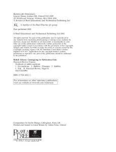
FIG. 1.12 Blood concentrations after oral administration of ethyl alcohol (2 ml/kg) The concentration declines in a zero-order fashion at an average rate of 190mg/L each hour.
OVERDOSE WITH ALCOHOLS ETHANOL (ETHYL ALCOHOL) (See also ch. 24 p. 1435) Ethanol, present in alcoholic drinks, is the alcohol most often taken in overdose. It is also found as a solvent in some cosmetic and antiseptic preparations. Its major toxicity is related to its central nervous depressant effects. The loss of the gag reflex max result in aspiration of stomach contents. Alcohols inhibit gluconeogenesis and may cause profound hypoglycaemia, particularly in children. Blood ethanol concentrations give a rough guide to the severity of overdose, but may be misleading because of tolerance in chronic alcohol users. Severe intoxication with stupor and marked incoordination is generally associated with blood concentrations above 3000 mg/L. The rate of metabolism is shown in Figure 1.12. Management Because alcohol is rapidly absorbed, lavage is unlikely to be of benefit and the compound is poorly adsorbed by activated charcoal. Metabolism is saturated even at low doses. The treatment is usually symptomatic and supportive, with correction of hypotension and hypoglycaemia. Protection of the airway is paramount. Although haemodialysis will remove alcohol efficiently, it is only indicated in severe and complicated cases of poisoning. Fructose accelerates the metabolism of alcohol, but it can potentiate the metabolic acidosis already caused by the alcohol and should not be used. Intravenous dextrose may be necessary to treat hypoglycaemia. The problems of alcoholism are discussed on page 247.
METHANOL Methanol is much more toxic than ethanol and is widely used as a solvent. Methylated spirit is 5% methyl alcohol
35
and 95% ethanol. The mode of toxicity is not clear, but accumulation of formaldehyde and formic acid is partly responsible. As little as 10 mL of methanol may cause serious illness. The initial features are those of mild inebriation, but 8 or more hours later the patient may develop abdominal pain and vomiting, dilated and unreactive pupils, a metabolic acidosis and, sometimes, signs of cerebral oedema. The optic disc is at first hyperaemic and then becomes oedematous; vision is progressively lost. Management The stomach should be emptied within the first hour of methanol ingestion; activated charcoal does not adsorb significant quantities. Intravenous sodium bicarbonate should be used to keep the arterial pH above 7.2, but metabolic acidosis may be resistant to treatment. Diazepam is indicated if convulsions are a problem. Ethanol competitively inhibits the metabolism of methanol to formaldehyde and formic acid, and is used in less serious poisoning. Haemodialysis may be necessary if the blood methanol concentration exceeds 500 mg/L, or if there is clinical deterioration despite other measures. Haemodialysis also removes ethanol, so that ethanol administration during this procedure has to be increased. Visual impairment is often permanent, although prompt treatment reduces its severity.
ISOPROPANOL (ISOPROPYL ALCOHOL)
Isopropanol is found in rubbing agents, sterilizing fluids and disinfectants, window cleaning fluids and some aftershave lotions. Features of poisoning resemble those of ethyl alcohol poisoning, but because some of it is metabolized to acetone, ketonuria and a characteristic smell of acetone on the breath may be observed. Management
The stomach should be emptied within an hour of ingestion, but treatment is otherwise symptomatic. Haemodialysis will remove isopropyl alcohol but is normally only indicated in severe poisoning.
overdose, but ophthalmoplegias, papilloedema and subsequent optic atrophy may also occur and the patient may become comatose and develop convulsions. After 12 hours respiratory and cardiovascular complications are seen, together with a metabolic acidosis, hypocalcaemia and hyperkalaemia. If the patient survives this phase, he or she may develop acute renal failure after approximately 24 hours. Ethylene glycol is not itself toxic but is metabolized by alcohol dehydrogenase to aldehydes, glycolates, oxalates and lactate, which are responsible for the toxic features. Management The stomach should be emptied within the first hour and arterial blood gases and serum calcium measured. Activated charcoal does not adsorb ethylene glycol. Any metabolic acidosis may respond to intravenous bicarbonate, and calcium gluconate may be necessary to treat hypocalcaemia. Ethylene glycol metabolism can be inhibited by administration of alcohol in doses sufficient to attain blood ethanol concentrations between 1000 and 2000 mg/L. In severe cases (ethylene glycol concentration >500mg/L, or clinical condition deteriorating despite other measures) haemodialysis is necessary to remove ethylene glycol metabolites. Ethanol administration may also be necessary for up to 4 days after the overdose to reduce their production. Dialysis may be required for renal failure. Fomepizole has recently been licensed for the treatment of ethylene glycol poisoning. 1 POISONING WITH CORROSIVE AGENTS
Fortunately, poisoning with corrosive agents is rare, and most incidents are accidental rather than deliberate. The majority of agents produce local tissue damage by protein denaturation, but some (e.g. paraquat) also have serious systemic toxicity. The patterns of damage with acids and alkalis are different and are therefore discussed separately.
ACIDS ETHYLENE GLYCOL
Antifreeze solutions contain ethylene glycol, a solvent that is sweet-tasting and therefore not unpleasant to drink. Death may occur after ingesting as little as 100 mL of ethylene glycol which, like alcohols, is metabolized by alcohol dehydrogenase. CNS toxicity is similar to alcohol
1
36
MCQ1.25
Acids produce severe pain on ingestion. They may cause asphyxia from epiglottal oedema and, although oesophageal damage has been described, they are more likely to cause gastric perforation, particularly in the region of the antrum. Late stricture formation is also a possibility. Lavage should not be performed unless the agents also have serious systemic toxicity. Any metabolic acidosis should be treated with intravenous bicarbonate, but weak alkalis should not be given by mouth. Endoscopy is of limited value in the acute phase and may cause perforation; in later stages, however, it may be helpful in identifying stricture formation. Corticosteroids have been used to try to prevent stricture formation, but their value is unclear.
SUMMARY 14 Acid poisoning • Acid ingestion may cause local corrosion, asphyxia due to oedema of the glottis and perforation of the stomach (less commonly of the oesophagus) • Metabolic acidosis and renal failure may also occur • Lavage should not be performed unless the agent produces severe systemic toxicity • Late stricture formation may occur but the value of corticosteroids in preventing this complication is not proven
Phenol (carbolic acid) can cause systemic toxicity as well as local corrosion, although the latter is normally less severe than with inorganic acids. The urine may be dark brown or black and the breath, urine and blood may smell strongly of carbolic. Haemolysis, renal failure, coma and convulsions, myocardial damage and metabolic acidosis may occur, and early cautious gastric lavage is indicated, provided the airway can be protected. Metabolic acidosis corrected with intravenous sodium bicarbonate and urine output and renal function carefully monitored. Cresols (methylphenols) have similar effects and treatment is therefore as for phenol poisoning. Formic acid is used in kettle descalers and destaining solutions. It too can produce a metabolic acidosis, intravascular haemolysis and renal failure, as well as local corrosion. Gastric lavage is therefore indicated early after ingestion, and any metabolic acidosis should be corrected with intravenous sodium bicarbonate. As with phenols, aspiration into the lung may cause haemorrhagic tracheobronchitis and the airway must therefore be protected during lavage.
ALKALIS Corrosion by alkalis (e.g. drain and oven cleaners) is generally more severe than by acids and is more likely to cause serious oesophageal, as well as gastric, damage. Stricture may develop weeks or months after the initial injury. Treatment is similar to that for acid ingestion; lavage should not be performed. Endoscopy by an experienced operator should be considered to assess the degree of injury and to prevent unnecessary hospitalization. The instrument should normally be passed only to the level of the first area of injury. It may also be useful in identifying alkalis in tablet form, which may adhere to the oesophagus (e.g. Clinitest tablets and some denture cleaners).
BLEACHES Most bleach contains sodium hypochlorite (up to 10%), which may produce corrosion in large or concentrated doses, and laryngeal oedema due to chlorine liberation. The stomach should be cautiously emptied within an hour if more than 300 mL has been ingested in an adult (100 mL
in a child), provided there is no evidence of oesophageal corrosion or severe gastritis. An H2-receptor blocking drug such as ranitidine should be given, and corticosteroids may be of value if laryngeal or pulmonary oedema is present. The sodium load resulting from ingestion of large amounts of bleach can cause hypernatraemia and hyperchloraemic acidosis.
1
DISINFECTANTS Although phenols and cresols (found in Jeyes fluid and some other disinfectants) have been largely replaced by dichlorometaxylenol, corrosive effects may occur after large or concentrated doses. Metabolic acidosis, coma, myocardial and renal damage and laryngeal oedema may also occur after severe overdose, and the stomach should therefore be emptied and the metabolic abnormalities corrected. Some disinfectants also contain isopropanol (discussed on p. 36).
POISONING WITH INSECTICIDES, HERBICIDES, FUNGICIDES AND RODENTICIDES INSECTICIDES The organochlorine group of insecticides (e.g. DDT) have now largely been replaced by compounds that are less persistent in the environment. These include the organophosphorus and carbamate insecticides.
Organophosphates The organophosphates act as inhibitors of cholinesterase in humans, causing symptoms of excessive cholinergic activity, with vomiting, abdominal pain, sweating, constricted pupils and hypersalivation. Muscle spasm, diarrhoea, convulsions and coma follow. Organophosphates can be absorbed from the stomach, skin or bronchial mucosa. Plasma cholinesterase activity, measured in several centres throughout the UK, is useful in the diagnosis of both organophosphate and carbamate insecticide poisoning. Severe toxicity is associated with a reduction in plasma cholinesterase activity to around 10% of normal. Treatment consists of atropine to antagonize the cholinergic (muscarinic) effects. The initial dose is 2 nig for adults and 0.02mg/kg for children, by intravenous injection. Large doses (up to 30 mg in 24 hours in an adult) may be necessary until there are signs of atropinization (dry skin, tachycardia, dilated pupils and dry mouth). Within the first 24 hours the use of an oxime (e.g. pralidoxime mesylate or P2S) will help to reactivate cholinesterase. It can be obtained from holding centres in the UK, details of which can be found on TOXBASE. The normal dose in moder-
37
ate or severe poisoning is 30mg/kg by intravenous injection over 5-10 minutes. Benefit should be seen within 30 minutes, but repeated doses at 4-6-hourly intervals may be required. Alternatively, it may be given by intravenous infusion at a rate of 8mg/kg/h. Intravenous diazepam (5-10 mg for an adult; 0.02mg/kg body weight for a child) is helpful in reducing twitching and may improve survival. A clear airway must be maintained and assisted ventilation may be necessary.
the patient presents within an hour of ingestion) with care because of the corrosive effects of the toxin. Charcoal (1 g/kg body weight) is left in the stomach as an adsorbent for paraquat. Oxygen therapy should be avoided if possible as it may enhance the toxicity of these agents. Finally, haemodialysis and haemoperfusion have been tried in an attempt to remove the body burden of drug, but there is no evidence that either has any effect on mortality, particularly when the more concentrated solutions of paraquat (e.g. Gramoxone) are ingested. 2
Carbamate insecticides
Dinitro compounds
Carbamate insecticides produce similar symptoms (CNS signs are less prominent) and atropine is again the treatment of choice. However, because these compounds have a reversible effect on cholinesterase, their duration of action is much shorter and pralidoxime has not shown clear evidence of benefit in poisoning with these agents. O
Dinitro compounds (e.g. dinitro-orthocresol) are generally used in the form of washes for fruit trees. They cause yellow staining of the skin but can be absorbed subcutaneously and produce toxicity by uncoupling oxidative phosphorylation. This results in pyrexia, hyperpnoea and tachycardia, with subsequent sweating, thirst, dehydration and collapse. Treatment consists of preventing severe pyrexia and replacement of electrolytes and fluid. Pentachlorophenol acts similarly to the dinitro compounds but does not produce yellow discoloration of the skin.
HERBICIDES The five major groups of herbicides are: chlorates, triazines, bipyridilium herbicides, dinitro compounds and phenoxyacetic acids.
Chlorates The chlorates are powerful oxidizing agents that can produce haemolysis, methaemoglobinaemia, haemorrhagic gastroenteritis and, in severe cases, renal failure. Treatment consists of emptying the stomach (if appropriate) and methylene blue to reverse methaemoglobinaemia.
Triazines Triazine herbicides (e.g. simazine) appear to be relatively non-toxic, and serious human poisoning has not been reported.
Bipyridilium herbicides (paraquat) The bipyridilium herbicides, particularly paraquat, are extremely toxic. Not only can they cause local corrosion, but systemic absorption leads to renal and liver damage and later progressive pulmonary fibrosis. Diquat, which is less well absorbed from the gut, can cause similar toxicity, although pulmonary fibrosis seems to be slightly less likely to occur. Treatment consists of immediate gastric lavage (if
Phenoxyacetic acids Phenoxyacetic acids are probably the most widely used herbicides. They are relatively non-toxic unless large doses have been ingested; signs of cholinergic hyperactivity are then evident, resulting in coma and occasionally ventricular arrhythmias. Other features in severe cases include hypoglycaemia, metabolic acidosis, convulsions, hypertonia, myoclonus, rhabdomyolysis and renal failure. The stomach should be emptied if appropriate, contaminated clothing removed and the skin washed with soap and water. With 2-4-D and mecoprop urinary alkalinization may enhance their rate of removal, but haemodialysis is more efficient and should be considered in severely poisoned patients.
FUNGICIDES Organic and inorganic mercurials, organotin derivatives and dithiocarbamates are all used as fungicides in industry and horticultural practice. Organotin compounds are poorly absorbed from the gastrointestinal tract and therefore have low toxicity; dithiocarbamates are also relatively non-toxic.
Organic mercurials 1
38
MCQ 1.26, 1.27
2
MCQ1.28
Inhalation or ingestion of organic mercurials can produce severe corrosion and irritation of the respiratory tract. CNS toxicity consists of tremor, memory and psychiatric disturbances, and visual impairment that may lead to blind-
ness. The chelating agents DMSA and DMPS may reduce the systemic toxicity, but dimercaprol is contraindicated.
Inorganic mercurials Inorganic mercurial fungicides (e.g. mercuric chloride) are also predominantly corrosive. Treatment includes gastric lavage, adequate analgesia and treatment of any complications. Mercurous chloride (calomel) and mercuric oxide (the most commonly used agents) are poorly absorbed and, although corrosive, systemic toxicity is unlikely. If albuminuria occurs, however, significant absorption may have taken place and plasma mercury concentration should be measured. Treatment involves the use of the chelating agents. Although dimercaprol is licensed for this use, it has to be given by deep, painful intramuscular injection, and two orally active chelating agents, DMSA (Succimer) and DMPS (Dimaval) have also been shown to be effective. Guidance on administration should be obtained from the regional poisons centre for each individual case.
Metallic mercury Ingestion of metallic mercury is of no major consequence. However, intramuscular injection of metallic mercury may cause systemic toxicity, and intravenous injection has resulted in pulmonary embolus. Inhalation of mercury vapour may produce cough, retrosternal discomfort and breathlessness due to an acute pneumonitis, and flu-like symptoms such as lethargy, fever, aching limbs and arthralgia. High-dose corticosteroids may be useful if signs of lung damage are present. Signs of systemic poisoning may be an indication for dimercaprol, DMPS or DMSA.
cerebral oedema, coma and convulsions and ventricular fibrillation have also been reported. The chelating agents dimercaprol, DMSA and DMPS may reduce the severity of poisoning. Strychnine is sometimes used as a rodenticide, and has been found as an adulterant in some recreational drugs. It is rapidly absorbed by mouth and gastric lavage is contraindicated, as a convulsion may supervene during the procedure or be precipitated by the lavage. The clinical picture is similar to tetanus, with increase in muscle tone in the limbs and facial muscles, with severe spasms, muscle rigidity and exaggerated tendon reflexes. Twitching of muscles precedes the onset of convulsions, which may typically last between 30 seconds and 2 minutes and occur repeatedly. Convulsions may be provoked by any stimulus, including touch, pain and noise. The patient may be conscious and lucid between the convulsions. Cyanosis and death may occur during the convulsions, and most patients do not tolerate more than five convulsive episodes. The increased muscle tone can lead to hyperthermia and muscle damage (rhabdomyolysis) in severe cases. Treatment involves urgently establishing and maintaining a clear airway and ensuring adequate ventilation. This may be difficult if muscle tone is increased or convulsions are occurring, and it may be necessary to paralyse the patient before an endotracheal tube can be inserted and the patient ventilated. The patient should be kept at absolute rest and any stimuli that may precipitate further convulsions should be avoided. Sensory stimuli should be minimized by nursing in a darkened room; convulsions should be treated with diazepam. In severe cases neuromuscular blockers may be needed to paralyse the patient while ventilation is supported mechanically.
RODENTICIDES Many rodenticides contain warfarin or related coumarin anticoagulants and, unless taken in huge amounts, generally cause few symptoms. If the INR does rise, vitamin K should be administered as for warfarin overdose. Alphachloralose is a CNS stimulant that can cause coma, convulsions and rhabdomyolysis in overdose. Treatment consists of managing the complications. Fluoride salts may be associated with severe toxicity, sometimes delayed in onset, including abdominal pain and diarrhoea, with hypotension and peripheral circulatory failure. Hypocalcaemia and hypomagnesaemia cause muscle spasms, weakness, convulsions and cardiac arrhythmias including ventricular fibrillation. Coma and respiratory failure may supervene. Treatment is symptomatic and supportive. Arsenic is sometimes used as a rodenticide as well as a component of some timber (tannalizing) preservatives. Abdominal pain and vomiting precede the onset of profound (sometimes bloody) diarrhoea. Hypotension,
1
POISONOUS PLANTS AND FUNGI Although the number of plants that have been suggested to be poisonous is very great, many of these contain only small amounts of toxins and generally do not cause severe poisoning. They can be classified into those: • • • • • • • • •
producing cholinergic (nicotinic) stimulation producing cholinergic (muscarinic) stimulation with anticholinergic activity with gastrointestinal activity with effects on the heart with convulsant activity with hallucinogenic effects with dermatological effects with toxicity on other organs.
Plants with cholinergic (nicotinic) activity Alkaloids similar to nicotine are found in hemlock (coniine) and laburnum (cytisine). Nausea and vomiting
39
may be followed in severe poisoning by confusion, hallucinations, convulsions, and finally coma and respiratory arrest. Treatment for severe poisoning is as for nicotine. The stomach should be emptied and activated charcoal administered. Monitoring of fluid and electrolytes and adequate replacement therapy, together with measures to assist the respiration and cardiovascular functions, are generally only necessary in severe poisoning (rarely caused by laburnum).
Plants with cholinergic (muscarinic) activity Plants with cholinergic activity include the clitocybe and inocybe fungi. Ingestion produces increased perspiration, abdominal pain and visual disturbances. Treatment is with atropine, 1.2mg intramuscularly, repeated if necessary.
and delphinium may cause bradycardia and muscular weakness.
Plants with convulsant activity Cicuta viirosa (cowbane) and Qenanthe crocata (hemlock water dropwort) contain potent alcohols with effects similar to picrotoxin in antagonizing the CNS inhibitory effects of y-amino butyric acid. In severe cases nausea, vomiting and hypersalivation are followed by convulsions and respiratory failure. Convulsions can be controlled with either short-acting barbiturates or diazepam; respiration may require support until the effects of the toxin wear off. Strychnine is found in the seeds of the tree Strychnos nux vomica, native to India. Features and management are described under rodenticides (page 39).
Plants with hallucinogenic effects Plants with anticholinergic activity Plants with anticholinergic activity include the deadly nightshade (Atropa belladonna}, the thorn apple and henbane. In addition to atropine these plants may contain hyoscine and hyoscyamine. The symptoms are similar to those caused by drugs with anticholinergic activity (e.g. tricyclic antidepressants) and management is supportive.
Plants with gastrointestinal toxicity Oxalates present in some plants can cause local gastrointestinal irritation and painful ulceration of the mouth if chewed by children. These include the household plants dumb cane (Dieffenbachia) and some of the Monstera and philodendron species. Other plants contain irritant resins and saponins (e.g. arum, yew, bryony and Daphne mezereum), which can cause severe vomiting and diarrhoea. The beans of the Abrus species (e.g. Ricinus and Robinia) can cause delayed gastrointestinal symptoms as they contain compounds that can impair RNA synthesis. Solanum species also produce delayed gastrointestinal symptoms: these include potatoes that have been allowed to sprout, and woody and black nightshades. Treatment is supportive, with replacement of fluids and electrolyte loss.
Plants causing cardiovascular disturbances Digitalis glycosides are found not only in the foxglove but also in lily of the valley (Convallarid). Treatment is as for digitalis poisoning. The aconitine present in the monksfoot
1
40
MCQ1.29
In addition to those anticholinergic agents with hallucinogenic effects, there are several other hallucinogens in plants and fungi. These include the psilocin and psilocybin in psilocybes (magic) mushrooms, the tetrahydrocannabinol in cannabis, and the ibotinic acid and muscimol found in Amanita muscaria. Tranquillizers may be necessary if the patient is a risk to himself or to others, and adequate supervision must be given until the hallucinations subside. Table 1.13 lists various plants and their toxicities.
Plants that cause dermatological toxic ity Plants contain substances that can cause either direct chemical irritation (e.g. histamine and oxalic acid) or delayed hypersensitivity reactions (e.g. Primula obconica and the hogweeds, that can produce photoallergic dermatitis and several other species). Treatment is symptomatic.
Plants with toxic ity on other organs The lectins present in the seeds of Ricinus communis cause delayed gastrointestinal tract symptoms. Haemolytic anaemia may also occur, and haemoglobin precipitation in the renal tubules may cause acute renal failure. The principal lectin, ricin, is the most potent toxin known to man. Management is largely symptomatic and supportive. The beans of Ricinus communis are also highly allergenic and may cause anaphylaxis. Severe liver damage can also be caused by the phallotoxins and amatoxins found in Amanita phalloides and some of the other Amanita fungal species. Cooking or drying does not destroy these toxins. Vomiting, abdominal pain and diarrhoea often occur 6-12 hours after ingestion, and albuminuria, haematuria, renal failure and liver failure may occur around 1-3 days later. Treatment consists of emptying the stomach and replacing fluid loss. Specific antidotes are of unproven value, but thioctic acid and high doses of penicillin may be of value in preventing the liver and renal damage.
Blue-green algae are actually cyanobacteria, which grow as blue-green or brown rafts or sediments, usually in stagnant water, and which produce endotoxins. Their disagreeable odour usually prevents human consumption, but deaths have occurred in cattle. In humans local irritation and allergic skin reactions may occur. Consumption of contaminated water by humans may result in acute gastroenteritis, which usually resolves in 1-2 days. Occasionally a hepatitic picture or atypical pneumonia has been reported. Treatment is normally symptomatic and supportive. O
TABU 1.13 Plants and toxicity Plant or fungus
Latin name
Toxic features
Black (garden) nightshade Blue-green algae
Solarium nigrum Microcystis/Anabaena/ Aphanizomenon/ Oscillatoria Bryonia dioica Ricinus communis Clitocybe sp. Cicuta vimsa Daphne mezereum
Gastrointestinal Hepatotoxic Dermatological
Atmpa belladona Amanita phal/oides Dieffenbachia Amanita muscaria Digitalis purpurea Heracleum mantegazzianum Conium maulatum Oenanthe cmcata Hyoscamus niger Inocybe sp. Laburnum anagroides Conva/laria Psilocybes sp. Aconitum nape/lus
Anticholinergic Hepatotoxic Gastrointestinal Hallucinogenic Cardiotoxic Dermatological
Nerium oleander Thevetia peruviana Solanum tuberosum Primula obconica Philodendron Monstera deliciosa Datura stramomium Arum maculatum Solanum dulcamara
Cardiotoxic Cardiotoxic Gastrointestinal Dermatological Gastrointestinal Gastrointestinal Anticholinergic Gastrointestinal Gastrointestinal
Taxus baccata
Gastrointestinal
Bryony (white/red) Castor oil plant Clitocybe fungi Cowbane/water hemlock Daphne/mezereon/spurge olive Deadly nightshade Death cap fungus Dumbeane/Leopard lily Fly agaric Foxglove Giant hogweed Hemlock Hemlock water dropwort Henbane Inocybe fungi Laburnum/Golden rain tree Lily of the valley Magic mushrooms Monkshood/aconite/ wolfsbane Oleander (pink) Oleander (yellow) Potatoes (sprouting) Primula obconica Sweetheart vine Swiss cheese plant Thorn apple Wild arum/cuckoo pint Woody nightshade/ bittersweet Yew
Gastrointestinal Gastrointestinal Cholinergic (muscarinic) Convulsant Gastrointestinal
POISONOUS ANIMALS
1
ADDER ENVENOMATION The adder, Vipera hems, is the only indigenous poisonous snake in western Europe. Only 50% of those bitten develop signs of envenomation, which may occur either immediately, with shock, vomiting and explosive diarrhoea, or several hours later. The initial collapse may be due to activation of the kinin system; the later shock is more often due to hypovolaemia, caused by increased capillary permeability and fluid loss into the swollen limb, and direct cardiotoxicity of the venom (Fig. 1.13). Management Treatment includes reassurance, as shock may be exacerbated by the fear of impending toxicity. The limb should be kept still, but a ligature should not be used unless there is likely to be a delay of more than half an hour between the patient having been bitten and transfer to hospital. If a ligature is used, it should be tight enough to prevent
Cholinergic (nicotinic) Convulsant Anticholinergic Cholinergic (muscarinic) Cholinergic (nicotinic) Cardiotoxic Hallucinogenic Cardiotoxic
FIG. 1.13 Adder bite on the finger of a child A Note the twin puncture wounds and B subsequent oedema involving the hand and arm.
41
venous return but not to obstruct arterial inflow. If there are signs of toxicity the patient should be admitted and observed for at least a day after the bite. Pulse and blood pressure should be recorded, together with urinary output and fluid losses from diarrhoea and vomiting. Local swelling should also be noted, and the white cell count, urea and electrolytes should be measured each day. The use of antivenom should be considered if hypotension persists, ECG signs occur, the white count rises markedly to above 20000/mm3 or, in an adult, severe swelling has extended up the limb within 2 hours after the bite. Anaphylactic reactions to the antivenom occur in 1 % of patients and so antivenom should only be used when the indications described above are present. A history of asthma or allergy is a relative contraindication to its use. Antivenom is given by slow intravenous infusion and is stopped at the first signs of allergic reaction. Adrenaline (epinephrine) solution must be drawn up prior to administration of the antivenom and given intramuscularly if a reaction occurs (intravenously in the case of a severe reaction). Deaths from adder bite are extremely rare but local tissue necrosis may be severe, particularly in adults.
FISH ENVENOMATION
In the UK the only venomous fish regularly found are the Weaver fish (Trachinus vipera), which is found around the coast in the summer months, and the Lion fish (an aquarium species). Both contain heat-labile venom in their spiny dorsal fins, which causes severe local pain. Hypotension, and myocardial and respiratory depression may develop. Management Treatment is to remove any barbs and clean the wound, immersing the limb in water as hot as can be tolerated without discomfort to destroy the toxin. The wound should be examined at a later stage for signs of secondary infection.
VENOMOUS INVERTEBRATES
The Portuguese man of war (Physalia physalis) is the most poisonous jellyfish found around the coasts of the UK. Its sting contains substances capable of releasing histamine and other kinins. Local pain is common, but more serious symptoms include abdominal pain, dyspnoea, hypotension, muscular paralysis and convulsions. Most other jellyfish found around the UK are harmless, but the compass jellyfish (Chrysaora hyoscella), the lion's mane (Cyanea O MCQ1.30
42
capillata) and the sea nettle (Cyanea lamarckii) can cause a sting. Management The wound should be bathed in vinegar solution (4-6% acetic acid), and any tentacles still adherent removed with forceps, a gloved hand or adhesive tape. Treatment is otherwise symptomatic.
ARTHROPOD ENVENOMATION Wasp and bee stings contain amines, kinins, peptides and enzymes that cause local pain, erythema and swelling. Systemic effects are rare unless the stings are numerous or the subject is hypersensitive, in which case even a single sting may kill. Initial symptoms are generalized urticaria, flushing, dizziness, bronchospasm, collapse and coma. Serum sickness may occur after a week or more. Management The sting of the bee should be removed by flicking or scraping, taking care not to squeeze it and inject more venom. Severe pain may respond to local anaesthetic or aspirin. Swelling may respond to antihistamines and corticosteroids. Anaphylaxis should be treated immediately with adrenaline (epinephrine) (0.5-lmL of a 0.1% solution intramuscularly for an adult and 10|ig/kg for a child). The dose may be repeated every 10 minutes if necessary. Stings in the mouth, even in patients who are not hypersensitive, may cause severe respiratory tract obstruction. In allergic individuals, specific venoms have been used in increasing doses to produce desensitization.
POISONOUS SEAFOODS
Seafood may contain bacteria (e.g. Vibrio, Salmonella) or viruses (e.g. hepatitis A); toxicity may also occur as a result of contamination by dinoflagellates or decomposition by bacteria. Antemortem infestation by dinoflagellates of certain subtropical fish (such as groupers and snappers) may occur, but contamination usually involves shellfish, such as mussels, oysters, scallops and clams. The dinoflagellates contain neurotoxins (e.g. saxitoxin) which can cause nausea, vomiting and diarrhoea, followed by circumoral paraesthesiae, ataxia, muscular weakness and, in severe cases, respiratory paralysis. Treatment is symptomatic, with respiratory and cardiovascular support. Postmortem contamination of some oily (dark-meat) fish, such as mackerel, skipjack and tuna, sardines and pilchards, by Proteus bacteria may result in the production of histamine and other unidentified toxins that produce urticaria, flushing, abdominal pain, nausea, vomiting and diarrhoea and, in some cases, bronchospasm. This 'scombroid fish' poisoning occurs around 4 hours after ingestion,
generally lasts only a few hours and is seldom severe. Treatment is symptomatic. Prevention involves keeping the raw fish frozen or on ice to prevent decomposition by bacteria.
POISONING BY METALS IRON Although mortality from iron poisoning has fallen in recent years, it remains one of the most dangerous metals in overdose. Children are particularly sensitive to its effects. Abdominal pain, nausea and vomiting are often followed by gastrointestinal bleeding, encephalopathy, metabolic acidosis, pulmonary oedema, coma, shock, acute renal and liver failure. Doses of elemental iron of more than 150mg/kg body weight produce severe and possibly fatal toxicity. A serum iron level greater than 5mg/L (90 pmol/L) within 4 hours of the overdose is associated with an increased likelihood of severe poisoning. Coma and shock are manifestations of severe toxicity.
Management The stomach should be emptied if more than 60mg/kg has been ingested within the previous hour. Radiography of the abdomen in the first 2 hours may reveal whether or not iron tablets have been taken. Blood should be taken for urgent measurement of serum iron concentration. If coma or shock develop, desferrioxamine should be considered immediately without waiting for the serum iron result. The dose is 15mg/kg body weight/hour for 5 hours. It often causes hypotension if infused more rapidly than this, and can occasionally cause rash and anaphylactic-like reactions. Pulmonary oedema and ARDS have been reported in patients treated with more than 80 mg/kg/day for longer than 24 hours. With prompt treatment the mortality should be less than 5% in severe cases. 1
HEAVY METAL POISONING Heavy metals can cause acute or chronic symptoms,
SUMMARY 15 Iron poisoning • Children are particularly sensitive to the toxicity of iron • Abdominal pain, nausea and vomiting may be followed by gastrointestinal haemorrhage, encephalopathy and renal and hepatic failure • Severe poisoning is normally associated with plasma concentrations around 4 hours after ingestion of more than 5mg/L, with CNS signs and shock • Intravenous desferrioxamine should be given as soon as possible in severe iron poisoning
depending on the dose and duration of exposure. Acute ingestion of most heavy metals will cause gastroenteritis. Generalized convulsions may also be seen, particularly with lead poisoning, and renal failure and cardiac arrhythmias have also been recorded. Inhalation of heavy metal fumes may cause chemical pneumonitis and, in severe cases, pulmonary oedema, and a syndrome known as 'metal fume fever' may occur several hours after the inhalation of some metallic oxides. Chronic effects of heavy metal exposure are similar but milder. Chronic gastrointestinal symptoms and CNS effects occur, particularly after exposure to lead, manganese and mercury. Lead, thallium, bismuth and arsenic can also produce peripheral neuropathy, and gold, lead, cadmium and mercury may cause renal damage. Inhalation of heavy metal fumes can cause pulmonary fibrosis, particularly with beryllium, cadmium, tungsten, titanium and cobalt. An emphysematous picture has been seen with cadmium, and an asthma-like syndrome may occur as a result of sensitivity to chromium, vanadium and platinum. Nickel, chromium, cobalt, platinum and beryllium may also cause skin sensitization and subsequent dermatitis. Taking a full occupational history helps diagnosis. Clinical examination may reveal specific signs of poisoning, such as the blue line on the gums in chronic mercury poisoning or the raindrop pigmentation of the skin in arsenic poisoning. Toxicological examination of the urine, blood, or hair and nail clippings may also help, but this often has to be performed in specialized centres.
1
Management Treatment consists of preventing further exposure to the heavy metal, treatment of the complications of poisoning, and chelation therapy to enhance the elimination of the metal already absorbed. It must be remembered, however, that most chelating agents are toxic and should only be used when their benefits are likely to outweigh the risks of their use. Sodium calcium edetate is the treatment of choice for lead poisoning. It must be given by slow intravenous infusion. Dimercaprol, DMSA and DMPS are effective in severe mercury and arsenic poisoning. Finally, Prussian blue has been used successfully in the treatment of thallium poisoning.
FURTHER READING British National Formulary. British Medical Association and Royal Pharmaceutical Society of Great Britain, published biannually. An excellent reference text and practical guide to prescribing, with useful tables to prescribing in special circumstances and drug interactions. Laurence D R, Bennett P N 1997 Clinical pharmacology, 8th edn. Churchill Livingstone, Edinburgh. Highly readable, with a practical therapeutic flavour. Ritter J M, Lewis L D, Mant T G K 1998 A textbook of clinical pharmacology, 3rd edn. Edward Arnold, London. A practical guide to clinical pharmacology and drug therapy.
43
This page intentionally left blank
2
Heat cramps
Physicaland
Environmental Disorders
Spasms of painful muscular contraction may occur with sudden rises in temperature during strenuous activities such as long-distance running, or during work in very hot environments. Muscles of the legs and arms are usually affected, typically after the exertion or heavy work is over. There is usually hyponatraemia, and severe cramps may be accompanied by a rise in creatine phosphokinase and evidence of rhabdomyolysis. Part of the pathogenesis appears to be due to replacement of fluid and salt loss by water with too low a salt content; the condition is preventable by drinking fluids containing 0.25% saline.
2
Heat exhaustion John Moxham and Robert L Souhami
Disorders due to heat and cold 45 Whole body radiation exposure 47 Disorders due to extremes of barometric pressure 49
Submersion and drowning 51 Electric shock 53 Smoke inhalation 54 Chemical warfare 54
DISORDERS DUE TO HEAT AND COLD Heat produced by metabolism is lost mainly through the skin and, to a lesser extent, through the gut, urine and breath. Inadequate heat loss leads to sweating and vasodilatation. Cold produces cutaneous vasoconstriction and shivering, which results in increased muscle metabolism and heat production. In moving from a cold to a hot climate, acclimatization occurs over a period of 1-2 weeks; this process is not well understood. There is increased sweating and the salt content of sweat diminishes. Aldosterone secretion increases, at least at first, causing a low urinary sodium with loss of potassium. A progressive rise in temperature above 38-39°C causes irritability and mental confusion. At temperatures above 40-41°C vascular, renal and hepatic damage occur.
HEAT EXCESS Three syndromes due to excess heat occur: heat cramps, heat exhaustion and heatstroke.
Heat exhaustion or heat collapse is the commonest syndrome and is especially likely to occur in individuals exposed to a hot environment for a few days. The very young and the elderly (especially those on diuretics) are particularly at risk. The syndrome is due in part to salt and water loss, and in part to a failure of circulatory adaptation to heat. When water loss predominates, there is intense thirst and weakness, agitation and confusion. Water depletion is more likely to occur in acclimatized people (whose sweat contains less salt). The plasma Na+ and Cl~ rise. With salt depletion, there are muscle cramps, headaches, vomiting and myalgia. Haemoconcentration occurs with a rise in haematocrit and blood urea. Usually, salt and water depletion occur together, and the patient has a normal temperature and appears pale and clammy. The disorder is treated by rest and oral fluids. In water depletion (where plasma sodium is elevated) this should be with water. Typically 5-8 L may be given in 24 hours by mouth or as i.v. glucose. When salt depletion is present the oral fluids should contain salt (e.g. 5L of fluid with 20 g salt).
Heatstroke Heatstroke is a serious and sudden disease which may be fatal unless treated urgently. It is caused by a failure of thermoregulation, resulting in a rapid rise in body temperature. In normal individuals it is usually due to exercise or work in a hot, humid environment. When it occurs in sedentary individuals the patient is usually elderly, often with underlying disease such as cardiac failure, diabetes (with autonomic dysfunction), obesity or alcoholism. Atropine-like drugs, p-blockers, phenothiazines and diuretics predispose to the syndrome. Heatstroke can also occur in fit men working in hot environments, and there are some clinical differences in these cases. Typically the patient develops headache, dizziness and faintness, and abdominal pain and delirium may occur. Sweating stops and the body temperature rises rapidly (and may reach 43°C). Hypotension, renal failure and
45
rhabdomyolysis may occur; the latter is especially common in heatstroke following severe exertion. In this form of heatstroke sweating may continue. On examination the skin is hot and usually dry, and the patient exhibits tachypnoea and flaccidity. Coma and shock are grave signs. The blood shows leukocytosis and respiratory alkalosis (but may be acidotic in the exertional form), the urea is raised, and hypokalaemia and hyperphosphataemia occur. Myocardial necrosis may give ST changes and even an infarction pattern on the ECG. Liver function tests are abnormal, and acute renal failure may occur. Disseminated intravascular coagulation is present in mild degree in the elderly, but may be severe in exertional heatstroke. Management Treatment is urgent. The patient must be cooled as quickly as possible. The most effective way is immersion in cold water, the skin being rubbed to increase the efficiency of cooling. An alternative is to blow cold air over dampened skin. Core temperature must be monitored using a rectal thermometer. Hypotension can be treated with small quantities of saline, taking care to avoid circulatory overload. Hypoglycaemia, if present, can be treated with glucose, and severe acidosis by sodium bicarbonate. More serious complications, such as acute renal failure and disseminated intravascular coagulation, are treated in the usual way (Chs 20 and 23). Cerebellar degeneration may occur, and coma with cortical damage is a terminal event. 1
SUMMARY 1 Clinical features of heatstroke Symptoms • Headache • Dizziness/faintness • Abdominal pain Signs • Increased body temperature • Hot, dry skin (sweating may be absent) • Tachypnoea • Flaccidity • Hypotension • Delirium -> shock/coma Results/complications • Hepatic and renal failure • Leukocytosis, alkalosis/acidosis, hypokalaemia • Rhabdomyolysis • Myocardial necrosis • Disseminated intravascular coagulation • Cerebellar degeneration • Coma, cerebral damage
as 'ecstasy'. Muscle contraction begins after the start of anaesthesia, and body temperature rises rapidly. The anaesthetic must be stopped, the patient cooled and ventilated, and i.v. dantrolene sodium given (2-10mg/kg). Acidosis should be treated. 0
HYPOTHERMIA
Neuroleptic malignant syndrome This syndrome is characterized by muscle rigidity giving rise to hyperpyrexia, tachycardia and impaired consciousness. It is an idiosyncratic reaction to drugs that block dopaminergic transmission, such as phenothiazines and butyrophenones. The increase in muscle tone develops over 12-72 hours. Death from respiratory failure may occur. Treatment is by muscle relaxants, including neuromuscular blocking agents, and ventilation if necessary. Recovery may take several days.
Significant hypothermia is an uncommon problem mainly affecting the elderly, in whom the response to cold may be defective (p. 178). Hypothermia contributes to the excess mortality in this population during winter months. It is defined as being present when the deep body temperature falls below 35°C. As the mouth temperature may fluctuate depending on the ambient air temperature, it is best to use a rectal thermometer when monitoring hypothermic patients. A low-reading thermometer is necessary. The rectal temperature is about 0.5°C higher than oral.
Malignant hyperpyrexia
Aetiology
This is an autosomal dominant disorder in which hyperpyrexia is produced by skeletal muscle contraction following anaesthetic agents such as halothane and suxemethonium and local anaesthetics. It can also occur with drugs such as amphetamines and derivatives such
Hypothermia may be due simply to an age-related impaired thermoregulatory response to a cold environment (Ch. 7, p. 178). In the majority of patients, however, there is an associated underlying pathological cause, including any cause of autonomic dysfunction (p. 178). Hypothyroidism and hypopituitarism are both associated with hypothermia, and the risk is also higher in immobile or demented patients, or in the presence of an acute illness such as bronchopneumonia. There is a clear association between phenothiazines and impairment of temperature regulation, and hypothermia may also be precipitated by sedative drugs, such as hypnotics and alcohol. Although
1
46
Case 2.1
2
MCQ2.1
3
MCQ2.2
a mild drop in body temperature is common, clinically important hypothermia is unusual. However even modest falls in body temperature may produce changes in coagulability, which may predispose to myocardial infarction. Immersion hypothermia is produced by shipwreck. Exposure to cold weather occurs in hillwalkers and climbers, especially if they have not eaten or if they drink alcohol.
Clinical features Hypothermic patients typically live alone in poor housing and have impaired mobility. A typical case would be that of an old person who gets out of bed in the middle of the night to go to the lavatory, falls, and then lies all night on the cold floor in thin nightclothes until discovered the next morning by a neighbour. The skin has a greyish look owing to a combination of vasoconstriction and cyanosis; the face may appear puffy, resembling myxoedema. The diagnosis is often first suspected during examination, when it is found that the patient's skin feels cold in normally warm areas, such as the axilla or the abdomen. There may be ataxia and slow mental response. The patient is often confused when the temperature falls below 35°C, and drowsiness and loss of consciousness occur when the temperature falls below 29-33°C. Shivering is absent below this temperature; instead, there is increased skeletal muscle tone with neck stiffness and abdominal rigidity. The heart rate is slowed and the ECG may show a prolonged PR interval and J waves (Fig. 2.1). Respirations are slow and shallow, and there may be hypoxia and hypercapnia. Hypoxia is exacerbated by a shift of the oxygen dissociation curve to the right, leading to impaired release of O2 from haemoglobin. Acute pancreatitis is often found at necropsy, but this is only rarely diagnosed in life. Impaired renal and respiratory function may lead to a raised blood urea or bicarbonate. Blood sugar levels may be raised, but do not indicate diabetes mellitus, and will fall as the temperature returns to normal. Serum asparate aminotransferase and creatine kinase may be raised due to muscle damage.
Management The lower the deep body temperature, the higher the mor-
SUMMARY 2 Clinical features of hypothermia • • • • • • • •
Contusion/impaired consciousness Cold, greyish skin Shivering absent Increased skeletal muscle tone Slow heart rate, prolonged PR interval and J waves Slow, shallow respiration, hypoxia and hypercapnia Raised blood urea or sugar levels Raised serum asparate aminotransferase and creatine kinase
2
tality (about 50% overall). This is because many hypothermic patients have a serious underlying illness. Patients with mild hypothermia (core temperature 32-35°C) should be wrapped in a space blanket and nursed in a side ward where the ambient temperature is 28-30°C. Their temperature should be allowed to rise gradually, at a rate of about 0.5°C per hour, as fast surface rewarming carries the risk of hypotension. For this reason, the patient's pulse and blood pressure must be monitored. If the blood pressure drops, the space blanket should be removed or the room temperature lowered so that the patient is temporarily cooled. Intravenous fluids are hazardous and may produce acute pulmonary oedema. A broad-spectrum antibiotic is usually given parenterally because bronchopneumonia is nearly always present, even if not clinically apparent. The ECG should be monitored, as both bradyarrhythmias and ventricular fibrillation may occur. The patient is at serious risk of developing a pressure sore and must be nursed on a suitable mattress and turned regularly. When the deep body temperature is below 32°C a more aggressive approach to resuscitation is sometimes advocated, although there is little evidence that this reduces mortality. These measures include positivepressure ventilation to correct hypoxia, measurement of central venous pressure, and active rewarming using a radiant heat cradle over the torso or warmed intravenous fluids.
Thyroid hormones should not normally be given to hypothermic patients. If there is good clinical or laboratory evidence of hypothyroidism, then triiodothyronine (5ug) may be given by slow intravenous injection every 12 hours. A particular problem is presented by patients who have become hypothermic as a result of immersion at sea. They may develop ventricular fibrillation on rescue if they exert themselves. They should therefore be kept still and rewarmed slowly. 3
WHOLE BODY RADIATION EXPOSURE
FIG. 2.1 Electrocardiogram showing J wave (arrowed) in a hypothermic patient
There are two broad types of ionizing irradiation: photons (y and X-rays) and particulate (a and |3 particles). Radiation dose is defined in a variety of ways. The energy
47
deposited in a tissue is measured in grays (Gy; IGy = 1 J/kg). Because different types of radiation have different tissue effects, a unit called the Sievert (Sv) is sometimes used, which weights the radiation dose according to the type of radiation. Particulate radiations are absorbed according to their mass and energy, but do not penetrate deeply into tissues (see also Ch. 6, p. 159). Particulate irradiation transfers more energy and is much more ionizing than Y or X-rays.
EXPOSURE Exposure to ionizing irradiation comes about in three ways: • Background irradiation. We are exposed to approximately 2-3 mSv per year from solar and geological irradiation. Cosmic rays are mostly very high-energy protons. Secondary radiations, from the upper atmosphere, are largely y-rays. Geological irradiation is largely due to radon. • Medical exposure. The therapeutic uses of ionizing irradiation are described in Chapter 6. Accidental exposure of patients or staff has been rare since the advent of stringent precautions following the radiation-induced damage to skin and eyes in the early days of therapeutic and diagnostic radiation. Exposure to whole-body radiation forms part of the treatment of leukaemia and lymphoma. The resulting bone marrow suppression is treated by allogeneic (or autologous) haemopoietic stem cell transplantation. • Military and industrial irradiation. The atomic bombs dropped on Hiroshima and Nagasaki led to numerous cases of acute leukaemia and other cancers. Fallout from nuclear tests has increased annual background radiation by 1%. Industrial exposures also occur, the most dramatic example of which has been the ingestion of radium by radium dial painters from 1916 to 1926, resulting in a great increase in risk of bone sarcoma and cancer of the air sinuses of the skull. The accident in the Chernobyl nuclear power station in 1986 resulted in many deaths due to acute radiation exposure, and an increase in background radiation over a wide geographical area.
TISSUE DAMAGE The tissue-damaging effect of ionizing irradiation is probably related mainly to damage to DNA, leading to strand
1 48
MCQ2.3
breakage and impaired reproductive integrity of the cell. At 0.1 Sv total body exposure there is a slight fall in blood lymphocyte count. At ISv there may be mild radiation sickness with slight nausea and a more profound lymphopenia, and a fall in neutrophils and platelets 2-3 weeks after exposure. Following greater degrees of whole-body exposure certain tissues are damaged acutely by relatively low doses: • Bone marrow. This can regenerate after exposure to 10 Gy, but above this dose permanent aplasia may occur. The white count and platelet count begin to fall within 10 days of exposure. • Intestine. Doses of 10 Gy or over cause severe loss of crypt cells, leading to loss of villi and extensive ulceration. The onset of nausea and vomiting is 1-2 hours after exposure, often recovering a few hours later, but above 10 Gy permanent and fatal damage to the gut occurs, with death in 4-14 days. Death is due to severe diarrhoea, consequent salt and water depletion, and severe Gram-negative sepsis. • Skin. Erythema occurs at doses below 10 Gy. At 20 Gy the skin starts to desquamate and ulcerate. • Lung. Above l0 Gy in a single fraction pneumonitis occurs, and is increasingly severe with increase in dose. Long-term changes include glomerular loss and interstitial nephritis, infertility, pulmonary fibrosis, neuronal loss and gliosis, intestinal stricture and hepatic fibrosis. After acute radiation exposure (such as at Chernobyl and Hiroshima) there is an increased risk of leukaemia at 5-10 years. The safe lower limit is not easily quantifiable but is probably below l0c Gy. Solid tumours may also occur, including bone cancer in radium dial painters, skin cancer, and lung and breast cancer. The risk of solid tumours appears to be 1 in 2000 following total body exposure of 1-2 cGy.
Clinical features After acute whole-body exposure, the clinical features depend on the received dose. Following an initial period of nausea and vomiting (sometimes with parotid swelling), the patient may be relatively well for 7-10 days and then develop a syndrome of haematological failure. There is depression of the white count and platelets, with infection, bruising and bleeding. At the same time skin reactions occur, with desquamation, alopecia and ulceration. If the whole-body dose is above 15 Gy the gastrointestinal syndrome may predominate and be the first sign of toxicity. In a patient with severe leukopenia severe diarrhoea and fluid loss are followed by septicaemia from gut bacteria. This is almost always fatal. Higher doses of whole-body radiation (25-60 Gy) cause cardiovascular collapse and shock.
Management Management is supportive. Antiemetics are given in the acute phase, and fluid losses from diarrhoea are replaced. Transfusion of blood and platelets and intensive antibiotic treatment for gut derived organisms are used during the phase of myelosuppression and infection. Haemopoietic growth factors such as GCSF may be of value. Allogeneic bone-marrow transplantation may help the minority of patients for whom there is a donor, and in whom the haematological toxicity is the major life-threatening complication. It is of no value if the major toxicity is in organs such as the lungs or gastrointestinal tract. Accidental exposure to radionuclides is treated according to the isotope. Potassium iodide is given for I31 I ingestion, and soluble phosphate for 32P overdose. Overdose with bone-localizing isotopes (caesium, radium, strontium) is treated with EDTA and large doses of oral calcium. There has been some interest in drugs that limit damage from radiation: unfortunately, these have to be given before or immediately after exposure. The best known is ethyol, which may also protect normal tissues from cytotoxic drug damage. Late effects occur 3-10 years after exposure. There is an increased risk of acute myeloid leukaemia. The risk of solid tumours is about 1 in 2000. Tumours include cancer of skin, breast and lung. This is a small increase compared to the background incidence in the population.
height. Commercial aircraft are maintained at a pressure equivalent to 2500 metres, so that no additional oxygen is needed unless a patient has severe cardiac or respiratory disease. If there is a loss of pressurization, extra oxygen is made available through masks. Acclimatization to altitude is a complex process. On rapid ascent the relative hypoxia stimulates ventilation, but this stimulus diminishes over a few weeks. This hyperventilation diminishes Paco2, with the consequent development of a respiratory alkalosis. The respiratory centre appears to adapt to a lower Paco2. The acute circulatory adaptation consists of a rise in cardiac output, a fall in renal, cardiac and cutaneous blood flow, and a rise in cerebral blood flow. These changes slowly reverse, and after several weeks cardiac output on exercise is lower than at sea level. The red cell mass increases greatly and with it the total oxygen-carrying capacity of the blood. The oxygen dissociation curve does not shift substantially (as the alkalosis shifts it to the right and the hypoxia to the left). There is an increased formation of tissue capillaries, greatly increasing tissue oxygenation. There are four recognized syndromes of altitude sickness: • • • •
2
Acute altitude sickness Acute pulmonary oedema Acute cerebral oedema Chronic altitude sickness.
Prognosis With adequate treatment, most patients will survive acute exposures up to 10 Gy. Above this dose, bone-marrow failure and the intestinal syndrome are usually fatal. 1
DISORDERS DUE TO EXTREMES OF BAROMETRIC PRESSURE
ALTITUDE SICKNESS The effects of reduced barometric pressure result from the low partial pressure of oxygen and reduced oxygencarrying capacity of the blood. Figure 2.2 shows this relationship. The barometric pressure at sea level is 100 kPa, and the partial pressure of oxygen is 21% of this, i.e. 21 kPa. At 5500 metres the barometric pressure is reduced to 50% (50kPa). The partial pressure of oxygen is, however, less than 20% of this (l0kPa) because the proportion of water vapour rises. Because of the sigmoid shape of the oxygen dissociation curve, oxygen saturation is reasonably well maintained until an altitude of 5000 metres. A precipitous fall in saturation then occurs with increasing
FIG. 2.2 Effect of altitude on alveolar Pao2 and oxygen saturation Because of the steep slope of the oxygen dissociation curve, increasing altitude above 5000 metres causes a precipitous fall in saturation. In an unacclimatized person, acute changes occur at: IT1 3000 metres (10000 feet) - slightly impaired memory and judgement, increased heart rate, abdominal cramps and nausea; [B] 3500 metres (12000 feet) - headache, nausea, diminished visual acuity, and possible pulmonary oedema; [C] 5500 metres (18000 feet) - impaired consciousness after several hours in many people; and [D] 6750 metres (22000 feet) - loss of consciousness.
49
Acute altitude (mountain) sickness This syndrome occurs in unacclimatized individuals who ascend rapidly to heights in excess of 2500 metres. People who are unfit, or very young, or those who have respiratory or cardiac disease, are especially prone. The symptoms usually begin at 12-24 hours after ascent. Lightheadedness is followed by lethargy irritability and severe headache, usually occipital in position. Other symptoms include nausea and vomiting, dyspnoea with periodic respiration, and abdominal pain. Physical examination reveals little, although ataxia (shown by walking heel to toe) may be present. The cause of acute altitude sickness is unknown. It is possible that the hypoxia causes mild cerebral and brainstem oedema. This is suggested on MRI scans and by multiple petechiae in the brain in fatal cases. The syndrome is preventable by slow ascent. Individual susceptibility varies greatly and mountainous hikes and climbs should never put pressure on participants to ascend rapidly, especially if they start to feel unwell. When starting an ascent above 3000 metres a reasonable pace is 300m/day, resting for a day every 3 days. Even this may be too fast for some people. Dehydration should be avoided, and a good diet maintained. Treatment is to avoid sedatives, to take mild analgesics for headache, and if the symptoms do not abate, to descend to 1000m to await recovery. Acetazolamide can alleviate the symptoms of acute altitude sickness by acting as a respiratory stimulant, lowering the Paco2 and raising the Pao2. If ascent cannot be made slowly it may be advisable to start acetazolamide (250 mg b.d.) before ascent, and climbers should be warned of the side-effects, particularly paraesthesiae. Acute pulmonary oedema Rapid ascent to heights greater than 3000 metres may provoke the sudden onset of acute pulmonary oedema. Hypoxia is the cause, but the pathogenesis is unclear. The symptoms usually begin 5-36 hours after ascent; acute dyspnoea, haemoptysis and widespread crackles in the lungs occur. Pathologically there is acute oedema and a 'hyaline' membrane in bronchioles. Treatment is with urgent evaluation to a lower altitude, oxygen and diuretics. Nifedipine has been shown to be useful prophylactically in those who have had a previous episode of pulmonary oedema. Acute cerebral and retinal oedema Acute hypoxia increases cerebral blood flow and intracranial pressure, and can cause cerebral oedema. This is asso-
50
1
Fig. 2.1
4
Fig. 2.2
2 Case 2.2 3 MCQ 2.4
ciated with headache and brain dysfunction. The patient may be incoherent, drowsy, ataxic, and progress to coma. There may be papilloedema. Deepening coma leads to death. The treatment is urgent descent to a lower altitude and oxygen. Dexamethasone (i.v.) should be given. Cerebral and pulmonary oedema often accompany each other. Many people ascending to high altitude develop asymptomatic retinal haemorrhage.1 Occasionally this may leave a permanent blind spot. Oedema of the retina is assumed to be the cause. Retinal oedema occurs in the absence of cerebral oedema. Pink optic discs and retinal haemorrhage are not in themselves a reason for urgent descent.
Chronic altitude (mountain) sickness Individuals (usually middle-aged men), who have lived for many years at altitudes above 4000 metres may develop polycythaemia and a chronic insensitivity of the respiratory centre to hypoxia. Such individuals hypoventilate and develop a raised Paco2. There is central cyanosis, with cough, headache and giddiness. Patients are cyanosed, with vascular conjunctivae and finger clubbing. There may be RV strain on ECG as a result of pulmonary hypertension. The CNS symptoms are similar to those of polycythaemia rubra vera and are due to decreased cerebral blood flow (a consequence of the raised haematocrit) and hypoxia. Not all people living at high altitude develop this syndrome, which appears in part to be an excessive response to decreased inspired Pao2. The syndrome can only be treated by permanent descent to a lower altitude.
INCREASE IN BAROMETRIC PRESSURE
An increase in barometric pressure is encountered in diving or in mining.
Breath hold diving During a breathhold dive the body directly encounters the increased pressure (1 atm for every 10 metres descent). The breathhold causes a rise in Paco2 and a fall in Pao2. The main respiratory stimulus is the raised Paco2, so that hyperventilation before diving leads to a lack of stimulus to breathe (owing to CO2 depletion). This may cause fatal loss of consciousness from hypoxia (which does not cause an irresistible urge to breathe and thus terminate the dive). As the diver surfaces, decompression lowers Pao2 and again may cause loss of consciousness.
Lung rupture, air embolism, pneumomediastinum During a breathhold dive the air in the lung is compressed and re-expands to its previous volume on ascent, with no risk of lung rupture. If a diver breathes air below the surface he must allow time for the excess air under pressure to be exhaled or the lung may rupture. Divers are taught not to ascend more rapidly than the gas they exhale. A diver breathing air may hold his/her breath while ascending rapidly, without breathing out. This often happens if the diver has accidentally lost the air supply. During the rapid ascent the lung may rupture. Lung rupture may be into the mediastinum or into the pleural space, causing pneumomediastinum and pneumothorax, respectively. The symptoms are breathlessness, cough and haemoptysis during the ascent. Air embolism may occur and cause dysphasia, paralysis, visual disturbance or convulsions. Mediastinal air may cause dysphagia and surgical emphysema in the neck. Treatment is with 100% oxygen to eliminate inert gas, and recompression.
Deep diving, decompression sickness In deep diving the gas must be delivered at the same pressure as the surrounding water. If the gas is air, the high pressure of N2 causes mental confusion below 50 metres. Helium/oxygen mixtures are better tolerated and descents as low as 700 metres can be undertaken. These mixtures must be used below 50 metres. At greater depths than 700 metres neurological disturbances occur owing to the high gas tensions. Very high oxygen concentrations in the inspired gas (even though they reduce inert gas narcosis and the risk of decompression sickness) are toxic to the lung and CNS. This toxicity is related to the level of inspired Pao2 and the duration of the dive, and must be carefully regulated. When breathing air, the nitrogen dissolved in blood (PaN2) increases and, on rapid decompression, forms bubbles, which come out of solution in small blood vessels. Ischaemia results, causing pain in joints, spinal cord injury, chest pain, cyanosis, confusion, visual defects, paralysis and fits. Minor confusion occurs with lesser degrees of compression injury. Treatment is by recompression, followed by very gradual decompression over days. Some instances of air embolization during diving occur in individuals who shunt blood from the right atrium to the left via a patent foramen ovale. About 25 % of the population have minor degrees of patency of the foramen ovale. Figure 2.3 shows the relationship between depth, duration of dive and the need for controlled ascent to the surface. QQ
REFERENCE Bennett PB. Elliott DH (eds) 1993 The physiology and medicine of diving. WB Saunders, London.
2
FIG. 2.3 Relationship between depth, duration of dive and need for controlled ascent to the surface
SUBMERSION AND DROWNING Drowning is a relatively common cause of accidental death, particularly in children, in whom only motor accidents and cancer are more common causes of death. In adolescents and adults, alcohol and drugs are frequently a contributory factor. Spinal injury is a relatively common problem, particularly after submersion following diving. There are considerable differences in the osmotic pressure of salt water (5% NaCl), fresh water (0% NaCl) and plasma (0.9% NaCl). It has been suggested that when large amounts of salt water are aspirated into the lung, water may be sucked into the alveoli from pulmonary capillary plasma, causing death from pulmonary oedema.4 When fresh water is aspirated, however, large volumes may pass into the pulmonary circulation, causing haemolysis and consequent potassium release, which may lead to cardiac arrest. In fact, in both cases death can result from acute reflex laryngospasm, causing asphyxia. The differences between submersion in fresh and salt water are probably trivial; the major factors determining survival are the duration of submersion and the severity of the consequent hypoxia.
RESUSCITATION FROM NEAR-DROWNING A patient who is nearly drowned needs urgent resuscitation. The basic techniques are the same as for any 'sudden death', but the physiology of drowning raises special issues. In both fresh- and sea-water drowning gastric aspiration must be performed once a cuffed endotracheal tube is in place. Large volumes of water tend to be swallowed during submersion, and vomiting during resuscitation is common.
51
Similarly, once in hospital, ventilation with positive endexpiratory pressure is required in an attempt to reinflate atelectatic and waterlogged lungs. Acute respiratory distress syndrome (ARDS) is a complication. Hypothermia is frequently an associated problem and septicaemia is a common complication. Defibrillation for VF or VT is less effective in severe hypothermia. Patients who are breathing and coughing have a good prognosis (Table 2.1). Sea-water drowning can be associated with cardiac arrest produced by the rapid absorption of Ca2+ and Mg+ into the blood. Treatment by basic life support is frequently effective in sea-water drowning, if begun within 5 minutes. Fresh-water drowning may produce massive intravascular haemolysis, associated with ventricular fibrillation and
TULEJh Mortality following submersion Mortality (%)
Grade
Definition
1
Normal lung auscultation with coughing Abnormal auscultation, with crackles in some regions Auscultation: acute pulmonary oedema, no hypotension Acute pulmonary oedema, plus hypotension Isolated respiratory arrest Cardiopulmonary arrest
2 3 4 5 6
0 0.6 5.2 19.4 44
93
Modified from Szpilrnan
CASE STUDY 2.1 SUBMERSION AND DROWNING A 26-year-old woman went for a swim with her friend at the local pool. After she had been in the water for some time swimmers at the side of the pool noticed her thrashing around beneath the surface. The onlookers and pool staff pulled her out of the water at the shallow end. She was unconscious, not breathing, and had no detectable pulse. One of the onlookers was medically qualified. Mouth-to-mouth resuscitation and chest compression was initiated and the emergency services were called. On arrival at the scene the ambulance crew documented ventricular fibrillation on the ECG, shocked the patient and restored sinus rhythm. The patient was transferred to the local accident and emergency department. On arrival in A&E the patient was breathing and had a satisfactory blood pressure. There were no signs of external trauma. The axillary temperature was 36.0°C. There was vomitus in the mouth and the patient had obstructed breathing. The Glasgow Coma Scale was 3. The patient became greatly
O MCQ 2.5
52
agitated. Arterial blood gases, breathing supplementary high-flow oxygen, showed ^302 27.3 kPa, Faco2 9.2kPa, bicarbonate 16, pH 7.07. The patient was sedated, intubated and ventilated. She was given intravenous antibiotics and steroids. Additional history obtained from her friend revealed that the patient had been depressed for some time but was not, as far as the friend knew, taking any medication. There was no past history of attempted suicide or epilepsy. Questions 1. Wharf is the prognosA for this patient? 2. On afrival it A&E, fifhat ire the most important management issues? 3. A chest X~fay was performed; what might it show? 4. What is ydur interptetatidn of the arterial blodd gas results? For the next few days the patient was managed in the intensive care unit.
The acid-base status returned to normal. A CT brain scan showed no abnormalities. The chest X-ray initially showed bilaterial patchy air space ©pacification, but subsequently cleared (CASE FIG. 2.1.1). Gas exchange gradually improved and the patient was extubated. Moderate hypoxia persisted; the patient was treated with CPAP and transferred to the high-dependency unit. The patient became febrile, was diagnosed as having a secondary chest infection and treated with antibiotics. One week after admission the patient's jPao2 was within normal limits. The patient was alert, mobilized, and discharged 9 days after admission. She had no recollection of the events that led up to her submersion in the swimming pool. Question 5. What would you regard as being the main factor or ,„, factors that ultimately y resulted in a good outcorne for this patient?
CASE STUDY 2.1
2
CONTINUED
CASE FIG. 2.1.1 Chest X-ray. There is bilateral air space shadowing throughout the lungs, affecting the left lung more that the right Discussion Submersion and drowning is a common cause of death in young people. The most important factor determining outcome is the severity and duration of hypoxia. Clearly, cerebral and spinal trauma may also be critical in patients who, for example, dive into shallow water. Severe hypoxia causes cardiac arrhythmias, cardiopulmonary arrest, brain damage and acute lung injury. Ventricular fibrillation and
cardiopulmonary arrest indicate a poor prognosis (Table 2.1). The most important management issues are to sustain and improve tissue oxygen delivery. This patient's ventilation was inadequate and the upper airway was compromised, hence the importance of intubation and mechanical ventilation. Hypothermia is often a severe problem, but not in this particular case. The patient was treated with steroids, ostensibly to reduce lung
hyperkalaemia, which can be difficult to treat. Survival after prolonged submersion during warm weather is unusual, but survival (with normal cerebral function) has been recorded in children, even after 30 minutes' immersion, particularly in low-temperature water. Sudden death due to a combination of vagal slowing of the heart and intense peripheral vasoconstriction may occur in previously fit individuals who dive into cold water. O
FURTHER READING Szpilman D. Near drowning and drowing classification: a proposal to stratify mortality based on the analysis of 1821 cases. Chest 1997:112:660-665.
injury, but there is little evidence to support such therapy. The chest X-ray showed bilateral airspace shadowing consistent with pulmonary oedema (figure). Hypoxia causes increased pulmonary capillary permeability, alveolar flooding, and can lead to established ARDS. Many patients rescued from submersion vomit and some aspirate, and atelectasis may occur. Acute laryngospasm stops large volumes of water filling the lungs and the hypoxic lung injury is the more important lung insult. The arterial blood gas results show a severe combined respiratory and metabolic acidosis (i.e. the Paco2 is raised and the bicarbonate is reduced). This reflects reduced ventilation and inadequate oxygen delivery to all tissues. Hence the crucial importance of restoring the circulation and ventilation. With appropriate therapy the acid-base abnormalities were rapidly corrected. This patient survived and avoided hypoxic brain damage because of the prompt and effective poolside resuscitation (mouth-to-mouth resuscitation, chest compression and cardioversion). Without such rapid intervention the prognosis would have been very poor.
ELECTRIC SHOCK Injury following electric shock depends on the voltage and current. Below 100 V sudden death is less likely. Alternating current appears to be much more dangerous than direct, as it produces muscle spasm and is more likely to stop the heart. The direction of flow is also important, as flow from hand to hand or hand to foot traverses the heart. At high voltage, the body heats up as the current flows. This heating is much more severe in a small cross-sectional area (fingers) than in a large one (trunk), and burns and charring are thus found mostly in the hands. The upper limbs and trunk are less affected, and deep tissues more damaged than the superficial. The skin burns do not correlate with the level of internal injury. Burns occur at the skin, and arcing of the current may ignite clothing, causing further burns.
53
Clinical features With high voltage, cardiac arrest occurs. Cardiac resuscitation is essential, as is a careful assessment for burns which may manifest themselves later. Arrhythmias may recur for up to 2 days after resuscitation. Intestinal perforation and liver and gallbladder injury may occur. There may be cerebral and spinal cord damage, and peripheral nerves may be permanently damaged. Spinal cord injury may have a delayed onset, with paraplegia or a tabes dorsalis-like syndrome. Acute muscle necrosis may result in renal failure.
Treatment After resuscitation the cardiac rhythm must be monitored for 2 days after the last rhythm disturbance. Hyperkalaemia may occur as a result of acute tissue necrosis, and may need immediate treatment with glucose and insulin or haemodialysis. Renal failure due to rhabdomyolysis must be treated. Intravenous fluids should be used to maintain urine flow, which will minimize the danger of renal failure due to massive myoglobinuria. Mannitol or a diuretic may be needed to maintain urine output. A surgical assessment must be made of burns and charring, and peripheral circulation. Debridement, fasciotomy and even amputation may be necessary. An extreme example of high-voltage injury is a lightning strike. Cardiopulmonary resuscitation is essential, as described above. Rhabdomyolysis and burns are treated in the same way as in other electric-shock injury. With effective resuscitation, over half the victims will survive.
SMOKE INHALATION
54
Smoke inhalation causes anoxia, frequently exacerbated by carbon monoxide poisoning and, if plastics are burned, hydrogen cyanide poisoning. Lung and airway injury is mainly chemical rather than thermal. Initial effects are mainly on the airways, including laryngeal oedema and bronchoconstriction; severe pulmonary oedema can develop. Factors predictive of severe injury include fire in an enclosed space, black sputum, burns around the mouth, reduced consciousness, altered voice, and symptoms and signs of respiratory distress. In fires involving plastics, carboxyhaemoglobin levels generally reflect hydrogen cyanide levels. Carboxyhaemoglobin levels should be measured in all cases and levels greater than 10% indicate significant exposure; above 25% indicates severe exposure. High CO levels cause hypoxia; the true arterial oxygen saturation is not demonstrated by pulse oxymetry, and the %CO must be subtracted from the oxymeter reading to give the correct value. Treatment is supportive, including humidified oxygen, bronchodilators and mechanical ventilation in severe cases. Steroids are useful when airways obstruction is severe and persistent. Severe hydrogen
cyanide poisoning requires treatment with sodium nitrite and thiosulphate. In patients surviving smoke inhalation injury the long-term prognosis for pulmonary function is good.
PASSIVE SMOKING Breathing other people's cigarette smoke is a much less serious cause of morbidity and premature death than active smoking, but it does represent one of the most serious causes of indoor air pollution that can affect health. Respiratory symptoms such as wheezing, coughing and the development of respiratory infections are increased in the children of smoking parents. The effect is particularly noticeable in children aged under 1 year. Various international expert committees have concluded that exposure to environmental tobacco smoke increases the incidence of lung cancer in non-smokers, the observed risk being some 30% higher than expected and contributing approximately 300 cases of lung cancer annually in the UK. There is also evidence that environmental tobacco smoke can exacerbate asthma in adults.
CHEMICAL WARFARE Chemical weapons were first used on a large scale during the 1914-18 war, and although a wide range of chemicals were subsequently developed they were not used extensively until the Iran-Iraq war. Their use, but not their possession, is banned by an international treaty. The agents most readily available are nerve gases and mustard gas (see below). Other chemical agents include lewisite, hydrogen cyanide, and riot control agents such as CS gas, phosgene and chlorine. Biological weapons such as anthrax and Botulinus toxin are also potential weapons. When the use of these weapons is threatened, protection of those involved in, or close to, the conflict with respirators and protective clothing is important. Obviously, civilian populations are very vulnerable. Rescue of casualties by medical teams may expose these personnel to risk, so they should also take preventative measures. For all casualties of poisoning by chemical and similar weapons, decontamination is essential, particularly before they are transferred to areas where unprotected staff may be working. Mustard gas (sulphur mustard) is an alkylating agent (as is nitrogen mustard). It is a 'vesicant', causing skin blistering, sloughing of respiratory epithelium, ocular damage and, in a small number of cases, subsequent bone marrow depression. There is no antidote and the treatment is supportive, including careful management of the skin damage, which often takes many months to treat. Overall mortality of mustard gas exposure is only about 2%.
Nerve agents are organophosphorus compounds, closely related to insecticides. They inhibit the action of acetylcholinesterase, leading to parasympathetic overactivity and ultimately neuromuscular blockade. Clinical manifestations are meiosis, bronchoconstriction, hypersecretion and hypersalivation, rapidly progressing to general paralysis (including the respiratory muscles), convulsions and death. These events may occur in minutes, so rapid treatment is essential; this includes the use of atropine and oximes (which reactivate acetylcholinesterase). If there is a risk of nerve gases being used, pretreatment with pyridostigmine, which binds reversibly to acetylcholinesterase and thus 'protects' it from the effects of the nerve gas, is useful. Many armies now equip personnel with individual antidote packs for use in the field.
FURTHER READING
2
Howard J, Tyrer F H 1987 Textbook of occupational medicine. Churchill Livingstone, Edinburgh. A comprehensive and very readable textbook describing environmental and work hazards. Raffle P A B, Lee W R, McCallum R I, Murray R 1987 Hunter's diseases of occupations. Hodder and Stoughton, London. A traditional and comprehensive text covering the entire field of occupational medicine. Skinner D, Driscoll P Earlam R (ed) 1991 ABC of major trauma. BMV Books, London. A concise, practical account of important topics in major trauma. Ward M, Milledge J S, West J B 1994 High altitude medicine and physiology, 2nd edn. Chapman and Hall, London. An excellent book covering all medical aspects of high altitude medicine.
55
This page intentionally left blank
3
link to form the backbone of each strand, and the bases on each strand face each other and pair by hydrogen bonding. The base pairing is specific: adenine always pairs with thymine and guanine with cytosine. The specific base pairing allows DNA molecules to be copied precisely when cells divide. A sequence of bases coding for a gene is usually divided into coding regions (exons) and non-coding regions (introns). DNA is transcribed by synthesizing a singlestrand ribonucleic acid (RNA) molecule, using one of the DNA strands as a template. The resultant messenger RNA (m-RNA) molecule is processed to splice out the noncoding regions and to modify each end, and then migrates into the cytoplasm, where it directs the synthesis of a specific protein by a process known as translation. This process involves the interaction of the m-RNA with both ribosomes in the cytoplasm and a series of molecules known as transfer RNA (t-RNA) (Fig. 3.2). t-RNA is a specialized molecule to which can be attached a specific amino acid. Each t-RNA carries a three-base coding region which pairs specifically with a corresponding sequence of three bases on the m-RNA. The sequence of three bases is known as a codon and is specific for each amino acid. The sequence of codons makes up the genetic code. There are also three codons which determine the end of a protein; these are known as stop codons. A gene may occupy a sequence of one to tens of kilobases (one kilobase = 1000 base pairs), depending on the length of the resultant protein and the number of introns in the gene. A chromosome consists of a continuous molecule of DNA coding for many thousands of genes. In a chromosome, DNA is intricately coiled and combined with proteins, including histones, which results in a compact structure that can be seen with a light microscope. Often, special stains are used which create a pattern of light and dark bands. These band patterns are specific for each chromosome, and reflect their macrostructure. Figure 3.3 demonstrates the relative size of a gene, in relation to a chromosome band and the chromosome itself.
TheGeneticBasis of Disease Robin M Winter
DMA, genes and chromosomes 57 Ways of looking at genes 58
Loci, alleles and segregation 63 Chromosomal disorders 65 Single gene disorders 69
Types of single gene mutation 60
Multifactorial disorders 72
Chromosome studies 61
Management of genetic disorders 74
Cell division 63
In the western world, chronic diseases with a substantial genetic component occur in 5-10% of the adult population. Furthermore, more than one in 50 babies has a recognizable congenital anomaly at birth, many of which will have a genetic cause. Genetic disorders and malformations account for around 30% of paediatric hospital admissions and up to 50% of deaths under the age of 15 years. The relative frequency of the different types of genetic disorders in the UK is shown in Figure 3.1. In certain populations, specific genetic disorders are of such high incidence as to represent significant public health problems, e.g. the thalassaemias in Mediterranean and oriental populations, and sickle cell anaemia and other haemoglobinopathies in Afro-Caribbean populations. The incidence of affected homozygotes with these disorders can be as high as one in 50 births in some areas.
DNA, GENES AND CHROMOSOMES The basic unit of inheritance is the gene. A gene is a length of deoxyribonucleic acid (DNA) that codes for a specific protein. The DNA molecule is a double helix, i.e. it is formed from two interwoven strands. Each strand consists of a chain of nucleotides, and each nucleotide is made up of a deoxyribose sugar and a nitrogenous base. There are four possible nitrogenous bases: adenine (A), thymine (T), guanine (G) and cytosine (C). The deoxyribose sugars
FIG. 3.1 The relative frequencies of different types of genetic disorder in the United Kingdom
57
FIG. 3.2 DNA transcription and translation (After Emery A E H 1984 An introduction to recombinant DNA. John Wiley, London.) See text of details.
FIG. 3.3 The size of a gene relative to a chromosome and chromosome band, illustrated by the b-globin gene This is situated on chromosome 11 in band 11p15.5, which may contain around 4000kb. The gene itself is around 1600 base pairs long and is part of a cluster of similar genes, the (3-like globin gene cluster.
WAYS OF LOOKING AT GENES SOUTHERN BLOTTING There are three basic steps in the process of Southern blotting for the analysis of the sequences of DNA that make up genes (Fig. 3.4):
58
1. Use of restriction enzymes. Restriction enzymes, which originate from bacteria, cut double-stranded DNA at
FIG. 3.4 DNA analysis S DNA is cut at specific points by restriction enzymes into different sized fragments. H These fragments are separated by electrophoresis on an agarose gel and blotted onto nitrocellulose paper (Southern blotting). Specific genes cannot be visualized at this stage, [c] A radioactive DNA probe is used to locate a specific gene. The position of the gene on the gel depends on the size of the DNA fragment in which it is contained.
specific base sequences. The distance between such restriction enzyme recognition sites may range from one to tens of kilobases, so that the genome is cut into fragments of differing sizes. 2. Electrophoresis and Southern blotting. Fragments generated using restriction enzymes can be separated by electrophoresis on an agarose gel. The fragments are then denatured (i.e. double-stranded DNA separates into single-stranded DNA) and a mirror image of the pattern of fragments is made on to nitrocellulose paper by a process known as Southern blotting. 3. Use of DNA probes. At this stage, single-stranded fragments of DNA have been separated according to size, and fixed on to the nitrocellulose filter paper. A gene probe must now be used to identify the individual gene sequences present in specific bands. A gene probe is a radioactively labelled single-stranded length of DNA whose sequence is specific for a particular gene, or length of chromosome. It is sometimes made using mRNA coding for a specific protein, by a process known as reverse transcription. The single stranded probe recognizes complementary sequences of DNA on the Southern blot, and anneals to them. Autoradiography can be used to locate the position of the gene probe, which will mark the position of all similar sequences on the Southern blot. If a gene-specific probe has been used, only a few bands will be seen on the autoradiograph, according to how many restriction enzyme recognition sites there are within the gene itself. Different
3
restriction enzymes will generate different patterns, using the same probe. The position of the various restriction enzyme cutting sites within the gene can be inferred, and a restriction map produced.
POLYMERASE CHAIN REACTION (PCR) The polymerase chain reaction (PCR) is a way of amplifying specific stretches of DNA so that further genetic analysis can be carried out on them. The revolutionary nature of the technique derives from the fact that large amounts of specific DNA sequence can be copied from only a few (or even a single) DNA molecules. At the start of the process the DNA double helix is denatured by heat to give two single strands. Two specific primers are then needed to amplify the specific sequence of interest. The primers are made up of single-stranded DNA with a specific complementary sequence to the sequences flanking the target DNA sequence (Fig. 3.5). DNA polymerase is then used to copy the target DNA, starting from the primer sequence on both of the original DNA strands. At the end of this process of DNA extension two double helices containing the target sequence will have been made. These double helices are then denatured and the process is repeated several times. At each step the number of copies of the target sequence doubles.
POLYMORPHIC MARKERS Restriction fragment length polymorphisms (RFLP) The restriction enzyme cutting sites in and around genes are variable, as a common, harmless base change may create or destroy a cutting site. Thus, in some individuals, different patterns may be seen on Southern blotting using the same restriction enzyme and the same gene probe. This is because the gene in question will be contained in fragments of different lengths according to the presence or absence of specific cutting sites. Such restriction fragment length polymorphisms (RFLPs) are extremely important because they act as genetic markers, allowing abnormal genes to be tracked through families (Fig. 3.6).
Short tandem repeat polymorphisms (STRPs) The first generation of DNA markers involved RFLPs in known genes or DNA sequences. Use of these markers required Southern blotting. More modern maps use short tandem repeat polymorphisms (STRPs), which are detectable by PCR. The genome contains many stretches of repeated di-, tri- or tetranucleotides of variable length. One of the commonest is the CA repeat. At a specific chromosomal location in an individual there may be (for
FIG. 3.5 Polymerase chain reaction
example) a (CA)10 repeat on one chromosome and a (CA)20 repeat on the other. This length difference can be detected and is the basis of a polymorphism that can be used for linkage studies. Tetranucleotide repeat sequences are easier to analyse and are the basis of the latest generation of genetic maps.
59
TABLE 3.1 Molecular mechanisms of single gene mutation, illustrated by the haemoglobinopathies Abnormality of DNA
Effect
Example
Deletion of gene
Diminished or absent protein, depending on the number of genes involved
Most forms of a-thalassaemia
Point mutation (i.e. single base change)
Substitution of a single amino acid in the protein
Sickle cell anaemia, Hb E, C and many other examples Hb McKees Rocks, some forms of (3-thalassaemia Hb Constant Spring (a-chain)
Creation of a stop codon leading to a short protein chain Removal of a stop codon, leading to an elongated protein chain Alteration of splicing signal
FIG. 3.6 Restriction fragment length polymorphism (1) Shows the two alleles E and H of a gene at a specific locus. The alleles are represented by the coloured blocks on each of the two chromosomes. The arrows indicate restriction enzyme recognition sites. In allele A there is an extra site in the middle of the gene, so that when a DNA sample from this individual is cut with the restriction enzyme and a Southern blot made, three bands will be seen (2). The largest band represents allele B and the two smaller bands allele A. (3) Shows a pedigree where alleles A and B are being passed from generation to generation. Allele B causes an autosomal dominant disorder (e.g. neurofibromatosis) in this family so that the RFLP can be used to track the abnormal gene.
Some forms of a- and p-thalassaemia
Insertion or deletion of a number of nucleotides that are not a multiple of three
Frameshift mutation
Hb Cranston, Tak and Wayne Some forms of p-thalassaemia
Fusion of genes
Absence of normal product and presence of a new protein resulting from the fused genes
Lepore haemoglobins involving (3- and 8-globin chains ((3-thalassaemias)
TRINUCLEOTIDE REPEATS Single-nucleotide polymorphisms (SNPs) The most common type of polymorphism in the human genome is a single base pair change known as a singlenucleotide polymorphism (SNP). These SNPs can now be detected by oligonucleotide microarrays (so called 'gene chips'). This will allow for rapid typing of thousands of polymorphic changes in marker loci and known genes that might predispose to disease. A major project is under way to map thousands of SNPs throughout the human genome.
TYPES OF SINGLE GENE MUTATION
60
At the molecular level there are many mechanisms that can give rise to a malfunctioning gene (Table 3.1). The haemoglobinopathies best illustrate the full range of molecular defects that can occur. A mechanism of mutation, only recognized in recent years, involves the expansion of trinucleotide repeats.
An unusual form of mutation consists of expansion of a CAG sequence within a gene, sometimes leading to a sequence of polyglutamine within the coded protein. This disrupts its function. Table 3.2 gives details of eight disorders caused by trinucleotide repeat mutations. For reasons unknown, they all involve abnormal neurological function. Trinucleotide repeat mutations often show the phenomenon of anticipation. This means that the severity of disease appears to increase as the gene is passed down the generations of a pedigree. The clearest example is in myotonic dystrophy, where a grandparent may just have cataracts without overt myotonia, a daughter may have the classic 'adult' form of the disease with myotonia, and her child may have the severe congenital form of the disorder with severe early hypotonia and mental retardation. The explanation for this is that the trinucleotide repeat expands (i.e. the number of repeat sequences increases) each time the gene is copied and passed on to the next generation. In fragile X syndrome premutation carriers can occur. These are males or females who have no clinical or cytogenetic abnormalities but who carry an increased number of trinucleotide repeats in the fragile X gene. When passed on by a female such a premutation can mutate to the full
3
TABLE 3.2 Triplet repeat mutations and human disease Disease
Clinical features
Chromosome location
Huntington's chorea
Choreoathetosis, dementia
4p16
Spinocerebellar ataxia typel
Cerebellar ataxia
6p21
Friedreich's ataxia
Cerebellar ataxia
9q13-21
Machado-Joseph disease
Cerebellar ataxia, extra-pyramidal signs, ophthalmoplegia
14q32
Myotonic dystrophy
Myotonia, frontal balding, cataracts, diabetes, male infertility
19q13
Dentato-rubropallido luysian atrophy
• Chorea, seizures, dementia
12p12
Chromosome 1
Spinobulbar muscular atrophy (Kennedy's syndrome)
Muscular atrophy/fasciculation, gynaecomastia
Xq21
Fragile X mental retardation
Non-specific mental retardation, fragile X chromosome on cytogenetic examination
Xq28
References Mandel J-L 1993 Questions of expansion. Nature Genetics 4: 8-9 Richards R I, Sutherland G R 1992 Dynamic mutations: a new class of mutations causing human disease. Cell 70: 709-712
mutation (where the repeat is expanded) and a fully affected child can be born.
CHROMOSOME STUDIES Chromosomes are usually visualized during mitosis, at metaphase (p. 63). At this stage the DNA is coiled and condensed, and the chromosomes consist of two identical chromatids, joined together at the centromere (Fig. 3.7). The position of the centromere is specific for each chromosome. In some chromosomes (1-3) the centromere is close to the centre (metacentric); in others (13-15, 21-22) it is near one end (acrocentric), and in the remainder the centromere is somewhere in between these two extremes (submetacentric). By convention, all chromosomes are divided by the centromere into a short arm (p) and a long arm (q).
CHROMOSOME BANDING Like other cell structures, chromosomes must be stained in order to visualize them with the light microscope. Using various methods, a pattern of bands can be produced which
FIG. 3.7 Chromosome nomenclature, using chromosome 1 as an example
is specific for each chromosome. A number of staining techniques are commonly used: • G (Giemsa) banding is the most widely used technique. Light and dark bands are produced. • Q (quinacrine) banding requires a fluorescent microscope. The band patterns are similar to G-banding. Certain chromosome structures, e.g. the long arm of the Y, are particularly well stained. • R (reverse) banding produces a negative image of G-banding. • T (telomere) banding preferentially stains the telomeres (ends of the chromosomes). • C (centromere) banding stains centromeric regions.
Band nomenclature The banding pattern is specific for each chromosome, and a convention exists for labelling each band. The long and short arms are divided into regions by particularly prominent bands (landmark bands) (Fig. 3.7). Within each region, individual bands are numbered from the centromere distally, and landmark bands are included in the distal region they demarcate. Thus, in Figure 3.7 band Iq32 means band 2 in region 3 of the long arm (q) of chromosome 1.
FLUORESCENT IN SITU HYBRIDIZATION (FISH) Fluorescent in situ hybridization (FISH) is an important technique for recognizing very small deletions of chromo-
61
Abnormalities in chromosome number The normal diploid number of chromosomes in humans is 46. Individuals with chromosome counts that are not multiples of the normal haploid number (23) are said to be aneuploid. A fetus can receive higher multiples of the haploid number of chromosomes to give 69 (x3) or 92 (x4) chromosomes. Such triploid or tetraploid fetuses usually miscarry early. If individuals carry one extra chromosome, they are said to be trisomic for the chromosome concerned. Very occasionally, an individual can have two extra chromosomes (double trisomy). Monosomy is the situation where one chromosome is missing. In liveborn children it is virtually only seen for the sex chromosomes, giving monosomy X (Turner's syndrome, p. 69).
Translocation, deletion and insertion
FIG. 3.8 Chromosome FISH in a patient with Williams' syndrome One probe (white arrows) identifies the two No 7 chromosomes. The other probe (elastin, black arrow) is missing from one chromosome 7 indicating a microdeletion.
some material. The technique involves preparing a DNA probe, specific for a particular gene or region of the chromosome. This probe is then labelled with a fluorescent tag. A chromosome slide is prepared and the probe is used to label the specific chromosomal region containing the gene under investigation. For a gene on the autosomes, two labelled tags should show up. If there is a deletion on one chromosome, then only one tag will be visible. FISH can detect chromosome deletions that are much smaller than those visible by conventional light microscopic techniques. Another technique involves chromosome painting. In this technique, probes derived from the whole length of a specific chromosome are labelled with a fluorescent dye (Fig. 3.8). If these probes are then used in the same way as the FISH technique, the whole chromosome is labelled with the fluorescent dye. By using different fluorescent dyes and computer image manipulation, each of the 24 chromosomes can be labelled in a different colour. Chromosome painting is useful for detecting subtle or complicated translocations of the chromosomes (see below).
TYPES OF CHROMOSOME ABNORMALITY
62
Chromosome abnormalities can be divided into those involving the number of chromosomes, and those involving breakage or rearrangement.
Breakage of chromosomes can result in loss of material (deletion), exchange of material between two nonidentical chromosomes (reciprocal translocation), inversion of a segment of chromosome (pericentric inversions involve the centromere, paracentric inversions do not), and joining of the two ends of the same chromosome (to give a ring chromosome). Horizontal splitting of the centromere can result in a chromosome with identical long and short arms, an isochromosome. A translocation between the long arms of two acrocentric chromosomes, joined at the centromere, is known as a Robertsonian translocation. These possibilities are illustrated in Figure 3.9.
KARYOTYPE NOMENCLATURE The convention in karyotype nomenclature is to give the number of chromosomes first, followed by the types of sex chromosomes, followed by the types of any additional, missing or abnormal chromosomes. Thus, for example: • • • • •
46,XX - normal female 45,X - Turner's syndrome 47,XY + 21- male with trisomy 21 69,XXY - triploidy, XXY sex chromosome complement 45,X/46,XX - mosaic Turner's syndrome.
For inversions or translocations, the numbers of the chromosomes involved are given in brackets (the chromosome with the lowest number being given first). This is followed by the bands or regions involved in further brackets. Symbols are used to indicate the type of rearrangement involved. Thus, 46,XX,t(9:21) (qll :pll) karyotype means that chromosome 9 has broken at band qll and chromosome 21 has broken at band pll. The small letter t (for translocation) indicates that material has been exchanged between the two chromosomes.
3
FIG. 3.9 Types of chromosome aberration ffl Two normal non-homologous chromosomes. DEI A reciprocal translocation between the two chromosomes. Note that material has been exchanged between the short arms. Q-] A pericentric inversion of the large chromosome. Breaks have occurred in the long and short arms and the middle segment of the chromosome has become inverted. [B] A deletion. The top part of the large chromosome has been lost. DEI An isochromosome. The small chromosome consists of two identical short arms due to misdivision after replication. DEI A ring chromosome. Both ends of the small chromosome have joined to form a ring.
CELL DIVISION MITOSIS Division of somatic cells is known as mitosis. This is divided into four stages (Fig. 3.10). Between cell divisions, in interphase, the chromosomes are extremely elongated and cannot be seen with the light microscope. 1. Prophase. As a preparation for cell division, the chromosomes replicate to give the classic structure with two chromatids, and they begin to condense. 2. Metaphase. The chromosomes line up at the centre of the cell. 3. Anaphase. Each chromosome separates longitudinally at the centromere, and the two chromatids pass to opposite poles of the cell. 4. Telophase. Two new daughter cells are created by the formation of a nuclear envelope around each new set of chromosomes.
FIG. 3.10 Mitosis and meiosis See text for details.
different gametes of an individual. This is brought about by two separate means: 1. Different gametes receive different combinations of maternally and paternally derived chromosomes. 2. There is an exchange of genetic material between the two members of an homologous pair of chromosomes, known as crossing over. This process results in the creation of a chromosome containing part of the maternal and part of the paternal member of an homologous pair, known as a recombinant chromosome. Without crossing over, alleles on the same chromosome would always be inherited en bloc. Crossing over takes place during the first step of meiosis (meiosis I), which is divided into prophase, metaphase, anaphase and telophase, as in mitosis. However, meiosis I differs in that homologous members of a pair of chromosomes line up and exchange genetic material during prophase, whereas at anaphase each member chromosome passes to an opposite pole without division at the centromere. Meiosis I is known as reduction division, because it results in the formation of two daughter cells containing 23 chromosomes, each of which has two chromatids that may not be identical because of crossing over. The second step, meiosis II, is similar to mitosis except that each 'parent' cell now has only 23 chromosomes (each with two chromatids), and each 'daughter' cell contains 23 chromatids at telophase. Fusion of parental gametes at fertilization restores the diploid number of chromosomes.
MEIOSIS During the production of gametes, the normal diploid complement of 46 chromosomes must be reduced to the haploid number of 23 in the gametes by the process known as meiosis. During meiosis there is the independent assortment of maternally and paternally derived genes in the
LOCI, ALLELES AND SEGREGATION The genes determining a particular trait are situated at identical points or loci on each member of a homologous pair of chromosomes. The two genes on each chromosome
63
FIG. 3.11A Inheritance of alleles at autosomal loci
FIG. 3.12A Autosomal recessive inheritance with first cousin parents The boy at the bottom of the pedigree is homozygous for an abnormal gene inherited from the great-grandmother through both parents. The double line indicates consanguineous marriage.
FIG. 3.11B Inheritance of alleles at X-linked loci
at a particular locus may not be identical, and are known as alleles. If two alleles at a locus are identical, the individual is said to be homozygous at that locus; if the alleles are not identical, the individual is said to be heterozygous. The chance of a child inheriting one particular allele from a heterozygous parent is 1 in 2 (50%). For autosomal loci, if both parents are heterozygous, the chance of a child being homozygous for one particular allele is 1 in 4 (25%) (Fig. 3.11A). If a locus is on the X chromosomes, males can only carry one allele (because they only have one X chromosome). They are said to be hemizygous for a particular allele. All daughters of a male hemizygous for an abnormal gene will receive that gene; on the other hand, none of the sons will receive the abnormal gene (because a male must pass on a Y chromosome to his sons, and not an X). Each time a heterozygous female has a child, there is a 50% chance it will be a boy, and if it is a boy, a 50% chance that he will be affected. Likewise there is a 50% chance of a girl and a 50% chance she will be heterozygous (Fig. 3.11B).Q
O MCQ 3.1
64
O MCQ 3.2
FIG. 3.126 Autosomal dominant inheritance Note that the disease is passed on from parent to child and that both males and females are affected. There is also transmission of the disease from father to son.
DOMINANT AND RECESSIVE INHERITANCE Disorders where it is only necessary to carry one abnormal allele in order for an individual to be affected are said to be dominant. In autosomal dominant disorders, the abnormal gene is carried on a non-sex chromosome; in X-linked disorders, the gene is carried on the X chromosome. In recessive inheritance an individual is only affected if he or she does not carry a normal allele at all. For autosomal recessive inheritance, affected individuals will be homozygous for the abnormal allele and both of the parents will be heterozygote carriers. In X-linked recessive disorders males hemizygous for the abnormal allele will be affected, whereas heterozygous females will be unaffected. Typical pedigree patterns are shown in Figures 3.12 and 3.13.2
RECENT ADVANCES THE HUMAN GENOME PROJECT
FIG. 3.13. X-linked recessive inheritance Note that half of the sons of a carrier female are affected and half of the daughters are carriers. All the daughters of an affected male are carriers and all the sons are normal, because a male passes on his one X chromosome to his daughters and his Y chromosome to his sons.
NEW MUTATION, PENETRANCE AND GENE EXPRESSION Abnormal alleles have to arise somewhere in a pedigree by the process of new mutation. Sometimes this can be inferred in a case where an affected individual for an autosomal dominant disorder has normal parents. However, many autosomal dominant disorders also display incomplete penetrance or reduced expression. This means the gene carriers sometimes show no signs, or only a few minor signs, of the condition. The two phenomena are therefore important in the context of genetic counselling. If a person has an autosomal dominant disorder but there is no family history, both parents must be examined very carefully in order to determine whether either of them carries the abnormal gene. If they do not, the chance of their having a further affected child will be small (as this would require a second, unconnected, new mutation). On the other hand, if one parent shows very minor signs of the condition the recurrence risk would be 50% for a further affected child.
GENE MAPS AND LINKAGE The presence of loci close to one another on chromosomes leads to the phenomenon of linkage. The closer together loci are on a chromosome, the lower the probability that a crossover will take place between them during meiosis. I, and the higher the probability that two alleles inherited from the parent will be passed on together to an offspring. Linkage between loci can be studied in suitable families by looking at the joint inheritance of specific traits, and an estimate can be made of the distance separating the loci. These family studies are informative for linkage but do not give information about the chromosomes on which the loci are situated. This problem is known as gene assignment and can be tackled by a number of different methods, such as somatic cell hybrids. Using these methods, a human gene map is being constructed giving information about the
3
There are thought to be around 100000 genes in humans, and the total number of DNA bases in the haploid genome is around 3000000000 (3000 megabases). The ultimate goal of the human genome project is to locate every gene and to understand its function. It may not be absolutely necessary to sequence all of the 3000 megabases, as genes are known to be separated by large tracts of non-coding DNA sequences that may have no function. The first phase of the project was to create dense genetic maps of polymorphic genetic markers so that genetic diseases and traits can be located by linkage analysis. Markers are evenly spaced every megabase or so. At the same time the genome was broken up into overlapping DNA fragments (contigs) to provide a physical map. Gradually the genetic markers, known genes, and also parts of the coding sequences from expressed genes of unknown function (Expressed Sequence Tags - ESTs), are being located on contigs. This means that if researchers map a disease to a marker by linkage studies they are able to locate the marker on a physical fragment of DNA (the contig) and ask if any candidate genes are present on the same piece of DNA. If a suitable candidate is not found, the overlapping DNA fragments on either side can be examined and so on. The first draft of the entire sequence of the human genome was completed in the year 2000 and contains 90% of the entire sequence. The final sequence, with 99.99% accuracy, is expected to be completed by 2003, or perhaps earlier. The entire gene sequence of model organisms is also being examined. The fruitfly (Drosophila), an important organism in studying the mechanisms of embryonic development, has already been sequenced. Progress is being made in sequencing the genome of the mouse, which provides important models for many medical disorders. assignment of various loci and their distance from others on the same chromosome.
ICHROMOSOMALDISORDERS FREQUENCY OF CHROMOSOMAL DISORDERS About seven per 1000 babies are born with detectable chromosomal anomalies. Of these, three will have sex chromosome anomalies, 1.5 autosomal trisomies (mainly Down's syndrome) and the remaining 2.5 will have chromosomal rearrangements. Most of the latter are balanced, i.e. no genetic material is lost or gained. Of the autosomal trisomies, only trisomy 21 (Down's
65
syndrome) cases are likely to survive into adulthood. The sex chromosome anomalies will frequently present with infertility or amenorrhoea. Unbalanced chromosomal rearrangements usually cause severe physical and mental handicap. However, balanced rearrangements have no effect on the carrier, although they predispose to recurrent miscarriage and children with unbalanced chromosome complements. An unbalanced karyotype means that there is duplication or deletion of some genetic material.O
TABLE 3.3 Chromosomal types of Down's syndrome Type of Down's syndrome
Chromosome complement
Comment
Standard trisomy 21
47,XX,+21 or 47,XY,+21
Approximately 95% of Down's syndrome children
Translocation Down's syndrome
e.g. 46,XY,-14,+t(14;21) 46,XX,-21,+t(21q;21q)
In about 2-3% of cases, an extra chromosome 21 is attached to another chromosome. It is essential to examine the chromosomes of the parents, as one may carry a balanced translocation
Mosaic Down's syndrome
e.g. 46,XX/47,XX,+21
Can be demonstrated in about 2-3% of cases
DOWN'S SYNDROME Between one in 700 and one in 1000 liveborn infants have Down's syndrome. The characteristic physical features are well described in paediatric texts and are present at birth, allowing immediate clinical diagnosis in most cases (Fig. 3.14). The head is foreshortened (brachycephaly) and there are usually three fontanelles. The eyes have an upward slant and crescents of skin are present covering the inner canthi (epicanthic folds). Brushfield spots are commonly seen. These are small white spots, situated around the outer third of the iris. The nasal bridge is flat and the tongue protuberant and deeply furrowed. A single transverse palmar crease is present on the hand, together with a short, incurved fifth finger (clinodactyly). A wide gap is present
between the first and second toes, often with a longitudinal plantar crease. Hypotonia and delayed development are universal. Congenital heart defects (particularly A-V canal defects) and duodenal atresia can occur. It is essential that the chromosomal type of a Down's syndrome child is obtained. There are three possibilities (Table 3.3): • In about 95% of cases there is an extra, free-lying chromosome 21. This form of Down's syndrome is associated with increased maternal age. The risk at a maternal age of 37 years is 1 in 250, at 40 years 1 in 100, and at 45 years 1 in 40. • In 2-3% of cases there is a chromosome translocation, usually involving Robertsonian translocation (p. 62). The parents of such individuals must be checked to see whether they carry a balanced translocation. • The remaining cases are mosaic. In these infants the degree of mental retardation can vary according to the proportion of abnormal cells. The recurrence risks for Down's syndrome are set out in Table 3.4.
TRISOMY 18 (EDWARDS' SYNDROME)
FIG. 3.14 Down's syndrome
1
66
Case 3.1
This autosomal trisomy occurs in approximately one in 5000 live births. There is usually low birthweight, hypotonia, a prominent occiput, a small chin, a short sternum and characteristic hand and foot abnormalities. The index and little fingers of the hand overlap the middle two and the nails are small; in the feet the hallux is dorsiflexed and the lateral profile of the foot is convex, with a prominent heel (a so-called 'rocker-bottom foot', caused by a vertical talus). Analysis of the dermatoglyphic patterns of the digits reveals an increased number of arch patterns. Internal malformations are common, especially congenital heart defects (ventricular septal defects, patent ductus arteriosus) and renal abnormalities (horseshoe kidney,
3
CASE STUDY 3.1 GENETIC COUNSELLING DOWN'S SYNDROME A couple is referred for genetic counselling because they have had a child with Down's syndrome. Question 1. What facts do you need to know before being able to provide accurate genetic counselling? It is crucial to know the age of the parents, particularly the mother. It would also be important to know whether there was any other family history of Down's syndrome. It is essential to know the precise karyotype of the baby. In this case, the karyotype was 46,XY,+t(14;21). This is a karyotype of a male with Down's syndrome, caused by a chromosome translocation of a chromosome 21 to a chromosome 14. Question 2. What further tests would you need to carry out before being able to counsel the parents?
Discussion Most cases of Down's syndrome have standard trisomy 21. In other words, there are 47 chromosomes, and the extra chromosome is a free-lying chromosome 21. The chances of having a baby with this form of Down's syndrome increases with the age of the mother. Recurrence risks after a baby with standard trisomy 21 are mainly related to the age of the mother, although they are slightly increased over the general population risk at any maternal age. Two to 3% of babies with Down's syndrome have a chromosome translocation. In this case it is essential to test the karyotype of the parents. In many cases both parents might have a normal karyotype, in which case the recurrence risk would be small. Nevertheless, one parent can carry a balanced chromosome translocation, as in the current case. Where the mother carries a balanced
14;21 chromosome translocation, the risks of a further baby with Down's syndrome are about 10%. In a future pregnancy an amniocentesis or chorionic villus sample (CVS) can be offered to check the chromosomes of the baby. Serum screening tests for Down's syndrome are not appropriate in this situation, as they do not provide a definitive answer as to whether the baby has Down's syndrome. The balanced translocation may also be carried by other family members. In this family, the wife's parents should be tested if possible, to see whether they carry the translocation. If one of them does, then testing should be offered to other family members, such as the wife's siblings. The other normal children of the present couple should be offered testing when they are of an age to give informed consent.
It is essential to examine the chromosome types of the parents, as one of them may be a translocation carrier. In this case, the mother had a 45,XX, +t(14;21) karyotype. This means that she was a balanced carrier for a 14;21 translocation (Case Fig. 3.1.1). Question 3. What risks would you give for a baby with Down's syndrome in a further pregnancy, and what tests could be carried out in a future pregnancy? 4. What further tests must be carried out?
CASE FIG. 3.1.1 A balanced 14;21 chromosome translocation (arrow) Note that there is only one chromosome number 21 and 45 chromosomes in all. Clinically this is a normal female.
hydronephrosis). Aplasia of the radius, facial clefts and exomphalos each occur in about 20% of cases. Prognosis is very poor, with 50% of cases dying before 2 months, 90% before 1 year and 99% before 10 years; long-term survivors are very severely retarded. Most cases are caused by an extra chromosome 18 (rather than a chromosome translo-
cation) and there is an association with increased maternal age. The risk of recurrence after a trisomic case is about 1 in 100 for a baby with a chromosomal trisomy (not necessarily trisomy 18); however, these risks might have to be modified for older mothers (see section on Down's syndrome).
67
TABLE 3.4 Recurrence risks for Down's syndrome Risk to other relatives (excluding offspring of an affected patient)
Type of Down's syndrome
Risk to siblings of affected patient
Standard trisomy 21 Mother 40 years old
Maternal age risk
Translocation (Parents have normal chromosomes)
Maternal age risk
Maternal age risk
10%
Depends on chromosomes of relative Depends on chromosomes of relative Depends on chromosomes of relative
Translocation (One parent carries the translocated chromosome) Mother carries 14:21 translocation Father carries 14:21 translocation Either parent 21:21 translocation
2% 100%
1%
Mosaic
Probably similar to maternal age risk
Maternal age risk
TRISOMY 13 (PATAU'S SYNDROME) This occurs in about one in 8000 live births. There are significant craniofacial abnormalities, with punched-out scalp lesions (aplasia cutis), microphthalmia, and low-set, malformed ears. Many cases have severe bilateral cleft lip and palate and some cases have hypotelorism or cyclopia (a single central eye with a proboscis-like nose), indicating underlying holoprosencephaly of the brain (failure of midline separation of the cerebral hemispheres). In the hands and feet an extra, hypoplastic digit on the ulnar or fibular side is common (postaxial polydactyly). Brain, heart and kidney malformations are very common. Survival past the first year of life is very unusual and retardation is severe. Most cases arise by standard trisomy, but some are the result of a Robertsonian translocation involving chromosome 13. The risk of recurrence after a trisomic case is about 1 in 100 for a baby with a chromosomal trisomy (not necessarily trisomy 13); however, these risks might have to be modified for older mothers (see section on Down's syndrome).
1
68
MCQ 3.3
FIG. 3.15 Turner's syndrome (45,X) Characteristic fades, webbing of the neck, absent breast development and widely spaced nipples.
SEX CHROMOSOME ABNORMALITIES Klinefelter's syndrome Klinefelter's syndrome (47,XXY) occurs in about one in 1000 males. Adult individuals tend to have eunuchoid proportions with female fat distribution and gynaecomastia. The testes are small. Pituitary gonadotrophins are raised and androgen production is low. Affected individuals are sterile, as they produce no sperm. Androgen therapy may help to improve libido and encourage the development of male secondary sexual characteristics. Intelligence is usually normal, although mild to moderate retardation may be seen.
XYY males About one in 1500 males is XYY. Such individuals are physically normal, although they tend to be tall. Although an increased tendency to psychopathic behaviour has been reported, prospective studies have shown that the majority of affected individuals lead normal lives.
Turner's syndrome The incidence of Turner's syndrome (45,X) is one in 5000
births. Affected individuals are short, tend to have a web of skin on the lateral borders of the neck (Fig. 3.15), and may have other physical abnormalities, such as a broad chest with widely spaced nipples and an increased carrying angle at the elbows. Coarctation of the aorta can occur. The vagina and internal genitalia are normally formed, but the ovaries consist of simple streaks of fibrous tissue without ovarian follicles, so that primary amenorrhoea is the rule. Treatment consists of oestrogen replacement around puberty to promote the development of secondary sexual characteristics.
cific oncogenes (see p. 149). In other cases the precise nature of the genetic change is not known. Occasionally, an individual with a specific chromosome abnormality will be predisposed to develop particular types of cancer (see Table 6.3, p. 150). A group of single gene disorders exists that result in an increased tendency to chromosome breakage, owing to abnormalities of DNA repair after damage by agents such as ultraviolet light and radiation. These disorders, most of which are autosomal recessive, also predispose to malignancies (see Table 6.3, p. 150).
3
47,XXX females Females with a chromosome constitution 47,XXX have no physical abnormalities. However, primary and secondary amenorrhoea may occur, and average IQ is 20-30 points lower than in XX females, so that learning difficulties are more common than in the general population. Nevertheless, the majority of affected females (about 70-80%) have normal intelligence. 1
CHROMOSOME DELETIONS AND DUPLICATIONS A large proportion of individuals with chromosome abnormalities have the normal number of chromosomes but a small piece of a specific chromosome is missing or duplicated. Sometimes there may be a combination of a chromosome deletion and duplication. Any part of any chromosome can be involved. Significant chromosome deletion or duplication usually causes a combination of congenital malformations, with developmental delay. It is often difficult to predict the precise clinical picture from knowing the specific chromosome deletion or duplication, but there are a number of specific syndromes caused by particular chromosomal regions being affected. Microdeletion syndromes are also being recognized, where the deleted chromosome material must be detected by FISH (see above, page 61). Any child with a combination of malformations, or an unusual facial appearance, together with developmental delay, should have his or her chromosomes examined.
CHROMOSOME ABERRATIONS AND CANCER Cytogenetic analysis of any malignant cell will frequently reveal a non-specific chromosome abnormality. However, there are certain malignancies where a characteristic abnormality is found; in others, the development of a specific chromosome change can be of prognostic significance. In the case of Burkitt's lymphoma and chronic myeloid leukaemia, the chromosome translocations are known to bring together the loci for immunoglobulin genes and spe-
SINGLE GENE DISORDERS Sir Archibald Garrod first put forward the concept of an 'inborn error of metabolism' in 1908 after studying alkaptonuria, a familial disorder in which patients excrete large amounts of homogentisic acid. This led to the concept that an enzyme controlling a metabolic step can be missing or reduced, leading to failure of production of essential metabolites further down the metabolic chain (e.g. in albinism), or the accumulation of toxic substrates of the enzyme (e.g. in alkaptonuria and phenylketonuria) (Fig. 3.16). Today, McKusick's catalogue of genetic disorders lists over 4000 defects involving every organ system. Only a few important groups of disorders are described here.
AMINOACIDOPATHIES Defects in the metabolism of amino acids include a form of congenital hypothyroidism, albinism, disorders of the urea cycle and organic acid metabolism, as well as the disorders described below.
Phenylketonuria Phenylketonuria is an autosomal recessive disorder affecting about one in 10000 infants at birth. It is usually caused by a defect of the enzyme phenylalanine hydroxylase, which converts phenylalanine to tyrosine. The most common clinical presentation, now rarely seen in developed countries because of screening, is vomiting in early infancy, with eczema, depigmentation of skin and hair, hypertonia, seizures, and mental retardation developing during the first years of life. Fortunately, screening at birth is now widely available, in which a drop of blood, obtained within the first few days of life, is placed on a filter paper and a bacterial inhibition assay used to measure phenylalanine levels (the Guthrie test}. Affected infants can be treated by giving an artificial diet low in phenylalanine until at least 8 years of age. If dietary control is strict the prognosis for mental development is good. Females with phenylketonuria run a very high risk of having abnormal children, unless a strict diet is reintro-
69
FIG. 3.16 Partial metabolic pathway of phenylalanine, tyrosine and related compounds showing position of 'metabolic blocks' caused by enzyme deficiencies DO Deficiency of phenylalanine hydroxylase results in an inability to metabolize phenylalanine, leading to phenylketonuria. E Tyrosinase deficiency leads to an inability to produce melanin, thus causing albinism. [3 Deficiency of homogentisic acid oxidase leads to the accumulation of homogentisic acid, and hence to alkaptonuria. OS Deficiency of any one of several enzymes at this point means that thyroxine cannot be synthesized. The result is familial hypothyroidism.
TABLE 3.5 transport disorders Disorder
Site
Metabolites affected
Clinical features
Inheritance
Intestinal disaccharidase deficiency Hypophosphataemic rickets
Small intestine
Maltose, sucrose, lactose
Autosomal recessive
Renal tubules
Phosphates
Abdominal pain and distension, diarrhoea Rickets (vitamin D-resistant)
Hartnup disease
Renal tubules
Amino acids (mono-aminomonocarboxylic group)
Red, scaly facial rash, ataxia
Autosomal recessive
Cystinuria
Renal tubules, small intestine
Cystine, ornithine, arginine, lysine
Renal stones
Autosomal recessive
Nephrogenic diabetes insipidus
Renal tubules
Insensitivity to ADH
Polyuria, polydipsia
X-linked dominant
duced throughout pregnancy. Their children do not have phenylketonuria themselves, but the abnormal metabolites cross the placenta during pregnancy and affect the developing fetus. Microcephaly, cardiac defects and mental retardation are common. Phenylketonuria is a heterogeneous disorder: the most common form results from a mutation at the phenylalanine hydroxylase locus itself. However, a rarer form caused by a defect in dihydropteridine reductase has been described. This enzyme is essential for the recycling of the cofactor tetrahydrobiopterine.
Homocystinuria Homocystinuria is an autosomal recessive disorder and affects one in 200000 infants. It is due to a defect of cystathionine (3-synthase and leads to the accumulation of homocysteine, methionine and other metabolites. Clinical 1 70
MCQ 3.4
X-linked dominant
manifestations include lens dislocation, mental retardation, osteoporosis, and thrombosis of arteries and veins. Affected individuals tend to be long and thin, making Marfan's syndrome part of the differential diagnosis. Treatment involves a low-methionine diet, and pyridoxine treatment in that proportion of patients who are vitamin B6 responsive.
Cystinosis Cystinosis, an autosomal recessive disorder, should be distinguished from cystinuria (see Table 3.5). There are abnormally high intracellular levels of free cystine, which lead to crystal deposition in the kidneys, conjunctiva, bones and leukocytes. Presentation is in childhood, with photophobia and crystalline deposits in the conjunctiva, and the effects of renal tubular malabsorption (Fanconi's syndrome). Renal failure can occur before the age of 10 years. Alkaptonuria Alkaptonuria is a rare autosomal recessive disorder caused by a defect in the enzyme homogentisic acid oxidase
(Fig. 3.16). Homogentisic acid, a byproduct of the metabolism of tyrosine and phenylalanine, cannot be metabolized and so accumulates in the body tissues and is excreted in increased quantities in the urine. Classically, the urine turns dark on standing and the sweat may be black, but these findings are not invariable. The homogentisic acid and resultant polymers also stain cartilage and other connective tissue; ochronosis is the name given to the generalized blue/grey pigmentation that results and is best seen in the cartilage of the ear and the sclerae. It can also be seen in the tendons of the hands, through the overlying skin, in older patients. The main clinical problem is arthritis, which affects the hips, knees, shoulders and spine. Onset is unusual before middle age. Radiological changes include narrowing and calcification of the intervertebral discs, together with some fusion of the vertebral bodies. Rarer complications include an increased incidence of ischaemic heart disease, calcification of the cardiac valves, and occasionally arterial aneurysms. Treatment is symptomatic.1
TABLE 3.6 Storage disorders Example
Symptoms
Glycogen storage disorders
12 types including: Von Gierke's disease (glucose-6-phosphate deficiency) Pompe disease (acid maltase deficiency)
Hepatomegaly, hypoglycaemia, muscle weakness, cardiac failure (Symptoms vary according to type)
Niemann-Pick disease Gaucher's disease Krabbe's disease Metachromatic leucodystrophy
Hepatosplenomegaly (in some), progressive neurological deterioration
Tay-Sachs disease
Progressive blindness and neurological deterioration
At least 8 types including: Hurler syndrome, Hunter syndrome, MaroteauxLamy syndrome, Morquio's syndrome, Sanfilippo's syndrome
Hepatosplenomegaly, abnormal bones (dysostosis multiplex), corneal clouding, mental deterioration
Sphingolipidoses (p. 1413)
Gangliosidoses (p. 1413)
Mucopolysaccharidoses (p. 1029)
3
The transport of metabolites across cell membranes often involves specific binding sites and transport proteins. Mutations can disrupt these mechanisms and result in a group of disorders characterized by the failure of essential metabolites to enter the appropriate body compartments. Some important disorders are outlined in Table 3.5.
STORAGE DISORDERS Macromolecules in the body are often in a perpetual state of accumulation and breakdown. Thus, if an enzyme needed to break down a macromolecule is deficient, increased accumulation occurs, leading to a variety of symptoms according to the type of molecule and the site involved. Storage material usually accumulates in the lysosomes. Several examples are given in Table 3.6.
CONNECTIVE TISSUE DISORDERS
Type
(p. 1028)
TRANSPORT DISORDERS
TABLE 3.7 Types of collagen and their functions
The main protein of connective tissue is collagen, of which there are at least four different types, each with its own functions (Table 3.7). Defects of collagen synthesis can cause a wide variety of disorders (Table 3.8). Some of these have been characterized at the molecular level, and various collagen probes are available for precise diagnosis. The specific collagen chain abnormality can sometimes be deduced from the clinical features (Table 3.8). Thus, some forms of osteogenesis imperfecta are caused by abnormalities of type I collagen, whereas type III collagen deficiency can result in a form of Ehlers-Danlos syndrome, with thin, atrophic skin and a tendency to spontaneous rupture of
TABLE 3.8 Connective tissue disorders Disorder
Clinical features
Ehlers-Danlos syndrome (At least 8 distinct forms)
Hyperextensible skin, loose joints, 'cigarette paper' scars, easy bruising, rupture of blood vessels, viscera etc.
Osteogenesis imperfecta (At least 4 distinct forms)
Multiple fractures of bones, blue sclerae, hyperextensible skin, easy bruising
Cutis laxa
Loose skin, premature aged appearance, hernias, diverticula and emphysema
(Several different forms)
Type
Function
Marfan's syndrome
I II III IV
Major collagen in skin, tendon, bones, heart valves and scar tissue Major collagen in cartilage Found mainly in blood vessels and intestine; also in skin Found mainly in basement membranes
Tall stature, span greater than height, loose joints, dislocated lens of eyes, aortic and mitral incompetence, dissection and aneurysms of the aorta
Pseudoxanthoma elasticum
Angioid streaks of retina, linear yellow raised skin lesions, vascular disease
71
CASE STUDY 3.2 LENS DISLOCATION IN A TALL BOY A 14-year-old boy was referred to the genetic clinic because dislocation of the lenses of both eyes had been diagnosed after an episode of blurred vision. He was also noted to be tall.
Marfan's syndrome. Other physical features of Marfan's syndrome are a high-arched palate, pectus excavatum 1 or carinatum, arachnodactyly (long thin fingers) 2, 3 skin striae (stretch marks), particularly over the shoulders, loins and buttocks, and loose joints. In Marfan's syndrome the lens of the eye classically dislocates upwards, whereas in homocystinuria it dislocates downwards. An echocardiogram should be carried out to look for mitral or aortic valve incompetence and dilation of the aortic root, which precedes a possible dissecting aneurysm of the aorta. Urine should be sent to look for homocystine, although a methionine loading test might be necessary where the diagnosis of homocystinuria seems likely. In the present case, span was 2 cm longer than height, which was on the 98th centile. There was a high-arched palate and significant arachnodactyly. The heart was normal. The patient's father was said to have been tall and had died suddenly at the age of 42 from a ruptured aortic aneurysm. There was no evidence of homocystine in the urine. The diagnosis of Marfan's syndrome
Questions ' What is the differential diagnosis?
What would you ask about inthe family history? What investigation would you carryout? Dislocation of the lenses associated with tall stature suggests a connective tissue disorder. The differential diagnosis lies between Marfan's syndrome and homocystinuria. In the family history, one would ask about tall stature in other family members, and heart defects or sudden death caused by ruptured aneurysms or other vascular problems. The latter would suggest Marfan's syndrome. One would also ask about parental consanguinity, which might suggest the recessive disorder of homocystinuria. In the clinical examination one would measure both height and span. A stature above the 90th centile with span greater than height suggests
blood vessels and other internal organs. Defects of the connective tissue protein elastin are thought to cause the different forms of cutis laxa. Both collagen and elastin are abnormal in pseudoxanthoma elasticum (Ch. 12, Table 12.39). Marfan's syndrome has been shown to be caused by a defect in fibrillin protein.
MULTIFACTORIAL DISORDERS Many common malformations, some diseases and many normal traits are said to have a multifactorial origin, in that both genetic and environmental factors contribute to their O Fig. 3.1 0 Fig. 3.2 3
72
Q Case 3.2 0 Fig. 3.4 6
Fig. 3.3 Fig. 3.5
therefore seemed very likely (De Paepe et al, 1996). Discussion Marfan's syndrome is an autosomal dominant condition caused by mutations in the fibrillin gene on chromosome 15. The gene can be examined to look for mutations in suspected cases, but this is technically difficult because of the size of the gene, and in many cases diagnosis relies on clinical findings. Apart from dislocation of the lens, other clinical complications include joint laxity and associated scoliosis, in addition to mitral and aortic valve incompetence and aortic root dilation leading to aneurysms. Prophylactic p-blocker therapy can be given to alleviate the risk of aneurysms. Regular follow-up is needed for cardiac, orthopaedic and ophthalmological problems. Affected individuals have a 50% chance of having similarly affected children.
Reference De Paepe A, Devereux RB, Dietz HC, Hennekam RCM, Reed E (1996) Revised diagnostic criteria for Marfan syndrome. Am J Med Genet 62: 417-426.
aetiology. Furthermore, the genetic component is thought to be due to several genes acting together - so-called polygenie inheritance. A good example is the inheritance of height. The genetic component is determined by several genes, and final height correlates between parents and children. Nevertheless, environmental factors (such as nutrition) can play an important role. The contribution of the many factors involved results in a gaussian (normal) distribution of height in the general population.
COMMON MALFORMATIONS There are many clues which suggest that a common malformation has a multifactorial origin: • After one affected family member, the risk to other firstdegree relatives is usually in the range 1-5%, depending on the condition.
TABLE 3.10 Some common disorders of multifactorial origin, with possible geneti c markers
TABLE 3.9 Recurrence risk (%) in some common multifactorial malformations Normal parent
Condition
Affected parent Risk of second Risk of third Risk of affected child affected child affected child
Cleft palate alone 2 Cleft lip ± cleft palate 4 Neural tube defect 4 Club foot 3 Dislocation of hip 6 Congenital heart disease 1-4 (varies with type) Hirschsprung's disease (long segment): Male index case 8 14 Female index case Pyloric stenosis: Male index case 3 7 Female index case
8
7
10 10 10 10
4 4 3 12
5-10
1-10
-
-
_
4 13
• The presence of additional affected family members increases the risk to subsequent offspring. • Where a condition can have varying degrees of severity (e.g. cleft lip, with or without cleft palate), risks are higher for relatives of an individual with a more severe malformation. • If there is a sex difference in incidence, risks are greater for relatives of an affected individual of the less commonly affected sex. • The risk declines rapidly as one passes from first- to second-degree relatives. It is thought that everyone inherits a liability to a particular malformation. The degree of liability can vary in different individuals and families, according to the number of adverse genes and environmental factors present. If the liability exceeds a given threshold, the malformation is manifested. Individuals from families where there is more than one affected member will, on average, have a higher liability, as will relatives of more severely affected individuals. Similarly, where a condition is less common in a particular sex, affected persons of that sex must carry a greater liability, as will their relatives. Some common multifactorial malformations with their recurrence risks are given in Table 3.9.O0O
COMMON DISEASES, GENETIC MARKERS AND DISEASE ASSOCIATION Many common diseases, e.g. the common allergies and some forms of epilepsy, have been proposed as candidates for multifactorial inheritance. As more research is carried out into these conditions, heterogeneity is recognized and subtypes are defined. Subdivision is aided by the identifi-
Disorder
Marker
Insulin-dependent diabetes mellitus
HLA haplotypes, e.g, B8, B15, DW3, DW4
Coeliac disease Ankylosing spondylitis Belter's disease Anterior uveitis Peptic ulceration Alzheimer disease
HLA-A1-B8-DW3
3
HLA-B27
Blood group 0 APoE
cation of genetic markers, which may indicate that a major contribution to the disorder is being made by a gene on a specific chromosome. These genetic markers are particular blood groups, tissue types or restriction fragment length polymorphisms (RFLPs) at specific gene loci which are found to occur more commonly in individuals with the disorder (Table 3.10). A good example of the complex interaction between environmental factors and genetic loci is provided by the various possible causes of atheroma. A proportion of patients with early-onset atherosclerosis show abnormalities of lipoproteins or lipoprotein receptors which are determined by various gene loci. In some patients a single gene defect (e.g. of the cell membrane low-density lipoprotein receptors) can strongly predispose to atheroma. In the majority of cases, however, factors such as diet, smoking and high blood pressure may play the predominant role, with minor abnormalities of the lipoproteins merely providing an extra predisposition. The genes that predispose to common disorders are now being sought using genetic linkage techniques.
Linkage disequilibrium and pleiotropy There are two possible explanations for the association of a genetic marker and a specific disease: linkage disequilibrium and pleiotropy. Linkage disequilibrium If a genetic marker (e.g. an SNP, p. 60) is extremely closely linked to a disease locus, specific marker alleles may not be separated from the disease allele by recombination over the generations and an association will be observed in random individuals. This is known as linkage disequilibrium. It should be noted that linkage disequilibrium is a special case, observed only with very close linkage. In most cases of linkage, recombination over many generations will mean that a particular marker allele will not necessarily be associated with the disease in individuals picked at random. Pleiotropy The allele at the marker locus itself may predispose an individual to develop the disease, owing to some function
73
RECENT ADVANCES IN GENE THERAPY Gene therapy, defined as the replacement of a defective gene product, has been carried out for many years, for example in the treatment of haemophilia A and insulindependent diabetes (see Table 3.12). However, recent attempts at gene therapy have involved replacing a defective gene itself, so that it functions in situ to produce the appropriate protein. This approach involves targeting normal genes to a specific tissue so that they are integrated into the genome and function under appropriate regulatory control. At present the accessibility of the affected organ or organs limits the feasibility of this form of therapy to a few diseases only. Genetic disorders affecting the bone marrow are obvious candidates because marrow can be removed and cultured and manipulated in the laboratory before being retransplanted; immunodeficiency diseases and haemoglobinopathies are therefore among the first disorders where attempts at gene therapy have been made. Other organ systems where gene therapy is being tried include the liver and the lungs. Gene therapy for neurological disorders and muscular dystrophies will be more difficult, but is being explored.
of the gene. Some of the disorders associated with specific HLA types may be examples of a pleiotropic effect. For example, a specific HLA type may predispose to infection with specific viruses, leading to some forms of insulindependent diabetes mellitus.
EEIS2 =E :ZZL IE :Z :y 32 SS22 =23 SCREENING
The treatment of many genetic disorders is often most successful if the disorder is diagnosed at an early, presymptomatic stage. In addition, the prevention of genetic disease by genetic counselling and prenatal diagnosis requires the identification of asymptomatic gene carriers. For these reasons, genetic screening programmes have been developed. Depending on the nature and frequency of the condition, screening can involve the entire population, subgroups of the population (for example particular ethnic groups), or specific families in which a genetic disorder is known to be segregating (Table 3.11).
O MCQ 3.5
74
2
Case 3.3
In order to introduce normal genes into the appropriate cells a vector must be used. Vector systems that are being tried include inactivated retroviruses, adenoviruses and liposomes (lipid vesicles containing the appropriate DNA that fuse with the host cell membrane). The Table below summarizes some of the disorders where gene therapy is being attempted. Diseases where gene therapy has been attempted or considered Genetic disorder
Target organ
Adenosine deaminase deficiency (immunodeficiency) Thalassaemia/sickle cell disease Hypercholesterolaemia Phenylketonuria Haemophilia A Cystic fibrosis Duchenne muscular dystrophy
Bone marrow Bone marrow Liver Liver Liver Lung Muscle
TREATMENT OF GENETIC DISORDERS It is still technically difficult to replace missing or defective genes; although it may be possible to isolate and replicate a gene in the laboratory, it is much more difficult to ensure that a replacement gene reaches the correct tissue, and is properly regulated. Efforts are now being made to introduce normal genes into cells to correct genetic defects (see Recent Advances box). There are also a number of treatment strategies for genetic disorders which aim to ameliorate the phenotypic effects. Many common malformations of multifactorial or chromosomal origin can be corrected surgically, and a variety of treatments have been devised for single gene disorders (Table 3.12).
GENETIC COUNSELLING Most doctors will be faced at some stage with questions about the risk of a genetic disorder in patients or their families. Straightforward situations, such as Down syndrome or neural tube defect, can be dealt with by the nonspecialist but complex problems, involving the diagnosis of rare disorders or the use of carrier detection tests and linkage studies, require a specialist opinion.O Whatever the problem, there are a number of essential steps in genetic counselling: 1. Detailed family history. A pedigree of at least three gen-
TABLE 3,11 Screening for genetic disorders Disease
Group screened
Method
Purpose
Phenylketonuria
Total population of neonates
Guthrie test
Early dietary treatment of affected homozygotes
Hypothyroidism
Total population of neonates
TSH levels from dried blood spot
Early thyroxine therapy
Cystic fibrosis
Individuals of reproductive age
Direct DMA analysis of gene mutations
Prenatal diagnosis where both parents are carriers
Sickle cell anaemia and other haemoglobinopathies
Offspring of heterozygote mothers (total population of neonates in some regions)
Haemoglobin electrophoresis
Prophylaxis and early treatment of complications Prevention by counselling and prenatal diagnosis
Tay-Sachs disease
Individuals of Ashkenazi Jewish extraction
Carrier detection by serum enzyme analysis
Prenatal diagnosis where both parents are heterozygous
Adult-onset polycystic renal disease
Relatives of affected individuals
Renal ultrasound, blood pressure, gene tracking in families using DNA probes
Early treatment of hypertension and renal failure
Multiple endocrine adenomatosis
Relatives of affected individuals
Calcitonin, urinary VMA, gene tracking in families using DNA probes
Early treatment of endocrine tumours
Familial polyposis coli
Relatives of affected individuals
Sigmoidoscopy, gene tracking in families using DNA probes, ophthalmoscopy for associated retinal lesions
Identification of gene carriers, early treatment of malignancy
Familial breast cancer
Females in families with multiple affected members
Mutation analysis of BRCA1 and BRCA2 genes
Identification of gene carriers. Prophylactic mastectomy or medical treatment
erations should be taken and details of racial background and consanguinity (i.e. whether parents have common ancestors) obtained. 2. Accurate diagnosis in affected family members. An accurate diagnosis in affected family members must be made. This should include both a pathological diagnosis (established where possible from original notes and investigations) and an assessment of any genetic subgroup of the disorder. 3. Ancillary tests. Chromosome analysis, DNA marker studies or other carrier detection tests may need to be carried out, according to the nature of the problem. 4. Risk assessment. For single gene disorders risk assessment is sometimes straightforward, providing a correct diagnosis is available. For instance, the recurrence of risk for a couple who have had one child with phenylketonuria is 1 in 4, because phenylketonuria is autosomal recessive. Difficulties arise where a disorder has different inheritance types (e.g. retinitis pigmentosa) and with X-linked disorders, where carrier detection tests may need to be applied, new mutations have to be considered and risks must be modified to allow for normal sons in the pedigree. In addition, disorders with a late age of onset (such as Huntington's chorea) or of variable expression (such as tuberous sclerosis) can present problems that may require a specialist opinion. Where linked DNA probes are used, risk assessment can become complex. If a condition is caused by a chromosomal abnormality or is multi-
factorial, empirical risks derived from follow-up studies must be used. 5. Prenatal diagnosis. The availability of prenatal diagnostic tests (Table 3.13) has to be assessed and any possible risks to the mother or fetus weighed against the risk of fetal abnormality.© 6. Communicating risks. Having marshalled all the facts, sufficient time must be set aside to discuss the risks in detail with the patient. A non-directive approach should be taken, whereby objective risks are given and discussed in detail, in the light of the severity of the condition and the availability and reliability of prenatal diagnosis. These factors may be crucial in the final decision of the parents. It is best to give the risks as a proportion (i.e. 1 in 2, 1 in 100 etc.) and to emphasize that a 1 in 20 risk of a child being affected means that 19 times out of 20 the child will not be affected. In order to put the risks into perspective, it is also worth pointing out that over 1 in 50 children are born with significant abnormalities at birth. In comparison to these background risks, a genetic risk of 1 in 20 is generally regarded as low, and those of greater than 1 in 10 as relatively high. However, the perception of an 'unacceptable' risk will depend on a large number of factors. The severity of the condition and the availability of treatment will obviously affect the willingness of a couple to have further children. For example, a 1 in 20 risk of a child with isolated cleft lip and palate will often be seen as acceptable, whereas the
3
75
TABLE 3.12 Therapy for single gene disorders*
TABLE 3.13 Techniques of prenatal diagnosis
Treatment
Procedure
Timing
Types of disorder detected
Villus biopsy
Transcervical 9-11 weeks Transabdominal 12-1 6 weeks
Cytogenetic abnormalities Enzyme defects Single gene defects (DNA probes)
Maternal ct-fetoprotein
16-1 9 weeks
a-fetoprotein Raised: neural tube defects Lowered: Down's syndrome Lowered: Edwards' syndrome (N.B. These are screening tests; diagnosis must be confirmed by further investigation)
Amniocentesis
16-20 weeks
Cytogenetic abnormalities Enzyme defects Neural tube defects Single gene defects (DNA probes)
Ultrasound
From 16 weeks
Anatomical defects Neural tube/hydrocephalus Body wall defects Limb reduction defects Hydrops Major urinary anomaly Bone dysplasias Cardiac defects Craniofacial anomalies
Fetoscopy/ cordocentesis
16-20 weeks
Fetal blood sample Clotting disorders Haemoglobinopathies Cytogenetic abnormalities Fetal skin biopsy Epidermolysis bullosa Ichthyosis Albinism Fetal liver biopsy Urea cycle enzyme defects Fetal visualization Craniofacial defects Digital defects Genital defects
Replacement therapy Replacement of deficient protein Replacement of deficient vitamin or coenzyme: Vitamin B12 Vitamin D Biotin Replacement of deficient metabolite: Cortisone Thyroxine Organ or cell replacement Kidney Bone marrow Blood transfusion Amelioration therapy Restriction of substrates in diet: Phenylalanine Methionine Galactose Drug therapy to remove harmful metabolites: Penicillamine Cholestyramine Iron chelating agents Avoidance of harmful drugs or dietary factors Preventive therapy Removal of potentially malignant tissues Removal of site of destruction of diseased cells (e.g. spleen)
Disorder
Haemophilias Diabetes
Methylmalonicacidaemia (some types) Vitamin D-resistant rickets Biotinidase deficiency Adrenogenital syndrome Congenital hypothyroidism Polycystic renal disease Immunodeficiencies Osteoporosis Thalassaemias
Phenylketonuria Homocystinuria Galactosaemia Wilson's disease Hypercholesterolaemia Thalassaemias G6PD deficiency Porphyria
Polyposis coli Spherocytosis
* After Emery & Malcolm (1995)
same risk for a child with severe, untreatable mental retardation may seem too high. 7. Sympathy and understanding. If a couple have given birth to an abnormal child, the genetic counselling clinic is often the place where considerable feelings of guilt are expressed. Alternatively, there may be an understandable tendency to blame poor medical care or environmental factors for the abnormalities. Time should be set aside to discuss all these feelings in a sympathetic and understanding manner, as they are part of the normal reaction to the birth of an abnormal child. Guilt and blame are usually the result of lack of information. Once a couple have been fully informed of the nature and causes of a child's problems, they can be helped to accept the fact that many genetic abnormalities are unavoidable.
76
Successful genetic counselling requires a combination of
communication skills, detailed genetic knowledge, and a sound clinical background in both adult and paediatric medicine, if couples are to be helped to make the most appropriate decisions about childbearing. It is this combination that makes the practice of clinical genetics so rewarding.
FURTHER READING Emery A E H, Malcolm S 1995. An introduction to recombinant DNA in medicine, 2nd edn. John Wiley, Chichester. A good introduction to the techniques and uses of DNA analysis in medicine. Rimoin D L, Connor J M, Pyeritz R E 1996. Emery and Rimoin's principles and practice of medical genetics, 3rd edn. Churchill Livingstone, Edinburgh. A comprehensive multiauthor text covering most of clinical genetics.
Harper P S 1998. Practical genetic counselling, 5th edn. Butterworth-Heinemann, Oxford. A comprehensive introduction to the problems of clinical genetics.
The UK Medical Research Council Human Gene Mapping Project Resource Centre (MRC HGMP-RC).
Lander E S, Schork N J 1994. Genetic dissection of complex traits.
This Centre exists to provide specialist resources and
Science 265: 2037-2047. An excellent review of the techniques for finding the genes involved in multifactorial inheritance. McKusick V A 1998. Mendelian inheritance in man, 12th edn. The Johns Hopkins Press, London. A catalogue of all known single gene disorders in man. . Mueller R F, Young I D 1998. Emery's elements of medical genetics, 10th edn. Churchill Livingstone, Edinburgh. An excellent
services
introduction to medical genetics.
Worldwide Web computer addresses A vast amount of free information is available through the Worldwide Web (WWW). A few useful addresses are given below:
a
URL: .. http://www.hgmp.mrc.ac.uk/
OMIM (Online Mendelian Inheritance in Man) This database is a catalogue of human genes and genetic disorders authored and edited by Dr Victor A McKusick and colleagues at Johns Hopkins and elsewhere. This is an excellent place to look for up-to-date clinical and molecular information about genetic disorders. URL: http://www3.ncbi.nlm.nih.gov/omim/
77
This page intentionally left blank
THEIMMUNESYSTEM
4
4
INNATE IMMUNITY
Immunological Disorders
The components of the innate immune system provide a constant, unchanging system of protection for the healthy individual (Table 4.1). These physical and chemical barriers are an essential contribution to good health.
David Edgar and Herb Sewell Physical barriers and phagocytes
The immune system 79
Hypersensitivity 96
Immunodeficiency
Immune-suppression 102
85
INTRODUCTION This chapter describes the components of the immune system and their interactions in the defence against infection. It is only intended to provide the reader with sufficient basic scientific detail to understand the clinical disorders described. Abnormal or inappropriate immune responses cause the clinical conditions of immunodeficiency and allergy, and these are discussed with an emphasis on clinical presentation, investigation and management. The principles of autoimmunity, along with conventional and novel immunosuppressive therapies, are also discussed. Throughout, the chapter highlights how recent advances in understanding the immune response at a molecular level have had an impact on clinical practice. The human immune system has developed to combat infection by a diverse range of potential pathogenic microorganisms: bacteria, viruses, fungi, protozoa and helminths. It is a complex but effective system comprised of multiple components which have both specific and overlapping functions. The immune system is conventionally subclassified into the innate and adaptive components, the essential functions of which are: • The differentiation of self from non-self; • The recognition of foreign proteins by specific molecules (antibodies, T-cell receptors); • The recall of previous exposure to specific organisms and the production of a rapid, more effective response upon re-exposure. The last two are characteristic of adaptive immunity.
The intact skin and mucous membranes constitute a barrier to the entry of microorganisms. These agents must therefore rely on physical damage, such as injuries or insect bites, to gain access to the body. Numerous natural antimicrobial substances also exist that fatally damage microorganisms. These include unsaturated fatty acids that are secreted on to the skin, lysozyme that is secreted in tears, and others, including gastric acid. Interferons are a further family of molecules (oc,P,y) that block viral replication and promote antiviral immune responses. However, some microorganisms have developed specific attachment and penetration mechanisms to bypass these initial defences. An example is rhinoviruses, which penetrate the respiratory mucosa by first attaching to a cell surface adhesion molecule ICAM-1 (intercellular adhesion molecule-1). If microorganisms are successful in breaching the physical defences, they are usually phagocytosed and killed by polymorphonuclear leukocytes (PMN) or macrophages. Some bacteria (e.g. pneumococci) can resist phagocytosis by virtue of their capsules, whereas others (e.g. staphylococci) release toxins that destroy phagocytes. Whereas some microorganisms can survive within phagocytic cells (e.g. mycobacteria), giving rise to chronic infection, most are killed in the phagolysosomes by a combination of acid pH, oxygen metabolites (e.g. hydrogen peroxide) and various cytotoxic proteins. However, not all killing is intracellular, and macrophages or PMN can secrete toxic molecules in order to kill large targets, e.g. worms (helminths). In the case of viruses and tumours, natural killer (NK) cells also play an essential role.
TABLE 4.1 Innate and adaptive immune systems Innate Physical protection (skin, mucous membranes) Chemical protection (lysozyme, gastric acid) Interferons Phagocytes Complement
Adaptive Antibody Lymphocytes
79
complement and phagocytes are recognized as primary immunodeficiency diseases (PID). In addition, individual elements of the innate system are essential in stimulating the adaptive response during antigen presentation. Although classification into innate and adaptive helps our understanding of the immune response, it is important to remember that these elements are interdependent and function as an integrated system.
ADAPTIVE IMMUNITY
FIG. 4.1 Complement activation pathways The central event in complement activation is cleavage of C3 with the subsequent formation of C5 convertase. The generation of cleavage products (indicated by blue lines) augments the inflammatory response by attracting and activating phagocytes and causing activation and degranulation of mast cells. The deposition of C3b on cellular surfaces to promote phagocytosis is known as opsonization. The terminal pathway components (C5-C9) create the membrane attack complex (MAC) which causes osmotic cell lysis.
Complement system A further line of innate defence is the complement system (Fig. 4.1), a cascade of serum proteins which, when activated in succession, mediate three important effects: • Release of small peptides that stimulate inflammation and attract phagocytes; • Deposition of the cleavage component (C3b) on microbial membranes, which promotes phagocytosis via C3b receptors on phagocytic cells; • Assembly of complement components (C5-C9) into lytic complexes which puncture cell membranes, causing osmotic death. This activation 'cascade' is triggered by numerous microbial surfaces, notably the endotoxins of Gram-negative bacteria, by C-reactive protein (CRP) and mannan-binding lectin (MBL). Through interactions with antibody, phagocytes and mast cells, the complement cascade has an important role in integrating the response to infection.
Summary
80
The innate immune system is essential in the defence against infection, and deficiency can result in serious clinical consequences. The vulnerability to infection of patients with severe burns emphasizes the protective value of normal skin. As will be described later, specific defects of
The adaptive immune system differs in two important respects from the innate, with its characteristics of specificity and memory. The adaptive system responds to the environment and 'remembers' specific encounters with foreign antigens. The most important cells involved in the adaptive response are lymphocytes (B and T cells). The antigen recognition molecules on the surfaces of B and T cells (antibody and T-cell receptors) confer the specificity of the adaptive system. These molecules recognize and bind to specific antigens through amino acid sequences that are complementary in structure to the target antigen. Immunological memory is generated after the first antigen exposure (primary response) because antigen-specific lymphocytes proliferate and generate an increased pool of cells. These memory cells, responsive to the specific antigen, remain relatively inactive until the same antigen is encountered again (secondary response). Subsequent stimulation of memory cells results in a secondary immune response that is more rapid and quantitatively greater than the primary response (Fig. 4.2).
Lymphocytes Lymphocytes originate from stem cells in the bone marrow. B lymphocytes complete their maturation in the bone marrow, but T lymphocytes require passage through the thymus, where they complete their maturation and education process. Crucially, it is in the thymus that T cells learn to identify 'self proteins', and any autoaggressive T cells are either allowed to die (by apoptosis) or are rendered non-reactive (tolerant). The bone marrow and thymus are referred to as primary lymphoid organs. The secondary lymphoid organs include lymph nodes, the spleen, Peyer's patches, tonsils and adenoids. These are strategically located to encounter microorganisms entering the body through external tissues, blood, gut, and upper airway, respectively. The structure of secondary lymphoid tissue is exemplified by the lymph node, where the cortex contains mainly B lymphocytes and the paracortex T lymphocytes, whereas antibody-secreting plasma cells are found in the medulla. Peripheral blood normally contains 70-90% T cells, 5-10% B cells and approximately 1-10% natural killer (NK) cells. A characteristic of lymphocytes is their ability to 'traffic' from peripheral blood into the tissues and secondary lymphoid organs. This property allows lym-
4
FIG. 4.2 Primary and secondary antibody responses Primary and secondary antibody responses to antigen. This demonstrates the characteristics of the adaptive immune response, i.e. specificity and memory. Upon re-exposure to antigen the immune system recognizes the antigen due to immunological memory. The resulting response has a shorter lag time (lag 2) and a quantitively greater antibody response.
phocytes to encounter antigen in almost all physical locations. The lymphoid tissues of the gut, genitourinary tract, breast and lungs behave, to a certain extent, autonomously, in that particular B cells (largely IgAsecreting) and T cells recirculate selectively through them. This recirculation is mediated by the expression of specific homing receptors and is referred to as the mucosal immune system. The result is that primary antigen exposure at one mucosal site will facilitate protective immunity at other mucosal sites. B cells
The main function of B cells is antibody production. Some B cells are stimulated directly by polymeric antigens and these responses are termed T-independent (e.g. bacterial capsular polysaccharides). Most B-cell responses are however T-cell dependent. For T-dependent responses, B cells are triggered to proliferate by interaction between their surface Ig and the antigen, but they require additional signals from T-helper cells in order to differentiate into plasma cells and secrete antibody. These signals consist of non-antigen specific cytokines (particularly IL-4 and -6) and cell-cell-mediated signalling. The interaction of these cell surface molecules, e.g. gp39 (CD40 ligand, CD40L) on T cells with CD40 on B cells, is essential in facilitating effective B-cell antibody production. During T-dependent responses some proliferating B lymphocytes are retained in germinal centres instead of differentiating 'terminally' into plasma cells. These are the memory B cells. Antibodies Antibodies are a family of five major classes of molecule which have a primary role in combating microorganisms free in the blood or tissue spaces, primarily viruses in their
FIG. 4.3 Immunological molecules V = variable, D = diversity, J = joining, C = constant, HC = heavy chain, LC = light chain, VH/L = variable region of heavy/light chain, CH1(2,3) = constant region 1 (2,3) of heavy chain, CL = constant region of light chain.
extracellular stage, and many bacteria. The antibody or immunoglobulin (Ig) molecule is ideally suited to its function of promoting the removal of foreign antigens. Its basic structure is seen in antibodies of the IgG class (Fig. 4.3). The N-terminal half is composed of heavy (H) and light (L) chains and is responsible for binding to antigen (Fab portions). The C-terminal half (Fc), which consists of portions of heavy chains, determines the class of antibody and is responsible for the other biological functions of the molecule. The major role of antibody molecules is to bind to microorganisms via the Fab portion, enhancing phagocytosis, as phagocytes possess Fc receptors. This process of coating microorganisms with antibody is known as opsonization. The basic four-chain structure is preserved in all five classes of immunoglobulin, but each class has certain characteristics that subserve its specific functions. • IgG molecules are involved in the activation of complement, attachment to phagocytic cells, passage into the tissues and transport across the placenta. The four IgG subclasses (IgG1, IgG2, IgG3 and IgG4) vary somewhat in these functions. • IgM is a large molecule with a pentameric structure. It is mainly confined to the bloodstream and is very efficient at activating complement and agglutinating foreign material because of its 10 antigen-combining sites per pentameric complex. • IgA is the predominant immunoglobulin class secreted at mucosal sites. It has a dimeric structure and is protected from proteolytic damage by a special polypeptide, the secretory chain (Sc). Sc is produced by the mucosal epithelial cells and acts as a receptor-transporter, binding to and taking the antibody into external sites. IgA can
81
TABLE 4.2 Immunoglobulin structure and function Class & subclass
Structure
Major functions*
Monomer Monomer Monomer Monomer
C&O
Pentamer
c
Dimer
Mucosal protection Mast cell activation Antigen binding
IgG
IgG, IgG2 IgG3 IgG4 IgM IgA
IgE IgD
Monomer Cell surface bound
c C&O 0
*C, complement activation; 0, opsonization.
therefore survive proteolytic attack in secretions of the eyes, lungs, nose, gut and urinary tract. • IgE attaches via its Fc region to receptors on mast cells. When two bound IgE molecules are cross-linked by antigen, intracellular signalling results in the release of mediators from mast cell granules which cause the clinical features of acute allergy. • IgD is found mainly on B-cell membranes and acts as an antigen receptor. Its soluble form is not thought to have a significant physiological role. The properties of the various Ig classes and subclasses are summarized in Table 4.2. Antibody genes Genes on three different chromosomes code for antibody molecules. The genes of heavy and light chains are not continuous in the germline, but consist of separate clusters of gene segments separated by non-informative regions called introns. The clusters are named according to the part of the immunoglobulin molecule that they encode (Fig. 4.3), i.e. V, variable; D, diversity (heavy chains only); J, joining; and C, constant. During the development of a B cell, the germline DNA encoding the heavy-chain genes undergoes somatic rearrangement and recombination such that a complete heavychain variable region gene, V-D-J, is assembled. After transcription, RNA is spliced to link the V-D-J segment with a C segment to encode a complete heavy chain (V-D-J-C). Once this is accomplished, the light-chain genes rearrange to encode a complete light chain (V-J-C). Heavy and light chains then link together to form a complete antibody molecule. If rearrangement is not successful, the developing B cell is aborted. Enzymes (recombinases) encoded by two genes termed RAG1 and
1 82
MCQ4.1
RAG2 largely control the recombination events in lymphocyte progenitors. Defects or mutations associated with these genes can profoundly and adversely affect the development of the immune system. After gene rearrangement, the B cell does not restore the germline configuration and daughter cells will retain this unique rearrangement, ensuring continued antigenic specificity of that clone and its entire subsequent offspring (progeny). This is the molecular basis of immunological memory, thus maintaining immunological specificity in the clonal progeny. The heavy-chain C gene can, however, change to allow class switching (e.g. IgM-IgG) during the immune response, as occurs in moving from a primary to a secondary response. During the life of a B cell, point mutations may occur within the variable regions (somatic mutation), resulting in a change of affinity of the antibody molecule. Increased affinity for antigen confers a selective advantage to any B cell. This phenomenon results in affinity maturation whereby, as an immune response progresses, antibodybinding affinity increases. The huge potential diversity (i.e. the ability of humans to respond to a multitude of different foreign antigens) of antibody binding specificity (up to 108 specificities) is thus achieved by a number of mechanisms: • Association of different heavy chains with either K or X light chains; • Association of V genes with different (D) and J genes; • Genetic joining errors; • Somatic mutation. Genetic differences between antibody molecules are known as isotypic if they occur in the constant region and as idiotypic if they reside in the hypervariable regions (the sites associated with direct binding to antigen; Fig. 4.3). The idiotype of an antibody is the portion defining the uniqueness of that variable region, and may include both the antigen-combining site and the adjacent regions. There are also allelic differences known as allotypes, mainly within the C regions, which enable the antibody molecules of different individuals to be distinguished.1
T ce//s The major effector role of T cells is the elimination of viruses, fungi and protozoa, but they also act as coordinators of the overall adaptive immune response. B and T lymphocytes are impossible to distinguish by light microscopy and therefore more sophisticated techniques are needed (e.g. flow cytometry) which can identify specific cell typespecific surface antigens (CD antigens). All T cells bear the CD3 antigen, which is associated on the cell surface with the T-cell receptor (TCR). Cytotoxic T cells (Tctx) are responsible for killing abnormal host cells (e.g. virally infected, malignantly transformed or transplanted cells) and, in addition to possessing CD3, are also positive for the surface molecule CDS. They destroy target cells by several mechanisms, including cell membrane interactions (such as that of the Fas antigen
4
FIG. 4.4 Interaction of T cell receptor and HLA molecules The T cell is 'restricted' in that CD8+ T cells only recognize antigen when it is presented in association with HLA class I molecules. CD4+ cells only recognize antigen in association with HLA class II molecules.
(CD95) with its ligand) and the release of cytotoxic substances: perforins, granzymes and other cytokines. Helper T cells (Th), in addition to expressing CDS, also express CD4. It is helpful to remember some of these more common CD numbers, as these are important in describing primary immune deficiency states and lymphoproliferative diseases.
FIG. 4.5 Antigen processing and presentation 2 = 2 -microglobulin, TAP = transporter of antigenic peptides, class 1, WV = peptide, =C = HLA class II.
= HLA
TABLE 4.3 Major HLA associations The T-cell receptor (TcR, Fig. 4.4) The T-cell receptor is a two-chain (a and , or y and 6) molecule, rather like a shortened immunoglobulin. However, it has a more complicated combining site than antibody, and recognizes linear fragments of small foreign peptides in combination with molecules of the human leukocyte antigen system (HLA). The TcR-associated CD4 or CD8 molecules determine whether the individual T cell recognizes antigen in association with class I or class II molecules. CD8-expressing T cells only recognize antigen when it is presented with HLA class I molecules, whereas CD4-expressing T cells are only stimulated by antigen in association with HLA class II molecules. The human leukocyte antigen (HLA) systems The HLA system is the major determinant of transplant rejection responses. However, its normal physiological function is mainly to control cell-cell interactions, particularly within an individual's own immune response. The HLA genes lie on the short arm of chromosome 6 and are grouped into two major sets, referred to as class I and class II. Each set contains three genetic loci that have a large number of alleles coding for HLA antigens. The genes for some complement components and cytokines also lie within the same region (the class III region), but their products are quite unrelated to class I and class II molecules. Class I molecules consist of a large oc chain, and a small P chain known as pVmicroglobulin. 2-Microglobulin is encoded on a different chromosome and shows no sequence variability. Class I antigens (i.e. alleles at the A, B and C loci) are expressed on all nucleated cells (two from
Disease
HLA-B
Ankylosing spondylitis Rheumatoid arthritis SLE Celiac disease IDDM* Hashimoto's disease
27
HLA-DR
4 3 8 8
3 3,4 5
*IDDM, insulin-dependent diabetes mellitus.
each locus on diploid cells). Class II HLA molecules are two-chain ( and ) structures both chains are encoded in the HLA locus and are polymorphic. Class II antigens (alleles at the DP, DQ and DR loci) are expressed on a more limited range of cells: classic antigen-presenting cells, B lymphocytes and activated T lymphocytes. Class I and II antigens are central to antigen presentation and the generation of an effective immune response. Knowledge of the HLA alleles expressed by an individual is clearly essential for most organ transplantation. In addition, particular HLA types are also known to be associated with certain diseases (Table 4.3). In some circumstances the HLA association is so strong as to be helpful in diagnosis, e.g. HLA B27 and ankylosing spondylitis. Antigen presentation (Fig. 4.5) Antigen-presenting cells (APC) include macrophages, dendritic cells of the spleen and lymph nodes, Langerhans' cells of the skin, and B cells. Antigenic fragments are pre-
83
FIG. 4.6 CD4 + cell activation after antigen presentation The binding of cell surface receptors to their cell-bound or soluble ligands results in a series of positive (+) or negative (-) signals transmitted to the CD4 T cell nucleus. Secretion of IL-2 activates both the secreting T cell and its near neighbours. sented to T cells in specialized 'antigen-binding grooves' in HLA class I and class II molecules. Class I molecules present peptides which are endogenously synthesized, e.g. viral proteins in an infected cell, whereas class II molecules present peptides from external proteins (e.g. extracellular microorganisms) which have been engulfed and degraded to peptides by the APC. There are distinct intracellular pathways involved for class I and class II presentation. Class II molecules are synthesized within the rough endoplasmic reticulum (RER) and, bound to a second molecule called the invariant chain, are transported to the endosomal compartment. There the invariant chain is replaced by antigenic fragment and the complex is transported to the cell surface. Class I molecules are also synthesized in the RER and are initially anchored on the inner surface of this organelle. Peptide fragments generated in cytoplasmic proteasomes are transported into the RER by specialized transporter proteins TAP-1 and TAP-2 (TAP = transporter of antigenic peptides). The peptide fragments then bind to the class I molecule and this complex associates with pVmicroglobulin before being transported to the cell surface (Fig. 4.5). * In addition to TcR recognition of the HLA/antigenic fragment, the CD4 molecule binds to a non-polymorphic region of the HLA class II molecule. Several other adhesion molecule pairs (LFA1/ICAM-l, CD2/LFA-3) link together, strengthening the cellular adhesion. The interaction of a T-cell surface molecule CD28 with B7 molecules on APCs is believed to be crucial to T-cell activation, as is the secretion by the APC of IL-1.
1 MCQ 4.2
84
Th-cell activation (Fig. 4.6) CD4+ T-cell activation is dependent on a series of 'costimulatory' signals consisting of cell-cell surface binding and soluble cytokines.* The result of these 'positive signals' is the generation of gene transcription factors (such as NFAT - nuclear factor of activated T cells) which 'switch on' genes (e.g. IL-2 gene) within the T-cell nucleus. Once IL-2 is secreted, it acts on both the responding and neighbouring cells. In contrast to CD28, a molecule termed CTLA4 appears to be crucial in donwegulating the T-cell immune response. CTLA4 appears on T cells late in their activation and also binds to B7 molecules (see above), but this binding delivers a negative signal to 'switch off responding T cells. Constructs of CTLA4 have recently been used in clinical trials (phase I and II) to downregulate unwanted T-cell responses in transplantation rejection and in autoimmune disorders. The increase in understanding of the key signalling molecules involved in antigen presentation will undoubtedly result in further attempts at therapeutic manipulation. Regulation of immune responses Both T and B cells are activated by recognition of specific antigens. Effective removal of antigen is therefore an important physiological endpoint for an immune response. However, the immune system has several additional mechanisms that regulate the nature and intensity of the immune response: • Apoptosis (programmed cell death) is now recognized as an important regulatory mechanism for the disposal of immune cells which have completed their function. • Anti-idiotype antibodies are directed against the V region of the Fab portion (idiotype) of other antibody
4
TABLE 4.4 The major cytokines aiid their functions Cytokine
Natural source
Functions
Potential clinical uses
TNF-a and p
Macrophages
Antitumour, antiviral, vascular damage, PMN activation, bone resorption, induces IL-1 etc.
Anti-TNF moAb used for rheumatoid arthritis, inflammatory bowel disease
Interferon-
Macrophages
Antiviral
lnterferonInterferon-y
Fibroblasts T eel Is
Antiviral Antiviral, activates macrophages, increases mycobactericidal activity
IL-2 IL-5 IL-6
T cells T cells, mast cells T cells, fibroblasts, ARC, hepatocytes T cells, monocytes Macrophages
Activates T, B and NK cells Eosinophil growth and activation B-cell growth and differentiation; induces acute-phase response Neutrophil activation and chemotaxis Granulocyte growth and maturation
Hepatitis B&C, papillomatosis, Kaposi's sarcoma, hairy cell leukemia, chronic myeloid leukaemia Multiple sclerosis Multidrug-resistant TB, lepromatous leprosy, visceral leishmaniasis, IFNyR deficiency, chronic granulomatous disease, hyper-lgE syndrome, chronic mucocutaneous candidiasis Melanoma, renal cell carcinoma Anti IL-5 moAb in phase II clinical trials for asthma
IL-8 G-CSF
IL-8 receptor antagonist in preclinical trials for airway disease Cyclical neutropenia, chemotherapy 'rescue'
After activation, however, there is a preferential commitment towards either a Thl or a Th2 pattern (Fig. 4.7). Thl cytokines are IL-2, IL-12 and IFN-y, favouring a cellular immune response, whereas Th2 cytokines, favouring an antibody response, are IL-4, IL-5, IL-6 and IL-10. There is mutual suppression between these two dominant patterns of cytokine secretion such that, once a response has become committed in either direction, this pattern will tend to be maintained. It is therefore currently believed that the 'suppression' of immune response seen in certain situations is the net result of a balance in the type of cytokines secreted in the microenvironment, rather than the function of a single cell type. This is important in understanding the influence of the immune response on the pattern of clinical diseases such as leprosy (see hypersensitivity section).1
FIG. 4.7 T cell differentiation The blue lines indicate secretion of soluble mediators, the black lines indicate suppressive activity. For explanation see text.
molecules and may block or modulate antibody function. • A specific T-suppressor cell subset has not been identified in humans. There is, however, increasing evidence, in both animals and humans, that the pattern of cytokines secreted by CD4+ T cells will preferentially induce either a predominantly cellular or an antibody response. The major cytokines, their actions and potential clinical importance are summarized in Table 4.4. Prior to activation, most CD4+ T cells are 'ThO' (i.e. they have the potential to secrete a wide range of cytokines).
IMMUNO DEFICIENCY
Immunodeficiency is classified asprnnory (where the cause is/was previously unknown) or secondary, where there is a recognized cause for the immune defect, such as cancer, infection, drug treatment, radiotherapy or malnutrition.
PRIMARY IMMUNODEFICIENCY Understanding of primary immunodeficiency has increased rapidly over recent years with the identification of specific molecular lesions in a number of these previously poorly understood conditions. Research into these conditions has increased our understanding of the cell biology of the immune response and led to new diagnostic strategies and experimental forms of gene therapy in humans.
85
Primary immunodeficiency diseases (PID) are subclassified according to the individual immunological components involved, hence there are antibody, T-cell or combined deficiencies, as well as phagocytic and complement component deficiencies. The defect in each condition leads to preferential susceptibility to certain types of organisms. Typically, antibody deficiency causes susceptibility to bacterial infection: T-cell deficiency to viral and fungal infections; and some complement deficiencies to meningococcal infections. These associations are important in consideration of the initial immunological differential diagnosis.
What are the characteristics of infection in PID? If the clinical history of infection can be described as serious, persistent, unusual or recurrent (SPUR), then immunodeficiency should be part of the differential diagnosis. 'Serious' suggests a potentially life-threatening infection (meningitis or septicaemia). 'Persistent' suggests that an infection is particularly resistant to conventional therapy. Infections may be 'unusual' in terms of site (liver/ brain abscess, or osteomyelitis) or organism (Pneumocystis, Aspergillus, Mycobacteria), and these should always raise the possibility of immune deficiency. Finally, 'recurrent' infection is particularly important. The child or adult who repeatedly presents with infection deserves further investigation of his or her immune status. It is difficult to be specific about how many infections constitute a 'recurrent' problem, and this inevitably involves an assessment of both frequency and severity of infection, evidence of organ damage, impact on systemic wellbeing and normal daily activity. The '10 warning signs of immune deficiency' are by no means exhaustive, but do give practical guidance on the clinical features that should prompt investigation for PID (Table 4.5)
When to suspect the diagnosis? Patients with PID may present with a history of infection or with a family history of an affected relative. More com-
TABLE 4.5 Ten warning signs of immune deficiency
86
1. 2. 3. 4. 5. 6. 7. 8. 9. 10.
8 or more new ear infections/year 2 or more serious sinus infections/year 2 months antibiotics without effect 2 or more episodes of pneumonia within a year Failure to thrive Recurrent, deep abscesses Persistent thrush after 1 year of age Need for intravenous antibiotics 2 or more deep-seated infections (sepsis, meningitis, cellulitis) Family history of PID
monly, they may present with the long-term complications of their condition (e.g. bronchiectasis). They may also present with a combination of clinical features and laboratory abnormalities that may not initially alert the non-specialist to the underlying diagnosis (e.g. chronic ill health, autoimmune phenomena). It is very typical for patients with PID to suffer a prolonged delay before the correct diagnosis is made, despite attending many doctors. During this period of delay they suffer significant morbidity and even mortality, and it is therefore essential that there is increased awareness of these conditions to allow earlier diagnosis and treatment. The simple guidelines from the history (Table 4.5) are useful in determining the most appropriate first line of immunological investigation. It is important that investigations are used appropriately and that expert advice is available, both in the selection of tests and in the interpretation of results. The general principles guiding investigation for immune deficiency include determining both that the relevant components of the systems are present, and that they are functionally intact. It is particularly important in the investigation of children that all immunological results are compared to appropriate, age-related normal ranges. Increasingly, specific molecular tests are used to confirm diagnoses, and therefore subtler variant forms of immune deficiency are being recognized.
Primary antibody deficiency diseases X-linked agammaglobulinaemia (XLA/Bruton's type) Clinical features Affected boys usually present between 6 months and 2 years of age. There may be a family history of affected male relatives. The most common complaint is of recurrent bacterial infections (typically Streptococcus pneumoniae or Haemophilus influenzae), usually affecting the respiratory tract, which if untreated lead to bronchiectasis. Boys may present with ear or joint infection and some have a nonspecific arthropathy that responds to the introduction of immunoglobulin replacement therapy. Chronic diarrhoea and a malabsorption syndrome may occur as a result of Giardia lamblia infection. A helpful physical sign is the absence of tonsillar lymphoid tissue or lymphadenopathy. A particular long-term complication in XLA is mycoplasma or ureaplasma arthritis. This is distinct from the more frequent non-specific arthropathy and is difficult both to diagnose and to treat. XLA patients are also susceptible to enterovirus infection (polio, coxsackie or echoviruses). Oral polio vaccine is therefore contraindicated, as it may cause paralysis, and a chronic meningoencephalitis may result from coxsackie or echovirus infection. This latter complication has become rarer since the advent of intravenous immunoglobulin therapy, but it still occurs either as a generalized condition or in a more limited form, causing sensorineural deafness. The prevalence of XLA is thought to be approximately 1 in 100000 in the UK, but
with the advent of molecular testing a milder, previously undiagnosed, form is being identified. Investigation Serum IgG level is usually less than 2g/L, with serum IgA, IgM and IgE being undetectable. Measurement of IgG subclasses is not usually helpful. Isohaemagglutinins are typically absent, as are specific antibodies to previously administered vaccines. Mature B cells are not usually detectable in peripheral blood, but T-cell numbers and function are normal. Neutropenia may be a feature. In the atypical, less severe, cases immunoglobulin levels are more variable and low numbers of circulating B cells are detected (typically in young teenage or adult males). In such cases the diagnosis can be difficult and test immunization responses to protein and polysaccharide antigens (usually tetanus toxoid and pneumovax II) are very helpful. Antigen-specific antibody levels are measured before and 4 weeks after immunization. Results require expert interpretation. The underlying molecular lesion has been characterized as a defect of the intracellular tyrosine kinase 'Bruton's tyrosine kinase' (Btk), coded for at X21.3-22. This kinase is essential for normal B-cell maturation, and B-cell development is therefore arrested at the pre-B stage. Family assessment to identify carriers is possible and should be undertaken, with appropriate genetic counselling. Rare autosomal recessive cases occur. Management of antibody deficiency Replacement therapy with regular intravenous immunoglobulin (ivlg) is now the standard treatment for primary antibody deficiency. This lifelong treatment is extremely effective in controlling infection, improving quality of life and preventing end-organ damage. The usual dose is 400mg/kg, given every 3 weeks. Regular preinfusion serum IgG levels are measured and should be maintained within the physiological range (usually >8g/L). Patients contribute to their own monitoring by completing diary sheets which record their general wellbeing, frequency of infections and antibiotic requirement. Patients must be weighed regularly and appropriate increments in ivlg dose made, because growth (in children) and weight (in adults) often increases significantly after the initial diagnosis and treatment. Various ivlg preparations differ in their con-tent. Once a patient is established on a particular product, it must not be changed without clear clinical indications. The main potential side-effect of treatment is reaction to the infusions. This may be related to infusion rate, intercurrent infection, or anti-IgA antibodies in IgA-deficient patients. Transmission of viral infection is now a very small risk because of careful viral screening and new antiviral steps in the production of ivlg from pooled donor sera. Recent advances include self-administration of ivlg at home and the development of subcutaneous administration (sclg), which is of particular value in the very young. Although immunoglobulin replacement is the mainstay of
treatment for antibody deficiency, there are other aspects of therapy. When respiratory infection occurs, patients should be treated over an extended period (usually 10 days) with high-dose antibiotics, guided by antibiotic sensitivity. Patients should be encouraged to undertake regular breathing exercises to assist with expectoration of sputum, and in those with established lung damage postural drainage should be undertaken daily. Regular assessment of pulmonary function is advised; however, 'routine' use of X-ray and CT examination (e.g. at annual review) is not advised in patients with PID. Many of these conditions predispose to a range of malignant diseases, and 'non-essential' exposure to radiation should therefore be avoided.
4
Common variable immune deficiency (CVID)/ acquired hypogammaglobulinaemla Clinical features This condition usually presents in adults with established lung disease or recurrent pyogenic infections. It is very typical for there to be a diagnostic delay of many years, during which respiratory function has deteriorated. Patients with CVID are at increased risk of autoimmune phenomena, including thrombocytopenia, haemolytic anaemia, malabsorption syndromes and organ-specific autoimmune disease. They have a particular tendency to granuloma formation and may present with lymphadenopathy or hepatosplenomegaly. The finding of typical granulomata on imaging may be helpful in establishing the diagnosis. These are usually non-caseating and steroid responsive, but treatment is not necessary if lesions are asymptomatic. A further characteristic complication of CVID is nodular lymphoid hyperplasia (NLH), in which lymphoid tissue of the gut becomes hyperplastic and produces characteristic filling defects on small bowel radiology series. NLH is thought to be a prelymphomatous condition, but CVID patients without NLH also have an increased risk of malignancy, particularly of lymphoma. CVID prevalence is estimated as 2-4 :100000 in the UK, but is probably underdiagnosed. IgG subclass and specific antibody deficiencies are thought to be related conditions, and some will progress to more typical CVID. Investigation Total serum IgG level is usually less than 3g/L; the levels of IgA and IgM are more variable. The diagnosis can therefore be difficult, and test immunization as described in investigation of XLA is required. In CVID the responses are poor or absent. Measurement of IgG subclasses is not usually helpful. Peripheral blood lymphocyte subset analysis may indicate a reduction in the proportion of CD4+ T cells that also carry the CD45RA antigen. Lymphocyteproliferative responses may be reduced. Unlike XLA, a single genetic defect has not been described for CVID, but B cells from these patients do not develop normally into plasma cells. It is thought that there is an abnormality of T-cell regulation of B cells in CVID.
87
Management CVID patients require immunoglobulin replacement therapy as for XLA, with the possible addition of steroids for symptomatic granulomatous disease. CVID patients who are deficient in IgA should always be prescribed an ivlg preparation that is known to be low in IgA in order to reduce the risk of infusion reactions (see below). IgA deficiency Clinical features IgA deficiency is the most common primary immune deficiency in the UK, with a prevalence of approximately 1: 600. Most people with IgA challenge are identified by chance and are asymptomatic. The challenge is therefore to identify those in whom there is a significant immune deficiency. The history is crucial because IgA deficiency is known to be associated with an increased risk of pulmonary/sinus infections, allergy, celiac disease, other autoimmune diseases and malignancy. One must therefore look carefully for any suggestive clinical features in patients presenting with incidentally identified IgA deficiency. Furthermore, IgA deficiency can be a feature of some combined immune deficiencies (e.g. ataxia-telangectasia (page 93) and Wiskott-Aldrich syndrome (page 92)), and there may be clinical features suggestive of these diseases; IgA-deficient patients may form anti-IgA antibodies, which create a high risk of transfusion reactions to IgAcontaining blood products. If immunoglobulin replacement therapy is required, a product containing the lowest possible concentration of IgA must be used. Investigation IgA deficiency is defined by a laboratory measurement of serum IgA 400
25.3
>40
10.5 16.3
>16 50% reduction in size) and not sustained for long. Most of the regimens used today have been derived empirically by oncologists with knowledge and experience. There are, however, some general principles governing selection of the drugs (Table 6.10). Using these general principles, combination chemotherapy has been introduced into the treatment of many cancers and forms the mainstay of management in an important minority of uncommon tumours. Unfortunately, most of the common adult cancers are only moderately sensitive to these drugs, and some are very resistant (Table 6.11). Cancer is a disease of the elderly and many patients are not fit enough to risk the toxicity of chemotherapy, especially if the intention is to palliate rather than cure. Complications of chemotherapy 1 There are both immediate and long-term complications of chemotherapy. Many patients greatly fear chemotherapy
161
TABLE 6.10 General principles governing the selection of drugs used in combination therapy • The drugs are known to be effective when used as single agents. • Where possible, drugs with differing modes of action are combined. • The major toxicity of each drug should be as different as possible from that of other agents. • Pulsed intermittent treatment is used to allow recovery of the gut and bone marrow. • If possible each drug should be used in its optimum dose and schedule, although in practice some reduction in dose is nearly always necessary. • There should be no known synergistic toxicity.
as they have often been told lurid stories about toxicity. However, with many regimens toxicity is very mild. The drugs are not equally toxic and it is incorrect for inexperienced physicians to give patients the impression that they are. Immediate The most troublesome immediate side-effects of some chemotherapy regimens are nausea and vomiting, mucosal ulceration, bone marrow depression and alopecia. Nausea and vomiting are probably initiated by a centre in the medulla. Intravenous alkylating agents tend to produce nausea at 12-18 hours, and cisplatin and doxorubicin typically produce nausea about 6 hours after administration. Many other drugs produce very little nausea. Nausea is often accompanied by vomiting, and the symptoms usually last 12-24 hours. These symptoms can be very troublesome but are tolerated by the majority of patients. Skilled, meticulous and flexible use of antiemetics is essential. 5HT3 antagonists have been a big advance in controlling nausea and vomiting, and are now the mainstay of antiemetic treatment. The most widely used agents are ondansetron and granisetron. These are the antiemetics of first choice for chemotherapy which regularly produces troublesome vomiting,
SUMMARY 2 Complications of chemotherapy Long-term Impairment of fertility Secondary cancers Pulmonary fibrosis Cardiomyopathy Nerve damage Loss of hearing Renal impairment
Immediate Nausea and vomiting Mucosal ulceration Bone marrow depression Immune suppression (opportunistic infection) Alopecia
1
162
Case 6.2
2
Figs 6.12-6.15
3
MCQ 6.4
TABLE 6.11 Relative chemosensitivity of tumours Highly sensitive tumours (which may be cured by chemotherapy) Teratoma of testis Hodgkin's disease High grade non-Hodgkin's lymphoma Wilms' tumour Embryonal rhabdomyosarcoma Choriocarcinoma Acute lymphoblastic leukaemia in children Ewing's sarcoma Moderately sensitive tumours (in which chemotherapy may sometimes contribute to cure and often palliates) Small cell carcinoma of lung Breast carcinoma Low grade non-Hodgkin's lymphoma Acute myeloid leukaemia Ovarian cancer Myeloma Relatively insensitive tumours (in which chemotherapy may sometimes produce palliation) Gastric carcinoma Bladder carcinoma Squamous carcinoma of head and neck Soft tissue sarcoma Cervical carcinoma Resistant tumours Melanoma Squamous carcinoma of lung Large bowel cancer
such as cisplatin (Table 6.12). Piperazine phenothiazines (e.g. prochlorperazine) are of some help when given in full dose, but aliphatic phenothiazines (e.g. chlorpromazine) have less antiemetic effect. Metoclopramide is of some value, and its effect may be increased by giving it in high dosage. Benzodiazepines enable the patient to sleep during the period of maximum nausea. Mucosal ulceration in the mouth and gut is a complication of chemotherapy which is particularly likely to occur following methotrexate and anthracyclines. It is dose related, but some individuals are especially susceptible and the dose may then need to be lower than usual. Alopecia is very common with many drugs especially doxorubicin and cyclophosphamide, and is caused by damage to the proliferating cells of the hair follicle. Although the hair regrows when treatment is finally stopped, many patients find this a distressing side-effect. With anthracycline the hair loss can be minimized by scalp cooling during drug administration. The patient must be warned of the likelihood of hair loss, and a wig ordered if desired. Bone marrow depression results in acute leukopenia
TABLE 6.12 Commonly used antiemetic agents Drug
Usual dose
Side-effects
5HT3 antagonists Ondansetron Granisetron
0.15mg/kg 3mg i.v.
Constipation, headache Constipation, headache
12.5mg i.m. or as suppository
Drowsiness Dyskinesia
Benzodiazepine Lorazepam
1-2 mg p.o, or i.v.
Sedation, dizziness, hypotension
Steroids Dexamethasone
6-8 mg i.v.
Restlessness
Phenothiazines Prochlorperazine
10mg p.o. or i.v., Metoclopramide or 1-2mg/kg i.v. (5HT3 and dopamine antagonist)
Dyskinesias, diarrhoea
and thrombocytopenia and the slower onset of anaemia (because of the greater lifespan of red cells). The nadir of the leukocyte count often comes at 7-9 days after intravenous chemotherapy, and it is at this time that the patient is most susceptible to serious infection. 1 Increasingly, oncologists are adjusting the dosage in each cycle of very myelosuppressive regimens according to nadir counts, even though this involves an extra blood count between visits. Patients must be forewarned of their susceptibility to infection and bleeding, so that they can report symptoms promptly. The commonest infections are of the respiratory and gastrointestinal tracts. Immune suppression is cumulative. Patients become increasingly susceptible to opportunistic infection. These are typically pulmonary (aspergillosis, Pneumocystis), oral and oesophageal Candida, and herpes zoster 2 and simplex, which can be severe, confluent and disseminated. Long-term The long-term sequelae of cancer chemotherapy are now becoming better defined as more children and adults are surviving previously incurable cancers. One in 1000 adults now has been cured of cancer in childhood or adolescence. Although complications may be an inevitable risk of regimens that are potentially curative, they are a reminder of the risks attached to increasing the intensity of treatment. Cytotoxic drugs impair fertility in adults. In men, complete and irreversible loss of fertility occurs with many regimens, particularly those that include an alkylating agent or procarbazine. Prepubertal boys do not become infertile, but Leydig cell function may be impaired and testosterone levels may fall. It is important to inform adolescent boys and men of this risk and to arrange sperm storage before chemotherapy begins. Curiously, in Hodgkin's disease, where there is greatest experience of
this problem, patients are often subfertile even before chemotherapy begins. For women, the problem is more complex. Amenorrhoea is common during chemotherapy, especially if an alkylating agent is used. Menstruation will usually return when treatment is finished, although subfertility is common. Chemotherapy may, however, induce the onset of menopause; this is more likely to occur in older women who are nearer the natural menopause. Even in younger women the period of reproductive life may be reduced. It is essential to discuss this potential problem. In both sexes, patients who are receiving chemotherapy should be advised of possible short-term teratogenic effects and to use a contraceptive during treatment. Second cancers are increasingly recognized as a longterm consequence of chemotherapy, particularly in ovarian cancer or Hodgkin's disease. In both cases the risk is especially of acute myelomonocytic leukaemia. Second cancers are also more common in patients receiving immunosuppressive therapy with cytotoxic drugs, and in these patients there is an increased incidence of brain lymphoma, cervical carcinoma and skin cancer. Pulmonary fibrosis is a complication of treatment with busulfan (e.g. for chronic myeloid leukaemia), and acute pneumonitis leading to fibrosis is a dose-related complication of bleomycin. Other cytotoxic agents rarely produce lung damage, although pulmonary fibrosis is a long-term complication of cyclophosphamide and nitrosourea treatment. Anthracyclines such as doxorubicin cause a cardiomyopathy, the risk being dose related. Above a total dose of 400mg/m2 of doxorubicin about 50% of patients will have measurable impairment of cardiac function, but only 5% will develop cardiac symptoms. Established cardiomyopathy leads to cardiac failure, which is irreversible on stopping the drug. Vinca alkaloids cause damage to peripheral and autonomic nerves. The earliest symptoms are of tingling paraesthesiae. There is loss of tendon reflexes and, later, sensory loss and motor weakness. Autonomic neuropathy produces ileus and postural hypotension. Vincristine and vindesine are particularly likely to produce neuropathy, and vinblastine less so. The neuropathy is usually reversible, is totaldose related and is more common and severe in the elderly. Cisplatin may cause irreversible loss of hearing, particularly of high frequencies, and peripheral neuropathy. Renal impairment is also common and irreversible. These toxic effects are a reminder that the treatment of cancer with cytotoxic drugs is a matter requiring skill and judgement if the benefits are not to be outweighed by the disadvantages. Nevertheless, the improvement in prognosis in some cancers as a result of cytotoxic drug treatment fully justifies the acute and long-term risks involved.
6
Hormone therapy3 The growth of many normal tissues is under hormonal regulation, and the cancers that arise in them often retain
163
sensitivity to changes in the hormone environment. Hormone therapy is an essential part of management of cancers of the prostate, breast and endometrium. In these cases, the steroid hormone binds to a hormone-binding molecule (receptor) in the cytoplasm and the receptor/ hormone complex is transported to the nucleus, where it modifies the activity of DNA. There is an increase in RNA polymerase activity, resulting in increasing production of cytoplasmic proteins followed by cell division (Fig. 6.17). In some cases the hormone provokes the increased synthesis of receptors for other hormones; oestrogen, for example, increases the synthesis of progesterone receptor in breast epithelium. Response to hormone therapy is strongly associated with the presence of cytoplasmic hormone receptors. In advanced breast cancer, the presence of oestrogen receptors is associated with a 60% response to hormone therapy but only a 5% response if the cancer contains very low levels of receptor. Response is also strongly associated with the presence of progesterone receptors. The following general approaches are taken in hormonesensitive tumours. Blocking the action of circulating hormones In breast cancer, the use of tamoxifen has been a major advance. It appears to exert most of its effect by binding to the oestrogen receptor, thereby preventing the activity of circulating oestradiol. Paradoxically, the drug is effective in postmenopausal women whose tumours are receptor positive, but in whom circulating levels of oestrogen are likely to be low. A direct growth-regulatory effect of the drug may be responsible. Reduction in concentration of circulating hormones Until recently, this was the mainstay of hormone therapy and was usually accomplished surgically. In breast cancer, oophorectomy was performed, or in premenopausal
164
women a radiation menopause induced. In those who showed an initial response but then relapsed, further responses could be obtained by adrenalectomy, which prevented sex hormone synthesis in the adrenal, and by hypophysectomy which produced a similar effect. The advent of aromatase inhibitors has led to a decline in frequency of these operations. Androstanedione is converted in peripheral tissues (and in the breast itself) to oestradiol by enzymes known as aromatases, and several powerful aromatase inhibitors can now be used to prevent oestradiol formation. In prostate cancer, luteinizing hormone-releasing hormone (LHRH) agonists cause a spurt of LH release followed by a profound fall, which reduces plasma testosterone levels to those seen after castration. Prostate cancer is usually very sensitive to a fall in testosterone levels. LHRH agonists are therefore a widely used medical alternative to orchidectomy.
Addition of hormones The hormonal environment can be altered by the administration of hormones which are judged to provide an unfavourable environment for the tumour. Stilboestrol has for many years been used to produce regression in carcinoma of the prostate, and androgens, glucocorticoids and progestogens may all slow the growth of the tumour in breast cancer. Progesterone derivatives will also produce regression in about 20% of carcinomas of the endometrium. In advanced breast cancer, endocrine therapy has the great advantage of low toxicity and low cost compared to chemotherapy. Tumour responses are often more complete and more durable than those achieved with cytotoxic drugs. On the other hand, the proportion of responding patients is lower (30% with endocrine therapy, 60% with chemotherapy). For these reasons, many physicians prefer to start treatment with hormone manipulation, moving to chemotherapy when the disease becomes unresponsive and progresses.
FIG. 6.17 The mechanism of oestrogen action on cell growth Tamoxifen acts by binding to the oestrogen receptor and blocking its action.
Adjuvant chemotherapy and hormone therapy There are many situations in which a cancer has been removed surgically or treated with radiotherapy, but the risk of local or systemic recurrence is high. Examples include breast cancer, where there is involvement of axillary nodes; gastric and other cancers, where the regional nodes are involved; or Ewing's sarcoma of bone, where the tumour is highly sensitive to radiotherapy but the risk of metastasis is very great. In these situations, advances have been made by the use of cytotoxic drugs or hormones in an attempt to delay or prevent recurrence. Even if these treatments are not very effective against advanced cancer (of the stomach or breast), they may be more so when the residual tumour mass is small, when the penetration of the drugs may be better and the degree of cell kill may lead to cure rather than temporary tumour regression. Many large-scale trials have been undertaken to assess the value of adjuvant cytotoxic or endocrine therapy. For the results to be accepted with confidence, there must be both sufficient numbers of patients in the study, and prospective randomization of the allocation of the treatment policy. (The difficulties involved in these trials are discussed below.) In patients with stage II breast cancer, both adjuvant endocrine therapy (using tamoxifen) and cytotoxic therapy (using drug combinations such as cyclophosphamide, methotrexate and 5-fluorouracil) have been shown to give a survival advantage of about 7% at 5 years and 10% at 10 years (Fig. 6.18). A follow-up overview of all randomized trials now shows that patients with stage I disease also benefit from chemotherapy if they are premenopausal, and from hormone therapy after the menopause. Adjuvant drug therapy after surgery or irradiation has become an essential part of management in most childhood tumours (rhabdomyosarcoma, Wilms' tumour, osteosarcoma, Ewing's sarcoma), and in a number of adult tumours, such as breast cancer, ovarian cancer, and in Duke's C colon cancer. There are many other tumours (gliomas, gastric carcinoma, operable lung cancer) in which adjuvant chemotherapy has not yet been adequately evaluated. As the survival benefit is likely to be small, it will only be demonstrated in very large-scale trials, which are still to be undertaken or in patients data-based metaanalyses. Even a 5 % improvement in survival in colorectal cancer is worthwhile, as the disease affects 1 million people a year in eastern and western Europe.
SUPPORTIVE CARE IN CANCER MANAGEMENT Patients with cancer need supportive care for both the psychological impact of the disease and the physical effects of the tumour and its management. Even if there is no
6
FIG. 6.16 Radiation planning for carcinoma of bladder The relationship of the three beams to the tumour and the dose levels (% of
prospect of cure, the patients' quality of life can be greatly improved by the attitude of the medical staff and by meticulous attention to the management of pain, anaemia, infection, nausea and anorexia.
Psychological support The last 20 years have seen an increasing readiness on the part of doctors to discuss the diagnosis of cancer with their patients and to explain the principles of management. Indeed, the complexities of modern cancer management make it difficult to withhold the facts of the diagnosis, even if it were desirable to do so. Nevertheless, the manner in which the diagnosis is explained and the words used are of great importance, and must be carefully judged in each case. Cancer is the only word that conveys the nature of the complaint accurately; other words used are often misinterpreted by patients as implying a less, or even more, serious condition. The problem is that in the mind of many patients cancer is invariably fatal. Explanation of the diagnosis must therefore be accompanied by an unhurried and easily comprehended account of management and the possibilities of cure. It is nearly always wrong to offer no hope of either cure or a period of normal life. Usually, the physician will need to talk to the patient on several occasions, as their understanding of the problems may be incomplete at the beginning. Time taken in unhurried, private conversation is always well spent and greatly appreciated. One of the advantages of having specialized oncology units in hospitals is that all members of the medical team - nurses, doctors, social workers and counsellors - become familiar with the problems that are associated with malignant disease and skilful in dealing with them. It is important that all members of this team know what has been said to the patient in the way of explanation, in order to avoid conflicting advice. A relapse of the disease or the appearance of a new symptom will often cause a slump in morale and confidence which will require more explanation and reassurance. The emotional demands of this aspect of cancer
165
medicine are considerable, and are one reason why some doctors have been reluctant to spend time in discussion with their patients. Nevertheless, the patient will need the help and support of the doctor at these difficult times. It is an essential aspect of cancer medicine and one of the most rewarding.
PAIN RELIEF This is a very important aspect of management, particularly in patients with widespread disease who are terminally ill. No single analgesic will suit all patients all of the time. A patient will need to change from one to another, depending on the severity of pain. A brief classification of some of the most useful analgesics is given in Table 6.13. When using opiates, the dosage should be adjusted to the patient's requirements. A wide range of dosages is encountered. Long-acting morphine and heroin are especially useful and should be given regularly, rather than 'as required'. The constipating effects of opiates can be partly relieved by regular laxatives. If pain is at a particular site, it may be possible to give relief by local measures, such as radiotherapy to a bone metastasis, a nerve block or a coeliac plexus block. The advice of colleagues specializing in these forms of pain relief is often helpful, and the procedures may allow a reduction in dosage of opiate.
Nutritional support
166
Patients lose weight because of the local effects of the tumour (especially if this is in the alimentary tract), metastases, and as a result of treatment. Successful treatment of the primary tumour (e.g. by surgery) will be accompanied by a return of appetite and weight gain. During radical treatment with surgery, radiotherapy and intensive chemotherapy, weight loss is common. Calorie supplements can be given both in the form of oral supplements and intravenously to patients undergoing cancer treatment. However, the value of nutritional support is difficult to demonstrate. It is usual practice to consider total parenteral (intravenous) nutrition in patients undergoing exceptionally intensive treatment, such as high-dose chemotherapy or total-body irradiation with bone marrow transplantation, or during the intensive phases of chemotherapy for acute leukaemia, where there is prolonged hypoplasia. At these times, mucosal ulceration, nausea and diarrhoea make it difficult to feed the patient by mouth, and nasogastric tubes may produce monilial infection or exacerbate acid reflux and oesophagitis. Parenteral nutrition is also used for patients undergoing extensive surgery, particularly gut resection. The aim is to maintain the patient's weight and nutritional status so that recovery is more rapid, allowing further treatment with chemotherapy or irradiation to begin as soon as possible. There is no evidence that additional enteral or parenteral feeding is of value in the management of advanced
TABLE 6.13 Useful analgesics Drugs Mild analgesics Aspirin Paracetamol Indole derivatives (e.g. indomethacin) Propionic acid derivatives (e.g. ibuprofen)
Duration of action (hours)
4-6 2-4 6-8 4-6
Moderate analgesics Codeine (and dihydrocodeine) Pentazocine Dipipanone Oxycodone
4-6 3-4 6 8
Strong analgesics Morphine sulphate Diamorphine Dextromoramide Pethidine Methadone
4-6 (up to 12 with sustained-release forms) 4-6 4-6 3 12-30
cancer where cure is impossible. It is, however, important to encourage the patient to eat by giving food that appeals (often in the form of smaller, more frequent meals), by controlling nausea with drugs such as metoclopramide, and by the careful use of corticosteroids which improve wellbeing and stimulate the patient's appetite.
Tumour lysis syndrome In some very sensitive untreated cancers, chemotherapy may produce massive tumour breakdown when treatment is first given. This process may be accompanied by severe metabolic disturbances, which constitute the tumour lysis syndrome. The syndrome is usually associated with the early phases of treatment of lymphoma and leukaemia in children and, less frequently, in other chemosensitive childhood tumours. In adults the syndrome is infrequent but may occur with treatment of lymphoma, especially if there is impairment of renal function (such as ureteric obstruction by lymph nodes), which diminishes renal excretion of the tumour products. Tissue destruction releases large amounts of urate, phosphate and potassium. The urate may be deposited in the renal tubule, causing reversible renal failure, which in turn diminishes further urate excretion. Severe hyperuricaemia and renal failure may necessitate dialysis. This complication can be largely avoided by using the xanthine oxidase inhibitor allopurinol (100-200 mg 8-hourly) before and during the early phase of treatment, and by establishing and maintaining a diuresis during the first 24-48 hours.
Hyperkalaemia may be severe, particularly if renal failure occurs, and may need treatment with glucose and insulin (see Ch. 21). Hyperphosphataemia can be partly prevented by hydration and diuresis before and during the initial treatment.
NEW APPROACHES TO MANAGEMENT Progress in cancer treatment is slow, and results usually improve after many small refinements in therapy, rather than by sudden steps forward. Since the late 1970s only modest progress has been made, and the search for new drugs and new approaches continues.
Haemopoietic growth factors (see also Ch. 23) The production of haemopoietic growth factors by recombinant technology has allowed their use in chemotherapy to reduce the period in which the white count is low. Proven advantages are a modest reduction in the period of neutropenia and somewhat diminished antibiotic use. It is possible that the intensity of chemotherapy may be increased by avoiding dose reductions. It has yet to be proved that the routine use of growth factors increases the cure rate of cancer by allowing increasing dose of chemotherapy. They are used if the dose of a curative treatment would otherwise have to be reduced but can be maintained if growth factors are given. Granulocyte colony-stimulating factor (GCSF) is most widely used. It is given for 7-10 days subcutaneously following chemotherapy. It may cause fever and bone pain. It is expensive and should only be given if there is a clear indication, and not if treatment is palliative.
High-dose therapy Allogeneic (from one individual to another) bone marrow transplantation was introduced for the treatment of acute myeloblastic leukaemia in the early 1970s. Allogeneic marrow was infused after an attempt had been made to eradicate the leukaemia by total-body irradiation and high-dose cyclophosphamide. The results (Ch. 23) showed that the leukaemia might be eradicated. This finding led to the consideration of total-body irradiation in the management of other disorders, such as lymphomas and myeloma. More recently, attempts have been made to eradicate leukaemia, lymphoma and some solid tumours with very high-dose chemotherapy (rather than total-body irradiation) using autologous (taken from, and reinfused into, the same individual) bone marrow, harvested just before treatment, as a means of preventing life-threatening myelosuppression. These approaches are based on the assumption that increasing doses of chemotherapy will eradicate residual disease when multiple conventional doses will fail.
This approach has been revolutionized in the last few years by the finding that the haemopoietic progenitor cells appear in large numbers in the peripheral blood following chemotherapy and the use of haemopoietic growth factors. These cells can be 'harvested' by a leukopheresis machine and used, instead of bone marrow, to reconstitute the blood count following high-dose treatment. This is called a peripheral blood stem cell (PBSC) transplant. High-dose therapy and PBSC transplant has now become a safe and much cheaper treatment allowing further exploration of the value of high-dose therapy.
6
Monoclonal antibodies to cell surface antigens Many tumours express oncofetal or differentiation antigens not usually found in the normal tissue from which the cancer is derived. An example are the antigens found on the surface of both acute lymphoblastic leukaemia cells, lymphomas and normal lymphatic progenitor cells. The development of monoclonal antibodies of defined specificity has resulted in the production of monoclonal reagents, which bind preferentially to antigens on the surface of some tumour cells (and to those normal cells that express the antigen). Cytotoxic drugs, radioactive isotopes, and enzymes capable of converting an inactive cytotoxic agent to an active one, can be attached to such antibodies with the aim of selectively destroying the tumour. There are considerable difficulties with this approach: heterogeneity of antigen expression within the tumour; variable uptake and distribution of antibody in normal and malignant tissue; and the production of antibodies by the patient against the foreign (usually mouse) protein. Nevertheless, there are interesting future possibilities for therapy and tumour localization using this approach.
TRIALS OF TREATMENT Many cancers have a long and unpredictable natural history: cancer of the breast, for example, is associated with an excess mortality for over 25 years after diagnosis. If survival is the endpoint of treatment, assessment of therapy in this disease will mean prolonged periods of observation. In small cell lung cancer or acute myeloblastic leukaemia, on the other hand, a majority of patients are dead within 2 years, and relapse in those who survive is less frequent beyond that point. Treatment trials will therefore give answers quickly. In assessment of therapy of a cancer (such as breast cancer), there may be more than one endpoint to be considered: does the treatment give effective local control: does the treatment influence the onset of metastasis; is the proportion of survivors at 5 and 10 years or longer increased? Comparison of a treatment with historical controls is very unreliable, as diagnostic and staging criteria are changing constantly and selection of cases is thereby
167
altered. Randomized prospective studies are now regarded as the only reliable way of making a comparison between two treatments. Such studies must include sufficient numbers of patients to allow the questions to be answered with confidence statistically. The studies require careful documentation and prolonged observation of the patient groups. The treatments being compared must be justifiable ethically and the design of the study capable of giving a clear answer to the central question. These considerations mean that treatment trials in cancer are complex and usually demand collaboration between many different centres: even, in the case of rare tumours, between different countries. To detect a 5 % difference in survival with confidence may require many hundreds or even thousands of patients in a study. Nevertheless, a 5% difference in long-term survival in breast cancer, if achieved by a non-toxic hormonal means, would mean 1000 lives saved a year in the UK alone - more than the number of patients cured of Hodgkin's disease. However, a 5% difference achieved by means of toxic chemotherapy might be regarded with less enthusiasm. At present, because progress in cancer management is slow and achieved only by small improvements, large-scale clinical trials are the only reliable way of validating new treatments.
CARE OF THE DYING
168
Many patients with cancer die from their disease; it is the responsibility of the oncologist to be sure that aggressive management of the tumour is not thoughtlessly continued beyond the point at which cure is possible. This does not mean that treatment is discontinued, but that the aim of management becomes palliative rather than curative. In skilled hands palliative treatment can often give the patient a period of happy life free from distressing symptoms. Palliation may require radiotherapy, e.g. for painful bone metastases or bronchial obstruction; chemotherapy at low dose to suppress constitutional symptoms in lymphoma; aspiration of ascites or pleural effusion; or surgical pinning of a bone weakened by metastasis. There is more to palliative management than pain relief. Nevertheless, relief of pain, control of nausea and treatment of depression become increasingly important as the disease progresses. The most important component of management at this stage is the psychological support of the patient and his or her family. Support comes from the nature of the relationship that the patient has established with the medical team throughout the illness. Extra help may be needed from domiciliary nursing teams, which are increasingly providing expert and readily available advice on symptom control to patients in their own homes. Many patients prefer to remain at home in the last few weeks of life, and the family doctor and specialist nurses can often make this possible. Alternatively, the patient's wishes or family circumstances may make admission to a hospital or hospice necessary.
The nursing practices and the approach to symptom control that have been developed in hospices are now being incorporated into hospital practice. In some hospitals, nurse specialists have been appointed to advise on these aspects of management. The gradual awareness of death produces many different reactions. The patient may become angry and direct this at the medical or nursing staff. Feelings of apathy, hopelessness and fatigue are common, and depression may be severe enough to need treatment with antidepressants. The support of relatives and friends is essential. Meticulous attention to detail and a willingness to take time to talk to the patient are just as important at this stage in management as during the early stages when the intention may have been curative. Even though active management of the cancer has finally failed, this does not mean it was not worthwhile, nor is it a reflection on the abilities of the medical or nursing staff. The fact of treatment failure leads some doctors to avoid seeing their patients at this stage. This is bad practice and increases the patient's feeling of isolation and helplessness. Many non-medical agencies are able to help the patient and his or her family. Patients with religious beliefs may gain great comfort from their clergyman and other members of their local religious community. Counselling and support groups have been set up in many towns to provide help during the illness and to the bereaved relatives afterwards.
ALTERNATIVE THERAPY A multitude of unorthodox approaches to management of cancer have been offered to patients over the years. These usually have echoes of current medical practice. Serotherapy was used after antitoxins were introduced for infectious disease; tooth extraction was practised, based on the interwar notions that dental sepsis was responsible for chronic inflammation. Nowadays, psychological ideas are translated into visualization therapy, and concern over pollution and diet are transmuted into strict vegetarian or fatfree diets. The uncritical assumption that answers will be found in nature has led to a wide range of herbal remedies, some of which (e.g. laetrile) are somewhat dangerous. As with many chronic diseases, the practitioners of homeopathy and faith healing offer their remedies. Most of these approaches share certain recognizable characteristics. These include a tendency to borrow the jargon of medicine without its science; a belief that all tumours can be treated by the same or similar remedies; a tacit or explicit assumption that there exists a medical conspiracy to conceal the truth about the remedy in question; and a complete failure to adopt any of the proven methods of validation of the results of treatment. What these approaches offer distressed and anxious patients is a feeling that they can themselves do something to combat their disease, a glimmer of hope that they might
be cured, and relief from the side-effects of conventional treatment if this is abandoned. These benefits are often a reflection of inadequacy in the relationship of the patient and doctor, rather than a clearly held view by the patient and family. The damaging aspects of these approaches are that they may induce the patient to abandon a worthwhile treatment, weaken the relationship between doctor and patient, and impose unpleasant constraints on the patient's life, such as rigid dietary control. These disadvantages should be discussed with the patient and, if patients are thinking of abandoning a potentially curative treatment, every attempt should be made to persuade them not to do so. Nevertheless, the doctor's role in management is to offer help, advice and expertise; the patient has the right to choose what to do. In many cases no great harm will be
done, and an experienced doctor will make it clear that the patient is welcome to continue under his or her care now and in the future.
6
FURTHER READING DeVita V T, Hellam S, Rosenberg S 1997 Cancer: principles and practice of oncology, 3rd edn. Lippincott, Philadelphia. A comprehensive cancer textbook. Souhami R L, Tannock I, Hohenberger P, Horiot J C 2001 Oxford textbook of oncology, 2nd edn. Blackwell, Oxford. A comprehensive textbook of oncology. Souhami R L, Tobias J S 1997 Cancer and its management, 2nd edn. Blackwell, Oxford. A medium-sized text covering most aspects of cancer treatment.
169
This page intentionally left blank
7 Ageing and Disease Peter W Overstall
Theories of ageing 172
Altered reactions to disease 189
Sociological aspects 173
Clinical approach 191
Special features of disease in old age 174
Investigation in the elderly 193
Autonomic dysfunction 178
Assessment of fitness for operation 196
Nutrition and ageing 179
Prescribing for the elderly 197
Balance and falls 180
Cholesterol lowering in stroke 200
Urinary incontinence 185 Faecal incontinence 187 Sleep and insomnia 187
Rehabilitation 201 Community care and long-stay care 202
Sexual function 189
Most ill health in developed countries now occurs in elderly persons. Not only are the elderly more prone to illness, but the proportion of elderly people has increased considerably in the last 100 years. The response of an individual to growing old depends more upon the previous pattern of life than upon anything in the ageing process itself, so that one of the features of old age is increased differences between individuals.
LIFE EXPECTANCY Life expectancy is the average observed years of life from birth, or from any stated age. Since 1900, life expectancy at birth in the UK has increased from 49 to 75 years for men and from 52 to 80 years for women (Fig. 7.1). This is mainly the result of reductions in infant mortality from the major
1 MCQ 7.1
infectious diseases. Tuberculosis, acute rheumatic fever, smallpox, diphtheria, tetanus and poliomyelitis now account for less than 2% of the ill health they caused in 1900. These changes are largely due to improvements in nutrition and to public health measures which have reduced water- and food-borne diseases. The influence of immunization and treatment on mortality is comparatively much smaller. Life expectancy may also deteriorate sharply. In the former Soviet Union and eastern bloc countries male life expectancy has fallen since 1980 from 62 to 58 years. This has been blamed on the disruption caused by the transition to market economies and the resulting poverty, unemployment, homelessness, excessive drinking and smoking. Life expectancy in sub-Saharan Africa has also fallen since the 1980s because of HIV/AIDS. Much interest was aroused by early studies in industrialized countries which suggested that there was a connection between income inequality and life expectancy, implying that income inequality is bad for the whole population. It has now been shown that there is no association at the population level, although household income has a powerful effect on mortality. In the UK life expectancy at birth for daughters of semiskilled and unskilled manual workers is 3 years less than for those born to professional and managerial classes. For their brothers there is a 5-year class divide.
CHANGES IN THE ELDERLY POPULATION The age structure of a population is affected by infant mortality and by fertility rates, late-age mortality and migration. Demographic ageing, i.e. a shift towards a larger proportion of elderly people in the population, began in Europe in the early 20th century, as a result of falling fertility. This change has already occurred in the UK, so that during the 1990s there was very little overall change in the population aged 60 years and over, but a rapid expansion of the over-85s. After 2001, the cohort of the post-1945 'baby boom' reaches retirement and there will then be a further rise. The proportion of elderly in the population will not increase thereafter unless there are further substantial falls in both fertility and late-age mortality. Fertility rates are declining worldwide and we can anticipate the most rapid changes in age structure of the population to be in the developing world (Fig. 7.2). Note the narrowing of the base of the pyramids during the 21st century due to lower fertility rates. The population pyramids in Europe and the western Pacific are starting to invert. Already over 60% of older people live in developing countries, and by 2020 this will reach 70%. Absolute numbers of elderly in the world will increase from 385 million now to 1.5 billion in 2050. Most of this increase will occur between 2025 and 2050, as children bom in the 1960s reach the oldest population cohorts. 1
171
FIG. 7.1 Life expectancy at birth (both sexes) in WHO regions (weighted by population)
SUMMARY 1 Epidemiology of ageing • In the UK, the increase in life expectancy at birth seen in the early part of the 20th century was due largely to a decrease in infant mortality. • The rising percentage of elderly in the population is due to falling fertility and lower mortality rates, particularly in the very old. • During the 1990s there was a pause in the overall growth of the elderly population in the UK, but the over 85s increased by onethird. • Developing countries face a growing number of elderly in the first half of the 21st century.
FURTHER READING ON LIFE EXPECTANCY Mackenbach JP 2002 Income inequality and population health. BMJ 324: 1-2. FIG. 7.2 Population pyramids of WHO regions
THEORIES OF AGEING
172
Ageing is a complex process resulting from intrinsic and extrinsic damage to the organism at a rate dependent on each particular individual's genetic background and environment. The resulting disability is not fixed or inevitable. For example, high-tone deafness and high blood pressure are common in elderly Britons, but are absent among
elderly persons in the Easter Islands. Osteoporosis is common in western Europe and the USA but rare in China. Thus descriptions of physical decline are too variable to be useful for defining ageing. A better approach, which allows populations to be compared and so highlights differences in environmental or intrinsic ageing factors, is to define ageing
as a process which brings about an ever-increasing age-specific mortality (i.e. the older the individual becomes the more likely he or she is to die). In the UK mortality is at its lowest around puberty, and ageing can be said to begin at this point, rising progressively throughout adult life. There is general agreement that ageing is at least partly genetically determined. Each animal species has a specific lifespan: the maximum recorded longevity of Marion's tortoise, for example, is over 152 years, for man 122 years, and for golden hamsters 2 or 3 years. In species of Drosophila, the Fl hybrid has a greater longevity than either parental strain. Female sex also generally confers increased longevity in a number of different species. Individuals whose parents live to the age of 75 years or more live longer than those whose parents died before the age of 60, and siblings of centenarians have a four times greater chance than average of surviving to their early 90s. The mean difference in longevity in dizygotic twins is twice that of monozygotes. Current theory sees the ageing process as non-adaptive, with ageing evolving as a late by-product of processes that benefit the individual during its earlier, more fecund lifespan. With limited energy resources, an organism has to strike the right balance between investing in bodily maintenance and repair, and producing and rearing its young. Oxidative metabolism plays a key role in cellular ageing. Over the years free radicals cause increasing damage to enzymes, structural proteins, and nuclear and mitochondrial DNA. Mitochondria are the main intracellular source of free radicals, and with increasing age more free radicals are generated while at the same time antioxidant production declines. This suggests that defective mitochondria are central to the process of ageing. The only proven way to delay ageing in higher animals is to eat a low-calorie diet. A 50% calorie diet restriction increases the lifespan of mice from 38 to 56 months. Whether humans would show a similar gain is unknown, but restricting calories by onethird results in lower blood pressure, cholesterol, glucose and white blood cell counts.
FURTHER READING ON THEORIES OF AGEING Kirkwood T 1999 Time of our lives: the science of human ageing. London: Orion Publishing.
SOCIOLOGICAL ASPECTS Retirement age is an administrative measure without relation to the capacity of individuals. Most people at retirement can expect good health for several years, and for developed countries the World Health Organization has introduced a new demographic indicator of 'life expectancy without incapacity'. At age 65 this is estimated to be between 8 and 11 years for men, and between 9 and 12 years for women. People in developed countries are main-
taining better health in later life than ever before, and severe disability in older people is declining at 1.5% per annum. For example, it is anticipated that in the USA the number of severely disabled elderly will halve between 2000 and 2050. The notion that the older you get the sicker you get has been challenged by the finding that centenarians appear to have lived most of their lives in good health, with a relatively rapid terminal decline. This suggests that the older you get the healthier you have been, and promises a potential reduction in morbidity and disability as people approach the limits of their lifespan. None the less, medical costs are four times greater in the first decade of retirement than during working life, and by the second decade of retirement they are nine times as great. The top diagnostic categories for healthcare costs for the over-70s are dementia, stroke, musculoskeletal disorders and falls.
7
ATTITUDES TO THE ELDERLY In Europe the falling birth rate, increased longevity and trend to early retirement has reduced the workerpensioner ratio. In 1901 4.7% of British citizens were aged 65 or over; today this figure is 15.6%. This striking change had occurred by the 1980s (since when it has levelled off) and was not accompanied by undue economic strain or intergenerational strife, despite concerns in the 1960s and 1970s about 'dependency ratios' (the ratio of non-working people to those of working age). Discrimination against elderly people springs from three main impulses: a fear of death and dying, a social belief that falsely associates success with productivity, and misconceptions about the inevitability of senility. There is, in fact, little or no sign of a significant decline in happiness or life satisfaction with age. Over 80% of elderly people maintain independent house-holds, three-quarters of those over 85 can wash all over unaided, 95% can go to the toilet alone, and only 5% of people over 75 have significant dementia. It is true that institutionalization rates for the very old have increased considerably in the last 30 years owing to a decline in the amount of family support available to the elderly. Women aged between 45 and 60 have carried the brunt of community care, and since 1900, when there were 83 women in this age group for every 100 people over 65, there are now only 45. None the less, families do still care for their elderly relatives and indeed provide far more support than is available from health or social services. Family support ratios compare the relative sizes of older and younger generations and indicate the availability of family support for the elderly (Fig. 7.3).
RETIREMENT Retirement is now a well established part of life in developed countries and is starting to appear in developing countries among government employees and urban
173
FIG. 7.3 Parent support ratio in WHO regions (weighted by population) The increase in the parent support ratio (population aged over 80 divided by the population aged 50-64) is most marked in developed countries, particularly Japan (increase in ratio from 8 to 41 between 1975 and 2025).
174
workers (Fig. 7.4). Most people no longer see it as a traumatic event marked by major losses to the individual, but instead prepare for it as a normal and expected part of the life-cycle and welcome it as a time of opportunity and increased leisure. The rate of early retirement is, therefore, rising. Since 1975 there has been a decline in the UK of about one-third in the number of men aged 60-64 in fulltime employment. Of course, not all of these early retirements are voluntary, and there has been a noticeable trend for businesses in economic difficulties to make their older rather than their younger workers redundant. The period of active retirement, in which many take up part-time or voluntary work, ends when the person becomes disabled by declining physical or mental health. The difference between these two stages of old age depends on functional capacity, not on chronological age. Some people do find that retirement produces troublesome stresses. The most frequent problem is adjustment to a lower level of income. Some suffer from a loss of selfesteem, others fear the onset of ill health, or are distressed by their idleness. Most of these problems fade as the new pattern of activity is taken up, and serious depression is not a particular feature of retirement. Claims of large numbers of super-centenarians living in certain geographical regions - such as Vilcabamba in Ecuador, Hunza in Pakistan and Abkhasia in Georgia have been shown to be exaggerated. However, it is true that the inhabitants of each of the above areas are relatively long-lived, and in each case the esteem and merit enjoyed by a person increases with age. There is no retirement age
FIG. 7.4 Percentage of the population aged 65 years and over that is economically active worldwide The developed countries have still to find a satisfactory role for their unemployed elderly.
and old people continue to play an active role in the economic and social life of their communities.
FURTHER READING ON SOCIOLOGICAL ASPECTS Grundy E 1996 Age, 'dependency' and intergenerational relationships. Rev Clin Gerontol 6:303-304.
SPECIAL FEATURES OF DISEASE IN OLD AGE Measurement of physiological functions in healthy young adults gives fairly uniform results, with a narrow range between the upper and lower limits of normal. From about 30 years of age, a functional decline in performance can be detected in most organs and body systems (Fig. 7.5). However, the organs and systems age differentially, so that the variation both within and between individuals increases with age. The chronological age of an old person may therefore bear little relation to his or her functional age (Fig. 7.6), and the response of an old person to the stress of illness will be more unpredictable than that of a young adult. Degenerative changes occur throughout the body with increasing age, but these may become sufficiently marked to constitute a pathological process. The distinction between this and normal physiological ageing is often difficult to make, and there is increasing recognition that so-called normal ageing is the result of occult pathology. These changes have an important bearing on the management of the patient, as will be seen in the examples that follow.
TABLE 7.1 Good practice guidelines in osteoporosis 1. Frail housebound elderly should be prescribed calcium and vitamin D, assessed and managed for falls risk and considered for hip protectors.
7
2. Patients with osteoporosis risk factors should have their bone mineral density measured (DXA at the hip). If their T-score is above -1 they can be reassured. If between -1 and -2.5 they should be given lifestyle advice and osteoporosis treatment if there is a history of a previous fracture. If the T-score is below -2.5 they should have lifestyle advice and osteoporosis treatment.
30
40 Age (years)
3. Patients with a previous fragility fracture should have routine investigations (blood count, ESR, bone and liver function tests, serum creatinine, serum TSH) and, because they are at high risk for a further fracture, should be started on osteoporosis treatment and given lifestyle advice.
FIG. 7.5 Age decrements in physiological functions in males A Fasting blood glucose; B resting cardiac index; C maximum breathing capacity; [6] maximum work rate.
cause of visual impairment is age-related macular degeneration. This is untreatable, but about 80% of patients can improve their reading performance with simple magnifying devices and brighter illumination.
THE AGEING EAR
FIG. 7.6 The relation between resting cardiac output and age in men without circulatory disorders Note that several men over the age of 70 have values similar to men in their 30s.
Impaired hearing increases with age and can contribute to poor health, social isolation, depression and (probably) paranoid psychosis. About 30% of the elderly regard themselves as having a hearing impairment, but audiometric testing reveals a much higher prevalence (60%) in those aged over 70. Presbyacusis is age-related loss of high-frequency hearing, but this cannot be reliably distinguished from the effect of ototoxic drugs, diuretics or vascular disorders. Some of the hearing loss usually regarded as an inevitable part of old age may be due to our noisy environment.
OSTEOPOROSIS (See Table 7.1)
THE AGEING EYE The force required to focus the eye increases considerably at about 40 years of age and, by 45 or 50, accommodation will be so poor that for most persons small print can be read only at arm's length (presbyopia). This is a result of both a reduction in elasticity of the lens capsule and a thickening of the lens, which prevents it adapting its shape to focus on near objects. Opacities and general yellowing of the lens, together with an age-related meiosis, cut down the amount of light entering the eye and reduce visual aculity. A loss of cells in all regions of the visual pathway affects perception and contrast sensitivity. About 20% of those aged 75 and over have a visual acuity of less than 6/12. With continuing advances in the management of cataracts, glaucoma and diabetic retinopathy the main
Postmenopausal (type 1) osteoporosis results from accelerated bone loss due to oestrogen deficiency. There is mainly a loss of trabecular bone, typically resulting in fractures of vertebral bodies and the distal forearm in women in their 60s and 70s. Senile (type 2) osteoporosis is a slower, age-related bone loss that occurs in both sexes. There is cortical as well as trabecular bone loss, which typically results in femoral neck fractures in men and women in their 70s and 80s. These fractures are a serious threat to older people and are associated with a 20% increased risk of either death or institutional care over the following year. Secondary causes of osteoporosis include endocrine (thyrotoxicosis, hypogonadism, hyperadrenocorticism) and gastrointestinal (malabsorption, primary biliary cirrhosis) disorders, rheumatoid arthritis, malignancy, hypertension, and drugs such as corticosteroids and heparin. Other risk
175
factors include a strong family history, inactivity, excessive alcohol and smoking. Unfortunately, clinical risk factor profiles correlate poorly with both bone density and fracture risk, and the most useful way of assessing increased osteoporosis risk is to measure bone density by dual energy X-ray absorptiometry. Ultrasound assessment of the skeleton is a promising alternative method. The predictive value of bone density is similar to that of blood pressure in determining the risk of stroke. The relative risk of fracture increases two to three times for each standard deviation decrease in bone density. Definite indications for bone density measurements include fracture after minimal trauma in previously fit individuals, past history of early menopause (7.5 mg prednisolone/day), as they should be treated regardless of their bone density. Prevention of osteoporosis depends on achieving the maximum peak bone mass (regular weight-bearing exercise and dietary calcium intake of 1000-1500 mg/day, between the ages of 15 and 25) and reducing the rate of bone loss by encouraging elderly people to keep exercising, maintain a daily calcium intake of 1000 mg, avoid falls (see p. 175), stop smoking and moderate alcohol intake. These are all worthwhile interventions at the population level. For the individual postmenopausal patient at risk, particularly if she has climacteric symptoms such as hot flushes, the single most effective intervention is hormone replacement therapy (HRT). The greatest benefit is gained by starting treatment soon after the menopause. For current users of HRT there is a 65% reduction in hip fracture risk but 5 years after stopping HRT most of the benefit is lost. For optimal protection HRT should be continued for long periods. Long-term use increases the risk of breast cancer; for women aged 50-70 the risk of developing breast cancer increases from 51 cases per 1000 women to 57. HRT reduces the risk of coronary artery disease but the benefit is not as great as early studies suggested. The risks of developing Alzheimer's disease appears to be reduced. Raloxifene is an alternative to HRT for the postmenopausal patient without climacteric symptoms, which can be exacerbated by raloxifene. It appears to be associated with a lower incidence of breast cancer and does not cause
1
176
MCQ 7.2
2
Fig. 7.1
endometrial bleeding, but, like HRT, it can cause deep vein thrombosis. For the elderly patient HRT is rarely indicated and bisphosphonates or calcium and vitamin D are used instead. The best evidence for efficacy in reducing spinal, non-vertebral and hip fractures is for alendronate and risedronate. Calcitonin has proven efficacy for preventing spinal fractures and is also effective in relieving the pain associated with vertebral fractures. Calcium and vitamin D have proven efficacy in preventing non-vertebral and hip fractures, and it has been argued that these supplements should routinely be prescribed for frail housebound elderly persons. Corticosteroid treatment doubles the risk of fractures of the hip and distal radius and quadruples the risk of vertebral fracture. All patients over 65 years taking prednisolone 7.5 mg a day or more should have treatment to prevent osteoporosis: the first choice is a bisphosphonate1212.
FURTHER READING ON OSTEOPOROSIS Royal College of Physicians, 2000. Osteoporosis: clinical guidelines for prevention and treatment. RCP, London.
DEMENTIA AND NORMAL AGEING Cross-sectional and longitudinal studies consistently demonstrate a decline in cognitive function with increasing age. A decline in cognitive ability is not confined to old age, and tests that measure the ability to solve novel problems rapidly (fluid intelligence) show a fall in performance by the late third and early fourth decades. Age-related changes in cognitive function appear to be related to a general decline in the speed at which information is processed. Thus, highly practised skills, such as vocabulary test scores, alter very little with age, but timed novel problem solving, reaction time tests and the ability to learn new material decline with increasing age. Ill health undoubtedly affects cognitive performance, particularly cardiovascular disease. Congestive cardiac failure doubles the risk of cognitive impairment, and systolic hypertension in middle age increases the likelihood in old age of developing white matter hyperintensities (on MRI) and dementia. Other predictors of cognitive decline are increasing age, 'old age forgetfulness', low educational status, lack of social and physical activities and possession of the ApoE-4 allele. The major cause of intellectual loss in old age is Alzheimer's disease (AD), followed by vascular dementia and Lewy body dementia. There is increasing recognition that vascular risk factors are important predictors of AD as well as of vascular dementia. Severe atherosclerosis carries a threefold risk of developing AD, and the Syst-Eur study showed that treating systolic hypertension halves the likelihood of developing dementia. AD is a distinct entity rather than a mere exaggeration of normal ageing, and prevention strategies
17
pathological decrements in SUMMARY 2 Normal vs physiological performance in old age Normal age-related functional decline
pathological process
Impaired sight (presbyopia)
Cataracts, macular degeneration
Impaired hearing (presbyacusis)
Effects of ototoxic drugs, diuretics or vascular disorders
Bone loss
'Accelerated' osteoporosis
Decline in intellectual function
White matter lesions due to hypertension; Alzheimer's disease
Impaired glucose tolerance
Diabetes
Minor gait slowing and balance impairment (increased sway)
Parkinson's, Alzheimer's or cerebrovascular disease; dementia; vestibular lesions; cervical spondylosis; visual problems
Altered sleep pattern
Insomnia due to nocturia (due to detrusor instability); pain, depression, etc.
need to concentrate on 'cognitive impairment, not dementia' in people aged 65 and over. This has twice the prevalence of all types of dementia combined and is associated with functional disability and the need for institutional care, yet little is known of its natural history.
IMPAIRMENT OF BIOCHEMICAL HOMEOSTASIS Most normal old people can, under resting conditions, maintain their internal environment at levels similar to those in the young. When stressed, however, it is apparent that homeostatic mechanisms in the elderly are impaired. Blood glucose control and osmoregulation are two good examples of this.
The control of blood glucose The average fasting blood glucose level increases only slightly with advancing age. When a glucose load is imposed, however, the rise in maximum blood glucose levels is clearly greater, and the return to resting levels slower, in old people (Fig. 7.7). The upward drift of blood glucose with age produces diagnostic difficulties. If criteria used to diagnose diabetes in young populations were applied to the elderly, there would be a considerable increase in the number of those diagnosed as diabetic from the glucose tolerance test. Thus, elderly patients with an impaired glucose tolerance test (i.e. a 2-hour post-glucose blood sugar of 7-llmmol/L) should probably not be regarded as diabetic if they are asymptomatic, but they do carry a
Minutes FIG. 7.7 Mean blood sugar levels after a 50 g oral glucose load in a cross-sectional population study Known diabetics have been excluded.
higher risk of developing frank diabetes. Recent American Diabetic Association (ADA) criteria propose the use of fasting blood sugar, with 6.1-6.9 mmol/L indicating impairment and 7.0 mmol/L as the new diagnostic cut-off (instead of the WHO fasting criterion of 7.8 mmol/L). The fasting ADA criteria are less predictive than the WHO criteria of cardiovascular risk in the elderly, and for older adults a fasting glucose cut-off of 7.8mmol/L is preferable.
Osmoregulation On average, young adults maintain their blood osmolality in the range 280-295 mmol/kg. Although resting plasma osmolality is unchanged in the elderly, the osmoregulatory response to 24-hour water deprivation is impaired, even in good health. Despite developing higher plasma osmolality and sodium concentrations in response to dehydration, elderly subjects do not respond with appropriate thirst and water intake. They do not drink sufficient water to replenish their body water deficit and return plasma sodium levels to normal. Not only is there reduced thirst and water intake following dehydration, but the old person's kidneys are less able to retain water, due to reduced renal responsiveness to arginine vasopressin (AVP). These changes can predispose even healthy old people to fluid and electrolyte disturbances when there is excessive salt and water loss. They are also at risk of overhydration and hyponatraemia because they are unable to excrete excess water, mainly because of a decline in glomerular filtration rate with age. Particular care is needed with intravenous infusion to avoid overhydration and hyponatraemia. Postoperative patients are at high risk, especially if they have been taking a thiazide diuretic and as a result have mild hyponatraemia. Surgical stress causes inappropriate ADH secretion, which increases the hypona-
177
traemia. Sympathetic blockade resulting from spinal anaesthesia lowers the blood pressure, and if in an attempt to counteract this large volumes of i.v. 5% dextrose/saline are given, serious hyponatraemia ensues, which carries a 20% risk of brain damage or death. Hypotonic fluids must not be given to elderly postoperative patients.
FURTHER READING ON SPECIAL FEATURES OF DISEASE IN OLD AGE Mosekilde L 1998 Aging of bone. Rev Clin Gerontol 8:281-296. O'Neill P A 1997 Aging homeostasis. Rev Clin Gerontol 7:199-211. Sorlie P, Gordan T, Kannel W B 1980 Body build and mortality: the Framingham Study. JAMA 243:1828-1831. Staessen J A, Fagard R,Tuijs L et al 1997 for the Systolic Hypertension in Europe (Syst-Eur) trial investigators. Randomised double-blind comparison of placebo and active treatment for older patients with isolated systolic hypertension. Lancet 350:757-764.
AUTONOMIC DYSFUNCTION This results from a number of factors: a reduction in nerve density and neurotransmitter concentrations in the autonomic nerves; reduced responsiveness of effector organs, such as blood vessels and stretch receptors in arterial walls; and a decline in cellular p-adrenergic sensitivity. Dysfunction may follow central lesions such as cerebrovascular disease, Parkinson's disease, Shy-Drager syndrome, Wernicke's encephalopathy (due to thiamine deficiency associated with alcoholism) and Alzheimer's disease. Peripheral autonomic neuropathy may occur in diabetes mellitus, chronic alcoholism and malignancy. Autonomic dysfunction may also be caused by drugs such as phenothiazines, tricyclic antidepressants and haloperidol. The important clinical consequences are orthostatic hypotension (Table 7.2), bowel and bladder disturbances, impotence and impaired thermoregulation.
AGEING AND THERMOREGULATION
178
Under normal circumstances the body core temperature is about 37°C with, in temperate climates, a skin temperature of 33°C, giving a core-skin temperature gradient of 4°C. Thermoregulatory responses are impaired in old age due to disordered autonomic function. This usually passes unnoticed, but in times of environmental stress the old person is less able to maintain a constant deep body temperature. Surveys of old people living at home in winter show that only 0.5% had frank hypothermia (deep body temperature less than 35°C) but that 10% had a deep body temperature below 35.5°C. The latter had impaired
TABLE 7.2 Orthostatic hypotension • Fall in systolic blood pressure of at least 20mmHg or a fall of 10 mmHg or more in diastolic pressure on standing • Prevalence in elderly between 10 and 24% • Age-related aetiology Impaired baroreceptor response Failure of cerebral autoregulation Increased arterial and arteriolar rigidity Autonomic dysfunction • Cardiovascular causes Low cardiac output (e.g. heart failure) Restricted cardiac output (e.g. aortic stenosis) Absolute hypovolaemia (e.g. blood loss) Relative hypovolaemia (e.g. venous pooling) • Drug causes Centrally acting (e.g. L-dopa, MAOIs, sedatives) Vasodilators (e.g. nitrates, alcohol) Diuretics, calcium channel blockers • Symptoms Typically these consist of faintness, giddiness and blurring of vision, and are precipitated by standing and exacerbated by exertion or a heavy meal. However, if there is a lesion of the parasympathetic system these symptoms may be absent, so that despite a fall in blood pressure on standing there is no warning giddiness. The patient remains upright and experiences symptoms such as profound weakness, falls, confusion and pallor • Management Raise head of bed 15-20°, leg and abdominal compression, 9afludrocortisone 0.1 mg daily initially, increasing by 0.4mg each week. Other drug treatments include mididrone, dihydroergotamine, indomethacin, domperidone (in Parkinson's disease) and octreotide (for postprandial hypotension)
thermoregulation, as shown by a core-skin temperature gradient only half that of normal individuals. The elderly may also suffer impairment of thermal perception. Young persons can detect temperature differences as small as 0.8°C, but old people only perceive differences of 2-5°C. Furthermore, the elderly shiver less efficiently in response to cooling, with little more than half the metabolic heat production seen in young subjects. Old people are thus not only less able to maintain their deep body temperature when subjected to cold stress, but may also be less aware of cold conditions and therefore fail to take the appropriate steps. This may explain why some old people can tolerate cold living conditions without discomfort. Although most British homes now have central heating, hypothermic patients are less likely than controls to have the heating on at the time of collapse. Typically, hypothermia occurring indoors results from a collapse due to illness, when the patient is alone, lightly clothed and not in bed. These conditions cause progressive core cooling. About 80% of the variation in mortality rates throughout the year is associated with changes in temperature
a
FIG. 7.8 Observed and expected deaths in the UK during the cold winter of 1984/5 Note the rise in death rate during the winter months and the excess deaths during the two exceptionally cold periods in early 1985.
FIG. 7.9 Relation of obesity to mortality found in the Framingham study The obesity index is the ratio of the subject's weight to a reference weight obtained from height and frame charts compiled by large insurance studies. Note that the lowest mortality occurs at an obesity index rather above 1.0.
FURTHER READING ON AUTONOMIC DYSFUNCTION (Fig. 7.8). For every degree change in the average winter temperature, the number of annual winter deaths rises or falls by about 8000. Thus, in the UK in the mild winter of 1983/4, about 30000 more elderly people died than during the summer months (a 14% increase). In the cold winter of 1984/5 this figure rose to 46000 (an increase of 20%). However, the majority of these 'excess' deaths are not directly due to hypothermia, but to coronary and cerebral thrombosis resulting from haemoconcentration and hypertension after cold exposure. Over the last 20 years there has been a decline in this excess winter mortality. About half of this decline is the result of non-seasonal factors, such as improved general medical care. The rest is due to improvements in home heating and greater car ownership, which reduces outdoor exposure to cold. The diagnosis and treatment of hypothermia are discussed in Chapter 2, p. 46. The elderly are also less able to cope with excessive heat, and the mortality rate during heatwaves rises with increasing age. In Chicago during the benign summers of 1992, 1993 and 1994 the heat-related mortality was 1, 3 and 9, respectively, but during the exceptionally hot summer of 1995 it rose to 515, with 162 deaths on a single day in July. It has been estimated that on the hottest day of a heatwave there are six extra deaths per 100000 population. There is much unrecognized heat illness and large variations in the mortality rates, as there are no international or even national agreements on the definition of heat-related death. Various criteria have been introduced, such as a core body temperature above 40.6°C at the time of death. Most models of global warming predict an increase in the frequency and intensity of heatwaves. The impact is likely to be greatest in mid-latitude cities with infrequent but extreme heatwaves, so that in cities such as Shanghai and New York there will be several thousand extra heat-related deaths annually by the middle of the 21st century.
Collin K J 1997 Aging, disease and the autonomic nervous system. Rev Clin Gerontol 7:119-126.
NUTRITION AND AGEING At retirement age in developed countries the average person is more likely to be obese than undernourished, but subsequently body weight and nutrient intake fall and, for the very elderly, the main concern is undernutrition. Moderate overweight is not associated with a high mortality in old age (Fig. 7.9), but low body weight is associated with a higher mortality in both fit old people and those admitted to hospital. Nutritional surveys have shown a marked decrease in energy requirements and nutrient intake with increasing age. However, this is due mainly to physical illness and reduced physical activity, which reduce appetite and energy expenditure. With increasing age there is a gradual decline in metabolism at rest, but this is relatively unimportant. For healthy active persons over 60 years of age the energy and nutrient requirements are similar to those of a 30-year-old. Longitudinal studies have shown that nutrient intakes are maintained in old people who remain in good health. Housebound elderly women consume 15% less carbohydrate and 46% less vitamin C than do active age-matched controls.
NUTRITIONAL DEFICIENCIES A survey of old people living at home found that 3% were suffering from malnutrition, including protein-calorie malnutrition, iron deficiency and specific vitamin deficiencies.
179
supplement of 800 IU vitamin D and calcium reduces fracture rates and should be widely prescribed to at-risk patients. 1
TABLE 7.3 Risk factors for malnutrition Unexpected weight change of more than 3 kg Physical disability Lack of sunlight Gastrectomy Mental confusion Chewing or swallowing difficulties Fewer than eight main meals, hot or cold, in a week Absence of fruit or vegetables in diet Food wastage Alcoholism Poverty (receiving financial support from state) Depression or loneliness
BALANCE AND FALLS
In most cases this was the result of some underlying medical problem and was only rarely due primarily to economic or social factors (see Table 7.3). Social factors are, however, likely to be important in cases of subclinical malnutrition. For example, 31% of those living alone had leukocyte ascorbic acid levels below 7.0ug/108wbc, compared to 8% of those living with a spouse. Scurvy remains rare even in the most isolated patients. Almost 10% of acute hospital admissions are seriously malnourished and this is associated with a slower rate of recovery, longer hospital stay and increased treatment costs. Patients should be weighed on admission and questioned about their diet. Megaloblastic anaemia due to a folate-poor diet is rare in old people in the UK. However, low serum folate levels are found in about 8% of elderly hospital patients, and often in patients with physical or mental disorders that interfere with shopping and cooking. Osteomalacia may contribute significantly to the skeletal rarefaction seen in old age (although this is more commonly due to osteoporosis). There are many causes of osteomalacia, but two of the most important are reduced vitamin D intake and low exposure to sunlight - two factors often present together in the elderly. More than half of elderly admissions to a general medical ward have 25-OHD concentrations below 37.5nmol/L and nearly a quarter of these have a level below 20nmol/L indicating severe deficiency. The current UK recommended intake of vitamin D for people aged 65 years and over is 400IU (10 ug) daily, and this is not easily achieved without fortified foods or adequate levels of sunlight. A daily
180
1
MCQ 7.2
0 Case 7.1 3
4
Fig. 7.3
0 Fig. 7.4 6
0 Fig. 7.5
Fig. 7.2 MCQ 7.3
During normal quiet standing balance is maintained when the centre of gravity is kept within the support base provided by the feet. Maintenance of this upright position is associated with body sway, mainly in the anteriorposterior (AP) direction, and this sway may be measured either as degrees of angular movement or, using force platforms, as changes in the pressure over the soles of the feet. Threats to balance are detected principally by vision, lower limb cutaneous sensation and proprioception, and to a lesser extent by the vestibular system. Following a pertubation sensory information is centrally processed and corrective responses, such as muscle stiffening and cocontraction, or protective responses, where one or more steps are taken or an arm is thrown out to catch hold of an external support, are triggered. Disease in any part of this system will impair balance. Loss of a single sensory input is rarely completely disabling because of good overall reserve, but closing the eyes always causes an increase in postural sway and it appears that peripheral vision in particular is necessary for normal balance.
CHANGES IN BALANCE IN OLD AGE Both AP sway velocity and area increase in normal elderly subjects (Fig. 7.10), (i.e. those who report that their balance is normal and who are functionally independent). Further increases in AP sway correlate with spontaneous falls, but a better predictor of falls is increased mediolateral sway. Age-related changes occur in all parts of the postural control system. There is reduced visual acuity, contrast sensitivity, depth perception and dark adaptation. There is decreased cutaneous sensitivity in the feet and proprioceptor loss in the ankles and apophyseal joints in the cervical spine. In the vestibular system there is loss of labyrinthine hair cells and vestibular ganglion cells. Centrally there is an age-related loss of neurons, resulting in slowing of information processing and depletion of neurotransmitters such as dopamine in the basal ganglia. The older person has to pay much more attention to their balance, and loss of concentration contributes significantly to the risk of falling. On the motor side there is an agerelated reduction in muscle strength due to decreases in the size and number of muscle fibres and motor neurons. Loss of strength of the ankle dorsiflexors and knee extensors (i.e. quadriceps) is particularly associated with an increased risk ofgn f There has been a long-running debate on what constitutes normal gait in old age and the cause of senile gait
TABLE 7.4 Risk factors for falls
a
More than one previous fall Lower limb disability (including muscle weakness) © Impaired balance or gait Use of sedative drugs Use of more than four drugs Cognitive impairment (mini mental state examination score ELEPHANTIASIS FIG. 9.44 Lifecycle of lymphatic filariasis
Aetiology and transmission W. bancrofti (Fig. 9.44) females measure 80-100 mm long by 0.3mm. The males are smaller, 40-50 mm long. The adults live in lymphatic channels and the female sheds about 50000 sheathed microfilariae per day; these are carried to the bloodstream in lymph. They circulate in the blood and are taken up by mosquito vectors of the genera Culex, Anopheles and Aedes. The infective larvae moult twice after they enter a human host when the insect bites. W. bancrofti exhibits periodicity of microfilaraemia throughout most of its geographic distribution, releasing microfilariae during the night hours; maximal counts are at about 1 am, in close relation to the nocturnal feeding habits of the mosquito vectors. Subperiodic strains release microfilariae throughout the 24-hour period. Brugia malayi and B. timori have similar adult morphology and a similar lifecycle. Distribution ana1 incidence (Fig. 9.41) Bancroftian filariasis has the widest geographical distribution, covering tropical Africa, Asia east of Pakistan as far as eastern China, Indonesia, the Philippines, Papua New Guinea, the Caribbean and the northeastern regions of South America (all periodic forms). Subperiodic forms are widely distributed in southeast Asia and the Pacific. Current estimates indicate that about 905 million people live in endemic areas, about 10% of whom are infected. Of these, 90% have W. bancrofti. Two-thirds of all those infected live in China, India and Indonesia.
Fig. 9.60
Pathology and pathogenesis Obstructive lymphoedema of the arms, breasts, genitalia
or legs is the main outcome of inflammatory changes in the main lymphatic channels elicited by the presence of adult worms. Adult worms may be found in lymphatics or lymph nodes. Distal to this obstruction the lymphatics are dilated. Inflammatory changes in the epididymis can be an early manifestation of the host response to W. bancrofti. Similar histological features occur in infection with Brugia species, but these most often affect the legs. In established infections episodic non-bacterial lymphangitis and lymphadenitis occur, evoked by allergy to filarial antigens. Eosinophils, giant cells, dead adult worms and fibroblasts are seen in the affected lymphatics and glands. Established lymphoedema predisposes to and is exacerbated by bacterial cellulitis and lymphangitis. Minor cuts and abrasions and tinea pedis are routes for entry of streptococci and staphylococci. Bacterial infections add to the irreversible changes caused by the filarial obstruction of lymphatic channels. In the chronic phase lymphatic channels are obstructed by fibrosis with distal lymphoedema. There is fibrosis in the lymphoedematous tissue. Clinical features Many infected people have no symptoms related to the disease. The earliest feature is often generalized painless swelling of the leg. There is no rash, and inguinal glands may be normal. The swelling is worst in the evening and declines while the patient is recumbent overnight. This occurs within months of infection, before microfilariae are shed, which indicates a response to adult worms. Without treatment this swelling may progress to the chronic lymphoedema of filariasis. Filarial lymphangitis is another presentation of filariasis. Lymphatics are red and tender, and inguinal glands are swollen and painful. The lymphangitis may spread distally in a limb, which is the opposite of spread in bacterial lymphangitis. Epididymitis, which may or may not relapse, is another manifestation of the same process. The end stage is non-pitting lymphoedema with thickened, lichenified skin. 1 The penis, scrotum or labia may be involved. The breast is sometimes involved. Infection with Brugia species tends only to affect the lower limbs. Filarial abscesses can develop along the line of lymphatic channels, more often proximally in the limb. These rupture and discharge, and the ulcerated area heals well. W. bancrofti infection may cause intermittent chyluria, in which anastomoses between intestinal and renal lymphatic channels are open; these are due to more proximal obstruction in main lymphatics leading to the cisterna chylae and thoracic duct. An uncommon presentation of filariasis is referred to as tropical pulmonary eosinophilia (TPE). It occurs with both bancroftian filariasis and with Brugia malayi infections. It takes 6 months before TPE develops with bancroftian infections, but only 3 months with Brugia. The manifestations of this condition are almost entirely pulmonary. Dyspnoea with cough, predominantly nocturnal, is the major
symptom. There may also be some night sweats. Wheeze is not a usual feature. Chest X-rays may show a fibronodular pattern and peripheral blood eosinophil counts are markedly raised (6.0 x 10⁹/L is not uncommon), with strongly positive filarial serology. Day and night bloods are free of microfilariae. It is thought that this is a manifestation of hypersensitivity on the part of the host's immune system to the microfilariae, resulting in retention and destruction of the nocturnally released larvae within the pulmonary vasculature.
9
Diagnosis Diagnosis is usually made on the history, physical signs and marked peripheral blood eosinophilia when patients are seen at the early lymphoedema stage. Later, when there is relapsing lymphangitis or epididymitis, microfilariae are found in the blood, 2 maximally at 1 am in the periodic forms, and by day and by night in the subperiodic forms. When there are many microfilariae they are readily found in stained blood smears. Filtration of blood through a filter to retain the microfilariae may demonstrate parasites in lighter infections. In patients with scrotal nodules, ultrasound scanning can sometimes visualize adults which are motile, the 'filarial dance sign'. The adult parasites may have died out in patients with end-stage disease, and so parasites cannot be found. Serological tests are not specific for filarial infections and cross-reactions with Strongyloides are common, so that a positive test should stimulate a further search for parasites. In TPE the history, with relevant exposure in endemic areas plus marked eosinophilia plus strongly positive filarial serology, are the features required for diagnosis.
Differential diagnosis Tuberculosis, malignancy and chronic bacterial lymphangitis may all cause lymphoedema in the tropics. Management Diethylcarbamazine (DEC) is used, as for onchocerciasis (p. 364) though 12 days' therapy is sufficient. Reactions due to death of worms do occur. Lymphoedema is best controlled by elevation of the affected limbs at night and pressure bandaging. Albendazole and ivermectin are also effective. Surgery to excise the lymphoedematous tissue is not satisfactory and is only a final resort. Prophylactic penicillin V, 250 mg twice daily, is helpful in patients who have had repeated attacks of bacterial lymphangitis. Meticulous care of the feet, and particularly the skin between the toes, is essential. Staphylococci and streptococci are the common causes of bacterial infection in the affected limbs. TPE responds to DEC in the same dosage regimen, though it may be necessary to give prednisolone as well to inhibit exacerbations of pulmonary symptoms. Prevention and control Traditionally these have depended on vector control and
367
treating cases. Recent work in Indonesia and Kenya has shown that low-dose DEC can be used to control the infections.
Loiasis Loiasis is a filarial infection characterized clinically by transient soft tissue swellings at sites of adult Loa loa worm death or migration of adult worms across the eye, subconjunctivally. Chrysops flies are the vectors. Aetiology Loa loa adults measure about 60 x 0.5mm (female) and 30 x 0.4mm (male). They migrate freely in subcutaneous tissues. 1 Six months after infection adults have developed and fertilized females start to shed microfilariae, which are found in peripheral blood during the daytime. Chrysops flies ingest microfilariae in blood meals. Development to adult worms takes place in subcutaneous tissues, and shedding of microfilariae begins by 90 days after infection. Adults live for up to 15 years. Epidemiology The endemic areas (Fig. 9.41) cover the tropical rainforest regions of West and Central Africa, extending as far east as the southern Sudan and Uganda. The vectors live in the forest canopy and descend to lower levels to feed. The prevalence in some populations is 100%. Animal reservoirs do not play a part in the cycle of transmission. Clinical features Transient itchy soft tissue swellings up to 7cm across, on the limbs or less often the face, are a common presentation; these are called 'Calabar swellings' and are most often seen in people who have lived in endemic areas for a relatively short time. In indigenous residents of endemic areas a more typical presentation is migration of an adult worm across the globe of the eye beneath the conjunctiva, causing local irritation, a feeling of movement and some alarm. 2 Occasionally a worm dies immediately beneath the skin, producing a linear swelling in the skin. Problems can arise in patients who have large numbers of microfilariae in the peripheral blood (over 25-50/mm3), beginning a short time after starting treatment with DEC. Encephalitis or encephalomyelitis can occur, probably because of dead microfilariae occluding vessels of the brain and spinal cord. Cerebral oedema and granulomatous reactions around microfilariae are found in fatal cases. Diagnosis The diagnosis is confirmed by finding typical sheathed microfilariae in peripheral blood samples taken during the day. 3 When there are few microfilariae, millipore filtra-
368
1
Fig. 9.61
2
Fig. 9.62 3 Fig. 9.63
4
MCQ 9.29
5
Fig. 9.64
tion of blood may show parasites (see above). Eosinophilia in the peripheral blood is usual and a positive serological test, e.g. filaria indirect fluorescent antibody test, provides only indirect evidence of infection. Management DEC (p. 365) is the most active drug in the treatment of loiasis. It kills microfilariae well and adult worms more slowly. While the patient is on DEC, Calabar swellings may appear. Also, worms dying close to the skin may be seen. When microfilariae counts are above 25-50/mm3 DEC cannot be given alone because of the danger of precipitating cerebral complications. Removal of microfilariae from the circulation is recommended. Whole blood exchange transfusion, or apheresis using a blood separator, can be used to remove large numbers of microfilariae. This makes it safe to give DEC. Prednisolone in doses of 40-60 mg/day, beginning 24 hours before starting DEC and continuing till the top dose is reached, is usually more convenient. Prevention and control Measures for the control of loiasis have not been successful. Vector control is difficult. Mass chemotherapy is not appropriate because of the need for individual supervision of treatment in people with heavy infections, and because of the coexistence of onchocerciasis in the same endemic areas. Other measures include protection from biting by clothing and the use of insect repellants. 4
ANIMAL INTESTINAL NEMATODE INFECTION' Trichinosis Aetiology Trichinella spiralis is a nematode parasite with very low host specificity that will infect over 100 species of animals, particularly carnivores. Human infection occurs by consumption of undercooked meat containing encysted larvae (Fig. 9.45). These hatch in the human gut and mature to the adult stage. Fertilized females shed viviparous larvae for up to 14 weeks until the adults are expelled. The larvae penetrate the gut mucosa to reach the circulation and are disseminated to all the tissues, where they excite inflammatory responses. Larvae encyst and survive in skeletal muscles. 5 Distribution, incidence and transmission T. spiralis is widely distributed in nature through the Americas, Asia, Africa and the Arctic. Sporadic cases and epidemics occur. Outbreaks also occur in Europe. All ages and both sexes are susceptible but children seem to have milder attacks, probably related to a lower infecting dose of larvae in their smaller meals. Exposure to infection is more likely among those nationalities whose cuisine involves eating raw or lightly cooked meats. Smoked sausages made from wild boar are common sources of infection in European countries.
positive. Differential diagnosis includes polyarteritis and other causes of eosinophilia. Eats infected food
Encysted larvae eaten in raw or undercooked pork
A/loncrgemenf Albendazole 400 mg twice daily for 10 days will eradicate adult warms from the gut and thus cut off the production of invasive larvae. There is evidence that anthelminthics affect larvae in the tissues. Steroids may be needed when inflammatory responses in the tissues are very marked.
Larvae moult in intestinal mucosa to become adults in gut
Prevention and control Thorough cooking or freezing (-15°C for 20 days) of meat will prevent trichinosis. Public health measures include boiling pig swill and inspecting meat.
PIG
HUMAN
Larvae encyst in striated muscle Larvae invade intestinal mucosa and travel to muscles via lymphatics and blood vessels FIG. 9.45 Lifecycle of Trichinella spiralis
Pathology Granulomas form around worms dying in the tissues. Focal interstitial myocarditis is found in the heart, with a marked infiltrate of eosinophils. A non-suppurative meningitis with granulomas and capillary thromboses is seen in fatal cases. Invasion of the eye muscles may contribute to the periorbital oedema. Skeletal muscles show encysted larvae, myositis with marked eosinophil infiltrate, and patchy degeneration of muscle fibres. Calcification may occur in the capsule of the cyst or in the larvae by 6 months after invasion, but larvae can remain viable in muscle for 10-15 years. Clinical features Four main features of the disease are fever, orbital oedema, myalgia and eosinophilia. Bowel upset is variable. Vomiting occurs, and the alteration in bowel habit may be diarrhoea or constipation. In the phase of muscle invasion the affected muscles are tender on movement. Fever, urticaria, splinter haemorrhages in fingers and toes, difficulty with swallowing and difficulty with breathing may all be present. Invasion of the nervous system can cause a meningitislike picture. Fits, paralysis, disturbed conscious level, difficulty with balance and personality disorders may occur. Heart failure can result from severe cardiac involvement. Symptoms due to reactions to larvae in the tissues start to resolve gradually from about 14 days onwards in milder cases, but can take several months to settle completely. Diarrhoea and a malabsorption syndrome have been described. Diagnosis Trichinosis is suspected clinically and confirmed by examination of muscle biopsy for encysted larvae. Muscle biopsies can be obtained under local anaesthetic using a Trucut needle and are examined immediately, squashed between microscope slides. Eosinophil counts are very high and muscle enzymes are raised. Trichinella serology is strongly
9
Toxocariasis Aetiology Toxocariasis is caused by infection with larval forms of Toxocara canis or T. cati, which are primarily parasites of dogs and cats. The adult worms are found in the gut lumen and eggs are shed in the faeces. Humans are infected by ingesting eggs, and the larvae released burrow into the gut wall to enter vessels and disseminate. Larvae mature outside the host over 14 days and then are infective to the definitive host or to humans. Distribution, incidence and transmission The distribution is worldwide in dogs and cats, although there is considerable variation in the frequency of animal infection and contamination of the environment. Studies in Britain showed that 17% of soil samples were contaminated with toxocara eggs. Moist soil conditions are more suitable for transmission than hot, dry conditions. Children are most often infected. Infection is likely to occur when fingers are contaminated with soil or sand containing eggs during play and then put in the mouth. Dog faeces deposited in public parks, especially around playgrounds, represent a potential source of infection. Dog breeders and people who work in kennels are at risk of infection. Toxocariasis due to T. cati is much less common. Pathology and pathogenesis Disease in humans relates to the number of infecting larvae and the host response. Clinical manifestations are due to dying and dead larvae, which evoke granuloma formation with eosinophils, macrophages and lymphocytes. The eye, brain, liver, spleen and lungs may be involved in toxocariasis, but granuloma formation may occur in any organ of the body. Clinical features Ocular toxocariasis and visceral larva migrans (VLM) are two clinical presentations of this disease, which is often a subclinical infection. Unilateral visual impairment is the usual symptom in ocular toxocariasis. Lesions directly on the visual axis will cause severe impairment. A child may
369
develop a squint. The granuloma can form in relation to the lens and ciliary body, or on the retina itself. A cataract may develop secondary to a granuloma affecting the ciliary body. Visceral larva migrans is due to a heavy infection with larvae. Fever, anorexia, chills, night sweats and weight loss are usual features. Examination shows hepatosplenomegaly as the main physical sign. Pneumonitis may also be present. There is usually a marked eosinophilia in the peripheral blood in VLM, but eosinophilia is less common with ocular disease. Serological testing is valuable, the ELISA technique using a toxocara secretory antigen being a sensitive and specific test. Differential diagnosis Toxoplasmosis usually causes bilateral choroidoretinitis with destruction of the retina. 1 Lymphoma, tuberculosis and sarcoidosis are usually considered in the differential diagnosis of VLM, although the gross eosinophilia is against the former conditions and supports a helminthic infection. The toxocara antibody test is strongly positive. Management DEC is the usual treatment, giving initial doses of 50 mg and doubling the dose on alternate days till the maximum (lOmg/kg/day in three doses for 21 days) is reached. Ocular disease is not specifically affected by DEC because the worm is dead. There may be some spontaneous improvement as inflammation and granuloma size reduce. DEC is given in these cases to kill any worms that are still migrating. Prevention and control Regular deworming of dogs, particularly pregnant bitches and puppies, reduces the numbers of eggs contaminating the environment. Dog owners should try to ensure that their dogs defaecate somewhere where the faeces will not contaminate open spaces, playgrounds and parks.
Dracunculiasis (Guinea worm infestation) Dracunculus medinensis, the Guinea worm, is a tissue nematode widely distributed through Africa and Asia. Humans are infected by drinking water containing minute crustaceans of the Cyclops genus infected with Guinea worm larvae. The larvae penetrate gut tissues and migrate through host tissues, maturing to the adult stage. The female is fertilized by the male, which dies, and the gravid female migrates out into a limb, producing a painful, fluid-filled blister about 3cm across which then bursts and about 5 cm of the female protrudes. 2 Vast numbers of larvae are released from the worm. Patients often put
370
1
Fig. 9.65
2
4
Figs 9.68, 9.69
Fig. 9.66
3
Fig. 9.67
the affected limb into cold water to relieve the pain; this stimulates the discharge of larvae, providing the opportunity for larvae to continue the lifecycle by infecting other cyclops. The diagnosis is made on the clinical appearances. Treatment comprises relief of pain, treatment of secondary bacterial infection with antibiotics, and administration of tiabendazole. This does not have any direct effect on the worm but reduces inflammation around it, allowing it to be gently wound out of the subcutaneous tissues on a stick. O Control of Guinea worm is a realistic prospect using very simple methods. Cyclops can be filtered from drinking water using simple filters with nylon mesh of appropriate pore size. These can be made locally from materials that can be supplied by Ministries of Health. Village communities can all be involved in disseminating information about the disease, making the filters, and how to avoid disease by simple water filtration.
TREMATODES Schistosomiasis Infection with digenetic flukes of the genus Schistosoma affects the bladder and urinary tract (S. haematobium) or intestine (S. mansoni, S. japonicum). These worms cause disease because of the host's response to eggs retained in the tissues. The severity of disease relates to the number of eggs in the tissues, which is proportional to the worm burden. Aetiology Eggs are passed in stools or urine; those deposited in still or slow-flowing fresh water hatch to release the ciliate miracidium, which can survive for up to 48 hours before it dies (Fig. 9.46). During this time it must find an aquatic snail of the appropriate genus: Biomphalaria for S. mansoni, Bulinus for S. haematobium, and Oncomelania for S. japonicum. Cercariae are released from the snail and these penetrate the skin of a suitable host, almost always human, becoming schistosomules during penetration. These migrate via the blood vessels to the pulmonary vasculature, where some traverse the pulmonary circulation to enter the systemic circulation. Schistosomules of mansoni and japonicum mature in the hepatic branches of the portal vein, and by 6 weeks after infection they are mature. They migrate out of the liver against the flow of blood in the portal vein to small veins around the colon and small intestine. The colon is mainly involved in mansoni infections, and both colon and small intestine are involved in japonicum infections. The lifecycle of S. haematobium is similar, but maturation takes place in pelvic veins. The worm pairs migrate into small branches of the internal iliac vein, principally around the bladder, although other pelvic structures such
Schistosomules reach venous circulation via lungs. Become sexually mature and mate
9l
Male and female migrate to lay eggs in vesicle veins (S.haematobium) cr intestinal veins (S.mansoni, S.japonicum)
Blood and lymphatics
Eggs reach water in urine (S.haematobium) or faeces (S.mansoni, S.japonicum) HUMAN
Cercariae penetrate skin
FRESH WATER Eggs hatch, liberating ciliate miracidia SNAIL
Sporocysts rupture on to external surface of snail, releasing cercariae
Miracidia penetrate foot piece of one of several species of snail (intermediate hosts), where sporocysts form
Cercariae develop within sporocysts FIG. 9.46 Lifecycle of schistosome parasites
as the prostate and seminal vesicles in men, and the uterus and adnexal structures in women, may be involved. Adult worms survive for about 7 years on average, but survival of over 30 years has been reported. Distribution and incidence The distribution is shown in Figure 9.47. There is considerable overlap in the endemic areas of haematobium and mansoni, but japonicum has a distinct Asian distribution. The prevalence and incidence of infection vary considerably, with rates up to 70% or more in the endemic areas. Infection rates tend to be highest in children and decline with increasing age. Transmission and epidemiology Freshwater contact is the major factor in infection and maintenance of transmission. Children are infected early in life by playing in infected water. They are also likely to urinate and defecate in and around pools and streams, further enhancing the local intensity of transmission. The highest rates and intensities of infection are found in the second and third decades of life. Pathology and pathogenesis The pathological changes relate to the presence of eggs in the tissues. Eggs laid in the small branches of veins may: • pass through the wall of the viscus to reach the exterior in faeces or urine
FIG. 9.47 World distribution of schistosomiasis
• be retained in the tissues • embolize through vessels to lodge in presinusoidal branches of the portal vein in mansoni and japonicum infections, or in pulmonary arterioles in haematobium infections. The egg excites a granulomatous response with macrophages, lymphocytes, plasma cells and eosinophils. Granuloma formation is important for the transition of the egg through the mucosa to the lumen of the viscus. The chronic inflammation they may cause in the epithelium may be a factor in carcinogenesis of the bladder (haematobium). Eggs retained in the tissues within granulomas gradually break down until all that may remain is remnants of the egg case. The granuloma heals by fibrosis. Early in the course of infection exuberant granuloma formation can cause colonic polyps in mansoni and japonicum infections, and bladder polyps in haematobium infection. These resolve after treatment and may also resolve spontaneously. Fibrotic polyps and strictures are not often seen in mansoni infection, although they are more common in japonicum infection. Haematobium infection causes fibrosis of the bladder, which is often shrunken, with a thick wall. Bladder stones may form and cystitis is common. Unilateral or, less commonly, bilateral ureteric strictures cause obstructive uropathy. Stones may form in the dilated ureters. Distortion of the ureterovesical junction may allow vesicoureteric reflux of urine with associated recurrent pyelonephritis. Squamous cell carcinoma of the bladder is common in highly endemic S. haematobium areas. It is suggested that schistosomiasis acts with dietary factors to produce malignancy. This has not been found in S. mansoni infection or in S. japonicum, despite earlier reports from China. Mansoni and japonicum eggs embolize to the liver. The granulomas that form around eggs 4 add to the volume of the liver and increase its size, and obstruct the flow of blood through the portal circulation in the liver, causing congestive hepatosplenomegaly in those with heavy worm burdens. Granulomas resolve with local fibrosis, and when
371
this process goes on over years periportal fibrosis results, causing irreversible portal hypertension. The anatomy and architecture of the hepatic lobules are not altered and there are no regenerating nodules, therefore cirrhosis is not caused. In the lungs granulomatous reactions in the vessels cause fibrosis and pulmonary hypertension. Involvement of the CNS occurs in S. japonicum, S. mansoni and S. haematobium, in descending order of frequency. Disease of the central nervous system is most probably the result of an ectopic worm pair laying eggs in or around the brain or spinal cord. Mansoni and haematobium infections are associated with spinal cord and cauda equina lesions. Clinical features Cercarial invasion of the skin may cause a local irritant papular eruption, but this is uncommon. Most patients have no symptoms related to the phase of migration and maturation. Occasionally non-immune people develop the Katayama syndrome with the phase of worm migration. There is malaise and lethargy, and fever, profuse sweats, muscle pain, abdominal pain, joint aches, unproductive cough, urticaria, swollen eyelids and hepatosplenomegaly occur in more severely affected patients. These symptoms begin 3-4 weeks after exposure and persist for up to 3 months, with reducing severity. Chest X-rays may show coin lesions caused by worms dying in vessels and evoking local inflammatory reactions. Marked eosinophilia is common. Eggs are not found until 3-6 weeks after infection. S. mansoni and S. japonicum Many infected patients go through life without any symptoms related to this infection. Rectal examination is normal; sigmoidoscopy is normal or shows scattered mucosal haemorrhages. Occasional patients present with anaemia and oedema because of bleeding and protein loss from schistosomal polyps. The most common physical sign in infected patients in endemic areas is hepatomegaly, which correlates with the intensity of infection in the first two decades but not in older age groups. Similar findings are noted in S. japonicum infection. Abdominal pain and subacute intestinal obstruction occasionally occur in the rare patient who develops fibrotic strictures in the colon or, less often, the small intestine. Hepatosplenomegaly with congestive splenomegaly suggests end-stage schistosomal hepatofibrosis. These patients often present with haematemesis and ascites. Portosystemic encephalopathy is not a usual feature after variceal bleeding in schistosomiasis. Severe pain over a grossly enlarged spleen may indicate splenic infarction.
372
1
Fig. 9.70
2
Fig. 9.71
4
Fig. 9.74
5
MCQ 9.30
3
Fig. 9.72, 9.73
S. haematobium Dysuria, frequency and haematuria occur in some patients at the start of egg laying. More commonly, the presenting symptom is terminal haematuria, i.e. the last drops of urine passed are bloodstained. Physical examination is usually normal. Most people have light infections and suffer no long-term adverse effects. Reversible granulomatous polyps and obstructive uropathy occur in the early stages of heavier infection. This appears to be spontaneously reversible, and is certainly reversible after treatment. Those with heavy infections develop fibrotic and obstructive complications which can cause recurrent urinary tract infections, stone formation, and finally renal failure. 1 In these patients the infection is associated with an increased risk of bladder cancer. Excretion urography often shows bladder polyps and obstructive uropathy, either unilateral or bilateral, in older children and teenagers. Chronic infection causes 'sandy' patches on the trigone as a typical cystoscopic finding. 2 Calcification in retained eggs is seen in the bladder wall as a rim of calcium in the pelvis on a plain abdominal X-ray. Tramline calcification in the ureter may also be seen. Other manifestations Pulmonary involvement may be subclinical up to the stage when significant pulmonary hypertension develops. Right heart failure develops, and this is difficult to treat. Cutaneous involvement is indicated by a collection of subcutaneous papules. In haematobium infection the skin of the scrotum and perineum may be affected. 3 Symptomatic involvement of the CNS is uncommon. Intracranial disease occurs with S. japonicum infections, with a frequency of 2-4%. It is much less common in mansoni and haematobium infections. Focal signs and jacksonian epilepsy may occur with involvement of the cerebral hemispheres, whereas posterior fossa lesions are associated with raised intracranial pressure, cerebellar signs and brainstem compression. Lesions of the spinal cord may present with features suggesting cord compression, transverse myelitis or spinal artery occlusion. Granulomas may also form around the cauda equina. The occurrence of focal signs in the neuraxis in a patient in or from an area endemic for schistosomiasis should prompt a search for schistosome eggs in stools and urine that would indicate active infection. Eosinophils in the CSF suggest the possibility of schistosomiasis affecting the CNS. The empirical use of antischistosomal drugs plus steroids also need to be considered. Diagnosis Diagnosis is made by finding schistosome eggs. Eggs of S. mansoni and S. japonicum are found in concentrates of faecal samples. Eggs of S. haematobium are found in the centrifuged deposit of terminal urine samples. Eggs of all three species may be found in rectal snips. Squash preparations can also be made from fragments of bladder mucosa obtained at cystoscopy.
Eosinophilia is a common finding in the peripheral blood but occurs in several helminthic infections. Serological tests can be helpful. An ELISA for antibodies to schistosome egg antigens is useful because it indicates that infection has progressed to the stage of ovideposition. The test takes 18 months at least to revert to negative after treatment, and so is not helpful in assessing cure. Excretion urography, isotopic renography, ultrasound and CT scanning give anatomical and functional detail of the urinary tract in chronic schistosomiasis. Colonoscopy and barium enema can be used to examine the colon in more detail. Ultrasound scanning of the liver demonstrates the presence of periportal fibrosis. 4 Chest radiographs show dilatation of the pulmonary arteries and right ventricular hypertrophy in pulmonary hypertension. MRI or CT allow localization of schistosomal lesions of the neuraxis. Eosinophilia in the CSF supports the diagnosis of CNS involvement in schistosomiasis. Management Three safe, effective drugs are available for use in schistosomiasis (Table 9.39). Their safety in pregnancy has not been confirmed, and so treatment should be delayed until after delivery unless there are urgent indications for prompt treatment. All cases should be treated, even the advanced ones, as further ovideposition causes further urinary tract, intestinal, liver or pulmonary damage. Ideally, stools and rectal snips should be examined for viable ova 3 months after treatment. Prevention and control Control measures are expensive and require a change in the behaviour pattern of the exposed population. Education regarding the reasons for the measures is essential. Dams and irrigation projects for the improvement of the
SUMMARY 20 Schistosomiasis
9
• Infection by exposure to freshwater pools, lakes • Invasion usually inapparent: occasionally itching at sites of invasion (swimmer's itch) • Worm burden determines morbidity and mortality • Tissue damage is caused by host granulomatous response to retained eggs • Bladder (S. haematobium): polyps, strictures, fibrotic and contracted bladder squamous cell carcinoma associated with heavy prolonged infections • Intestine (colon S. mansoni; colon and small intestine S. japonicum) Polyps and stricture in gut uncommon • Liver: schistosomal hepatofibrosis causing portal hypertension, congestive splenomegaly, hypersplenism with variceal bleeding and ascites • Ectopic disease related to the site of a worm pair with CNS disease is the most serious manifestation
economy of an area may create new habitats for snail hosts of schistosome parasites. Molluscicides can be used, but are expensive and may have detrimental effects on other water creatures. Regular, community-based treatment with praziquantel has reduced morbidity. 5 Clonorchiasis
Aetiology Clonorchis sinensis is a hermaphroditic trematode parasite of the biliary tract in humans. The adult fluke is 10-25 mm long by 3-5 mm wide. The lifecycle is shown in Figure 9.48. Transmission ana1 incidence Fishponds into which human faeces are poured as fish feed are an important source of infection, as eggs, snails
TABLE 9.39 Drug treatment of schistosomiasis Drug
Active against
Dose
Side-effects/contraindications
Oxamniquine*
Schistosoma mansoni
West Africa and South America 15mg/kgx1 (adult) 20mg/kgx1 (child) Elsewhere 20mg/kg per day x 3
Occasional febrile episode 5 days post treatment
Metrifonate*
S. haematobium
7.5mg/kgx3 (2 weeks between doses)
Anticholinesterase actions may prolong neuromuscular blockade after surgery. Avoid elective surgery for 3 days post treatment and provide ventilatory support after emergency surgery within 48 hours of administration
Praziquantel*
S. mansoni S. haematobium S. japonicum
40mg/kgx1
Dizziness, nausea and occasional vomiting beginning 1 hour after dosing, lasting up to 4 hours
20mg/kgx3 (4 hours between doses)
*The safety of these drugs has not been confirmed in pregnancy; treatment should be delayed until after delivery unless there are urgent indications for prompt treatment.
373
FIG. 9.48 Lifecycle of Clonorchis and Opisthorcis
and fish are conveniently found together. The disease occurs in Asian countries. Dogs and cats are also important hosts. Pathology The worms obstruct small branches of the biliary tree, and bile accumulates in cysts proximal to the obstruction. Hypertrophy of the bile epithelium with adenomatous proliferation occurs. Later there is cellular infiltrate of the bile duct wall with chronic inflammatory cells. Pigment stones and biliary sludge form. Flukes obstructing the pancreatic ducts can cause pancreatitis. Long-standing heavy infections are associated with malignant change in the adenomatous hyperplasia, leading to cholangiocarcinoma. Clinical features Clinical features relate to the worm burden. Mild infections cause no symptoms. Upper abdominal discomfort, diarrhoea and weakness occur with heavier infections; with the heaviest worm burdens upper abdominal pain, diarrhoea, weight loss, obstructive jaundice, hepatomegaly, cholangitis and Gram-negative septicaemia occur. Diagnosis The eggs are found in faeces or in duodenal aspirate. Eosinophilia is usual in the peripheral blood. Endoscopic retrograde cholangiopancreatography or percutaneous transhepatic cholangiography shows dilated branches of the biliary tree, and the worms appear as filling defects.
1
374
Fig. 9.75
FIG. 9.49 Lifecycle of Taenia species
Management Praziquantel is the treatment of choice, giving 25 mg/kg on three occasions 4 hours apart on one day. When there is obstructive jaundice with cholangitis and septicaemia, relief of biliary obstruction, vigorous treatment of the bacterial infection and circulatory support for the patient are needed. Prevention and control Transmission can be interrupted by safe disposal of faecal wastes. Usually this involves a major change in social habits, which is difficult to achieve. Thorough cooking of fish before it is eaten prevents infection, as does freezing fish, -10°C for 5 days, and salting in 10% saline.
Opisthorciasis Opisthorcis viverrini and O. felineus have similar lifecycles to that of C. sinensis, and in humans they parasitize the intra- and extrahepatic branches of the biliary tree (Fig. 9.49). Raw, pickled, smoked and undercooked fish are the sources of infection. The pathological changes and consequences are those of chronic irritation and obstruction of the biliary tree, with ascending cholangitis in severely affected patients. Cholangiocarcinoma occurs in O. viverrini patients. The diagnosis is made by finding eggs of the parasite in stools or duodenal aspirate. Praziquantel is used for treatment as for clonorchiasis.
Fasciolidsis Fascioliasis is an infection of the biliary tree with the
hermaphroditic trematode Fasciola hepatica. It is primarily an infection of sheep. The lifecycle is similar to that of Clonorchis (Fig. 9.46). Aetiology and distribution The parasite measures 3 x 1.5cm. 1 Eggs are released in the biliary passages and pass to the exterior in the faeces. The miracidium hatches in fresh water and penetrates the tissues of a freshwater snail (Lymnaea species). After a cycle lasting 4 months in the snail, cercariae are released. These encyst on the leaves of watercress and other types of aquatic vegetation. Humans are infected by eating watercress with encysted metacercariae. Maturation to the adult stage in bile ducts takes about 4 months until egglaying starts. Sheep are the normal hosts of this infection, and human infections are likely wherever sheep are herded in wet pasturelands. Pathology There is hepatic parenchymal necrosis in the phase of migration through the liver. The liver tissue regenerates. The worm burden determines the severity of disease. In the bile ducts there is chronic inflammation, with regenerative hyperplasia of the epithelium and some degree of duct obstruction. Clinical features Pain in the liver area, fever, chills, tender hepatomegaly and marked eosinophilia are usual features during the tissue migration phase. Upper abdominal discomfort and hepatomegaly are present in mild to moderate infections, whereas heavy infections may cause obstructive jaundice and cholangitis as well. Diagnosis Eggs of the parasite are found in stools or duodenal aspirate, though they can be difficult to find in relatively light infections. Endoscopic retrograde cholangiopancreatography may show filling defects in the biliary tree and flukes may be extracted from ducts using a stone basket. Eosinophilia is usual and serological tests for this infection are usually positive. Management and prevention Triclabendazole, 10mg/kg b.d. x 1 day, or bithionol, 40 mg/kg in 2 divided doses on alternate days for 15 dosage days, are effective. Watercress production must be separate from sheep pastures. Wild watercress should not be sold for human consumption.
when these are eaten uncooked. After about 3 months the adult has matured in the small intestine; egg production begins and lasts for most of the 6-month lifespan of the adult. Upper abdominal discomfort, diarrhoea, preprandial upper abdominal pain relieved by food, and symptoms and signs of intestinal obstruction may occur, more marked symptoms being found with heavier infections. Eggs are found in the faeces. Tetrachloroethylene is effective in treatment. Praziquantel may prove to be effective.
pj
Paragonimiasis Paragonimus westermani is a hermaphrodite fluke usually found in the lungs but occasionally in other sites, e.g. CNS. It has a wide distribution in South America, West Africa, south east Asia and the Pacific. Human infection occurs when raw or undercooked freshwater Crustacea with metacercariae encysted in their muscles are eaten. The development cycle outside the host involves a freshwater snail: from the snail the cercaria is released and then finds a suitable crustacean host. In humans the metacercaria excysts and penetrates the full thickness of the gut wall to enter the peritoneal cavity. The phase of tissue migration continues with the parasites reaching and crossing the diaphragm to enter the lungs. Where the worm finally comes to rest in lung tissue it elicits first a cellular response and then fibrosis, so that the worm is enclosed but for a connection to an airway. Dead worms calcify. Radiological appearances comprise pleural thickening, infiltrates at the site of recent invasion, nodular lesions (encysted adults), fibrosis and finally calcification. Eggs released by the worms are coughed up or swallowed. Cough productive of sputum, dyspnoea and chest discomfort are usual features. Bacterial infection of the affected area causes fever, systemic upset, purulent sputum and haemoptysis on occasions. Abdominal organs and brain are affected less often. Diagnosis and management Eggs are found in the sputum in most cases. Serological tests are available. Praziquantel is effective in doses of 25 mg/kg given three times daily for 3 days.
CESTODES
HUMAN INTESTINAL CESTODE INFECTION Taenia saginata
Fasciolopsiasis Fasciolopsis buski is the largest trematode parasite, measuring up to 7.5cm long by 3.0cm wide. It has a wide geographic distribution throughout eastern Asia, with a reservoir of infection in pigs. The metacercariae are found on bamboo shoots or water chestnuts, and infection occurs
Human infection with T. saginata is common in Africa, the Middle East, Asia and South America. The infective stage is the cysticercus in beef, and humans are infected by eating rare or undercooked beef (Fig. 9.46). The cyst is released in the gut and the scolex everts from the cyst. It adheres to the gut wall and proglottids start to develop. Each segment
375
has testes and ovaries, and is self-fertile. The worm may be 5-10 m long. The distal segments are full of eggs. Lengths of worm are passed in the stools, and when defaecation occurs in pasture, cattle may eat the gravid segments and develop cysticercosis bovis. The infection causes no symptoms in humans although often a variety of vague abdominal symptoms have been attributed to it. Occasionally a patient will present with pruritus ani and find segments in underwear, the motile segments having wriggled out of the anus. The diagnosis is confirmed by examining segments. T. saginata segments have 15-20 lateral branches to the uterus. The eggs are occasionally found in the stools or on the perianal skin, but are identical in saginata and solium infections (see below). Treatment with praziquantel (10-20 mg/kg as a single dose) is effective in 96% of patients. Niclosamide is also effective. Both praziquantel and niclosamide destroy the scolex and it is not present in the expelled worm remnants. If segments have not been passed by 4 months after treatment the infection is cured. Thorough cooking of meat and freezing meat to -10°C for 10 days kills cysticerci. Meat inspection is also an effective control measure.
Taenia solium The lifecycle is the same as that of T. saginata, except that the pig is the intermediate host (Fig. 9.46). Infection with T. solium in adults causes no symptoms. The segments are distinguished from those of T. saginata by the small number of lateral branches of the uterus: 7-13. Treatment is the same as for T. saginata, with the addition of an antiemetic prior to dosing and a purge 2 hours after dosing. Thorough cooking of pork or freezing to -10°C for 10 days prevents infection. Cysticercosis is discussed below in the section on larval cestodes.
Diphyllobothrium iatum
376
The fish tapeworm has a more complex lifecycle than the other intestinal cestodes: coracidia released from eggs infect water fleas (copepods). Further development takes place in fish that eat the copepods, and in bigger fish that eat plerocercoid-infected fish. Mammals are infected by eating the plerocercoid-infected fish. Digestion releases the plerocercoid, which adheres to the gut wall and starts to produce proglottids. Proglottids break up, releasing eggs which are passed in the stools. Humans are infected by eating uncooked fish. A variety of abdominal and general symptoms are attributed to this infection, including abdominal discomfort, fatigue, weakness, sensations of hunger and diarrhoea. It is associated with deficiency of vitamin B12 and, less commonly, with overt megaloblastic anaemia. When the worm is sited in the upper gastrointestinal tract it can take up both free and intrinsic factor-bound vitamin B12. Features of vitamin B12 deficiency appear when body stores are exhausted.
The diagnosis is made by finding eggs or, less often, typical segments in faecal samples. Purging may provide a sample of segments. Praziquantel gives a high cure rate. Niclosamide is also effective. Thorough cooking of fish and freezing fish to -10°C for 48 hours kills plerocercoids.
Hymenolepiasis Hymenolepis nana and H. diminuta are two species of small tapeworm. H. nana is predominantly a parasite of humans, with person-to-person transmission, whereas H. diminuta is predominantly a rodent parasite, with humans as an occasional host. Children are most often infected with H. nana. Heavy infection causes abdominal pain, anorexia, diarrhoea, irritability, pruritus ani and urticaria. Eosinophilia is found in heavy infections. Treatment with praziquantel (15 mg/kg as a single dose) is effective. H. diminuta has a more complex lifecycle involving an insect intermediate host that ingests eggs with rodent faeces. Most infections cause no symptoms and are diagnosed when eggs are found in faeces. Treatment with niclosamide is effective.
LARVAL CESTODE INFECTION Cysticercosis Infection with the intermediate state of the T. solium parasite (see above) can occur in two ways: • By ingestion of the eggs of the worm, which hatch in the gut to release the cysticercus; • By regurgitation of gravid segments into the stomach to initiate the process of egg digestion and release of cysticerci. The cysticerci released by either route of infection then invade host tissues. The first route is more likely. Occasionally patients are seen with a vast number of cysticerci in a wide range of sites, in those instances it is most likely that the patient has swallowed a proglottid. The development of subcutaneous lumps is a common presentation. Uniocular disturbances of vision occur with cysticerci in the eye. The most serious consequences occur in cerebral cysticercosis, causing epilepsy, raised intracranial pressure and localizing signs related to space occupation. Frequently numerous cysts are found in the brain. The diagnosis is based on the geographic history, a positive serological test for cysticercosis on serum, and the finding of cystic lesions by CT or MRI (Fig. 9.50). In endemic areas where CT is not available the diagnosis would be made on the clinical picture. Antibodies to cysticerci can be detected in CSF, which contains increased amounts of protein, a normal glucose and a normal or increased cell count. Skeletal muscles are often involved in cysticercosis. Usually this causes no symptoms, and calcified cysticerci are seen on X-rays. There may be muscle pain at the
9
FIG. 9.50 MRI scan of cerebral cysticercosis, showing cystic lesions
time of invasion. If cysticerci are found at any site in the body a CT brain scan should be done to detect cerebral involvement. Praziquantel, 50mg/kg/day in divided doses for 10-14 days, and albendazole, 20mg/kg/day in divided doses for 14 days, are both effective in killing cysticerci, and when this happens surrounding inflammation may increase enhancing neurological signs. For this reason steroids are started prior to treatment with either drug to reduce these effects. This should be carried out in a centre where neurological expertise is available. Two-thirds of cerebral cysts disappear after treatment, and clinical improvement follows. The efficacy of drug treatment in this condition has not been proven in prospective studies.
FURTHER READING ON CYSTICERCOSIS Del Brutto O et al 1993 Therapy for neurocysticercosis: a reappraisal. Clin Infect Dis 16:730-735
Hydatid disease Aetiology and distribution Echinococcus granulosus is a tapeworm of dogs. The lifecycle is shown in Figure 9.51. The ingested eggs release the onchosphere, which penetrates the gut mucosa and spreads by vascular or lymphatic channels to other organs, most often the liver, with lung, spleen, brain, eye, bone and other tissues infected less often. Cyprus, Turkey, Middle Eastern countries, the Turkana area of Kenya, and South America are among the endemic areas. Clinical features The clinical presentation is very variable. Unexplained painless hepatomegaly is fairly common, but an expanding cyst can cause right upper quadrant abdominal discomfort.
FIG. 9.51 Lifecycle of Echinococcus granulosus
Rupture of the cyst may produce pleuritic discomfort, abdominal pain and tenderness, and allergic manifestations such as urticaria and anaphylaxis. Rupture of hydatid cysts is infrequent, although the consequences of release of daughter cysts to seed the peritoneal cavity are serious. Pulmonary hydatids may be symptomless and may be found on chest X-ray. They may rupture spontaneously. Hydatid material, membranes and scolices with booklets may be coughed up and disseminate endobronchially to seed the lungs bilaterally, causing numerous small cysts in both lung fields. Bone and joint hydatids cause pain, swelling, and sinuses that discharge hydatid material. The latter may follow surgical exploration. The cyst wall may calcify. Diagnosis There may be blood eosinophilia and positive serological tests, but a viable cyst may be present without eosinophilia or positive serology. Ultrasound and CT scanning (Fig. 9.52) allow accurate localization and measurement of hydatid cysts in internal organs. Bone hydatid produces areas of lucency surrounded by sclerosis. Joints are destroyed. Management Management is difficult. Albendazole is given in doses of 400 mg twice daily for 12 weeks and appears to be effective. However, surgical treatment is probably needed for most cases. There is increasing experience with percutaneous aspiration of cysts under ultrasound guidance. Solitary pulmonary hydatids can often be excised with the relevant segments or lobe. Excision of hepatic hydatids is
377
able, vigorous antibiotic treatment is needed first, followed by safe surgical drainage of the cysts. Hydatid disease of bone is very difficult to treat, as it causes pain and destruction of affected areas. Neither drug treatment nor surgery is very effective, although on occasion, when disease is limited to the femur, total excision of the affected bone with prosthetic replacement, including hip and knee joints, can be carried out.
FIG. 9.52 CT scan showing hydatid cysts in the liver
more difficult. Surgery for hydatids of the CNS requires great care to avoid rupture. Complications Secondary bacterial infection of a hydatid cyst is not uncommon. Amoebic abscess, pyogenic abscess and perihepatic sepsis must be considered in the differential diagnosis when liver cysts are concerned. Gallium scanning may help to indicate bacterial infection and blood cultures should be taken. Amoebic serology is strongly positive in amoebic liver abscess. When infected hydatid cyst is prob-
378
Prevention and control Regular deworming of dogs, killing of stray dogs, and safe disposal or boiling of offal are important control measures. These measures, plus effective communication of the dangers of hydatid disease among populations in Iceland and New Zealand, have controlled the disease.
FURTHER READING ON INFECTIOUS, TROPICAL AND PARASITIC DISEASES Cook G C 1996 Manson's tropical diseases, 20th edn. London: W B Saunders Lambert H P, Farrar W E 1982 Infectious diseases illustrated. London: W B Saunders Mandell G L, Douglas R G, Bennet J E, Dolin R 1994 Principles and practice of infectious diseases, 4th edn. New York: Churchill Livingstone Strickland G T, ed. 2000 Hunter's tropical medicine, 8th edn. London: W B Saunders
Skin Disease Cameron T C Kennedy and Robin A C Graham-Brown
Biology of the skin 379 Examination of the skin 381 Dermatological diagnosis 381 Dermatological therapy 382 Bacterial diseases 384
Internal malignancy and the skin 420 Sunlight and the skin 426 Pigmentation 427 Pressure sores 428 Leg ulceration 429
description of those skin diseases that are either common or important. Diseases presenting as changes in the skin but whose major impact is on other systems, are discussed in other chapters. Because of the great importance of the visual aspects of dermatology, a colour atlas or, whenever possible, patients, should be viewed in conjunction with the text. Therapy is considered in terms of general principles.
BIOLOGY OF THE SKIN The skin is a protective barrier between a hostile environment and internal tissues. Its principal functions are to reduce the loss of water, electrolytes and other solutes, and the entry of unwanted molecules, microbes and radiation. It has a complex and specialized sensory innervation and contains a network of antigen-processing cells. The cutaneous vasculature and sweat glands are vital to heat regulation. Vitamin D is synthesized in the skin. By virtue of its smell, colour and texture, the skin has psychological and sexual roles. The skin is composed of three layers: epidermis, dermis and fat (Fig. 10.1).
Superficial fungal diseases 389
Acne vulgaris 431
Viral diseases 393
Hyperhidrosis 431
Arthropods and the skin 394
Hidradenitis suppurativa 432
Psoriasis 395
Rosacea 432
Epidermis
Eczema and dermatitis 399
Pruritus 433
Ichthyosis 405
Alopecia 433
Reaction patterns 405
Hirsutism and hypertrichosis 434
Blistering diseases 410
Nails and disease 435
Miscellaneous disorders of unknown cause 413
Psychological causes of skin disease 436
Skin changes in diabetes 415
Genodermatoses 436
The epidermis is a stratified epithelium comprised mainly of keratinocytes, which are of ectodermal origin. These divide in the basal layer, where they are separated by the basal lamina from the dermis, progressively produce the sulphur-rich fibrous protein keratin, and become flattened and dead by the time they reach the surface. These cells have a lipid-rich envelope and are linked to one another by specialized cell junctions, notably the desmosomes, and in health the horny layer is tough, flexible and relatively impermeable. At the surface they are constantly shed as squames. Removal of the horny layer virtually destroys the barrier function of the skin with respect to water and solutes. The effectiveness of the horny layer is reduced if its production is faulty (e.g. in psoriasis), if the water content drops below a critical level (e.g. chapping), or if it is damaged (e.g. by detergents and lipid solvents). Resident in the epidermis are the melanocytes, Langerhans' cells and Merkel's cells.
Drug eruptions 416
Skin disorders are a common reason for seeking medical advice (Table 10.1). Apart from diseases that primarily affect the skin, many systemic diseases may produce dermatological manifestations. When a skin disease is widespread and interfering with skin functions, systemic consequences follow - sometimes, as in untreated pemphigus vulgaris, with fatal results. More limited skin diseases can cause distress or hardship. The psychological impact of readily visible skin lesions, even if biologically trivial, should not be underestimated. Another common problem associated with skin disorders is fear (often not expressed) that a skin disease is contagious, or a manifestation of cancer. This chapter gives a brief account of the biology of the skin, the principles of diagnosis and therapy, and then a
• Melanocytes migrate early in fetal life from the neural crest and become established along the basal layer, so that there is approximately one melanocyte per 10 basal keratinocytes. Via their dendritic processes, the yellow or brown-pigmented protein melanin is passed in membrane-bound particles, called melanosomes, into the keratinocytes; here most of it is distributed over the surface of the nucleus that faces the sun. Melanin absorbs ultraviolet radiation and thus helps protect against DNA damage. In its absence, in albinism, skin cancer on exposed sites is common at an early age.
379
TABLE 10.1 Prevalence of skin diseases* Rates of consultation (general practice) per 1000/year All skin diseases Alopic dermatitis and related conditions Contact dermatitis and other eczema Viral warts Psoriasis Urticaria Chronic ulcer Malignant neoplasms Some comparisons Asthma Osteoarthritis Irritable bowel Diseases of stomach (functional) Rheumatoid (and other inflammatory) arthritis
229 43 27
18 11
9 4 1.4
91 58 32 22 11
* Date from Morbidity Statistics from General Practice, Fourth National Study 1991-2. OPCS, HMSO, 1995.
FIG. 10.1 Cross-section of the skin
RECENT ADVANCES IN MEASURING DISABILITY IN SKIN DISEASE An important new approach to the assessment of skin disease has been the application of techniques that measure the impact of the process on the quality of life (see Further Reading). Such methods are being used in other branches of medicine too, and can help doctors: • Gain an understanding of what having a chronic skin disease such as psoriasis or eczema is really like; • Gauge the usefulness of treatments in terms of improvements in life quality (which may improve the ability to judge therapy in terms of risk/benefit ratios and cost-effectiveness); • Compare the effects of skin disease with other medical problems and apportion resources.
FURTHER READING Finlay AY 1997 Quality of life measurement in dermatology: a practical guide. Br J Dermatol 136:305-314. Anderson RT, Rajagopalan R 1997 Development and validation of a quality of life instrument for cutaneous diseases. J Am Acad Dermatol 37:41-50.
1
380
MCQ 10.1
• Langerhans' cells, also dendritic, are of bone marrow origin. They are located in the mid-epidermis, function as antigen-trapping and antigen-presenting cells, and can migrate to regional lymph nodes. Together with lymphocytes, they constitute a vital outpost of the immune system. • Merkel's cells, best recognized on electron microscopy by their dense-cored granules, are closely associated with sensory nerve endings, and may represent a mechanoreceptor system. They are most numerous on the lips, on digital pads and in hair follicles. The epidermis is separated from, and attached to, the dermis by a basal lamina, across which all nutrients must pass. A component of the basal lamina is the target of immunological attack in the blistering disease bullous pemphigoid (p. 411).
Dermis The dermis is a three-dimensional fibrous tissue network of collagen and elastin associated with water-rich glycosaminoglycans, in which are embedded the blood and lymphatic vessels, neural elements and epidermal appendages - sweat glands, sebaceous glands and hair follicles (Fig. 10.1). The dermis is manufactured by fibroblasts, and other cells seen in normal tissue include dermal phagocytes and mast cells. The dermis has viscoelastic properties,
allowing body movements without permanent distortion, yet considerable mechanical strength. It is essential to the wellbeing and repair of the epidermis. The superficial part of the dermis, containing capillaries and nerve endings, is more loosely woven than deeper layers, and projects into the undersurface of the epidermis as papillae. Blood flow through the skin can be greatly in excess of nutritional demands, and skin vasculature contributes to thermoregulation and the maintenance of blood pressure. There are networks of blood vessels at the levels of the deep fascia, the fat-dermis junction and in the papillary dermis, where capillary loops follow the contour of the dermoepidermal junction. There are also numerous arteriovenous shunts that allow blood to bypass capillaries, thereby conserving heat. Vascular control is by autonomic nerves and chemical mediators. The skin is also provided with an extensive plexus of lymphatics, which act both as a drainage system for lymph and in the recirculation of lymphocytes. There are afferent sensory nerves subserving touch, pain, temperature and itch, and also the specialized Meissner and Pacinian corpuscles, which are probably mechanoreceptors. The autonomic supply is sympathetic, with adrenergic, cholinergic and purinergic terminals. Collagen and elastin disorders are described on page 71.
Epidermal appendages Eccrine sweat glands consist of secretory coils connected by straight ducts to the surface. There is a rich blood supply and a predominantly cholinergic sympathetic innervation. Because of the sodium-resorptive properties of the ducts, sweat is hypotonic. The basal level of fluid loss through the skin of about 600mL/day can be greatly increased in response to the need for heat loss. Apocrine sweat glands are found in the axillae, anogenital regions, on the breast and on the scalp. They open into hair follicles and on to the skin surface. Their secretion is low in volume, and after bacterial alteration it has a characteristic odour. Very similar glands produce wax in the ears and occur on the eyelids. The pilosebaceous follicles produce both hairs and sebaceous glands. In humans hair is largely of cultural and sexual significance. There are three types: lanugo, which covers the fetus; vellus, which is fine and short; and terminal hair, which is coarse and occurs on the scalp, eyebrows, axillae and pubic regions. A residual protective value of hair is seen when its absence in bald males is accompanied in later life by sun-induced neoplasms. An important characteristic of hair follicles is their cyclical activity: for each follicle a period of active growth (anagen) is followed by a transition period (catagen) and then a resting phase (telogen), when the hair is shed and a new hair begins. Individual follicles are normally asynchronous, hence humans do not moult. Nails are hard plates of keratin which protect the ends of the digits and facilitate many of the functions of the hands. 1
FURTHER READING ON BIOLOGY OF THE SKIN
10
Eady R A J , Leigh I M, Pope F M 1998 Anatomy and organisation of human skin. In: Champion R H et al, eds. Textbook of Dermatology, 6th edn. Oxford: Blackwell Science, pp. 37-111.
EXAMINATION OF THE SKIN It is essential to examine all the skin, together with the mouth, genitalia, hair and nails, in a good light. The skin should be palpated as well as inspected, to appreciate the thickness of lesions and of the whole skin. Being touched can help some patients to shed a 'leper complex'. Skin lesions should be described in terms of: • The individual characteristics (shape, colour, etc.) • Their arrangement in relation to each other (if any) • Their distribution. The terms used have precise meanings (Table 10.2). Distribution The following points should be noted: • Is the rash localized, regional or generalized? • Is there symmetry? Endogenous disorders are often symmetrical. • Is there localization by exposure to light, cold or some other environmental factor? • Has non-specific trauma, e.g. scratching, caused new lesions (Koebner's phenomenon), as in psoriasis and lichen planus? • Is there an underlying anatomical basis, e.g. the vascular or nerve supply, the flexures or hair follicles? Many conditions have characteristic patterns of distribution, the basis for which is obscure, e.g. dermatitis herpetiformis on elbows and knees, scalp, shoulders and buttocks.
DERMATOLOGICAL DIAGNOSIS There is a great variety of dermatological diagnoses. However, the practised observer can describe changes that suggest a diagnosis or differential diagnostic group. The history is important, narrowing the possibilities (Table 10.3). Diagnostic procedures and investigations may be useful. Diascopy When the skin is pressed with a microscope slide or Perspex spatula, purpura (which does not blanch) can be distinguished from erythema (which does). Nikolsky sign The Nikolsky sign involves the production or extension of a blistering process by the combination of pressure with a
381
TABLE 10.2 Terms used to describe skin lesions Skin lesion Appearance Erythema Macule Papule Plaque Nodule Vesicle Bulla Pustule Weal Angioedema Purpura
Telangiectasia Atrophy Sclerosis Ulcer Excoriation Crust Scaling Burrow
Definition Redness due to vasodilatation Flat, circumscribed, impalpable area of colour change. There may be scaling, as in pityriasis versicolor Small (less than 0.5cm), solid, elevated lesion, most of which is above the plane of the skin Elevated lesion with relatively large surface area in relation to height Large (greater than 0.5cm diameter) solid lesion Blister less than 0.5cm diameter Blister greater than 0.5cm diameter Elevation of the skin containing fluid and leukocytes, usually yellow or green in colour, but not necessarily infective Elevation due to dermal oedema, lasting minutes to several hours Massive oedematous reaction in loose dermis or subcutaneous tissue Extravasated red cells (see Diascopy, p. 381). Small purpuric lesions are called petechiae, large ones ecchymoses. They are not obliterated by pressure. The colour varies from red, through purple and brown, to yellow, depending on the age of the lesion Permanent dilatation of capillaries Thinning, often with loss of normal skin markings, and increased transparency Hardening due to changes in the dermis Loss of (at least) the epidermis Scratch mark Dried exudate Desquamating horny layer Irregular linear elevation of horny layer, characteristic of scabies
Shape and arrangement Linear In a line Annular Ring-shaped Erythematous ring or rings separated by relative pallor with a Target purplish centre Herpetiform Clustered, like herpes simplex Zosteriform Clustered and following a dermatome Reticular Net-like
sliding action. It is characteristic of pemphigus and some other blistering diseases.
382
Wood's light Wood's light is produced by a long-wave UV lamp, which shows red fluorescence with erythrasma, green fluorescence with some common scalp fungal infections, and pale yellow fluorescence with pityriasis versicolor. It enhances the pallor of vitiligo and is helpful in screening for the ash-leaf macules of tuberous sclerosis.
TABLE 10.3 Taking a history History of the skin eruption When and where did the problem start? Has it spread, and how? Has it changed in character? What was it like if different before? Does it itch? Does it come and go? What has been used or taken for it (prescribed and self-medication)? Have any other medications - oral, injected or topical - been used? Is there any relation to sunlight? Any past history of skin disease? Does anyone the patient knows have anything similar? What does the patient think is the cause? Background Age, sex, race. Pregnant? Pubertal? General medical, past and family history Personal and family history of eczema, asthma or hay fever Social history: occupation, hobbies, travel Any known allergies?
Biopsy Biopsies are carried out for histopathology and, when relevant, immunological studies, microbiological culture and electron microscopy, cell culture and molecular biological studies such as gene analysis. Mycological techniques Mycological techniques are described on page 389. Patch testing Patch testing is described under 'Contact
dermatitis'
(p. 400). Scabies mite scraping Scabies mite scraping is described on page 395.
DERMATOLOGICAL THERAPY Dermatological treatment includes topical and systemic pharmacy, physical modalities such as liquid nitrogen cryotherapy, ultraviolet radiation, radiotherapy, laser and surgical procedures, and dietary measures. Topical therapy is the traditional province of the dermatologist.
Vehicles Active ingredients are applied to the skin in a vehicle (Table 10.4). A vehicle may be used therapeutically: a lotion to cool and dry, a cream to soothe, an ointment to moisturize dry skin, and a paste to protect. When an active ingredient is included, the vehicle must
TABLE 10.4 Composition and properties of vehicles for topical treatment Vehicle
Composition
Properties
Lotion
Liquid (water or alcohol)
Cools inflamed skin. Useful in hairy areas. Miscible with exudate
Shake lotion
Water + powder
Powder increases area for evaporation, so has cooling properties
Cream
Water + grease + emulsifier
Aqueous phase gives some cooling effect and miscibility with exudate. Grease increases hydration. Properties will vary depending on whether the emulsion is oil-in-water or water-in-oil
Ointment
Grease
Moisturizes dry skin by trapping water passing through the epidermis. Hydrophobic (e.g. vaseline) or hydrophilic (e.g. lanolin)
Paste
Grease + powder
Moisturizing and protective
Dusting powder
Powder
Reduces friction. Absorbs water
deliver it appropriately and maintain its integrity against oxidation and bacterial contamination. Therefore vehicles, especially creams, often contain preservatives and stabilizers. Some preparations contain substances to enhance drug penetration, e.g. urea. When adverse reactions to topical agents occur, it should be remembered that not only the active ingredient but also the components of the vehicle should be considered as possible causes. For most active ingredients the major barrier to penetration is the horny layer. Penetration is greater through facial and genital skin and flexures (e.g. axillae, crural folds) and is increased by occlusion (e.g. by rubber gloves), diseases that alter the horny layer (e.g. eczema and psoriasis), and pharmaceutical penetration enhancers such as urea. In contrast, when the horny layer is very thick, as on the palms and soles, there is reduced penetration.
TABLE 10.5 Potency of topical corticosteroids Group
Potency
Examples
1 2
Very high High
3 4
Moderate Low
Clobetasol propionate Betamethasone valerate Hydrocortisone butyrate Clobetasone butyrate Hydrocortisone
10
TABLE 10.6 Adverse effects of topical steroids Adverse effects
Factors increasing risk
Pituitary-adrenal axis suppression; growth suppression in children
Potency group 1 Potency groups 1 and 2 with occlusion Widespread skin inflammation Infancy Hepatic failure
Spread of skin infection
Overt infection Body folds
Atrophy (thin, red, fragile skin)
High risk sites, e.g. face, body folds Occlusion Prolonged and continuous use
Striae (stretch marks)
Potency groups 1 and 2 Body folds
Increased hair growth (hypertrichosis)
Potency groups 1 and 2 Prolonged use
Peri-oral dermatitis
Face
Exacerbation of skin disease on withdrawal
Psoriasis Facial dermatoses
tration, the vehicle, and also by factors favouring increased absorption. Adverse effects (Table 10.6) are very unlikely with topical 1 % hydrocortisone.
Other topically used drugs
Topical corticosteroids
A selection of drugs used topically is shown in Table 10.7.
Topical corticosteroids have anti-inflammatory and other actions, with useful suppressive effects in a number of skin disorders, but they do not cure disease. They can worsen skin infection and modify physical signs, and the more potent preparations easily produce side-effects. In general, the efficacy of topical steroids goes hand in hand with the ability to cause side-effects, and a preparation of the lowest potency that is effective should be used (Table 10.5). Applications are usually once or twice daily, with bland creams or ointments at other times if necessary. Efficacy is determined by the steroid molecule, its concen-
Cryotherapy Many benign and some malignant skin lesions can be quickly and successfully treated by accurately applied low temperature. The most satisfactory agent is liquid nitrogen, whose temperature is -196°C, applied on a cottonwool swab, by a spray or a probe. The procedure can be painful and is often followed by swelling and blistering, but longterm cosmetic results are generally good. Hypopigmentation and some sensory loss can be troublesome consequences.
383
TABLE 10.7 Some topical preparations used in skin diseases Topical drug
Use
Tars Dithranol Calcipotriol Benzoyl peroxide Adapalene Metronidazole Calamine Aminobenzoic acid esters Salicylic acid
Chronic eczema, psoriasis Psoriasis Psoriasis Acne Acne Rosacea Pruritus Sunscreens Viral warts, other localized hyperkeratotic conditions Viral warts Scabies Some dermatophyte infections Some dermatophyte and yeast infections; seborrhoeic dermatitis Dermatophyte infections Candidiasis Localized superficial Gram-positive bacterial infection Some herpes simplex and zoster infections
Podophyllum Permethrin Compound benzole acid ointment Antifungal imidazoles Terbinafine Nystatin and amphotericin B Antibiotics, e.g. fusidic acid, mupirocin Aciclovir
BACTERIAL DISEASES Bacterial infection can produce skin lesions at or near the portal of entry, as in impetigo. Lesions may also be due to organisms deposited in the skin during septicaemia, bloodborne spread of a toxin, and hypersensitivity phenomena, such as the vasculitic Osier's nodes and splinter haemorrhages of subacute bacterial endocarditis's. Normal intact skin is colonized by diphtheroids and nonpathogenic staphylococci and, in some sites such as the anterior nares, axilla and perineum, by potentially pathogenic Staphylococcus aureus. In moist areas there are often some Gram-negative bacilli. Overt infection nearly always follows some kind of injury, albeit trivial. If it is associated with some obvious pre-existing skin lesions, such as eczema, it is referred to as secondary infection. Host factors, particularly the immunological status, are important in determining the degree and extent of infection. Some skin diseases, such as hidradenitis suppurativa, are a non-specific reaction to bacteria, which, although contributory, are not the primary cause. The types of staphylococcal and streptococcal skin infection are shown in Table 10.8. 1
384
1
MCQ10.2
2
Fig. 10.1
3
Fig. 10.2
4
Fig.10.3
RECENT ADVANCES IN DERMATOLOGICAL THERAPY There have been a number of important advances in the treatment of skin diseases in the last 5 years. • The introduction of newer 'azole' derivatives (itraconazole, fluconazole) and the allylamine terbinafine has resulted in a wider range of more effective and safer systemic options for the treatment of cutaneous (and other) fungal infections. • Ciclosporin, which suppresses T-lymphocyte activity, has an established role in the therapy of severely affected patients with psoriasis and atopic dermatitis, and can be effective in several other skin diseases in which immune dysfunction is important, e.g. pyoderma gangrenosum. Other drugs with immunomodulatory effects useful in dermatology include tacrolimus (similar in action to ciclosporin) which can be used topically (e.g. for atopic dermatitis) and thalidomide (helpful in Behcet's syndrome and cutaneous lupus erythematosus). • The vitamin D3 analogues can now be regarded as first-line agents in the management of plaque psoriasis. Examples include calcipotriol and tacalcitol. • Retinoids are the most important cell-signalling molecules in dermatology. Two newer retinoids of clinical value are tazarotene for psoriasis and adapalene for acne vulgaris. • Better laser technology has improved the results of treatment for vascular lesions such as port wine stains. Other lasers are used to eliminate tattoos and some pigmented lesions.
Impetigo Impetigo (Fig. 10.2) is a primary superficial bacterial skin infection, initially vesicular or bullous and later crusted. It may only involve the openings of hair follicles and is caused by Staph. aureus, Streptococcus pyogenes group A, or both. In some tropical regions the streptococci can cause nephritis. Bullous impetigo is usually due to phage group II staphylococci. Impetigo occurs predominantly among children and is highly contagious. Spread is mainly from skin to skin, although in streptococcal cases infected individuals may acquire the disease from pharyngeal colonization. Trauma, for example from scratching insect bites, and infestation commonly predispose to the infection. Exposed parts are mainly affected. The early lesions are clear or turbid blisters which soon become crusts, often golden yellow, with a surrounding zone of erythema. In bullous impetigo there are larger blisters with no erythema. The lesions tend to heal centrally but spread peripherally. Regional lymphadenopathy is common. Diagnosis is by Gram stain and culture of a swab, which will help to distinguish impetigo from other lesions such as tinea and varicella.
10
FIG. 10.3 Staphylococcal scalded skin syndrome This shows characteristic crusting and radial fissuring around the mouth. FIG. 10.2 Impetigo: ruptured flaccid blisters and crusts on a red base
TABLE 10.8 Staphylococcal and streptococcal bacterial infections of the skin Due to direct bacterial infection Staphylococcus aureus Impetigo Ecthyma Follicular infections Streptococcus pyogenes Impetigo Ecthyma Erysipelas Cellulitis Necrotizing fasciitis
Due to toxin
Due to hypersensitivity
Staphylococcal scalded skin syndrome Toxic shock syndrome Staphylococcal scarlatina Scarlet fever
Erythema Vasculitis Erythema nodosum
Treatment Gentle removal of crusts is helpful. If localized, a topical antibiotic, such as fusidic acid or mupirocin, is adequate. For widespread impetigo, oral flucloxacillin (or erythromycin if the patient is allergic to penicillin) is required. Improvement of hygiene may be advisable, e.g. not sharing towels.
Ecthyma Ecthyma is a vesiculopurulent condition similar to impetigo, but the infection extends deeper, producing ulceration 2; it is caused by Strep, pyogenes, often with Staph. aureus. The condition is common in hot, humid climates, affects mainly children and the elderly, and is often associated with poor hygiene and/or insect bites. The initial vesicular lesion soon becomes a punched-out ulcer with an overlying crust. The legs are commonly affected. The differential diagnosis includes ecthyma gangrenosum 3, caused by Pseudomonas septicaemia (see below),
and other causes of ulcers (p. 430). Treatment is with a penicillinase-resistant antibiotic, such as flucloxacillin or erythromycin. If there is surrounding cellulitis penicillin should be added.
Staphylococcal scalded skin syndrome Staphylococcal scalded skin syndrome is a generalized skin reaction to bloodborne Staphylococcal epidermolytic toxins, which cause a split in the superficial epidermis; unlike impetigo, there are no organisms in the accumulating fluid. It is caused by a focus of infection of Staph. pyogenes of phage group II, which may be cutaneous but can be elsewhere. Children, especially infants, are most susceptible (Fig. 10.3). Clinical features A few days after the initiating Staphylococcal infection there is a sudden onset of widespread tender erythema with fever. Twelve hours or so later there may be widespread flaccid bullae, and the horny layer begins to detach in sheets. 4 Differential diagnosis Toxic epidermal necrolysis due to drugs (p. 417) can be distinguished by the histology of a blister roof, which shows a deeper level of split in the epidermis in the drug-induced type, and by the history. Treatment Treatment is with parenteral flucloxacillin (or equivalent), and careful attention to fluid balance and temperature regulation.
Erysipelas Erysipelas (Fig. 10.4) is a dermal infection, usually with group A streptococci. There is often a recognizable portal of entry, such as a fungal infection between the toes or a fissure at the corner of the mouth. There may have been a previous upper respiratory tract streptococcal infection. The onset is often sudden, with fever, malaise and rigors,
385
FIG. 10.5 Cellulitis Spreading soft tissue infection due to B haemolytic streptococcuslor which the portal of entry was a venous leg ulcer.
Cellulitis
FIG. 10.4 Erysipelas: tender erythema and oedema
together with a bright red, tender swelling. The face and lower legs are common sites. The skin becomes oedematous and a well-defined, raised, sometimes blistering edge advances rapidly. There are often red streaks due to lymphangitis. Diagnosis Leukocytosis is usual. Surface culture is useless, but the organism can sometimes be cultured from aspirated fluid. There is usually an antibody response to streptococcal proteins, with rising litres of antideoxyribonuclease B; antistreptolysin O is less often elevated. Differential diagnosis Acute contact dermatitis and angioedema of the face may simulate erysipelas but do not produce fever. Herpes zoster, before the outbreak of vesicles, is associated with pain or paraesthesiae. Erysipeloid (p. 387) occurs in meatand fish-handlers, and systemic symptoms are much less severe. Complications Erysipelas is usually self-limiting but can produce suppurative and widespread infection. There is involvement of the lymphatics, and this predisposes to persistent swelling (lymphoedema) and recurrent attacks. Treatment The treatment of choice is penicillin, parenterally in severe infections, unless contraindicated by hypersensitivity, in which case erythromycin or another macrolide should be suitable.
1
386
Fig. 10.4
Cellulitis is a deeper dermal/subcutaneous infection than erysipelas, usually with group A streptococci. In practice there is no absolute distinction between erysipelas and cellulitis. There is usually an obvious portal, e.g. a leg ulcer (Fig. 10.5) or wound, or oedema of lymphatic, venous or renal origin. There is spreading erythematous oedema as in erysipelas, but without the sharply denned edge or vesiculation. There may be similar constitutional symptoms and lymphangitis. Untreated, gangrene may occur. Treatment is the same as for erysipelas, unless the cellulitis is caused by bacteria other than group A streptococci.
Necrotizing fasciitis Necrotizing fasciitis (streptococcal gangrene) is a necrotizing process usually caused by group A streptococci; it involves the deep fascia and vessels within it, with secondary death of overlying skin. This uncommon process can occur in healthy subjects, but arterial insufficiency and diabetes predispose. Necrotizing fasciitis usually begins like cellulitis, but after about 2 days the area becomes purplish, haemorrhagic bullae appear, and there is tissue death. 1 Untreated, there is a high mortality. The diagnosis is often based on clinical suspicion alone, but streptococci can commonly be found in exudate or the blood. Treatment by urgent and adequate surgical debridement is essential. There has been some debate over the antibiotic of choice in necrotizing fasciitis, with some recommending vancomycin or clindamycin. However, most authorities recommend high-dose intravenous benzylpenicillin, with vancomycin being reserved for the penicillin-allergic individual.
Progressive bacterial synergistic gangrene Progressive bacterial synergistic gangrene is gangrenous ulceration due to synergistic infection with microaerophilic
streptococci and Staph. aureus, usually associated with an abdominal or thoracic operation wound. One to 2 weeks after surgery there is a slowly spreading area of ulceration which has a rim of gangrenous skin surrounded by purplish erythema. The main differential diagnosis is pyoderma gangrenosum (see p. 407). Treatment is by surgical debridement plus i.v. highdose penicillin, gentamicin and metronidazole. For the penicillin-allergic patient, clindamycin and gentamicin are recommended.
10
Follicular infections A furuncle (boil) is an acute necrotizing infection of a follicle. A carbuncle is more extensive, involving contiguous follicles and the tissues around them. Infection of the hair follicle opening is termed superficial folliculitis. The usual infecting organism is Staph. aureus, and there is often nasal, axillary or perineal carriage of the organism. Friction, as at the nape of the neck, and moisture, as in the flexures, are important predisposing factors. Obesity, poor hygiene, widespread skin disease, immune deficiency and diabetes mellitus may predispose in severe cases. Superficial folliculitis can be non-infective; causes include contact with oils and other irritant chemicals. Furuncles are tender red papules or nodules, which become pustular centrally and often heal with some scarring. Furunculosis may be chronic, as in sycosis barbae, a pustular eruption due to Staph. aureus that occurs on the male face. Carbuncles are often associated with fever and malaise, and with an underlying systemic illness such as diabetes or immune suppression. Follicular infections can produce bacteraemia and, rarely, bony and cerebral abscesses, and endocarditis. Swabs should be taken from pus and from the anterior nares, axilla and perineum of both the patient and household contacts (potential reservoirs of infection) in chronic cases. Differential diagnosis The differential diagnosis includes dermatophyte infection (the fungi can be demonstrated by microscopy and culture of hairs) and pseudofolliculitis, a non-infective papulopustular condition on the face and neck caused by cut hairs growing back into the skin. The latter is very common in black people because their hair is tightly curled. Acne vulgaris (p. 431) can usually be distinguished by the presence of comedones, and the pustules are sterile. Hidradenitis suppurativa is discussed on page 432. In anthrax (see Ch. 9) there is a haemorrhagic crust and vesicular margin, and a swab will establish the diagnosis. Treatment Furuncles and carbuncles may need incision and drainage. Superficial staphylococcal folliculitis should respond to flucloxacillin (or an equivalent) for 1-2 weeks, but chronic cases may need treatment for longer, and treatment of
FIG. 10.6 Erythrasma Well-defined brownish erythema which fluoresces pink by Wood's light.
reservoir sites, e.g. chlorhexidine for the skin and mupirocin for the nose.
Erythrasma Erythrasma (Fig. 10.6) is a common surface infection caused by Corynebacterium minutissimum. Sharply defined red or reddish-brown patches, sometimes with slight scaling, are seen in the axillae, groins, toe webs, and sometimes other body folds. There are usually no symptoms. The diagnosis is readily made with Wood's light; the affected skin fluoresces pink. Most cases respond to a topical imidazole, e.g. clotrimazole, but if the condition is widespread oral erythromycin for 10 days may be more effective.
Erysipeloid Erysipeloid is an acute cutaneous infection with Erysipelothrix insidiosa. This Gram-positive bacillus causes infection in several animals and in salt-water fish. Transfer to the skin occurs mainly to veterinary surgeons, butchers, fish-handlers and housewives.
387
The hand is the common site. A few days after inoculation there is a well-defined, slowly spreading, dusky erythematous oedema with little or no general upset (in contrast with erysipelas). Treatment is with penicillin or tetracycline.
Pseudomonas infection of the skin Pseudomonas aeruginosa is not normally found on the skin but can become pathogenic, with the production of a characteristic bluish-green pus and a fruity odour in circumstances of increased moisture. Examples may be found at the base of the nail in those whose hands are often immersed in water, in toe spaces, beneath dressings for ulcers, and very importantly on raw surfaces following burns. Prior eradication of Gram-positive flora may also predispose. A pruritic pseudomonas folliculitis can be acquired from contaminated whirlpool baths. Localized infection usually responds to simple antiseptic measures, such as acetic acid, silver nitrate or silver sulfadiazine, together with drying. Pseudomonas septicaemia can produce solitary or widespread vesicles with surrounding red haloes. Subsequent necrosis, often with haemorrhage into the tissues, producing black crusts, is known as ecthyma gangrenosum. The lesions, as well as the blood, should yield Pseudomonas on culture, and prompt treatment can be life-saving.
Mycobacterial and treponemal diseases Tuberculosis (see also p. 640) leprosy (p. 325) and syphilis (p. 458) all have important cutaneous manifestations. Cutaneous tuberculosis Involvement of the skin by Mycobacterium tuberculosis is relatively uncommon in developed countries but is often seen in the developing world, where the overall prevalence of tuberculosis is much higher. The condition still needs to be considered in the west, particularly in those who have migrated from the Indian subcontinent and Africa. Many descriptive terms have been applied to the wide variety of clinical presentations that can occur, and to some extent these reflect the mode by which the skin may be affected: haematogenous spread from a deep source (most lupus vulgaris); direct spread from underlying organs, e.g. lymph nodes, bones, respiratory or gastrointestinal tract (scrofuloderma, tuberculosis cutis orificialis); direct inoculation from outside (some lupus vulgaris, tuberculosis verrucosa cutis and warty TB). The commonest form is lupus vulgaris, which presents as an area of indurated, reddish-brown skin, characteristically on the head and neck (Fig. 10.7), although lesions may
1
388
Fig. 10.5
FIG. 10.7 Typical lupus vulgaris involving ear and cheek
occur elsewhere. The lesions gradually spread and have a tendency to produce scar tissue, which results in distortion of normal anatomy and loss of hair follicles in hair-bearing areas. When the microorganism is inoculated directly into the skin, lesions sometimes have a warty surface. Tuberculosis may result in a number of skin changes in which the microorganisms are not directly involved (known as tuberculides}: • Erythema nodosum (see p. 407), in which the lesions usually appear in association with primary infection; • Erythema induratum (or Bazin's disease), in which the nodular, ulcerative lesions appear on the lower legs, involving the calves as well as the shins; 1 • The rare papulonecrotic and lichenoid forms. Investigation of suspected cutaneous tuberculosis involves biopsies for histology and culture and a search for involvement of internal organs (see Ch. 13). Treatment should be with standard antituberculous chemotherapy (p. 645), even if a deeper focus is not found. Atypical mycobacterial infections The skin may become infected by direct inoculation of other mycobacteria, especially Mycobacterium marinum, an organism responsible for a systemic infection in fish. An indurated nodule or nodules appear, usually on the hand or arm of a fish-fancier (Fig. 10.8). In some patients, further nodules appear along the lines of lymphatic drainage (socalled sporotrichoid spread). Occasionally lesions may be more widespread, especially in those who catch the organ-
10
FIG. 10.8 'Fish-tank granuloma' These lesions are caused by mycobacteria acquired from fish.
ism from swimming. Biopsy for histology and culture should be performed, but the tissue must be incubated on special media at low temperature for the organism to grow. Lesions often resolve spontaneously, but this is a slow process and therefore treatment with antibiotics may be indicated. Various drugs have their advocates: minocycline, co-trimoxazole and rifampicin are all considered to be effective. Treatment should be for about 6 weeks.
Fungal infections involving the skin are broadly classified into superficial and deep. The former include the ringworm fungi (dermatophytes), candidiasis and pityriasis versicolor. Except for Candida, these fungi rarely invade deeper than the horny layer of the epidermis. Nails and hair, as well as the skin surface, may be involved (see Fig. 10.12, p. 391). The clinical manifestations depend partly on the infecting species, on the body part affected, and on the host response to substances diffusing from the fungi into the skin. Deep fungal infections are discussed in Chapter 9. Diagnosis Diagnosis is by microscopic examination of scrapings to show fungal hyphae, and species identification is by culture. The dermatophytes are grown on Sabouraud's medium. With skin lesions scale is collected by scraping with a blade, hair is plucked and clippings are taken from nails. Material is sent to the laboratory dry in folded paper. Microscopy is performed on small samples after clearing with a few drops of 30% potassium hydroxide. For some species of scalp dermatophyte infection and for pityriasis versicolor the Wood's light is valuable (p. 382).
Tinea The fungi that cause the characteristic annular lesions
FIG. 10.9 The annular lesions of tinea (ringworm)
known as tinea or ringworm (Fig. 10.9) are also known as dermatophytes, as they have the ability to digest keratin. There are three genera - Trichophyton, Microsporum and Epidermophyton - and numerous species, which are recognized by their culture characteristics. Humans are the primary host for some (the anthropophilic species), but with others the fungi are incidental pathogens to humans, being primarily animal parasites (zoophilic) or found in soil (geophilic). The immune response is most marked against zoophilic species, and these tend not to recur. With very inflammatory ringworm there can be so-called 'id' reactions, owing to the immune response. These are widespread, often follicular, papular lesions which do not contain fungi. Probably for similar reasons, a vesicular eruption on the hands can be associated with ringworm of the feet. The distinctive appearance of ringworm infections can be greatly modified by topical steroids. Treatment Successful treatment requires attention to sources of reinfection and measures to deal with acutely inflamed macerated skin, such as aluminium acetate soaks, as well as specific antifungal preparations. Topical agents, including Whitfield's ointment, and various imidazoles are suitable for localized skin infection. The most rapid, effective and expensive agent is terbinafme. Oral terbinafine is the treatment of choice for hair, nail, widespread and chronic skin infections, but is not yet licensed in the UK for use in children. Itraconazole and griseofulvin are alternatives. A weak topical steroid, together with antifungal therapy, may be useful if there is much inflammation. Tinea pedis Tinea pedis is a dermatophyte infection of the feet, some-
389
FIG. 10.10 Tinea pedis Macerated hyperkeratosis and scaling in the lateral toespace.
times with concurrent bacterial infection. This very common, often chronic condition is frequently acquired where bathing facilities are shared, and infection is favoured by maceration. It is much commoner in males than in females. The most common pattern is an itchy, inflamed, fissured, moist toe space, usually between the fourth and fifth toes (Fig. 10.10). It is often unilateral. A vesicular pattern may occur involving the instep, dorsum of the foot or sides of the toes. A very chronic pattern may be seen, with hyperkeratosis and fine white scaling accentuating skin creases on the soles. 1 Tinea pedis may be accompanied by cellulitis. Tinea pedis can resemble eczema, contact dermatitis, candidal infection and psoriasis. Endogenous eczema and psoriasis are more likely to be bilateral. Most toe space infections respond to a topical antifungal agent, but extensive involvement of the soles needs oral treatment. Regular cleansing of communal bathing facilities and prophylactic use of antifungal powders help reduce transmission of infection. Tinea cruris Tinea cruris is a dermatophyte infection of the groins and adjacent skin. Like tinea pedis, with which it is often associated, tinea cruris is much more common in males. Itching is very common. There is inflammation, with a well-defined margin which is scaly, vesicular or occasionally pustular, often with central clearing. The differential diagnosis includes candidiasis, in which there is a less well defined edge and outlying tiny pustules. Erythrasma is not usually inflammatory. Intertrigo, an inflammatory process common in the obese, in which friction sweating and minor bacterial infection cause erythema
1
390
Fig. 10.6 2 Fig. 10.7
FIG. 10.11 Tinea corporis Ring-shaped lesions with erythematous scaly borders and some central clearing.
TABLE 10.9 Types of tinea capitis Representative species
Wood's light fluorescence
Patchy baldness, with broken-off hairs and scaling of the scalp
Microsporum canis
Blue-green
Microsporum audouini
Green
Puppies, kittens Humans
Patchy baldness with black dots
Trichophyton tonsurans Trichophyton violaceum Trichophyton verrucosum Trichophyton schoenleinii
-
Humans
-
Humans
-
Cattle
Dull green
Humans
Clinical pattern
Kerion Favus
Source
in body folds, can mimic tinea cruris, but scrapings will be negative. Eczema and psoriasis can usually be recognized by their characteristic appearances on non-flexural skin elsewhere. For limited infection treatment with a topical imidazole is sufficient. For extensive involvement, topical steroidtreated cases, or when therapy fails, however, oral therapy is needed for 3-6 weeks. Tinea corporis Tinea corporis is often of animal origin, and is then selflimiting. Lesions are usually multiple and often, but not always, ring-shaped (Fig. 10.11). Because the appearances are so variable, scrapings should be taken from any red, scaly rash that cannot be readily diagnosed. If the disease is limited in extent, a topical agent is usually sufficient; if widespread, then oral terbinafine or itraconazole is preferable. Tinea capitis Many different fungal species can infect hair and scalp skin (Table 10.9). In the UK, tinea capitis is nearly always a
10
FIG. 10.12 Distal thickening and yellow discoloration due to fungal infection FIG. 10.13 Candidiasis, a superficial fungal infection
disease of children. Affected hairs may break off a few millimetres above the scalp, producing short stubble, or flush with the scalp, giving bald patches with black dots in dark-haired patients. 0 When there is a marked degree of inflammation, producing an appearance like that of a carbuncle, the lesion is called a kerion, and in this circumstance there may be concurrent staphylococcal infection. An appearance known as favus is now very rare in the UK. In this form yellowish cup-shaped crusts develop, together with hair loss. Both kerion and favus can produce permanent alopecia. Fungal infection can be distinguished from other causes of patchy hair loss by microscopy and culture.
Tinea of the hand A diffuse hyperkeratosis with fine powdery accentuation of the palmar creases, usually of only one hand, is the commonest pattern of tinea of the hand. If there is diagnostic doubt, scrapings should be taken.
Tinea of nails In many cases nail infection (onychomycosis) occurs in conjunction with fungal disease elsewhere. Predisposing factors include previous trauma and poor peripheral circulation. The toenails are more commonly affected than fingernails. The changes spread proximally from the free edge. The nailplate becomes discoloured and usually thickened, and may crumble away. It may also separate from the nailbed (onycholysis) (Fig. 10.12). Confirmation of the diagnosis by microscopy and culture of clippings should precede treatment. The differential diagnosis includes psoriasis, in which there is often a distinctive pattern of fine pitting. Candidal infection produces changes that begin proximally. Eczema can simulate fungal infection. Treatment is with oral terbinafine or griseofulvin.
Candidiasis Most human candidal infection is caused by Candida albicans (Fig. 10.13). This yeast is a common commensal in the
TABLE 10,10 Some factors facilitating candidal infection Local Maceration of skin Topical steroids Dentures Poor oral hygiene High oral carbohydrate levels
General Debility Extremes of age Immunosuppression by drugs or disease, particularly HIV Broad-spectrum antibiotics Corticosteroids Diabetes mellitus Iron deficiency Cushing's syndrome Hypocalcaemia/hypoparathyroidism Pregnancy
gastrointestinal tract, mouth and vagina, but not on the skin. However, it may become pathogenic (Table 10.10). Unlike the dermatophyte fungi, Candida invades living tissue. In some circumstances serious systemic infection can occur (see also p. 332). Some of the clinical patterns of candidal infection of the skin and mucosal surfaces are shown in Table 10.11. When infection is widespread and refractory to treatment this may be due to an immune deficiency. The skin lesions in such patients often resemble ringworm, and nails as well as nailfolds tend to be affected. There are several clinical/genetic types. In group I there is a well-defined major primary immune defect, e.g. Swiss-type agammaglobulinaemia. Group II comprises those cases without such a clear-cut primary immune defect. Subgroups include: • The Candida endocrinopathy syndrome, with hypoparathyroidism and Addison's disease being the commoner associated organ-specific autoimmune diseases • Autosomal recessive • Autosomal dominant
391
TABLE 10.11 Clinical patterns of Candida infection Type Oral Thrush Atrophic candidiasis Chronic hyperplastic candidiasis Angular stomatitis Skin Flexural
Anogenilal Vulvo-vaginitis Balanitis
Nails Paronychia Onychia
Pattern of infection
Features
White patches (Fig. 10.9) Painful red atrophic mucous membrane Thickened white adherent plaques Soreness and fissuring at the angles
Can be scraped off to reveal inflamed mucous membrane Common with antibiotic treatment and dentures Needs differentiating from leukoplakia Occurs in folds, usually due to ill-fitting old dentures
Sore, red, marginated skin, often with outlying pustules
Due to maceration
Itchy, sore with curd-like discharge Tiny red papules and pustules on glans and prepuce
Commoner in pregnancy
Painful swollen nail folds with loss of adhesion of cuticle Thickened, discoloured nails
Maceration predisposes. May have poor peripheral circulation Uncommon, often associated with immune detects
Usually from sexual partner
• Diffuse (severe) mucocutaneous candidiasis • Late-onset candidiasis, in which thymoma and HIV infection should be considered. Treatment of Candida infections Any remediable predisposing factors should be dealt with. Locally active agents include nystatin, amphotericin B and the imidazoles. For chronic oral candidiasis prolonged treatment is often necessary. Oral agents, including itraconazole and fluconazole, are usually required for chronic mucocutaneous candidiasis, a common problem in patients with symptomatic HIV infection. Long-term treatment is required.
Pityriasis versicolor Pityriasis (tinea) versicolor (Fig. 10.14) is a superficial infection caused by the mycelial form of the commensal yeast Pityrosporum orbiculare (also known as Malassezia furfur).
1 MCQ 10.3 2 Figs 10.8,10.9 392
FIG. 10.14 Slightly scaly macules of pityriasis versicolor; brown on a white skin (A) and pale on a tanned skin (B)
For most patients who have the disorder it is assumed that there has been a change in the pathogenicity of their surface commensal yeasts. Young adults are the most affected, and it is seen more in the tropics and subtropics. The distinctive hyper- and hypopigmentation is due to the diffusion into the epidermis of azelaic acid, a fatty acid from the fungus, which affects the melanocytes. Clinical features The upper trunk, arms and neck are the commonest sites. Lesions are macular and sharply demarcated, and have fine scaling. They may become confluent over large areas. On sun-protected 'white' skin the patches are skin-coloured or pale brown, but after sun exposure and in darker races they are hypopigmented. Scrapings show a mixture of clustered spherical yeasts and short mycelia. Under Wood's light the affected areas
fluoresce, and it is often apparent that the infection is more widespread than can be seen by visible light. Treatment Selenium sulphide 2.5% in a detergent base applied to affected areas and the scalp (a reservoir of yeasts), left on overnight and repeated a week later, usually clears the condition. Alternatives include topical imidazoles and oral itraconazole. 1 FURTHER READING IN SUPERFICIAL FUNGAL DISEASES Elewski B E. Hay R J, eds. 1999 Novel treatment strategies for superficial mycoses. J Am Acad Dermatol 40: Sl-42. Evans G V, Dodman B, Williamson D M, Brown G J, Bowen R G 1993 Comparison of terbinafine and clotrimazole in treating tinea pedis. Br Med J 307: 645-647.
VIRAL DISEASES
dysplasia verruciformis, predispose to squamous carcinoma. The hands, feet, face, around the knees, perianal and genital skin are the commonest sites for warts (Table 10.12). Because most warts resolve spontaneously, caution is needed in assessing therapy, and methods such as scalpel surgery and radiotherapy should be abandoned. For many non-genital warts, salicylic acid paints (or plasters for large plantar warts) are effective. Cryotherapy (p. 383) is uncomfortable but often more rapidly successful. Recently, intralesional bleomycin has proved effective. In selected cases curettage and minimal cautery may be needed. Genital and anal warts are frequently associated with other sexually transmitted diseases, requiring appropriate investigations. These lesions usually respond to liquid nitrogen cryotherapy or careful applications of podophyllin paint at weekly intervals, but this agent should be avoided during pregnancy because of the enhanced risk of absorption and fetal damage.
10
Molluscum contagiosum Skin lesions are a common feature of many viral infections and may represent the direct consequences of virus replication (e.g. warts), the immune response to a virus, or interaction between the two. Only those where the skin or adjacent mucous membranes are the sole target are discussed here; the remainder, including herpes simplex and zoster, 2 are considered in Chapter 9.
Warts Warts arise because of hypertrophy of the prickle cell layer of the skin (or adjacent mucous membranes) induced by human papilloma virus. Warts are transmissible, inoculation being favoured by mild trauma. They are commoner in children and young adults, and are usually self-limiting and benign. However, some subtypes of virus (defined by DNA hybridization techniques), particularly those found in the genital tract and the rare condition epidermo-
Molluscum contagiosum (Fig. 10.16) is a contagious papular eruption caused by a DNA poxvirus. It occurs mainly in children but is also common in AIDS patients. The lesions are smooth, dome-shaped papules, often craterlike, with a central keratinous plug or depression. When multiple, they are typically in groups. Lesions are ultimately self-limiting. Direct microscopic examination of the cheesy contents or an entire curetted lesion reveals the characteristic eggshaped molluscum bodies. (These are epidermal cells filled with virus particles.) Electron microscopy can also be used. Single lesions can mimic benign or malignant epithelial tumours, e.g. keratoacanthoma or basal cell carcinoma. In young children it is often best to leave the lesions. Treatment is local destruction, e.g. with liquid nitrogen or by mildly traumatizing the papules and then painting with iodine.
TABLE 10.12 Viral warts Type of wart
Main site
Appearance
Common Plane (flat)
Hand Face and backs of hands Face and neck Scalp and neck Soles
Dome-shaped, papilliferous surface Flat-topped smooth papules (Fig. 10.15) Elongated Elongated with horny cap Circumscribed, often painful papules or nodules with altered surface pattern, sometimes with black dots Aggregation of numerous adjacent plantar warts Soft, velvety, vegetative papules and plaques
Filiform Digitate Plantar (verruca)
FIG. 10.15 Plane warts Multiple reddish-brown barely elevated lesions with a rough surface.
Mosaic plantar
Soles
Anogenital
Anal and genital regions
393
FIG. 10.17 Scabies burrows on wrists
FIG. 10.16 Domed umbilicated papules of molluscum contagiosum
Orf and milkers' nodules Orf is a poxviral disease of sheep, usually acquired directly from the perioral lesions that occur on lambs. Milkers' nodule is a similar condition acquired from infection of the teats of cows and ulcers in the mouths of calves. It is caused by a parapoxvirus. Several days after animal contact, a red papule appears and evolves into a 1-2 cm diameter smooth, bluish-red nodule, the centre of which may develop a crusted surface. There is often a whitish ring around the central crust with orf nodules 1 There may be lymphangitis and lymphadenopathy, and sometimes a rash on the limbs resembling erythema multiforme (p. 407). Resolution occurs in 3-4 weeks. Diagnosis is usually evident from the history but can be confirmed by electron microscopy. No treatment is required.
ARTHROPODS AND THE SKIN Numerous arthropods can inflict skin lesions on man, by the mechanisms listed below:
394
1
Fig. 10.10
4
Fig. 10.12
2
Fig. 10.11
3
MCQ10.4
• Mechanical trauma (e.g. horsefly, tungiasis) • Injection of toxins (e.g. some spiders, bees, wasps, ants) • Hypersensitivity (local and generalized) to injected materials (e.g. many arthropods, such as fleas, bedbugs); contact reactions (e.g. locusts); to an invading parasite (e.g. scabies); to retained mouth-parts, etc. (e.g. tick bite granulomas) • Secondary infection (e.g. many insect bites, scabies) • Transmission of disease (e.g. leishmaniasis, erythema chronicum migrans - caused by a spirochaete). Insect bite reactions should be suspected when there are grouped or linear arrays of itchy weals or papules. The lesions last longer than urticaria. Fleas and mites, often from domestic pets, mosquitoes, gnats, midges and bedbugs are frequent causes of this common problem. A careful history and examination of brushings from pets, their bedding and house dust can help identify the cause.
Scabies Scabies (Fig. 10.17) is an itchy dermatosis caused by the human mite Sarcoptes scabiei. Clinical features The female mite, just visible to the naked eye, makes the necessary journey from one human to another during close bodily contact and burrows into the horny layer, producing a serpiginous greyish elevation 1-10 mm long. Burrows are most commonly seen on the finger webs, wrists, elbows, around the nipples in women and on the male genitalia. In children the palms, soles and even the face may be colonized, and vesicles and pustules may be evident. In addition there is a widespread papular rash and extensive excoriation 0, probably the result of an allergic reaction to mite products. Generally there are only a few burrows at a time, but in so-called crusted or Norwegian scabies there are vast numbers, particularly on the hands. This mainly occurs in patients with learning difficulties, the immunosuppressed, the elderly and those who are unable to scratch.
With an initial attack there is a latent period of about 2 months before itching begins, but this period is reduced in subsequent episodes. The itching is often severe and tends to be worse at night. The distinctive burrows are usually overshadowed by widespread excoriations, eczematization and secondary infection.
The lesions following their bites often do not appear until well into the next day. They occur mainly on the face and upper limbs. The environment should be treated with an insecticide.
Diagnosis Mites can be extracted from burrows with a needle; somewhat easier is scraping the area after applying a drop of mineral oil or 30% potassium hydroxide and viewing the material microscopically for mites, eggs, faeces etc.
Lice are obligate human parasitic wingless insects. The itchy reactions are a result of sensitization. There are two species: Pediculus humanus, which occurs in two varieties - the head and the body louse - and Phthirus pubis, the pubic or crab louse.
Treatment The patient and all close contacts should be treated with a scabicide such as permethrin, lindane (gamma benzene hexachloride), malathion, or benzyl benzoate from the neck downwards. Properly applied, one dose is usually curative, but sometimes two applications 3 days apart are needed. Pruritus may continue for up to 2 weeks and can be helped by crotamiton (a less effective scabicide which also has antipruritic properties) or a topical steroid. 3
Other mites Dogs, cats, birds, grain and stored foodstuffs are sometimes sources of mites that can attack humans, producing itchy papules, often topped by tiny vesicles, and weals. Harvest mites, minute red creatures found in grass and other low vegetation, can produce similar and often very florid lesions. Diagnosis involves finding the offending mite, e.g. from brushings of pets.
Fleas Fleas include varieties that can burrow into skin (e.g. Tunga penetrans, found only in the tropics) and those that feed on the skin. In general the human flea is unusual, but occurs in overcrowded communities with poor hygiene. Bites from fleas living on pets, birds and occasionally wild animals, such as hedgehogs, are very common. The reaction to a flea bite is a hypersensitivity response. Individual lesions are like urticaria, but often have a haemorrhagic central punctum and last a week or two. They characteristically occur in groups, and often leave hyperpigmentation as they resolve. Troublesome itching can be treated with an oral antihistamine and secondary infection with an antibiotic. When the domestic pet is the source, the diagnosis can often be confirmed by examination of brushings from the animal. Flea faeces in this material stain damp white blotting paper red. Both the pet and the environment need appropriate treatment.
Bedbugs Bedbugs (Cimex lectularius) are nocturnal, wingless, bloodsucking insects that live in crevices in the walls, floor and furniture, and can best be seen in the middle of the night.
10
Lice
Head lice Head louse infection is very common in children, presenting as intense irritation, particularly at the back of the scalp. Secondary infection is frequent and may be mistakenly diagnosed as impetigo of the scalp. The insect is 3-4 mm long, but usually the eggs and egg cases (nits) are much more easily seen, firmly cemented to the hairs. Treatment is with malathion, a pyrethroid or carbaryl. Contacts should also be treated. Body lice The body louse is rare in developed countries, except in cases of self-neglect. There is widespread irritation, with papules and excoriation. The lice and their eggs are found on the clothing, particularly the seams, and are best treated by hot tumble drying. Pubic lice Pubic lice have legs somewhat like crabs' pincers, with which they are often found clinging to hairs. Secondary infection is common, and as these creatures are often acquired during sexual intercourse other venereal disease should be excluded. Treatment is with lindane (gamma benzene hexachloride), malathion or a pyrethroid.
PSORIASIS Psoriasis is a non-infective, usually chronic inflammatory skin disease with a number of clinical manifestations, the most common of which is red plaques covered by silvery scales (Fig. 10.18). 4 A distinctive arthritis may occur, and nails are often involved (Fig. 10.19). Genetic and environmental factors interact to account for the capricious natural history.
Aetiology Psoriasis occurs in about 2% of the population in Britain, but is less common in other countries, notably West Africa and Japan. The condition may present at any age, but onset is most common at puberty and the menopause. Additional triggering factors, of both onset and recurrence, are infection, especially streptococcal pharyngitis, which character-
395
Pathology and pathogenesis In the well-established psoriatic lesion there is epidermal thickening with increased mitoses, retention of nuclei in the horny layer (parakeratosis) accounting for the silvery scales, small collections of neutrophil polymorphs in the epidermis, tortuous dilated dermal capillaries and a dermal inflammatory infiltrate. Cell kinetic studies show that epidermal turnover occurs in 5-7 days, compared with the normal 30-45 days. Epidermal maturation is also abnormal, not just as a consequence of the rapid throughput. There is no comprehensive theory to account for the many abnormalities demonstrated in the epidermis, dermis, circulating granulocytes and lymphocytes, and the humoral immune system. CD4 helper lymphocytes in the dermis produce cytokines which initiate many of the inflammatory and proliferative events. From work in which psoriatic skin is explanted on to athymic nude mice it seems that the increased proliferation is intrinsic to the skin. It is likely that circulating factors determine the overall activity of psoriasis and maintain the inflammatory process in the plaques.
FIG. 10.18 Psoriasis Well-defined red plaque with silvery scales.
Clinical features The different patterns of psoriasis result from variations in expression of the disease (guttate, erythrodermic and pustular types) and modification of the features at certain sites, notably the scalp, flexures, palms and soles. The extent and duration are usually unpredictable, although some patterns are more likely to improve spontaneously, e.g. guttate psoriasis. Psoriasis means a condition (-iasis) of itching (psor-), but pruritus is not inevitable. 1 FIG. 10.19 Psoriatic arthritis Red swollen tender terminal interphalangeal joints with associated dystrophic nails.
istically provokes guttate psoriasis; trauma to the skin, generating new psoriatic lesions (Koebner's phenomenon, see below); and some drugs, notably lithium salts, systemic corticosteroids followed by their rapid withdrawal, and antimalarials. In some subjects UV radiation can initiate psoriasis on exposed skin. Rarely, hypocalcaemia provokes psoriasis. The importance of stress is disputed, but sudden psychological trauma can play a part in initiating the disease and causing relapse. The tendency to develop psoriasis is genetically determined but the mode of inheritance remains unclear. There is a close association with HLA-CW6 and HLA-DR7, the former conferring a 5-10-fold increased risk. HLA-B27 is seen in 70% of those patients with psoriasis and an ankylosing spondylitis pattern of spinal arthritis.
1 396
MCQ 10.5
2
Fig. 10.13
3
Fig. 10.14
Discoid or plaque psoriasis The well-defined, silvery-scaled red plaques may be found at any site, but the knees, elbows, extensor surfaces of limbs and the lower back are commonly affected. Scratching the scales reveals many tiny bleeding points. Flexural psoriasis Psoriasis of the axillae, groins and beneath the breasts is less readily recognized because the plaques are smooth, but usually remain more sharply defined than eczema. Lesions are typically symmetrical. Scalp psoriasis Scaling often becomes very thick, beneath which are telltale areas of well-defined erythema. 2 Psoriasis of palms and soles This may be more hyperkeratotic, without the silvery scale, but usually remains well defined. Guttate psoriasis There are numerous scaly papules and small plaques. 3 This type of psoriasis mainly occurs in children and adolescents. Erythrodermic psoriasis Total involvement of the skin can cause hypothermia and hypoproteinaemia (see also p. 410).
Pustular psoriasis Pustules occur when the neutrophil collections become clinically visible. The main varieties are a form localized to the palms and soles, in which yellow, white or greenish pustules (sterile) arise on erythematous scaly skin (Fig. 10.20), and a generalized type. In the latter the patient is ill, with fever and leukocytosis, and large areas of erythema develop myriad small sterile pustules which soon dry up and desquamate, to be followed by further waves of pustules. As with erythroderma, management entails rest, the prevention of hypothermia, and maintenance of fluid, electrolyte and protein balance, as well as control of the disease process. Localized pustular psoriasis of the palms and soles is probably genetically distinct, there being no association with HLA-CW6 or HLA-DR7, and often occurs without psoriasis elsewhere. Tar and dithranol are contraindicated in generalized pustular psoriasis, and generally unhelpful in palmoplantar pustular psoriasis. Psoriatic nail changes Common patterns are: • Multiple small pits, resembling a thimble (Fig. 10.21) • Onycholysis (separation of the nail), often with an adjacent zone of orange discoloration • Subungual (nailbed) keratosis • Thickening and distortion of nails. Nail changes are very common with psoriatic arthropathy involving the fingers.
FIG. 10.20 Pustular psoriasis of the soles Circumscribed areas of erythema, some scaling and pustules.
Psoriatic arthropathy Probably about 5% of psoriatics have a seronegative arthritis, although a much higher proportion have abnormalities detected by isotopic bone scans. Different patterns include:
10
• Distal arthritis - terminal interphalangeal joints of fingers and interphalangeal joints of toes • Simulation of rheumatoid arthritis, but more asymmetrical • Sacroiliitis and ankylosing spondylitis • Arthritis mutilans, involving multiple joints with marked bone resorption. Diagnosis and management are discussed on page 1151.
Management The choice of treatment will depend on many factors. These include the capabilities and lifestyle of the patient, as well as the type, location, extent and severity of the psoriasis. The options shown in Table 10.13 will not be appropriate for every patient, and are often used in combination. Some patients will prefer not to have any treatment. Nails do not respond well to topical therapy, but may improve with the rest of the psoriasis, especially when systemic treatments are used. Topical therapy Rest and bland applications are important in acutely inflamed psoriasis, erythrodermic and generalized pustular psoriasis. Many cases with discoid lesions respond well to tar and dithranol preparations, although, particularly with the latter, it is wise to begin with a weak preparation and gradually increase the potency to minimize irritation. Topical steroids are less likely to clear psoriasis completely, but with due attention to their potential hazards they are useful for areas that are readily irritated, such as the flexures, genitalia, ears and face; they are often more effective on the palms and soles. The most potent preparations are
FIG. 10.21 Psoriatic nail showing thimble-like pitting
397
CASE STUDY 10.1 A WIDESPREAD RASH IN A 35-YEAR-OLD WOMAN FOLLOWING AN UPPER RESPIRATORY TRACT INFECTION A 35-year-old married woman presented complaining of the sudden onset of a widespread rash, beginning 2 weeks after a severe sore throat for which she had received a course of penicillin. There was no previous history of skin disease but her mother and older sister both had psoriasis. On examination there were numerous well circumscribed, discrete red scaly plaques on the trunk, elbows and knees. There were also lesions in the scalp (Case Fig. 10.1.1).
Questions 1. What is the most likely diagnosis? 2. What other condition should be considered? 3. What treatment should be instituted?
Discussion The most likely diagnosis is guttate psoriasis. The sudden onset following a sore throat (probably streptococcal) is typical and the patient has a positive family history. Pityriasis rosea can also present in this way and can sometimes be difficult to distinguish, especially in the first week or two. However, lesions on the limbs and the scalp are rare in pityriasis rosea. The most effective treatment in this case would probably be the combined use of topical tar preparations and ultraviolet B (UVB) phototherapy, although some dermatologists would also use moderately potent topical steroids and/or vitamin D analogues (calcipotriol or tacalcitol), either as well as or instead of the tar. The phototherapy would normally be administered in a specialist facility
1
398
MCQ 10.6
twice weekly, initially for 6-8 weeks, before an assessment of the response was made. If the condition failed to settle satisfactorily, further measures might need to be considered. Questions 4. What therapies might be offered to this woman if firstline therapy fails? 5. What additional factors need to be considered in reaching the appropriate decision? Discussion It may be necessary to commence topical therapy with dithranol, an anthracene derivative which can eradicate psoriatic plaques completely. However, the treatment is messy, time-consuming and can, if mishandled, result in burns and staining of both skin and clothes. This woman was a busy executive with an international marketing company and was unable to accept the inconvenience dithranol would have caused. There is a case to be made for at least a short course of
CASE FIG. 10.1.1 Plaques of psoriasis
systemic therapy with acitretin (a retinoid), methotrexate or ciclosporin. It is important to discuss the implications of each of these drugs with the patient and her husband. They need to understand that acitretin is teratogenic. It may also cause hyperlipidaemia and a (usually mild) hepatitis. It is important to avoid pregnancy while on the drug and for 2 years after stopping. Methotrexate can, in exceptional circumstances, cause severe bone marrow suppression, but its main problem is hepatotoxicity if used over long periods. Ciclosporin is expensive and may cause renal and cardiovascular damage. In the circumstances this woman and her husband agreed that it was sensible, after all, to try to use dithranol at home under the supervision of a specialist nursing team from the hospital. Although progress was slow, the psoriasis reduced considerably in extent and the woman was able to manage her disease effectively thereafter with a combination of topical steroids and vitamin D analogues.
10
TABLE 10.13 Treatment choices in psoriasis Form
Site
First choice
Second choice
Third choice
Plaque
Face, flexures genitalia Scalp Trunk, Limbs: few plaques Trunk, limbs: many plaques
Topical steroid Tar gel or pomade Dithranol or calcipotriol Calcipotriol Dithranol ± UVB UVB Topical steroid Tar Dithranol
Mild tar or weak dithranol Dithranol or calcipotriol Tar PUVA
Topical steroid Topical steroid Systemic therapy, e.g. methotrexate cyclosporin
PUVA
Oral retinoid
Guttate
Calcipotriol ± UVB
UVB ± tar
Topical steroid
Erythrodermic with systemic complications
Oral retinoid
Methotrexate
Systemic steroid
Topical steroid Oral retinoid
PUVA Methotrexate
Oral retinoid Other systemic therapy
Palms and soles
Pustular psoriasis
Palms and soles Generalized
best avoided, except on the palms and soles, and even there they should be restricted to short courses. The vitamin D analogue calcipotriol is effective and provides an effective alternative to tar and dithranol. Ultraviolet (UV) radiation and photochemotherapy Ultraviolet B (p. 426) is used both alone, e.g. for guttate psoriasis, and together with other modalities. Photochemotherapy (also known as PUVA) is the combination of long-wave UVA and the photosensitizing drug psoralen, usually taken orally 2 hours previously. It is a powerful treatment for psoriasis, but prolonged use is likely to age the skin prematurely and to induce non-melanoma skin cancer. Photochemotherapy is valuable for extensive plaque psoriasis when topical therapy is poorly tolerated or has failed, and can be successful for erythrodermic and pustular forms. Systemic therapy For the most severe forms of psoriasis, antimitotic and immunosuppressant drugs are used. Methotrexate is effective and reasonably safe. In long-term use hepatic fibrosis can occur and liver biopsies are recommended every 1-2 years to identify this before it is irreversible. Alcohol should be avoided. Ciclosporin has recently been introduced for the treatment of cases where methotrexate is unsuitable. This drug is not antimitotic, and probably works by modulating the immune system. The main hazard is nephrotoxicity. Vitamin A derivatives (retinoids) with less toxicity than vitamin A itself have been found to be of value in pustular and erythrodermic psoriasis, and useful in combination with other treatments, e.g. photochemotherapy. Retinoids have a number of side-effects and, like methotrexate, are teratogenic, but they are not cytotoxic or immunosuppressant. They probably influence the disturbed maturation of the epidermis in psoriasis. 1
FURTHER READING ON PSORIASIS Greaves N W, Weinstein G D 1995 Treatment of psoriasis. N Engl J Med 332:581-588. Stern RS 1997 Psoriasis. Lancet 350:349-353. Workshop of the Research Unit of the Royal College of Physicians of London: Department of Dermatology, University of Glasgow, British Association of Dermatologists 1991 Guidelines for management of patients with psoriasis. Br Med J 303: 829-835.
ECZEMA AND DERMATITIS Eczema is a pattern of inflammation with many possible causes, rather than a disease. The different types are unified histopathologically by the presence of spongiosis in the epidermis. The term 'dermatitis' is often used to imply an eczema of external origin, but it should always be qualified, as in 'contact allergic dermatitis', as 'dermatitis' is used in many other contexts, e.g. dermatitis artefacta, dermatitis herpetiformis etc. Eczema is usually itchy and red; when the inflammation is intense, vesicles are seen and there is oozing from the surface (Fig. 10.22). If the process continues, scaling and thickening occur, and with rubbing and scratching lichenification (the development of leather-like plaques) is common. The earliest and most distinctive pathological change is spongiosis, the accumulation of oedema fluid within and between keratinocytes. Viewed in microscopic section, this produces an appearance somewhat like a sponge. In acute eczema the process extends to produce intraepidermal vesicles. In the dermis, and to a lesser extent the epidermis, there is an inflammatory infiltrate in which lymphocytes
399
contact allergen. The face is often affected, even when not directly exposed to a causative agent.
EXOGENOUS ECZEMA Irritant contact dermatitis When the skin changes are a direct result of exposure and allergic mechanisms are not involved, the rash is termed irritant contact dermatitis. Not all reactions to irritants are eczematous; for example, the agent may produce necrosis or urticaria. Because obvious irritants tend to be avoided, the main problems occur with those that require prolonged and repeated contact, e.g. detergents, alkalis, mineral oils and organic solvents. Atopic individuals are particularly susceptible, as are housewives, hairdressers and those in a number of industrial occupations. Even when a period of avoidance produces healing, chronic irritant dermatitis tends to recur readily with re-exposure.
Allergic contact dermatitis
FIG. 10.22 Eczema With intense inflammation there are vesicles and oozing. If the process continues, scaling and thickening occur.
are prominent. With time, both the epidermis and dermis become thickened. Aetiological factors in eczema include: • • • • • • •
400
Skin irritants Contact allergens Friction, low humidity, UV light Infections (bacterial, dermatophytes, yeasts) Atopic constitution Drugs and foods Local factors: venous stasis, ichthyosis.
Several factors may act in concert, particularly with eczema of the hand and lower leg, and not infrequently treatment is an inadvertent contributory factor. A careful history is essential in the evaluation of eczema. Several different clinical patterns of eczema and dermatitis (with eczematous features) are recognized. The basis for these patterns is well understood in some instances (e.g. contact allergic dermatitis) but obscure in others (e.g. discoid eczema). Because successful management depends on recognizing external factors that are contributing to the skin disease, it is helpful to classify according to whether the disease process is primarily exogenous or endogenous, although these distinctions are somewhat artificial. Whatever- the cause, while active, eczema tends to spread, often beyond any recognizable stimulus such as a
Allergic contact dermatitis is the response to a substance to which the individual has become sensitized. Most such substances are of low molecular weight and penetrate the skin to behave as haptens. After conjugation with a skin protein the complete antigen is taken up by Langerhans' cells, which migrate to regional lymph nodes and act as antigen-presenting cells in a T lymphocyte-mediated (type IV) immune response. All subsequent contact with the sensitizer results in an acute eczematous reaction. This type of response is uncommon in children, although contact urticaria is frequent in atopies (see below). Contact allergic dermatitis usually begins at the site of contact, but the clinical picture can at times be deceptive, e.g. nail varnish resin, which causes no trouble locally but gives rise to eczematous patches when it is transferred to the face and neck by the fingers. Many factors determine the acquisition of contact allergy. Some substances, e.g. epoxy resin and poison ivy, are inherently more likely to sensitize. Occlusion (e.g. a hand in a glove) and skin damage (e.g. from irritants) enhance penetration. It is possible that some individuals are more likely than others to be sensitized. The thick horny layer of the palms renders them less likely than the dorsa of the hands to be affected. Once initiated, repeated exposure tends to produce increasingly severe and widespread dermatitis, and sites far removed, such as the eyelids, can be involved. The allergic state usually persists for life. Some common sensitizers are listed in Table 10.14. Investigation: patch testing The allergic state is reproduced in miniature by the application of a non-irritating dose of a suspected chemical beneath an occluding disc or patch placed on the back for 48 hours. Because some allergens are ubiquitous, a
CASE STUDY 10.2 A 25-YEAR-OLD NURSE WITH AN ITCHY RASH ON HER HANDS A 25-year-old nurse developed an itchy rash that particularly involved her hands. She had been working on the labour ward for the previous 3 months. She reported to the hospital occupational health department one morning because of pain and swelling in her right hand. She had been feeling unwell, had been shivery, and had had aching muscles for the previous few hours. She had a past history of eczema in childhood and urticaria when given penicillin. She had bouts of sneezing when near furry animals. In her family history there was a sister with asthma. On examination she appeared unwell, had a temperature of 38.5°C, an erythematous, swollen, tender right hand, and both hands showed areas of erythematous thickening, fissuring and yellow crusts (Case Fig. 10.2.1). There were erythematous and excoriated patches and plaques on the chest and, back, and behind the knees. Questions 1. What is the most likely reason for the febrile illness and swollen right hand? 2. What investigations would be appropriate in this case? 3. What would be your immediate management? She has cellulitis secondary to a breach in the skin owing to hand eczema. The most likely cause is Bhaemolytic streptococcus group A, but Staphylococcus aureus, both organisms, or less probably some other bacteria, e.g. from her workplace, are also possible causes. Bacteriological confirmation is valuable, especially since she is systemically unwell; works in a hospital environment; and has a history very suggestive of allergy to penicillins. Useful investigations at the outset are swabs from crusted lesions, blood cultures, a full blood
count and serum antistreptolysin O titre (ASOT) and antideoxyribonuclease B (ADB). It was decided to admit her to hospital for parenteral therapy with i.v. cefuroxime. She tolerated this well and made a full recovery over the next 5 days. Investigations gave the following results: Hb 13.5 g/dL, WBC 23 x 10"/L, neutrophils 95%, ASOT 300, ADB >1200. Cultures from the skin showed Staph. aureus, sensitive to flucloxacillin. Blood cultures were negative. Note that she had a neutrophil leukocytosis, and of the two serological tests for evidence of acute streptococcal infection only the ADB was unequivocally positive. In general, ASOT is more commonly positive with pharyngeal infections. She was discharged and returned to work 1 week later. Shortly thereafter, she found that her hands became red and itchy again, but this time the eruption was over both hands and extended on to the wrists. She saw her GP, who prescribed betamethasone valerate cream (a potent topical corticosteroid) to use twice daily and an emollient for the dryness, but her hands did not improve. A referral
10
letter was sent to the local dermatologist but before she could be seen she had an episode of acute shortness of breath and collapsed while eating a salad in a restaurant. She was taken to the nearest A & E department and was found to be hypotensive, dyspnoeic, with grossly swollen lips and tongue, and with an extensive urticarial eruption. Anaphylaxis was diagnosed; she was treated with parenteral adrenaline (epinephrine), chlorphenamine (chlorpheniramine) and hydrocortisone, and made a rapid recovery.
Questions 4. What is the likely mechanism for the acute anaphylactic reaction? 5. How can it be investigated? Release of inflammatory mediators, especially histamine, from mast cells and basophils is the most common cause of acute anaphylaxis. In this case the release was probably IgE mediated. Other mechanisms include the direct action of some pharmaceutical agents (e.g. opiates) and circulating immune complexes.
CASE FIG. 10.2.1 Lymphoedema of the hands following recurrent eczema and infection
401
CASE STUDY 10.2 CONTINUED Investigations for IgE-mediated reactions include prick tests and in vitro methods such as the RAST (radioallergosorbent test). When the patient was evaluated by the dermatologist, the history revealed that she had had an acute swelling of the lips when blowing up balloons a few weeks earlier, and had also noted that her hands were becoming red and itchy immediately after she put rubber gloves on. Serum was sent for a RAST to latex proteins, and was strongly positive. As well as being relevant to her immediate reactions to rubber gloves and balloons, her hypersensitivity to latex proteins probably explains her episode of anaphylaxis in the restaurant: she had eaten avocado in the salad, and this is one of a number of plants which have proteins that cross-react with latex protein. Others include banana, chestnut and kiwi. She was also patch tested and had a positive reaction to sorbic acid (the preservative in her emollient cream). Discussion This case illustrates a number of problems that can occur in the atopic individual. Having had self-limiting eczema as an infant, the patient developed it again as an adult when her hands were subjected to repeated hand-washing with detergents and soaps in her work as a theatre nurse.
The skin in someone with atopic eczema commonly becomes colonized with Staph. aureus and sometimes Strep. pyogenes group A, and this led on to acute soft tissue infection (cellulitis). It is common for streptococcus not to be cultured from either the surface or the blood, and diagnosis only to be confirmed serologically. The interpretation here is that the cellulitis was due to B haemolytic streptococcus; the Staph. aureus was a superficial infection only. In a patient known to be allergic to penicillins there is some risk of cross-hypersensitivity with other antibiotics containing the (3lactam ring in their structure, but the risk was thought to be low because she had had urticaria without systemic symptoms many years ago. There is also a possibility that the causative organism would not be sensitive to a macrolide (e.g. erythromycin), which would otherwise be a reasonable choice. Hence the decision to admit her and give cefuroxime, a cephalosporin, albeit with a small risk of an allergic reaction. If she had been working on a medical ward there could well have been a significant risk of her being infected with a multiresistant Staph. aureus (MRSA), in which case i.v. vancomycin would have been the drug of choice. In a patient with continuing hand eczema, patch testing was the
selection of common sensitizers is tested as well as any chosen on the basis of the patient's history. A positive reaction does not prove the cause of a patient's rash, but does help confirm suspicions and suggest unsuspected materials. Positive reactions may be of past relevance only. Light-induced eczema Eczema on light-exposed skin may be due to:
1
402
Fig. 10.15
appropriate step to take to evaluate whether contact allergy (type IV hypersensitivity) was playing a part in addition to atopy and irritant exposure. She was tested for a large number of potential allergens, including those in her treatments. When it was found that she was allergic to the preservative in her emollient, this was changed to a soft white paraffin product with no preservatives, and all the areas where she had been applying the bland cream to dry skin improved. Type I allergy to the low molecular weight proteins in latex has become increasingly common over the past decade, especially in atopies, and is a significant hazard for nurses and doctors because of their frequent use of rubber gloves. Direct exposure of the allergenic material to internal surfaces (e.g. introduction of a rubber catheter) can be fatal. In this patient, whereas contact with rubber produced urticarial reactions, limited to the skin (her hands and lips), the ingestion of avocado led to acute anaphylaxis. With avoidance of allergens, use of a soap substitute, soft white paraffin as emollient and a mild corticosteroid ointment, the patient's hands recovered and she was able to return to work. She now uses vinyl gloves, rather than rubber ones, for hand protection.
• Photocontact allergy, e.g. sunscreen medicaments. The essential role of UV radiation is shown by patch testing with and without UVR • Photosensitive drug eruption, e.g. chlorpromazine, sulphonamide, thiazide • Exacerbation of existing eczema, e.g. atopic dermatitis. Phototoxic reactions, which resemble sunburn rather than eczema, can occur following contact with tar and its derivatives, some dyes and plants, and with drugs (p. 418). Sensitivity to airborne allergens can closely mimic light reactions, but areas normally spared from sun exposure, such as the skin beneath the ears, under the chin and
10
TABLE 10.14 Some common sensitizers Chemical
Source
Nickel Chromate Epoxy resin Paraphenylenediamine Rubber antioxidants Preservatives Lanolin Perfumes Topical drugs
Jewellery, watch bands, silver coins Wet cement, many engineering processes, leather Two-part glues Hair dyes Gloves, shoes Cosmetics and medicaments Cosmetics and medicaments Cosmetics and medicaments Neomycin, antihistamines, sulphonamides, some local anaesthetics Sticking plasters, newsprint
Colophony
around the eyes, are involved. Dermatomyositis may occasionally mimic a light-induced eruption.
ENDOGENOUS ECZEMA Atopic eczema/dermatitis Atopic eczema/dermatitis (Fig. 10.23) is closely associated with asthma, hay fever, urticaria and dry skin. There is usually a family history of one or more of these. The separate condition ichthyosis vulgaris (p. 405) is probably more common in atopic individuals than in the rest of the population. The skin readily becomes itchy in response to a number of allergic and non-allergic stimuli, and the pruritus may be disproportionate to the physical signs. High levels of circulating IgE antibodies are very common, as are multiple positive prick tests. The disorder is very common, most cases beginning in infancy, and at least 10% of the population are affected to some degree. There is often a fluctuating course, but most cases clear up by adulthood, the majority within the first 5 years of life. Aetiology There is much evidence for a genetic basis for the atopic state, although how this is translated into the various clinical manifestations is unknown. A number of immunological abnormalities have been described, including the overproduction of IgE, reduced numbers of suppressor/cytotoxic T lymphocytes, and cutaneous basophil hypersensitivity, e.g. to house dust mite antigen. Environmental factors are important determinants of fluctuations. These include ingested proteins (e.g. dairy products and eggs), and exposure to house dust mites, pollens and pet hair. Wool next to the skin frequently causes irritation. Extremes of climate often worsen the disease. Beyond a certain level of colonization, Staph. aureus may produce flares of erythema and pruritus.
FIG. 10.23 Atopic dermatitis in a young child: erythema, weeping and crusting
Clinical features Onset is often in the first 3 months of life. Itching is the major symptom and at times there may be little rash. In infancy the skin is often patchily dry and red, with episodes of vesiculation and crusting. By 1-2 years the elbow and knee flexures, wrists, hands and tops of the feet are typically involved. With prolonged rubbing and scratching, papules and then leathery plaques (lichenification) are seen. 1 At any stage secondary infection may occur, with superimposition of impetigo and follicular pustules. Urticaria from contact, e.g. with a pet or foods, is also common. Complications Eczema may be complicated by erythroderma (p. 409). Severely affected children are often growth retarded. Herpes simplex infection can be widespread (eczema herpeticum) and, particularly in the very young, may be disseminated to internal organs, with potentially fatal outcome. If eczema herpeticum is suspected, treatment with aciclovir (i.v. if the patient is unwell) should be commenced immediately. When eczema begins in infancy or early childhood the prognosis for recovery is generally good. Adults can be affected, especially when the skin is exposed to irritants, e.g. in nursing and hairdressing. Management The mainstays of management are the avoidance of irri-
403
SUMMARY 1 Classification of eczema Exogenous Irritant contact dermatitis Allegic contact dermatitis Light-induced dermatitis
Endogenous Atopic eczema/dermatitis Discoid (nummular) eczema Seborrhoeic eczema/dermatitis Pompholyx Asteatotic eczema Gravitational eczema
tants such as bubble bath, mild topical steroids, plentiful use of emollients when the skin is dry, antibiotics when necessary, tar preparations for the more chronic and less inflamed lesions, and a sedative antihistamine at night. Cotton clothes and the use of a soap substitute, e.g. emulsifying ointment or aqueous cream BP, are helpful. Current evidence suggests that children of atopic families should be breastfed, at least up to 3 months if possible. Subsequent supervised dietary manipulation may be helpful.
Discoid eczema Discoid (nummular) eczema consists of discrete rounded patches of eczema, usually symmetrically distributed, for which there is no apparent cause. The aetiology is unknown and treatment is symptomatic. A group 1 or 2 potency topical steroid is often needed, and infection, if present, should be treated with an antistaphylococcal antibiotic.
Seborrhoeic eczema/dermatitis Seborrhoeic eczema/dermatitis (Fig. 10.24) is distinctive in both appearance and location. The cause is unknown, but Pityrosporum yeasts play a part. There is usually no correlation with sebaceous activity, although it is a disorder of areas where sebaceous glands are plentiful. Severe seborrhoeic dermatitis is an early feature in about one-third of AIDS patients. There is less itching than with other eczemas. The lesions are yellowish-red, scaly away from flexures, and involve some or all of the following sites: scalp, eyelids, eyebrows, sides of nose, ears, axillae and groins, beneath the breasts, front of the chest, and upper back. The differential diagnosis will depend on the sites affected. In the scalp, psoriasis can be very similar. In the flexures, psoriasis, contact allergic dermatitis, dermatophyte infections and candidiasis may need to be excluded; the truncal form can resemble pityriasis rosea and pityriasis versicolor (the latter diagnosed from a scraping). Methyldopa can produce a seborrhoeic dermatitis-like drug eruption.
1 Fig.10.16 404
2
MCQ 10.7
FIG. 10.24 Nasolabial erythema and scaling of seborrhoeic dermatitis
The addition of sulphur, a mild tar, salicylic acid or an imidazole to a suitable topical steroid is often helpful. Shampoos containing ketoconazole, selenium sulphide or zinc pyrithione are useful for scalp involvement.
Pompholyx Pompholyx is an eruption of vesicles on the sides of fingers, palms and soles. Itching may be severe. The condition is usually episodic. Causes of pompholyx include the onset of warm weather; dermatitis or fungal infection of the feet (producing pompholyx of hands); ingestion of a contact allergen, e.g. nickel; and stress. Often no cause can be found. Treatment is symptomatic. If weeping is present, potassium permanganate soaks are helpful, followed by a group 1 or 2 potency topical steroid. Secondary infection is common and should be treated with a systemic antibiotic.
Asteatotic eczema Asteatotic eczema is a mildly inflammatory condition that occurs mainly in the elderly on the shins, as erythema with a crazy paving-like pattern of superficial fissuring and scaling. It may be a result of reduced epidermal lipids. Hypothyroidism, uraemia, dehydration, low humidity and overzealous cleansing may contribute. It often responds to less bathing and to emollients. A mild topical steroid ointment can be added if necessary.
Gravitational eczema 1
10
Eczema often occurs on the lower legs as a response to chronic venous hypertension, in association with haemosiderin staining, patchy atrophy, sclerosis, oedema, and perhaps ulceration. It is particularly common near the medial malleolus in patients with previous deep vein thrombosis, but may also be seen in relation to varicose veins and incompetent perforating veins (p. 429). Patches indistinguishable from those of discoid eczema may spread elsewhere. Important in management are improvement of the venous hypertension, e.g. with support hosiery, and the avoidance of potent topical steroids and potential sensitizers, such as neomycin, lanolin and preservatives. Symptomatic treatment of eczema Any causative and aggravating factors should be removed if at all possible. Acute weeping eczema can be soothed with wet compresses, e.g. aluminium acetate lotion BNF or 1:10000 potassium permanganate solution, followed by a topical steroid cream. In some cases, particularly contact allergy, when a cause has been recognized and dealt with, a short course of systemic steroids is justified. For less acute eczema, a topical steroid of the lowest effective potency is needed, often in a greasy base. If the surface is dry, additional applications of an emollient are helpful and reduce the likelihood of steroid overuse. For chronic lichenification, tar preparations (e.g. as impregnated bandages) are valuable. Secondary infection is usually best treated with a systemic antibiotic, e.g. flucloxacillin or erythromycin. 2
FURTHER READING ON ECZEMA AND DERMATITIS Brehler R et al 1997 Recent trends in the treatment of eczema. J Am Acad Dermatol 36:983-994 Charman C 1999 Clinical evidence: atopic eczema. Br Med J 318:1600-1604 Friedmann P S 1998 Allergy and the skin II - contact and atopic eczema. Br Med J 316:1226-1229 McHenry P M et al 1995 Management of atopic eczema. Joint Workshop of the British Association of Dermatologists and the Research Unit of the Royal College of Physicians. Br Med J 310:843-847
ICHTHYOSIS The term ichthyosis is used for a number of conditions in which there is either a disorder of keratinization or shedding of the horny layer, producing persistent, generalized scaly skin (Fig. 10.25). Most types of ichthyosis are inherited and present in infancy or childhood. Onset in adulthood can be associated with an underlying neoplasm (p. 423). Ichthyosis vulgaris is the only common type, occurring in about 1 per 300 of the population. It is inherited as an auto-
FIG. 10.25 Rough skin and scaling of ichthyosis
somal dominant trait and has distinctive histology: absence of the granular cell layer of the epidermis. Individuals have rough skin with white scales, maximal on the extensor surfaces and sparing the antecubital and popliteal fossae. There are often rough follicular papules (keratosis pilaris) on the upper arms and thighs. The physical signs are worse in winter. Avoidance of soap and regular use of emollients are helpful measures. Sex-linked recessive ichthyosis occurs in about 1 per 6000. Female carriers may show mild changes, but the fully developed condition, as seen in males, is more severe than ichthyosis vulgaris, with large brownish scales; the flexures are involved. The condition is characterized by steroid sulphatase deficiency. Acquired ichthyosis has a similar clinical pattern to ichthyosis vulgaris. As well as being associated with neoplasia, particularly Hodgkin's disease, it can occur in gross nutritional deficiency.
REACTION PATTERNS Despite the large number of named diseases, as an organ the skin has a limited range of reactions to noxious stimuli such as circulating immune complexes and infections. Some of these reaction patterns have sufficiently few common causes that their recognition can be helpful in arriving at a diagnosis. This may be particularly true when the patient has other symptoms, e.g. cough or malaise, for which there is no obvious cause.
405
SUMMARY 2 Reaction patterns • • • • • •
Urticaria and angioedema Erythema multiforme Erythema nodosum Pyoderma gangrenosum Cutaneous vasculitis Erythroderma
A single aetiological factor may produce one or more of the reaction patterns described below. For example, the hepatitis B virus may lead to urticaria if the effect on dermal blood vessels is fully reversible, but purpuric vasculitis if the damage is more profound. FIG. 10.26 Urticaria: arcuate weals
URTICARIA AND ANGIOEDEMA The characteristic lesion in urticaria is a weal (Fig. 10.26). This is caused by oedema in the dermis, so weals are raised and often pallid but surrounded by a zone of erythema. Urticaria is evanescent, usually lasting from minutes to a few hours (rarely more than 24 hours) and fades without trace. Itching may be severe. Lesions initially like urticaria may occur in vasculitis, arthropod bites and stings, and lupus erythematosus, but in these circumstances they are more long-lasting. If there is doubt, a skin biopsy can be helpful in distinguishing these conditions. Urticaria lasting more than 3 months is regarded as chronic, and in most cases no cause is found. Angioedema occurs in the subcutaneous tissues, particularly where they are rather loosely organized, such as in and around the mouth, around the eyes and on the scrotum. 1 The lesions are larger, less well defined and not necessarily itchy. Common causes of urticaria and angioedema are listed in Table 10.15. Many cases of chronic idiopathic urticaria are now recognized to be due to IgG autoantibodies directed against the high-affinity IgE receptor and, less commonly, IgE itself. The basis for urticaria and angioedema is vasodilatation and extravasation of fluid owing to release of mediators from mast cells and/or basophils. In some circumstances the mediators responsible can cause hypotension and bronchospasm, constituting anaphylactic shock (p. 97). Mechanisms include IgE and immune complex-mediated reactions, and direct effects of chemicals, e.g. toxins and drugs. Symptomatic treatment is with H1 antihistamines. Hereditary angioedema is a rare but potentially lifethreatening condition, in which non-pruritic swellings
TABLE 10.15 Common causes of urticaria and angioedema Drugs Penicillin, aspirin, X-ray contrast medium Foods Azo dyes, benzoate preservatives (chronic urticaria) Arthropod reactions Transfusion of blood products and foreign proteins
Plants Stinging nettles Systemic disease Hepatitis B, other virus infections systemic lupus erythematosus Contact urticaria Some foods and pet saliva on atopies Latex Inhalants Grass pollens, house dust
Physical stimuli Light pressure (dermographism), cold, light, increase in body temperature (cholinergic urticaria)
occur in the gastrointestinal and respiratory tracts as well as the skin and adjacent mucous membranes. A careful family history may suggest the diagnosis. There is either deficiency or an abnormality of the inhibitor of activated complement component Cl. The best laboratory investigation is the measurement of Cl esterase inhibitor by antigenie activity or functional assay. Also useful is the demonstration of low C4 and C2, the substrates of activated and unopposed Cl. Acute episodes may respond to Cl esterase inhibitor concentrate, and danazol can be used to suppress attacks. © FURTHER READING ON URTICARIA
406
1 Fig. 10.17 4 Fig. 10.19
2
MCQ 10.8
5
MCQ 10.9
0 Fig. 10.18
Kaplan A P 2002 Chronic ruticaria and angioedema New Engl J Med 346:175-179. Sabroe R A, Greaves M W 1997 The pathogenesis of chronic idiopathic urticaria. Arch Dermatol 133:1003-1008.
10
FIG. 10.27 Erythema multiforme: target lesions with central blisters
ERYTHEMA MULTIFORME AND STEVENS-JOHNSON SYNDROME The causes of erythema multiforme include: • • • • •
Herpes simplex Other infections, e.g. mycoplasma Drugs, e.g. sulphonamides Pregnancy Collagen vascular disease, e.g. SLE.
The distinctive features of erythema multiforme are the target lesion (Fig. 10.27), the distribution of the rash and the histology. It is an acute eruption that usually lasts about 3 weeks, maximally involving the hands and feet, forearms, 3 elbows and knees. Among a variety of erythematous lesions there are usually some target lesions. These have a purplish or blistering centre inside one or more red rings. The mucosae of the mouth, 4 genitalia and eyes may be involved (Stevens-Johnson syndrome, Fig. 10.28) with blistering and haemorrhage. Sometimes internal organs, e.g. the kidney and lungs, are affected. There is an interval of about a week between the stimulus and the eruption, but this is shorter with recurrences. The pathogenesis is not fully understood. When erythema multiforme is recurrent, the most common cause is herpes simplex. The condition is self-limiting but supportive care is needed, especially if there is eye and oral involvement. This will include oral toilet, analgesia, antibiotics, treatment of ocular complications and, in cases with widespread blistering, management as for burns on an intensive care unit.
FIG. 10.28 Stevens-Johnson syndrome Severe muco-cutaneous erythema multiforme due to mycoplasma infection. Note the conjunctivitis, haemorrhagic crusting on the lips and target lesions.
nodosum is often accompanied by fever, malaise and painful joints. Lesions disappear over 3-6 weeks, with the same colour changes as in a resolving bruise. It is mainly a condition of young adults. Most cases are probably due to circulating immune complexes lodging in or near the venules in the deep dermis and subcutaneous fat. Causes include: • • • • •
Sarcoidosis Streptococcal infection Ulcerative colitis and Crohn's disease Tuberculosis Other infections, e.g. yersinia, deep mycoses (where e.g. blastomycosis occurs commonly), Chlamydiapsittaci and lymphogranuloma venereum • Drugs (uncommon), e.g. oral contraceptives, sulphonamides • Leukaemia and Hodgkin's disease. 0 Similar but smaller nodules are seen in Behcet's disease (p. 1153). Erythema nodosum leprosum is clinically and histologically quite different (p. 327). The lesions of nodular vasculitis are more persistent and occur more on the calves. Bed rest is advisable at first, and a non-steroidal antiinflammatory drug may be helpful. Further management will depend on the cause.
ERYTHEMA NODOSUM The plaques and nodules of erythema nodosum (Fig. 10.29) are red, hot and tender. They typically occur over the shins but may be more widespread on the legs, on the outer aspect of the arms and sides of the neck. Erythema
PYODERMA GANGRENOSUM Pyoderma gangrenosum (Fig. 10.30) is an uncommon, noninfective ulcerating condition frequently associated with an
407
FIG. 10.30 Pyoderma gangrenosum: ulcer with purplish undermined edge
small lesions topical and intralesional corticosteroids can be effective; minocycline can be a valuable agent in milder cases.
CUTANEOUS VASCULITIS FIG. 10.29 Erythema nodosum Tender erythematous nodules over the shins in a young woman due to streptococcal pharyngitis.
underlying systemic disease. 1 The lower limbs and face are the most common sites, but any part of the body may be affected. The necrotic area often begins as a sterile pustule which breaks down and spreads, sometimes to form a huge ulcer, with a characteristic ragged, purplish overhanging edge. Lesions may be multiple. The pathogenesis is unclear, but many patients have evidence of a depressed immune system. Causes include: • • • • •
Ulcerative colitis Crohn's disease Rheumatoid arthritis Other forms of arthritis Leukaemia, lymphomas, monoclonal gammopathies and myeloma.
The differential diagnosis includes infective causes of ulceration (e.g. postoperative gangrene, p. 386) and vasculitic disorders (e.g. Wegener's granulomatosis). Cultures for aerobic and anaerobic bacteria and other organisms, and a biopsy, should be taken before treatment. Treatment is with high-dose corticosteroids, although for
1
408
4
Case 10.1 MCQ 10.10
2 5
Fig. 10.20 Fig. 10.22
0 Fig. 10.21
Palpable purpura is the hallmark of cutaneous vasculitis (Fig. 10.31) 2 but, depending on the type and degree of vessel wall damage, other lesions, not all of which are purpuric, may occur; these include livedo reticularis, weals, papules, pustules, infarcts and ulcers. Aetiological factors of cutaneous vasculitis include: • Bacteria, e.g. streptococci, gonococci, Mycobacterium tuberculosis (erythema induratum) and M. leprae (erythema nodosum leprosum) • Viruses, e.g. hepatitis B • Neoplasia, e.g. lymphoma • Drugs, e.g. sulphonamides,thiazides,captopril (see p. 418) • Food additives, e.g. tartrazine • Autoimmune diseases, e.g. lupus erythematosus, polyarteritis nodosa, scleroderma. The initial event is damage to the endothelium of the vessel. Both circulating and resident extravascular inflammatory cells are then activated to release mediators, some of which have destructive effects whereas others contribute to repair. The net result depends on many factors; these include the nature of the initial insult, the type and location of the vessels affected, and the adequacy of the collateral blood supply. One of the most common histological appearances is the accumulation of neutrophils, their breakdown products, and nuclear debris around damaged venules, an appearance termed leukocytoclastic vasculitis. Sometimes vasculitis occurs because there is inadequate clearance of a potential cause, such as circulating immune complexes, from the circulation, or there is a deficiency in repair mechanisms, e.g. removal of fibrin is
TABLE 10.16 Some multisystem vasculitides that may involve the skin Clinical patterns
Other organs commonly involved
Henoch-Schonlein purpura Polyarteritis nodosa
Joints, gut, kidney Kidneys, gut, cardiovascular system, nervous system, muscles and joints Eyes Kidneys and respiratory tract Gut and brain
Temporal arteritis Wegener's granulomatosis Malignant atrophic papulosis (Degos' disease) Lupus erythematosus
10
Widespread
are characterized by a leukocytoclastic vasculitis, and in many cases there is deposition of IgA around venules. There is no specific treatment, but some authorities use high-dose corticosteroids if there is severe renal disease. O
FIG. 10.31 Vasculitis: purpuric papules and nodules
too slow to maintain patency of affected vessels. A defect in the reticuloendothelial system may account for the occurrence of vasculitis in lymphomas, other malignancies and sarcoidosis. Sometimes the localization of vasculitic lesions can be explained by circumstances that slow blood flow, e.g. in the lower limbs and where skin is cooled by overlying fat. As well as attempting to find the cause, it is clinically important to know whether other organs are being affected, especially the brain, heart, kidneys, lungs and gut. There is no entirely satisfactory classification of multisystem vasculitis (see Ch. 22, p. 1181 for a further account of the vasculitides). Most multisystem vasculitides can involve the skin (Table 10.16).
Clinical features Henoch-Schonlein purpura Henoch-Schonlein purpura comprises arthralgia, abdominal pain and vasculitic rash, often with renal involvement (see p. 1076). Children are predominantly affected. A streptococcal sore throat and upper respiratory virus infections are the most common recognizable causes. The rash is mainly on the buttocks and extensor surfaces of the limbs. ©As well as purpura there are usually erythematous macules and papules, and urticarial weals. The lesions
Polyarteritis nodosa Polyarteritis nodosa, or PAN (see pp. 1076,1181), is a multisystem disorder in which there is necrotizing vasculitis of small and medium-sized arteries. 0 The distinctive feature in the skin is the occurrence of nodules along the course of subcutaneous arteries, best felt on the lower limb. In addition to nodules there are often purpuric papules, weals, or plaques of gangrene. Livedo reticularis, a net-like arrangement of bluish venules, is common. Malignant atrophic papulosis Malignant atrophic papulosis (Degos' disease) is a rare but very distinctive condition in which insignificant-looking red papules become slowly necrotic with a greyish-white central scale, and heal with porcelain-like white scars. Similar lesions occur internally, particularly in the intestine, and much of the high mortality is due to perforation or haemorrhage. The brain and kidney may also be affected. Rheumatoid disease Small purple or black spots around the nailfolds are very characteristic of rheumatoid disease (p. 1138), although they are also seen in other vasculitic diseases.
ERYTHRODERMA Erythroderma, or exfoliative dermatitis, is redness of all or nearly all the skin. There is usually some degree of scaling. Most cases of erythroderma evolve from more limited eczema or psoriasis. The principal causes include eczema; psoriasis; drugs, especially gold, allopurinol and sulphonamides (see p. 417); and lymphoma and leukaemia. The cause can usually be determined from the history and a skin biopsy, but in some cases no aetiology is found.
409
SUMMARY 3 Causes of erythroderma • • • • •
Eczema/dermatitis Psoriasis Drugs, e.g. gold, allopurinol Lymphoreticular neoplasia, e.g. mycosis fungoides Miscellaneous
The skin is red, hot to the touch and often oedematous; itching can be severe and scaling is variable. Patients feel uncomfortably cold and shiver. Erythroderma can have serious metabolic consequences, particularly in the elderly. The greatly increased blood flow through the skin, with loss of normal capacity for vasoconstriction, leads to hypothermia in a cold environment and can cause high-output cardiac failure. Blood flow to other organs can be greatly diminished. There is increased fluid loss from the skin surface, with compensatory thirst and oliguria. Anaemia is common, and in part is due to a state of malabsorption secondary to the skin disease (dermatogenic enteropathy). Serum albumin falls as a result of loss from shed skin scales and reduced synthesis. The skin is more prone to infection, and thrombophlebitis is common. Lymphadenopathy is usual. Systemic corticosteroids are needed for severe cases. Maintenance of fluid balance and body temperature is important and use of appropriate emollients can be helpful (p. 383). More specific treatment will depend on the underlying cause.
BLISTERING DISEASES Blisters are focal collections of free fluid in the skin. By convention small ones are called vesicles and larger ones bullae. They are the result of a defect or disturbance in the normal mechanisms that hold the components of the skin together. Extensive blistering produces a serious loss of normal skin function, and before effective treatment was available some of the bullous diseases had a high mortality. Many different pathological processes can result in the clinical appearance of a blister. The diagnosis may be obvious from the history, such as in an insect bite reaction, but often there is a differential diagnosis and investigations are necessary. Light microscopy of an early blister, preferably one less than 24 hours old (before re-epithelialization has begun), will provide useful evidence of the level of the split in the skin and sometimes details of the pathogenetic mechanism, e.g. spongiosis (Fig. 10.32). Electron
1
410
Fig. 10.23
2
MCQ 10.11
3
Fig. 10.24
A Sub-corneal: impetigo B Mid-epidermal: viral infections (e.g. herpes simplex, varicella zoster), eczema, friction blister C Supra-basal: pemphigus vulgaris D Basal lamina: pemphigoid, dermatitis herpetiformis E Sub-basal: porphyria F Variable: erythema multiforme, epidermolysis bullosa, blistering drug reactions G Variable: thermal burns (depends on severity of burn) FIG. 10.32 Examples of blistering diseases at different levels in the skin
microscopy allows a much more detailed analysis of blister formation and is particularly important in the accurate diagnosis of epidermolysis bullosa, a heterogeneous group of inherited blistering disorders. Immunological phenomena are central to some of the bullous diseases, particularly pemphigus, pemphigoid and dermatitis herpetiformis, and these are best demonstrated on a sample of unfixed skin from just beyond the edge of a blister (peribullous skin). Other tests that may be indicated include serum for antibodies (see below), bacteriological and viral studies on blister fluid, and, when appropriate, porphyrin estimations and patch testing.
Pemphigus vulgaris Pemphigus vulgaris (Fig. 10.33) is an uncommon, chronic intraepidermal blistering disease of unknown aetiology, characterized by the presence in affected skin and mucosa of an IgG antibody which localizes to the cell wall of keratinocytes (Fig. 10.34). A split forms within the epidermis. Aetiology and pathogenesis The disease is most common in middle age and in Ashkenazi Jews. The characteristic antibody is usually found in the bloodstream, and antibody titres correlate approximately with disease activity. Relapse is sometimes shown to be preceded by an increased titre. The pemphigus antibody probably brings about separation of keratinocytes by
10
FIG. 10.33 Erosions due to pemphigus vulgaris
activating proteinases. Occasionally pemphigus is associated with other immunological diseases, e.g. thymoma, myasthenia gravis and SLE. Clinical features Pemphigus often begins in the mouth and sometimes other mucous membranes, with non-healing erosions. Blisters appear sooner or later on previously normal-looking skin, and when the disease is active sideways pressure with a finger on unblistered skin can produce new blisters (Nikolsky sign). Increasing numbers of flaccid blisters appear and, when they burst, leave erosions. The resultant problems of infections and fluid loss are similar to the consequences of extensive burns. 1 2 Diagnosis The diagnosis is made from characteristic histology, showing a suprabasal split and free-floating keratinocytes (acantholysis). 3 Immunohistochemical techniques show that the keratinocytes are coated with IgG. In all but localized or treated disease the pemphigus antibody can be demonstrated in the serum. Treatment Treatment is with high doses of corticosteroids (about 80mg prednisolone daily), together with an immunosuppressant such as azathioprine, with subsequent gradual reduction of the steroid. Gold salts have also been used with success, but toxicity is a problem. Even with treatment pemphigus remains a serious disorder, up to 25% of patients dying from the disease or the consequences of treatment. Most patients need maintenance therapy for lengthy periods, although in some a true remission occurs.
FIG. 10.34 The lesions of pemphigus vulgaris A, pemphigoid B and dermatitis herpetiformis C (IF = immunofluorescence).
Bullous pemphigoid Bullous pemphigoid (Fig. 10.28) is a fairly common subepidermal blistering disease characterized by an antibody that localizes to the lamina lucida of the basement membrane (Fig. 10.35). Pathology In pemphigoid the complement system is activated, and C3a and C5a attract and activate eosinophils. Eosinophil products and mast cell mediators contribute to the split that occurs in the basement membrane and the clinically evident inflammatory reaction. Clinical features Pemphigoid is mainly a disease of the elderly. It is usually preceded by an erythematous itchy rash, which can be mistaken for urticaria or eczema. It may be localized for some weeks before becoming generalized. The blisters are tense, clear or bloodstained, and often remain intact for several days. Unlike pemphigus, pemphigoid does not usually involve the mucous membranes. When blisters do
411
RECENT ADVANCES IN UNDERSTANDING BLISTERS AT THE MOLECULAR LEVEL The structural integrity of cells is greatly dependent on the three-dimensional network of intermediate filaments within them, and defects in these filaments can result in mechanical weakness. The keratins are a family of intermediate filament proteins maximally expressed in the keratinocytes, and contribute greatly to the strength of the skin. Some forms of epidermolysis bullosa, a group of mostly inherited blistering diseases, have now been attributed to mutations in keratin genes, and the split in the epidermis occurs at the site where the particular keratin is maximally produced. The molecular genetic basis for the most severe types of epidermolysis bullosa, EB letalis and recessive dystrophic EB, has also been discovered recently. EBL is usually due to a defect in the basement membrane protein laminin 5, and RDEB is due to mutations in the gene coding for collagen type 7. In some families it is now possible to make a prenatal diagnosis from a firsttrimester chorionic villus sample. The molecular basis for the immunobullous diseases is now better understood. In pemphigus vulgaris the antigen is desmoglein 3, a transmembrane component of the specialized keratinocyte attachment zones, the desmosomes. In pemphigus foliaceus the antigen is desmoglein 1. In bullous pemphigoid the target antigen is either a 230 or 180 kDa protein in the basement membrane zone.
FURTHER READING Huilgol S C, Bhogal B S, Black M M 1995 Immunofluorescence of the immunobullous disorders. Eur J Dermatol 5:186-195. Mellerio J E 1999 Molecular pathology of the cutaneous basement membrane zone. Clin Exp Dermatol 24:25-32.
break, the skin usually heals and the prognosis is better than that of pemphigus. There is a localized variant of pemphigoid, involving the mucous membranes, in which scarring occurs. Treatment Treatment of pemphigoid is with prednisolone and azathioprine to facilitate steroid withdrawal.
Dermatitis herpetiformis Dermatitis herpetiformis is an uncommon, chronic, intensely itchy disorder in which subepidermal blistering usually occurs.
1 412
MCQ 10.12
2
Fig. 10.25
FIG. 10.35 The blisters of bullous pemphigoid are tense, clear or bloodstained, and often remain intact for several days
Aetiology and pathogenesis There is a close association with HLA-B8 and DRW3, gluten-sensitive enteropathy, and deposition of IgA in the dermal papillae throughout the skin. The enteropathy may not be symptomatic, but it is likely that absorbed products derived from gluten are important in the pathogenesis of the skin lesions. Abnormal immunity in patients with dermatitis herpetiformis is suggested by an increased incidence of organ-specific autoimmune disease, and a few patients (as with coeliac disease) develop small bowel lymphoma. The skin lesions are a result of activation of dense clusters of neutrophils in the dermal papillae, producing damage and then fluid accumulation. Clinical features Dermatitis herpetiformis usually begins in young adults, but can begin at any age and can persist indefinitely. The initial lesions are very itchy grouped weals or papules, on which arise small blisters (Fig. 10.36). Extensor surfaces are the characteristic sites, especially the elbows, knees, shoulders, buttocks and scalp. Oral lesions are sometimes seen. 1 Diagnosis The diagnosis is best made from finding typical histology in an early lesion and IgA deposition in unaffected skin. (In the lesions the IgA can disappear as a result of the inflammatory infiltrate.) A jejunal biopsy usually shows
10
FIG. 10.37 Pityriasis rosea Pink plaques with characteristic scaling centrally.
suggests an infective cause, and a viral aetiology is likely but unproven.
FIG. 10.36 Dermatitis herpetiformis: itchy papules and vesicles
subtotal villous atrophy. Potassium iodide, both systemically and topically, provokes dermatitis herpetiformis and has occasionally been used as a diagnostic test. Treatment Dapsone has a dramatic beneficial effect in this condition, probably by modulating the neutrophil myeloperoxidase enzyme system, and/or the alternative pathway of complement activation. Haemolytic anaemia can occur, in which case sulphamethoxypyridazine is an alternative. A glutenfree diet is helpful in most patients and may reduce the likelihood of intestinal lymphoma.
Clinical features The first lesion to appear, known as the herald patch, is red, circumscribed, slightly scaly and larger than those that will follow about 10 days later. The generalized eruption is maximal on the trunk and proximal aspects of the limbs, and usually includes distinctive ovoid patches (Fig. 10.37) whose long axes are parallel to the major skin creases. 2 This produces a pattern somewhat like a Christmas tree on the back. The pink borders of these lesions are separated from the slightly brown centres by collarettes of scale. There is usually an admixture of smaller pink papules. Itching may occur, and occasionally there is malaise and lymphadenopathy. The rash usually lasts 4-6 weeks. Recurrences are very unusual. Differential diagnosis There are no diagnostic tests. Conditions that may resemble pityriasis rosea include drug eruption, seborrhoeic dermatitis, guttate psoriasis, tinea corporis and secondary syphilis. Treatment A mild topical steroid may be helpful.
FURTHER READING ON BULLOUS DISEASES
Lichen planus
Nausari H C, Anhalt G J 1999 Pemphigus and bullous pemphigoid. Lancet 354:667-671.
Lichen planus (Fig. 10.38) is a fairly common disease that may affect the skin, hair, nails and mucous membranes. The histology is distinctive and shows damage to the basal epidermal cells, with a dense band of lymphocytic infiltrate immediately beneath. There are close similarities to the skin changes seen in graft-versus-host disease and some drug reactions (p. 418).
MISCELLANEOUS DISORDERS OF UNKNOWN CAUSE Pityriasis rosea Pityriasis rosea, a common self-limiting disorder, occurs mainly in children and young adults. Clustering of cases
Clinical features The skin lesions are usually very itchy, and typically form purplish, polygonal, shiny, flat-topped papules. They are usually found on the flexor aspect of the wrists, the lower
413
FIG. 10.38 The lesions of lichen planus typically form purplish, polygonal, shiny, flat-topped papules
back and the shins. Individual lesions often show distinctive fine white lacy patterning (Wickham's striae). Larger, more warty plaques are sometimes found on the lower legs. Induction of new lesions by injury to the skin (Koebner's phenomenon) is often seen. In the mouth lichen planus is seen mainly as a network of white lines on the buccal mucosa and white patches on the tongue, 1 but occasionally can cause chronic ulceration. The scalp may be involved, producing patches of inflammation from which hair fall is usually permanent. The nails can also be affected, usually with longitudinal areas of thinning; in some cases nails also can be permanently lost. Lichen planus often lasts a year or more, and to some extent it is steroid responsive. Postinflammatory hyperpigmentation is common. Treatment Topical steroids may help the pruritus. For widespread symptomatic lichen planus a course of prednisolone, 2030 mg daily for 4-6 weeks, then tapering, can shorten the course of the disease.
Granuloma annulare There are two forms of granuloma annulare, localized (Fig. 10.39) and generalized; as a rule both are asymptomatic. Histologically there is palisading granuloma formation around focal areas of dermal necrosis. Localized granuloma annulare presents as rings of smooth firm papules. The backs of the hands and the tops of the feet are common sites. The commonest misdiagnoses are warts and fungal infection, in which epidermal changes are prominent, in contrast to the smooth surface of granuloma annulare.
414
1
Fig. 10.26
2
Fig. 10.28
2
Fig. 10.27
3
MCQ 10.13
FIG. 10.39 Granuloma annulare, localized form Note the ring-shaped arrangement of dermal papules.
In generalized granuloma annulare the papules are much more widespread and not necessarily in ring-shaped configurations. 2 This is often associated with diabetes mellitus. Diagnosis is confirmed by biopsy. Treatment A potent corticosteroid, topically or by injection, can be used for symptomatic lesions, but granuloma annulare is self-limiting and most cases need no treatment.
Necrobiosis lipoidica This condition (Fig. 10.40) occurs in about 3 per 1000 diabetics, and more than half of patients with necrobiosis lipoidica are overtly diabetic (see Ch. 19). The pathology is similar to that of granuloma annulare but with more pronounced dermal necrosis. The most common site is the shin. The lesion begins as a purplish plaque which gradually enlarges and becomes atrophic, with yellowish areas centrally. Ulceration is quite common. The condition is chronic, and when there is associated diabetes careful control of blood glucose has no effect on the progress of the skin lesions. Treatment The active advancing edge may be responsive to topical or intralesional corticosteroid. Older lesions can sometimes be disguised by cosmetic camouflage.
Sarcoidosis The skin can be invaded in sarcoidosis (Fig. 10.41). Sarcoidosis is discussed on page 682.
10
FIG. 10.40 Necrobiosis lipoidica Subcutaneous vein visible due to central atrophy, bordered by dermal inflammation.
Lichen sclerosus Lichen sclerosus is a distinctive inflammatory condition in which lesions are usually white. The commonest site is the anogenital skin. The cause is unknown. Clinical features Most patients are female, and present either in middle or later life or, less commonly, before puberty. Whereas patients in the older age group tend to have a more protracted course, in prepubertal girls the disease may resolve spontaneously at or around puberty. Usually the vulva is affected, often together with the perianal skin, giving a figure-of-eight appearance. The initial lesion is a white papule; lesions tend to coalesce, and involved skin often becomes wrinkled and atrophic. In the vulva, purpura is common and resorption of the labia may occur. Pruritus is very common. Scratching will often produce excoriations and erythema, which can mask the characteristic whiteness. Dysuria, dyspareunia and, in children, constipation are complications. In the male, involvement of the foreskin and glans can lead to balanitis, phimosis and meatal stricture. Squamous carcinoma is a risk in both sexes. Extragenital lesions occur in a minority. There is an increased likelihood of organ-specific autoimmune diseases. Differential diagnosis Vitiligo can involve the genital skin but does not cause pruritus, and apart from the pallor the skin is otherwise normal. When the pallor is not evident, eczematous conditions, intraepidermal neoplasia and lichen planus may need to be excluded. A biopsy is usually diagnostic.
FIG. 10.41 Sarcoidosis Note the shiny brownish-red indurated swelling of the ala and the two hyperpigmented dermal nodules on the left cheek.
Treatment A superpotent topical corticosteroid twice daily for several weeks initially, and then for shorter periods as necessary, can reverse clinical and histological manifestations in the genital area. Circumcision is often indicated for phimosis. Long-term follow-up is worthwhile in view of the risk of carcinoma. ©
SKIN CHANGES (N DIABETES There are a number of cutaneous problems that occur in diabetics, one of the most common being recurrent infections. Mucosal candidal infections may occur, 4 as may recurrent staphylococcal boils (p. 387). Indeed, in developed societies, the carbuncle (p. 387) is only seen with any frequency in diabetics. Injection sites may become infected, and occasionally deep abscesses may form. Patients with sensory neuropathy may fail to notice repetitive trauma (usually from footwear). This predisposes to ulceration, commonly on the sole (p. 1016). Necrobiosis lipoidica (Fig. 10.33) and granuloma annulare (Fig. 10.32) have histological features in common, and both occur in diabetics (see above). The term 'diabetic dermopathy' refers to small, duskybrown, scar-like lesions that often appear on the shins in diabetics. The patients usually have microangiopathy.
415
TABLE 10.17 Pathogenesis of drug eruptions - some examples
Mechanism Direct effects Immunological IgE-mediated Immune complex Effector pathway stimulation Side-effect Overdosage Cumulative toxicity FIG. 10.42 Xanthelasma Creamy yellow dermal lipid deposits in a diabetic with hyperlipidaemia.
Indirect effects Drug interaction Metabolic effects Ecological disturbance
'Diabetic sclerosis' or 'cheiroarthropathy' occurs in at least 30% of juvenile-onset insulin-dependent diabetics. The fingers and toes become stiffened and sclerodermatous. The patient is unable to straighten the fingers completely, and cannot place them flat on a table. A rarer condition is scleroedema, in which the skin of the neck and upper back suddenly becomes stiff and thickened. These changes gradually spread on to the face, trunk and upper arms. Rarely, diabetics develop distinctive non-inflammatory blisters on the extremities. Hyperlipidaemia is a common complication of diabetes, and lipid deposits in the skin may occur in the form of eruptive xanthomata, most commonly on the buttocks and extensor surfaces. Palpebral xanthelasmata may also be seen in diabetics (Fig. 10.42). Injection-site lipoatrophy and lipohypertrophy are discussed on page 992. Rarely, glucose intolerance and diabetes are part of a constellation of changes, including generalized fat reduction (lipodystrophy) and acanthosis nigricans. Such patients often have marked insulin resistance. There is an association between vitiligo and maturity-onset as well as insulin-dependent diabetes.
DRUG ERUPTIONS
416
Drug eruptions are a frequent occurrence in hospital practice and can produce skin changes in many ways (Table 10.17). Some of these, such as the predisposition to candidal infection by a broad-spectrum antibiotic, are not direct effects of drugs on the skin, but are none the less a consequence of drug exposure. Often the exact mechanism of a drug eruption is unknown. Some drugs - such as antibiotics, thiazides and sulphonamides, gold, allopurinol, phenylbutazone and other non-steroidal anti-inflammatories - are relatively
Exacerbation of existing disease
Drug
Clinical pattern
Penicillin Sulphonamides Acetyl salicylic acid
Urticaria Vasculitis Urticaria
Cytotoxic drugs Anticoagulants Corticosteroids
Alopecia Purpura Striae
Aspirin-displacing warfarin Isoniazid Broad-spectrum antibiotics Hydralazine
Purpura
Lithium
Pellagra Candidiasis Lupus erythematosus Psoriasis
SUMMARY 4 Skin changes in diabetes mellitus • Cutaneous infections
• • • • • • • • •
Neuropathic ulcers Necrobiosis lipoidica (diabeticorum) Disseminated granuloma annulare Diabetic 'dermopathy' Diabetic sclerosis (cheiroarthropathy) Scleroedema adultorum (of Buschke) Bullae (bullosis diabeticorum) Xanthomata and xanthelasmata Effects of insulin injections
- candidiasis - staphylococcal infections - injection site infection
lipoatrophy lipohypertrophy (Insulin tumours') infections and abscesses
• Generalized lipodystrophy (lipoatrophy not due to insulin injection)* • Acanthosis nigricans* • Vitiligo * When these cutaneous signs accompany diabetes, there is usually gross insulin resistance
common causes of rashes, and others very rare, but in all patients who develop a rash the drug history is of great importance. Not only prescribed drugs, but also medicines and tablets bought over the counter, must be considered.
TYPES OF DRUG ERUPTION Urticaria The mechanisms whereby a drug can cause urticaria include IgE-mediated immediate hypersensitivity, immune complex-mediated generation of activated complement components (serum sickness), a direct action on mast cells causing them to release histamine, and modulation of arachidonic acid metabolism. The drugs commonly implicated in urticaria are: • Penicillin and related antibiotics • X-ray contrast media • Enzymes • Blood products
• Opiates • Non-steroidal antiinflammatory drugs • Pollen vaccines • Iodides.
Angioedema and anaphylaxis may accompany urticaria, particularly when the drug has been injected, and can be life-threatening. Urticaria usually begins within minutes or hours of the drug being given, but when it is immune complex-related it occurs several days after the challenge and is often associated with fever, lymphadenopathy, joint symptoms and haematuria as a result of renal damage.
Ampicillin, amoxicillin and derivatives, e.g. talampicillin, are common causes, especially when the patient has infectious mononucleosis or lymphatic leukaemia, or is also taking allopurinol, and can begin up to a few days after the antibiotic has been stopped. The common causes of morbilliform (exanthematic) drug eruptions are: • • • • • •
Allopurinol Antituberculous drugs Captopril Carbamazepine Gold salts H2 antihistamines
• • • • • •
10
Penicillamine Penicillins Phenothiazines Phenylbutazone Sulphonamides Thiazides.
Erythroderma When caused by a drug, erythroderma, or exfoliative dermatitis (p. 409), tends to begin several weeks after the drug has been started. Important causes of drug-induced erythroderma are: • • • • • •
Allopurinol Carbamazepine Phenytoin Isoniazid Lithium Gold salts
• • • • • •
Chloroquine Barbiturates p-aminosalicylic acid Captopril Sulphonamides Methyldopa.
Morbilliform eruption A widespread, symmetrical, blotchy, maculopapular erythematous rash (Fig. 10.43) is probably the commonest drug eruption. There is often a mild fever, but serious consequences are uncommon. The mechanism is usually obscure but does involve delayed hypersensitivity in some cases, especially when the onset is within a few days of the drug being started. Morbilliform eruptions usually begin within a week of the onset of the drug, and may progress to erythroderma if the drug is continued.
Erythema multiforme Erythema multiforme (p. 407) is a reaction distinguished by target lesions. Most cases are not due to drugs. The commonest drugs to cause erythema multiforme are: • • • •
Sulphonamides Phenytoin Phenylbutazone Barbiturates
• • • •
Penicillins Carbamazepine Rifampicin Gold salts.
Toxic epidermal necrolysis
FIG. 10.43 Morbilliform drug eruption Maculopapular erythema brought on by co-trimoxazole.
Toxic epidermal necrolysis is a rare drug reaction which has a high mortality. There is often a brief prodrome of malaise and fever, followed by widespread, tender erythematous areas which then blister. Large sheets of epidermis readily rub off with light pressure, to leave painful denuded dermis. Mucous membranes as well as skin may be involved. Fluid imbalance, septicaemia and pneumonia are the most common problems. Differentiation from staphylococcal scalded skin syndrome (p. 385) can be made histologically on a blister roof because in toxic epidermal necrolysis the roof consists of the whole epidermis. Butazones, Sulphonamides, allopurinol, gold salts and phenytoin are examples of drugs that can cause the syndrome, which can also be due to infections, graft-versus-host disease and lymphoma. Management is similar to that of widespread burns, with treatment of fluid and protein loss and of infection.
417
Allopurinol Thiazides Phenytoin Thiouracil Non-steroidal antiinflammatory drugs
Photosensitivity The most common photosensitivity drug reaction resembles sunburn and is usually phototoxic, i.e. does not involve immunological mechanisms. Occasionally drugs produce photoallergic reactions which may look like eczema on light-exposed skin. Drugs which can induce phototoxic and photo-allergic reactions include: Phenothiazines Thiazides Sulphonamides Tetracyclines Sulphonylureas
• • • • •
Nalidixic acid (bullous) Amiodarone Azapropazone Protriptyline Psoralens.
Drugs can also induce photosensitive diseases, e.g. procainamide can induce lupus erythematosus, and isoniazid pellagra.
Fixed drug eruption The characteristic feature of a fixed drug eruption is that inflammation occurs in exactly the same place or few places each time the drug is taken. The reaction is usually a round red patch, which may blister (Fig. 10.44), and after the inflammation has subsided there is often prolonged hyperpigmentation. The causes change with variations in drug use, but a recent survey in the UK identified several common offenders - paracetamol, non-steroidal anti-inflammatory drugs, fluconazole, terbinafine, sulfasalazine, tetracylines, trimethoprin, diltiazem and proton pump inhibitors.
• • • •
Sulphonamides Hydralazine Quinidine Captopril.
Erythema nodosum Drugs are rarely responsible for erythema nodosum, and other causes should be sought (p. 407). Oral contraceptives and Sulphonamides are the drugs most likely to be involved.
Pigmentation changes induced by drugs Colour change (Table 10.18) can be produced by deposition of the drug in the skin and mucous membranes, stimulation of melanin production and alteration of the distribution of pigment so as to make it more apparent, as in postinflammatory hyperpigmentation (p. 427). The hyperpigmentation after fixed drug eruption may be the presenting feature. In some situations the pigmentation is only evident in light-exposed skin. In many examples of drug-related pigment change the mechanism is unknown.
Lichenoid drug eruption
Vdsculitis
The rash in lichenoid drug eruption is similar to that of lichen planus (p. 413). It usually begins weeks to months after the drug is started. As in idiopathic lichen planus, hyperpigmentation is common. Important causes of lichenoid drug eruptions are:
Drug-induced vasculitis (p. 408), usually in the form of palpable purpura, and often with urticarial and blistering lesions, can be accompanied by similar lesions in other organs and may be a serious illness. The commonest drugs to cause allergic vasculitis are:
• • • • •
Gold salts Antimalarials Thiazides Phenothiazines Methyldopa
• • • •
Furosemide (frusemide) p-Blockers Penicillamine Chlorpropamide.
Drug-induced lupus erythematosus
418
FIG. 10.44 Fixed drug eruption: dusky erythema with central blister
Drug-induced lupus erythematosus has some differences from the naturally occurring condition (p. 1172). It is likely that affected individuals have a genetic predisposition. This has been best documented in association with hydralazine and procainamide. With hydralazine the disorder is broadly dose-related, being seen in 5% of those on 100 mg daily, in 10% of those on 200 mg daily, but not in patients on 50 mg daily. Other common causes include penicillamine, phenytoin, methyldopa, B-blockers, sulfasalazine and oral contraceptives. In most cases druginduced lupus erythematosus occurs after the drug has been taken for at least a few months. On discontinuation, the disease resolves in most cases, but can persist for years. The condition is relatively less common in black people and more common in the elderly; renal and central nervous system disease are less likely to occur than in idiopathic
TABLE 10.18 Pigmentation caused by drugs
TABLE 10.19 Eczertia induced by topical drugs
Patterns
Eliciting drug
Source of topical exposure
Antibiotics, e.g. penicillin, streptomycin Antihistamines Aminophylline
Contents of medical and veterinary ampoules
Drug
Brown Similar to Addison's disease Similar to chloasma Light-exposed areas
Generalized Generalized, with patchy pigmentation Blue-grey Light-exposed areas
Generalized, maximal on light-exposed areas
Shins, nails and palate (and sometimes light-exposed areas) Yellow Generalized
Red Generalized
ACTH Phenytoin Oral contraceptives Phenothiazines Psoralens Cytotoxics Cytotoxics e.g. busulfan Pyrimethamine Arsenic
Minocycline Amiodarone Phenothiazines Gold Silver Bismuth Antimalarials
Mepacrine
Clofazimine Methysergide
lupus erythematosus. Antihistone antibodies are characteristic, and antinative DNA antibodies are not found. Complement levels remain normal.
Purpura When caused by a drug, purpura is usually a result of thrombocytopenia, vascular damage or both. Mechanisms include both toxic and allergic effects.
Pruritus Itching without a rash is considered on page 433. Drugs can cause generalized pruritus and the most common causes are: • Opiates • CNS stimulants • Antidepressants
• Oral contraceptives • Hepatotoxic drugs • Chloroquine.
For many drugs the mechanism is unknown, but some produce intrahepatic cholestasis.
Acne and acneform eruption In true acne there are comedones, papules and pustules in
10
In antipruritic creams, e.g. mepyramine Cross-reaction with ethylenediamine in Tri-adcortyl cream
a characteristic distribution (p. 431). Drugs exerting an androgenic stimulus, such as testosterone and some progestogens, can exacerbate or induce acne. An acne-like eruption, in which there are follicular lesions but in which the morphology or the distribution differs from true acne, is seen with a number of drugs, including corticosteroids and ACTH, halogens, antiepileptic drugs, isoniazid and lithium salts.
Alopecia Partial or complete hair loss is very common and appears early with cytotoxic drugs, but can occur, rarely and late, with other drugs, e.g.: • Cytotoxic agents (especially cyclophosphamide and anthracyclines) • Retinoids
• • • • •
Anticoagulants Oral contraceptives Antithyroid drugs Phenytoin Valproate.
Hypertrichosis Corticosteroids and androgenic steroids can produce hirsutism, but some drugs can induce increased hair growth on areas not dependent on a sex hormone stimulus. Minoxidil, ciclosporin, diazoxide, phenytoin and penicillamine are the main causes.
Eczema If sensitized via the skin to a 'drug' and then 'challenged' by an internal route, the patient can develop an often widespread eczematous eruption. Examples are given in Table 10.19.
Vesicular and bullous drug eruptions Blistering can occur in several different types of drug eruption already described, e.g. fixed eruption, erythema multiforme, photosensitivity and vasculitis. Bullae are common at sites subjected to prolonged pressure in drug-induced coma. Porphyria (p. 1026) can be precipitated by drugs, as occasionally can pemphigus (e.g. rifampicin) and acquired epidermolysis bullosa (e.g. furosemide [frusemide]).
419
INVESTIGATION AND TREATMENT OF DRUG REACTION The evaluation of a possible drug reaction should include the following: • A careful assessment of the risk to the patient, from the rash itself and from the involvement of other organs, e.g. kidneys and brain in a vasculitis; an assessment of the airway and maintenance of adequate circulation in urticaria/anaphylaxis. • A full drug history, including all current and recently completed treatments (tablets, mixtures, injections, suppositories etc.) and their timing in relation to the onset of the rash. Some drugs can cause a rash several days after they were last given, e.g. ampicillin, gold and depot preparations. • Past drug history and associated rashes. • Other possible explanations for the rash, e.g. a virus infection. • Consideration of which drugs can be stopped. Nonessential drugs should be stopped and essential drugs changed to structurally different ones if possible. • Laboratory investigations. Except for the investigation of drug-induced blood dyscrasias, these are disappointing. Blood eosinophilia, if present, is supportive of drug eruption. • A skin biopsy may help in some cases. • Treatment. The most immediately serious drug reaction is anaphylaxis. Emergency treatment is with 1:1000 adrenaline, 1 mL i.m., or slowly i.v. if the patient is moribund. Parenteral antihistamine and hydrocortisone are also given, and if necessary the patient is intubated and managed on ICU.
and vasculitis will require treatment with systemic corticosteroids. FURTHER READING ON DRUG ERUPTIONS Breathnach S M, Hintner H 1992 Adverse drug reactions and the skin. Oxford: Blackwell Scientific.
INTERNAL MALIGNANCY AND THE SKIN Skin abnormalities may be the first indicator of a systemic malignancy (Table 10.20). The cause of many other skin disorders associated with malignancy is unknown. Some are: • Generalized pruritus without a primary rash (p. 433). • Dermatomyositis, adult onset 1(p- 1180). • Acanthosis nigricans (Fig. 10.45). The skin becomes hyperpigmented and warty but feels soft. Body folds, e.g. axillae, are affected. There are other causes, such as obesity, some drugs and some inherited syndromes, but onset in a lean adult is highly likely to be associated with an adenocarcinoma or lymphoma. • Acquired hypertrichosis lanuginosa. This is a rare condition but one that has a strong association with malignancy, especially Hodgkin's disease. The patient
Depending on severity, some patients with erythroderma, bullous erythema multiforme, toxic epidermal necrolysis TABLE 10.20 Skin manifestations of malignancy Direct involvement e.g. Direct spread from breast carcinoma; plaques and nodules from lymphoma Genetic predisposition e.g. Multiple cysts and benign skin tumours occurring with colonic carcinoma with autosomal dominant inheritance (Gardner's syndrome) Carcinogen (also causing skin disease) e.g. Vinyl chloride skin eruption and angiosarcoma of the liver following exposure to vinyl chloride monomer (used in plastic manufacture) Metabolic products of tumour e.g. Flushing due to malignant carcinoid; migratory necrolytic erythema with glucagonoma - partly due to essential amino acid deficiencies induced by metabolic abnormality
420
1
Fig. 10.29
2
Fig. 10.30
3
Fig. 10.31
4
Figs 10.32,10.33
FIG. 10.45 Hyperpigmentation and velvety thickening due to acanthosis nigricans
•
• •
•
develops blond downy hair over the whole face and then elsewhere. Erythema gyratum repens. Here the skin develops irregular wavy bands of erythema, the overall appearance resembling wood grain. Nearly all cases have an associated malignancy. Acquired ichthyosis. The unexplained occurrence of dry, scaly but non-inflamed skin is often associated with lymphoma (p. 405). Pachydermoperiostosis with finger clubbing. The skin becomes thickened, with extra folds; the hands, elbows, knees and tongue enlarge and there may be painful periosteal new bone formation (see hypertrophic pulmonary osteoarthropathy, p. 619). These changes are associated with bronchial carcinoma. Pancreatic panniculitis. Tender red nodules of distinctive histology, owing to the effect on subcutaneous fat of circulating lipases from the pancreas, indicate either malignancy or inflammation in that organ. There is often arthritis and eosinophilia.
NAEVI AND TUMOURS OF THE SKIN Most primary tumours are either clearly benign or malignant, but occasionally some benign lesions evolve into malignant patterns of behaviour and are termed premalignant. Some of the known causes are listed in Table 10.22 (p. 424). A naevus is a circumscribed developmental defect, and the term has a similar meaning to hamartoma. Naevi are often, but not necessarily, evident at birth. There may be more than one cell type involved in the process. Each of the cell types in the skin can proliferate to produce a tumour, and the exact diagnosis usually rests with the pathologist. Many benign tumours and naevi are of little more than cosmetic importance, but in some instances they are pointers towards a genetic syndrome or multisystem disorder. Malignant skin tumours can be fatal, and often come to attention during a routine medical examination. Malignant metastases in the skin can occasionally be the presenting feature of a cancer elsewhere, and the histology can provide a valuable indication of the organ of origin.
The blue naevus is a slate-coloured lesion composed of a localized proliferation of melanocytes in the dermis which never reached the epidermis during development. Malignant melanoma (p. 424) may arise from a seemingly benign melanocytic naevus, but this is rare except from giant congenital pigmented naevi.
10
Vascular naevi There are several types of vascular naevi, some of which resolve in infancy or childhood (e.g. the salmon patch and strawberry naevus). The port wine stain is an area of permanent vascular dilatation, often on the face, where it may produce a disfiguring purplish discoloration. When the area affected is in the distribution of the ophthalmic division of the trigeminal nerve, there may be an associated vascular anomaly in the brain or meninges which can cause epilepsy, hemiplegia or retardation (Sturge-Weber syndrome), and various ocular abnormalities, especially glaucoma, can occur. The skin change can be successfully treated with the dye laser. Epithelial naevi Developmental malformations involving epidermal keratinocytes affect at least 1 in 1000 live births. They are usually circumscribed verrucous plaques which are often linear on the limbs, but may assume quite bizarre, whorled configurations, especially on the trunk. Occasionally lesions may be widespread. Such multiple lesions may be associated with developmental defects in the CNS and skeleton.
Naevi and multisystem disease
Naevi
Tuberous sclerosis Tuberous sclerosis is an autosomal dominantly inherited disorder in which mental deficiency is associated with an abnormality of many other organs. The first skin lesions to appear, often in early infancy, are the 'ash leaf macules. These well circumscribed, often irregularly shaped pale patches are best seen with Wood's light. From later childhood onwards, one or more of the following may appear: red angiofibromatous papules on the face (misnamed adenoma sebaceum), 2 thickened plaques on the trunk (shagreen patches) and periungual fibromas 3 (smooth nodules beside the nails). The skin lesions can be important in recognizing carriers of the gene.
Pigmented naevi Pigmented (melanocytic) naevi, or common moles, are occasionally evident at birth, but more commonly appear in childhood or adolescence. The naevus is initially brown, with clusters of proliferating melanocytes at the base of the epidermis. Melanocytic naevi that develop after birth are usually symmetrical in shape and colour and rarely grow to more than 1cm in diameter. In time the cells migrate into the dermis, and the lesion often becomes a fleshcoloured nodule.
Von Recklinghausen's disease (neurofibromatosis) The many features of von Recklinghausen's disease, or neurofibromatosis (Fig. 10.46), an autosomal dominantly inherited condition, include acoustic neuroma, glioma, spinal deformities and phaeochromocytoma. The skin shows multiple pale brown patches (cafe au lait macules}, especially in the axillae (a pathognomonic sign), and soft nodules (neurofibromas) 4 composed of nerve sheath cells.
421
FIG. 10.46 Neurofibromatosis is associated with localized areas of hyperpigmentation
Benign tumours and cysts The growth of these is usually slow, does not destroy normal tissues and is eventually self-limiting. The lesion produced is generally symmetrical in shape. Seborrhoeic warts are one of the commonest skin abnormalities, occurring with increasing frequency with advancing years. The lesions vary in colour from pale brown to very dark brown/black, and usually look as though they have been 'stuck on'. The surface is irregular and rather greasy to the touch. The simplest treatment is freezing with liquid nitrogen, although this is only necessary if the lesion is causing symptoms. Dermatofibromas (histiocytomas) normally present as firm papules, 0.5-1 cm in diameter (although occasionally they are significantly larger). The lesion moves easily within the skin and is never fixed to deeper tissues. If the diagnosis is certain dermatofibromas can safely be left, but excision is the treatment of choice if this is deemed necessary. Pyogenic granulomas grow rapidly, often following tauma and often on a digit. They are more common in children and young adults, but may be seen at any age.
1
422
Fig. 10.34
The surface is red, friable, and bleeds easily, consisting as it does of vascular proliferative tissue. In most instances a thin epithelium eventually extends to cover the raw surface, and lesions may involute spontaneously. It is more practical, however, to remove the whole lesion by curettage or by excision; the tissue should always be sent for histopathology. Smaller tumours may respond to cryotherapy. Skin tags develop in clusters around the neck, axillae and groins, more commonly in the obese (although there appears to be an inherited tendency in some families). Occasionally there may be a background of mild acanthosis nigricans (see p. 420). Lesions catch on clothing and jewellery and can be removed easily with a pair of scissors or by diathermy. Lipomas may be solitary or multiple, a tendency to numerous lipomas being inherited as an autosomal dominant trait in some families. They feel soft, subcutaneous and, as they enlarge, lobulated. Some lesions are mildly painful when knocked. There is no satisfactory therapy other than formal excision. Kemtoacanthomas are rapidly growing and, by definition, self-limiting tumours with a histopathology that may be difficult to distinguish from a well-differentiated squamous cell carcinoma. They arise most commonly on the head and neck, the hand and forearm and on sun-damaged skin in general. The normal course of events is for the lesion to grow rapidly over 4-6 weeks, reaching a maximum of about 2-3 cm in diameter. Growth then ceases, and a period of quiescence is followed by equally rapid shrinkage and disappearance. There is often a small pit left in the skin. The lesions are highly characteristic in shape, annular with a central keratin plug, 1 but these feaures are shared by some invasive squamous cell carcinomas (see below) and complete removal is recommended unless the patient is very old and frail. Epithelioid cysts and pilar cysts are both frequently called 'sebaceous cysts' by the terminologically inexact. Both consist of spheres of epithelium producing keratinous material internally. This results in a smooth, round swelling which moves easily over deeper structures in most instances. Occasionally inflammation and subsequent scarring leads to a tethered feel. Epithelioid cysts may occur anywhere, but are most common on the face, neck and trunk. They frequently follow in the wake of severe acne and there may be a small punctum on the surface. Pilar cysts are essentially a malformation of hair root sheath and are normally found on the scalp. There is often a positive family history. Another common keratinizing cyst is the milium, which are usually multiple (milia). They may arise in scars or other areas of trauma, but are most commonly found on the face, either in clusters around the eyes or more widely scattered, when they may be associated with signs of chronic sun damage. Keloid. A keloid is an excessive growth of connective tissue after an injury (including surgery or infection). Unlike a hypertrophic scar, it tends to grow beyond the limits of the initial trauma and can occur spontaneously.
Keloids are very long lasting. They are firm, smooth, rounded, protuberant swellings, often itchy and sensitive. In whites they tend to be purplish, and in dark-skinned patients they are often hyperpigmented. They are common in black races and at certain sites, e.g. the sternum, shoulders, upper back and the beard area. Treatments include injections of corticosteroid, compression and applications of silicone sheeting.
sites. In some cases there is an association with internal malignancy. Bowen's disease presents as a slow-growing, well-defined, red scaly or crusted patch. It is often solitary, although it can be multiple. The major differential diagnosis is psoriasis. Small lesions can be treated as for actinic keratosis, but for larger lesions excision is preferable.
10
Malignant and premalignant tumours Precursors to squamous carcinoma Actinic keratosis Actinic (solar) keratosis is a lesion that arises on skin chronically damaged by the harmful effects of UV radiation, in particular UVB (p. 426). The face (Fig. 10.47), bald scalp, backs of hands and forearms are common sites. Actinic keratoses are often multiple; they are more likely in the blue-eyed, fair or red-haired, and rare in darkskinned people. The individual lesion usually presents as a papule or plaque of hard, often brownish keratin. Transformation to squamous carcinoma is uncommon. Actinic keratoses can be treated by a number of modalities, e.g. cryotherapy, curettage and 5-fluorouracil cream. Bowen's disease Bowen's disease can be a consequence of previous ingestion of inorganic arsenic, usually given as a tonic many years ago. It is more common on covered than on exposed
FIG. 10.47 Solar keratosis A focal area of hard keratin accumulation with a zone of underlying erythema on sun-damaged skin.
Squamous carcinoma More than in other skin malignancies, there is likely to be an identifiable cause (Table 10.21) for squamous carcinoma (Fig. 10.48). The tumour often arises from a premalignant condition and is distinguished clinically by induration of the base and surrounding normal tissues. With growth, ulceration usually occurs. Squamous carcinoma is more liable to metastasize to regional lymph nodes than is basal carcinoma, and distant metastases can occur. Diagnosis is by biopsy. Keratoacanthoma (see Table 10.21) can be difficult to distinguish both clinically and histologically from squamous cell carcinoma. Treatment will usually be by surgical excision, although in some circumstances radiotherapy may be preferable. Basal cell carcinoma Basal cell carcinoma (BCC), or rodent ulcer (Fig. 10.49), arises from cells that resemble the basal layer of the epidermis, and may have its origin from skin appendage epithelium. It is the commonest malignant skin tumour in white skin, and although sunlight exposure is clearly a major aetiological factor, unlike squamous cell carcinoma it is not often seen on the backs of the hands. The face is the commonest site, although when there has been exposure to a systemic carcinogen, such as inorganic arsenic, BCC can be multiple and widespread.
FIG. 10.48 Squamous carcinoma is characterized by induration of the base of the tumour and eventual ulceration
423
TABLE 10.21 Factors predisposing to primary skin malignancy • • • • • • • •
Ultraviolet radiation X-rays Chemicals, e.g. hydrocarbons in mineral oil, inorganic arsenic Immunosuppression Genetic, e.g. xeroderma pigmentosum Scars, e.g. old burn scar Long-standing skin disease, e.g. lupus vulgaris, chronic leg ulcer Some human papilloma viruses (rare)
TABLE 10.22 The rising tide of melanoma USA lifetime risk (to age 75) 1935
1:1500
1991
1:105
2000
1:75
Melanoma is: • the commonest cancer in young adults aged 25-29 • the commonest cancer in males aged 30-34 • the second commonest cancer (to breast) in females aged 30-34
FIG. 10.50 Superficial spreading melanoma usually grows with an irregular edge and variable pigmentation
FIG. 10.49 Basal cell carcinoma is often translucent, with overlying telangiectatic vessels
The tumour is usually slow-growing, evolving from a translucent papule. Various growth patterns may occur, e.g. superficial spreading, nodular, infiltrative and sclerosing. Most nodular and infiltrative tumours ulcerate. Except for the sclerosing variety, BCC tends to retain a translucent raised margin as it extends into surrounding tissues. Telangiectatic vessels are often visible on the surface and
424
Fig. 10.36
1
Fig. 10.35
2
4
Fig. 10.37
0 Fig. 10.38
7
Fig. 10.39
3
Case 10.2
6 MCQ 10.14
may bleed on contact. 1 In some tumours there is an admixture of melanocytes, and the resultant lesion can simulate malignant melanoma. Local destruction can be extreme, 2 and very rarely BCC metastasizes. Treatment is usually by excision, radiotherapy, curettage or cryotherapy. Malignant melanoma Malignant melanoma (Fig. 10.50) usually arises either from previously normal-looking skin, 3 4 or from a benign melanocytic naevus. It may also arise from a naevus in a nailbed, in a mucous membrane or from the choroid or the iris. At present there is a rapidly rising incidence among white people (Table 10.22) and, aetiologically, short bursts of sun exposure, as during a hot, sunny holiday, may be important. There is not the same association with chronic sun damage as with squamous cell carcinoma, and to a lesser extent BCC. Conditions that can give rise to melanoma are lentigo maligna (Hutchinson's freckle) and dysplastic naevus. Lentigo maligna evolves as a flat, brown, variably pigmented patch, which slowly spreads on markedly sun-damaged skin. 5 Dysplastic naevi are
unusual-looking, often larger than average, with a distinctive histology; they are sporadic or inherited as an autosomal dominant characteristic, and may be a precursor to melanoma. Melanoma is often suspected when an enlarging pigmented lesion has one or more of the following: irregular notched border; irregular pigmentation, often with red and white as well as brown/black areas; itching or prickling sensation; inflammatory halo; ulceration and bleeding. The malignant cells spread laterally, in and just beneath the epidermis, and inwards. The prognosis depends on the depth of invasion, being excellent for completely excised, very superficial tumours. Metastasis occurs both to regional lymph nodes and distantly. ©
plaques and tumours appear (Fig. 10.51). Itching usually occurs. Erythroderma (p. 409) can develop, and if there are circulating neoplastic T cells this constitutes Sezary's syndrome. Lymphadenopathy and visceral involvement occur late and indicate a poor prognosis.
10
Metastatic malignancy Nodules, often ulcerating, may occur with many visceral malignancies. Paget's disease of the nipple The epidermis of the nipple becomes invaded by malignant cells arising from an underlying intraduct carcinoma of the breast tissue. The lesion is red, crusted and well defined, like Bowen's disease.
Lymphoma and leukaemia in the skin B-cell lymphoma, Hodgkin's disease and leukaemia may occasionally metastasize to the skin, or rarely present with cutaneous nodules. The skin is usually involved rather late in the course of the disease. The diagnosis is made from biopsy and other characteristic features of these diseases. T lymphocytes have an affinity for the epidermis and its appendages, and therefore T-cell neoplasia often involves the skin from the outset and may appear to be localized to the skin in some cases. Mycosis fungoides is the best-characterized T-cell cutaneous neoplasm. There is often a long phase of poikilodermatous patches (poikiloderma is a combination of erythema, atrophy and reticulate pigmentation) before
Kaposi's sarcoma Kaposi's sarcoma (Fig. 10.52) probably has a viral aetiology - humanherpesvirus 8. The lesions of this multicentric vascular neoplasm are usually purplish patches, plaques or nodules. Leakage of blood readily produces purpura and brown staining of the skin. The lymphatics can be affected, producing lymphoedema. Initially the lesions may be insignificant-looking, flat, reddish-brown or purple patches. ©A distinctive feature of AIDS-related Kaposi's sarcoma (p. 449) is a tendency for ovoid lesions to orientate along major skin creases, which on the trunk resembles the arrangement seen in pityriasis rosea.
FIG. 10.51 Mycosis fungoides This patient has numerous infiltrated plaques of T cell lymphoma in the skin.
FIG. 10.52 Kaposi's sarcoma in an AIDS patient There are multiple purple plaques and nodules.
425
Treatment, when indicated, is radiotherapy for localized lesions and, in some cases, cytotoxic chemotherapy for extensive disease. Primary sarcomas Malignant tumours arising from the various mesenchymal elements in the skin are all rare and present as enlarging masses.
AIDS AND THE SKIN
TABLE 10.23 Skin and oral lesions in AIDS Seborrhoic dermatitis (early feature in 30%) Itchy folliculitis (Fig. 10.53) Kaposi's sarcoma Infections Molluscum contagiosum Perianal warts Superficial fungal infection Herpes zoster and simplex Atypical mycobacteria
Severe drug eruptions Exacerbation of psoriasis Vasculitis Diffuse alopecia Dry skin (late) Oral hairy leukoplakia (see Fig. 11.9) Oral candidiasis Oral Kaposi's sarcoma
Skin and oral lesions are common features in patients with symptomatic HIV infection (Table 10.23). TABLE 10.24 Electromagnetic spectrum
SUNLIGHT AND THE SKIN For descriptive purposes, the continuum of electromagnetic radiation emitted by the sun is divided into segments according to wavelength (Table 10.24). Of the energy reaching the earth's surface, 99% is ultraviolet through to middle infrared. The shorter wavelengths of ultraviolet are filtered out by ozone in the stratosphere and by the atmosphere. The response of skin to solar radiation depends on the wavelength and on natural defences, the most important being melanin deposition. Ultraviolet B (UVB) is responsible for acute sunburn and the chronic changes of sun exposure: atrophy, dryness, blotchy pigmentation and wrinkling, solar keratoses and many skin cancers. The consequences of failure to repair damage inflicted on cellular DNA by UVB are seen in the rare autosomal recessively inherited condition xeroderma pigmentosum, in which sun-exposed skin becomes prematurely aged and skin cancers can appear in childhood. UVA has a much less obvious effect on normal skin, but is often responsible for photosensitive drug reactions and polymorphic light eruption. A clinical clue that UVA is playing a part is the provocation of a sun-related skin disorder behind window glass, as this filters out UVB. The action spectrum, i.e. the determination of exactly which wavelengths are responsible for a photosensitivity disorder, is known for only a few diseases. Visible light does not produce reactions on normal skin, but is responsible for photosensitivity in some diseases, notably the porphyrias, where the maximal reactivity is around 400 nm. Although most of the consequences of solar radiation on the skin appear harmful, one benefit is the synthesis of vitamin D3. UV radiation is essential to break the steroid
Type
Wavelength
Gamma Vacuum UV Ultraviolet C (UVC) Ultraviolet B (UVB) Ultraviolet A (UVA) Visible light Near infrared Middle infrared Far infrared Microwaves and radiowaves
0.1-100A 10-200 nm 200-290 nm 290-320 nm 320-400 nm 400-760 nm 0.74-1.5 um 1.5-5.6 um 5.6-1000 um >1mm
B ring and convert 7-dihydrocholesterol to previtamin D3, which then isomerizes spontaneously to vitamin D3. The immune system in the skin is modified by UV exposure, which may be important in the development of skin cancer.
Polymorphic light eruption Polymorphic light eruption is a common disorder that occurs mainly in females. Usually in spring, a day or so after sun exposure, itchy red papules, plaques and sometimes vesicles appear on sun-exposed sites. The condition gradually settles with continued sun exposure.
Drug photosensitivity It is usually not known whether allergic or toxic mechanisms are involved in drug photosensitivity. The drugs commonly responsible are given on page 418.
Phytophotodermatitis 1 426
1
Fig. 10.40
4
MCQ 10.15
2
Fig. 10.41
0 Fig. 10.42
Psoralens, a group of chemicals found in many plants, can be activated by UVA to produce inflammation and hyperpigmentation; 8-methoxypsoralen with UVA is used
TABLE 10.25 Causes of widespread hyperpigmentation Condition
Cause
Hypoadrenalism Cushing's syndrome
ACTH excess
Renal failure
Vitamin B12 deficiency
MSH Carotenoids Melanin Haemosiderin Melanin
Any debilitating diseasf disease
Melanin
Drugs
Melanin and sometimes the drug Keratin
Haemochromatosis
FIG. 10.53 Itchy folliculitis Excoriated follicular papules in a patient with AIDS.
therapeutically as PUVA (p. 418). Contact with a plant containing a psoralen (e.g. common rue, giant hogweed), together with sun exposure, produces erythema, often blistering, and then considerable hyperpigmentation.
Photoallergic contact dermatitis A number of cosmetics and even sunscreens contain chemicals which are activated by UV and, in combination with skin constituents, generate photoallergy. Investigation is by patch testing using two sets of patches, one of which is exposed to UV after a period of contact with the skin. Photocontact allergy produces positive results only in the set that has been irradiated. In some patients the state of photosensitivity persists long after the allergen has been withdrawn.
Metabolic disorders with light sensitivity Skin sensitivity to sunlight may occur in porphyrias (except the acute intermittent variety 2)> pellagra, carcinoid syndrome 3 and Hartnup disease.
Disorders aggravated by sunlight Many skin diseases may be worsened or triggered by sunlight. Common examples are lupus erythematosus and recurrent facial herpes simplex. Careful use of sunscreens can be helpful. 4
Acanthosis nigricans
10
Pattern Maximal in flexures, creases of palms and soles, sites of friction and pressure, and buccal mucous membranes Diffuse, maximal on hand and face Exposed skin and flexures Widespread, accentuated over knuckles Hypomelanosis of hair Generalized or Addisonian See p. 419, Table 10.18 Flexures, sides of the neck
the melanocytes and distributed in cellular organelles called melanosomes via cell processes (dendrites) to the keratinocytes of the skin and hair. All races have the same number of melanocytes - about 1 per 10 basal keratinocytes - the differences in skin colour being due to the amount of melanin formation in the melanosomes (maximal in dark races) and the way these are dispersed (singly in black people; in membrane-bound packages in white people). Apart from genetic factors, melanin production is stimulated by UV radiation and certain hormones, e.g. melanocyte-stimulating hormones (MSH), the chemically related ACTH, and female sex hormones. Hyperpigmentation is a common sequel to many inflammatory skin diseases, especially those in which the lower epidermis is damaged. There is both stimulation of melanogenesis and passage of melanin into dermal macrophages. Sometimes hyperpigmentation is due to substances other than melanin, e.g. haemosiderin (brownish-red) and drugs, e.g. amiodarone (grey). Many conditions associated with localized areas of hyperpigmentation are described elsewhere. These include neurofibromatosis (Fig. 10.38), xeroderma pigmentosum and Peutz-Jeghers disease. In practice the commonest are freckles, benign melanocytic naevi and postinflammatory hyperpigmentation. The causes of generalized or extensive hyperpigmentation are given in Table 10.25.
PIGMENTATION LOSS OF PIGMENT Normal skin colour is determined by haemoglobin, both oxygenated (red) and reduced (blue),carotenoids (yellow) and melanin (brown). Melanin is a polymer, synthesized from tyrosine in
Complete or partial loss of melanin pigment can have many causes. It is helpful to consider localized and generalized loss of pigment separately (Table 10.26).
427
TABLE 10.26 Localized and generalized pigment loss Condition
Causes
Clinical features
Localized hypopigmentation Vitiligo Loss of melanocytes
Pityriasis versicolor
Toxic effect on melanocytes
Well-defined white patches, often with hyperpigmentation of the bordering skin Personal or family history of organ-specific autoimmune disease common Slightly scaly macules mainly on the trunk, paler after sun exposure
Postinflammatory
(i)
(ii)
Faulty melanin transfer to keratinocytes Loss of melanocytes
Tuberous sclerosis 1 Poorly developed melanosomes Leprosy Decreased melanocytic activity Generalized pigment loss Albinism Faulty production of melanin in skin, hair and eyes Phenylketonuria Competitive inhibition of tyrosinase by phenylalanine ACTH and MSH Hypopituitarism deficiency
Pale patches with or after inflammation, e.g. eczema e.g. scarring after lupus erythematosus Leaf-shaped, pale macules and fibromas Pale patches are anaesthetic FIG. 10.54 Vitiligo shows sharply outlined areas of pigment loss Pale skin and hair, pink irises Pale skin with mental deficiency Associated endocrine deficiencies
Vitiligo Vitiligo (Fig. 10.54) is an acquired disorder in which melanocytes are lost from the basal layer of the epidermis. It occurs in about 0.4% of the population. A genetic basis is likely, as about 40% have a family history. In the individuals and their families there is an increased frequency of organ-specific autoimmune diseases, e.g. hypo- and hyperthyroidism. The precise pathogenesis is unknown. Clinical features Vitiligo has often begun by the age of 20. It is characterized by sharply defined areas of pigment loss, sometimes with mild hyperpigmentation of the adjacent normal skin. 0 The texture is normal and there is no scaling. In paleskinned individuals their first awareness may be sunburn in the depigmented skin. Any site can be affected and symmetrical involvement is common. Occasionally vitiligo
428
1
Figs 10.30, 10.31
3
MCQ10.16
0 Fig. 10.43
follows a dermatomal pattern. The face, axillae, groins, backs of hands, knees and elbows, and the genitalia are common sites. The condition is usually progressive, but repigmentation can occur spontaneously. 3 Diagnosis Diagnosis is usually straightforward. By Wood's light (p. 382) pityriasis versicolor (which is scaly) fluoresces yellow, and vitiligo is more strikingly white than the other causes of hypopigmentation (Table 10.27). The pale macules of leprosy are hypoanaesthetic. Treatment No treatment is entirely satisfactory. Cosmetic camouflage is helpful in a few. Sunscreens should be used on exposed areas to prevent burning and chronic sun damage, and in the pale-skinned this measure can improve the appearance by reducing tanning of the surrounding normal skin. Other measures used include psoralens and UVA (see PUVA treatment of psoriasis, p. 399) and the short-term application of potent topical corticosteroids.
PRESSURE SORES A pressure sore (bed sore, decubitus ulcer) is the consequence of sustained pressure to the tissues and is usually seen over a bony prominence, such as the ischial tuberosity, greater trochanter and elbow. The normal response to sustained pressure is movement to relieve that pressure. If
TABLE 10,27 Pathogenesis of pressure sores
TARLE 10,28 Causes of leg ulceration
Major factors Prolonged immobility e.g. Paraplegia Arthritis Operations and intensive care Plaster casts Apathy Loss of sensory stimuli e.g. Coma Multiple sclerosis Vascular disease e.g. Atherosclerosis
Cause
Example
Venous hypertension Arterial diseases Vasculitis Pyoderma gangrenosum Trauma Infections
Venous thrombosis Atherosclerosis, diabetes mellitus Rheumatoid disease, Wegener's granulomatosis See p. 407 Mechanical injury, burns, bites Ecthyma, tuberculosis, syphilis, deep fungal infection Leprosy, diabetes mellitus Disseminated intravascular coagulation, platelet disorders, sickle cell anaemia and spherocytosis, polycythaemia Cryoglobulinaemia Radiodermatitis Cutaneous carcinomas, Kaposi's sarcoma
Contributory factors Anaemia Hypoproteinaemia Severe weight loss
Neuropathy Blood disorders Plasma protein disorders Scarring disorders Neoplasia
discomfort is not felt or movement is not possible, then ischaemia is likely to occur. Friction (lateral pressure) exacerbates the tissue damage. Because fat and muscle are more susceptible, subcutaneous necrosis may be much greater than the surface changes suggest. Factors responsible for pressure sores are listed in Table 10.27. The earliest clinical sign is redness on an area subjected to pressure, the erythema not fading within 30 minutes. Action at this stage may prevent further damage. Progressive damage is manifested by blistering, ulceration and the formation of slough.
Management For any patient at risk, prevention is the chief aim. Frequent changes of position - with care not to exert friction by dragging the skin, and careful choice of the surface on which the patient is lying or sitting - are crucial. For the established pressure sore, these measures are equally important. In addition, anaemia and malnutrition should be corrected, slough should be removed and the sore kept scrupulously clean. In some cases excision of the entire necrotic area, with closure or grafting, should be considered.
10
VENOUS LEG ULCERATION Venous leg ulceration is also known as stasis or gravitational ulceration. The skin changes are due to the consequences of raised pressure in the venous system.
Aetiology and pathogenesis In most cases venous return is compromised by previous deep vein thrombosis. In a few cases ulceration is associated with varicosity of the superficial veins, or with defective communicating veins. Pressure in the venous system is raised because the valves are absent or destroyed, and this increased pressure is transmitted back to the capillary bed in the skin. There is extravascular deposition of fibrin, followed by fibrosis, and these changes are thought to impair transfer of oxygen and nutrients to the skin. It is also likely that venous distension produces reflex arteriolar constriction. This phenomenon occurs in normal individuals when standing still, and in them is promptly relieved by exercise, but this relief does not occur in those with persistently raised venous pressure. Patients who develop leg ulcers may also have decreased ability to lyse the fibrin, and this may contribute to the pathogenesis.
LEG ULCERATION
Clinical features Ulceration of the lower leg can have many causes, some of which are shown in Table 10.28. In the developed world most cases of leg ulceration are wholly or partly due to disorders of the venous system, but arterial insufficiency often coexists and may be the major factor in many patients. In the tropics, infective causes are much more important. Particularly in the elderly there can be multiple aetiological factors.
Venous ulceration is most common in middle-aged and elderly women. 'Interestingly, leg ulcers occur frequently and early in Klinefelter's syndrome (p. 68). Venous ulcers are usually preceded by varying combinations of oedema, prominent venules around the ankle, brown discoloration due to extravasation of red blood cells, eczema (p. 405), fibrotic thickening of the dermis and subcutaneous fat, and white plaques stippled with telang-
429
tional deficiency, anaemia, diabetes, obesity, hypertension, cardiac and renal disease, myxoedema, and any disease causing immobility. It is most important to assess the arterial system in the legs, as the compression that is valuable for venous insufficiency can be harmful if there is a poor arterial inflow. A simple Doppler device should be used, and if the ratio of inflow pressure in the ankle or foot to that in the arm is less than 0.8, compression should not be used without a vascular surgical assessment. Raised venous pressure can be counteracted by elevation of the legs at night and for periods during the day, and by careful use of compression bandages combined with exercise. If the ulcer is due to incompetent superficial or communicating veins rather than to deep vein thrombosis, surgical treatment may be indicated. Numerous treatments are available for the ulcer itself, and in most circumstances materials that do not contain potential sensitizers and maintain a moist surface should be used. Desloughing is often best achieved with a hydrocolloid dressing. Cellulitis requires systemic antibiotics. When venous ulceration has healed, the patient should continue to exercise and maintain compression with suitable stockings indefinitely. FIG. 10.55 Venous hypertension Ankle showing changes of venous hypertension: dilated veins, haemosiderin staining and atrophie blanche.
iectases (atrophie blanche) (Fig. 10.55). These signs occur most commonly near the medial malleolus, 1 but may be seen also on the lateral aspect of the ankle and over communicating veins. Another characteristic site is over the dorsum of the foot near the base of the toes. Ulceration may occur spontaneously or after trauma, which is often trivial. 2 Unlike ulceration due to arterial disease or vasculitis, venous ulcers are often painless. The base of the ulcer usually shows red granulation tissue and the edge is oedematous. The ulcer may be complicated by infection; cellulitis, usually due to Staph. aureus and/or Strep. pyogenes, can develop rapidly. Another common complication is contact allergic dermatitis due to medicaments applied to the ulcer, e.g. antibiotics and antiseptics (p. 400). In time the fibrotic process causes lymphoedema, and loss of movement at the ankle joint often occurs. Rarely, squamous and basal cell carcinoma can develop in chronic venous leg ulcers.
Management Patients with venous leg ulcers often have other disorders, correction of which facilitates healing. These include nutri-
1
430
Fig. 10.44
2
Case 10.3
3
Fig. 10.45
ARTERIAL ULCERATION When the arteriolar supply to a region of skin is insufficient or interrupted necrosis occurs, with resultant ulceration. The more profound process of gangrene is discussed on page 603. In general, vascular occlusion can occur because of changes outside the vessel wall, within the wall itself (as in atherosclerosis and vasculitis), and as a result of changes in the blood (as in cryoglobulinaemia and platelet thrombi). The symptoms and signs will be determined by the size of the ischaemic area and the speed with which the process occurs. The most common cause of arterial ulceration is atheromatous disease of the aorta and its tributaries.
Clinical features of ulceration due to atherosclerosis Arterial ulcers tend to be painful, sometimes severely so. There may be coldness, and pallor exaggerated by elevation of the limb, but cyanosis and blotchy erythema are also seen. Chronic ischaemia results in dryness and atrophy of the skin, loss of hair, and thickened, distorted nails. Intermittent claudication may be present (p. 602). Common sites for arterial ulceration due to atherosclerosis are the front or lateral aspect of the ankle, 3 and the toes. The ulcer tends to be well demarcated, with a grey sloughcovered base, which may expose deeper structures such as tendons. The management of arterial ulceration is usually the province of the vascular surgeon.
10
ACNE VULGARIS Acne vulgaris primarily involves the pilosebaceous follicles, i.e. the sebaceous glands, ducts, and the distal part of the hair follicles into which they open. The lesions include keratinous plugs in the ducts (comedones), inflammatory papules, pustules, nodules, cysts and scars.
Pathogenesis The best-defined factors determining the occurrence of acne vulgaris are androgenic stimulation of the sebaceous glands and the commensal anaerobic bacterium Propionibacterium acnes, which heavily colonizes active pilosebaceous follicles. Although the onset of acne around puberty is explained by the increased output of androgens at that time, it is much less clear why the disease becomes quiescent without any measurable change in the hormonal milieu or numbers of propionibacteria. The basis for the disorder of keratinization in the sebaceous ducts which produces the comedones is uncertain. Inflammatory lesions are mainly derived from closed comedones (see below). Possible mediators of inflammation include free fatty acids, bacterial cell wall components and enzymes, and the patient's complement system. The pus in acne lesions is sterile and a consequence of inflammation, which is often so severe that scarring results.
Clinical features Acne can occur in infants, but is usually mild and due to the influence of transplacental hormonal stimulus. In older children acne often represents the beginnings of puberty, but may not occur until the mid-teens or beyond. The areas affected are those with maximal numbers of pilosebaceous follicles: the face, upper trunk and shoulders. Comedones are either open (blackheads) or closed (small whitish papules). The inflammatory lesions are erythematous, varying from papules through pustules to nodules and large collections of pus (wrongly) called cysts (Fig. 10.56). The more destructive lesions heal with scarring, which is usually pitted but is hypertrophic or keloidal in those so predisposed. The course of acne can be erratic and there are often premenstrual exacerbations. Lesions resembling acne can be provoked by halogenated hydrocarbons, mineral oils, tars, greasy cosmetics, and drugs (p. 419).
Management Acne can have serious psychological effects and should not be ignored on the basis that it will get better sooner or later. Mild cases often respond to topical agents alone, e.g. benzoyl peroxide 2.5-10% gel, or topical retinoic acid. The latter is more irritant, but often more effective when comedones are the predominant feature. If there is insufficient
FIG. 10.56 Acne Note the papules, pustules, cysts and nodules.
response after a few weeks' treatment, or if the acne is of moderate severity, an oral antibiotic should be used in addition (or instead, if topical therapy proves too irritant). Tetracyclines and erythromycin are equally effective, usually in a dose of 0.5 g twice daily for several months. Topical antibiotics, e.g. 1 % clindamycin, can also be effective. For females with moderate acne, hormonal modulation with the antiandrogen cyproterone acetate may be more effective. This drug must be given with an oestrogen in the form of a low-dose oral contraceptive pill. Patients with severe and destructive acne usually need treatment with oral isotretinoin. This vitamin A analogue is highly effective but has a number of adverse effects, notably teratogenicity, and in the UK is restricted to hospital use. Other measures sometimes used include intralesional corticosteroids for inflammatory nodules and cysts, and dermabrasion, a surgical technique to improve scarring.
HYPERHIDROSIS Excessive sweating can be a feature of several systemic illnesses, or an isolated complaint.
Generalized hyperhidrosis Sweating from much of the body surface is a feature of many febrile illnesses and may be the presenting symptom (e.g. of tuberculosis and malaria). Other diseases
431
associated with a similar pattern of widespread sweating include lymphoma, hyperthyroidism, hypoglycaemia, phaeochromocytoma, the menopause, diabetic autonomic neuropathy and acromegaly. Drugs that may cause sweating include alcohol (commonly) and fluoxetine (rarely).
Palmoplantar and axillary hyperhidrosis Emotional stress and mental activity typically produce excessive sweating to varying degrees from the palms, soles and armpits. Other areas that may be involved include the face and groin. This pattern of hyperhidrosis is common, not associated with underlying disease, but can cause significant disability.
Asymmetrical hyperhidrosis This can be caused by a variety of neurological lesions, from the brain downwards. A particular example is sweating on one side of the face associated with eating (gustatory hyperhidrosis). This often occurs after an injury that results in loss of the normal nerve supply to the parotid gland, the regenerating fibres instead supplying the sweat glands.
Treatment
and sinus tracts. The areas affected are the axillae, anogenital area and breasts. Tender inflammatory nodules, which discharge pus, are seen and blackheads are often present early on. Because bacteria play a perpetuating role in this disorder, courses of antibiotics (if possible governed by sensitivity testing) are helpful but not curative. Incision, marsupialization and even wide excision may be needed in chronic cases. Hormonal modulation, e.g. with antiandrogens in females, may have a role in treatment, and isotretinoin may help in some cases.
ROSACEA Rosacea is a common condition of the middle-aged and elderly, in which there are varying combinations of redness of the face with telangiectasia, papules and sterile pustules (Fig. 10.57). 1The facial erythema is often easily exacerbated by spicy foods, alcohol and emotional upset; chronic UV exposure may have an aetiological role. Unlike acne there are no comedones and no elevation of sebum excretion rate. Common associations are eye disorders (e.g. conjunctivitis and keratitis), facial lymphoedema and rhinophyma, a hypertrophy of the nasal skin and its appendages. Despite the cause remaining unknown, treatment with oral oxytetracycline (erythromycin is an
Axillary hyperhidrosis can sometimes be helped by 20% aluminium chloride in alcohol applied at bedtime. Palmar and plantar hyperhidrosis are usually better treated with iontophoresis, either using water alone or with the anticholinergic drug glycopyrronium bromide in solution. This treatment utilizes DC electricity to drive ions into the skin. Psychological methods and even psychotropic drugs are useful in some patients in whom anxiety is a prominent trigger. Transthoracic endoscopic sympathectomy can be very effective in selected patients with palmar hyperhidrosis. Recently, success has been claimed for localized injections of botulinum toxin.
HIDRADENITIS SUPPURATIVA Hidradenitis suppurativa is a chronic suppurative, inflammatory and scarring condition of blocked apocrine glands. There may be a genetic basis for this disorder, which has some similarity to acne vulgaris and may be associated with it. Following keratinous plugging of the apocrine glands there is bacterial infection, subsequent gland rupture, damage to surrounding tissues, and healing with scarring
1
432
Fig. 10.46
FIG. 10.57 Rosacea Rosacea is characterized by erythema, papules and pustules but there are no comedones. 1
TABLE 10.29 Systemic causes of generalized pruritus
TABLE 10.30 Causes of prurifus ani
Hepatic disease Obstructive biliary disease Pregnancy: oestrogen-induced cholestasis
Anorectal disease Fissure Haemorrhoids Rectal carcinoma Faecal soiling Skin disease Psoriasis Atopic dermatitis Irritant and allergic contact dermatitis (especially medicaments)
Endocrine disease Hyper- and hypothyroidism Diabetes mellitus (rare) Renal Chronic renal failure Parasitic Trichinosis Onchocerclasis Malignancy Hodgkin's disease Leukaemia and lymphoma Polycythaemia rubra vera Other neoplasms
Haematological Iron deficiency Polycythaemia rubra vera Drugs Opiates Subclinical drug sensitivity Psychiatric Neurosis Psychosis, e.g. delusion of parasitosis
alternative) is effectively suppressive in most cases. Topical metronidazole is also helpful. It is usually helpful to minimize the effects of sun exposure. Rosacea is slowly worsened by the use of topical steroids on the face, and a similar condition, perioral dermatitis, can be initiated by the more potent topical steroid preparations.
PRURITUS Pruritus, or itching, is the distinctive sensation whose outward manifestation is the act of scratching. Except for the role of histamine in some disorders, and bile salts in obstructive liver disease, we know disappointingly little about the mechanisms of this common symptom. When severe, pruritus can surpass pain in the distress it can cause. Many skin diseases can be itchy, and some, such as scabies, dermatitis herpetiformis, lichen planus and urticaria, severely so. Dry skin of whatever cause tends to be itchy, and this is a particularly common problem in the elderly and one of the reasons for pruritus in the atopic individual. Occasionally an external cause (such as fibreglass or scabies) can produce widespread pruritus with little to see. Itching can occur without any primary rash, the skin changes, if any, being those caused by rubbing and scratching. Generalized pruritus may be a pointer to systemic disease (Table 10.29). Wherever possible, the underlying cause of generalized pruritus should be treated. When symptomatic measures are called for the use of emollients can be helpful, probably because dry skin has a lower threshold for pruritus. Oral HI antagonists can provide some relief, particularly when sedative antihistamines are used. Uraemic pruritus
10 Infection Candidiasis Infestation Threadworms Psychogenic Anxiety Depression Unknown
TABLE 10.31 Causes of pruritus vulvae Diseases special to vulvaf skin Lichen sclerosus et atrophicus Leukoplakia Carcinoma Skin disease Psoriasis Atopic dermatitis Irritant and allergic contact dermatitis (especially medicaments)
Infection Candidiasis Trichomonas Infestation Pediculosis Psychogenic Anxiety Depression Unknown
can be helped by UVB. Pruritus due to cholestatic liver disease often responds to cholestyramine or colestipol. The itch in polycythaemia rubra vera may respond to antiserotonin agents, cyproheptadine or pizotifen. Localized pruritus without primary signs of skin disease is common in the perianal (Table 10.30) and vulval areas (Table 10.31). Sometimes there is a psychological basis for this.
ALOPECIA The evaluation of alopecia, or hair loss, takes account of the distribution, any abnormalities of the hair shafts, and the scalp.
Androgenic alopecia (common baldness) The prerequisites for this very common condition are an inherited tendency; some degree of ageing; and postpubertal androgen levels. The longer, coarser, pigmented terminal hairs on the scalp are progressively replaced by small, fine vellus hairs. In males there is recession of the anterior hairline and thinning over the crown, and ulti-
433
mately terminal hair may only be present at the back and sides of the scalp. In an endocrinologically normal female there is thinning over the top of the scalp, but baldness to the extent seen in males is uncommon, except in the elderly, and if present warrants careful evaluation. Treatment In a few, with a short history of hair loss, topical minoxidil can induce modest regrowth of hair, but even with sustained application the hair usually falls out eventually, and the treatment is expensive. Some resort to hair transplantation.
Diffuse alopecia Diffuse alopecia is a decrease in hair density over the whole scalp without changes in the skin or hair morphology. In some instances, e.g. a febrile illness, childbirth and severe emotional upset, the causative factor synchronizes a proportion of the follicles so that hairs are shed 2-3 months later, a phenomenon called telogen effluvium. Regrowth usually takes place in about 6 months. Normal hair growth requires a adequate nutrition, and diffuse alopecia accompanies malnutrition and wasting conditions. Iron deficiency, even in the absence of anaemia, has been associated with hair loss. Dry and progressively sparse hair is a feature of hypothyroidism. Many drugs (p. 419) can produce diffuse alopecia, sometimes (as with cytotoxic agents) within days of administration, or after several weeks (as with anticoagulants, antithyroid drugs and the retinoids). Stopping the oral contraceptive has been associated with mild hair loss, similar to the effect of childbirth.
Alopecia areata Alopecia areata (Fig. 10.58) is a common condition in which there is loss of hair in patches, typically with no skin changes. Hair on the face and body as well as the scalp may be affected. A distinctive feature is the presence of shortened hairs which taper markedly where they emerge from the scalp (exclamation-mark hairs). White hairs tend to be spared the process causing the loss. Alopecia areata is usually reversible, and when hair grows back it is often initially white. When the entire scalp becomes affected the condition is called alopecia totalis, and if all the body hair is lost as well the term alopecia universalis is applied. Regrowth usually occurs, but is less likely with these more severe forms. Nails may show a distinctive pattern of pitting and ridging. 1 There is sometimes an association with one or more organ-specific autoimmune diseases in the patient or family, such as vitiligo. The cause of alopecia areata is not known. Topical and intradermal corticosteroids can be helpful in selected cases. Treatment by induced contact allergic dermatitis, e.g. to diphencyprone, can be useful.
Inflammatory alopecia Fungal (p. 389) and bacterial infection can cause patchy hair loss with varying degrees of erythema, oedema and pustules. Lichen planus (p. 413) and lupus erythematosus (p. 1172) may affect the scalp, and characteristically produce atrophy. Occasionally metastatic neoplasia presents as inflammatory alopecia.
Traumatic alopecia Hair can be damaged by chemicals, e.g. those used in permanent waving, and physical trauma such as twisting between the fingers and pulling.
HIRSUTISM AND HYPERTRICHOSIS
FIG. 10.58 Alopecia areata A distinctive feature of alopecia areata is the presence of shortened 'exclamation mark' hairs which taper markedly where they emerge from the scalp.
Hair growth can be excessive by being coarser, longer and/or more profuse than is normal for age, sex and race at any particular body site. The term hirsutism is used to describe excessive hair growth in females on those areas where secondary sexual hair occurs in males, i.e. the upper lip, beard area, chest, around the nipples, upper back, lower abdomen etc. Hypertrichosis designates other patterns of excessive hair growth. Both hirsutism and hypertrichosis can have a familial basis and are commoner in certain races.
Hirsutism 1
434
Fig. 10.47
2
Fig. 10.48
When hirsutism occurs together with other symptoms and signs suggestive of an underlying endocrine cause, such as menstrual irregularities, infertility and virilization (clitoromegaly, deepening of the voice and male pattern baldness),
10
TABLE 10.32 Endocrine causes of hirsutism Ovarian Adrenal Pituitary Exogenous
Polycystic ovaries Virilizing tumours Congenital adrenal hyperplasia Adenoma and carcinoma Cushing's syndrome Acromegaly Corticosteroids, androgens, progestogens
appropriate investigations should be carried out. Table 10.32 lists some endocrine causes. In most cases hirsutism is an isolated finding, probably caused by an increased sensitivity of hair follicles to normal levels of circulating androgens, and in the absence of other symptoms investigation is unnecessary.
Hypertrichosis There is no known underlying cause for hypertrichosis. It can occur as an isolated event, e.g. over the lower spinal column in spina bifida occulta, in association with melanocytic naevi, and in areas of chronic inflammation; or as a more generalized phenomenon, e.g. in malnutrition, some porphyrias, and with certain drugs, notably ciclosporin and minoxidil.
Treatment When there is an underlying endocrine cause of hirsutism, treatment of this may help. For idiopathic hirsutism, treatment should be tailored to the needs of the individual. For mild cases counselling alone may be sufficient. Excessive hair can be removed by depilatory creams, waxing or shaving. Follicles can be destroyed by electrolysis or certain types of laser therapy. For some patients the antiandrogen cyproterone acetate (given together with ethinyl oestradiol to maintain a normal menstrual cycle) can gradually reverse hirsutism, but the effect only lasts as long as the treatment.
NAILS AND DISEASE The nail is a specialized sheet of keratin largely produced by an invagination of the epidermis called the matrix, which is located beneath the posterior nailfold and extends beneath the nailplate as the lunule (halfmoon). Disorders of the nails may result from local causes such as psoriasis, fungal infection and lesions beneath the nail (e.g. melanoma), or reflect a systemic disorder (see below).
Clubbing Normally the angle between the nail and the posterior nail-
FIG. 10.59 Koilonychia The nail plates are thinned and spoon-shaped depressions.
fold is less than 180°. In clubbing this angle increases, often becoming more than 180°. The causes are discussed on page 619.
Koilonychia Koilonychia is flattening or even depression of the normal slight convexity of the nail, and is usually due to iron deficiency (Fig. 10.59).
Beau's lines Beau's lines are transverse grooves in all the nails, reflecting a transient reduction in nail growth due to a severe illness. They may also occur after childbirth.
Yellow nail syndrome In yellow nail syndrome the nails become yellow, thickened and excessively curved, and virtually stop growing. 2 Cuticular attachment is lost and there is usually some separation of nail from nailbed. The nail changes are associated with lymphoedema, bronchiectasis and pleural effusions. It is more common to find yellow nails in circumstances such as psoriasis and dermatophyte fungal infection, these conditions being recognized by the associated skin changes and mycological investigations. Splinter haemorrhages Splinter haemorrhages are usually longitudinal or dotshaped areas of haemorrhage beneath the nails (Fig. 10.60). Most are the result of minor trauma, are painless, and appear beneath the distal third of the nail. Psoriasis and fungal infection of the nails may be associated with similar distal splinters. Painful and proximal splinter haemorrhages are much more likely to be associated with medical causes, of which the best established are subacute bacterial endocarditis, trichinosis, chronic mountain sickness and indwelling radial arterial catheters.
435
Dermatitis artefacta Dermatitis artefacta is mainly seen in females. The lesions are self-inflicted but this is denied, and they may indeed be produced subconsciously. They are often bizarre in appearance, resistant to therapy (except occlusive dressings, in which case lesions may appear at new sites), and often regarded with indifference. The patients tend to be immature or insecure, but the motivation behind dermatitis artefacta is often difficult to elucidate and the condition can be protracted. Self-inflicted damage is also seen in psychotics and the Lesch-Nyhan syndrome, a rare X-linked inherited disorder of purine metabolism associated with mental retardation and spasticity.
Delusions of infestation
FIG. 10.60 Splinter haemorrhages Elongated deposits of blood beneath the nail plate.
Paronychia Paronychia is a painful swelling of the posterior nailfold. When acute, staphylococcal infection is likely. A more gradual onset and chronic course is usually associated with Candida albicans. 1 Women are especially prone to chronic paronychia. Common predisposing factors are occupations in which the hands are frequently wet, diabetes mellitus and poor peripheral circulation.
This is the conviction that there are parasites on or in the skin, often supported by offerings of excoriated skin fragments. Such a delusion may be one element in a broader presentation of psychosis, including that due to organic disorders such as vitamin B12 deficiency, but can present in an isolated form (monosymptomatic delusional hypochondriasis). It is of course essential to exclude infestation such as scabies and pests in the home. The monodelusional state often responds well to the psychotropic drug pimozide.
Dermatological non-disease The term 'dermatological non-disease' describes a disproportionate anxiety about minor and often physiological skin changes. Such patients are difficult to manage, but can be seriously disturbed and at risk of suicide.
PSYCHOLOGICAL CAUSES OF SKIN
DISEASE
GENODERMATOSES
Many bona fide skin diseases, such as eczema and psoriasis, may worsen or be precipitated during periods of psychological stress. Similarly, anxiety and depression commonly accompany distressing skin disease, and can often be ameliorated by explanation, reassurance and encouragement. There is a direct psychological cause with dermatitis artefacta, many cases of delusions of infestation, and psychogenic pruritus. A diagnosis of psychogenic pruritus rests on exclusion of an organic cause as well as association with other psychoneurotic symptoms and signs.
1
436
Fig. 10.49
The skin is affected in a number of genetically determined syndromes, some of which are covered in this chapter or, because they most frequently present with the systemic manifestations, elsewhere in this book. However, there are some important disorders that often present with cutaneous features that should be discussed briefly here. Darier's disease is an autosomal dominant condition in which clusters of warty papules are seen on the face, neck and trunk, in association with abnormal nails (classically linear streaks and 'v'-notching) and palmar pits. The term palmoplantar keratoderma is used to describe diffuse or patchy thickening of the palms and soles. There are many variants, some of which are simply a nuisance and affect only the hands and feet. In some, however, there may be associated abnormalities, e.g. mental retardation and carcinoma of the oesophagus. Ehlers-Danlos syndrome is a name given to a group of disorders in which there are
10
TABLE 10.33 Syndromes associated with cancers Syndrome
Features
Cowden's syndrome Gardner's syndrome Rothmund-Thomson syndrome Xeroderma pigmentosum Xeroderma pigmentosum variant Bloom's syndrome Howel-Evans syndrome Muir-Torre syndrome Peutz-Jeghers syndrome
Multiple trichilemmomas; fibromas of the lips; acral keratoses; carcinoma of breast, pancreas, thyroid Multiple epidermoid cysts; intestinal polyposis Poikiloderma; photosensitivity; short stature; skin and soft tissue cancers (e.g. osteosarcoma) Early-onset photosensitivity; excessive freckling; multiple skin cancers; some variants also develop internal cancers and CNS defects Similar, but less severe changes; later onset Growth retardation; pigmentary anomalies; leukaemia Palmoplantar keratoderma with carcinoma of the oesophagus Sebaceous gland carcinomas; colonic carcinomas Intestinal polyps occasionally undergo malignant transformation
FIG. 10.61 Hereditary haemorrhagic telangiecstasia Papular telangiectases on the forehead. Unlike many spider naevi, these lesions do not fill from the centre when emptied by pressure.
abnormalities of connective tissue (see also p. 71). Some forms are mild, consisting largely of hyperextensible joints and rather lax, friable skin. Others, however, are associated with major vascular problems, including arterial rupture. Pseudoxanthoma elasticum (see also p. 71) is a condition in which the skin of the neck, the elbow folds and occasionally other sites, gradually becomes more and more papular. The final appearance is said to look like the skin of a plucked chicken. The condition may be entirely benign, but in some pedigrees the connective tissue defects lead to 'angioid' streaks in the retina, blood vessel rupture and mitral valve prolapse. Hereditary haemorrhagic telangiectasia presents with multiple telangiectases on the face (Fig. 10.61), especially the lips. There are often lesions in the respiratory and gastrointestinal tracts as well, which may bleed. The skin is affected in a number of inherited immunodeficiency syndromes, either with multiple infections (e.g. in agammaglobulinaemia and severe combined immune deficiency) or with more direct cutaneous changes (e.g. eczema in Wiskott-Aldrich syndrome or the telangiectases of ataxia telangiectatica). There are also a number of syndromes associated with cancers which have significant cutaneous features. Some of these are listed in Table 10.33.
437
This page intentionally left blank
11
the western world male homosexuals are more likely to contract hepatitis B, syphilis and HIV infection than are their heterosexual counterparts. However, the incidence of newly acquired syphilis and gonorrhoea in homosexual men fell as trends in sexual behaviour changed in the 1980s. The basis of any STD service is the control of sexually transmitted infections. There are basic requirements:
Genitpironary
Disease I G Williams
Sexually transmitted diseases 439
Acquired immune deficiency syndrome (AIDS) 439
HIV neurological disease 448 Sexually transmitted infections 456
SEXUALLY TRANSMITTED DISEASES Sexually transmitted diseases (STDs) are caused by infections transferred through sexual contact. Our appreciation of the range of agents capable of being transmitted sexually has increased.
Epidemiology The prevalence and incidence of sexually transmitted infections vary in different parts of the world. This is due to variations both in the diseases found and in the methods of reporting such infections. The type of sexual behaviour and the sexual orientation of the person involved also influence the transmission of the infectious agents. This is clearly demonstrated by the fall in incidence of syphilis and gonorrhoea after the introduction of penicillin (Figs 11.la, 11.1b), the rise in incidence in the 1960s due to an increase in the number of people's sexual partners, followed by a substantial fall in the 1980s, when the AIDS epidemic resulted in a change in sexual behaviour in homosexual men. Figure 11.2 shows the most commonly found STDs treated at genitourinary medicine clinics. This is an underestimate of the problem. Morbidity associated with STD is more common in women and the agents responsible are more difficult to identify. Reported STDs are observed mostly in the 20-24-yearold age group, followed by 25-29 and 15-19-year-olds. In
• The patient needs ready access to a specialist centre for accurate diagnosis and treatment of any infection. Attendance needs to be voluntary. Treating the index case only partly helps in controlling an infection, and therefore sexual partners need to be identified and treated. • Patients are much more likely to cooperate in these matters if they are assured of absolute confidentiality. The STD service is the most confidential are in the health service, and details are generally only released with the patient's consent. • Education about STDs, both for an individual and as part of general health education, is important in helping to control the spread of infection. • The clinics keep a record of the number of patients seen with each infection, and these figures form part of a detailed national epidemiology report.
ACQUIRED IMMUNE DEFICIENCY SYNDROME (AIDS) Definition The first recognized cases of the acquired immune deficiency syndrome (AIDS) occurred in the summer of 1981 in America. Reports began to appear of Pneumocystis carinii pneumonia and Kaposi's sarcoma in young men, who, it was subsequently realized, were both homosexual and immunocompromised. Even though the condition became known early on as AIDS, its cause and modes of transmission were not immediately obvious. The virus now known to cause AIDS, the human immunodeficiency virus (HIV), was discovered in 1983. The definition of AIDS has changed over the years as a result of increasing appreciation of the wide spectrum of clinical manifestations of infection with HIV. Currently AIDS is defined as an illness characterized by one or more indicator diseases (Table 11.1). In the absence of another cause of immune deficiency, and without laboratory evidence of HIV infection (if the patient has not been tested or the results are inconclusive), certain diseases, when definitely diagnosed, are indicative of AIDS. Regardless of the presence of other causes of immune deficiency, if there is laboratory evidence of HIV infection, other indicator diseases that require a definitive (or in some cases only a presumptive) diagnosis also constitute a diagnosis of AIDS.
439
TABLE 11.1 AIDS indicator diseases
FIG. 11.1 Incidence in England and Wales of A gonorrhoea 1918-1990, B infectious and early latent syphilis 1931-1998
Recurrent/multiple bacterial infections - child aged under 13 Candidiasis - pulmonary *Candidiasis - oesophageal Cervical carcinoma - invasive Coccidioidomycosis - disseminated Cryptococcosis - extrapulmonary Cryptosporidiosis - with diarrhoea persisting for more than one month *Cytomegaiovirus retinitis Cytomegalovirus disease - not in liver, spleen or nodes HIV encephalopathy Herpes simplex virus (HSV) infection - mucocutaneous ulceration lasting for more than one month or pulmonary, oesophageal infection Histoplasmosis - disseminated Isosporiasis - with diarrhoea persisting for more than one month *Kaposi's sarcoma tymphoid interstitial pneumonia - child aged under 13 Non-Hodgkin's lymphoma - Burkitt's or immunoblastic Primary cerebral lymphoma *Disseminated mycobacteriosis, e.g. Mycobacterium avium *Mycobacterial tuberculosis - extrapulmonary, pulmonary * Pneumocystis carinii pneumonia *Recurrent pneumonia within a 12-month period Progressive multifocal leucoencephalopathy Salmonella septicaemia - recurrent *Cerebral toxoplasmosis Wasting syndrome due to HIV * These diseases may be diagnosed presumptively if laboratory evidence of HIV exists.
Disease Genital warts Non-specific genital infection Chlamydia infection Genital herpes Gonorrhoea Trichomonas Syphilis FIG. 11.2 Number of cases of sexually transmitted disease seen in STD clinics in England and Wales, 1998 (excluding HIV)
440
In 1993 the Centers for Disease Control in the USA extended the definition of AIDS to include all persons who are severely immunosuppressed (a CD4 count 500x10 /L
A1
B1
2. 200-499 x10 6 /L
A2
B2
3. 100) or a bradycardia (pulse 3.0mm in size may be detected (see bacterial endocarditis, p. 574). It is the method of choice for the demonstration of pericardial fluid and atrial tumours, and can reveal intracardiac thrombus following myocardial infarction. Using peripheral venous injections of fluids containing microscopic bubbles (usually agitated normal saline in a bolus of 1025 ml) intracardiac shunts can be demonstrated - contrast echocardiography. Newer commercial contrast agents have been developed with echogenic particles of >6um which can pass through the pulmonary capillary bed (10 um). These agents may become useful in defining left ventricular function and viability. Echocardiography can be used to map regional left ventricular function to compare images obtained at rest with those immediately after exercise or during infusion of an inotropic agent such as dobutamine. Stress echocardiography can indicate the extent of regional dysfunction produced by ischaemia in coronary artery disease, particularly in patients with atypical symptoms and normal LV function at rest. An improvement in contraction of a region of LV with inotropic stimulation differentiates stunned from dead myocardium, an important difference if surgery or angioplasty (PTCA) is being considered.
12
Doppler ultrasound The technique of Doppler ultrasound makes use of the change in frequency of the backscattered ultrasound energy from the red blood cells. This change of frequency is proportional to the blood velocity; thus, if the beam of ultrasound is aligned with the jet of blood within the heart, accurate measurements of blood velocity can be made and gradients across the valves calculated (Fig. 12.20B) from the equation P = 4V2
where P is the pressure gradient in mmHg and V the velocity in ms. The predicted pressure gradient from Doppler measurements is the instantaneous pressure gradient, whereas cardiac catheterization usually measures the peak-to-peak pull-back gradient (Fig. 12.22).
483
FIG. 12.18 M-mode echocardiography The diagram shows the position of the transducer and directions of the ultrasonic beam. In A the beam traverses the left ventricle (LV) below the mitral valve. Systole (S) and diastole (D) are indicated. In B the beam is aimed to demonstrate the mitral valve (MV). In C the aortic valve (AV) and aorta (Ao) are shown.
FIG. 12.19 Two-dimensional echocardiography Parasternal long axis views of the heart in systole and diastole are shown. (The direction of the beam is equivalent to that shown by line B in Figure 12.18A.) The opening and closure of the aortic and mitral valves are clearly shown.
Doppler ultrasound is used predominantly for measuring pressure gradients and can also be used to demonstrate the presence and severity of valvular regurgitation. It is extremely sensitive in detecting minor degrees of regurgitation, which are often unexpected. Colour flow Doppler imaging Colour flow Doppler uses a form of pulsed wave Doppler to superimpose a colour image of the blood velocity and
1
484
Figs 12.7, 12.8
direction on a two-dimensional image of the heart. By convention, blood flowing away from the probe is coded blue and that towards it red. Very high velocities are associated with multicoloured pixels. This type of imaging does not lend itself to quantification, but is very sensitive at detecting regurgitant jets and unusually directed high-velocity jets such as appear in leaks around prosthetic heart valves. Colour flow Doppler has now become a routine part of the echocardiographic examination; it gives a rapid overall assessment of intracardiac flows and makes the rest of Doppler technology easier to use and appreciate. Transoesophageal echocardiography (TOE) All ultrasound techniques are limited by the size of the acoustic windows, which must avoid bone and lung. The windows are large in babies and children (almost complete in the fetus) but become smaller in adults, especially those with lung disease. Miniaturized probes mounted on endoscopes allow some of these deficiencies to be overcome (Table 12.3). Virtually all the imaging planes available transthoracically can be achieved, and in addition the atria, their appendages and the descending aorta can be seen. The technique is more invasive, being equivalent to an upper gastrointestinal endoscopic procedure. Doppler tissue imaging (DTI) This is a modification of conventional colour flow imaging where low-frequency motion is encoded and the high velocities of the red cells are filtered out. The velocity, direction and acceleration of motion of the myocardial
12 FIG. 12.20 Two-dimensional echocardiogram and Doppler ultrasound A Apical four-chamber view of the heart. B The continuous wave Doppler signal shows a diastolic jet (upwards towards the transducer). An abnormal systolic jet away from the transducer, due to mitral regurgitation, is also shown. The velocity trace indicates peak velocities of 2ms and 5ms respectively (corresponding to peak gradients of 16 and l00mmHg).
TABLE 12.3 Indications for transoesophageal echocardiography • • • • • • • • •
Poor views on transthoracic echocardiography Investigations of prosthetic heart valves, especially mitral and aortic Endocarditis (better resolution, to 1 mm with high-frequency transducers) Aortic dissection or aneurysm (to view descending aorta) Mitral stenosis - intra-atrial clot can be excluded prior to balloon valvuloplasty Mitral valve disease prior to surgical repair Systemic embolism (looking for a cardiac source) Suspected atrial mass (tumour or clot) Intraoperative, e.g. exclusion of mitral regurgitation during valve repair
cells and valve structures are colour coded, as in conventional colour flow imaging. This technique is used to detect regions of wall motion abnormality and is particularly useful in stress echocardiography as well as in quantifying regional diastolic dysfunction.
Limitations on use The combined techniques of echocardiography and Doppler ultrasound save many patients, particularly children, from invasive studies, as in many cases the complete pathology can be demonstrated with ultrasound. These techniques cannot, however, measure chamber pressures, such as the left ventricular end diastolic pressure, or show the coronary arterial anatomy.
NUCLEAR IMAGING Two major nuclear techniques are used in cardiology, myocardial perfusion imaging (MPI) and radionuclide ventriculography. Isotopes, principally y-emitting agents such as technetium-99m and thallium 201, are used to image heart function. The commonest use for nuclear techniques in cardiology is in coronary heart disease, where perfusion imaging can differentiate normal from reversibly ischaemic from infarcted myocardium.
Ventriculography, using gated pool scanning, is used to quantify ventricular function by calculating the ejection fraction (EF) of the ventricle. Very short-acting positronemitting isotopes have been used to measure myocardial metabolism and its alteration in ischaemic states, but they remain a research tool as the isotopes must be produced on site by a cyclotron.
Nuclear ventriculography Nuclear ventriculography uses intravenously injected 99m Tc-pertechnetate bound to erythrocytes. In the first-pass technique, a discrete bolus of labelled blood is followed through the heart and data are collected by an externally placed gamma camera for 30-50s. A fresh injection is required for each view. Short-lived isotopes such as 195 Au enable rest and exercise studies to be performed with relatively low cumulative exposure and background. In an equilibrium study the whole of the circulating blood is labelled and data collection can take much longer, with serial views obtained over time. Resolution of the gamma camera image is increased by taking a large number of images over several cardiac cycles, and the images from different phases of the cardiac cycle are averaged. This process is accomplished by 'gating' with reference to the ECG as a time marker and dividing the cardiac cycle into a series of time intervals. This provides a non-invasive assessment of the ventricular ejection fraction (EF) and wall motion. 1 Derived images and measurements Ejection fraction (EF) The ejection fraction is normally above 50%, and should rise with exercise by at least 5%. Regional wall motion abnormalities are' shown by quantitative colour coding of emission over the heart. Limitations of nuclear ventriculography In severe right heart failure, a peripheral venous injection of isotope is dissipated through a large mixing volume before it reaches the heart. Tricuspid regurgitation aggravates the problem, leading to greatly degraded imaging. Time-gating over many cycles is not usually possible in patients with atrial fibrillation or other significant arrhyth-
485
mias. More than a minor degree of movement of the thorax relative to the gamma camera also tends to limit studies during exertion.
Myocardial perfusion imaging (MPI) The technique of myocardial perfusion imaging depends on the radionuclide being taken up by viable myocardium that is normally perfused. Generally two sets of images are required, one obtained at peak stress and the other at rest. 1 Areas of viable myocardium which are not perfused during stress appear as areas that 'fill in' during the resting scan. Stress defects on the scan that do not perfuse on the resting images represent infarcted myocardium. Although bicycle ergometry is the commonest stress used in MPI, those patients who are unable to exercise are given vasodilator agents such as adenosine or dipyridamole by infusion. Alternatively, catecholamine derivatives, dobutamine or arbutamine are used. Radionuclides used for MPIU studies are thallium-201 (201T1) and 99mTc-MIBI (Cardiolite) on 99mTc-tetrofosmin (Myoview). The latter two agents require reinjection, but no overall superiority of one agent over the others has been established. • Perfusion imaging has a high prognostic capability. The risk of a future cardiac event in a patient with normal myocardial perfusion is less than 1% per year, even in the presence of angiographically proven coronary artery disease. The converse is true, severe defects being associated with a poor prognosis. • In addition to providing vital prognostic information in patients with known or suspected stable coronary disease, MPI performed within 3 days of myocardial infarction can identify low-risk patients. It is used to stratify the risk of non-cardiac surgery in patients likely to have widespread vascular disease.
MAGNETIC RESONANCE IMAGING (MRI) Cardiovascular MRI (CMR) has the capacity to provide high-quality tomographic images of the heart and great vessels, combined with reproducible functional assessment. New technology means that scans may take only 20 minutes to acquire. The advantages of CMR include its non-invasive nature and freedom from ionizing radiation. Cost and patient tolerability (because of claustrophobia) have been limitations which are diminishing with speedier image acquisition. CMR has a particular role in the diagnosis and assessment of anatomical abnormalities, such as aortic aneurysm, dissection and coarctation. Its use in complex congenital
A Fig. 12.9 0 Fig. 12.10
486
TABLE 12.4 Indications for MPI in chronic coronary artery disease • Detection and quantification of ischaemia in symptomatic or selected asymptomatic patients • Risk stratification prior to non-cardiac surgery • Selection of 'culprit' coronary lesion prior to PTCA • Symptom evaluation after PTCA or CABG • Evaluation of viable myocardium
heart disease is becoming routine in some centres. CMR can provide imaging where echocardiography is impossible, and it can usefully quantify valve regurgitation to a degree unobtainable with other techniques. Regional and global ventricular function can be obtained, as well as unique insights into myocardial tissue in diseases such as sarcoid and iron overload due to transfusion (e.g. thalassaemia) or haemochromatosis. Angiography of the neck vessels and the renal arterial system is now well established. Imaging of coronary arteries is improving but only superior to current radiological methods in certain rare circumstances, such as the assessment of aberrant coronary arteries.
COMPUTED TOMOGRAPHY X-ray technology using ultrafast CT scanners has developed to the point of allowing rapid scans of the heart, flow and morphological detail to be obtained. The spiral CT scan is now the diagnostic tool of choice in suspected moderate or large pulmonary embolism.
CARDIAC CATHETERIZATION Cardiac catheterization is a diagnostic procedure in which a flexible tube is introduced under local anaesthetic, into the brachial or femoral veins and passed into the right side of the heart, or from the brachial or femoral arteries to the left side of the heart. In children, the procedure may require a general anaesthetic. In right heart catheterization the catheter can be passed to the right atrium, right ventricle and pulmonary artery in turn. Using an end-hole catheter, an indirect LA pressure can be obtained by wedging the catheter in a small pulmonary artery. Left heart catheterization from a peripheral artery enables cannulation of the aorta, the coronaries and, by passing the catheter retrogradely through the aortic valve, the LV cavity.
Use of cardiac catheterization Catheterization allows pressure measurements to be made and cine angiograms performed by the injection of contrast agents. Selective catheterization of the coronary arteries provides high-quality angiographic views of the coronary
12
FIG. 12.21 Radionuclide ventriculography: the multiple uptake gated acquisition (MUGA) scan The blood pool is labelled and counts over the left ventricle are related to defined proportions of the cardiac cycle (1-15 in the diagram). These are summated to give an average radioactivity for end systole (B) and end diastole (A), from which the ejection fraction can be calculated.
tree to demonstrate its anatomy. 2 This has become the major use of cardiac catheterization, complementing noninvasive techniques such as echocardiography and nuclear imaging. In addition to its diagnostic role catheterization is now the tool by which many therapies are achieved, in particular the use of angioplasty balloons and stents to dilate narrowed blood vessels in the heart and circulation. Devices can be introduced to close shunts, e.g. atrial septal defects or patent ducts, as well as instruments to retrieve foreign bodies lost in the circulation. In valvular heart disease many of the uses of catheterization have been supplanted by the non-invasive techniques discussed in this chapter. Nevertheless, catheterization may still be required to obtain the following information: • To measure chamber pressures and valve pressure gradients (Fig. 12.22); • To estimate valve areas in stenoses from the approximation: Valve area in cm2 =
Cardiac output in L/min Pressure gradient in nimHg mmHg
• To perform angiography by using radio-opaque contrast agents to show cardiac anatomy, assess valve or conduit regurgitation and assess ventricular function. • Measurement of oxygen content for demonstration of shunts (Fig. 12.23). Sampling in the different chambers of the right side of the heart can be used to demonstrate the presence and size of a left-to-right shunt by the rise in oxygen content (saturation) of the blood. Persistent arterial desaturation, despite the patient breathing 100% oxygen, demonstrates the presence of a right-to-left shunt. Measurement of pulmonary artery and systemic arterial blood oxygen content, together with mea-
FIG. 12.22 Cardiac catheter pressure tracing in aortic stenosis As the catheter is withdrawn from the left ventricle into the aorta, there is an abrupt drop in pressure demonstrating a gradient across the aortic valve of 140mmHg. Lower tracing is the ECG.
TABLE 12.5 Complications of cardiac catheterization • Peripheral arterial damage (dissection, haematoma) • Cardiac arrhythmia (SVT, VT, VF) • Thromboembolism, due to clot on catheter, air embolism, dislodged atheroma • Dissection of a coronary artery • Cardiac tamponade • Renal dysfunction - especially in diabetes, or - when performed in the presence of other nephrotoxic drugs, including biguanides.
surement of oxygen uptake by timed expired air gas collection (or, in many centres, estimated oxygen uptake from normograms of patient's surface area), are used to calculate cardiac output. The use of cardiac catheterization in coronary angiography is described on page 546.
Complications of cardiac catheterization The complications of cardiac catheterization are shown in Table 12.5. The risks are small (100mmHg set up an i.v. infusion of nitrate: - i.v. GTN 1 mg/h (or isosorbide dinitrate). Titrate upwards to 10mg/h. Dose can be increased each 5min according to response, heart rate (aim 90)
the cause of heart failure (valvular lesions, acute infarction, rhythm or conduction defects, pericardial effusion or constriction). Frequently, a chest X-ray, ECG, and, increasingly, echocardiography, are needed to diagnose the primary defect.
ACUTE HEART FAILURE Diagnosis and management
Pulmonary oedema Although the commonest cause of pulmonary oedema is acute left ventricular failure following acute myocardial infarction, there are many important non-cardiac causes. These include overtransfusion, shock lung in septicaemia,
O Figs 12.11,12.12
492
0 Case 12.1
If systolic BP200 mol/L) is a reason to withdraw this therapy. Nitrates In patients with heart failure secondary to coronary heart disease nitrates have a distinct role, particularly as ACE inhibitors do not have any specific antianginal activity. Long-acting nitrates are used, leaving a 6-8-hour nitratefree period to avoid tolerance to the haemodynamic effects of nitrates. Isosorbide mononitrate modified release
40-120 mg o.d.
Other agents The combination of high-dose nitrate with hydralazine (50-75 mg q.d.s.) has been shown to improve function and prognosis for severe heart failure, but the drug combination can be difficult to tolerate. Calcium antagonists are not generally useful in heart failure. p-Blockers With indication for use in heart failure, p-blockers can now be claimed to be a ubiquitous cardiological drug. Trial data have shown that they can improve the prognosis, in terms of survival and hospitalization. There are no conclusive data to show that they improve functional capacity or quality of life. Clinician concerns (which led to these drugs being previously categorized as contraindicated for use in heart failure) are still valid, as inappropriate use in some patients may precipitate exacerbation of heart failure. It is therefore vital that -blockers should be introduced by trained personnel, starting with the lowest doses, which may be increased very gradually if tolerated. Carvidelol is a non-cardioselective -blocker with ocreceptor blocking activity, which produces vasodilatation. It was the first to be reported to provide remarkable survival improvements in heart failure.
12 Blood tests: Biochem FBC TFT Chest X-ray ECG Urine dipstix Cause known? eg. previous Ml previous myocarditis elderly patient with BP
Another cause for symptoms No
No
Yes
Echo Yes
Regional ?IHD
Surgical cause e.g. aortic stenosis pericardial constriction
Global 3 cardiomyopathy
Look for reversibilit; e.g. FDG nuclear medicine scan Cardiac catheter
Revascularization for CAD
Primarily systolic failure? e.g. MI/IHD alcohol myocarditis or diastolic failure? e.g. hypertension hypertrophy HCM
No
Don't know
Echo
Yes
Surgical candidate?
No
Yes
REFER
Caution with diuretic ACE
Diuretic ACE I
May still need antifailure treatment
Sinus rhythm
?p-Blocker verapamil
No
YES
If severe HF
AF
Bradycardia & heart block
Tachyarrhythmia
Dual-chamber pacemaker
Antiarrhythmic agent ±AICD
FIG. 12.26 Management of chronic heart failure
Not all -blockers are equally prognostically beneficial to heart failure patients. Bucindolol has recently been shown not to confer similar benefit. Bisoprolol and metoprolol have been shown to be beneficial in improving survival.
Inotropic agents Positive inotropic agents are not without potential dangers (e.g. arrhythmogenicity, and liability to induce worsening cardiac myocyte necrosis). In viral myocarditis they should
497
TABLE 12.8 Digoxin dosage Digoxin • Loading dose 15 g/kg in 3 doses every 6 hours • Maintenance doses 62.5-3.75 go.d. depending on age and renal function • Dose adjusted according to plasma levels (therapeutic range 1.0-2.6 mmol/L) Digitoxin Mainly non-renal metabolism. Very long half-life and rarely used
be avoided, unless as a bridge to transplantation. With the exception of digoxin (see below and Table 12.8), chronic oral inotropic therapy (with 2 stimulants and phosphodiesterase inhibitors) has been very disappointing. Digitalis preparations (digoxin, medigoxin, digitoxin, ouabain) are now well established as of value as mild positive inotropic agents in chronic heart failure. Digoxin is still the only oral positive inotropic agent available. Antiarrhythmic agents These drugs often have to be used in heart failure patients. Most intravenous antiarrhythmic agents are cardiodepressant to varying degrees. The most negatively inotropic are p-blockers, verapamil and disopyramide, and these are generally avoided in severe heart failure. In therapeutic doses the following intravenous agents exert little negative inotropism, and may be used in heart failure: lidocaine (lignocaine), mexiletine and procainamide. Orally, amiodarone, digoxin, mexiletine and procainamide are virtually devoid of negative inotropic effects. In heart failure the most effective and least proarrhythmic antiarrhythmic agents for supraventricular and ventricular tachycardia are -blockers and amiodarone. The oral preparation of amiodarone is virtually devoid of negative inotropism and is therefore suitable for use in patients with severe heart failure who are intolerant of (3blockers. Their concurrent use with amiodarone requires a reduction in dose of digoxin and warfarin. O
CARDIAC TRANSPLANTATION Since the first human heart transplant in 1967, the technique has become an accepted form of treatment for patients dying of heart failure and for whom no other form of treatment offers any help. The current annual rate of cardiac transplantation worldwide is approximately 3000. The number of patients requiring transplantation each year in the USA alone is estimated to be 35 000-70000. The shortfall is mainly due to the shortage of donor hearts, and
Q MCQ12.6
498
is responsible for the plateauing of the transplantation rate in the USA and Europe. Cardiac transplantation will not become the panacea of heart failure treatment, and selection of recipients will necessarily remain strict.
Indications The vast majority of patients who undergo cardiac transplantation have either terminal idiopathic dilated or ischaemic cardiomyopathy. Other indications include peripartum cardiomyopathy, congenital heart disease and cardiac tumours. The patient must have reached end-stage heart failure and have a very limited life expectancy. The best available method of measuring the prognosis of these patients is estimation of their cardiac reserve (see p. 494). Contraindications to transplantation of the heart alone include increased pulmonary vascular resistance, blood group incompatibility, or any coexisting systemic illness that may significantly limit life expectancy.
Surgery There are two main surgical procedures: • Orthotopic transplantation. The recipient's heart (except the venous attachment side of the atria) is excised and replaced by the donor's heart. This is the commonest procedure. • Heterotopic transplantation. The recipient's heart is not excised and the donor's heart is anastomosed side to side to the corresponding atria and great vessels of the recipient's heart. Contrary to popular belief, the surgery itself is simpler than most open heart surgical procedures. The main operative problem is the preservation of the donor heart during transit.
Management By far the most difficult part of cardiac transplantation is the postoperative care. The two major complications are graft rejection and infection, which account for most of the early mortality. There is as yet no early and accurate noninvasive method of detecting rejection. Apart from clinical suspicion and loss of R wave in the ECG, the most reliable method of diagnosing rejection is by percutaneous endomyocardial biopsy. Immunosuppressive regimens include combinations of ciclosporin, azathioprine, corticosteroids, cyclophosphamide, antithymocyte globulin (ATG), use of monoclonal antibody against the CD3 molecule on mature T cells, and occasionally vincristine or methotrexate. The need for immunosuppression means that patients are at increased risk from bacterial infection. Opportunistic fungal infections and reactivation of primary viral infection (notably cytomegalovirus) occur. Late complications include accelerated coronary disease in the donor heart, hypertension, and an increased incidence of malignancies (especially
lymphoproliferative), and bone diseases (osteoporosis, a vascular necrosis).
Prognosis Current survival rates of cardiac transplantation are as good as those of renal transplantation, with overall 1-
and 5-year survival rates of about 80% and 70%, respectively, for orthotopic transplantation. The survival rate for heterotopic transplantation is less good, with a 5-year survival of about 50%. Factors that negatively influence prognosis include major HLA mismatch, prolonged cold preservation of the donor heart, the presence of preformed circulating antibodies and advanced donor age.
12
CASE STUDY 12.1 ACUTE ONSET OF BREATHLESSNESS AND ARRHYTHMIA A 66-year-old man presented to his GP feeling generally unwell and breathless. He felt he had 'flu'. Initially he had felt feverish with mild nausea and diarrhoea. This had been some weeks before his current presentation, which consisted of significant limitation in exercise tolerance. Anything other than minor exertion produced breathlessness without chest pain. His sleep was very poor. He would fall asleep then wake in the middle of the night with breathlessness. His abdomen felt bloated and he was unable to eat comfortably. He was aware that his heartbeat was much more forceful, fast and irregular. In the past he had had no significant medical history except for being a heavy cigarette smoker which he had managed to stop 10 years prior to presentation. Twenty years previously he had had a period of excess alcohol consumption, but this had been resolved and he was now teetotal. He was taking no medication. The GP found him to be dyspnoeic on minimal exertion. He had a pulse rate of 104 which was irregularly irregular. Blood pressure was 110/70. The jugular venous pressure (JVP) was elevated to the angle of the jaw and he noted mild ankle swelling and some abdominal distension. His heart sounds were rapid on auscultation and there was a suspicion of a systolic murmur. The chest revealed bilateral absent breath sounds and basal dullness above which there were a few fine crepitations. The GP referred him up to the cardiology clinic with a diagnosis of cardiac failure and atrial fibrillation.
Questions 1. Is this diagnosis appropriate? What differential diagnoses should be considered? 2. What investigations should be arranged? 3. What would be the principles of treatment for this patient assuming that there were no surgically remediable cause for his heart failure?
Discussion Heart failure is a clinical syndrome which was diagnosed confidently and correctly by the GP. The features in support were the symptoms of exercise limitation. His nocturnal symptoms could represent orthopnoea with some features of paroxysmal nocturnal dyspnoea (PND). The GP noted the findings consistent with right heart failure in that the JVP was elevated and he had peripheral oedema. The abdominal distension could represent ascites and possible enlargement of his liver. He had dull lung bases which could be bilateral pleural effusions and the crepitations heard above this area of dullness could be evidence of left heart failure producing pulmonary oedema. He was noted to have a fast irregular pulse most likely due to atrial fibrillation complicating the heart failure or indeed contributing to it. There was a possible systolic murmur. Having detected the clinical syndrome, it is important for the doctor to establish remediable causes. These might include non-cardiac causes of heart failure such as thyrotoxicosis, anaemia and excessive
alcohol intake. Cardiac causes of heart failure that need to be excluded would be ischaemic heart disease, valvular disease or pericardial disease. Investigations are designed to establish a diagnosis behind the presenting clinical syndrome of heart failure. Blood tests will exclude anaemia and thyrotoxicosis. The plasma biochemical profile should include renal and liver function to give baseline values because treatment may affect renal function adversely. When the presentation is relatively acute it is important to include cardiac enzymes in the investigation to exclude silent myocardial infarctions as a possible cause of heart failure. An ECG will confirm the presence of atrial fibrillation and also give clues to the aetiology of heart failure particularly if there are any changes consistent with either acute or chronic ischaemic heart disease. Chest X-ray would confirm the presence of pleural effusions and the clinical suggestion of pulmonary oedema. Calcification around the pericardial sac, or elsewhere in the heart, might be seen. The size of the cardiac silhouette and any contribution from coexistent pulmonary disease will be established. An echocardiogram can be used to establish whether the heart failure relates to valvular disease or is a primary problem of the heart muscle. In this patient the problem is likely to be due to viral myocarditis leading to the appearance of thin-walled, very poorly functioning left and right ventricles. A degree of mitral regurgitation was noted secondary to
499
CASE STUDY 12.1
CONTINUED
left ventricular dilatation and this accounted for the systolic murmur. In a man of this age, exclusion of myocardial ischaemia is important even in the absence of any other pointers such as chest pain or ECG change. This is because occasional silent presentations of the severe ischaemia can be adequately treated by revascularization, usually by cardiac bypass surgery. Consideration therefore would be given, in this age group, to a myocardial perfusion scan using thallium and to cardiac catheterization with coronary angiography. The treatment would begin by management of the acute symptoms produced by his presumed post-viral myocarditis. Fluid retention would be
treated by the use of loop diuretics such as furosemide (frusemide) with the addition of the potassium-sparing diuretic spironolactone. His atrial fibrillation should be treated. The heart rate should be controlled with digoxin and anticoagulation with warfarin is indicated. After adequate anticoagulation subsequent DC cardioversion to sinus rhythm would be an option if mitral regurgitation was not severe and if the left atrium was not severely dilated. ACE inhibitors should be introduced as they improve survival from myocardial failure and will allow the use of lower doses of diuretics in the long term. In addition -blockers should be introduced into treatment, although careful titration starting at
Heart-lung transplantation Patients with end-stage pulmonary vascular hypertension (primary or Eisenmenger's syndrome) and with parenchymal lung disease (mostly cystic fibrosis) have the option of heart and lung transplantation. The two classes of disease each constitute about half of the transplanted cases. The 1- and 3-year survival rates are currently about 65% and 50%. A major difficulty in management is that the diagnosis of rejection is more difficult in the lung than in the heart. FURTHER READING ON TRANSPLANTATION O'Connell J B, Bourge R C, Costanzo-Nordin M R, Driscoll D J, Morgan J P, Rose E A, Uretsky B F 1992 Cardiac transplantation: recipient selection, donor procurement, and medical follow-up. A statement for health professionals from the Committee on Cardiac Transplantation of the Council on Clinical Cardiology, America Heart Association. Circulation 86:1061-1079.
ARRHYTHMIA Arrhythmia is a general term for any cardiac rhythm or conduction abnormality. Anatomically the arrhythmia
O Fig. 12.13
500
very low doses is necessary in this context. The particular -blockers of use in this situation are carvedilol and propranolol. In randomized trials both of these drugs have shown improvement in prognosis when given in heart failure. Cardiac transplantation is an option although in this age group the success rate is greatly diminished. For this reason some centres do not accept patients in this age group for transplantation. Acute post-viral cardiomyopathy has been treated successfully by temporary mechanical ventricular assistance devices. This patient's chronic and slightly less severe problem is unlikely to be suitable for this management.
may originate supraventricularly (atria or AV junction) or ventricularly. The most compelling clinical feature is whether the arrhythmia causes haemodynamic disturbance; however, its implications depend more on the type of arrhythmia, so that ventricular tachycardias have a poorer prognosis than supraventricular tachychardias.
INVESTIGATIVE TECHNIQUES The principal method of analysis of arrhythmia is the 12-lead ECG. The correct interpretation of the ECG may depend on comparison with previously acquired recordings.
24-hour ECG (Holter monitor) Recorders can monitor two channels of an ECG in ambulant patients over a continuous 24-hour period (Fig. 12.27). Some rhythm disturbances may occur very infrequently and require alternative equipment. Event recorders can be applied by the patient during symptoms and record a few seconds of ECG in a solid-state memory. This can subsequently be replayed or transmitted by telephone to a central station, where a doctor can advise on the nature of the event. For those patients who receive no warning of impending rhythm disturbance, there are newer implantable automatic solid-state recorders (ILR, implantable loop recorders) that sample the ECG over long periods but only memorize significant events, or events triggered by the patient.
but not the ventricular, activity; this is most commonly encountered in complete heart block (p. 503 and Fig. 12.31). Sinus arrhythmia is the quickening of the heart rate that occurs cyclically with each inspiration. It is most marked in individuals (often young) with high vagal tone.
12
EGTOPIC OR EXTRASYSTOLIC BEATS These are defined as beats that arise outside the normal heart pacemaker. They may be generated in the atria, the AV node, the bundle of His or the ventricles. They may also arise as 'escape' beats in sinus bradycardia.
Atrial ectopics FIG. 12.27 24-hour continuous ambulatory ECG monitoring At 19.20 hours the sinus rhythm is interrupted by ventricular ectopic beats and a short run of ventricular tachycardia. O
Exercise EGG The exercise test has a role in managing certain patients with arrhythmia. Those with recurrent ventricular tachycardia, or Wolff-Parkinson-White syndrome, may undergo exercise testing to check the efficacy of their drug treatment. However, not all these patients have exerciseinduced arrhythmia and may instead require more sophisticated electrophysiological (EP) studies.
Intracardiac electrophysiological testing The techniques of cardiac catheterization enable localized intracardiac ECGs to be recorded, using catheters similar to pacing catheters but with electrodes mounted distally. The activity of the atria, AV node, bundle of His and ventricles can be recorded independently. By combining recording with artificial stimulation from intracardiac pacemakers, a detailed analysis of intracardiac conduction can be made. This has proved useful in studying patients with tachycardia, in whom the abnormal rhythm may be induced and its origin (ventricular or supraventricular) confirmed. The site, number and potential danger from accessory AV bypass tracts (e.g. in Wolff-Parkinson-White syndrome) can be mapped and the tracts ablated. The effectiveness of drug regimens and the potential for using antitachycardia pacemakers are also tested using EP studies.
NORMAL SINUS RHYTHM Each ventricular (QRS) complex is preceded by a P wave with a normal P-wave axis (i.e. from the sinus node, P-wave axis = 0° to +90°) and normal PR interval (0.11-0.20s). In some circumstances sinus rhythm may control the atrial,
Atrial ectopics or atrial premature beats (Fig. 12.28A) are usually preceded by a P wave of different shape, different PR interval and, frequently, a different axis to the sinus P wave. The following QRS complex is normal. Atrial ectopics are common even in normal hearts, although, like all extrasystoles, they may be felt as uncomfortable fluttering, palpitation or 'skipped beats' in the chest. Stimulants, such as alcohol or caffeine-containing drinks and cigarettes, may be precipitants.
Junctional ectopics Junctional ectopics may arise from the AV node or proximal bundle of His, although this differentiation cannot normally be made, hence the less specific term 'Junctional' rather than the older 'nodal beat'. Atrial activation occurs retrogradely, so that the P wave is positive in aVR and negative in II, III and aVF. The delay between ectopic activation and ventricular activity depends on its site of origin and will vary the timing of the P wave. This can precede, be buried in, or follow the QRS complex, which has a configuration identical to that in sinus rhythm.
Ventricular ectopics Ventricular ectopic (VE) beats, by definition, are not preceded by a P wave and have a broad QRS configuration (Fig. 12.28B). There is no prognostic significance attached to whether the ectopic beat has a left or right bundle branch block pattern. However, when frequent or multiform VEs are found in association with underlying coronary heart disease or cardiomyopathy, there is an increased risk of sudden death. Control of VEs with drugs may improve symptoms, but may not improve the prognosis; the severity of the underlying heart disease is probably a more important influence on survival. In addition, the drugs used to suppress VEs may themselves precipitate serious ventricular arrhythmias.
Clinical features Ectopic beats, whether from atria or ventricles, are commonly found in normal individuals and generally not per-
501
FIG. 12.28 ECGs showing ectopic beats fAl An atrial ectopic beat, demonstrated by the varying shape of the P wave. B Coupled ventricular ectopics (bigeminy).
ceived by the patient. In some patients the frequency of ectopics is increased and they may produce the feelings of a 'missed beat' or even atypical chest pain (see p. 467). Ectopics are frequently more obvious to the patient at rest, presumably owing to the relative bradycardia allowing an ectopic focus to activate. Suppression with exercise that increases the heart rate is common and a good prognostic sign. Explanation of the cause of the symptom and avoidance of certain common precipitants of ectopics, such as alcohol, caffeine and cigarette smoke, may be all that is needed to treat the patient. Some patients improve when given (3blockers (e.g. metoprolol 25-100mg b.d., sotalol 20-80mg b.d. or bisoprolol 2.5-5.0mg o.d.)
BRADYCARDIA AND HEART BLOCK Bradycardia Defined as a heart rate less than 60bpm, bradycardia may be due to vagal tone in athletes or, more commonly, be an unwanted effect of -adrenoreceptor-blocking drugs. It generally requires no treatment, unless severe and producing symptoms. In an emergency (e.g. severe bradycardia complicating a myocardial infarction), it responds to atropine (600 g i.v. repeated to a maximum of 2.4 mg in 2 hours). If persistent, a temporary artificial pacemaker should be implanted. Chronic bradycardia may be a manifestation of the sick sinus syndrome (p. 516) and may benefit from permanent pacemaker implantation.
Sinus node block and sinus arrest Both sinus node block and sinus arrest are associated with a pause in the normal train of sinus rhythm, leading to a delay in the appearance of the P wave. In sinus node block the resultant pause between P waves is approximately double the preceding sinus (P-P) interval. The essential pacemaker rate remains constant but a normal discharge does not occur. In sinus arrest the pause is not a Fig. 12.14
502
FIG. 12.29 ECG showing first-degree heart block Note prolonged PR interval (0.28s).
multiple of the sinus interval, implying a transient change in pacemaker automaticity which has led to the loss of the P wave. Chronotropic incompetence is a poorly defined condition affecting some patients whereby they are unable to increase their heart rate in response to exercise. Their maximum heart rate may reach only 75-80% of the agepredicted maximum (220/age). Atrial rate-responsive pacemakers are indicated in this relatively uncommon condition. Many cardiac drugs may produce sinus node dysfunction, particularly those used as antiarrhythmics. (3Adrenoceptor blockers, the calcium antagonists verapamil and diltiazem, as well as amiodarone, propafenone and flecainide, are all potential causes. Occasionally cimetidine, lithium and phenytoin may interfere with sinus node function. Although idiopathic degeneration of the sinus node, particularly with ageing, may be the commonest cause apart from these drugs, hypertension, myocardial ischaemia and rarer conditions such as sarcoid may account for sinus node dysfunction.
First-degree heart block In first-degree heart block (Fig. 12.29) the PR interval is prolonged (>0.20s). This may occur in otherwise normal patients and is caused by delayed propagation within the AV node. A modestly prolonged PR interval may also accompany vagally induced bradycardia. However, long PR intervals in the presence of a normal or fast heart rate imply an intrinsic defect in the AV node. This is generally benign. The development of first-degree heart block in the context of infective endocarditis usually means aortic root abscess formation. Similarly, widespread conducting tissue disease is suggested by the combination of firstdegree block, left axis deviation and bundle branch block. Symptomatic patients may require a pacemaker. Firstdegree block may also occur following myocardial infarction (p. 562).
Second-degree heart block Second-degree heart block (Fig. 12.30) is defined as intermittent failure to conduct an impulse from the atria to the ventricles, and may be temporary or permanent. It occurs in two major forms. The Wenckebach phenomenon or Mobitz type I block The Mobitz type I block (Wenckebach phenomenon)
12
FIG. 12.30 ECGs showing second-degree heart block S3 Mobitz type I showing progressive lengthening of the PR interval until the P wave is not conducted to the ventricles (arrow). B Mobitz type II. Four sinus beats are followed by cessation of conduction following the P wave (arrowed).
produces successively increasing PR intervals until a P wave is not conducted; the cycle then repeats itself. There are usually 3-5 beats in a cycle. This block occurs within the AV node and is probably relatively benign. It may complicate acute inferior myocardial infarction and generally does not require specific treatment. It may also be seen in patients with generalized conducting tissue disease, and as part of the sick sinus syndrome (p. 516).
FIG. 12.31 Three rhythm strips demonstrating complete heart block A Atrioventricular dissociation with a relatively rapid ventricular rate and narrow QRS complex (with acute inferior infarction). B A slow idioventricular rate with broad QRS complexes, [c] The unusual combination of complete heart block and atrial fibrillation (no P waves).
Mobitz type II block In the Mobitz type II block an unheralded failure to conduct a P wave occurs. The site of the block is below the AV node in the proximal bundle of His. This condition has a tendency to progress unpredictably to complete block, which may cause Stokes-Adams attacks or even death. This conduction disturbance is much less common than type I block, and because of its implications usually requires treatment with an artificial pacemaker.
may be within the AV node, when the escape ventricular rhythm will be within the lower regions of the node or bundle of His, giving narrow, normal configuration QRS complexes and heart rates of about 40bpm. Complete heart block may also occur at the level of the bundle of His, so that ventricular or distal bundle pacemakers control the heart rate, producing a much slower pulse (about 30bpm) and wide bundle branch block QRS complexes. O
Other special forms of second-degree block During atrial tachycardia (e.g. atrial flutter) there may often be conduction of alternate beats to the ventricles in a ratio of 2 P waves to 1 QRS complex. When a tachycardia is present this block constitutes the normal physiological function of the AV node, and the block can often be increased further (to 3:1 or 4:1 or more) by manoeuvres that stimulate vagal activity, such as carotid sinus massage. In the absence of an atrial tachycardia, established 2:1 conduction represents a form of second-degree block. Highgrade block is present when the ratio of P waves to QRS is 3:1 or greater. In either case, artificial permanent pacemaker implantation is the treatment of choice when the arrhythmia persists.
Clinical features
Complete or third-degree heart block In complete or third-degree block (Fig. 12.31) the atrial impulses fail to be conducted to the ventricles. The block
Symptoms First-degree heart block is asymptomatic, as are many cases of second-degree block, but patients may be aware of the dropped beat and the stronger post-pause beat in Mobitz types I and II. In complete heart block the most dramatic symptoms are related to recurrent asystole or the ventricular arrhythmias that complicate severe bradycardia. However, complete heart block may present without symptoms, despite a marked bradycardia; with heart failure secondary to a low cardiac output; and in the elderly as a confusional state secondary to poor cerebral perfusion and multiple syncopal episodes. Stokes-Adams attack Stokes-Adams attacks can be caused by transient asystole or a transient burst of tachycardia. They are usually unheralded and the patient may first be aware of lying on the ground or falling. Recovery is usually rapid and complete.
503
Skin pallor may be followed by flushing. If the anoxia lasts more than a few seconds a convulsion may occur and, on occasion, incontinence. Incomplete attacks are common, presenting with transient giddiness (without true vertigo) or near fainting (presyncope). Attacks are sporadic and random. There may be months to years between symptoms in some patients with intermittent block, so that 24-hour ambulatory ECG recordings may have to be made repeatedly for the diagnosis to be made. Occasionally, treatment with a pacemaker is warranted on the basis of the history alone, although intracardiac electrophysiological studies may help in these patients. Signs There are few clinical signs in heart block. The first heart sound is quiet in first-degree block, and intermittent dropped beats will be felt at the pulse in second-degree block. In complete heart block a slow pulse and intermittent cannon waves will be seen in the JVP, caused by occasional simultaneous contraction of the atria and ventricles. The large stroke volume and collapsing pulse with a systolic murmur may mimic aortic valve disease.
Management In congenital complete heart block there is a surprising degree of tolerance to the slow heart rate, which may show modest rate responsiveness with exercise. Nevertheless, adults with this condition may risk sudden death, and permanent pacemaker implantation is warranted. In acute inferior myocardial infarction complete heart block is due to transient ischaemia of the SA and AV nodes, and may not need a temporary pacemaker unless symptoms of heart failure occur (p. 562). In extensive anterior myocardial infarction complete heart block is related to extensive muscle damage and carries a poor prognosis. Drug treatment of complete heart block with isoprenaline slow-release capsules is now hardly ever justified, and permanent pacemakers should be offered to virtually all patients. Like sinus node disease, idiopathic fibrotic degeneration of the AV node is the most frequent cause of heart block. It may be precipitated after ischaemic damage or secondary to calcific degenerative aortic or mitral valve disease. Aortic valve surgery and congenital heart defect repairs may cause heart block, acutely or after delays of months to years. O
THE PERMANENT ARTIFICIAL PACEMAKER The basic pacemaker consists of a small battery container which can be implanted under the skin and connected by O MCQ12.7
504
an insulated electrode to the heart. Small electrical impulses can be transmitted through the electrode to stimulate the heart to contract. Since 1960 there has been a great increase in their use, and 1 million people worldwide now live with an implanted pacemaker. The initial sole indication for pacing was the treatment of chronic complete heart block, but now a major indication is the treatment of the sick sinus syndrome (p. 516). More recently, units able to treat tachycardias and administer internal DC cardioversion have been developed. Pacemakers that respond to the patient's activity by changing the pacing rate are now in common use. Early pacemakers were fixed rate. Modern pacemakers, however, are almost exclusively demand (or standby) units, providing a pulse only when the heart rate falls below certain predetermined limits, and have battery lives of 8-15 years. They may offer considerable adjustment and analysis via transcutaneous telemetry. The complexity of cardiac pacemakers has increased for a number of reasons: • To increase battery life by reducing unnecessary pacing and by reducing output to the lowest safe limit; • To allow fine control of the pacing heart rate and, with dual-chamber devices, to restore the atrial contribution to filling, which may be very important to individuals with impaired ventricular function or valve disease; • To detect when the patient needs an increase in heart rate (e.g. with exercise); • To detect tachycardias and abort them by programmed bursts of pacing (antitachycardia). A letter coding system, used internationally, describes the type of pacemaker used. In some pacemakers a facility allows the interval between the last natural beat and the paced beat to be longer than the pacing interval. Thus, only when a pause corresponding to a very low heart rate is exceeded will the pacemaker step in and gradually accelerate to its programmed paced rate. This stops the pacemaker inserting beats for trivial slowing of the heart (e.g. during sleep).
Dual-chamber pacemakers Dual-chamber pacemakers have been developed to achieve a more physiological situation than is possible with ventricular pacing alone. These require the implantation of electrodes in the right atrium and right ventricle. They use more complex, and therefore more expensive, generators. In addition, the capacity to 'fine-tune' the multiplicity of programmable functions to each individual patient requires both time and the availability of skilled pacemaker technicians or doctors in pacing clinics. Nevertheless, these units are justified in certain groups: • Patients with AV block but normal SA activity, in whom a true physiological situation can be achieved, with normal rate increase with activity, and maintained AV sequential contraction. This is usually reserved for young or active elderly patients.
• Patients with poor ventricular function or valve disease, where the atrial contribution to filling is of prime importance. • Patients with the 'pacemaker syndrome': a small number of patients develop severe symptomatic hypotension with pacing of the right ventricle alone. • Patients with carotid sinus sensitivity. Dual-chamber pacemakers should be avoided in patients with unstable SA activity or atrial fibrillation.
Dual-ventricular pacemakers It has been recognized that improvements in cardiac output and hence patients' symptoms may be gained by pacing the left ventricle in some individuals with conduction disturbance (LBBB). Electrodes are placed in a branch of the great cardiac vein via the coronary sinus in conjunction with the usual leads in RA and RV. The pacemaker thus provides synchronized LV and RV contraction, with claimed benefits in patient exercise capacity.
Pacemaker implantation and complications Permanent pacemakers are almost always inserted by a cardiologist using local anaesthesia and full surgical sterile technique. Like temporary pacemakers (p. 562), the pacing electrode is passed down a central vein (usually the cephalic or subclavian) to the right heart. The electrode and generator box are then buried subcutaneously, above the pectoralis major on the high anterior chest wall. Complications of implantation are similar to those of temporary pacemakers. Infection is rare, but when it occurs it requires major revision of the entire system and accounts for considerable morbidity and a small mortality. Lead displacement and fracture are very rare with modem pacing systems. Specific pacemaker malfunction is now very rare. With current telemetry the pacemaker (and lead) function can be assessed transcutaneously, so that premature battery failure can be predicted, problems with lead impedance
TABLE 12.9 Pacemaker description Letter position Function Pacemaker types
1
2
3
4
Chamber paced A = atrium V = ventricle D = dual A+V
Chamber sensed A V D
Response to sensing
Rate responsiveness
T = triggered I = inhibited D
R = Rate responsive
An entry of '0' in one position denotes the absence of that function, e.g. VOO would be an 'old-fashioned' ventricular pacemaker with no sensing and hence no response to sensing - the old fixed-rate pacemaker.
(electrical resistance), current leak and failure to sense can often be diagnosed, and the unit reprogrammed to compensate for problems until replacement is required. Failure to sense the patient's electrical activity and intermittent failure to capture may sometimes cause problems. Dualchamber pacemakers, although potentially much closer to normal physiology, carry their own problems, the most serious being the capacity to generate pacemaker-induced arrhythmia. Fortunately, these problems are rare. Interference with pacemaker function by external sources of high magnetic and electromagnetic fields is less of a problem than might be expected; few problems arise in the normal environment of most patients. However, magnetic resonance imaging is contraindicated, and cautery during surgery has to be performed with care, avoiding placing the ground electrode near the generator and altering the unit to a fixed-rate mode.
12
THE TACHYCARDIAS Tachycardia is defined as a heart rate above 100 bpm, and is divided according to the origin of the pacemaker impulse. A classification is given in Table 12.10 and a guide to the use of antiarrhythmic agents in Table 12.11. A decision tree to aid in diagnosis is shown in Figure 12.32.
Mechanisms of production of tachycardias Three basic mechanisms have been established: abnormalities of automaticity; early after-depolarizations with triggered activity; and abnormal impulse conduction or re-entry. Abnormalities of automaticity Under normal circumstances only SA and AV node cells display automaticity. Abnormal automaticity may be displayed by diseased atrial and ventricular muscle. Stretch, alkalosis, temperature increases and hypoxia all increase the rate of repolarization and reduce the resting membrane potential (Vm), thereby accentuating the tendency to abnormal automaticity. Cardioactive drugs also influence heart rate and rhythm via these mechanisms. Therapeutic doses of some antiarrhythmics achieve their beneficial effects by decreasing the rate of depolarization (Table 12.11). Toxic doses may cause atrial and ventricular tachyarrhythmia by increasing the diastolic depolarization slope and lowering Vm. Early after-depolarizations ana triggered activity Following an action potential there may be a further spontaneous depolarization, which can occur early during repolarization or late. Single 'after-depolarizations' can precipitate further spontaneous beats, to produce a rapid cycle of spontaneous activity. Factors that encourage their occurrence include hypoxia, hypercapnia, high circulating
505
CASE STUDY 12.2 AN EPISODE OF COLLAPSE A 78-year-old woman presented to casualty having collapsed in the street whilst walking to a restaurant. The collapse was witnessed by her niece. They were walking at a good pace along a normal pavement on the flat. The patient was talking when she became silent and then slumped to the ground without warning. She struck her forehead on the pavement causing a small graze. She looked very pale at this moment but recovered within seconds. She was not incontinent and had no limb twitching. On recovery she was distressed but completely orientated. They attended casualty where she was found to be well orientated with no significant injuries. Physical examination was normal. She was normotensive and had a regular heart rate of 86 beats per minute with occasional pauses noted at the radial pulse. An ECG confirmed sinus rhythm, the PR interval was 334 msec and the QRS complexes were narrow and normal. During the 12-lead ECG, a 1.8 second pause in heart rhythm was detected. This appeared to be sinus arrest and the patient was not aware of this pause. During history taking she admitted that for the previous 2 weeks she had had waves of 'panic' attacks. These consisted of short-lived episodes when she felt very uncomfortable and was aware of a rapid fluttering in her chest associated with feeling faint. She was clear that the fall precipitating this admission was not preceded by any fluttering in her chest. She had seen her GP for these 'panic attacks' and
he had ordered a 24-hour halter monitor ECG. That revealed a background of sinus rhythm with a long PR interval and occasional pauses up to 2.3 seconds. Several episodes of narrow-complex, irregular tachycardia of up to 150 beats per minute were noted during the 24 hours. One of these which lasted 4 minutes was associated with the patient's symptoms in the chest.
Question What is the diagnosis and how should this patient be managed? Discussion This very fit 78-year-old woman has had a single syncopal fall which had the characteristics of a Stokes-Adams attack. This did not appear to be preceded by symptomatic arrhythmia on this occasion. Her 24-hour ECG revealed that she has episodes of tachycardia which punctuate normal sinus rhythm which itself was interrupted by occasional pauses of up to 2.3 seconds on this 24-hour recording. Her baseline ECG confirmed sinus rhythm but revealed a very long PR interval with no other conduction abnormality. This patient has the tachycardia-bradycardia or 'tachy-brady' syndrome, otherwise known as sick sinus syndrome. As is often the case, it is occurring in an otherwise healthy person with no other predisposing illnesses. Management must include the control of the tachycardia as well as
catecholamines, non-specific muscle injury and stretch. There is evidence that this activity occurs frequently and is important in producing tachycardia, particularly in the long QT syndrome (p. 517) and with drugs of the VaughanWilliams classes IA and III (Table 12.11).
Abnormal impulse conduction and re-entry
506
A wave of depolarization may sometimes circle endlessly
prevention of further syncopal episodes. It can be assumed that her collapse leading to the casualty admission represented a prolonged pause with sino-atrial arrest. Even without such a presentation, the current resting ECG indicates that it would be difficult to give her prophylactic treatment against paroxysmal atrial fibrillation (AF) because of the very long PR interval and occasional pauses. The risk would be that standard treatment of AF, such as a -blocker such as sotalol, or a class one agent such as flecainide, or a class three agent such as amiodarone, may all exacerbate the pauses and lead to the development of more profound arrhythmia. This patient received a permanent pacemaker. A dual chamber system was used to maintain atrioventricular synchrony. It is a demand pacemaker which will be inhibited during normal sinus rhythm but provide synchronized atrial and ventricular pacing if it detects a pause of greater that 1 second's duration. In order to prevent further bouts of paroxysmal AF she has been successfully treated with sotalol 40 mg b.d. Modern dual pacemakers have algorithms which may also help with the prevention of paroxysmal AF that has been generated by bradycardia pauses and atrial ectopics. These are known as DDRP pacemakers and their use is increasing although it is not yet determined whether their use will be an advantage over standard dual chamber pacemakers combined with drug therapy in this clinical setting.
around a loop of cardiac tissue. The mechanism of re-entry is shown in Figure 12.33. The loops of myocardial tissue may be found as a combination of alterations in anatomy (congenital or acquired), and abnormal function of parts of the heart. The best-known is that due to the presence of anomalous pathways connecting the atria with ventricular myocardium. This is the basis of the pre-excitation syndromes, so called because of the tendency of the aberrant pathways to excite
12
TABLE 12.10 Features and classification of tachycardias Clinical
ECG
Type Supraventricular Sinus Atrial Paroxysmal atrial tachycardia Atrial fibrillation Atrial flutter
Rate (bpm)
>100
120-250
>140
150
Response to carotid sinus pressure
Symptoms and signs
P waves
QRS complex
Normal Abnormal shape
Normal Regular and normal aberrant conduction produces broad QRS of RBBB or, less commonly, LBBB type. Broad QRS also if BBB already present Irregular Very regular QRS complexes
Transient minor slowing May terminate tachycardia
Palpitation Palpitation, dizziness, breathlessness, chest pain may occur Diuresis on cessation
Transient slowing Conduction may decrease from usual 2:1 to 4:1 or less
As above As above
Absent Flutter waves; sawtooth pattern in lead V1
Junctional AVNRT* AVRT**
180-240 180-240
None seen (buried in QRS) Inverted P waves after QRS; QP interval < PR
Normal and regular Normal and regular
None, or abrupt cessation None, or abrupt cessation
As above As above
Ventricular Ventricular tachycardia
150-250
Dissociated P waves diagnostic, but can be difficult to see None seen
Regular broad QRS > 120ms sometimes >140ms. QRS look similar in V-leads 'concordance' Loss of regular discernible QRS features
No response
May cause collapse; sometimes well tolerated
Inappropriate
Collapse with rapid loss of consciousness
Ventricular fibrillation Ventricular flutter
150-300
* AVNRT = AV nodal tachycardia **AVRT = AV re-entrant tachycardia
areas of adjacent ventricle and produce the delta wave on the ECG (Fig. 12.34); the clinical syndrome that results is the Wolff-Parkinson-White (WPW) syndrome (p. 516). Slow conduction in the anomalous pathways is the basis for the arrhythmia in WPW, whereas in ischaemia segments of diseased ventricular muscle with slow conduction or unidirectional block are the cause.
SUPRAVENTRICULAR TACHYCARDIAS Supraventricular tachycardias (SVT) arise in the atria or AV node. They are characteristically associated with narrow QRS complexes, but there are important exceptions: • When there is pre-existing bundle branch block - a situation impossible to diagnose with certainty unless there are ECGs available in normal sinus rhythm - before or after the arrhythmia; • When there is rate-dependent bundle branch block; • In the pre-excitation syndromes (although the delta waves often do not show during tachycardia, as the anterograde conduction may be normal), orthodromic tachycardia.
Clinical features Common symptoms of SVT are an unpleasant awareness of rapid heartbeats or palpitations in the chest. Associated cardiac disease may dominate the picture, causing the arrhythmias to lower cardiac output, producing complaints of breathlessness, dizziness, fatigue or chest pain. The tachycardia may precipitate acute LV failure. Patients who suffer paroxysmal SVT, particularly those with structurally normal hearts, may notice polyuria during, or after, the attack.
Diagnosis Differentiating between the many forms of SVT may occasionally be difficult. The ECG is the basic diagnostic tool, although aspects of the history and, occasionally, physical examination can give clues. Very fast heart rates may produce ST and T-wave changes on the ECG which persist for some hours after cessation of the arrhythmia.
Sinus tachycardia Sinus tachycardia is a persistent sinus rate over 100 bpm. It is regarded as pathological when inappropriate and
507
TABLE 12.11 Guide to use of antiarrhythmic drugs VaughanWilliams classification
Electrophysiological action On action potential
Increased threshold for activation Slow rate of rise of action potential? (Inhibit fast sodium channel)
QRS and QT prolongation, especially quinidine
Disopyramide
Comment
Toxicity now limits use. Effective in VT, and may convert AF or A flutter to sinus rhythm
Gl disturbance Proarrhythmogenic Interacts with digoxin and numerous other drugs
Now rarely used in UK practice
Not used long term, owing to side-effects. Acute i.v. for ventricular and supraventricular arrhythmia Ventricular and supraventricular arrhythmia Prophylaxis in paroxysmal AF Ventricular arrhythmia during and after myocardial infarction Phenytoin useful for digoxin toxicity arrhythmia
Lupus syndrome Other autoimmune disorders Negative inotropy Anticholinergic side-effects
IB Lidocaine • (lignocaine) Mexiletine Phenytoin .
Inhibit fast sodium channel Shorten action potential
Usually none
1C Flecainide 1 Propafenone
Inhibit fast sodium channel Inhibit His-Purkinje network with QRS widening
QRS duration prolongation and QT lengthening
Mainly ventricular arrhythmia
Bradycardia
SVT Exercise-induced VT Rate control of AF, as adjunct to digoxin For i.v. use, ultra short acting
Transient i LV function
Pneumonitis, proarrhythmia, interference with thyroid function, photosensitivity Profound hypotension p-blocker side-effects
Class II: p-Adrenoreceptor blockers e.g. Propanolol Slow diastolic depolarization Atenolol
Esmolol Class III: Amiodarone
CNS side-effects
Apart from lidocaine (lignocaine) in the acute situation little use in UK
Major proarrhythmic potential with flecainide
Have largely taken over class 1 treatment
Hypotension, wheeze Negative inotropy Claudication worsens
Prolongs action potential
QT prolongation
Prolongs action potential Additional p-blocking effects Prolongs AP
QT prolongation QT prolongation
Extremely wide-spectrum against SVT, VT and VF For life-threatening arrhythmia i.v. for resistant VF SVT and VT
QT prolongation
Acute termination AF
Proarrhythmia
Class IV: Calcium antagonists e.g. Verapamil Inhibit slow calcium channel
None
SVT, except acute AF
Hypotension Profound AV nodal block
Unclassified Digoxin
Inhibits Na/K ATPase
None
Control of AF rate
Adenosine
AV nodal block
None
SVT. Differentiation of SVT andVT
Digoxin toxicity Proarrhythmic Transient block; angor animi
Bretylium tosylate Sotalol Ibutilide
chronic, e.g. with diseases such as thyrotoxicosis, or when secondary to heart failure. Under these circumstances treatment consists of control of the underlying disorder. A number of diseases of sinus node function produce a chronic sinus tachycardia, such as the dysautonomia of diabetes, cardiac infiltration with amyloid, and extrapyramidal disease of the Shy-Drager syndrome.
Atria I tachycardia 508
Toxic effects
On ECG
Class I: Membrane-stabilizing drugs IA Quinidine
Procainamide
Indications
In atrial tachycardia (Fig. 12.35) the ECG shows abnormal
Not in general use in UK
P waves at rates from 120 to 250 bpm. The mechanism of the arrhythmia is usually enhanced, or abnormal, automaticity in the SA node or other site in the atria. Causes include coronary heart disease, cardiomyopathy, rheumatic heart disease and the sick sinus syndrome. The normal AV node may not conduct at rates much greater than 200 bpm, so that very fast atrial rates may be associated with intermittent failure of contraction. This may lead to an atrial tachycardia with 2:1 conduction or higher degrees of block. Conduction through the AV node may be further impaired by disease or drugs such as digoxin. Indeed, one
12
509
FIG. 12.33 Mechanism of re-entry tachycardias Q] Normal conduction. B An area of abnormal conduction affects one limb of the circuit. This interrupts the antegrade conduction (large grey arrow) but is able to conduct retrogradely some moments later (black dashed line). This combination allows re-entry of the impulse (black arrow) and a tachycardia ensues.
FIG. 12.35 ECG showing atrial tachycardia with 2:1 conduction Dots indicate position of P waves. (Atrial rate is 300/min.)
FIG. 12.34 ECG characteristics of the Wolff-Parkinson-White syndrome Note the short PR interval and pre-excitation of the ventricle, producing the slurred upstroke of the R wave (delta wave, arrowed in V4). O
of the manifestations of digoxin toxicity is atrial tachycardia (at a relatively slow rate) with block.
Management Withdrawal of digoxin is important if toxicity is suspected. Otherwise, measures to control ventricular rate include intravenous verapamil (slow bolus of 5mg repeated to a maximum of 10-20mg in 20min) when there has been no prior -blocker therapy, and intravenous digoxin when glycoside toxicity is not the cause. Patients who have received -blocker therapy should be given verapamil with great caution, for fear of precipitating severe bradycardia or high-grade AV block. Intravenous -blockade (e.g. atenolol 2.5 mg i.v. over 2-5 min, repeated 2-3 times over 30min; esmolol 500 g/kg for 1min then 50 g/kg/min for up to 10 min) may also terminate the tachycardia, as will DC cardioversion or atrial overdrive pacing.
Atrial fibrillation Atrial fibrillation (AF) is probably the most common arrhythmia, and in many patients may be permanent. It is characterized by a chaotic, rapid and low-amplitude wave-
Q Fig. 12.15
510
Fig. 12.16
FIG. 12.36 Rhythm strips showing atrial fibrillation and flutter H Atrial fibrillation. Note the irregular ventricular rate. B Atrial flutter with typical sawtooth P waves and a regular ventricular response (4:1). C Atrial flutter with a variable ventricular response. ©
form on the ECG (Fig. 12.36). The ventricular response is characteristically completely irregular in rhythm, and usually shows a narrow QRS complex unless there is preexisting disease or rate-dependent bundle branch block. Despite an atrial activity of 350-600 impulses/min, the AV node conducts only intermittently and randomly. The nodal rate is usually less than 200 impulses/min, or even less in the presence of AV nodal disease or drugs suppressing nodal activity (e.g. digoxin). An exception to the rule of irregular ventricular activity in AF is when it coexists with complete heart block, when a subnodal pacemaker provides a slow, regular ventricular complex (Fig. 12.31). The rhythm is believed to be caused by localized re-entry circuits within the atria, and is a frequent consequence of chronic atrial distension from any cause. The causes of AF are shown in Table 12.12. The most common underlying condition is probably hypertension. Some patients develop
TABLE 12.12 Causes of atrial fibrillation With structural heart disease • Rheumatic mitral valve disease • Hypertension • Cardiomyopathy Dilated Hypertrophic • Atrial septal defect • Coronary heart disease
Without structural heart disease Alcohol Thyrotoxicosis Acute pericarditis Constrictive pericarditis Pulmonary embolism Sick sinus syndrome Myocarditis Coronary heart disease
there is associated rheumatic mitral valve disease, especially mitral stenosis. Stroke due to embolism is nearly five times more common in patients with AF than in those in sinus rhythm, and this relative risk rises to 17-fold if there is associated mitral valve disease.
12
Clinical features Clinically, the symptoms are determined by the rate of the arrhythmia and the underlying cardiac state, but they do not differ significantly from those of any SVT. The pulse is of variable rhythm and volume. Very early beats, especially with fast AF, may not produce sufficient stroke volume to produce a pulse at the wrist, producing an apex to radial pulse deficit in 'uncontrolled' AF. The venous pulse shows only a single waveform, corresponding to ventricular systole (the v wave).
RECENT ADVANCES ATRIAL DEFIBRILLATOR
Patients with intractable and poorly tolerated paroxysmal atrial fibrillation, especially those with left ventricular dysfunction, are being considered for nonpharmacological interventional therapy. These may include AV nodal ablation followed by dual-chamber pacemaker insertion, or operative procedures (e.g. 'maze or corridor' operation) to restore sinus rhythm while preserving atrial transport. An alternative currently being explored is the insertion of an implantable atrial defibrillator, similar to the implantable ventricular cardioverter/defibrillator. This device detects atrial fibrillation with satisfactory sensitivity and specificity. With improvements in electrode design, and by keeping the shock duration short and energy low ( 130bpm. The aim is to control ventricular rate to achieve a resting AF rate of 90bpm or less, and produce a 'chemical cardioversion' within 48 hours. If this conversion is not achieved the patient is discharged on maintenance drugs designed to control the ventricular rate, and anticoagulants prior to elective DC cardioversion. Oral digoxin is used to initiate ventricular rate control: digoxin 15 g/kg divided into three doses over first 24 hours. The maintenance dose of digoxin is established with reference to the level of this drug in the blood (see p. 518). In patients with good left ventricular function improved control of the heart rate may be achieved by using oral verapamil 40-80 mg 8-hourly. Caution should be exercised with combinations of digoxin and verapamil, because verapamil increases the plasma levels of digoxin. In the absence of structural heart disease or ischaemia, flecainide by i.v. bolus (slow injection of 2mg/kg up to 150 mg) is gaining popularity. Anticoagulation. All patients should be anticoagulated with heparin unless there are specific contraindications, such as very recent surgery. Heparin is given as a bolus of 5000U followed by 24000-48000U/24h adjusted according to the clotting times, aiming for a 2.5-3.0-fold increase over control clotting times. If cardioversion is not achieved the patient should be converted to oral anticoagulation with warfarin (aiming for an INR = 3.0) with a view to elective cardioversion after 3-6 weeks' adequate anticoagulation. Chemical cardioversion. This means using drugs to achieve what is usually undertaken with DC shock. It may be achieved with oral amiodarone or intravenous flecainide. The regimens are: a. Amiodarone 200 mg oral t.d.s. for 1 week, 200 mg b.d. for the second week, maintenance 100-200 mg daily. (NB: It is necessary to reduce the digoxin and warfarin doses if they are used concurrently.) Amiodarone is effective but may take several days to restore sinus rhythm. It may be combined with digitalization, but there is an interaction between these drugs, so digoxin doses should be reduced and plasma levels checked. b. Flecainide: 2mg/kg i.v. slow bolus over 10 minutes minimum. Maintenance with oral flecainide 50 mg 12hourly, increasing to a maximum 300mg/day. A recent report suggests that oral loading with 300 mg flecainide, followed by standard maintenance regimens, is as effective as i.v. treatment. In patients with good ventricular function (3-blockade with esmolol, the ultra-short-acting agent, has been successfully used (see p. 518 for treatment regimen). Maintaining sinus rhythm. If cardioversion is immediately successful, the patient with very recent-onset AF in
Fig.
512
12.16
an otherwise normal heart does not require anticoagulation. Digoxin is not continued, and if there has been an obvious precipitating event to the AF, such as excess alcohol, the appropriate advice is given and no preventative drugs are prescribed. If the AF, albeit of recent onset, was poorly tolerated, owing, for instance, to the coexistence of ventricular hypertrophy and poor LV compliance, treatment designed to prevent further attacks is advisable (see below under Paroxysmal AF). Patients who required anticoagulation or were electively cardioverted after some weeks in AF should continue to receive anticoagulants for up to 3 months, as well as prophylactic antiarrhythmic treatment continued for that time. If they remain in sinus rhythm and do not have severe cardiac abnormalities as the substrate for the rhythm, all antiarrhythmics may be discontinued at the 3-month review. Patients with severe cardiac abnormalities, or those in whom AF was poorly tolerated, should probably continue treatment in the long term. Chronic sustained AF Treatment aims to reduce the resting ventricular rate to about 90 bpm, with a modest increase on exertion. Digoxin alone, although effectively controlling the resting heart rate, is insufficient to control the response to exercise in a large proportion of patients. A combination of digoxin with verapamil (or digoxin with a -blocker) is superior to digoxin alone in these circumstances. Anticoagulation with warfarin reduces the risk of emboli and is essential when rheumatic valve disease is present, or if the heart (particularly the left atrium) is enlarged. It is indicated in patients with a previous history of systemic emboli, and where the cause of AF is thyrotoxicosis. Paroxysmal AF can be disabling. Commonly used prophylactic treatments are shown in Table 12.13. Digoxin is less effective at preventing attacks. The symptoms may be
SUMMARY 5 Digoxin points
Rapid loading
I.V.
Oral
500 g diluted over 30 min Second 500 g over 60 min
15|ig/kg in 3 divided doses in 24 h
Slow loading
125-250 g oral b.d. for 1 week
Maintenance
62.5-375 g o.d. (therapeutic range 1.0-2.6 mmol/L)
Reduce all dosages in: • elderly patients • renal impairment (by 30-70%, according to GFR) • combination with verapamil • combination with quinidine • combination with amiodarone
12
TABLE 12.13 Drug treatment of paroxysmal atria I fibrillation Amiodarone • Loading dose - 200 mg t.d.s. (to a total of 1.2-1.6 g) Maintenance dose -100-400 mg/day Flecainide • Upto150mgb.d. Propafenone • Up to 900 mg/day in divided doses Disopyramide • 250-800 mg/day in divided doses • Must be used with digoxin • Valuable in bradycardia-dependent AF
helped by verapamil, which lowers the peak heart rate during a paroxysm of AF. Ablation of the AV node and pacemaker implantation are used in intractable cases; this does, however, render the patient pacemaker dependent. Atrial flutter Atrial flutter is an arrhythmia caused by multiple re-entry circuits producing atrial contraction at 300 ± 50 bpm. Conduction block at the AV node (most commonly 2:1) is almost always present, producing a regular pulse of 150 bpm. This pulse rate should always lead to the clinical suspicion of atrial flutter. Vagal slowing of AV conduction produced by carotid sinus massage will temporarily increase the degree of AV block, and the sudden halving or more of the ventricular rate can easily be detected at the bedside. On the ECG monitor, P waves previously concealed within T waves become visible as a sawtooth pattern, thus establishing the diagnosis (Fig. 12.36C). O Physical examination may show flutter waves in the neck. The causes of atrial flutter are the same as those of AF (Table 12.12), but flutter is rarely permanent. Drugs are less effective in flutter compared with fibrillation, and DC cardioversion is the therapy of choice. Energies of 25-50 J are frequently sufficient to achieve successful cardioversion to sinus rhythm. Intravenous verapamil will successfully convert many patients to sinus rhythm, as will pacing the atria at or below the tachycardia rate (underdrive pacing), or rapidly for a few minutes (overdrive pacing). Chaotic atrial tachycardia and wandering atrial pacemaker Chaotic atrial tachycardia is characterized by multiple wave shapes and PR intervals. It is quite common in elderly patients, whereas in younger patients it is usually associated with severe respiratory disorders. It requires no specific intervention, except for control of any associated illness and withdrawal of digoxin if this has previously been prescribed. Conversely, a sustained rhythm may be slowed by digoxin or verapamil.
FIG. 12.37 AV nodal re-entry tachycardia Note rapid narrow QRS complex tachycardia (rate 185 bpm).
The wandering atrial pacemaker is a slower arrhythmia often associated with variations in vagal tone, allowing various subsidiary atrial pacemakers to become established. It requires no special treatment.
JUNGTIONAL TACHYCARDIAS AV nodal and AV re-entry tachycardia or paroxysmal supraventricular tachycardia The mechanism involved in these tachycardias is re-entry through a functionally distinct area of the AV node or tissues surrounding it. They are common, particularly in (young) patients with structurally normal hearts. There is a 1:1 relationship between atrial and ventricular activity. P waves generally cannot be identified; if they can be seen, they will be buried within the QRS or immediately follow it (Fig. 12.37). The onset is abrupt, usually precipitated by an atrial extrasystole, and an equally abrupt cessation is the rule, although the latter may not be appreciated by the patient as a sinus tachycardia may follow the arrhythmia. The usual rates are about 180-240 bpm, with narrow QRS complexes, except if there is pre-existing or rate-dependent bundle branch block. If manoeuvres or drugs induce a lower degree of conduction than 1:1, AVNRT is excluded and a paroxysmal atrial tachycardia or flutter is the more likely diagnosis. Management Treatment may not be necessary if the attacks are transient and infrequent. Several vagotonic manoeuvres may be helpful (Table 12.14), all of which can be taught to the patient who suffers paroxysmal attacks. Failing these simple approaches, drug therapy is usually successful. Intravenous adenosine is the drug of choice, although intravenous injections of verapamil, -blocker, disopyramide and amiodarone adenosine may also prove useful. Atrial and
513
TABLE 12,14 Simple procedures to terminate paroxysmal SVT
!
• Carotid sinus massage. If effective, the rhythm is abruptly stopped; occasionally only moderate slowing occurs • Cold water splash on face (to mimic the diving reflex). This may be effective, but is often difficult to administer • Performance of Valsalva's manoeuvre (often effective) • Swallowing cold drinks • Inducing vomiting (a few patients discover this themselves)
ventricular pacing may also be used, the former following a similar procedure to that used to convert atrial flutter. A more elegant approach is to interrupt the re-entry circuit with critically timed ventricular or atrial ectopic impulses using chronically implanted antitachycardia pacemakers. Long-term prevention can often be achieved with (3blockers, amiodarone, flecainide and propafenone. Transcatheter ablation of accessory pathways or the AV node can be undertaken. These latter techniques need a prior definitive intracardiac electrophysiological study (p. 501), but with the advent of radiofrequency catheter ablation are the treatment of choice in significantly symptomatic patients.
TABLE 12.15 Differentiation of VT from SVT with aberration or bundle branch block (BBB) Feature
Value in differentiation
Broad QRS complex (>120ms)
Very broad (>160ms) - almost always VT Broader than 140ms favours VT, but may be due to pre-existing BBB
Bizarre QRS morphology, similar in V1-V6 and not resembling any BBB pattern - concordance
Characteristic of VT when present
Independent P-wave activity
Diagnostic of VT, when found
Fusion beat with QRS between sinus and tachycardia morphology
Diagnostic of VT, when found
Variability of rate from 10 to 20ms; except for fusion/capture beats
Not good
Hypotension, shock and collapse
No value
When V-A conduction occurs with 2:1 block
When ventricular rate is shown to be greater than atrial, the rhythm is VT
Non-paroxysmal AV nodal or functional tachycardia Non-paroxysmal AV nodal tachycardia is a narrowcomplex tachycardia associated with inverted P waves in II, III and aVF. Characteristically, it is caused by ischaemia and digoxin toxicity, although junctional rhythms may be precipitated by most forms of heart disease; the mechanism is enhanced automaticity. Because the atria and ventricles are stimulated virtually simultaneously, the P waves may precede the QRS complexes with a short PR interval. Alternatively, they may be buried within the QRS complex or follow after it. The onset of the tachycardia is gradual and the established rates, between 60 and 130bpm, considerably slower than the paroxysmal forms of tachycardia. Therapy is along the same lines as for AV nodal and reentry tachycardias, although lignocaine may also be useful. Withdrawal of digoxin may be all that is required when toxicity is implicated.
BROAD-COMPLEX TACHYCARDIAS The hallmark of ventricular rhythm is the broad QRS complex. Ventricular fibrillation and flutter do not cause diagnostic difficulties, but ventricular tachycardia (VT) is often wrongly diagnosed as SVT with rate-related aberra-
FIG. 12.38 Ventricular tachycardia (monomorphic) A rapid, broad QRS complex tachycardia illustrating the similarity of appearance in complexes from V1 to V6 (concordance). O
tion, or with pre-existing bundle branch block. There is often a reluctance to diagnose VT, particularly on the part of less experienced physicians, probably because the implications for the patient are so much worse than for SVT. Guidelines for their differentiation are given in Table 12.15. In the context of acute infarction, or when there is severe underlying myocardial disease, the observation of a broad-complex tachycardia should always be assumed to be ventricular in origin until proved otherwise.
Ventricular tachycardia Q Fig. 12.17
514
VT (Fig. 12.38) is defined as the appearance of three or more ventricular ectopic beats in a sequence. When sustained, the rate is between 150 and 250 bpm and regular.
Unsustained VT can be seen in people with normal hearts, but organic heart disease is suspected if the rhythm is sustained or produces symptoms. Causes are coronary heart disease, cardiomyopathy or myocarditis. VT carries the risk of sudden death, usually due to degeneration of the rhythm to ventricular fibrillation (VF). Even with the guidelines to diagnosis given in Table 12.15, VT can in some circumstances be difficult to diagnose, and prolonged examination of the ECG or the use of an oesophageal ECG lead may be necessary. In about half of patients the sinus node continues to fire independently, so that atrial P waves dissociated from ventricular activity can sometimes be observed, if not buried within the QRS complex. O In a few patients, retrograde activation of the atria occurs; this may be recognized by finding inverted P waves. In paroxysmal VT (150-250bpm) the onset is abrupt, usually following a critically timed ectopic beat. A slower form of VT, accelerated idioventricular rhythm (rate 60130bpm), occurs as an enhanced automatic rhythm during periods of suppressed sinoatrial activity. It is usually seen in acute infarction and its prognosis - unlike that of paroxysmal VT - is generally good. Management Acute In the presence of acute haemodynamic disturbance or acute myocardial infarction, immediate DC cardioversion with a synchronized shock is the treatment of choice; this is followed by an intravenous lidocaine (lignocaine) bolus dose of 100 mg (or 1 mg/kg), followed by an infusion, starting at 4 mg/min for 30 min, reducing to 2 mg/min for 2 hours and continued at 1 mg/min thereafter, for approximately 24 hours. This approach is generally successful, particularly in acute infarction, when there is rarely any indication to continue to chronic oral antiarrhythmic therapy. In resistant or recurrent cases an alternative therapy is amiodarone. Mexiletine, flecainide or bretylium may be required in certain instances, although the acute use of many different antiarrhythmic drugs should be avoided. The serum potassium must be maintained at or above 4.5mmol, and acidosis and hypoxaemia reversed. Chronic Episodic or paroxysmal VT complicating organic heart disease (e.g. hypertrophic cardiomyopathy, chronic coronary heart disease and cardiomyopathy) requires prophylactic therapy. In choosing the drug, information on its
SUMMARY 6 Treatment of tachycardia where doubt persists as to SVT or VT
EQ
• DC cardioversion if haemodynamics are compromised. • Do not use verapamil. • Try i.v. adenosine (0.05-0.25 mg/kg) - it will terminate an SVT but will not affect VT or haemodynamics. • Try i.v. lidocaine (lignocaine) (1 mg/kg bolus) - it may terminate VT.
efficacy in the individual patient should be taken into account. The response to exercise testing and monitoring the effectiveness of therapy with Holter 24-hour ECG recordings is helpful. In those patients in whom VT is inducible by exercise, -blocking drugs may be useful; sotalol, with its class III effects (Table 12.11), may be the rational first choice. Patients with poor ventricular function are probably most at risk from VT. The choice of drug for these patients is very limited, owing to the negative inotropy of most antiarrhythmic agents. Amiodarone or mexiletine are the most useful in these patients. A new form of therapy for those patients in whom drug therapy fails is the use of small implantable automatic defibrillators (AICD) which detect the arrhythmia (VT or VF) and, after an appropriate pause, deliver a DC shock. Surgery and catheter ablation Certain patients have an anatomical basis for their ventricular arrhythmia - either a ventricular aneurysm or a localized area of ischaemically injured myocardium. Treatment options now include excision of the aneurysm or electrically abnormal tissue by surgery, or ablation using catheters capable of delivering radiofrequency ultrasound.
Torsade de pointes ventricular tachycardia Torsade de pointes VT is an uncommon but important arrhythmia, characterized by a VT, often of modest rate but associated with an undulating QRS height owing to a slow but continual variation in the QRS axis (Fig. 12.39). Its importance is owing to its precipitation by antiarrhythmic drugs, particularly if electrolyte disturbances coexist.
Management Acquired forms are treated by withdrawal of the offending agent and correction of electrolyte abnormalities. In
FIG. 12.39 Rhythm strip illustrating torsade de pointes, a broad-complex ventricular tachycardia Note the fluctuation in size of the complexes.
515
OTHER ARRHYTHMIC SYNDROMES Sick sinus syndrome
FIG. 12.40 Rhythm strip showing ventricular fibrillation
less stable situations an infusion of isoprenaline, or increasing the heart rate with a temporary pacemaker, is the appropriate remedy. Congenital syndromes can be caused by abnormalities in sympathetic supply to the heart. These are best treated with (3-blocking agents.
Ventricular fibrillation and ventricular flutter VF is characterized by rapid, irregular and uncoordinated electrical activity in the ventricles, probably due to re-entry circuits within localized areas of myocardium. The ECG shows more or less coarse, irregular waveforms, without discernible P, QRS or T waves (Fig. 12.40). In ventricular flutter the entire tracing appears as a rough sawtooth with a rate of 160-250 bpm, without normal QRS morphology. If either rhythm occurs effective contraction and cardiac output cease, leading to unconsciousness within seconds. These rhythms do not usually revert spontaneously and are often precipitated by an ectopic beat or runs of VT, particularly when they complicate acute myocardial infarction. VF is probably the arrhythmia responsible for most cases of sudden death in the community (p. 561). In the context of acute myocardial infarction, when VF occurs early, within 24-48 hours of onset of the illness (primary VF), and is successfully treated, the prognosis for the patient is not adversely affected. However, VF occurring late in the course of acute infarction (secondary VF) often follows extensive muscle damage and carries a grave prognosis. Management Witnessed collapse due to VF (usually seen only in coronary care units) requires immediate action; a precordial chest thump (p. 519) may convert up to 15% of cases without the need for electrical defibrillation. In all other circumstances basic cardiopulmonary resuscitation should begin until the treatment of choice is available, namely defibrillation by the application of an external DC shock (p. 519). Correction of acidosis, hypoxaemia and electrolyte imbalance improves the likelihood of conversion to a stable rhythm. Drug treatment and long-term prophylactic therapy are the same as for VT. Primary VF does not generally require subsequent prophylactic therapy. O O MCQ12.8
516
O
MCQ12.9
The sick sinus syndrome (SSS) is a relatively common condition, characterized by episodic arrhythmias combining episodes of profound bradycardia and heart block with episodes of tachycardia (it is sometimes called the tachycardia-bradycardia syndrome). The bradycardias may be paroxysmal or sustained sinus bradycardia, episodes of sinus arrest, or SA block producing long pauses in pulse rate and pauses after atrial ectopics. Immediately following a tachycardia there may be a prolonged pause or very slow sinus rhythm sufficient to cause dizziness or a Stokes-Adams attack. The tachycardias are mostly supraventricular, with AF, flutter, chaotic atrial rhythm and atrial tachycardia being the most common, although VT (especially torsades de pointes) is occasionally seen, as is chronic AF with a slow ventricular rate. The pathology is probably degeneration and fibrosis in the sinus node and conducting tissues. The AV node is often involved in the degenerative process. Ischaemic heart disease and amyloid infiltration of the heart may be the cause in some patients, and SSS has also been associated with many other pathologies, from cardiomyopathy to Friedreich's ataxia. Drugs such as digoxin, (3adrenoreceptor blockers or quinidine may mimic SSS. Apart from discomfort and Stokes-Adams episodes, the condition carries a risk of systemic embolization and is the cause of episodic heart failure. Sick sinus syndrome is often unmasked when drugs are given to treat tachycardia, and profound bradycardia or heart block is produced, necessitating the implantation of a pacemaker. Management and prognosis Treatment is with a combination of antiarrhythmic drugs and pacemaker implantation. The prognosis is generally good, but mainly dependent on any underlying condition.
Wolff-Parkinson-White syndrome The ECG abnormality of short PR interval (4m/s) indicates a surgically significant gradient, but as jet velocities can be underestimated, a low jet velocity record is a less reliable finding. Cardiac catheterization Measurement of the valve gradient by cardiac catheterization is not essential if the diagnosis is well established. Catheterization is usually indicated where there is doubt about the site or degree of stenosis, a not-infrequent occurrence in older patients, who may be poor ultrasound subjects. It also allows analysis of ventricular function. Coronary angiography is now considered essential prior to operation in all but the very elderly.
Management
528
tomy in children may gain sufficient time for them to reach a reasonable size before having aortic valve replacement. For adults, valve replacement is the only treatment. If the ventricle has already been badly damaged the results are less good, but in the patient with a normal ventricle the left ventricular hypertrophy regresses fairly rapidly. Lesser degrees of stenosis need serial follow-up, as the stenosis may become more significant with time at a rate averaging about 8mmHg/yr, although this may be very variable.
Surgical Tight aortic stenosis has such a poor prognosis that surgery should be offered to all patients. Valvuloplasty or valvo-
The aortic valve cusps normally overlap on closure to the extent of about 25% of the cusp area. Aortic regurgitation can arise from: • Dilatation of the valve ring so that the cusps no longer meet adequately to prevent leakage • Damage to the cusps themselves • In some diseases, a combination of these factors.
Clinical features Symptoms and pathophysiology Aortic regurgitation can be well tolerated for years if it develops slowly. Up to 80% regurgitation can be found in severe chronic aortic regurgitation, but acute regurgitation (such as follows aortic dissection or acute infective endocarditis) is poorly tolerated. The severity of the regurgitation is normally indicated
12
by a low diastolic blood pressure and wide pulse pressure. These may appear more 'normal' in heart failure as the stroke volume falls and the end-diastolic pressure of the left ventricle rises. As aortic regurgitation progresses, the increasing leakage requires a larger and larger forward flow. This is usually achieved not by a tachycardia but by the left ventricular end-diastolic size increasing with an increased stroke volume. Initially, the ejection fraction of the left ventricle is well maintained and exercise tolerance is excellent. However, after an unpredictable time there is a steady or sudden deterioration in left ventricular function, usually with a great increase in heart size and the development of symptoms of left ventricular failure. Eventually, right heart failure follows, with the development of pulmonary hypertension. The development of heart failure indicates serious dilatation of the left ventricle and the need for urgent consideration of valve surgery. Heart failure in aortic regurgitation is associated with rapidly worsening left ventricular function, which may never recover even with valve replacement. Angina can develop because the dilated left ventricle has increased oxygen requirements (as in congestive cardiomyopathy), but is usually associated with coronary artery disease. AF occurs in about 15% of cases, usually those with long-standing failure. Physical signs The physical signs of aortic regurgitation (Fig. 12.45) are related to the large stroke volume, the peripheral vasodilatation and the compensatory increase in size of the left ventricle. The pulse may be collapsing or bisferiens (p. 471), or feel normal if there is heart failure. The blood pressure indicates the large pulse pressure and low diastolic pressure. If the diastolic pressure is well maintained in the presence of severe aortic regurgitation, coincident hypertension should be suspected. The left ventricle is very active, and in severe cases the apex beat is displaced. The increased forward flow is often accompanied by a systolic flow murmur, which, of itself, does not indicate coincident stenosis. The early diastolic murmur is notoriously difficult to hear. The murmur is best heard with the diaphragm of the stethoscope, with the patient sitting forward having breathed out. It may be best heard at the left sternal edge, nearer the apex or in the aortic area, depending on the direction of the jet. Typically, valve ring dilatation regurgitation is better heard in the third right interspace rather than the third left. An Austin Flint murmur may be associated with aortic regurgitation. This is an apical diastolic murmur, similar to that of mitral stenosis, arising from the anterior cusp of the mitral valve, which vibrates in the jet of aortic regurgitation. Because aortic regurgitation may be both difficult to hear and is a frequent lesion in infective endocarditis, a patient with a fever and steadily widening pulse pressure is regarded as having aortic regurgitation and endocarditis until proved otherwise.
FIG. 12.45 Features of aortic regurgitation [A] Pressures in the aorta (blue) and left ventricle (black) are illustrated with the accompanying murmurs (3 = third sound). B Direction of regurgitant flow (arrow) and orientation of M-mode and Doppler beams (dotted lines), C Continuous-wave Doppler signal showing a regurgitant jet throughout diastole with a maximal velocity of 4m/s (upwards signal). The small downward signal excludes aortic stenosis. D M-mode echocardiogram showing flutter of the anterior mitral valve leaf (arrowed), which may close prematurely in severe aortic regurgitation, causing an early first heart sound.
Investigation ECG shows left ventricular hypertrophy of the diastolic overload type, in that, initially at least, voltage changes with prominent Q waves over the lateral leads predominate over T-wave changes. Later, the T waves invert. Chest X-ray shows increasing size of the left ventricle and often some dilatation of the proximal aorta. Generalized cardiac enlargement may follow and, eventually, changes of raised pulmonary venous pressure. Echocardiography. M mode shows the dilated left ventricle and its wall thickness. Calculation can be made of the stroke volume and ejection fraction, which are useful for following progress. In many cases vibration of the anterior cusp of the mitral valve or the septum can be seen, confirming the diagnosis. Doppler ultrasound will confirm the diagnosis but cannot quantify the lesion. Cardiac catheterization is necessary to examine left ventricular function, the severity of the aortic regurgitation and its anatomy, as well as looking for other pathology, such as mitral regurgitation and coronary artery disease.
529
SUMMARY 9 Clinical features of aortic regurgitation Symptoms Left heart failure: breathlessness Myocardial ischaemia, angina Atrial fibrillation (less frequently) Physical signs Large stroke volume with vasodilatation Collapsing pulse Wide pulse pressure Head nodding, capillary pulsation Enlarged left ventricle with dynamic displaced apex Auscultatory High-pitched early diastolic murmur Forward flow systolic murmur Premature first heart sound (in severe cases) Austin Flint murmur
Management Surgery Those in heart failure have been left too late to obtain the maximum benefit from surgery, as a dilated 'myopathic' heart never returns to normal and is associated with a high risk of sudden death. The currently used criteria for surgery are based on echocardiographic dimensions; others are now being developed based on the use of isotope left ventricular angiography. A fall in the ejection fraction of the left ventricle on exercise has been suggested as a criterion for surgery, as the ejection fraction is normally well maintained or even increases. Acute aortic regurgitation requires very close attention and urgent valve replacement at the first sign of heart failure. Delay can result in catastrophic heart failure and death, as the degree of regurgitation and size of the heart rapidly increase.
Medical Medical management of chronic regurgitation consists of treatment of heart failure and control of other problems, such as arrhythmias and endocarditis prophylaxis (p. 578). There is some evidence that using vasodilators such as nifedipine can delay the need for valve replacement when begun in moderate regurgitation. O
Investigation and management Assessment of combined lesions has been greatly helped by Doppler ultrasound and echocardiography. The gradient measured by Doppler ultrasound may appear much greater than the pull-back pressure gradient measured at cardiac catheterization. Surgery for combined lesions is based on the same criteria as for single lesions. Onset of symptoms means that there is severe left ventricular hypertrophy and probably fibrosis, with consequent poor left ventricular function. Surgery is therefore ideally timed to anticipate symptoms and allow the left ventricular hypertrophy to regress. A gradient of over 50mmHg, the development of a left ventricular strain pattern on ECG, left ventricular dilatation and any evidence of heart failure are all considered indications for aortic valve replacement.
COMBINED MITRAL AND AORTIC VALVE DISEASE Over 50% of patients with rheumatic mitral valve disease will eventually develop evidence of aortic valve disease, most commonly aortic regurgitation, but sometimes stenosis as well. The presence of disease in both valves makes assessment of the severity of the individual lesions more difficult. Mitral valve disease in particular tends to reduce cardiac output and stroke volume, which may make the physical signs of aortic valve disease, such as the carotid pulse, far less evident. Aortic regurgitation and mitral regurgitation both cause a volume load on the left ventricle, so the contribution of each lesion to this is difficult to assess. The auscultatory signs may also be far more difficult to elicit, particularly if the patient is in heart failure.
MIXED AORTIC VALVE DISEASE
Investigation and management
About one-third of patients with aortic valve disease have a combined lesion with significant regurgitation and stenosis. As previously described, regurgitation makes a great difference to the valve gradient (p. 521, Table 12.19), and a stenotic valve with a gradient of 16mmHg in systole would have a gradient of over 60mmHg if twice the flow
The presence of aortic valve disease should be suspected in every patient with rheumatic mitral valve disease, particularly if they have ECG evidence of left ventricular hypertrophy, or a slightly sustained carotid pulse. Fortunately, echocardiography and, in particular, Doppler ultrasound enable the valves to be individually examined for evidence of stenosis and regurgitation. This is far more accurate than inspection of the valves at surgery. If a patient is being considered for mitral valve replacement in the presence of aortic valve disease, it is unwise for the surgeon to leave anything but trivial aortic valve disease.
O MCQ 12.13 530
were accommodated, because of 50% regurgitation. The physical signs of the combined lesion are distinctive, with a large-volume sustained or bisferiens pulse, a markedly hypertrophied left ventricle which is both pressure and volume loaded, and both systolic ejection and early diastolic murmurs. The second sound is single. A combined lesion cannot be caused by syphilis or other pathologies that primarily dilate the aortic root.
12 FIG. 12.46 Features of tricuspid regurgitation fAl Illustration of regurgitant flow and direction of Doppler beam (dotted line). PA = pulmonary artery; SVC and IVC = superior and inferior vena cava. Q] and [C] Continuous-wave Doppler signals, both showing tricuspid regurgitation (downwards signal). In B there is severe (organic) regurgitation with normal pulmonary artery pressure and increased tricuspid forward flow. In C there is pulmonary hypertension with a high-velocity regurgitant jet through the tricuspid valve.
What may seem a trivial degree of aortic regurgitation or stenosis at presentation may become clinically important with replacement of the mitral valve.
TRICUSPID REGURGITATION Tricuspid regurgitation most commonly arises as a result of dilatation of the valve ring secondary to right heart failure with pulmonary hypertension. Less common causes are actual rheumatic involvement of the tricuspid valve leaflets producing 'organic tricuspid disease', and rightsided endocarditis, which is rare in anyone other than intravenous drug addicts. Floppy tricuspid valve may be associated with floppy mitral valve, and some congenital heart disease is associated with tricuspid regurgitation either as a primary phenomenon or secondary to right heart enlargement (e.g. atrial septal defect). Endomyocardial fibrosis, a disease occurring in the tropics, especially Africa, causes mitral and tricuspid regurgitation. Doppler ultrasound has shown that a right-sided cardiac enlargement in rheumatic heart disease is almost invariably associated with a regurgitant jet through the tricuspid valve, very often in the absence of any physical signs. Similarly, it may be the cause of an 'innocent systolic murmur'.
Clinical features Pathophysiology and symptoms The leakage of a small percentage of the right ventricular stroke volume is easily accommodated by the right atrium. As the volume of regurgitation increases, there is increasing enlargement of the right atrium and right ventricle, and the movement of the interventricular septum becomes dominated by the right ventricle and so 'paradoxical'. The large systolic pressure pulse in the right atrium leads to
stagnation and, eventually, reversal of blood flow in the great veins; the liver becomes distended and pulsatile (Fig. 12.46). The high hepatic venous pressure leads to cardiac cirrhosis, and this may occasionally lead to secondary portal hypertension and splenomegaly. The poor venous return, high venous pressures and poor hepatic function are associated, in most patients, with a degree of cardiac cachexia, poor wound healing and difficulties with haemostasis. Tricuspid regurgitation due to disease of the valve is much better tolerated, as the regurgitation is at a much lower pressure than when it is secondary to pulmonary hypertension. The principal symptoms are those of right heart failure, with low cardiac output, fatigue, oedema and pain from hepatic congestion which may arise on exercise - so-called 'hepatic angina', i.e. epigastric or right-sided subcostal pain arising on exercise and gradually (more gradually than 'true' angina) disappearing with cessation of exercise. Signs The patient may have AF and is often slightly jaundiced. In moderate to severe cases there is a highly pulsatile jugular vein with a large v wave and rapid y descent. The height of the v wave peak may be as much as 15cm above the sternal angle, and in these circumstances the jugular vein is palpably pulsatile and can be confused with an arterial pulse. The right ventricle is very active and gives rise to a right parasternal impulse. A pansystolic murmur is heard at the left sternal edge, or even at the cardiac apex, which may be formed by the right ventricle under these circumstances. The murmur is often lower in pitch than a mitral murmur. There can be a right ventricular third heart sound. Abdominal examination reveals a pulsatile liver, which is often visible but is best felt bimanually.
531
Investigation ECG may be unhelpful and is usually dominated by other factors associated with the underlying cardiac disease. Chest X-ray shows right-sided cardiac enlargement, and there may be a fullness in the region of the distended superior vena cava. O Echocardiography shows an enlarged right ventricle with paradoxical movement of the interventricular septum suggesting 'volume load' of the right ventricle. The tricuspid valve leaflets are more easily seen with an enlarged right heart. Doppler ultrasound will confirm a regurgitant jet through the tricuspid valve. The jet velocity will give an indication of the gradient across the valve, and hence the right ventricular pressure and the severity of the pulmonary hypertension (if any).
Management As tricuspid regurgitation is often secondary to pulmonary hypertension, the treatment is frequently that of the underlying cause. Bed rest and diuretics will be effective in many cases, with the murmur and pulsatile jugular veins disappearing. Intravenous diuretics may be required, as the oral drugs may be poorly absorbed. Organic tricuspid valve disease may require surgical treatment by replacement or annuloplasty at the same time as other cardiac surgery.
TRICUSPID STENOSIS Tricuspid stenosis is a very uncommon manifestation of rheumatic heart disease. It mimics mitral stenosis, but produces right heart failure rather than left. It almost always complicates rheumatic involvement of the mitral and aortic valves, and should be suspected if there is a high venous pressure with a slow y descent in the JVP. The diastolic murmur is reputedly enhanced on inspiration and is best heard at the left sternal edge. There can be a tricuspid opening snap, and often coincident tricuspid regurgitation.
Investigation and management The diagnosis can be confirmed by echocardiography, which will show the thickened cusps. Doppler ultrasound can be used to measure the jet velocity and thus calculate the diastolic gradient. Cardiac catheterization with a double-lumen catheter is needed to measure the rather small valve gradient. Treatment is by valve replacement.
O Fig. 12.19
532
PULMONARY REGURGITATION Pulmonary regurgitation is usually caused by dilatation of the pulmonary valve ring in pulmonary hypertension. It may also follow pulmonary valvotomy or valvuloplasty. In severe cases the right ventricle is hyperactive from volume loading, and the murmur is difficult to distinguish from that of aortic regurgitation, being an early diastolic murmur at the left sternal edge starting immediately after the pulmonary component of the second sound. It may have a slightly lower pitch and be more obvious on inspiration, but the two can easily be confused. The Graham Steel murmur of pulmonary regurgitation in mitral valve disease is far more frequently due to aortic regurgitation than was formerly believed. Pulmonary regurgitation is usually well tolerated but can be one reason why the heart remains enlarged after surgical correction of Fallot's tetralogy.
PULMONARY STENOSIS Pulmonary stenosis is the second most common form of congenital heart disease in adults; it is commoner in women. Mild cases, with gradients of less than SOmmHg, are common. Stenosis may occur at valvular or subvalvular level, or even in the pulmonary arteries. There is a raised pressure in the right ventricle and, in severe cases, reduced lung blood flow and poor effort tolerance.
Clinical features Most cases are asymptomatic. With gradients of over SOmmHg, the most common symptom is dyspnoea on exertion. In very severe stenosis symptoms of exertional syncope and angina become prominent, similar to those associated with aortic stenosis. In children and young adults, pulmonary stenosis may present with severe right heart failure. The physical signs are of a small volume pulse, a large a wave in the JVP, a right ventricular substernal heave and a harsh systolic murmur in the pulmonary area. The latter appears very long and drowns the heart sounds in severe cases of infundibular stenosis, but can be a short ejection murmur in mild cases. The timing of pulmonary valve closure, which is delayed in severe cases, is a useful indication of severity.
Investigation and management ECG shows right ventricular hypertrophy. The chest X-ray shows oligaemic lung fields; a large poststenotic dilatation with dilatation of the pulmonary conus is common in valvular, but not infundibular, stenosis. The right ventricle is not significantly enlarged unless it has failed. Doppler ultrasound assesses the valve gradient. Echocardiography will show the hypertrophy of the right ventricle and the site of the obstruction.
Surgical valvotomy has been largely replaced as a treatment for pulmonary stenosis by balloon valvuloplasty (see below).
SURGICAL MANAGEMENT OF ACQUIRED HEART VALVE DISEASE The choices in surgical management of heart valve disease have recently become more complex, with closed valvotomy, open valvotomy and balloon valvuloplasty all being possible approaches.
Cardiac bypass In the technique of cardiac bypass the circulation is arrested and there is extracorporeal circulation from a roller pump and external oxygenator. The brain is perfused at relatively low pressure, and the oxygenator and perfusion system introduce microemboli into the arterial circulation. There is therefore a morbidity associated with prolonged cardiac bypass. Most of the morbidity seems to be reversible (e.g. intellectual impairment) but patients with cerebrovascular disease, such as carotid stenosis, are at particularly high risk from cerebral infarction.
Balloon valvuloplasty In balloon valvuloplasty a balloon catheter is inserted through the valve orifice and then dilated to split open the valve. The aortic valve can be dilated in children, and balloon valvuloplasty has been especially successful for pulmonary and mitral stenosis. If torrential regurgitation is produced during mitral valvuloplasty, valve replacement is essential.
Valve repair and replacement Plastic surgical repair to a damaged valve uses pericardial or other tissue to repair defects in the valve cusps, or suturing of the valve ring to a prosthetic former to correct a grossly dilated valve ring. The latter is commonly performed in severe tricuspid regurgitation. The procedures are time-consuming and technically demanding and have had a history of variable results. Prosthetic and biological valves The type of valve used for replacement depends very often on local preference and expertise. Mechanical valves require permanent oral anticoagulation with warfarin. Biological valves do not, but are prone to early failure and calcification. The original prosthetic Starr-Edwards valve essentially a ball in a cage - was built for durability, but early models formed thrombus on the struts and were also rather noisy. Numerous attempts have been made to improve on this, but many have developed mechanical problems and the Starr valve remains the most durable prosthesis currently available. In order to avoid the anticoagulation essential with
mechanical valves, various attempts have been made to use biological ones. Fresh human homografts have been used very successfully in the aorta, but availability has been a problem. The most widely used biological valves are xenografts. Porcine or bovine tissue is usually mounted on a ring or stent; such valves have the advantage of not requiring long-term anticoagulation, nor do they produce the loud prosthetic clicks of mechanical valves. Their disadvantages are that they have an unpredictable lifespan and can calcify and become stenosed very rapidly in young patients. However, most of them last at least 5 years. They may be considered in women of childbearing age who wish to have a family without the teratogenic risks of anticoagulation. Such patients must be prepared to face the subsequent reoperation for valve replacement when the biological valve fails. Under current UK legislation a heavy-goods or publicservice vehicle licence holder cannot drive if he or she is on anticoagulants, and so these patients may have to opt for biological valves if they wish to maintain their occupation.
12
FURTHER READING ON VALVULAR DISEASE Guidelines on the investigation and management of valvular heart disease July 1996 Prendergast B, Banning A P, Hall R J C on behalf of a working group of the British Cardiac Society and the Royal College of Physicians, London
CONGENITAL HEART DISEASE
Incidence and aetiology Congenital heart abnormalities are present in about 8-12 per 1000 live births (excluding bicuspid aortic valve and floppy mitral valve). The simpler abnormalities are commonest, with ventricular septal defect (VSD) accounting for 25-30% of cases and patent ductus arteriosus 10% (Table 12.21). Pulmonary stenosis, atrial septal defect (ASD), coarctation of the aorta, aortic stenosis and tetralogy of Fallot together account for another 35% of cases. Of the complex lesions, transposition of the arteries, AV septal defects and the hypoplastic left heart syndrome are the most frequently seen. The fetal circulation is shown in Figure 12.47. Only a minority of these patients (10-15%) would survive to adolescence and adulthood without the help of cardiac surgery and interventional cardiology. Over the last 15-20 years babies have survived who would previously have died, so that cardiologists and physicians are now more likely to see adult patients with medically or surgically corrected lesions than to diagnose a congenital defect for the first time in adult life. The relative frequency of different lesions at different ages depends on the compatibility of the lesion with longevity. ASD, for example, can present in the patient's
533
TABLE 12.21 Relative frequency of types of congenital heart disease Congenital defect
Frequency (%)
Live births Atrial septal defect Pulmonary stenosis Tetralogy of Fallot Ventricular septal defect Eisenmenger syndrome Patent ductus arteriosus Coarctation of the aorta Other
30 25 10 9 7 7 5 7
Stillbirths Ventricular septal defect Univentricular heart (with tricuspid and mitral atresia) Atrial septal defect Coarctation of the aorta Complete transposition Atrioventricular septal defects Hypoplastic left heart Tetralogy of Fallot Other
35 13 10 9 9 9 6 3
TABLE 12.22 Causes of congenital heart disease Causes
Defect
Rubella
Patent ductus, atrial septal defect, pulmonary stenosis
Drugs Alcohol
Ebstein's anomaly Tricuspid atresia
Thalidomide
Multiple defects
Inherited disease Connective tissue disorders Marfan's Ehlers-Danlos Inborn metabolic errors Mucopolysaccharidoses Homocystinuria Pompe's disease Chromosomal abnormalities Down's syndrome Trisomy 13 and 18
6
Turner's syndrome Other inherited syndromes Di George's Friedreich's ataxia Holt-Oram Kartagener's Tuberous sclerosis
FIG. 12.47 Diagram of the fetal circulation SVC and IVC = superior and inferior vena cava.
OMCQ 12.14 534
Ventricular septal defect
Lithium
Aortic dilatation and incompetence; mitral incompetence Mitral regurgitation; arterial dilatation Valve disease, cardiomyopathy Aortic and pulmonary dilatation Glycogen deposition in myocardium Atrial and ventricular septal defect; Fallot's tetralogy; endocardial cushion defect Ventricular septal defect; right ventricular anomalies; pulmonary stenosis Coarctation of aorta, bicuspid aortic valve; pulmonary stenosis Fallot's tetralogy; aortic arch anomalies Cardiomyopathy Atrial septal defect Dextrocardia Cardiomyopathy
70s, but congenital aortic stenosis or Coarctation of the aorta presents much earlier, either to the physician or as a cause of sudden death. As the cohort of patients with surgically palliated or corrected hearts reaches maturity, so special issues arise involving counselling, the advisability of pregnancy, the likelihood of inheritance in the next generation, and the timing of heart or heart-lung transplantation in the disabled or deteriorating patient. The commonest forms of congenital heart disease seen after birth are shown in Table 12.21. Spontaneously aborted fetuses have a much higher incidence of severe congenital heart disease than do normal-term infants. Congenital heart defects may occur as part of more complex syndromes; there are often associated minor thoracic abnormalities. Most cases of congenital heart disease remain unexplained. Some causes are listed in Table 12.22. Most lesions develop in the first trimester of pregnancy, many at about the 7th week. Many involve 'mistakes' in the complex
IB FIG. 12.48 Diagrammatic representation of blood flow in the systemic and pulmonary circulation A Normal. B Left-to-right shunt. El Right-to-left shunt; the pulmonary blood flow is reduced as a result of development of high resistance. RH = right heart; LH = left heart.
embryological processes of flexing, twisting and formation of septa and the formation and closure of foramina (Fig. 12.48). The formation of a patent ductus arteriosus (more correctly a persistent ductus arteriosus) and coarctation of the aorta appear to be due to physiological mechanisms that go wrong after birth.
CHROMOSOMAL ABNORMALITIES Chromosomal abnormalities can be accompanied by congenital heart disease. Down's syndrome (mongolism or trisomy 21, see Ch. 3, p. 66) is associated with endocardial cushion or AV canal defects (ASD mitral and tricuspid regurgitation), but VSDs and pulmonary stenosis also have an increased incidence. Turner's syndrome (XO) - characterized by stunted growth, web neck, female sexual stunting and cubitus valgus - has a greatly increased incidence of coarctation of the aorta and pulmonary stenosis. Maternal viral illnesses Viral infection in early pregnancy, typically rubella, appears to be the cause of some congenital heart defects. Rubella produces a characteristic association of defects, including deafness, cataract, and often persistent ductus arteriosus, although other lesions may occur. Although rubella is the best-documented viral agent, mumps and influenza epidemics have been shown to increase the risk of congenital defects if the mother has the illness early in pregnancy. Other agents, e.g. cytomegalovirus, herpes simplex and coxsackie virus, have also been implicated. About 5% of cases of congenital heart disease can be explained by viral illness. Maternal illness and drug effects Maternal illness, especially SLE, may predispose to congenital AV block. Drugs taken by the mother can be teratogenic, and drugs now associated with congenital heart disease include alcohol, lithium, amphetamines, phenytoin, oestrogen/progesterone and trimethadione. Counselling, pregnancy and contraception A mother who has congenital heart disease herself, or who has one affected child, can usually be counselled that sub-
sequent pregnancy carries a low risk (1-5%) of congenital heart disease. Concordance rates for identical twins are between 15 and 20%, and for non-identical twins between 3 and 5%, suggesting environmental as well as genetic factor(s) in the development of congenital heart disease. In families with affected first-degree relatives the risk to the fetus increases to 50% when there are three affected relatives. When both spouses have congenital heart disease the risk may be up to 15%. In all these instances professional help from clinical geneticists is invaluable. Fetal echocardiography can be used to screen high-risk pregnancies. Mendelian dominant inheritance is seen in Marfan's syndrome and some families with hypertrophic cardiomyopathy; both conditions present in adulthood. Pregnancy in cyanotic congenital heart disease carries a high risk of complications and accelerated deterioration, and frequently ends in spontaneous abortion. At particular risk are women with haemoglobin above 17g/dL and those with pulmonary hypertension or the Eisenmenger reaction (p. 536). Patients with lesser degrees of cyanosis and polycythaemia may occasionally be brought through successful pregnancies by a combination of rest and prevention of thrombosis, infection or prolonged labour. Sterilization is usually recommended for high-risk (Eisenmenger) women. Even this relatively minor surgical procedure can be dangerous, but is less so than pregnancy. High-oestrogen-containing contraceptive pills increase an already high risk of thrombosis and embolization; it is not clear if the low-oestrogen pills are any safer in this group of women. The potential associated infective risk prevents the use of intrauterine devices. Vasectomy for the male partner and barrier methods are alternatives, but are too unreliable. O
ACQUIRED COMPLICATIONS OF CONGENITAL HEART DISEASE Apart from the simplest conditions of ASD, PDA and VSD, where early surgical correction is associated with few long-term consequences, many operated and unoperated patients develop complications. Pulmonary vascular disease, myocardial failure, arrhythmia, systemic embolization, endocarditis and degeneration of prosthetic valves
535
and conduits are some of the complex problems posed by the survivors of congenital heart defects. Their optimal management calls upon many skills within cardiology and other disciplines. In the future long-term management of this group of patients is likely to be concentrated in a few centralized units containing all the expertise required and a large enough cohort of patients to further define treatment strategies - the so-called GUCHD units (grown-up congenital heart disease).
CYANOTIC CONGENITAL HE ART DISEASE Central cyanosis occurs with an arterial oxygen saturation of 5.2mmol/L, HDL/LDL 2.0mmol/L should be given an appropriate diet (Ch. 5, p. 136) until repeat estimations are within the recommended ranges. First-degree relatives of young patients with CHD should have their lipid levels measured. The patients with angina of effort, a normal resting ECG and chest X-ray, and whose symptoms are well controlled on simple treatment can probably be managed adequately without further investigation. The prognosis in CHD is dependent on the severity of the stenoses, the number of coronary arteries involved and on left ventricular function. This information can only be obtained accurately at cardiac catheterization, which should be offered to patients below 40 years of age, whose risk is highest. The risk of death is approximately 4% per year in other patients, but increases to between 7 and 9% if there are associated abnormalities in the resting ECG. Thus, although a resting ECG may be sufficient investigation for many patients, further determination of the risk to the individual is obtained by non-invasive tests, especially the exercise test and myocardial perfusion scanning with Tl201 (p. 486).
12
TABLE 12.23 Drugs commonly used in the treatment of angina Drug
Route
Dose
Frequency
Nitrates Glyceryl trinitrate (GTN) Tablets Spray Buccal Patches
Sublinguai Oral Buccal Skin
0.3-0.6 mg 0.4mg/spray 1-1 Omg 5-10 mg
PRN and prophylaxis PRN and prophylaxis PRN and prophylaxis Daily (omit for 8-12 hours)
Isosorbide dinitrate (ISDN)
Oral (some sustained release)
10-80mg
4-8-hourly
Isosorbide mononitrate
Oral (some SR)
10-1 20 mg
6-24-hourly
Parenteral preparations GTN ISDN
i.v. i.v.
0.6-1 2 mg/h 1-12mg/h
Infusion Infusion
fi-Blockers. Many alternatives, including: Atenolol (fa selective) Bisoprolol Propranolol (non-selective)
Oral Oral Oral
50-200 mg 5-1 Omg 30-360 mg/day
Daily Daily 6-hourly
Calcium antagonists Nifedipine Long-acting Diltiazem Modified release Verapamil Slow release
Oral Oral Oral Oral Oral Oral
10-20mg 30-90 mg 60-120 mg 90-240mg 40-120 mg 90-240 mg
8-hourly (12-24-hourly for sustained release) Daily 8-hourly (12-24-hourly for sustained release) 12 hourly-daily 8-hourly (12-24-hourly for sustained release form) Daily
New agents Nicorandil (ATP-sensitive K+ channel opener) Trimetazidine
Oral Oral
10-20mg Not available in UK
12-hourly
Catheterization may then be required for those with a positive test at low workloads, those with large reversible Tl201 perfusion scan defects, and those in whom medical treatment is not being successful.
Drug treatment The drugs commonly used in the treatment of angina are listed in Table 12.23.
Aspirin Although not a symptomatic treatment, all patients should be given 75-150 mg of aspirin per day as an antithrombotic agent. Nitrates Therapy begins with sublingual tablets or aerosol spray nitrates. These are used to treat an anginal episode and can be successfully used in prophylaxis by patients with predictable symptoms. The aerosol spray has the advantage of a long storage life compared to glyceryl trinitrate (GTN) tablets, which deteriorate rapidly in their container once
opened. The onset of action is very rapid (within 1-2 min) and its duration can be up to 20 minutes. With all nitrates troublesome headache can prevent their use in certain susceptible individuals, although this side-effect usually stops after a few days of treatment. Longer-acting nitrate preparations include glyceryl trinitrate given via the skin, using sustained-release plasters or patches. Isosorbide dinitrate is absorbed from the gut and metabolized by the liver to the active mononitrate. The duration of action is longer than short-acting nitrates, but 4-8-hourly doses are needed to maintain blood levels. Mononitrates are absorbed in the gut and do not require hepatic conversion; thus, some variability in effectiveness due to liver metabolism is avoided. Their duration of action is longer, so one or two daily doses are adequate. An idiosyncratic reaction to GTN (and also other nitrates) is syncopal attacks. Clinically, this has to be distinguished from syncope secondary to angina, which is suggestive of an arrhythmia, whereas nitrate-induced syncope is associated with bradycardia and hypotension (a vasovagal type of reaction).
547
RECENT ADVANCES IN ISCHAEMIC PRECONDITIONING There is good experimental evidence that heart muscle is able to adapt to conditions of ischaemia, so that short bursts of ischaemia (in the order of 2-5 minutes, singly or repeated over a short period) will render the myocardium resistant to a much longer period of lack of blood supply. In laboratory conditions 30-60 minutes of coronary occlusion will produce infarction of 40-60% of the muscle supplied by that coronary artery. Merely occluding the artery temporarily, for 2-3 minutes once or twice before the long occlusion, can reduce infarct size to just 10-20% of the muscle at risk. This phenomenon is called preconditioning, and is associated not only with reduced infarct size, but also with a dramatic reduction in reperfusion arrhythmia. These observations are being extended to human heart muscle suffering global ischaemia, as in cardiac surgery, or regional ischaemia, as in angina and PTCA. The mechanisms underlying preconditioning are being sought, but there is good evidence to suggest adenosine receptors and opening of ATP-sensitive potassium channels may be involved. This opens up the prospects of using drugs to mimic ischaemia-induced preconditioning.
Tolerance Tolerance to nitrates develops as sulphydryl (SH) groups on the vessel walls become oxidized by constant exposure; this prevents the production of nitric oxide and stimulation of guanilate cyclase, which is believed to be fundamental to the smooth muscle relaxation produced by these drugs. A nitrate-free period of 6 hours in every 24 is sufficient to allow recovery of SH groups when longer-acting preparations are used. The tendency for patients to develop a tolerance to longacting nitrates (tachyphylaxis) limits their usefulness. No controlled trials have demonstrated an improvement in mortality with stable angina or after myocardial infarction. Their primary use therefore remains as an effective symptomatic treatment of coronary heart disease.
548
-Blocking drugs |3-Adrenoreceptor blockade has become established as a cornerstone in the treatment of angina. Although many -blocking drugs with varying pharmacological properties are available, there is no overwhelming evidence to suggest that one is any better than the others. Some patients find they are unpleasant owing to a 'sedative' action and an interference with exercise capacity. They are specifically contraindicated in patients with asthma, and relatively contraindicated in diabetes and in those with peripheral vascular disease, bradycardia or heart block. A number of patients may have been mislabelled as asthmatics in the past. A safe way of testing whether -blockade may be used in these patients is by infusing esmolol (50-
200 g/kg/min), because it has the shortest half-life and any detrimental effects may be reversed relatively rapidly. A cardioselective -blocker (selective for blockade of 1 receptors > p2) may then be used as maintenance treatment, e.g. bisoprolol (2.5-5mg o.d.). In angina, these drugs achieve their benefit by reducing myocardial oxygen demand consequent on a fall in heart rate, blood pressure and myocardial contractility. There is good evidence that -blockers improve prognosis after myocardial infarction. However, no prospective trial has extended this observation to the treatment of stable angina. Calcium antagonists Calcium antagonists are effective in angina, achieving their effect by smooth muscle relaxation in the coronary arteries and peripheral circulation, increasing myocardial supply and reducing myocardial work. The three types of calcium antagonist available for clinical use are the dihydropyridines (e.g. nifedipine, nicardipine, amlodipine, felodipine), diltiazem and verapamil. They vary in their pharmacological properties, and so their indications may be different. The mode of action varies with the type of drug, all of which inhibit cellular calcium entry but differ in their tissue specificity. The dihydropyridines act primarily by arteriolar vasodilatation with minimal effects on the SA node and the myocardium. They may cause a reflex tachycardia. The action of diltiazem is more marked on the SA and AV nodes, producing a mild bradycardia, but less negative inotropy than verapamil. It is advisable to avoid using a dihydropyridine as monotherapy in angina. These drugs are best used in combination with a |3-blocker. If a |3-blocker is contraindicated, or when monotherapy with a calcium antagonist is desirable, then the use of verapamil (40-120mg t.d.s., SR 240mg o.d. or b.d.) or diltiazem (SR 90-120 mg b.d.) is recommended. The negative inotropic and chronotropic effects of verapamil are useful in antianginal treatment in those without heart failure or heart block. In heart failure the least negatively inotropic calcium antagonists are to be used, such as nicardipine, felodipine or lacidipine. Calcium antagonists are a useful symptomatic treatment for angina, with added beneficial blood-pressure-lowering action. As with nitrates, there are no prospective trial data that suggest any effect of these drugs on prognosis. There are some data to suggest that patients with good LV function may do better on diltiazem or verapamil after transmural and non-Q-wave myocardial infarction.
Nicorandil Nicorandil is now accepted as another class of antianginal agent. It is the first potassium channel opener (KCO) that has been shown to be effective in the treatment of angina. It can be used in preference to some of the above drugs in coronary artery spasm, angina in asthmatics and diabetics, and in patients with heart failure or peripheral vascular disease. It is an arterial and venous vasodilator which combines the arteriolar vasodilation of ATP-sensitive
potassium channel opening with a nitrate-like venodilatory capacity. A major prospective trial on the drug in stable angina will report in 2001/2002. Other compounds Amiodarone was introduced as an antianginal preparation but its use is now limited to the treatment of serious arrhythmia. Perhexilene alters the metabolism of cardiac muscle from FFA to glucose, and can be effective in refractory angina. In certain identifiable genotypes, however, it can have serious side-effects, including hepatotoxicity and peripheral neuropathy. Trimetazidine is marketed in some parts of Europe. It has a poorly characterized mode of action. ACE inhibitors Although there has been a demonstrable mortality benefit when these drugs are used in patients following MI, particularly when LV function is impaired, there are no convincing data on their use in stable angina. Drug combinations in angina There is a progression in therapy before medical treatment can really be said to have failed. Most patients begin treatment with nitrates or KCO and a (3-blocker. A calcium antagonist is used if -blockers are contraindicated. Elderly patients may well be adequately controlled with nitrates and a reduction in activity, and many find that -blockers induce unpleasant lethargy. Younger patients may not wish to reduce their activity, and find the prophylactic action of -blockers or calcium antagonists a considerable advantage. Verapamil and diltiazem produce profound bradycardia or heart block when used with a (3blocker, so these combinations should be used cautiously. Patients with severe disease commonly require 'multiple therapy' to control their symptoms, but the benefits of these three- or four-drug combinations are dubious. Practical prescribing in angina Provided there is no contraindication to the use of the drugs in individual patients, the schema of drug therapy shown in Figure 12.59 may be adopted: • It is generally wise to start antianginal medication by selecting the shortest-acting drugs and small doses several times a day, and build up the doses gradually if clinically indicated and in the absence of adverse reactions. In this way any unexpected adverse reaction will not be as prolonged or as severe as if larger doses and longer-acting versions had been used. Drug compliance is generally not problematic (as it can be in the treatment of hypertension). • The exception to the above rule occurs when treating coronary arterial spasm and arrhythmia secondary to myocardial ischaemia. In these conditions it is imperative to institute full therapeutic cover for the entire 24 hours using known effective doses, which can subsequently be reduced if side-effects supervene. • In vasospastic or Prinzmetal's angina the aim of treat-
ment is to prevent the powerful vasoconstriction: combination therapy using long-acting isosorbide mononitrate, dihydropyridine calcium antagonists and nicorandil may be required. p-Blockers may have to be added to counteract the reflex tachycardia and to reduce the intensity of angina by reducing oxygen demand during an attack. • In patients with other conditions concomitant with angina, drugs that precipitate angina (e.g. theophylline, digoxin, thyroxine) may have to be reduced or stopped, and an alternative therapy sought. In angina patients with heart failure and low blood pressure, or with hypertension, it may be necessary to replace their prescribed vasodilators (e.g. ACE inhibitors, a-blockers, hydralazine) with those possessing antianginal properties.
12
Coronary artery bypass graft surgery (CABG) Effective surgical treatment has had a profound effect on the management of CHD in the developed world. Coronary artery bypass grafting can now be performed with an overall operative mortality of under 1 %. Symptoms can be eradicated in 80-90% of patients and improved considerably in a further 5-10%; a few are no better or, indeed, are worse following surgery. It might therefore be asked why this form of therapy, which aims to 'revascularize' the heart, is not offered to all patients with angina. Several factors are responsible, including: • Finite morbidity from surgery • Deterioration of grafts • Survival advantage for surgical treatment has been proven for only moderate- and high-risk subgroups of patients • Benefit in symptoms has to outweigh the cost in wellbeing for up to 12 weeks postoperatively. Deterioration of grafts Saphenous vein bypass grafts have an appreciable rate of deterioration: more than 90% are patent immediately after surgery, but then occlude at a rate of approximately 2% of grafts per year between 5 and 10 years, and at 4% per year thereafter. About 60% of grafts are patent after 10 years. This implies that surgery is not a permanent cure, merely another method of symptom control in these patients. Improvements in early vein-patency rates have been achieved by the use of antithrombotic agents, such as aspirin. Even more significant has been the use of the internal mammary arteries (IMA) as grafts. This is more demanding technically, but has been shown to provide greater than 90% patency rates at 10 years. Unfortunately, complete cardiac revascularization is not often possible even when both IMA are used. Survival advantage Survival is improved by surgery in those with left main stem disease and those with three-vessel coronary disease and impaired left ventricular function (Table 12.24). It is
549
FIG. 12.59 Schema for medical antianginal therapy
very likely to improve survival in patients with severe angina, three-vessel coronary disease and normal ventricular function. The situation is less clear for patients with less marked symptoms and those with little in the way of symptoms but with a positive exercise test, although evidence is mounting that this group of patients also benefit from surgery. Surgery appears to be a more effective treatment for patients with continued symptoms not controlled by drugs. There is a diminution in fatal heart attacks, but probably not in non-fatal events. Randomized studies have failed to show any differences between medical and surgical treat-
O MCQ12.19
550
0 Fig. 12.23
Case 12.5
TABLE 12.24 Indications for coronary artery surgery in angina Indication
Comment
Left main stem stenosis Symptoms uncontrolled despite maximal medical treatment Impaired LV function especially with three-vessel coronary artery disease Proximal stenosis in left anterior descending coronary artery with positive exercise test at low workload
Proven survival benefit of surgery Improved survival with surgery in some subgroups of patients Improved survival with surgery Likely improved survival with surgery
ment in the numbers of patients who return to work, or in the amount of recreational activity they take up. However, the sociological measures may not reflect the changes in perceived quality of life in the two groups of patients.
TABLE 12.25 Indications for coronary angioplasty Indication
Comment
All indications for surgery (except left main stem stenosis) where coronary lesions suitable* Symptomatic single-vessel coronary disease
Single- and double-vessel PTCA. Low risk, high efficacy
Coronary disease where surgery impossible ('salvage PTCA') In AMI, especially if thrombolysis contraindicated
PTCA is as good and safe as surgery but patients face a higher incidence of recurrence A suitable approach in the very elderly or infirm Survival advantage over thrombolysis
* Suitable coronary lesions: proximal, discrete, not involving major branches, recent onset, non-calcified, circumferential lesions.
Thus, in general, patients are considered for coronary artery surgery if medical therapy fails to control their symptoms adequately, or if they fall into the above higherrisk groups. These broad guidelines conceal a large number of patients in a rather grey area, in whom individual assessments must be made. These might include young patients with severe stenoses causing highly positive exercise tests, in whom the consequences of occlusion might be a disabling infarction and in whom an internal mammary graft might be expected to be a highly successful treatment. Patients with extensive left ventricular aneurysm may often improve, in terms of heart failure control and anginal symptoms, with plication or resection of the aneurysmal area.
Changes in surgical technique A number of new, less invasive methods of coronary revascularization are currently in use. They share the aim of avoiding the requirement for cardiopulmonary bypass and its attendant complexity and morbidity. Their application is more limited than that of conventional CABG and the long-term advantages over CABG, or indeed angioplasty, have not yet been assessed by trial. O
Percutaneous transarterial coronary angioplasty (PTCA) (Table 12.25) © The indications for percutaneous transarterial coronary angioplasty (PTCA), first introduced in 1977, are similar to those that select for surgery. In most centres one or two stenoses are tackled by angioplasty, and patients with severe three-vessel coronary disease are generally considered for CABG. Patients with left main stem disease are also not considered for PTCA, unless surgery is impossible for technical reasons or because of coincidental disease. Intracoronary stents are now routinely deployed following balloon angioplasty. Debate continues regarding their optimal use, but the great improvement in immediate angiographic appearances, the virtual eradication of the
need for urgent CABG in emergencies, and the rapid reduction in the cost of stents has fuelled their adoption in advance of randomized trials.
IB
Restenosis Initial experience with PTCA revealed a >30% angiographic restenosis rate, with a need for further intervention in up to 25% of patients. The routine use of stents has halved these incidences, and in selected cases symptomatic restenosis rates may be less than 10% at 1 year. Patients with small arteries (4.0mm diameter) are best managed without stents, but they form less than 10% of the PTCA population. The determinants' recurrence are unknown, but most patients are given antithrombotic treatment with aspirin and newer antithrombotic agents, clopidogrel or ticlopidine. Some patients receive calcium antagonists and/or nitrates as treatment for vasospasm. Immediate complications such as vessel occlusion, myocardial infarction or the requirement for urgent surgery have diminished dramatically with the development and increased use of coronary stents. Overall, complications for elective PTCA now should approximate 1 %. O
Newer techniques Interventional cardiology is a rapidly evolving field. In
RECENT ADVANCES - INHIBITORS OF PLATELET AGGREG/VTORY RECEPTORS Despite the availability of aspirin, heparin, warfarin and fibrinolytic agents (streptokinase, tPA, urokinase), the treatment of certain conditions such as unstable angina and the result of angioplasty of acute ischaemic syndromes are still unsatisfactory because of unpredictable tendencies to thrombus formation and acute occlusion. Indeed, it has been suggested that intracoronary infusion of fibrinolytic agents can, paradoxically, hasten thrombus formation. There is therefore a need to find more effective agents that will prevent intravascular thrombus formation without causing significant bleeding complications. Platelet IIB/IIIA receptors are necessary to form a meshwork of interlinked platelets, which is an essential process to induce new thrombus formation. Inhibitors of this receptor prevent this and have shown promising results in the treatment of unstable angina and in reducing the incidence of acute occlusions peri- and postangioplasty. Three types of such inhibitors are being tested: monoclonal antibody against the receptor, peptide inhibitors, and chemical non-antibody and nonpeptide inhibitors. The relative efficacies of these agents in the treatment of diverse manifestations of the acute ischaemic syndrome have yet to be elucidated.
551
farction. Symptomatic treatment alone is associated with a mortality of about 12-15%, with a further 12% of patients suffering full transmural myocardial infarction within 12 months. An enormous amount of effort has gone into establishing the most appropriate protocols to manage these patients, particularly as many of the options are technically sophisticated or demand the use of very expensive new drugs.
FIG. 12.60 Technique of coronary angioplasty A The guide catheter is introduced into the coronary ostium. B A steerable guide wire is advanced across the lesion. C The balloon catheter is advanced from the guiding catheter over the guide wire and positioned so that the balloon straddles the narrowing. D Balloon inflation splits and compresses the atheromatous plaque. E After deflation, the balloon is removed and the arterial wall shows irregularity at the site of the narrowing. F A balloon-mounted stent is deployed G The stent remains in situ to be re-endothelialized within weeks.
order to extend the principles of PTCA to more demanding lesions, various devices have been developed. These include laser-assisted PTCA (mostly for occlusions on calcified lesions), atherectomy devices, devices to remove thrombos, and catheters capable of delivering drugs or radiation therapy directly to the lesion. Their precise roles continue to be defined.
ACUTE CORONARY SYNDROMES(ACS)
552
These syndromes, which include the clinical diagnoses of unstable angina, non-Q-wave myocardial infarction and rest angina, share much of the pathophysiology of acute myocardial infarction. Rupture of the fibrous cap over an atheromatous plaque, intracoronary thrombus formation and myocardial ischaemia are common to all. Numerically, the acute coronary syndromes now account for more hospital admissions than acute myocardial in-
Management The diagnosis relies on the clinical history of crescendo angina (p. 544) or rest pain, which is not associated with ST segment elevation on the ECG or with significant cardiac enzyme elevation (CK or CKMB). The patient should be admitted into a coronary care unit where bed rest is imposed. Light sedation can be helpful. Medical therapy includes oxygen, aspirin (150-200mg orally) and heparin with i.v. nitrate. Close monitoring of blood pressure is required during nitrate infusion, and the systolic pressure should not be reduced by more than 20%. p-Blockers and calcium antagonists may be added when symptoms persist despite increased doses of i.v. nitrate. Use of thrombolytics in unstable angina does not improve the prognosis and may even worsen outcome. Opiates should be used if pain is persistent. Heparin and aspirin are well established in the treatment of ACS. Low molecular weight heparin has the advantage of ease of use (subcutaneous injection rather than i.v. infusion), more predictable effects on clotting parameters and probably better outcomes in ACS than with conventional i.v. unfractionated heparin. It is, however, more expensive, although increased drug cost may be mitigated by savings in overall patient care (no i.v. pump, fewer laboratory tests, etc.). Recently, more powerful antiplatelet agents, including the oral agents ticlodipine and clopidogrel and the parenteral platelet glycoprotein Ilb/IIIa receptor inhibitors, have been used in ACS. The former can be used when there is aspirin intolerance or allergy. Several trials of IIb/IIIa receptor antagonists have shown that these parenterally given drugs can reduce the combined complications of death and myocardial infarction. They are very expensive and will initially be used in the highest-risk subgroup of patients, as well as those undergoing urgent angioplasty. The timing of cardiac catheterization and angioplasty in ACS is hotly debated. In the USA and some countries in Europe early investigation and PTCA is the norm. In countries such as the UK, with less well-developed cardiac intervention facilities, fewer patients are investigated, and in those that are, the tests are performed later, sometimes electively in the post-discharge recovery period. Long-term outcomes appear comparable whichever strategy is used.
Risk stratification in ACS (Table 12.26) Straightforward clinical features can be used to identify patients at high risk of death or myocardial infarction from
12
CASE STUDY 12.3 INCREASING CHEST PAIN A previously well man of 68 years came to the emergency department with a history of increasingly severe chest pains. The pains were central and radiated to the angle of the jaw. Although he had initially noticed milder pains precipitated by activity and emotion, for the previous 2 nights he had been woken from sleep with increasingly severe episodes of the same discomfort. The last bout of pain had been about 8 hours previously and had lasted altogether for 40-45 minutes. It was not associated with sweating, nausea or other sensations. The pain had woken him from sleep. Initially he thought it was indigestion, but its persistence, and lack of response to antacids, led him to seek advice. He had no significant past medical history but was having treatment with a statin for a hyperlipidaemia detected by a routine screen by his GP. He was an ex-smoker, having given up 6 months prior to presentation, and he had a strong positive family history of ischaemic heart disease with three first-degree relatives suffering myocardial infarction before the age of 60. In the emergency department his initial ECG was normal, but half an hour later he began to suffer chest pains and a further ECG revealed flattening of the T wave in lead AVL and 1.5mm horizontal ST segment depression in leads V4, V5 and V6. He was given sublingual glyceryl trinitrate (GTN), the pain resolved and ECG returned to normal after 10 minutes.
Question How might the diagnosis of an acute coronary syndrome be confirmed and how should the patient be subsequently managed?
Discussion Acute coronary syndromes rely upon a classical history related to myocardial ischaemia in the absence of transmural infarction. Infarction should be excluded by the absence of evidence from the ECG (ST elevation and subsequent development of Q waves and T-wave changes). In addition, to diagnose acute myocardial infarction a 2-3 fold elevation in creatine kinase enzyme and its isoform CK-MB should be demonstrated. The characteristics of the chest pain in infarction also suggest the diagnosis in that the pains tend to be more prolonged than in an acute coronary syndrome, not responsive to GTN and often associated with other symptoms such as nausea, sweating and breathlessness. Acute coronary syndrome is often therefore a diagnosis of exclusion. In this case the association of ST segment depression with pain is a highly important prognostic factor. Additional important prognostic features include the presence of an elevated myocardial specific enzyme in the blood, either troponin T or troponin I. Elevations of these enzymes during acute coronary syndrome presentation is associated with a much higher incidence of
ACS. Continuation of chest pain despite adequate treatment, the presence of ST depression with chest pain, or the development of T-wave changes identify a high-risk group. An early indicator is the detection of elevations in the cardiac-specific troponins T or I. These patients will generally benefit from cardiac catheterization with a view to revascularization, usually
complications. Indeed patients with angina-type pains in whom the troponins are negative 6-8 hours after the symptom have a very low risk of subsequent events and can usually be discharged safely from hospital for further outpatient evaluation. This patient is in a high-risk subgroup which requires stabilization. Mainstays of treatment include intravenous nitrate therapy plus heparinization using low molecular weight heparin. Beta-blockers are used and help to control the pain. The smooth-muscle potassium channel activator nicorandil causes arterial dilatation and may also improve the immediate situation. There is discussion as to whether all patients with acute coronary syndromes in the higher risk groups should automatically be offered invasive investigation with cardiac catheterization and angiography. This is the most common approach in Europe, the USA and Australia. Cardiac catheterization and angiography are essential if patients have further episodes of pain with or without ECG change despite pharmacological therapy. A case can be made for delaying angiography in those patients whose symptoms disappear on the initial treatment. If patients require cardiac catheterization, additional treatment with drug inhibitors of platelet aggregation (by binding to the Ilb/IIIa cell surface receptor), such as clopidogrel or ticlopidine, further improves their prognosis and decreases the risks of acute . angiography and angioplasty treatment.
by PTCA. Angioplasty and stent treatment is further improved, in terms of both immediate and long-term outcome, if these patients are given bolus plus infusions of Ilb/IIIa receptor inhibitors immediately before or during the coronary intervention. Those patients - often the majority - without ECG changes on troponin elevation (Fig. 12.61) may safely be
553
RECENT ADVANCES IN LASER
TRANSMYOCARDIAL REVASCULARIZATION
FIG. 12.61 Cardiac specific troponin I levels to predict risk of mortality in patients with acute coronary syndromes
TABLE 12.26 Risk stratification in ACS, The presence of just one feature may be sufficient to classify a patient who is clinically at high risk of death or Ml from ACS. Feature
Low risk
High risk
ST depression 3=1 mm with pain 8 hr troponin T or troponin I
Absent
Present
Negative
Failure to control episodes of chest pain T-wave changes Haemodynamic disturbance with pain Conventional risk attributes in acute Ml/ischaemia, e.g. old age, poor LV Previous Ml
Absent
Positive - the higher the enzyme, the higher the risk Present
Absent Absent
Present Present
Absent
Present
discharged early for conventional outpatient investigation by exercise testing or Tl201 scanning, and management similar to that for those with stable angina. The diagnostic work-up should include identification of other conditions that may exacerbate angina, such as anaemia, hypertension, thyrotoxicosis, severe anxiety and cocaine abuse.
FURTHER READING ON CORONARY HEART DISEASE 554
Braunwald E 1998 Unstable angina: an etiologic approach to management. Circulation 98: 2219-2222.
One of the prerequisites of coronary artery bypass surgery or angioplasty is that the coronary arterial stenoses should occur proximally in epicardial coronary arteries. If the stenoses occur distally, especially if the distal intramyocardial vessels are affected, neither of these two procedures will produce much benefit. Certain medical conditions, such as diabetes mellitus, predispose to such distal disease. Hitherto, the only option has been cardiac transplantation, for either severe cardiac failure or intractable angina. Recently, laser transmyocardial revascularization has been developed as a potential alternative. During open chest surgery, thin rays of powerful laser beams are directed from the epicardial surface towards the left ventricular chamber in a myocardial region that has been shown to be viable, and either ischaemic or hibernating. Through a process which has not yet been elucidated, the epicardial portion of these artificial channels spontaneously seals up, leaving intramyocardial channels communicating with the ventricular chamber, and effectively forming new coronary vessels. Preliminary studies suggest that perfusion and oxygenation of these myocardial regions can be restored towards normal, and that symptoms and functional status can be significantly improved in selected patients. The effects on ventricular function need investigation. Clinical studies are currently in progress, and the applicability of this technique in practice will depend on the results.
Shepherd J, Cobbe S M, Ford I et al. 1995 Prevention of coronary heart disease with pravastatin in men with hypercholesterolaemia. West of Scotland Coronary Prevention Study Group. N Engl J Med 333:1301-1307. The Bypass Angioplasty Revascularisation Investigation (BARI) Investigators. 1996 Comparison of coronary bypass surgery with angioplasty in patients with multi-vessel disease. N Engl J Med 335: 217-225. Yusuf S, Zucker D, Peduzzi P et al. 1994 Effect of coronary artery bypass graft surgery on survival: overview of 10-year results from randomised trials by the Coronary Artery Bypass Graft Surgery Trialists Collaboration. Lancet 344: 563-510.
MYOCARDIAL INFARCTION In the UK about 200000 people suffer a myocardial infarction each year. Half die from the infarction, and, of these, half do so before they reach hospital (Fig. 12.62). Most of this group suffer an arrhythmia, and death occurs instantaneously or within 1 hour of the first symptom. Clearly, these deaths will only be avoided by measures that prevent the development of coronary disease, although
TABLE 12.27 Mortality from acute myocardial infarction Class
Definition
1
Uncomplicated, without clinical signs of LV failure Generally asymptomatic, but manifesting a ventricular third sound, basal crackles or raised venous pressure Clinical evidence of pulmonary oedema Cardiogenic shock indicated by a systolic BP below 90 mmHg and at least one of: oliguria (4) post MI. These drugs should not be given while patients are hypotensive, particularly during the acute stages of myocardial infarction. Benefit from ACE inhibitors is probably not adequately explained purely by their vasodilatory and antihypertensive effects.
HMG-CoA reductase inhibitors - the statins More than 70% of myocardial infarction survivors in the UK have a total cholesterol >5 mmol/L. There is now convincing evidence that the majority will benefit from being treated with a statin.
Bed rest, mobilization and rehabilitation Bed rest during the acute phase of infarction is aimed at minimizing harmful cardiac remodelling processes. However, prolonged bed rest is unnecessary and can cause complications of its own (such as deep vein thrombosis, pulmonary embolism, deconditioning and bedsores). Prophylactic subcutaneous heparin (12 500 U b.d.) may be indicated if prolonged bed rest is necessary. Mobilization can begin within 24 hours in uncomplicated infarction. Patients can generally be discharged on the 10th day, having demonstrated their ability to walk and manage a flight of stairs before returning home. Mobilization should be slowed if there is a return of chest discomfort, a heart rate below 50bpm or above 120 bpm, the return of ischaemic ST elevation on the ECG, or an increase of more than 15mmHg in systolic pressure with gentle ambulation. Acute myocardial infarction is quite a distressing experience. Patients should be counselled individually and many of them appreciate a clear explanation of the condition and prognosis in simple layman's terms. Advice on risk factors (e.g. stopping smoking) can be initiated at this stage.
559
TABLE 12.29 Thrombolytic agents in myocardial infarction Feature
t-PA
Streptokinase
Anistreplase (APSAC)
Reteplase
Plasma clearance time (min) Fibrin specificity Piasminogen binding Antigen icity Risk of hypotension Method of administration
4-8
15-25
50-90
>20
Minimal Indirect Yes Yes i.v. infusion (1 h)
Minimal Indirect Yes Yes Bolus
High Direct No No Bolus
Licensing
Moderate Direct No No Bolus + i.v. infusion Licensed
Licensed
Licensed
Expense
High
Low
Moderate
Unlicensed in UK ??
TABLE 12130 Contraindications to thrombolytic therapy
TABLE 12.31 Benefits of treatment during and after acute myocardial infarction
Major contraindications Any previous history of haemorrhagic stroke History of stroke, dementia or CNS damage within 1 year Head trauma or brain surgery within 6 months Known intracranial neoplasm Suspected aortic dissection Internal bleeding within 6 weeks Active bleeding or known bleeding disorder Major surgery, trauma or bleeding within 6 weeks Traumatic cardiopulmonary resuscitation
[ACUTE Ml] Deaths prevented/1000 patients treated Treatment i.v. p-Blocker
Relative contraindications Oral anticoagulant therapy Acute pancreatitis Pregnancy or within 1 week postpartum Active peptic ulceration Transient ischaemic attack within 6 months Dementia Infective endocarditis Active cavitating pulmonary tuberculosis Advanced liver disease Intracardiac thrombi Uncontrolled hypertension (SBP >180mmHg, DBP >110mmHg) Puncture of non-compressible blood vessel within 2 weeks Previous Streptokinase therapy (for repeat treatment with SK)
FURTHER READING ON MYOCARDIAL INFARCTION
560
Ball S G (ed) 1995 Myocardial infarction: from trials to practice. Petersfield: Wrightson Biomedical, p. 194 Task Force on the Management of Acute Myocardial Infarction of the European Society of Cardiology 1996. Acute myocardial infarction: prehospital and in-hospital management. Eur Heart J. 17: 43-63
7
Oral -blocker Aspirin
[POST Ml] Major event prevented (including death and Ml) per 1000 patients treated
21 deaths 21 reinfarction 24
7 deaths 9 reinfarction 3 strokes
Streptokinase and aspirin
52
ACE inhibitor HMG-CoA reductase inhibitor
Smoking cessation
5-8 -
? 7 deaths 11 CABG/PTCA 12 Ml 3CVA 4 heart failures 15 deaths 46 reinfarction
(Adapted from British Cardiac Society ACCSAP Commentary)
•
COMPLICATIONS OF MYOCARDIAL INFARCTION ARRHYTHMIAS Arrhythmias are a common complication of myocardial infarction.
Tachycardias Sinus tachycardia Sinus tachycardia (heart rate greater than 100 bpm) is associated with a poor prognosis if it persists despite control of pain, anxiety and cardiac failure. -Blockers will reduce its incidence but cannot be given to patients with severe impairment of ventricular function or hypotension. Ventricular fibrillation VF is a lethal arrhythmia which can complicate acute myocardial infarction from the moment of coronary occlusion. It is the cause of most cases of sudden death due to CHD. Its peak incidence is in the first few hours of the attack, when it is termed primary VF and, if successfully dealt with in an otherwise uncomplicated infarction, has a good prognosis. Secondary VF occurs several days or weeks after infarction, usually in patients who have suffered extensive muscle damage and heart failure. Its prognosis is then very poor. Treatment is by DC defibrillation; the earlier it is used, the more likely is a successful outcome. The procedure itself is described fully on page 519. Lidocaine (lignocaine) is also given after successful defibrillation to prevent further episodes. Limitations on the use of lidocaine (lignocaine) include its narrow therapeutic margin, central nervous system side-effects being the most common. Doses have to be reduced in the elderly and in those with poor hepatic function. The use of lidocaine (lignocaine) to prevent VF is controversial. Many episodes of VF are preceded by multiple or frequent ventricular ectopic beats, or early ectopics falling on the vulnerable period of the ECG - the R on T phenomenon. Warning arrhythmias give frequent false positive and false negative associations and their treatment does not reduce the incidence of VF. Ventricular ectopic beats At an early stage (6-72 hours) in an acute myocardial infarction, frequent ventricular ectopic (VE) beats (>10VE/h) are seen in 70% of patients. Ectopy then diminishes, so that only 10% of patients have frequent VEs at the time of discharge. Frequent VEs at the time of discharge from hospital carries an increased (two- to threefold) risk of death in the first year. This association appears to be independent of ventricular function. Although drugs to effectively inhibit ectopic activity are available, controlled trials have not shown any benefit and, indeed, some worsening survival associated with treatment of frequent or complex VE. Treatment of frequent VEs is indicated if their occurrence causes symptoms due to an effective bradycardia or diminished output. Ventricular tachycardia Haemodynamic disturbance and degeneration to VF is common with sustained VT in acute infarction. DC shock therapy is indicated in almost all cases, to be followed by intravenous or oral therapy (p. 515). Lidocaine (ligno-
caine) is usually used in the acute situation, followed by other Vaughan-Williams class I drugs and amiodarone. Although drug therapy may suppress the arrhythmia the effect on survival is less certain. Patients with non-sustained VT should probably be monitored carefully, and receive a predischarge exercise test and oral antiarrhythmic therapy for the first few weeks after infarction. The risk of death is increased if VT is present at discharge.
12
Accelerated idioventricular rhythm Occasionally a ventricular rhythm at a rate of 60-90 bpm can arise. It generally has broad complexes, but they may be narrow if the origin is high in the bundle of His. This rhythm rarely requires specific treatment, but may be an indication of profound sinus and AV nodal suppression, which may respond to atropine (0.6-1.2mg). Investigation and adjunctive therapy Patients suffering from VF or early ventricular arrhythmia may be treated conventionally. Those with late or secondary arrhythmia deserve very careful inpatient assessment, including angiography. Revascularization by CABG or PTCA may be necessary. Increasingly, patients who remain at risk of sudden death post MI will be offered treatment with an implantable cardioverter defibrillator (AICD), especially if an arrhythmic tendency persists after adequate revascularization. Supraventricular tachycardia Atrial fibrillation (AF) is the commonest supraventricular tachycardia seen in acute infarction, atrial flutter being relatively uncommon. Tachycardias causing severe haemodynamic disturbance are usually best dealt with by DC cardioversion. In atrial flutter this is generally the treatment of choice, as low energies are often required to convert to sinus rhythm. In AF, digoxin is given by oral loading (p. 511) (0.5-1.0mg a.d.) followed by 0.125-0.25 mg o.d. Control of AF rate may require the concurrent administration of another drug (p. 511). -Blockers are often used, and verapamil or diltiazem are also effective. Paroxysmal atrial tachycardia may complicate acute infarction; if hypotension is severe, DC cardioversion is the therapy of first choice. Intravenous adenosine or verapamil is effective in terminating many of these episodes, and intravenous -blockade (atenolol 5mg or metoprolol 515 mg) may be an alternative. If the first-choice drug fails to produce a satisfactory slowing of the rhythm or conversion to sinus rhythm, it is best to use DC cardioversion to avoid additive negative inotropic effects of antiarrhythmic drugs. An alternative therapy for atrial flutter and atrial tachycardia is overdrive pacing; this has the advantage of avoiding negatively inotropic drugs but requires a temporary transvenous pacemaker placed in the right atrium.
Bradycardia Bradycardia is often transient and requires no treatment.
561
When it produces symptoms or impairs the haemodynamics, treatment with a pacemaker or drugs is required. Sinus fyradycardia Up to 80% of patients with acute inferior infarction develop a heart rate less than 60bpm in the very early stages of the condition. This usually responds to atropine (0.5-1.Omg i.v.), but if symptomatic bradycardia persists a temporary pacemaker is required.
Heart block First-degree heart block The lengthening of the PR interval may be seen during the evolution of an infarction, particularly inferior infarction. When pre-existing it requires no action. However, lengthening PR intervals deserve observation, as this implies interference with AV node function, and progression to higher grades of block may occur. Second-degree heart block Both Mobitz types I and II (p. 502) require very careful observation. Progression to complete heart block and sudden death is uncommon with inferior infarction, where Mobitz type I is a frequent, though transient, complication (about 15% of cases). In anterior infarction with extensive involvement of the interventricular septum, Mobitz type II block may be seen and carries a poor prognosis (80% mortality) which owes more to the associated large mass of muscle loss than to the conduction disturbance itself. Temporary pacemakers should be used in all cases where second-degree block produces symptoms (e.g. syncope) or haemodynamic disturbance, or when antiarrhythmic drugs are to be given. A permanent pacemaker should be considered for survivors of anterior infarction with persistent block. Third-degree or complete heart block Complete heart block is a common complication of inferior infarction (about 10% of cases), when treatment is along the same lines as for second-degree block. It needs no specific treatment if well tolerated, but temporary cardiac pacing should be employed if heart failure, or rates less than 50bpm, occurs. In anterior infarction, complete block (incidence approximately 5% of cases) may occur suddenly or be preceded by bifascicular block (see below). It generally occurs in large infarctions involving the septal tissue where the conducting tissue is concentrated, and always requires treatment with a permanent pacemaker. This may not always improve survival but management is considerably eased. Complete heart block rarely persists, but if it does persist beyond 7-14 days after infarction a permanent pacemaker is required.
562
Bifascicular and trifascicular heart block Interruption of conduction through the right bundle and
either fascicle of the left bundle constitutes bifascicular block. When the anterior fascicle is involved (left anterior hemiblock, LAH) the QRS axis is greater than -30° with small r waves in II, III and aVF and absent initial q waves in V5 and V6. Posterior fascicular block (left posterior hemiblock, LPH) produces a rightward axis shift (greater than +100°) with an S wave in I and Q in III. Trifascicular block is any combination of bifascicular block associated with a long PR interval. The association of right bundle branch block with LPH in anterior infarction indicates considerable ischaemic damage, and its appearance is an indication for prophylactic use of a temporary pacemaker, as progression to complete block may be sudden. Right bundle branch block with left anterior hemiblock does not carry such a poor prognosis, and is a slightly less powerful indication for pacing.
Temporary transvenous cardiac pacemakers The indications for implanting a pacemaker in acute infarction are summarized in Table 12.32. There are various methods by which pacing can be achieved, but in general transvenous routes are the most stable and can be maintained for the few days that are required. The methods require the insertion of a bipolar electrode into the right ventricular apex, usually under radiological control. Pacing thresholds of under 1 V at 2 ms pulse width duration should be established, as the threshold tends to rise after implantation. To achieve this, it may be necessary to try various positions in the right ventricular apex. The pacemaker is an external box which is always set in the demand mode; this ensures the patient's ventricular activity is sensed and pacing only begun when the intrinsic heart rate falls below a preset rate (usually between 70 and 90bpm). A stable position of the temporary electrode is confirmed by establishing pacing at an output of 1 V, and ensuring ventricular capture is not lost during maximal inspiration, coughing or sniffing. There should be sufficient slack in the electrode to accommodate these movements of the thorax without displacing the tip of the electrode. Once the electrode position is finalized it is secured to the skin under a sterile dressing. Except in an emergency, it is rarely justified to manipulate a temporary pacemaker after it has been inserted for some time, as sterility cannot be assured. If displacement has occurred, a replacement pacemaker will be required. Threshold measurements should be checked daily, or more frequently if the patient is pacemaker dependent. The pacemaker settings are adjusted to provide a voltage output of at least three to four times threshold. A threshold of over 2 V is an indication to replace the electrode. Atrial and dual-chamber pacing Although pacing via a ventricular electrode is sufficient in most instances, there are circumstances where atrial or dual-chamber pacing is required. The former can be used to treat atrial flutter or bradycardia-dependent rhythms
TABLE 12.32 Indications for temporary pacemaker in acute myocardial infarction (Ml) A. Indicated regardless of Ml site Symptomatic and drug-resistant bradycardia due to:
B. Indicated in anterior Ml All indications in A
C. Not indicated First-degree heart block alone
sinus bradycardia, sinus arrest, SA block
RBBB + LPHB
Asymptomatic Mobitz type I
second-degree heart block
RBBB + LAHB
Asymptomatic bradycardia or junctional rhythm
third-degree heart block
Trifascicular block:
Sick sinus syndrome with bradycardia-tachycardia Drug-resistant tachycardia DC shock-resistant tachycardia Ventricular standstill Alternating LBBB with RBBB
long PR + LBBB long PR, RBBB + LPHB or LAHB Second-degree heart block, especially if Mobitz type II
12
LAHB ' LBBB RBBB
in isolation
LPHB .
LAHB = left anterior hemiblock; RAHB = right anterior hemiblock; LPHB = left posterior hemiblock; LBBB = left bundle branch block; RBBB = right bundle branch block
when AV nodal conduction is intact. Electrode positioning is more difficult and stability can be a problem. In dual-chamber or physiological pacing, sequential pacing of first the atria, followed, after a suitable pause, by the ventricles, offers a near physiological system maintaining the atrial contribution to cardiac filling. This can be very important in severe ventricular impairment, right ventricular infarction, or when there is associated valvular disease, such as aortic stenosis. The technique requires suitable dual-chamber pacemakers and the expertise to implant atrial as well as ventricular electrodes; availability is thus likely to be limited to specialized cardiac centres.
CARDIAC FAILURE Dyspnoea, basal crepitations and the radiographic signs associated with pulmonary oedema (p. 492) are frequently encountered in the early stages of infarction.
Management Prompt treatment with intravenous loop diuretics (furosemide (frusemide) 20-40 mg or bumetanide 1-2 mg) has been the mainstay of treatment. Larger initial doses are often given, but are rarely necessary and may produce hypotension, worsening the patient's overall condition. Diamorphine 2.5-5.0 mg i.v. is also helpful, and oxygen, given by a close-fitting, low dead-space mask (e.g. MC mask at 4L/min) is indicated. The preferred treatment is now with vasodilatation using intravenous nitrates, reducing preload. Patients with low blood pressure and heart failure are best managed using arterial and indirect left atrial pressure measurement. The latter is obtained with a Swan-Ganz catheter placed in a pulmonary arterial branch (see below). The aim of treatment should be to decrease the indirect left atrial pressure (or pulmonary capillary wedge pressure) to less than 20mmHg, while maintaining systolic blood pressure above lOOmmHg and diastolic
pressure over 60mmHg. Nitrates with inotropes (dobutamine ± dopamine) may be better than diuretics in these difficult cases. A week after infarction, introduction of ACE inhibitors has been shown to improve prognosis.
CARDIOGENIC SHOCK Cardiogenic shock in myocardial infarction is defined as hypotension with a systolic pressure under lOOmmHg; it is invariably accompanied by ventricular failure, peripheral vasoconstriction and oliguria ( 30mmol/L), and a serum sodium which is high or at the upper limit of normality (Na+ > 145mmol/L). Investigation of primary hyperaldosteronism is aimed at differentiating adenomas from adrenal hyperplasia, as the former may be removed surgically. Radionucleotide adrenal scanning with labelled cholesterol, CT scanning and adrenal vein catheterization to measure the production of aldosterone offer the best methods of diagnosis. Explorative surgery may be necessary in some instances. Hyperplasia is treated successfully with antialdosterone drugs, such as spironolactone or amiloride.
Phaeochromocytomas Phaeochromocytomas are tumours, usually found in the adrenal, that secrete a mixture of catecholamines. They are responsible for clinical syndromes where hypertension may be episodic and alternate with episodes of postural hypotension. Paroxysms of hypertension are often associated with a constellation of symptoms, including headache, sweating, palpitations, feelings of apprehension and tremor. These symptoms are more sensitive in making the diagnosis than are most biochemical tests. The majority (90%) of the tumours are benign, and 10% arise outside the adrenals, most commonly at the bifurcation of the abdominal aorta. There are associated phaeochromocytomas in Sipple's syndrome (often a familial disorder with the combination of medullary carcinoma of thyroid and
593
parathyroid adenoma) and in neurofibromatosis or von Recklinghausen's disease (p. 421). Investigation and management The diagnosis is confirmed by the detection of increased amounts of catecholamines and their breakdown products - normetadrenaline, metadrenaline and vanillylmandelic acid (VMA) - in urine or plasma. Localization of the tumour is by CT scanning and radionucleotide scans with MIBG (131I meta-iodobenzylguanidine). Treatment is surgical when possible, but can be hazardous owing to surges of catecholamine secretion during tumour manipulation. Preliminary treatment with a- and {3-blockade is essential, as are agents to raise blood pressure and prevent vascular collapse when the levels of these hormones fall rapidly as the tumour is removed.
Drug-induced hypertension Mineralocorticoid-induced hypertension with hypokalaemia and alkalosis accompanies the use of steroids, and may occur with carbenoxolone and excessive liquorice consumption. Hypertension induced by the contraceptive pill is generally mild and reversible on cessation of therapy, although this may take several months.
Coarctation of the aorta In coarctation of the aorta, children or young adults present with asymptomatic upper limb hypertension and are noted on physical examination to have characteristically late and low-volume femoral pulses compared with the radial pulses (radiofemoral delay). Hypertension may be difficult to treat medically and, if left until the patient is a teenager, is often only partially helped by surgical repair of the narrowed aortic segment. Those operated on late (after 20 years of age) frequently suffer the complications of uncontrolled hypertension, and about 12% may die in their 30s of cerebral haemorrhage or ruptured aortic aneurysm. Surgery is now recommended before the age of 6, but balloon angioplasty has recently been successfully used in this condition and may be used more frequently in the future if the initial good results are maintained. The hypertension of coarctation is not fully explained, but renal hypoperfusion, aortic baroreceptor activity and mechanical effects of aortic obstruction may all play a part. CLINICAL FEATURES OF HYPERTENSION
Until complications appear, essential hypertension has no symptoms and, by definition, no associated physical signs save the elevated blood pressure. Headaches (once widely
regarded as indicators of hypertension) are no more common in hypertension than in the general population, although very severe hypertension, associated with cerebral oedema, does produce headache. Breathlessness may be present, owing to elevated left ventricular and enddiastolic pressure and pulmonary venous congestion produced by left ventricular hypertrophy. Once the complications of essential hypertension are present, they are reflected in the symptoms and physical signs. Ventricular hypertrophy may lead to breathlessness, orthopnoea and frank cardiac failure; coronary disease to angina pectoris or myocardial infarction; cerebrovascular disease to stroke and dementia; and renal disease to all its associated symptoms. The presence of some symptoms (e.g. those associated with phaeochromocytoma) will suggest a secondary cause. Symptoms of renal impairment (e.g. nocturia and polyuria) may reflect the consequence of long-standing hypertension rather than its cause. Certain points in the examination of the hypertensive deserve emphasis, and require careful observation at diagnosis and during follow-up. Pulse Rate and rhythm. Atrial fibrillation is a late consequence of hypertension. A resting tachycardia might imply cardiac decompensation or a secondary cause, such as phaeochromocytoma or hyperthyroidism. Arterial wall. If thickened arteries are palpated in an under-50-year-old, aggressive treatment of blood pressure and other risk factors would be justifiable. Peripheral pulses. Radiofemoral delay is a sign of aortic coarctation, which may be a cause of hypertension in young patients. In older patients atherosclerotic disease may cause peripheral vascular bruits and absent distal pulses. Apex beat A displaced and thrusting apex beat indicates left ventricular hypertrophy (LVH) and therefore hypertension worthy of aggressive treatment. The absence of this sign does not exclude LVH. Abdomen Palpable kidneys might be found with polycystic renal disease. A systolic and diastolic abdominal or flank bruit raises the possibility of renal arterial stenosis. Optic fundus Haemorrhage, exudates and papilloedema (grades 3 and 4) are all signs of accelerated or malignant hypertension. Early signs of arterial wall thickening are narrowing of the arterial lumen (grade 1, which is difficult to identify) and arteriovenous nipping (grade 2). General aspects Other important general signs include:
1
594
Fig. 12.31
• Signs of risk factors for vascular disease (xanthomata), which might tip the balance in favour of treatment because of the increase in vascular risk;
Signs suggesting Cushing's syndrome or neurofibromatosis (associated with phaeochromocytoma); Indications of congestive cardiac failure.
INVESTIGATION The aims of investigation are: • To establish the diagnosis • To identify a cause of the hypertension • To determine the presence and extent of complications from hypertension • To assess efficacy of treatment. The history and clinical examination provide clues as to which investigations are appropriate. Numerically, attempting to find an aetiology is far less rewarding, as only about 5% of all cases of hypertension have an identifiable cause. This proportion is higher in young adults, all of whom should be investigated accordingly.
Persistent proteinuria requires detailed testing of renal function (p. 1037). The discovery of haematuria should be followed by tests for inflammatory and neoplastic disease of the kidney. A 24-hour urine collection is required to quantify protein excretion; this may be helpful in hyperaldosteronism, Cushing's disease and phaeochromocytoma.
12
Blood tests Routine haematological and biochemical tests are seldom rewarding. However, it is usual to obtain a full blood count and biochemical profile, which may occasionally provide useful information, for example: • An indication of renal function (urea and creatinine); • Electrolyte disturbances (low K+, raised HCO-3 and Na+) that might indicate hyperaldosteronism; • Other associated risk factors, such as hyperuricaemia or hyperlipidaemia; • Anaemia due to chronic renal disease, or polycythaemia secondary to renal disease.
ECG Ambulatory BP monitoring The difference between ambulatory BP (ABP) monitoring and clinic BP readings is similar to that between cinematography and snapshot photography. There are two ways of monitoring ambulatory BP: • With intra-arterial cannulation and direct pressure measurement • With indirect cuff manometry both of which record readings in a portable recorder which can subsequently be fed into a computer for display and analysis. The former method is now considered too invasive and is reserved for research purposes. Some patients with elevated blood pressure have 'white coat hypertension' (see above). The characteristic pattern is that they have significantly elevated blood pressure when the ambulatory BP monitor is first applied, and subsequent blood pressure readings at home or elsewhere show normal levels. A proportion of them show a further elevation of blood pressure as they make their way to hospital to return the equipment. Patients with essential hypertension possessing a 'white coat' component show elevated pressure at home, and further elevation during visits to hospital or clinic. The correlation between the effects of hypertension, such as left ventricular hypertrophy with isolated clinic BP reading, is much poorer than that with ABP. ABP provides information on diurnal variation in blood pressure. Blood pressure falls by about 20mmHg during sleep. The absence of nocturnal sleeptime falls in blood pressure is also associated with adverse prognosis. Urine testing Even though the yield of abnormal results by dipstick testing is not high, this simple test is essential in all cases.
An initial ECG may be helpful as a baseline for future changes, as well as in deciding whether to offer treatment. Features to be documented are signs of left atrial enlargement ('P mitrale', p. 477) and, with more severe levels of hypertension, evidence of LVH (Fig. 12.69) possibly associated with abnormalities in the ST segments and T waves that denote the 'strain' pattern. With effective blood pressure reduction these changes usually resolve. Chest X-ray The chest X-ray is helpful in establishing cardiac size and may reveal cardiac failure. 1 In most Caucasian patients it is normal, but cardiac enlargement is common in hypertensives of African descent. In young hypertensives, signs of a coarctation (p. 540) may be found.
FIG. 12.69 Typical ECG in severe hypertension showing left ventricular hypertrophy
595
Ultrasound techniques The echocardiogram can determine the presence and extent of left ventricular hypertrophy. The presence of LV hypertrophy in a new patient without other causes for hypertrophy (e.g. athletism, aortic stenosis) is supportive of the diagnosis that the hypertension has been substantial and chronic. In epidemiological studies, left ventricular hypertrophy has been found to be an independent indicator of adverse prognosis. These patients benefit from more attentive management to achieve as normal a blood pressure level as possible. Abdominal ultrasound examination shows kidney size and ureteric anatomy and gives some information on the renal substance. Doppler study of the renal arteries can detect stenosis, the suspicion of which should lead to radionuclide renal scintigraphy to assess residual renal function, or to renal angiography. Renal artery stenosis is a contraindication for ACE inhibitor, renin inhibitor and angiotensin receptor antagonist therapy. Nuclear medicine techniques Nuclear medicine techniques are used to measure dynamic aspects of kidney and cardiac function. These specialized methods should be reserved for those in whom a specific problem has been identified. Renal angiography A renal arteriogram is the definitive test to determine renal artery stenosis. Angioplasty may be performed if the lesion is deemed suitable for intervention. Renal venous sampling may be performed to measure plasma renin activity, which provides complementary information about the extent and effects of arterial stenosis. Venous sampling may also be indicated to identify the locations of tumours producing vasoactive agents.
COMPLICATIONS OF HYPERTENSION
596
The development of LVH in hypertension is an adaptive response, but the effects on diastolic function may give rise to pulmonary venous congestion and, with increasing hypertrophy, inner layers of myocardium become ischaemic. The associated acceleration of atheromatous coronary disease contributes to the progression to left ventricular dysfunction and cardiac failure. Atheroma deposition is accelerated by hypertension, and causes the most numerous of the complications of coronary artery disease; it also is responsible for most of the cases of stroke. Aortic dissection is a consequence of vessel wall degeneration and cystic medial necrosis. A pathologically distinct process affects smaller vessels, particularly of the renal and cerebral circulations; this consists of medial and intimal proliferation. Renal impairment may follow, although renal failure is usually a complication of the fibrinoid necrosis of the resistance
vessels, which characterizes malignant or acceleratedphase hypertension.
SUITABILITY FOR TREATMENT At the highest levels of blood pressure (diastolic greater than 115 mmHg) the risks to the individual patient are so high that treatment is mandatory. However, confirming that it is beneficial to treat patients with diastolic pressures between 90 and 114mmHg has been more difficult. The combined results of randomized trials suggest that treatment has decreased the odds of stroke significantly, by about 40%, but only managed to reduce the risk of heart attack by 10%. These large trials have provided no evidence to favour any one particular drug regimen over any other. Patients with diastolic blood pressures above 105 mmHg are recommended for treatment, particularly if they demonstrate any ventricular hypertrophy or renal impairment. For the 10-15% of the middle-aged population with diastolic pressures between 90 and 104 mmHg a more complex strategy is necessary. This is because halving the annual risk of a stroke, from 0.1 to 0.05% in an asymptomatic middle-aged man may not be worthwhile, particularly when set against the requirement for lifelong treatment. However, if the individual has already suffered a complication, such as a transient ischaemic attack, halving the annual stroke risk from 20 to 10% is a worthwhile goal. A strategy for patients with diastolic blood pressure between 90 and 104 mmHg should aim to identify those with the highest risk of complications. Treatment begins with measures to reduce associated risk factors, such as smoking, high blood lipids and obesity. Drug therapy of mild hypertension should therefore be reserved for those in whom all other measures have failed and those who carry the greatest risk of progression to severer forms of blood pressure or cardiovascular complications.
Systolic hypertension Although epidemiological studies have shown that systolic blood pressure is at least as good a predictor of subsequent complications as diastolic pressure, there is less information on the levels of systolic pressure that should be treated. In most cases, systolic and diastolic pressures follow each other closely. However, difficulties arise in elderly patients, where isolated systolic hypertension is more common. Precise guidelines are not available, but the fears that treatment might reduce cerebral blood flow and worsen cerebrovascular risks have not been borne out. Reduction of high systolic pressures (greater than 180 mmHg) might therefore be expected to accrue benefits by reducing the stroke rate, and this indeed has been the finding in the few studies of elderly hypertensive patients.
METHODS OF TREATMENT The importance of attention to associated risk factors, such as smoking and hyperlipidaemia, has already been emphasized, and is an important part of all treatment strategies. Blood pressure-lowering drugs may not be needed if other methods are successful.
Non-pharmacological methods Drug withdrawal. Withdrawal of drugs that may contribute to hypertension, e.g. NSAIDs, steroids and oestrogencontaining compounds such as the oral contraceptive pill, should be considered where possible. Patients with a high alcohol intake should be advised to abstain or reduce their intake. Dietary measures. For each kilogram loss of weight, blood pressure can be expected to fall by 2.5-3.0mmHg systolic and 1.5-2.3 mmHg diastolic. In general, salt restriction has not been found to be an effective treatment for hypertension at the levels of intake achievable with a tolerable western diet. However, high salt intakes can negate
the effectiveness of the thiazide diuretics used in treatment, so it is important to ensure patients moderate their intake. Relaxation. Teaching patients to relax - using formal programmes administered by non-medical personnel in general practices and continued at home by the patient has been shown to produce a modest sustained blood pressure reduction. Exercise. Aerobic exercise maintained for some 15-20 minutes will lower subsequent blood pressure readings by 5-10 mmHg for about 12 hours. Exercise may also improve associated cardiac risk factors, including obesity.
12
Drug treatment of essential hypertension The principles of drug treatment for hypertension are theoretically simple. Therapy with a single drug given once a day is the ideal, but combination treatment is frequently necessary. A step-by-step approach is frequently used, starting with a first-line preparation alone, and then in combination with another, before third- and fourth-line drugs are used (Table 12.45).
TABLE 12.45 Indications for specific antihypertensive drugs and their common side-effects Indication
Advantages
Symptomatic side-effects
Other side-effects
First line or adjunct to p-blockers or Ca antagonists
Effective, cheap, usually once-daily treatment
Gout, impotence
Spironolactone Amiloride Loop
First line in Conn's or adrenal hyperplasia In renal disease
Potassium-sparing
Gastrointestinal upset
Powerful diuretics
Urinary urgency
Hypokalaemia Glucose intolerance Lipids Hyperkalaemia in renal impairment As for thiazides
B-Adrenoreceptor blockers
First line
Antianginal and antiarrhythmic
Asthma in some subjects, tiredness, claudication and cold hands/feet
? lipid effect LDL HDL
Calcium antagonists
First or second line
Antianginal and antiarrhythmic
Flushing, oedema, gastrointestinal upset
Heart block and negative inotropy with some examples
Angiotensin-convertingenzyme inhibitors
First line in renal disease First line in heart failure
Well tolerated and very powerful; useful in heart failure
Cough, postural hypotension
Profound hypotension Renal impairment in renovascular disease
Angiotension II receptor antagonists
Second or third line
As ACEI, but no cough
Dizziness
As ACEI, hepatic impairment
a-Adrenoreceptor antagonists
First or second line
Largely superseded by Ca antagonists and ACE inhibitors
Postural hypotension
-
Can be used alone effectively
Sedation, impotence
Haemolytic anaemia
Hydralazine
Second/third line Methyldopa in pregnancy Third line
Headache
SLE-type reactions
Diazoxide
Third line
Oedema
Glucose intolerance
Minoxidil Bethanidine/guanethidine
Third and fourth line Fourth line
Largely superseded by Ca antagonists and ACE inhibitors Largely superseded by Ca antagonists and ACE inhibitors Extremely powerful vasodilator Rarely indicated
Hirsutism, oedema Postural hypotension/impotence
Fluid retention Hypotensive infarction
Type of drug Diuretics Long-acting thiazides
Others Methyldopa/clonidine
597
Diuretics The long-acting thiazide diuretics such as chlorothiazide and bendroflumethiazide (bendrofluazide) are effective, although symptomatic side-effects such as impotence and gout can occur. They are less powerful in renal failure, when loop diuretics such as frusemide and bumetanide have an advantage. Thiazide diuretics cause biochemical changes which have given concern about their long-term safety. In particular, mild hypokalaemia is common and may be associated with a risk of cardiac arrhythmias. Hyperuricaemia may produce symptomatic gout and, in conjunction with glucose intolerance and increases in cholesterol and triglyceride concentration, an overall worsening of the cardiovascular 'risk factor profile' is produced by these drugs. It is not at all clear whether these concerns are justified, although the lack of improvement in the incidence of myocardial infarction in the vast majority of hypertension treatment trials has been attributed in part to these unwanted biochemical effects of the thiazide diuretics. B-Adrenoreceptor blockers Chance clinical observations led to the recognition that the B-adrenoreceptor blockers are effective antihypertensives. These drugs are specifically contraindicated in obstructive airways disease, because of the production of asthma even by drugs with supposed cardiac selectivity. Other troublesome side-effects are related to the B-receptor blocking action, with bradycardia, tiredness, claudication and cold limbs being prominent. The demonstration of their protective effect following myocardial infarction, as well as their antiarrhythmic and antianginal actions, make B-blockers a rational choice when CHD complicates hypertension. They have to be used with great caution, if at all, in heart failure. Plasma lipids are altered with B-blockade, potentially worsening the cardiovascular risk. However, whether these alterations are deleterious in the long term has not been clarified. B-Blockers with diuretics have become established as the mainstays of the management of hypertension. There are many examples of these drugs. In the UK, atenolol, metoprolol, propranolol, bisoprolol and pindolol are commonly used examples. Calcium antagonists The antihypertensive effectiveness of calcium antagonists is related to the fall in peripheral vascular resistance that follows resistance vessel vasodilatation. Side-effects of nifedipine can be attributed to its vasodilator activity and include flushing, headache and oedema; in contrast, verapamil has more gastrointestinal side-effects and ought not to be combined with B-blockade because of its propensity 1
598
MCQ 12.24
to produce heart block and impair ventricular contractility. Diltiazem fills an intermediate position, with fewer peripheral effects and more cardiac effects than nifedipine, but fewer myocardial effects than verapamil. Although these drugs have not been subjected to close analysis in large trials, there is no doubt of their efficacy and popularity, even as a first-line therapy. Angiotensin-converting-enzyme inhibitors The ACE inhibitors were specifically manufactured to inhibit the renin-angiotensin system by blocking the conversion of angiotensin I to its active form, angiotensin II. These drugs are subjectively very well tolerated, and their associated efficacy, in all but the groups of patients with renal arterial stenoses, has led to their adoption as the first of the second-line treatments to be used in the more severe degrees of hypertension. Cough is a common (>10%) sideeffect of ACE inhibitors, but at higher doses severe neutropenia, rashes and taste disturbance have been seen with captopril. Their hypotensive effect may be severe in relatively hypovolaemic patients and in those with hyponatraemia (which might occur in those taking diuretics). ACE inhibitors will cause a deterioration in function or renal failure in renal arterial stenosis. Methyldopa In pregnancy methyldopa still maintains a place in the treatment of hypertension, but the associated CNS sideeffects have made it less popular now that less sedative preparations are available. Sympathetic ganglion blocking drugs In the past, the sympathetic ganglion blocking drugs (guanethidine and bethanidine) were important as early effective treatments, but their use is now limited by their troublesome postural hypotensive effects, lack of supine blood pressure control and the sexual dysfunction that they produce in men. Drug selection in therapy Despite decades of antihypertensive drug therapy, the only drugs that have been shown to improve prognosis are thiazide diuretics and B-blockers. In newly diagnosed hypertensive patients without contraindications, these drugs should be considered as first-line therapy. Newer agents have numerous claimed advantages, but whether these will translate into improved prognosis is uncertain. Their use may be more sensibly reserved for patients with contraindications for, or intolerance of, diuretic or B-blocker usage, unless they can provide specific advantages, as shown in the following conditions: • Patients with angina benefit from antihypertensive drugs which are also antianginal. • Diabetic patients with proteinuria may benefit more with ACE inhibitors as an antihypertensive agent. • Patients with systolic heart failure have a better
• •
•
•
prognosis with ACE inhibitor or vasodilating B-blocker therapy. Patients with significant left ventricular hypertrophy or diastolic ventricular failure may benefit more from ACE inhibition as antihypertensive therapy. Thiazide diuretics may worsen diabetic control and (3blockers may blunt the sensation of hypoglycaemia, and so these drugs should be reserved as second- or thirdline antihypertensive therapy in diabetics. Thiazide diuretics may result in hypokalaemia and/or hypomagnesaemia, which exacerbate or precipitate serious cardiac arrhythmia, and should be avoided in susceptible patients. In black hypertensives, (3-blockers and ACE inhibitors are less effective as monotherapy than diuretics and calcium antagonists.
Combination therapy Adequate control of hypertension with monotherapy should be sought before any combination therapy is considered. Once it has been established that monotherapy is inadequate, then combination therapy may be required. The choice of combinations is largely empirical in order to attain acceptable blood pressure levels, but, from published trials, p-blockers plus dihydropyridine calcium antagonists, ACE inhibitors plus diuretics or calcium antagonists appear to offer advantages, whereas [3-blockers plus ACE inhibitors or diuretics plus calcium antagonists appear less efficacious. Combination of a (5-blocker and verapamil should be avoided as the combination is very negatively dromotropic and inotropic. Both diuretics and ACE inhibitors are liable to alter electrolyte balance, and there is no standard formula to calculate combination dosage - there is no substitute but to monitor serum electrolytes serially.
Drug treatment of hypertension in pregnancy Pre-eclampsia The treatment of pre-eclampsia (p. 592) requires delivery of the fetus. Nevertheless, drugs may be needed to control the blood pressure before delivery can be achieved. Methyldopa (250-500 mg, 6-hourly by mouth) is used and, if necessary, can be given parenterally. It has the advantage of being effective and harmless to the fetus, although its sedative side-effects can confuse neurological assessment and its onset of action is slow (1 hour). Hydralazine, orally ( H2CO3 catalysed by carbonic anhydrase, and the carbonic acid then dissociates by the reaction: H2CO3 - H+ + HCO3Accordingly, a rise in arterial Pco2 tends to increase H+ and decrease pH; this mechanism is of fundamental importance in acid-base balance and the regulation of ventilation (p. 1114).
Carriage of gases by the blood Oxygen is transported in the blood mainly in combination with haemoglobin; a small amount is present in solution. The relationship between the percentage saturation of haemoglobin and the partial pressure (or tension) of O2 (Po2) in the blood is shown in Figure 13.3. The shape of the curve is important: blood is nearly fully saturated when the Po2 is greater than 8kPa (60mmHg), as it usually is in arterial blood; furthermore, when the prevailing Po2 is low, 5.3kPa (40mmHg), as it is in the tissues, haemoglobin readily gives up its O2, which is then available for metabolic activities. The position of the curve is not fixed, and shifts occur to the right (decreased affinity) and left (increased affinity) in many common clinical conditions. Shifts of the curve to the right, resulting from an increase in Pco2, H+ concentration, temperature and organic phosphates (particularly 2,3-diphosphoglycerate), are beneficial because they facilitate O2 transfer to the tissues and thus serve to maintain tissue oxygenation (see also p. 1203).
Control of ventilation The rhythmic pattern of breathing is driven by the spontaneous discharge of neurons located chiefly in the medulla. The activity of these cells is modulated by signals from the pons, hypothalamus and cortex, and from central chemoreceptors and receptors in the lung. Although the physiological mechanisms involved are largely unknown, there is a remarkable matching of metabolic activities and ventilation.
PATHOPHYSIOLOGY
1
It is convenient to consider generalized lung disorders under the following headings: • • • •
Airflow obstruction Restrictive lung disease Abnormalities of diffusion Abnormalities of pulmonary blood vessels.
These functional categories often overlap and, of course, do not account for localized abnormalities such as those caused by tumours, pneumonia or pulmonary infarction.
Airflow obstruction Airflow obstruction is common and occurs in acute and chronic asthma, chronic bronchitis, emphysema, cystic fibrosis and bronchiectasis. Small peripheral airways are obstructed in patients with obliterative bronchiolitis and fibrosis, and inflammation can be demonstrated in the small airways of young smokers. Localized obstruction can be due to tumours, adenopathy, inhaled material, tracheal damage and pressure from an enlarged thyroid gland. FIG. 13.3 Oxygen dissociation curve In normal subjects a large fall in arterial oxygen tension causes a small reduction in oxyhaemoglobin saturation (and oxygen carriage). In a hypoxic patient (for example, with a PaO2 of 6 kPa/45 mmHg) a small fall in oxygen tension causes marked further desaturation.
Determinants of bronchial calibre The total cross-sectional area of the tracheobronchial tree increases progressively as each generation of airways branches because the sum of diameters of the daughter branches exceeds that of the parent branch. Therefore,
607
resistance to airflow, which depends on cross-sectional area, is greatest in the large or central airways and least in the small or peripheral airways. Airway calibre is influenced by the tone of bronchial smooth muscle, which in turn is regulated by a variety of neurohumoral mechanisms. Normally, there is a small amount of resting tone because parasympathetic-bronchoconstrictor impulses predominate over sympathetic-bronchodilator impulses. In addition to bronchoconstriction, airway calibre may be decreased by mucosal oedema, muscle hyperplasia, bronchial secretions and loss of elastic recoil. Bronchial calibre will also be reduced by external compression or intraluminal tumour. Tests of airflow obstruction There are simple tests to assess the severity of airflow obstruction that can be performed at the bedside, in the doctor's surgery or in the laboratory. A simple handheld meter is used to measure the maximum or peak expiratory flow rate (PEFR). PEFR measurements are widely used to assess the severity of asthma (see Fig. 13.33) A portable spirometer can be used to measure the forced expiratory volume in 1 second (FEVi - Fig. 13.4A) and the vital capacity (VC). Normally the lung can be rapidly deflated during a maximal expiratory effort and the FEV1 is >75% of the maximum volume that can be expelled (VC), but with airways obstruction expiratory flow rates are reduced and the FEV1 becomes a smaller percentage of VC. Both tests depend on patient cooperation and effort, and predominantly reflect the function of relatively large airways. Additional useful information on the calibre of smaller airways can be obtained by recording flow against volume during a forced expiratory manoeuvre, producing a maximal expiratory flow-volume curve (Fig. 13.4B). Maximum expiratory flows (PEFR) are achieved from close to full inspiration (total lung capacity) and flow becomes progressively less as the lung volume falls. This is partly due to the narrowing of normal airways that occurs as the lungs get smaller. In patients with widespread airways obstruction (e.g. chronic bronchitis) the narrowing of bronchi means that at low lung volumes there is a marked increase in airways resistance and a fall in maximum flows. The flow-volume curve is therefore convex. Diffuse airways obstruction causes an increased volume of gas to be trapped in the lung at the end of expiration. Hyperinflation is therefore an important feature of airways obstruction, and can be demonstrated in the laboratory as an increase in residual volume (RV) and an increase in the ratio of RV to total lung capacity (Fig. 13.5). In all patients with airways obstruction, reversibility in response to an inhaled (32 agonist should be assessed. Rapid improvement after inhalation of bronchodilators is characteristic of asthma (see Fig. 13.34) and may occur to a lesser degree in some patients with chronic bronchitis, emphysema and bronchiectasis.
608
Upper airway obstruction This may be caused, for example, by tumours of the larynx
A
B FIG. 13.4
A Forced expiratory spirogram Starting from a full inspiration, the volume expired during a forced expiration (forced vital capacity, FVC) is reduced in both obstructive and restrictive pulmonary disease. However, the volume expired in 1 second (FEV1) is disproportionately reduced in patients with airways obstruction, but not in those with restriction. B Expiratory flow-volume curves In patients with airways obstruction expiratory flow is strikingly reduced as residual volume is approached. In restrictive disorders the shape of the flow-volume curve (although not the magnitude) is normal. (TLC = total lung capacity; RV = residual volume; PEFR = peak expiratory flow rate.)
or trachea, by post-tracheostomy strictures or by inhaled foreign bodies. Clinically, it can be difficult to distinguish upper airway obstruction from other causes of airflow limitation (although severe narrowing of the upper airway causes stridor). The flow-volume loop is diagnostically helpful (Fig. 13.6). In patients with asthma or chronic bronchitis expiratory flow is more reduced than inspiratory flow; when upper airway obstruction is present, inspiratory flow may also be equally reduced.
Restrictive lung disease Limitation of lung expansion can result from diseases of
IB
FIG. 13.5 The effect of obstructive and restrictive disorders on lung volumes Both obstruction and restriction reduce vital capacity (VC). With obstruction, residual volume (RV) and functional residual capacity (FRC) increase due to airway narrowing and closure during expiration, and the patient becomes 'hyperinflated'. With restrictive disorders total lung capacity (TLC) and FRC are reduced due to reduced compliance, or reduced respiratory muscle strength. (TV = tidal volume; IC = inspiratory capacity; ERV = expiratory reserve volume.)
FIG. 13.6 Flow-volume curves with airways obstruction With obstruction of the upper airway (e.g. tracheal tumour) maximal flow rates are reduced, resulting in the characteristic 'plateau' shape of the flow-volume curve. When breathing out from full inspiration (total lung capacity: TLC), peak expiratory flow rate (PEFR) is reduced. Typically PEFR is reduced to a greater extent than the forced expiratory volume during the first second (FEV1). (RV = residual volume.)
the bony ribcage, pleura or lung parenchyma, and of the respiratory muscles. A restrictive defect is therefore a feature of disorders as diverse as kyphoscoliosis, the pneumoconioses and polyneuritis. Pleural thickening or effusion also limits lung expansion. The most common causes of restrictive lung disease, however, are diffuse infiltrative processes within the lung, such as pulmonary fibrosis or extensive sarcoidosis, which reduce pulmonary compliance. Regardless of the underlying cause, pulmonary function tests show small lung volumes (Figs 13.4 and 13.5) and, often, a reduced transfer factor. Airways resistance is not increased and the ratio of FEV1 to vital capacity (VC) is not reduced.
increased blood volume and haemoglobin content of the lungs, as in left-to-right intracardiac shunts, polycythaemia, early left heart failure and pulmonary haemorrhage. The TLCO is raised in asthma, perhaps reflecting the widespread inflammation of the small airways. Transfer factor may be expressed as the carbon monoxide transfer coefficient (KCO), which is the TLCO per litre of alveolar volume. When lung volume is reduced (e.g. pneumonectomy) but the remaining lung is normal, TLCO is reduced but KCO is normal. When lung volume is small because of an inability to expand the lung (e.g. weakness, obesity) TLCO is low but KCO is normal or increased.
Abnormalities of diffusion
Abnormalities of pulmonary blood vessels
Abnormality of any of the parameters that determine diffusion will reduce TLCO (see p. 606). Tests of diffusing capacity are affected by the concentration of haemoglobin; thus TLCO is low in patients with anaemia and high in those with polycythaemia. When corrected for changes in haemoglobin concentration, decreased TLCO values are found most commonly in patients with destruction or obliteration of pulmonary capillaries and/or maldistribution of alveolar ventilation (emphysema, pulmonary vascular disease, interstitial infiltrative diseases). Increased TLCO values are not common but are found in patients with
The function of the pulmonary vascular system is to expose mixed venous blood to adequately ventilated alveoli (Fig. 13.2). Local vasoconstriction in response to hypoxia acts to divert blood flow from poorly ventilated to betterventilated regions. Conversely, if blood flow to part of the lung is compromised, ventilation of that region is reduced. Both these compensatory mechanisms tend to preserve the normal balance between blood flow and ventilation that is essential for efficient gas exchange. The most common disorder of the pulmonary circulation
609
is pulmonary embolism. Chronic obliterative disease of pulmonary arteries may cause severe dyspnoea on exertion but is difficult to diagnose at an early stage because physical examination, chest X-rays and spirometry show no abnormalities. In such disorders, gas transfer is reduced and hypoxaemia intensifies, or may only be apparent, on exercise.
Arterial blood gases and pH Arterial blood gases and pH are also discussed in Chapter 21. The prime function of the lung is to add O2 and remove CO2 from blood. Thus the ultimate test of the adequacy of ventilation, diffusion, pulmonary capillary blood flow and control of ventilation is measurement of Po2, Pco2 and pH of arterial, or arterialized earlobe, blood. 1 'Arterial Po2 Arterial oxygen tension (Pao2) is normally between 11.3 and 13.3 kPa (85 and lOOmmHg) in healthy adults breathing air, at rest and at sea level. The main causes of hypoxia are ventilation-perfusion (V/Q) abnormalities (e.g. pneumonia), hypoventilation (e.g. narcotics or sedatives), impaired diffusion (e.g. interstitial lung disease) and right-to-left shunts (e.g. pulmonary arteriovenous malformation). V/Q abnormalities are the most common cause; hypoventilation is characterized by hypercapnia, and impaired diffusion is rarely an important mechanism except on exercise. Breathing at altitude causes hypoxia (see Ch. 2, p. 49). Reduced delivery of oxygen to tissues, and therefore cellular hypoxia, occurs in severe anaemia, poor tissue perfusion, and (rarely) with respiratory chain enzyme abnormalities (e.g. mitochondrial myopathies, cyanide poisoning). Patients may have significant arterial hypoxaemia without symptoms. Central cyanosis only occurs when the saturation of arterial blood is less than 85%. Thus the clinical signs and symptoms of mild to moderate, but nevertheless important, arterial hypoxia are unreliable, and the diagnosis must be made by analysis of arterial blood gases. With chronic hypoxia patients develop secondary polycythaemia and pulmonary hypertension. Arterial Pco2 The arterial CO2 tension (Paco2) at rest is 4.6-6.0 kPa (35-45 mmHg). Regulation of arterial Pco2 is determined according to the equation: Arterial Pco2 =Kx
CO2 production Alveolar ventilation
Only two variables, CO2 production and alveolar ventilation, are involved. Deviation from normal values of arter-
1
610
MCQ 13.3
ial Pco2 can be viewed as a failure of alveolar ventilation to increase or decrease in accordance with metabolic changes. Hyperventilation, an increase in ventilation in excess of metabolic need so that arterial Pco2 decreases, occurs in patients with a variety of common lung diseases (asthma, pneumonia, pulmonary embolism), presumably from reflexes arising from the chest wall or within the diseased lung. Hyperventilation also results from stimulation of the central nervous system by drugs (aspirin), irritative lesions (cerebral tumours, infections) or anxiety. When Paco2 falls below about 20 mmHg there may be a sudden onset of dramatic symptoms such as paraesthesiae, blackouts and chest pain (owing to vasoconstriction and increased nervous irritability). Some patients hyperventilate and have symptoms due to hypocapnia, without any demonstrable cause. Symptoms are often misattributed by both the patient and the attending doctors to serious organic disease. Correct early recognition and reassurance are helpful. Inappropriate breathlessness may be a separate syndrome which is often, but not invariably, associated with excessive breathing and hypocapnia during exercise. The causes are obscure, but about one-third of such patients are depressed. There are few management strategies and response to treatment is poor. It should be stressed that, following the documentation of hyperventilation (i.e. hypocapnia), recognized disorders that cause excessive ventilation should be excluded before attributing the condition to a psychological cause. Hypoventilation can result from disorders that involve the airways (chronic bronchitis), lung parenchyma (advanced diffuse interstitial fibrosis), chest wall (kyphoscoliosis), respiratory muscles (myasthenia gravis) or central nervous system (sedative drugs). Commonly, in chronic diseases that can eventually lead to hypoventilation, hypercapnia first occurs during sleep. Primary causes of sleep-disordered breathing (e.g. obstructive sleep apnoea) can lead to profound hypoxia by night and eventually persistent hypercapnia by day. Like arterial hypoxia, mild to moderate changes in arterial Pco2 produce no or few signs and symptoms. Thus, analysis of arterial blood gases is required to determine the presence and severity of disturbances in Pco2.
Arterial pH The arterial pH (Fig. 13.7) at rest is 7.38. It can also be expressed as H+ content, and the normal value is 3545 nmol/L. The arterial pH value represents the net effects of both respiratory and non-respiratory factors that regulate Pco2 and HCO3 (and other buffers). When ventilation increases acutely, Pco2 decreases and pH increases; conversely, when ventilation decreases acutely, Pco2 increases and pH decreases. If the ventilatory changes persist, pH gradually returns towards normal values as the kidney makes appropriate adjustments in blood concentration of HCO3-. Thus the pH value depends on both the magnitude of the change in Pco2 and whether or not compensation has had time to occur.
TABLE 13.1 Indications for lung function testing
ED
Diagnostic To evaluate symptoms, signs and abnormal laboratory tests Screening 'at-risk' individuals (smokers, occupational exposure) Assessing preoperative risk Assessing prognosis Monitoring To evaluate the effects of treatment (e.g. bronchodilators, corticosteroids) To assess progress in chronic disease (COPD, pulmonary fibrosis, drug-induced lung disease) Measurement of disability Assessment as part of rehabilitation programmes Medicoiegal issues, insurance claims Epidemiological surveys (Adapted from Crapo, R. N Engl J Med, 1994, 331: 25-30).
FIG. 13.7 Acid-base diagram The relationship of arterial pH [H+], Pco2 and bicarbonate with disturbances of acid-base balance. For example, acute ventilatory failure leads to a raised arterial Pco2 and a reduction in pH; compensatory renal mechanisms then increase bicarbonate and restore pH to normal. (Modified from Flenley D C 1971 Another non-logarithmic acid-base diagram? Lancet 1961.)
Lung function testing (Table 13.1) Patterns of lung function abnormality are easily identified (Table 13.2). Airflow limitation without reversibility and with a normal TLCO is compatible with chronic obstructive bronchitis; if emphysema is present, however, TLCO and KCO will be reduced. A more than 15% improvement in FEY1 or peak flow after inhaled bronchodilator is suggestive of asthma. Restrictive lung disease due to pulmonary fibrosis will show low lung volumes with a normal or high FEV1/VC ratio and a reduced TLCO, whereas if the restriction is due to chest wall disease the TLCO may be normal or slightly reduced and the KCO raised.
lessness. Patients with lung disease stop exercising before achieving their maximum predicted heart rate and have levels of ventilation that are disproportionately high for a given oxygen uptake. Assessment of respiratory muscle strength is useful in patients with neuromuscular diseases and when evaluating selected patients with unexplained breathlessness (see p. 615). Vital capacity is a particularly useful simple test. Maximum inspiratory and expiratory mouth pressures can be measured using simple hand-held pressure meters. Specialist laboratories can undertake more complex techniques (e.g. phrenic nerve stimulation and diaphragm strength measurements). Overnight pulse oximetry and, in selected cases, polysomnography, is required to properly investigate sleep-disordered breathing (see pp. 668-670).
FURTHER READING ON STRUCTURE AND FUNCTION OF THE RESPIRATORY SYSTEM Hughes J M B, Pride N B 1999 Lung function tests. Philadelphia: WB Saunders.
Exercise testing
Simple tests can be of great clinical value in patients limited by lung disease. The 6- or 12-minute walking distance, in which the patient walks as far as he can during the time allowed, gives an indication of overall exercise capability. In some patients it may not be certain that lung function is impaired, and in such instances the monitoring of oxygen saturation, by finger probe, during treadmill or cycle ergometer exercise, is a sensitive technique for detecting minor abnormalities of oxygenation. Formal laboratory exercise testing - measuring oxygen uptake, CO2 production, heart rate and ventilation during progressive treadmill or cycle ergometer exercise - can be helpful, particularly in patients with unexplained breath-
SYMPTOMS AND SIGNS OF RESPIRATORY DISEASE The most important symptoms of respiratory disease are: Cough Sputum Haemoptysis Breathlessness Wheeze Chest pain.
611
TABLE 13.2 Patterns of abnormality in lung function tests Feature
Asthma
Chronic bronchitis
Emphysema
Pulmonary fibrosis
Chest wall abnormalities*
Airflow obstruction Reversibility Hyperinflation Reduction of lung volumes TLCO KCO Lung compliance
Yes Yes Yes No Normal or increased Normal or increased Normal
Yes Slight Yes No Normal Normal Normal
Yes Slight Yes No Reduced Reduced Increased
No No No Yes Reduced Reduced Reduced
No No No Yes Normal or reduced+ Normal or increased Normal or reduced
* e.g. kyphoscoliosis, muscle weakness. + (See p. 609)
Cough
612
Cough is initiated when irritant receptors in the mucous membrane of the respiratory tract are stimulated. Cough is by far the most common respiratory symptom, and is characteristic in heavy smokers. Frequently, cough is triggered by the presence of sputum in the respiratory tract, and is useful in helping to clear infection from the bronchial tree. A wide variety of inhaled irritants in addition to cigarette smoke (e.g. noxious gases or cold air) may stimulate coughing, and this is more likely if the airways are already irritable because of inflammation as a consequence of infection. Similarly, the irritant receptors in the bronchial tree may be stimulated by tumours, inhaled foreign bodies, allergens and the asthmatic response, pulmonary oedema and external compression by lymph nodes. In non-smokers the most frequent causes of chronic cough are asthma, sinus disease and oesophageal reflux. With neurological disease laryngeal function may be impaired or oesophageal motility abnormal (e.g. achalasia), and cough may be due to repeated aspiration. Drugs can cause cough, by damaging the lung (p. 696) or causing asthma (p. 651). A characteristic persistent dry cough can occur with ACE inhibitors. Cough after drinking can also indicate an oesophagobronchial fistula. In some patients cough is worse at night, particularly in asthma or pulmonary oedema. Prolonged coughing reduces venous return, causes a transient fall in cardiac output and cerebral oxygenation, and leads to cough syncope. Damage to the recurrent laryngeal nerve, commonly at the left hilum due to bronchial carcinoma, leads to vocal cord paralysis and an inability to produce a normal explosive cough, which becomes 'bovine'. A dry cough, sometimes following an upper respiratory tract infection and often persisting for weeks or months, for which no cause can be found, is a common clinical problem. It can present a formidable management difficulty, especially as patients' expectations of 'cure' are high (Table 13.3, Figure 13.8).
TABLE 13.3 Investigation of chronic cough Chest X-ray/HRCT Pulmonary function testing Sinus X-ray/CT Skin prick testing Blood/sputum eosinophils Sputum culture/AFB Oesophageal function - Barium swallow, pH, manometry, Speech therapist evaluation Bronchoscopy (when occult endobronchial lesion suspected) Common causes of refractory cough Asthma Chronic bronchitis/smoking Post viral URTI Gastro-oesophageal reflux Chronic sinusitis/post nasal drip Bronchiectasis Endobronchial - tumours foreign body exposure to irritants Parenchymal - pulmonary fibrosis sarcoidosis Cardiac - left ventricular failure Drugs-ACE inhibitors Inhaled irritants Neuromuscular disorders Psychogenic
Sputum In healthy subjects the bronchial tree produces approximately 100 mL of mucus each day; this is carried upwards by ciliary action and is then unconsciously swallowed. This
TABLE 13.4 Important causes of haemoptysis Pulmonary infections Bronchiectasis (particularly upper lobe bronchiectasis from past pulmonary tuberculosis) Lung abscess Pneumonia Tuberculosis Aspergilloma Pulmonary infarction Tumours Carcinoma Adenoma (Haemoptysis is unusual with pulmonary metastases) Pulmonary oedema Particularly due to mitral stenosis Pulmonary haemorrhage Goodpasture's syndrome Idiopathic pulmonary haemosiderosis Systemic lupus erythematosus Pulmonary vasculitis
Trauma Needle biopsy Transbronchial biopsy Pulmonary contusion Vascular abnormalities Arteriovenous malformation Hereditary haemorrhagic telangiectasia Bleeding disorders Usually severe thrombocytopenia from any cause Miscellaneous Pulmonary endometriosis (catamenial haemoptysis)
13
Disorders of the upper airways Epistaxis Tumours
FIG. 13.8 Algorithm for investigation of chronic cough
'escalator' is a normal part of the mechanism for clearing debris and pathogens from the bronchial tree. In disease processes causing the production of excess mucus, irritant receptors are stimulated and sputum is coughed up. Sputum is not described reliably by patients and it is always best to inspect it. Sputum may be clear, white or mucoid, as in chronic bronchitis, or purulent, in which case pus is mixed with mucus and the sputum is yellow or green. Sputum may contain blood, which may be bright red (e.g. pulmonary infarction), a rusty colour (acute pneumonia) or pink (pulmonary oedema due to left heart failure). In asthma the sputum may contain mucus plugs. Microscopically, sputum may contain bacteria, pus cells, eosinophils (as in asthma and pulmonary eosinophilia) or malignant cells. It is helpful to know the volume of sputum produced each day, and this can be particularly large: greater than 20 mL in bronchiectasis, cystic fibrosis, and lung abscess when there is a bronchopulmonary fistula. Clinical progress can be monitored by documentation of sputum volume. Occasionally patients with alveolar cell carcinoma produce very large volumes of clear watery sputum
(bronchorrhoea). Anaerobic infection results in foulsmelling sputum.
Haemoptysis Patients may have difficulty in being sure that blood has been coughed rather than vomited. Coughed blood may be from a lesion of the nose, nasopharynx and vocal cords. When infection causes haemoptysis, blood will be mixed with purulent sputum, whereas in non-infective causes (e.g. pulmonary infarction) there is usually frank blood and no sputum. In patients with chronic bronchitis it is common for the sputum to contain occasional specks of blood, but more substantial haemoptysis is unusual (Table 13.4). A cause for haemoptysis should be carefully sought in all cases, but a definite diagnosis is only achieved in 60-70%. Occasionally haemoptysis can be severe and lifethreatening. This occurs most frequently in cavitating tuberculosis and bronchial carcinoma. In those patients in whom no definite cause for haemoptysis is found, careful follow-up is necessary, with repeated chest X-rays. The importance of haemoptysis as the presenting feature of lung cancer must be emphasized. In particular, frank haemoptysis in a smoker, even in the presence of a normal plain chest X-ray, may be the earliest warning of a
613
CASE STUDY: 13.1 PERSISTENT SEVERE HAEMOPTYSIS A 45-year-old Afro-Caribbean woman was admitted to hospital with persistent severe haemoptysis. She had originally attended the hospital 10 years previously with sharp central chest pain, bilateral hilar adenopathy, and enlargement of the mediastinal lymph nodes. At that time a lymph node biopsy, via a mediastinoscopy, demonstrated non-caseating granulomata, consistent with a diagnosis of sarcoidosis. Subsequently the patient developed bilateral pulmonary infiltrates and increasing breathlessness. She was treated with oral prednisolone, with substantial symptomatic improvement. Over the next few years, on varying doses of prednisolone, the adenopathy resolved but the interstitial lung disease persisted. The patient developed a moderately severe restrictive defect on lung function testing. She found the adverse effects of steroid therapy, particularly the weight gain and hirsutism, troublesome, and for a period of several years did not attend regularly for follow-up and took her steroid treatment intermittently. Eighteen months prior to her current admission she first experienced haemoptysis. At that time bronchoscopy showed blood throughout the bronchial tree, but probably originating from the left upper lobe bronchus. Chest X-rays and a CT scan showed extensive bilateral upper lobe fibrosis with spaces within the lung and cavity formation (Case Fig. 13.1.1).
Questions 1. Should the diagnosis of sarcoidosis be reconsidered? 2. What is the cause of the haemoptysis? 3. What would be the future management 01 this patient?
614
CASE FIG. 13.1.1 [70 Chest X-ray showing bilateral upper zone volume loss, fibrosis and cavitation (best seen on the right). B CT scan There are large spaces in the upper zones of both lungs.
The original presentation is typical of sarcoidosis. It is not uncommon for patients with extensive intrathoracic lymphadenopathy to experience chest pain, often with a sharp quality. The histology of the lymph node makes the diagnosis virtually certain. Tuberculosis was possible and culture of the biopsy material was essential. In this patient the culture did not yield mycobacteria and a certain diagnosis of sarcoidosis was therefore made. Although sarcoidosis can effect all racial and ethnic groups it can be particularly persistent and severe in Afro-Caribbeans. A reasonable question is whether the development
of upper zone spaces and cavitation should lead one to suspect an additional diagnosis, particularly tuberculosis. Occasionally patients with sarcoidosis do develop tuberculosis (and vice versa); however, extensive sarcoidosis also leads to cavity formation, usually in the mid and upper zones. The poor control of this patient's disease has led to extensive fibrosis and lung destruction, which can also cause a degree of bronchiectasis. Thus, although tuberculosis should be considered as a cause for the haemoptysis, so should bacterial infection within damaged and bronchiectatic lung. Crucial investigations would include sputum examination and culture to exclude tuberculosis and a tuberculin skin test, usually positive in TB and characteristically negative in sarcoidosis. This patient had bronchial lavage, which showed no evidence of TB, and a Heaf test which showed no reaction. She was treated with antibiotics and steroids and the bleeding settled. Over the next 18 months there were several episodes of substantial bleeding. Bronchial arteriograms showed abnormal bronchial artery circulations in both upper lobes, and on two occasions she was treated by bronchial artery embolization. This treatment produced only a temporary reduction in haemoptysis. In the months leading up to the current admission the patient had coughed blood during most weeks, often up to a cupful 'daily, and was highly symptomatic because of a persistent disabling cough for much of the day and night. Question 4. What other complication of her cavitatpry disease could explain her haemoptysis?
13
CASE STUDY 13.1 CONTINUED A repeat CT scan demonstrated soft tissue shadowing within the cavitatory systems in the upper lobes, particularly on the left, strongly suggesting aspergilloma formation (Case Fig 13.1.2). The patient underwent surgery and the complex mycetoma cavity in the left upper lobe was cleaned out and the space packed with intercostal muscle. She did well postoperatively, with resolution of both her cough and haemoptysis. If bleeding returns the management would be to operate to remove the mycetoma cavities within the right lung. Question 5. How should severe haemoptysis be managed? The main danger of severe haemoptysis is hypoxia, as the bronchial tree becomes flooded with
blood. Large-volume bleeding, combined with coughing, rapidly fills the large and small airways. It is helpful to identify which lung is bleeding and for the patient to lie on their side with the bleeding lung dependent, thereby limiting soiling of the good lung. If the PaCO2 is not raised mild sedation is appropriate. The patient should be discussed with anaesthetic colleagues because urgent intubation may be necessary. Intubation should be with a doublelumen tube to enable the bleeding lung to be isolated. Definitive treatment of haemoptysis will depend on the cause. For example, bleeding endobronchial tumour can be treated by laser therapy; bleeding from bronchiectatic lung by bronchial artery embolization; arteriovenous malformation by coil insertion; and, if necessary, refractory bleeding can be treated by lung resection.
CASE FIG. 13.1.2 IT] Chest X-ray. Note that compared with Figure 1 the left upper zone is more opaque, as a result of mycetoma formation. B CT scan. Note the large round opacity within a cavity in the left lung (arrowed). This is a large mycetoma.
tumour in a large airway which has not yet occluded the bronchus.
Breathlessness Breathlessness is an unpleasant awareness of the effort of breathing. This sense of effort usually reflects an increased ventilatory load (e.g. bronchoconstriction), a reduced breathing capacity (e.g. respiratory muscle weakness due to motor neuron disease) or both (e.g. severe COPD, in which respiratory muscle function is impaired because of hyperinflation and load is increased). The history should document severity: How far can the patient walk on the flat? How many steps can the patient climb without stopping? Breathlessness may have characteristic
features: in left heart failure breathlessness is worse when lying flat; in asthma it is frequently episodic, worse at night. With psychogenic breathlessness the patient complains of difficulty in taking air in, difficulty in fully filling the lungs with air and an inability to take a deep breath. These patients are often breathless at rest but not on exercise. Such 'behavioural breathlessness' causes a low arterial CO2, and symptoms of the hyperventilation syndrome (e.g. faintness, chest pains) can be reproduced by asking patients to breathe deeply for 20-30 seconds. The period over which breathlessness has developed is of great importance (Table 13.5). The patient with chronic bronchitis who continues to smoke has gradually progressive breathlessness; an acceleration in the pace of the symptom may reflect a bronchogenic
615
CASE STUDY 13.2 CHRONIC BREATHLESSNESS ON EXERTION IN A YOUNG PROFESSIONAL FOOTBALLER The patient was a 29-year-old man from Kenya who had come to the UK 4 years previously. He was a professional football player. For many years he had noted breathlessness on exertion, gradually becoming more severe, and despite his footballing talents he was now finding it difficult to complete a full game. Three months previously he had been admitted to hospital with cough, purulent sputum and fever. He was diagnosed as having a chest infection, treated with antibiotics, and made a full recovery, albeit slowly, after a period of 6-8 weeks. There was no relevant past medical history, and particularly no history of respiratory or cardiac illnesses. He was a non-smoker. On examination the patient was comfortable at rest; BP 125/80, heart rate 64, JVP not raised, apex beat not displaced, no murmurs or added sounds heard. There was no cyanosis, no clubbing, the trachea was central and the respiratory rate 14. Expansion of the lower chest on the left was reduced. The breath sounds were slightly reduced over the left lower zone anteriorly, laterally and posteriorly. Vocal resonance and tactile vocal fremitus were reduced at the left base. Occasional inspiratory crackles were audible at the left base.
Question 1. What is the likely explanation for the patient's physical signs and breathlessness?
Discussion There are no signs of cardiac or pericardial disease. The signs indicate chest pathology. The reduced expansion of the lower left hemithorax, combined with reduced breath sounds, tactile fremitus and vocal resonance, would be consistent
616
with pleural effusion, collapse or pleural thickening. The normally positioned apex beat and the easily audible breath sounds make a substantial pleural effusion unlikely. The central position of both the upper and lower mediastinum is against collapse. The pathology best explained by the physical signs is extensive pleural thickening. The chest X-ray is illustrated in Case Figure 13.2.1. What abnormalities are shown? Note the reduced transradiancy of
linear, mainly vertical opacities, typical of pleural thickening and calcification. There is dense calcification of the left diaphragmatic pleura. A CT scan of the thorax is shown in Case Figure 13.2.2 and demonstrates a rim of thickened
CASE FIG. 13.2.1 Chest X-ray. (See text for description).
the left lower zone compared to the right. The vascular pattern is nevertheless detectable, indicating that the abnormal shadowing is outside the lung (i.e. pleural). Pleural thickening is apparent, running down the inner border of the left chest wall and over the left hemidiaphragm, making the diaphragm indistinct. The mediastinum is central. Note the reduced space between the left lower ribs compared to the right, indicating that the changes are chronic. In the left lower zone, adjacent to the heart and behind it, are irregular
CASE FIG. 13.2.2 CT scan of thorax. A Scan through lower thorax, showing pleural thickening, calcification and reduction in volume on the left side. B Scan at the level of the diaphragm; on the left there is gross thickening, and calcification of the pleura. pleura with calcification, surrounding the left lung. Note the reduction in the area of the left hemithorax compared to the right. The cause of
this patient's chronic pleural thickening and calcification is almost certainly tuberculosis, which would have caused a tuberculous pleurisy when he was a child. Adult patients often have no knowledge of having been unwell when young. A traumatic haemothorax can cause a similar picture, but there was no history or signs of severe thoracic trauma. The lung function test results for this patient are shown in the table below. Measured
FEV! (L) VC (L) FEVj/VC % PEFR(L/min) TCO (mmol/ kPa/min) KCO
2.2 2.3 95% 455 7.8 2.3
Predicted
4.5 5.4 606 12.3 1.6
Question 2. What is the interpretation of these results?
The marked reduction of vital capacity, combined with the high FEV\/VC ratio, indicates a severe restrictive ventilatory defect. The small reduction in PEFR reflects the reduced TLC, and there is no airways obstruction. The difficulty in expanding the lung and recruiting alveoli for gas exchange causes the low overall gas transfer (TCO), but the lungs themselves are normal and relatively well perfused, and when TCO is corrected for the small lung volumes to derive the transfer coefficient (KCO) this is supranormal. A reduced TCO and normal or increased KCO is typical
13
of extrapulmonary restriction (e.g. pleural disease, obesity, muscle weakness).
Question 3. Why is the patient breathless?
The causes of breathlessness are multiple and poorly understood. Hypoxia and hypercapnia can contribute, but changes in blood gases per se are often not of great importance. Breathlessness is an unpleasant awareness of the difficulty of breathing and is most commonly the consequence of disproportionate respiratory effort. Whenever the load on the respiratory system is increased (e.g. lung fibrosis, airways obstruction) and/or ventilatory capacity is reduced (e.g. respiratory muscle weakness), neural respiratory drive is increased and becomes a larger proportion of maximum available respiratory drive. Thus, breathing is perceived as being a great effort. Pleural thickening encasing the lung, as in the present case, dramatically reduces chest wall compliance, imposes a high load on the respiratory system, and makes ventilation difficult despite great efforts. Case Figure 13.2.3 shows ventilation and perfusion lung scans of the patient. Note the marked reduction in ventilation of the left lung. Perfusion of the left lung is also reduced, but less so than ventilation, and such ventilation/perfusion mismatch contributes to hypoxia, especially on exercise. Treatment for chronic pleural thickening is limited. In selected cases it is possible for a thoracic
carcinoma, narrowing a bronchus or causing a pleural effusion. Most causes of breathlessness are relatively easily diagnosed by history, physical examination, chest X-ray, blood
ANT
VENT
CASE FIG. 13.2.3 Lung ventilation and perfusion scan. Both ventilation and perfusion are reduced in the left lung. The left lung is receiving 23% of total ventilation and 34% of perfusion.
surgeon to dissect the thickened pleura off the lung. This is a difficult operation, often only partially successful, and not appropriate in most cases. In the present case the patient was advised that surgery was not appropriate and he resolved to seek educational training for a different profession. The case emphasizes how important it is that acute pleural disease (particularly empyema) is correctly managed to avoid whenever possible the development of chronic extensive pleural thickening.
gas analysis and lung function tests. In difficult cases an exercise test may be helpful. Although orthopnoea and paroxysmal nocturnal dyspnoea suggest left heart failure, many patients with
617
TABLE 13.5 Time course for the development of breathlessness Immediate Pneumothorax Pulmonary oedema due to cardiac arrhythmias Pulmonary embolism Inhalation of foreign body
Weeks Pleural effusion Anaemia Muscle weakness Months Tumours Pulmonary fibrosis Thyrotoxicosis Muscle weakness
Hours Asthma Left heart failure Pneumonia Laryngeal oedema Days Pneumonia Acute respiratory distress syndrome Left heart failure
Years Muscle weakness Chronic airways obstruction Pulmonary fibrosis Primary pulmonary hypertension Chest wall disorders
asthma are worse at night and many patients with severe airways obstruction are more comfortable sleeping with several pillows. Diaphragm weakness can cause profound orthopnoea.
Wheeze Wheeze is generated by increased turbulence of airflow through a narrowed airway. Patients with airways obstruction frequently complain of wheeze, which in asthma, chronic bronchitis and emphysema is predominantly expiratory. Patients may notice that a wheeze is inspiratory, indicating narrowing of the larynx, trachea or main bronchi, and occasionally they will remark that the wheezing sound is from only one side of the chest, as when a tumour narrows a major bronchus. Diffuse expiratory wheezes can occur in left heart failure.
Chest pain The many causes of chest pain can usually be distinguished by careful history taking, paying particular attention to the duration of the pain, its site and radiation, and the relationship to movement, breathing and exercise (see Ch. 12, p. 467). The quality of pain is also diagnostically important, especially if it is pleuritic in nature. Diseases within the
618
1
MCQ 13.4
3
Fig. 10.48
2
Figs 13.1-13.3
lung are usually painless, as there are no pain receptors in lung tissue. Pleural pain This usually indicates involvement of the parietal pleura by inflammation or malignancy, but is also a feature of pneumothorax. Inflammation of pleura can be caused by: • • • • • • •
Pneumonia Pulmonary infarction Lung abscess Tuberculosis Rheumatoid arthritis Systemic lupus erythematosus Uraemia.
Pleural pain is sharp, often well localized, worse on breathing, particularly with deep inspiration, and causes the patient to catch his breath. Pain from inflammation of the central diaphragm is referred to the shoulder; pain from inflammation of the lateral diaphragm is referred to the lower lateral chest wall and upper abdomen. Mediastinal pain Central chest pain may occur with primary carcinoma of the lung, particularly when there is involvement of mediastinal structures including lymph nodes. Adenopathy due to other disorders, such as sarcoidosis, can cause pain. The differential diagnosis will include angina, pericarditis, oesophagitis and bone pain. Severe sudden central chest pain can occur with pulmonary embolism (p. 682). Chest wall pain With rib fractures there is local pain and tenderness. Occasionally coughing leads to fracture, particularly of the middle ribs posterolaterally. Osteoporotic ribs and those involved by tumour fracture easily. Cough fractures in patients with chronic asthma treated with long-term oral steroids are relatively common, as are fractures in the elderly. Infiltration of the chest wall by tumour causes pain and is an important feature of pleural malignancy, particularly mesothelioma. Perichondritis causes pain and swelling of the costochondral junction, often of the second ribs bilaterally (Tietze's syndrome). Pain within the distribution of thoracic nerves occurs with herpes zoster, the pain frequently preceding the vesicular rash. Nerve root pain, often described as burning, also occurs with an intervertebral disc or malignant and inflammatory diseases of the spine. Local pain and tenderness of the muscles of the chest wall is caused by Coxsackie B viral infections (Bornholm's disease) which may sometimes involve the diaphragm. Upper chest wall and shoulder pain is a feature of neuralgic amyotrophy, and phrenic nerve involvement can lead to unilateral or bilateral diaphragm paralysis. Rib pain occurs in sickle cell crisis, and these patients are more likely to develop sickle lung. Localized muscle pain without an important underlying pathological process is common in breathless, coughing patients. 1
13
EXAMINATION OF THE RESPIRATORY SYSTEM In thoracic medicine the history, physical examination, chest X-ray and pulmonary function tests all contribute to diagnosis and assessment in a complementary manner. The chest X-ray gives accurate information on thoracic anatomy, whereas the clinical examination is much more useful in assessing pathophysiology. Before the clinical examination, many relevant observations will have been made while talking to the patient concerning breathlessness, pain, cough and general appearance. In addition, the history will have served to focus attention on particular diagnostic probabilities and possibilities that subsequent examination will confirm or refute. If already available, the information from the chest X-ray will similarly direct the physician's examination of the patient.
Examination of the hands Examination of the hands may give valuable information: • Finger and toe clubbing (Fig. 13.9) is an important physical sign. The four criteria for documenting finger clubbing are: increased sponginess of the nailbed; loss of the usual acute angle between the nail and the nailbed; increased nail curvature; and increased bulk of the soft tissues over the terminal phalanges. The majority of patients with clubbing have pulmonary disease (Table 13.6). Clubbing is occasionally familial in otherwise normal subjects. It can develop very rapidly, within a few weeks (e.g. with an empyema), but its onset is usually gradual. The mechanism of clubbing is not understood. The bones of the fingers and toes are normal. If the underlying cause is successfully treated clubbing usually resolves. • Hypertrophic pulmonary osteoarthropathy (HPOA). Virtually all patients with this unusual condition have clubbing, but only a few patients with clubbing have HPOA. There is arthralgia and joint swelling affecting particularly the wrists and ankles. X-rays show subperiosteal new bone formation in the long bones of the lower limbs and forearm, the bone scan demonstrates increased activity, and often the serum alkaline phosphatase is raised. Ninety per cent of cases are associated with bronchogenic carcinoma, particularly peripheral squamous cell tumours. • Examination of the nails. This may show that some or all of them are thickened and yellow or greenish in appearance. This occurs in the yellow nail syndrome, 3 in which clubbing is associated with lymphoedema, exudative pleural effusions, bronchiectasis and sinusitis. • Carbon dioxide retention. When there is carbon dioxide retention, peripheral vascular dilatation greatly
FIG. 13.9 Finger clubbing
TABLE 13.6 Important causes of clubbing Bronchial carcinoma (particularly squamous cell) Mesothelioma Benign intrathoracic tumours
Cyanotic congenital heart disease Bacterial endocarditis Atrial myxoma
Pulmonary arteriovenous fistula
Cirrhosis
Lung abscess Empyema Bronchiectasis Fibrosing alveolitis Asbestosis Advanced pulmonary fibrosis of any cause
Ulcerative colitis Crohn's disease
Familial
enhances blood flow and the hands are strikingly warm. With severe and acute carbon dioxide retention there may also be an irregular flapping tremor of the outstretched hands. The fingers may be blue (peripheral cyanosis), and if the limb is warm this is likely to reflect central cyanosis rather than poor peripheral perfusion.
Cyanosis In patients with a normal haemoglobin concentration, oxygen saturation must fall to 85%, corresponding to an arterial oxygen tension of 7-8 kPa (approximately 5560mmHg), before cyanosis can be clinically detected. As a consequence of the sigmoid shape of the oxygen dissociation curve (Fig. 13.3), further small falls in oxygen tension then produce large, dangerous falls in saturation. In anaemia, severe hypoxia is required to produce the quantity of reduced haemoglobin (about 1.5g/dL) necessary to cause cyanosis, whereas patients with polycythaemia become cyanosed at higher arterial oxygen tensions. The bluish colour of central cyanosis is best appreciated by examination of the tongue in adequate daylight.
619
Cyanosis is a difficult physical sign, and estimates of severity are so unreliable that measurement of the arterial oxygen tension is the next logical step. Cyanosis cannot be reliably detected in many black and Asian patients, in whom severe hypoxaemia may go undetected. In most cases central cyanosis is due to lung disease. Right-to-left shunts within the heart or within the lungs (arteriovenous malformation) cause central cyanosis, as do pulmonary shunts in advanced cirrhosis. Rarely patients appear cyanosed due to methaemoglobinaemia or sulphaemoglobinaemia, most commonly caused by drugs.
Pulsus paradoxus The curious term 'pulsus paradoxus' is used to describe a greater than normal fall in blood pressure during inspiration. In normal subjects the systolic blood pressure falls by a few mmHg during inspiration. This fall is greater (not paradoxical) when venous return to the right heart is impaired, e.g. in patients with hypovolaemia, cardiac tamponade or massive pulmonary embolism. In respiratory disorders pulsus paradoxus is seen when there are large pressure swings within the thorax during the respiratory cycle. This is particularly likely to occur in severe acute asthma, when the extent of paradox is related to the severity of airways obstruction, and systolic blood pressure may fall by 40 mmHg on inspiration. The degree of paradox is best documented using a sphygmomanometer: as the pressure of the cuff is reduced, the systolic sound is initially only audible on expiration, but with a further reduction in cuff pressure it becomes audible throughout inspiration too. The pressure difference between the measurement taken when the systolic sound is first detected and the measurement taken when it is present throughout the breathing cycle is termed the degree of paradox (recorded in mmHg). In asthma, paradox is dependent on respiratory muscle contraction and is reduced when hyperinflation is so severe that the generation of large inspiratory pressures is no longer possible because of exhaustion, as well as with improvement in airways obstruction.
Jugular venous pressure The jugular venous pressure is raised in right heart failure, which is itself frequently due to pulmonary disorders such as chronic bronchitis. The venous pressure is also elevated if the resting pressure in the thorax is raised, as with a tension pneumothorax. When there is severe airways obstruction the pressure swings within the thorax are large; intrathoracic pressure is positive during expiration, owing
620
1 Fig. 13.4
2
Fig. 13.5
3
Fig. 13.6
4
5
Fig. 13.8
6
Fig. 13.9
Fig. 13.7
to active recruitment of the expiratory muscles and the positive recoil pressure of the respiratory system when expiration is halted by airway collapse at a lung volume well above normal FRC. This positive intrathoracic pressure elevates the jugular venous pressure, which then falls during inspiration. Thus, interpretation of the jugular venous pressure in the tachypnoeic patient, particularly with severe airways obstruction, is difficult. The jugular venous pressure is raised and not pulsatile in superior vena cava obstruction, most commonly due to malignant nodes or a tumour mass compressing the vein. 1 The most common cause is an extending bronchial carcinoma, but lymphoma, thymic tumours and metastases can all cause compression. Very rarely non-malignant lesions are responsible, e.g. mediastinal fibrosis, or aortic root aneurysm or dissection. Superior vena caval obstruction produces drowsiness, a sense of fullness in the head, swelling and cyanosis of the face, neck and arms, epistaxis, and sometimes papilloedema.
Peripheral oedema Peripheral oedema associated with severe pulmonary disease (cor pulmonale) is relatively common, and is most often seen with chronic bronchitis, in which there is hypercapnia as well as hypoxia. Patients with hypoxic normocapnic respiratory failure seldom have oedema. In some patients with oedema the jugular venous pressure is not elevated, the cardiac output is normal and fluid accumulation is related to factors other than right heart failure, perhaps mediated through the action of hypoxia and hypercapnia on renal blood flow and function.
Examination of the chest Examination of the chest (Fig. 13.10) may reveal that the chest wall shows an abnormality of shape (kyphoscoliosis, a barrel chest reflecting hyperinflation or pectus deformity) or an abnormality of symmetry (a reduction in the volume of one hemithorax reflecting underlying chronic fibrosis, or perhaps an increase in volume reflecting a pneumothorax). Much can be learned about respiratory function by observing the patient's breathing pattern. An increase in respiratory rate is a sensitive index of cardiorespiratory disorders and is not sustained above 14-20 breaths per minute in normal adults. Disorders that cause widespread functional impairment, such as asthma, pulmonary fibrosis and pulmonary oedema, invariably increase respiratory rate. Conversely, respiratory rate is reduced by central nervous system injury or central depressant drugs. Visible or palpable contraction of the accessory muscles is abnormal. Scalene and sternomastoid activity is particularly obvious in severely hyperinflated patients, whereas in patients who are not overinflated, accessory muscle activity is an indication of a greatly increased respiratory effort. In severely breathless patients abdominal muscle activity is vigorous.
Abnormality
Consolidation 2
Pleural effusion 3
Lobar collapse 4
Pneumothorax
5
Yes away from effusion
Yes towards collapse
No without tension Yes with tension
Pneural thickening 6
13
Chest radiograph
Mediastinal shift
No
Chest wall movements
Normal or
Breath sounds
(Bronchial)
Added sounds
Crackles
Percussion note duller =more resonant) Tactile fremitus Vocal resonance
No
Normal or
Occasional rub
No
Occasional click
No
Normal or
| most distinguishing sign FIG. 13.10 Signs of most important focal abnormalities
Chest wall movements Both sides of the thorax should be seen to expand equally during tidal and full inspiration. Hands placed flat on either side of the sternum can appreciate the predominantly forward movement of the sternum and upper ribs, whereas hands placed around the lower thorax can best detect the normal outward and upward movements of the mid and lower ribs. Overall movement of the ribcage is reduced by hyperinflation (e.g. emphysema), reduced pulmonary compliance (e.g. lung fibrosis), reduced chest wall compliance (e.g. ankylosing spondylitis) and weak inspiratory muscles (e.g. myasthenia gravis). Local chest wall movement is reduced in pleural effusion, pleural thickening, pulmonary collapse, and to a lesser degree in consolidation and pneumothorax. The pattern of breathing is altered by disease. For example, patients with stiff lungs, a poorly compliant ribcage or weak respiratory muscles breathe rapidly and shallowly. It must be stressed that clinical assessment of the depth of respiration is notoriously inaccurate. Patients with airflow obstruction have a prolonged expiratory phase and patients with left heart failure may have marked periodic respiration. With severe weakness or paralysis of the diaphragm the anterior abdominal wall moves paradoxically inwards during inspiration when supine (see Fig. 13.76).
Trachea and apex beat The trachea is an important anatomical landmark indicating the position of the upper mediastinum. It is best pal-
pated by one finger gently placed in the suprasternal notch. Loss of volume in one hemithorax, as with pulmonary flbrosis, pulls the trachea to the same side. Expansion of a hemithorax (as with tension pneumothorax) or local masses (e.g. an enlarged thyroid) pushes the trachea towards the opposite side. When examining the trachea, the distance between the suprasternal notch and the cricoid cartilage is noted and is normally 3-4 fingers, and a reduction in this distance is a reliable sign of hyperinflation. Assessment of tracheal shift is difficult in patients with hyperinflation because the displacement is within the thorax. A shift of the lower mediastinum displaces the cardiac apex. However, a displaced apex must be interpreted with caution because ventricular enlargement also moves the apex beat. In some patients the apex beat is not palpable, usually because of obesity or hyperinflation of the lungs. Less commonly the apex is difficult to palpate because of pericardial disease or poor left ventricular function, and the apex will be located on the right side with dextrocardia. Palpation at the left sternal edge may elicit the heave of right ventricular hypertrophy, common in patients with severe pulmonary disease causing pulmonary hypertension. In patients with hyperinflation heart sounds are much reduced at the apex, but are well heard over the lower sternum in the midline.
Percussion When percussing the chest, comparison should be made between corresponding areas on both sides in an attempt to detect differences in percussion note. The percussion note is resonant over aerated lung, and hyperresonant with emphysema, large bullae or pneumothorax. Percussion is
621
dull over solid organs such as the liver and heart (except with hyperinflation, when the aerated lung covers the heart). In the normal subject the upper level of liver dullness when supine is at the sixth rib in the midclavicular line. The percussion note becomes dull when there is fluid in the pleural space, when there is collapse of a lobe or lung, and when there is extensive consolidation. Less marked dullness occurs with pulmonary fibrosis, pleural thickening and large peripheral tumours. Basal dullness as a consequence of diaphragm elevation is easily confused with pleural fluid. When the palm or side of the examiner's hand is placed on the chest and the patient talks (e.g. says ninetynine) vibrations are easily felt (tactile fremitus); and if a stethoscope is used the voice sounds are well heard (vocal resonance). Tactile fremitus and vocal resonance are increased with pulmonary consolidation and decreased by pleural fluid, but are relatively crude physical signs.
Auscultation Listening to the chest is of the greatest clinical importance: a chest X-ray can subsequently provide detailed structural information, but auscultation is crucial for providing functional data. At the bedside the breathing of patients with airways obstruction is noisy, and this correlates well with the degree of obstruction documented by pulmonary function tests. This noisy breathing is particularly prominent in chronic bronchitis and asthma, but is less marked in emphysema because inspiratory resistance is not increased. On auscultation by contrast, breath sounds are generally reduced in patients with airways obstruction, particularly emphysema, and there may be local areas of greatly reduced breath sounds over bullae, a pneumothorax, pleural effusions, and when a lobe or lung is poorly ventilated, as when there is substantial bronchial obstruction due to tumour. Expiratory breath sounds are prolonged with airways obstruction, whereas expiration is rapid in patients with severe restrictive disease. In normal subjects expiration is followed by a pause before the next inspiration; this disappears as respiratory rate increases. Bronchial breathing is when the breath sounds become harsh and high-pitched, owing to enhanced transmission of sound through the abnormal lung. Whispered sounds are then easily heard as high-pitched 'whispering pectoriloquy'. Bronchial breathing is most commonly heard over areas of consolidated lung or over large peripheral cavities. In addition to alteration of the normal ('vesicular') breath sounds the stethoscope may detect additional sounds:
622
Crackles Crackles (crepitations or rales) are short explosive sounds thought to represent the equalization of intraluminal pressure as collapsed small airways open during inspiration. Crackles therefore occur in disease processes causing small airway closure, whether due to airway damage (chronic bronchitis) or to increased interstitial volume (pulmonary oedema, pulmonary fibrosis). With diffuse disease the effect of gravity makes crackles more prominent in dependent parts of the lungs. In immobile patients small airways at the lung bases close, giving rise to crackles that clear once deep breaths are taken; the same also occurs postoperatively. The crackles of chronic bronchitis and emphysema are characteristically early in inspiration and are transmitted to the patient's mouth, where they can be easily heard by the unaided ear. In bronchiectasis the crackles are maximum in midinspiration, and in pulmonary fibrosis or oedema they occur during mid and late inspiration; in both circumstances the crackles are not transmitted to the mouth. Wheezes Wheezes are musical sounds reflecting airway narrowing, such that the airway oscillates between being open and closed like the reed of an oboe. In asthma, chronic bronchitis and emphysema, multiple polyphonic wheezes are heard in expiration. A localized area of narrowing due to tumour or foreign body produces a single monophonic wheeze. Wheezes caused by intraluminal secretions often disappear following coughing. The causes of wheeze are listed in Table 13.7. Narrowing of the upper airway (larynx and trachea) causes a wheeze in inspiration as well as on expiration, which may be audible at the bedside and is then described as stridor. Stridor is an important clinical sign that indicates severe, potentially critical, narrowing of the upper airway. Pleural rub A pleural rub is always pathological and is the sound produced when the two layers of pleura move over one another in a jerking motion, generating a creaking sound similar to that produced by bending stiff leather. Rubs are sometimes palpable, and the rubbing sensation is often appreciated by the patient. Pericardial sounds are discussed in Chapter 12.
SUMMARY 1 Common causes of crackles
• Crackles • Wheezes
1
• Pleural rub • Clicks.
MCQ 13.5
• • • • • • •
Bronchiectasis Pneumonia Left heart failure Acute respiratory distress syndrome Pulmonary fibrosis Chronic bronchitis and emphysema Bronchiolitis
TABLE 13.7 Causes of wheeze
Viewing the chest X-ray 1
Generalized
Localized
A normal chest X-ray is illustrated in Figure 13.11. The following points should be considered.
Asthma Chronic bronchitis Emphysema Pulmonary oedema
Tumour Extrinsic compression Foreign body Bronchial secretions
SUMMARY 2 Clinical examination: points to note • • • • • • • • •
Hands Cyanosis Respiratory rate Peripheral oedema Trachea and apex beat Jugular venous pressure Chest wall structure and movements Percussion note Breath sounds
Clicks A clicking sound with each heartbeat is occasionally heard with a small left pneumothorax.
FURTHER READING ON EXAMINATION OF THE RESPIRATORY SYSTEM Braman S S (ed). Pulmonary signs and symptoms. Clin Chest Med 1987; 8:2.
IMAGING THE THORAX THE CHEST X-RAY In respiratory medicine the chest X-ray is of fundamental importance in demonstrating anatomy and is a direct extension of the physical examination. Chest X-rays should be of good quality. Underexposure of films is a much greater problem than overexposure because it cannot be compensated for by any viewing conditions. Digital imaging systems have improved the quality of films, especially in difficult clinical situations (e.g. supine patients in ICU). Digital films can be viewed in depth and online, and may greatly change respiratory clinical practice. All available films should be studied in sequence: past films are frequently the most important in understanding the patient's clinical problems. The chest X-ray should be taken with the patient properly centred and the medial end of the clavicles equidistant from the spinous processes. The film should be taken in full inspiration, in which case the dome of the diaphragm will normally be at the level of the sixth rib anteriorly and the tenth rib posteriorly in the midclavicular line.
13
Trachea The translucent tracheal air column is readily seen in the neck and superior mediastinum; it is placed centrally or slightly to the right. The tracheal air column may be narrowed by intraluminal disease (e.g. tumour), external compression (e.g. enlarged thyroid), or displaced to the right or left (e.g. upper lobe collapse or fibrosis). The paratracheal regions of the superior mediastinum are common sites of pathology (e.g. lymphadenopathy, apical pulmonary tumours). Diaphragm Downward displacement of the diaphragm indicates largevolume lungs (e.g. emphysema); upward displacement suggests small-volume lungs (e.g. pulmonary fibrosis), diaphragm dysfunction (commonly unilateral) or abdominal pathology. Both hemidiaphragms have a curved shape and a clear edge, and can be seen throughout their length. The right hemidiaphragm is 2 cm higher than the left. The level of the left hemidiaphragm, however, is influenced by the volume of gas in the bowel beneath it. The diaphragm becomes flattened by hyperinflation, best appreciated on the lateral chest X-ray. Costophrenic angle This sharp angle is obliterated and the lateral aspect of the diaphragm obscured by small pleural effusions. Chronic pleural abnormalities (e.g. from past infection) also blunt the costophrenic angle. Cardiophrenic angle The cardiophrenic angle is blunted by pleural effusion or thickening, and also obscured by pericardial fat pads. Sub diaphragmatic region Posteriorly the lung extends below the level of the diaphragm as viewed on the PA film, and pathology in this recess is easily missed; this area is best inspected on the lateral chest X-ray (see Fig. 13.13). On the left, gas in the stomach is commonly seen immediately below
SUMMARY 3 Viewing the chest X-ray Points to consider • Trachea (check the patient is not rotated). • Diaphragm • Costophrenic angle • Cardiophrenic angle • Subdiaphragmatic region • Cardiac silhouette • Behind the heart • Hilar shadows
• • • • • • • •
Horizontal (minor) fissure Vessels Lymphatics The lung fields Behind the clavicle, and lung apex Pleura Ribcage Soft tissues
623
cardiac silhouette may be enlarged because of a pericardial effusion, as well as dilatation of cardiac chambers. The cardiac diameter is usually less than 50% of the transthoracic diameter, but this is a crude measure and absolute measurements are more valuable; the cardiac diameter is less than 14.5cm in females and less than 15.5cm in males on a standard size X-ray, and a change in diameter of more than 1.5cm between two X-rays is significant. Emergency chest X-rays taken in the AP plane magnify the cardiac silhouette, as do supine films. Behind the heart This is a notorious blind spot on the PA chest X-ray, particularly if the film is underpenetrated. Digital techniques have improved the imaging of this region. The retrocardiac space is well seen on the lateral chest X-ray. A retrocardiac shadow can be caused by a hiatus hernia, which may contain a fluid level.
Fig. 13.11 Normal chest X-ray Careful inspection will include: 1. Trachea; 2. Diaphragm; 3. Costophrenic angle; 4. Cardiophrenic angle; 5. Subdiaphragmatic region; 6. Cardiac silhouette; 7. Behind the heart; 8. Hilar shadows; 9. Horizontal (minor) fissure; 10. Vessels; 11. Lung fields; 12. Behind the clavicle, and lung apex; 13. Ribcage (Fig. 13.65); 14. Soft tissues (Fig. 13.65).
the diaphragm and provides some indication of the position of the diaphragm even when it is obscured by pleural or pulmonary pathology. Perforation of an abdominal viscus causes free gas below the diaphragm, as may abdominal surgery. Cardiac silhouette The mediastinum requires careful inspection to confirm that it is centrally placed and that the cardiac silhouette is of normal configuration and size (see Fig. 12.16, p. 481). The heart may be displaced by pleural effusions, collapse of a lung or lobe, extensive fibrosis or pneumothorax. The heart is displaced to the left by a depressed sternum, obvious on clinical examination and confirmed by lateral chest X-ray. Many disorders alter the configuration of the heart and mediastinum (e.g. cardiac and aortic disease, mediastinal tumour and lymphadenopathy) and the
1 Fig. 13.10 2 Fig. 13.11 624
Hilar shadows The left hilum is always higher than the right. The hilar shadows are vascular and the midpoint (the 'hilar point') is where the upper lobe veins appear to cross the basal pulmonary artery. The lateral border of the hilum is concave. Both hila are similar in size, shape and density. Adenopathy (e.g. tumour) is a common cause of hilar enlargement, with increased density and convexity of the lateral border. Volume loss in the lung (e.g. fibrotic shrinkage) distorts the hilum and moves the hilar point towards the loss of volume. Any rotation of the chest X-ray will make the hila asymmetrical. Horizontal (minor) fissure This fissure is visible in 60% of normal chest X-rays, running from the centre of the right hilum, laterally and horizontally, to meet the sixth rib in the mid-axilla. The fissure is more easily visible when there is increased pleural fluid (e.g. cardiac failure), and encysted pleural fluid, within any fissure, can mimic a mass lesion. Localized volume loss in the right lung (e.g. collapse of the right upper lobe) moves the fissure from its normal position, and the position of the fissure is therefore an important landmark when assessing the chest X-ray for possible obstructing lesions in the right bronchial tree. Vessels On the X-ray of normal lungs virtually all pulmonary shadows, apart from a few central bronchi, are vascular. Arteries can be traced back to the hilum, but this is more difficult with pulmonary veins. As expected, vessels branch asymmetrically and taper towards the periphery. In the erect posture blood flow to the base of the lungs is greater than to the upper zones, and vessels at the bases are therefore larger. Vessels in the second intercostal space are usually less than 3 mm in diameter on a standard size Xray. The right basal artery measures approximately 15mm. If vessels are enlarged, this implies that either pressure or flow is increased. Loss of vascularity (e.g. in emphysema) makes the affected area of lung abnormally transradiant. Following
13
lobar collapse the remaining lobes of the lung enlarge, the vessels per unit volume of lung are reduced, and the chest X-ray shows increased transradiancy. The vascular pattern of the lung is therefore an excellent index of localized volume loss. It is not possible to see any vessels in a large bulla, cyst or pneumothorax (see Fig. 13.36). Lymphatics These are not normally visible on the chest X-ray, but when distended by fluid and surrounded by oedema (left heart failure) or infiltrated by tumour (lymphangitis carcinomatosa, see Fig. 13.64) they are seen as linear shadows. In left heart failure the distended lymphatics are seen as 1-2 cm horizontal lines extending inwards from the pleura, best seen just above the costophrenic angles (termed Kerley B lines, or septal lines). On the lateral film, septal lines are best seen immediately behind the sternum. The lung fields Pathological processes within the lung increase its density (with the exception of pulmonary embolism, emphysema and bullous disease). The abnormality may be localized (e.g. tumour mass, pneumonic consolidation) or generalized (e.g. diffuse fibrosis, pulmonary oedema). Abnormal shadowing is often characteristic of a particular condition or group of disorders, e.g. the bilateral apical shadowing with cavitation characteristic of pulmonary tuberculosis. Two particularly important radiological signs arise from increased pulmonary shadowing: the air bronchogram and the silhouette sign. • Air bronchogram. An air bronchogram (Fig. 13.12) is visible when alveoli are filled with exudate, transudate or other substance and the bronchi remain patent and filled with air. Important causes of an air bronchogram are pneumonia, cardiogenic pulmonary oedema, acute respiratory distress syndrome and alveolar cell carcinoma. • The silhouette sign. The outline of many structures on the chest X-ray is visible because of an interface between opaque tissue (e.g. heart or diaphragm) and air in the lung. If the lung adjacent to such structures also becomes opaque the 'silhouette' is lost (Fig. 13.13). Thus, collapse of the left lower lobe obscures the left or right hemidiaphragm and collapse of the lingula or right middle lobe obscures the left and right heart borders. 1
Fig. 13.12 Air bronchogram The chest X-ray shows bilateral air-space consolidation in a patient with pneumocystis pneumonia and the acute respiratory distress syndrome. Note that the major airways (arrowed) are seen as an 'air bronchogram' against the background pulmonary shadowing. 0
Behind the clavicle, and the lung apex The lung apex is an important site for pulmonary tumours and tuberculosis. If necessary, an apical view should be taken. Apical lesions are clearly demonstrated by CT scans. Pleura Pleural effusions usually obliterate the costophrenic angle, but loculated effusions and pleural thickening (e.g. mesothelioma, Fig. 13.74) are easily missed unless care is taken to look all around the inner aspect of the thoracic cage. In some cases the pleura may calcify (asbestos plaques, tuberculosis, past empyema). With pneumothorax the line of the visceral pleura is seen.
Fig. 13.13 Silhouette sign On the normal chest X-ray (Fig. 13.11) there is a clear silhouette of the structures making up the left mediastinum. Collapse of the left upper lobe (including the lingular division) causes an opacity adjacent to the mediastinum and the silhouette is lost. Note the volume loss of the left lung and shift to the left of the trachea.
625
Ribcage The ribcage should be symmetrical. All bones should be inspected, looking particularly for fractures and destructive processes (Fig. 13.65). Destruction of ribs in the axillary line is easily missed unless care is taken to follow the curve of each bone - the 'skyline' - down the outer aspect of both sides of the thoracic cage. Soft tissues Inspection of the soft tissues not uncommonly provides valuable information which is helpful to the interpretation of lung field abnormalities. Thus, a mastectomy may be noted in a patient with a pleural effusion.
The lateral chest X-ray A lateral chest X-ray is necessary in many patients with cardiorespiratory problems in whom a comprehensive assessment of thoracic structure is required. On the lateral film some areas are better seen than on the PA view, and three-dimensional localization of pathology is possible (Fig. 13.14). Retrosternal area This area is transradiant and should be of the same density as the retrocardiac area. With hyperinflation (e.g. emphysema) there is an increase in the size and transradiancy of the retrosternal space. Conversely, there is increased retrosternal shadowing with anterior mediastinal masses (see Fig. 13.70).
SUMMARY 4 Viewing the lateral chest X-ray Points to consider • • • •
Retrosternal area Retrocardiac area Vertebral bodies Diaphragm
Greater (oblique) fissure Horizontal fissure Trachea Cardiac silhouette
Retrocardiac area Careful inspection of this area, which should be transradiant, is useful because much of the retrocardiac space is not easily visible on the PA chest X-ray. Vertebral bodies Superimposed structures make it difficult to visualize the upper thoracic vertebral bodies, but they are seen progressively more clearly towards the lower thoracic spine. If this is not the case this suggests that there is pathology (e.g. consolidation or tumour) overlying the spine (see Fig. 13.72). Diaphragm Both hemidiaphragms should be visible. The gastric gas bubble is beneath the left hemidiaphragm (see Fig. 13.72). Greater (oblique) fissure The greater fissure passes from where the anterior quarter of the diaphragm meets the posterior three-quarters, through the hilum, and up to the level of the fourth thoracic vertebral body. Displacement of the fissure is an important sign of volume changes within the lobes of the lungs. Horizontal fissure The horizontal fissure between the right upper and middle lobes passes anteriorly and horizontally from the level of the hilum. Its position is altered by volume loss, particularly of the right upper and middle lobes. Trachea The tracheal air column is frequently better visualized on the lateral chest X-ray than on the PA film. Cardiac silhouette Intracardiac, aortic and pericardial calcification is often better seen on the lateral than the PA chest X-ray.
Supplementary radiological examination of the chest
626
FIG. 13.14 Lateral chest X-ray 1. Retrosternal area; 2. Retrocardiac area; 3. Vertebral bodies; 4. Diaphragm; 5. Greater (oblique) fissure; 6. Trachea; 7. Cardiac silhouette.
Films taken in expiration are useful for demonstrating small pneumothoraces not easily visible on the PA film at full inspiration and gas trapping in bullous disease or severe airways obstruction. Screening of diaphragm movement can detect paralysis (p. 718). For the differential diagnosis of pleural effusion from pleural thickening it can be useful to request a chest X-ray taken in the lateral decubitus position, in which free fluid will run along the lateral wall of the thorax.
CT scanning Many patients with cardiorespiratory disorders can be satisfactorily investigated with simple radiological techniques. However, CT scans of the thorax can provide remarkably detailed information of thoracic anatomy (Figs 13.15, 13.28, 13.35, 13.38, 13.53, 13.59, 13.70, 13.75). CT scanning is particularly useful for imaging the mediastinum and demonstrating pleural and chest wall disease. For many patients with bronchogenic carcinoma CT scanning is necessary to stage the disease adequately, particularly when surgery is being considered. Scanning is also valuable in the diagnosis and assessment of emphysema, bullous disease, bronchiectasis, lymphangitis carcinomatosis, alveolar proteinosis, interstitial lung disease, and pulmonary fibrosis. Spiral CT is often useful for the diagnosis of pulmonary emboli. Magnetic resonance scans are particularly good for
demonstrating lesions of bone lymphadenopathy.
and
for
detecting
13
Ultrasound Ultrasound is not generally a suitable technique for visualizing the lung but it is a good method for imaging pleural disease, both pleural fluid and pleural thickening, subdiaphragmatic regions (particularly when investigating a subphrenic abscess), and for assessing diaphragm movement.
Positron emission tomography (PET) The key role of PET is in differentiating malignant from benign lesions, at presentation or following therapy with surgery or radiotherapy. PET can avoid alternative difficult and invasive procedures. Tumours are metabolically active and will take up an appropriate tracer, the most commonly used being fluorodeoxyglucose (FDG). The tracer emits positrons, and therefore tumour issue can be imaged by the PET technique. Clinical studies of PET in the diagnosis of pulmonary lesions, including the common problem of evaluating solitary pulmonary nodules, demonstrate near 100% sensitivity and high specificity for the detection of malignancy. Occasional false positive scans are because non-malignant processes can have high metabolic activity (e.g. tuberculous granulomas). Some tumour types (carcinoid, bronchoalveolar cell) can have a relatively low metabolic activity and are not always identified by PET. PET may not diagnose very small nodules. Overall, PET is probably superior to CT in staging nodal disease. It is useful for detecting distant metastases and identifying that some abnormalities demonstrated by CT are benign rather than malignant. It is a good technique for the follow-up of patients after surgery and radiotherapy, both to detect residual disease and to diagnose tumour recurrence early.
FURTHER READING ON IMAGING THE THORAX Armstrong P A, Wilson A G, Dee P, Hansell D M (eds) 2000 Imaging of diseases of the chest, 3rd edn. London: Mosby.
BRONCHOSCOPY AND LUNG BIOPSY Fibreoptic bronchoscopy FIG. 13.15 CT scans of the thorax [A] Scan demonstrating the normal pulmonary vascular pattern. B CT scan in arantitrypsin deficiency. Bullous air spaces occupy most of the left lung and much of the right posteriorly: the peripheral vascular markings are lost and the mediastinum is shifted to the right.
Fibreoptic bronchoscopy is an invaluable technique for diagnosis, assessment and therapy in respiratory medicine. For most patients the procedure is undertaken using local anaesthesia and on a day-case basis. As skills in the technique have increased, so the risks have become small and the indications for fibreoptic bronchoscopy have widened
627
TABLE 13.8 Major indications for fibreoptic bronchoscopy • Investigation of symptoms, signs or radiological appearances suggesting pulmonary tumour (e.g. haemoptysis, hoarseness, mass, persistent consolidation, or volume loss on chest X-ray) • Diagnosis of pneumonias, tuberculosis and pulmonary shadowing in the immunocompromised host • Removal of mucus, pus and aspirated material from the bronchial tree (frequently on the intensive care unit) • Diagnosis and assessment of activity in diffuse interstitial lung disease (e.g. sarcoidosis)
(Table 13.8). Fibreoptic bronchoscopy is of great value in many patients with seemingly inexplicable respiratory problems. The development of video-assisted bronchoscopy has improved the views obtained, and the hardcopy colour images help in management (Fig. 13.16). Bronchogenic carcinoma Bronchogenic carcinoma is commonly visible as an obvious endobronchial tumour mass (Fig. 13.16B), in which case bronchial biopsy will confirm the diagnosis in 90% of cases. The flushing of saline over the surface of tumours and the subsequent aspiration of this fluid, or the brushing of the surface of tumours, provides cells for cytological examination, and these investigations have a diagnostic yield of 60-80% for visible endobronchial disease. In assessing the extent of malignant disease fibreoptic bronchoscopy is of great importance, allowing visualization of the proximal spread of the tumour, which is one of the factors that determines the feasibility and likely extent of any surgical resection. In patients with extrabronchial tumours situated adjacent to a large airway, a needle can be passed through the bronchial wall to acquire a cytological specimen. In selected patients endobronchial tumour can be partially destroyed by a laser beam, transmitted through a fibre passed down the biopsy channel of a fibreoptic bronchoscope. In patients with large central tumours causing breathlessness or haemoptysis that has not responded to radiotherapy, such palliative therapy can be of substantial benefit. In appropriate patients bronchoscopy can be used to introduce stents for the dilation of narrowed airways (usually caused by extrinsic compression by tumour) or treatment with local intrabronchial radiotherapy. Haemoptysis All patients with haemoptysis who are at risk of carcinoma of the bronchus should be bronchoscoped; approximately 10% of patients with a chest X-ray that does not obviously
1
628
Fig. 13.12
2
Fig. 13.13-13.17
B FIG. 13.16 Bronchoscopy
1
A View of right and left main bronchi at fibreoptic bronchoscopy; the appearance is normal. B View of right main bronchus, occluded by bronchogenic carcinoma. ©
suggest tumour will nevertheless have malignancy. When bleeding is not due to tumour (for example in bronchiectasis) bronchoscopy is useful to determine the site of bleeding, which is important should bleeding become a serious problem and bronchial arterial embolization or thoracic surgery be considered. Interstitial lung disease Sarcoidosis is the most common cause of interstitial lung disease and in the majority of cases the diagnosis is easily confirmed by bronchoscopy. Overall, a bronchoscopic
antibiotics, bacterial culture is frequently negative and staining techniques give the most valuable information. Similarly, patients with pulmonary tuberculosis do not always have sputum, and in these cases bronchoscopy and lavage of the affected lobes is an efficient technique for confirming the diagnosis. In miliary tuberculosis BAL is commonly positive, and TBB will confirm the diagnosis in virtually all cases. Bronchoscopy with BAL and TBB is of great value in the investigation of pulmonary shadowing in the immunocompromised host (p. 638). The importance of these techniques has increased since the advent of AIDS (p. 638).
13
Research indications for bronchoscopy Much important research data can be acquired through bronchoscopy. Bronchial biopsy and lavage can yield information on inflammatory changes in the airway and lung parenchyma and its response to treatment.
Bronchoscopy on the intensive care unit FIG. 13.17 Bronchoscopy Using the flexible fibreoptic bronchoscope, biopsy forceps can be passed into peripheral bronchi and samples of lung obtained by transbronchial biopsy.
transbronchial biopsy has a positive yield of 90% (p. 683). With the technique of transbronchial biopsy (TBB) a small fragment of lung tissue is obtained when the forceps removes the bronchial wall of the carina of two small peripheral bronchi (Fig. 13.17). In pulmonary fibrosis, including cryptogenic fibrosing alveolitis (p. 695), TBB is less helpful. The small samples obtained are often not representative, and if histological confirmation of the diagnosis is required, thoracoscopic or open lung biopsy is preferable. TBB can be diagnostic in the uncommon conditions of alveolar proteinosis, histiocytosis X and pulmonary disease due to inorganic dusts. Bronchoalveolar lavage At fibreoptic bronchoscopy saline is instilled into the periphery of the lung, with the tip of the bronchoscope wedged in the segmental or subsegmental bronchus, and the fluid is subsequently gently aspirated. The lavage fluid contains thousands of cells (macrophages, lymphocytes, neutrophils and eosinophils), the numbers of which, and their differential count, give information on the nature of the interstitial lung disease. In occasional circumstances bronchoalveolar lavage (BAL) can be diagnostic, as in alveolar proteinosis, pulmonary haemosiderosis and histiocytosis X. Pulmonary infections Some patients with pneumonia fail to show an adequate response to chemotherapy, yet do not have sputum for microbiological analysis. In such cases BAL provides helpful information, although in patients already receiving
Many patients on the intensive care unit, as well as other ill patients, often in the immediate postoperative period, develop sputum retention, basal atelectasis or pulmonary collapse. Bronchoscopy and BAL are effective at removing impacted secretions. BAL is frequently superior to conventional catheter suction in the diagnosis of intensive care unit pneumonias. Bronchoscopy is also valuable for the inspection of the bronchial tree, particularly the trachea, for catheter or endotracheal tube trauma, or stenosis.
Rigid bronchoscopy As a diagnostic technique, rigid bronchoscopy, which requires a general anaesthetic, has been largely superseded by fibreoptic bronchoscopy, but remains of great value in the following situations: • In the bronchoscopy of smaller children; • In patients who have a foreign body impacted in the bronchial tree; • For the biopsy of vascular tumours, particularly adenomas; • For obtaining a large biopsy sample to confirm submucosal malignancy not demonstrated by the small superficial biopsies that can be taken using the fibreoptic bronchoscope.
Transthoracic needle biopsy Several techniques are available to biopsy the lung and diagnose pathological processes, most commonly tumours, within it. The usual technique is fine needle aspiration or biopsy under radiological control. If it is performed using local anaesthesia, patients seldom experience pain and major complications are rare. A small pneumothorax is common (30%), particularly in patients with emphysema,
629
and a few patients (85 20-30 10-15 6 15-25 5-20 5-20 5 3-8 8 hours - Depressed or compound skull fracture - Base of skull fracture - Signs of rising ICP
TABLE 14.10 Targets for treatment of head injured patients • • • • •
Pao 2 >13.5kPa Paco2 4.0-4.5 kPa Mean arterial blood pressure >100mmHg Cerebral perfusion pressure >70mmHg Intracranial pressure 70mmHg, and if the ICP is elevated it will be necessary to increase systemic blood pressure. If adequate blood pressure cannot be achieved with fluid resuscitation alone, inotropes or vasopressors should be introduced. Maintenance of adequate CPP is one of the most important aspects of treatment of brain-injured patients on the ICU. Control of intracranial pressure Provision of adequate analgesia and sedation (see below) prevents rises in ICP during mechanical ventilation and invasive interventions on the ICU. If patients are well sedated but continue to cough and strain, muscle relaxants may be used. Good patient positioning, with a 20° head-up tilt, the neck in the neutral position and neck veins free from obstruction, allows maximum venous drainage from the brain and a reduction in ICP. Surgical evacuation is the primary goal in the presence of an expanding mass lesion causing effects on ICP.
However, in some cases the lesion may be inaccessible or the brain swelling diffuse, in which case other methods must be used. Insertion of a catheter into the lateral ventricle of the brain allows small amounts of CSF to be withdrawn to control raised ICP. Drugs such as mannitol have been used for some time and decrease brain bulk by an osmotic effect. In an acute situation a single dose of mannitol (0.5g/kg) can be lifesaving because it 'buys time' prior to definitive surgical treatment. It can also be used on the ICU to treat increases in ICP, but long-term treatment does not improve outcome. As well as having an osmotic effect, mannitol also lowers blood viscosity and might increase cerebral microcirculatory flow. Excessive use of mannitol results in fluid depletion and hypernatraemia. Barbiturates have been the mainstay of treatment for raised ICP for many years. They increase cerebrovascular resistance and reduce CBF and ICP secondary to dose-dependent suppression of brain electrical activity and oxygen consumption. However, controlled studies have not shown that barbiturates improve outcome after head injury. They cause hypotension that might be detrimental to the injured brain, and are also long-acting. It can therefore be difficult to assess neurological status because of residual sedation when the drug is discontinued. Propofol also causes a dose-dependent fall in ICP and, because of its rapid metabolism and favourable pharmacological profile, is associated with good control of sedation level and ICP. It allows rapid wake-up when it is discontinued, and has become the first-line sedative for brain-injured patients on the ICU. For many years hyperventilation has been used as a treatment for raised ICP. Lowering Paco2 reduces CBF and ICP secondary to constriction of cerebral vessels. However, excessive hyperventilation should be avoided because it can cause a dangerous reduction in CBF and precipitate cerebral ischaemia. It is now known that overzealous hyperventilation is associated with worsened neurological outcome after head injury. Modern practice uses a target Paco2 of 4.0-4.5 kPa. In selected patients lower levels of Paco2 might be beneficial, but hyperventilation to such levels should only be undertaken in association with appropriate monitoring to check the adequacy of cerebral oxygenation (see below).
CEREBRAL PROTECTION There have been many attempts to develop a 'magic bullet' to block the cellular events that occur during cerebral ischaemia and therefore prevent secondary brain damage. It is unlikely that such a solution will be reached in the near future, but there are many strategies available on the ICU that protect the brain from further damage. Maintenance of cerebral perfusion and oxygenation is the cornerstone of cerebral protection, and control of cerebral metabolic rate with sedative drugs prevents oxygen demand outstripping delivery. It is also important to
prevent hyperglycaemia. A high level of plasma glucose leads to elevated levels of lactate in areas of ischaemic brain, and this is associated with poor neurological outcome. Plasma glucose levels should be maintained within the normal range. High temperature is also associated with worsened neurological outcome, and pyrexia must be treated aggressively. It has been suggested that modest reductions in temperature to 33-34°C might improve neurological outcome. Large studies are under way to determine the exact role of hypothermia in the management of brain injury.
14
GENERAL THERAPY Other organ system failure is common in patients with brain injury. Acute lung injury occurs in about 25% of head-injured patients because of pulmonary complications such as aspiration, or because of direct complications of the brain injury itself, such as neurogenic pulmonary oedema. Electrolyte disturbances occur in almost 60% of patients after severe head injury. In particular, disorders of sodium balance occur secondary to diabetes insipidus (DI) or the syndrome of inappropriate antidiuretic hormone secretion (SIADH). Urine and plasma electrolytes must be closely monitored and appropriate treatment initiated (see Ch. 17). Early provision of an adequate calorie intake is associated with improvement in outcome after traumatic head injury, and enteral feeding should be instituted soon after admission to the ICU. An orogastric tube should be used for feeding in the presence of a fracture of the skull base. Because many brain-injured patients require longer-term enteral nutrition, early consideration should be given to placement of a percutaneous gastrostomy. Coagulation abnormalities can also be associated with brain injury because the ischaemic brain releases tissue thromboplastin. Coagulation studies guide replacement of clotting factors. Brain-injured patients fall into a high-risk group for the development of venous thromboembolism. Heparin prophylaxis may increase the risk of intracranial bleeding following craniotomy, and so physical methods of prevention, such as antitrombotic stockings and calf compression, should be used. The physiotherapist plays a key role in the management after brain injury, and in the early stages will be active in the prevention and treatment of chest infection. Early physiotherapy to the limbs and trunk prevents later contractures, and the patient must be sat and stood up as soon as their general condition allows. Such manoeuvres ensure the return of autonomic control of blood pressure and heart rate reflexes, and maintain body and limb alignment.
MONITORING Routine monitoring should include ECG, Spo2, measurement of direct arterial blood pressure, CVP, temperature,
745
urine output and regular blood gas analysis. In the presence of cardiovascular instability an oesophageal Doppler probe or pulmonary artery catheter will allow optimization of circulatory support. Treatment of brain-injured patients with appropriate amounts of sedation removes the clinical signs that would otherwise indicate changes in their neurological status. Brain monitoring is therefore crucial to assess the effects of treatment. Monitoring of ICP allows measurement of CPP and guides therapy. The 'gold standard' for ICP monitoring is measurement of the pressure in the lateral ventricles via a catheter and pressure transducer system. However, under most circumstances ICP is monitored using a miniature sensor placed in the brain tissue via a small burr hole through the skull. Such monitoring devices are safe and reliable. A well as measuring ICP and CPP, pressure monitors are able to detect abnormal ICP waveforms that reflect decreased intracranial compliance. Persistent rises in ICP above 25 mmHg are associated with poor outcome. Measurement of cerebral oxygenation is now standard practice in the ICU management of brain-injured patients. When perfusion of the brain is not adequate, the brain tissue extracts more oxygen from the blood than usual. This results in a reduction in the amount of oxygen in the blood leaving the brain, reflected by a fall in the cerebral venous oxygen saturation. A small catheter can be placed in the internal jugular vein so that its tip lies in the jugular venous bulb. Intermittent blood samples can be withdrawn via the catheter to measure jugular venous oxygen saturation (Sjo2) and venous lactate levels. A reduction in Sjo2 associated with a rise in lactate suggests inadequate cerebral perfusion. Specialized catheters, incorporating fibreoptic technology, allow continuous measurements of Sjo2 to be made in a similar manner to the measurement of mixed venous saturation by pulmonary artery catheters. This allows reductions in cerebral saturation to be monitored in real time and therapy to be initiated early. A reduction in Sjo2 to 6.65kPa - Confirm no spontaneous respiration - Reconnect ventilator
TABLE 14.12 Suitable organ donors • • • • • • •
Age 0-70 years Patient has suffered irreversible brainstem death Ventilator-dependent No malignancy (except primary brain tumour) No systemic sepsis Hepatitis and HIV negative ABO compatibility
ORGAN DONATION Patients who are BSD might be suitable to become organ donors (Table 14.12). In the UK this is coordinated by UK Transplant (UKT). Regional transplant coordinators are available to support and advise relatives, to offer advice on the management of the potential donor, and to organize the transplant of suitable organs. These can include heart, lungs, liver, kidneys and pancreas, as well as corneas, skin and bone.
747
TABLE 14.13 Physiological complications after brainstem death Hypotension Diabetes insipidus Disseminated intravascular coagulation Cardiac arrhythmias Pulmonary oedema Hyperglycaemia Acidosis Seizures
There is a high incidence of complications likely to jeopardize vital organ function after BSD (Table 14.13). The many physiological changes that occur following cessation of brainstem function cause the multiple organ donor to become very unstable and active support is required. Following the diagnosis of BSD there is a change in the emphasis of care. Therapy has previously been aimed at preserving residual brain function, but after BSD has been confirmed the emphasis of care switches to optimizing organ function for subsequent transplantation. If it is the family's wish that their relative should become an organ donor, it is the duty of the ICU staff to provide organs in optimum condition.
RENAL PROBLEMS
The development of renal failure is a common and serious complication in the critically ill, associated with high mortality. The most common cause of renal failure is relative hypotension (prerenal), although this often proceeds to acute tubular necrosis. Although the major insult to the kidney may have occurred prior to admission, rapid and effective resuscitation may prevent further deterioration of renal function. Incipient and established renal failure are discussed in Chapter 20. Sepsis, sometimes occult, is a common cause of renal failure on the ICU. Other causes must not be forgotten, however, and a renal ultrasound to exclude obstruction is important at an early stage. Commonly, the first indication of renal dysfunction is reduced urinary volume. Assessment and manipulation of the CVS are often important. Despite hypovolaemia, the blood pressure may be normal because of vasoconstriction, but the urine will be concentrated, with a urinary sodium often less than 20mmol/L.
1 748
MCQ14.9
2
MCQ 14.10
Once an adequate circulation has been established some clinicians try to enhance urinary output with lowdose dopamine (7.8mmol/L should be followed by a full glucose tolerance test. A fasting glucose >5.5mmol/L or a 2-hour glucose >9.0mmol/L probably warrants treatment. A team approach to treatment is essential, with the dia-
betes specialist nurse and dietitian working alongside the physician and obstetrician. Patients should be taught blood glucose monitoring (preferably using a meter) and asked to test blood both fasting and 1 hour after meals. Targets are 20 mmHg), loss of the normal sinus arrhythmia during deep breathing (15mmol/L) and suffer greatly accelerated atherosclerosis, often dying of myocardial infarction in their teens or 20s. Heterozygotes (who have moderately reduced LDL receptor activity) have lower LDL cholesterol levels (>9mmol/L) and present with coronary heart disease in their 40s. Both genotypes often have tendon xanthomata - cholesterol deposits that thicken the Achilles tendon or cause nodules over the patellar or triceps tendons - as well as the corneal arcus and xanthelasmata that can occur in milder degrees of hypercholesterolaemia, and also in people with normal cholesterol levels (see Table 19.14). Poly genie hypercholesterolaemia This is much commoner than FH and causes a similar but milder pattern of hypercholesterolaemia with normal (type Ila) or elevated (type lib) triglyceride levels. Inheritance is polygenic; coronary heart disease appears in the 40s and xanthomata do not develop. Familial combined hyperlipidaemia This is a variable combination of types Ila, lib and IV, which is inherited as an autosomal dominant and which predisposes to atherosclerosis. Familial hypertriglyceridaemia This is due to either increased endogenous triglyceride synthesis (excess VLDL is produced by the liver, Fig. 19.28), or failure of exogenous triglyceride in chylomicrons to be cleared by LPL; defects in LPL itself or in apo CII, which normally activates it, can be responsible. Plasma triglycerides are grossly elevated (10-100mmol/L); the fraction responsible can be identified simply by storing plasma for 18 hours at 4°C, when chylomicrons will form a creamy supernatant, whereas VLDL or IDL cause uniform turbidity. Severe hypertriglyceridaemia (>10mmol/L) causes acute pancreatitis, eruptive xanthomata (itchy, reddish
1023
TABLE 19.13 Primary hyperlipidaemias (WHO modification of Fredrickson classification) Type
Total plasma cholesterol
I
Normal
Ha
(LDL)
llb III IV V
(LDL)
Normal or slight Normal or slight
Total plasma triglyceride (chylo) N
(VLDL) (IDL) (VLDL) (chylo and VLDL)
Defect
Inheritance
Prevalence
Atherosclerosis risk increased
Lipoprotein lipase deficiency or apo Cll deficiency LDL receptor defect
Recessive
Rare
No
Dominant (FH) or polygenic Dominant Dominant Recessive
Polygenic is common Common Rare Common Rare
Yes
LDL receptor defect Apo E abnormality Overproduction of VLDL Lipoprotein lipase deficiency or apo Cll deficiency
TABLE 19.14 Features of hyperlipidaemia Coronary heart disease and peripheral vascular disease Lipid deposition in soft tissues: • Tendon xanthomata always indicate hypercholesterolaemia • Palmar xanthomata • Xanthelasmata • Corneal arcus
also found in subjects with normal cholesterol
• Eruptive xanthomata Lipaemia retinalis Acute pancreatitis
in hypertriglyceridaemia
triglyceride deposits) and lipaemia retinalis (a milky appearance of the retinal vessels); coronary heart disease is not generally increased (see Table 19.14). Secondary hyperlipidaemias These may arise in hypothyroidism, untreated diabetes mellitus, oral oestrogen or thiazide diuretic therapy, alcohol abuse, the nephrotic syndrome and liver disease. Those causing an atherogenic lipid profile predispose to vascular disease. These conditions must be considered and excluded in cases of lipid abnormality (Table 19.15).
Management of hyperlipidaemias 1 Hyperlipidaemia must be managed as part of a general attack on cardiovascular risk factors. Essential general measures therefore include stopping smoking (one of the most important independent risk factors); reducing excessive alcohol intake; controlling hypertension (avoiding drugs that raise blood lipid or glucose levels); lowering body
1
1024
Case 19.4
Yes Yes No No
TABLE 19.15 Secondary hyperlipidaemias Cholesterol Disorder
VLDL
Diabetes Obesity Hypothyroidism Nephrotic syndrome Cholestasis (e.g. primary biliary cirrhosis) Alcohol abuse
LDL HDL Triglycerides Complications
4 4 —
or -
-
CHD CHD CHD CHD CHD
or
Pancreatitis; not CHD
CHD, coronary heart disease.
weight towards a BMI of 6.5 mmol/L) • Severe symptomatic acidosis • The need to remove fluid to allow intensive feeding with high-energy/high-nitrogen diets or total parenteral nutrition (TPN). Such feeding usually requires an intake of at least 2500 mL/day. Slow continuous haemodialysis or slow continuous haemofiltration is usually the preferred treatment, especially in the presence of multiple organ failure and circulatory instability. Intermittent haemodialysis is suitable for stable patients and, less commonly, peritoneal dialysis is used this has the advantage of simplicity and, unlike the others, avoids the need for anticoagulation.
ACUTE NEPHRITIC SYNDROME Acute nephritic syndrome is characterized by the abrupt appearance of blood and protein in the urine, in association with a decline in GFR, sodium and water retention and hypertension. The haematuria may be macroscopic or microscopic, and the spun deposit of fresh urine contains red blood cells and casts which are cellular, granular, or both. The syndrome is often mild and transient, but may also be severe enough to present as ARF. The renal lesion is usually acute GN, which may be primary or secondary (discussed on pp. 1068 and 1072), although a minority of patients will be found to have acute tubulointerstitial disease (p. 1080). The causes of acute nephritic syndrome are listed in Table 20.17. 1 The management of the disease causing this syndrome is discussed on pages 1069-1075.
RAPIDLY PROGRESSIVE GLOMERULONEPHRITIS The pathological process of rapidly progressive glomerulonephritis (RPGN) often appears confined to the kidney
TABLE 20.17 Causes of acute nephritic syndrome Primary postinfectious Bacterial Streptococci Bacterial endocarditis Ventriculo-atrial shunt ('shunt nephritis') Typhoid Brucella Meningococci Viral Cytomegalovirus Hepatitis virus Epstein-Barr virus
20
Secondary with underlying multisystem disease SLE Polyarteritis Wegener's granulomatosis Goodpasture's syndrome Henoch-Schonlein purpura Haemolytic-uraemic syndrome Other Idiopathic (RPGN) Acute tubulointerstitial nephropathy
Parasitic Plasmodium falciparum
(idiopathic RPGN), but may also be present on a background of a multisystem disease such as polyarteritis, Henoch-Schonlein purpura, systemic lupus erythematosus (SLE) or infective endocarditis. There is usually a distinctive epithelial crescent within the glomerulus, leading to the alternative designation 'crescentic nephritis'. Fibrin deposition may be prominent. In this syndrome, renal function deteriorates progressively over days, weeks or months, usually with urine containing many casts, red blood cells and protein. The course is relentlessly downhill, and although some patients appear to respond to aggressive therapy with steroids, cytotoxic drugs, plasma exchange and, in some cases, antiplatelet and anticoagulant drugs, many progress to ESRD and require dialysis. Diagnosis is based on renal biopsy. The individual diseases causing RPGN, and their management, are discussed on pages 1068-1075.
CHRONIC RENAL FAILURE Aetiology Chronic renal failure (CRF) can result from any cause of parenchymal renal disease, including unrelieved obstruction (Table 20.18). 2
Clinical features and investigation Assessment of the patient with suspected CRF is described in Table 20.19. In all cases it is important to decide whether the renal impairment is indeed chronic or whether it is acute (Table 20.16). CRF rarely has the potential to improve significantly (although appropriate treatment may slow or arrest progression), but many patients with ARF have potentially recoverable lesions. It follows that a diag-
1051
TABLE 20.18 Causes of chronic renal failure Vascular Renal artery stenosis (only when bilateral or affecting solitary functioning kidney) Hypertensive nephrosclerosis Systemic sclerosis Haemolytic uraemic syndrome/thrombotic thrombocytopenic purpura Vasculitis (polyarteritis, Wegener's) Glomerulopathy All causes of primary glomerulopathy (except minimal change) All causes of secondary glomerulopathy (with associated multisystem disease, e.g. SLE, Henoch-Schonlein purpura) Toxic glomerulopathy (gold, penicillamine) Tubulointerstitial nephropathy (TIN) Idiopathic Associated with multisystem disease (Sjb'gren's, sarcoidosis) Associated with metabolic disorders (e.g. urate, nephrocaicinosis) Toxic TIN, e.g. analgesic abuse, Bence-Jones' protein in myeloma Infection and/or reflux Chronic pyelonephritis Renal tuberculosis Cystic disease Adult polycystic disease Obstructive nephropathy Diabetic nephropathy Amyloid Renal dysplasia, hypoplasia, agenesis
nosis of CRF does not usually require more than general information regarding the renal pathology. Thus, renal biopsies are seldom of value in patients with CRF. There are, however, certain factors that require careful consideration, and attention should be directed in particular to the following: • Is a prerenal factor exacerbating the CRF? The most likely of these is salt and water depletion (which may itself be a consequence of a concomitant renal concentrating defect), and its correction may dramatically improve the level of renal function. • Has coexisting hypertension been adequately controlled? • Have postrenal factors been adequately excluded? The importance of this is clear: relief of an obstructing lesion is likely to halt progression of the CRF, and may lead to its partial reversal. Advanced CRF is always associated with the development of the uraemic syndrome. 1
1052
MCQ 20.9
TABLE 20.19 Assessment of the patient who appears to have chronic renal failure Clinical Symptoms or signs of uraemia Length of history Family history of renal disease Pointers to underlying multisystem disease (diabetes, SLE, etc.) Symptoms and/or signs of obstruction Palpably enlarged kidneys Volume depletion or expansion Blood pressure/signs of accelerated hypertension Laboratory Urine analysis
- for blood/protein - microscopy of fresh spun deposit - culture Assessment of GFR (plasma creatinine/clearance) 24-hour protein excretion Full blood count, ESR Immunological studies (optional, depending on case), e.g. immunoglobulins, complement, immune complexes, autoantibodies, ANCA Infection - HBsAg, Hep. C virus antibodies, and blood cultures (where appropriate) Imaging Plain abdominal X-ray with/without nephrotomography (renal size) Ultrasound of kidneys (renal size, obstruction) Radionuclide studies (level of renal function, obstruction) IVU (renal size, gross morphology and function) Hand X-rays (secondary hyperparathyroidism) Histology Renal biopsy - only if kidneys are not small and there is doubt over chronicity
Uraemia The uraemic state eludes precise definition. A working description would be 'the constellation of symptoms, signs and altered body physiology and chemistry that arises when renal function is substantially impaired'. These features are listed in Table 20.10 (p. 1045). Uraemia results mainly from the toxic accumulation of waste products, but depletion of essential compounds and failure of biosynthetic functions of the kidney also contribute. Most organ systems are affected in one way or another. The conservative management of CRF is outlined in Table 20.20. Uraemic toxins There are many potential uraemic toxins, although urea itself is probably not one of them, at least in clinical practice. Other candidates are peptide hormones (PTH, gastrin, glucagon and calcitonin), purine metabolites, aliphatic and aromatic amines, phenols and indoles, all of which are demonstrably toxic in vitro. The demonstration of increased quantities of compounds of molecular
TABLE 20.20 Conservative management of the uraemic syndrome Item
Comments
Identify and treat prerenal factors
Many patients with CRF cannot conserve sodium/water
Identify and treat postrenal factors
Exclude obstruction in all cases
Restrict dietary protein
GFR usually less than 25mL/min, Early restriction might protect remaining nepnrons
Adjust dietary sodium/ potassium
Severe restriction unnecessary except in oliguria. Sodium supplementation needed in some cases
Fluids
Aim for urine flow rate of 1500-2000 ml/day
Prophylaxis or treatment of osteodystrophy
Restrict dietary phosphate. Give phosphate binders (calcium carbonate and/or aluminium hydroxide). Give calcium supplements. Consider vitamin D analogues (calcitrol, alfacalcidol)
Control blood pressure
Adjustment to body sodium/water by diet and diuretics. Beta-blockers, angiotensin-converting enzyme inhibitors, calcium antagonists if necessary
Look out for deficiency of vitamins and minerals
Iron is the most likely to require supplementation
Acidosis
Treat with sodium bicarbonate if (a) severe and (b) no contraindication
weight 0.5-5kDa (so-called 'middle molecules') in uraemic patients has led to speculation that one or more of these may be involved. Both small molecules, such as urea, and middle molecules are removed by dialysis (p. 1059), and this is associated with amelioration of the uraemic syndrome. However, evidence relating these compounds directly to clinical uraemia remains circumstantial. Haematopoietic system in uraemia Anaemia The majority of patients with significant renal impairment have normochromic normocytic anaemia. The following contribute to the anaemia (see also Ch. 23, p. 1205): • Decreased production of erythropoietin by the diseased kidney • Direct marrow suppression by uraemic toxins • Shortened red cell survival • Increased blood loss. Most patients approaching ESRD or being treated by haemodialysis have haemoglobin concentrations in the region of 6-10 g/dL, although in those who have been nephrectomized the anaemia is usually more severe. Patients with polycystic kidney disease often continue to
make erythropoietin and may have completely normal haemoglobin concentrations, even when on dialysis. Haematinic deficiency may exacerbate the anaemia of CRF: iron and folate supply is often marginal in patients on restricted diets and, in addition, renal patients malabsorb iron and have increased iron needs as a result of mucosal bleeding. 1
20
Bleeding Bleeding in uraemia is mainly from capillaries and is due to abnormal platelet function (platelet numbers are usually normal in uraemia). Treatment of uraemia by dialysis improves, but does not normalize, the platelet defect. The bleeding tendency manifests itself clinically with cutaneous ecchymoses and mucosal oozing. Cardiovascular system in uraemia Cardiovascular disease is by far the most common cause of death in patients with renal failure, regardless of whether they are treated by haemodialysis, peritoneal dialysis or transplantation. Heart Many patients with CRF, particularly those maintained for long periods on dialysis, appear to develop impaired cardiac function. Although this usually occurs as a consequence of hypertension or coronary artery disease, this is not always the case. Chronic anaemia, volume overload, nutritional deficiency, high circulating levels of angiotensin II and uraemic toxins may all contribute to a 'uraemic cardiomyopathy'. Rigorous control of intravascular volume, blood pressure (by dialysis and/or drugs) and anaemia (using erythropoietin) offers the best hope of prevention or amelioration of the condition. If necessary, symptomatic ischaemic heart disease and valve disease can be managed surgically with no great excess in morbidity or mortality. Arteries Chronically uraemic patients, whether treated by dialysis or not, have a greatly increased incidence of arterial disease, and cardiovascular disease is by far the largest single cause of death among patients on dialysis programmes. This is paralleled by striking increases in vascular calcification affecting the coronary arteries and other vessels. Uraemia is often accompanied by hyperlipidaemia, in which plasma triglycerides and very low-density lipoproteins are elevated and high-density lipoproteins are reduced (p. 1022). These abnormalities, in combination with hypertension, may account for the accelerated arterial disease that appears to be part of the uraemic state. It is not yet known whether biochemical improvements from lipid-lowering diets and drugs translate to a reduction in vascular disease in uraemia, but current practice is to treat both hypertension and hyperlipidaemia vigorously. Pericardium Haemorrhagic pericarditis is a frequent complication of terminal uraemia, but may also arise in the less uraemic patient approaching ESRD or receiving dialysis. Its occur-
1053
FIG. 20.14 Renal osteodystrophy The process is dominated by a combination of phosphate retention (its consequences shown on the left side of the figure) and failure of the diseased kidney to synthesize 1,25-dihydroxyvitamin D (its consequences shown in the centre and right of the figure).
rence is loosely related to the severity of uraemia, as judged by plasma urea concentration. The onset may be insidious or abrupt, the latter leading to severe pain or cardiac tamponade, which may be fatal. Management is that of any pericardial effusion (p. 587) combined with intensive dialysis. Surgical relief of tamponade or chronic constrictive pericarditis is occasionally needed. Skeletal and mineral metabolism in uraemia: renal osteodystrophy Skeletal abnormalities are an invariable accompaniment of uraemia and comprise hyperparathyroid bone disease, osteomalacia and osteosclerosis. The pathogenesis of these conditions is dominated by two distinct processes: phosphate retention and lack of 1,25-dihydroxyvitamin D (calcitriol) (Fig. 20.14), both leading to secondary hyperparathyroidism.
1054
Deficiency of 1,25-dihydroxyvitamin D/calcitriol Calcium malabsorption develops when renal damage has progressed to the point where the production of 1,25dihydroxyvitamin D, the active hormonal form of vitamin D, from its precursor 25-hydroxyvitamin D (under the influence of 25-OHD-loc-hydroxylase) becomes significantly impaired (Fig. 20.15). Early in renal failure plasma concentrations of 1,25-dihydroxyvitamin D are normal, probably because the accompanying hyperparathyroidism has the effect of stimulating the diseased kidney
FIG. 20.15 Renal and hormonal control of calcium metabolism PTH, secreted in response to decreasing ECF calcium, stimulates the production of 1,25(OH)2D in the kidney. 1,25(OH)2D in turn stimulates bone resorption and intestinal calcium absorption, and feeds back negatively to inhibit PTH secretion.
to maintain 1,25-dihydroxyvitamin D production (pp. 962, 964). In advanced renal failure (GFR 3 g/day) is exceedingly uncommon. In general (although by no means always), tubulointerstitial diseases are less likely to be associated with oliguria or anuria and are more likely to give rise to one or more of the renal tubular syndromes than are the glomerulopathies. Many nephrotoxins act primarily on the interstitial tissues. In some cases toxicity may be confined to the kidney, whereas others may involve several organs. Drugs are an important cause of toxicity, antibiotic and antirheumatic agents being particularly important (see p. 1086). Analgesic nephropathy is a tubulointerstitial disease that results from prolonged exposure to certain mild analgesics. Phenacetin (now withdrawn) was implicated, but there is also evidence that aspirin and NSAIDs may be involved in some cases. The disease eventually leads to ischaemic necrosis of the renal papillae and CRF; in the past, analgesic nephropathy was one of the major causes of ESRD in a number of developed countries. Conditions such as renal obstruction (p. 1090), reflux nephropathy and infection (p. 1086) and multisystem diseases of the kidney (p. 1086) may lead to severe interstitial damage. Obstruction (p. 1090), reflux and infection (p. 1083) are discussed in detail elsewhere, but nevertheless fall into the broad category of tubulointerstitial nephropathy.
Diagnosis and management The frequency of drug-induced acute or chronic tubulointerstitial nephropathy indicates that the diagnosis should be carefully considered in any patient presenting with an unexplained acute or chronic renal disturbance. A history of relevant drug exposure should be sought, and in many cases a renal biopsy will be needed to define the renal lesion and the potential for recovery. In most cases treatment is expectant, with the removal or avoidance of any relevant nephrotoxin if possible, and treatment of any underlying immunological disorder. Acute idiopathic cases and those associated with hypersensitivity reactions to drugs, such as penicillin, may benefit from a short course of steroids and generally have a good prognosis. Chronicity and the presence of marked tubular atrophy or interstitial fibrosis on biopsy are adverse features, although progression may be very slow.
CYSTIC DISEASES OF THE KIDNEY Renal cysts are relatively common and vary greatly in significance. Table 20.37 gives a classification of the important types, of which the most important are: • Adult polycystic kidney disease • Simple cysts (single or multiple) • Neoplastic cysts. Less common are medullary cystic disease (MCD) and juvenile nephronopthisis.
ADULT POLYCYSTIC KIDNEY DISEASE (PKD) Adult polycystic kidney disease is an important cause of renal failure, accounting for 5-10% of all cases of ESRD. It is inherited as an autosomal dominant and the penetrance is 100%. Genetic counselling is therefore important. Two main variants exist. In PKD-1 the gene has been localized to chromosome 16 close to the oc-globin locus, enabling presymptomatic and prenatal diagnosis by DNA markers in some families. The PKD-2 variant has the same inheritance pattern but tends to present later.
Clinical features Presentation may be with microscopic or macroscopic haematuria, hypertension, renal pain (associated with
TABLE 20.37 Cystic diseases of the kidney Cystic disease Polycystic kidney disease Adult polycystic kidney disease Juvenile polycystic kidney disease
1080
2
MCQ 20.18
Common. Dominant inheritance. Renal failure. Associated with hepatic cysts but no hepatic disease Rare. Recessive inheritance. Hepatic cysts, renal and hepatic failure
Simple cysts Single Multiple
Common. Often asymptomatic
Neoplastic cysts
Renal cell tumour (hypernephroma)
Medullary cysts Medullary cystic disease Juvenile nephronopthisis
1 Fig. 20.10
Clinical and pathological features
Medullary sponge kidney
Usually dominant inheritance; presents in adolescence. Renal failure Recessive; presents in childhood. Renal failure Tubular ectasia. Nephrocalcinosis. Recurrent urinary infections
haemorrhage into a cyst or infection), CRF, or identification of enlarged kidneys. Progressive enlargement of the cysts leading to impairment of renal function is the rule, about 10-20 years elapsing between presentation and ESRD in most cases. About 30% of patients also have hepatic cysts; these are not associated with hepatic dysfunction. There appears to be an increased incidence of subarachnoid haemorrhage in patients with adult polycystic kidney disease, and 10-22% of patients may have intracranial aneurysms.
Diagnosis and management Diagnosis is easy in advanced cases, where the enlarged kidneys may be palpated and their gross morphology assessed by renal ultrasound. However, in early cases, particularly in children and young adult relatives of affected patients presenting for screening, diagnosis may be extremely difficult. Good-quality renal ultrasound offers the best combination of accuracy, sensitivity and acceptability to the patient, although the sensitivity of CT scanning may be slightly better than that of ultrasound. Renal biopsy is not appropriate in these patients. Complications include hypertension, urinary infections and stone formation, and bleeding into cysts or the urine. Energetic treatment of these problems probably retards progression, but at present there are no means of slowing or preventing the underlying process of cyst enlargement and renal damage. ©
SIMPLE CYSTS Simple cysts may be single or multiple. If large enough, they may present with loin pain or a loin mass, but they are more frequently detected incidentally during the course of renal imaging for other reasons.
NEOPLASTIC CYSTS Some renal tumours may exhibit cyst formation and it is therefore most important to distinguish accurately between benign simple cysts and neoplastic cysts. This can often be achieved with acceptable certainty by careful imaging, ultrasound or CT. However, it may be necessary to aspirate the cysts percutaneously and perform cytological examination of the fluid or renal arteriography in doubtful cases.
JUVENILE POLYCYSTIC KIDNEY DISEASE Juvenile polycystic kidney disease is an uncommon recessively inherited disorder also associated with hepatic fibrosis and cysts. Renal and/or hepatic failure develop early.
MEDULLARY CYSTIC DISEASE AND JUVENILE NEPHRONOPTHISIS
20
Medullary cystic disease (autosomal dominant) and juvenile nephronopthisis (autosomal recessive) are causes of renal failure in early adulthood and childhood, respectively. The prominent tubulointerstitial involvement leads to urinary concentrating defects and other tubular syndromes. There is no specific treatment.
FURTHER READING ON CYSTIC DISEASES OF THE KIDNEY Brenner B M (ed) 1995 The kidney, 5th edn. Philadelphia: W B Saunders. Reeders S T et al 1985 A highly polymorphic DNA marker linked to adult polycystic kidney disease on chromosome 16. Nature 317:542-544.
RENAL DISEASE AND HYPERTENSION The kidney and blood pressure interact in a number of ways, and the final common paths of these interactions are such that: • The kidney may cause hypertension • The kidney may be damaged by hypertension.
RAISED BLOOD PRESSURE AS A CONSEQUENCE OF RENAL OR RENAL VASCULAR DISEASE Hypertension is much commoner in patients with renal diseases than in the population at large, and the incidence of hypertension in such patients increases as renal function declines. By the time that CRF occurs, at least 80% of patients will be, or will have been, hypertensive. It appears that two major processes, acting either in isolation or (probably more frequently) in concert, underlie the raised blood pressure in these patients. These are: Increases in body sodium and water content Inappropriately increased activity of the reninangiotensin-aldosterone system. Increased body sodium and water content. The most clear-cut example is the anephric patient receiving regular dialysis treatment. Here, the renin-angiotensin system is essentially non-functioning and blood pressure is found to be closely related to the level of ECF expansion, rising as body sodium/water are allowed to increase and falling when sodium/water are removed by dialysis. Inhibition of the renin-angiotensin system by converting-enzyme inhibitors has only minor effects on blood pressure. Increased activity of the renin-angiotensin-aldosterone system. In some patients in whom there is no evidence of
1081
increasingly severe glomerular ischaemia is interrupted by means of effective hypotensive treatment.
Management' Hypertension with underlying renal parenchymal disease The aim of treatment is the normalization of blood pressure, achieved in most cases by a combination of dietary manipulation and drugs. The following possibilities must be considered: • Is the patient fluid-overloaded? (Physical signs include raised JVP, peripheral oedema, pulmonary oedema.) • Is the patient in the benign or the accelerated phase of hypertension? (The signs are mainly in the optic fundi: grade III or grade IV changes indicate accelerated phase and the need for immediate treatment.) FIG. 20.26 Renal hypertension
excess body sodium and water, ACE inhibition dramatically reduces blood pressure, implying dependence of the elevated blood pressure on the renin-angiotensinaldosterone system. In practice, hypertension in most renal patients is probably a result of both mechanisms outlined above. Impaired ability to excrete sodium/water favours ECF expansion and a rise in blood pressure. In a normal individual this would trigger a pressure-induced natriuresis and neurohumoral responses (including decreased renin release and increased release of atrial natriuretic peptide (ANP), both of which favour natriuresis (Fig. 20.26). The abnormal kidney, however, may show a blunted natriuretic response and may also fail to demonstrate appropriate suppression of renin release under these circumstances. Thus, volume expansion develops, the renin-angiotensin-aldosterone system is unsuppressed, and hypertension ensues.
RENAL DAMAGE AS A CONSEQUENCE OF RAISED BLOOD PRESSURE
1082
The effect of raised blood pressure on the kidney depends on its severity and chronicity, and whether or not the hypertension is in the benign phase or the accelerated phase (p. 592). It is doubtful whether hypertension in the benign phase poses a serious threat to renal function in the short or medium term, although there is evidence that black races may be more susceptible to renal damage in this setting. In contrast, the accelerated phase of hypertension is associated with florid changes in the renal circulation, with fibrinoid necrosis and very marked endothelial cell proliferation giving rise to the so-called 'onion peel' appearance of the small blood vessels. These lesions are remarkably similar to those seen in patients with systemic sclerosis (p. 1076). Rapid progression to renal failure is the rule, unless the spiral of increasing blood pressure and
Those patients in whom volume expansion is a major factor will respond well to a combination of dietary sodium restriction and a loop diuretic such as furosemide (frusemide) (thiazides do not work well in patients with significant impairment of renal function). It is important not to cause excessive reduction in body sodium/water: to do so would introduce a 'prerenal factor' which could further compromise renal function. Patients in whom there is no evidence of volume overload, or who have not responded adequately to therapeutic reduction of body sodium/water, will usually respond well to inhibition of the renin-angiotensin system (ACE inhibitors), reflecting the overactivity of this system in the genesis of their hypertension. Hypertension with underlying renal vascular disease Regardless of the site of the arterial lesion (renal artery, branches of the renal artery, arcuate or interlobular vessels), the result is reduced renal blood flow, reduced pulse pressure at the baroreceptor in the JGA, and consequently increased release of renin by the JGA. In these patients, overactivity of the renin-angiotensin system is usually the dominant factor. Body sodium/water status is variable and successful inhibition of the renin-angiotensin system with a ^-blocker or ACE inhibitor is likely to control blood pressure, if necessary with the help of a diuretic or additional hypotensive agents. ACE inhibitors may cause a profound (and usually reversible) reduction in GFR in patients with critical renal artery stenosis, and are contraindicated if the stenosis is bilateral, or affects a solitary functioning kidney. Disease of the renal artery Disease of the renal artery (see Fig. 20.10) poses a particular therapeutic problem because in some cases correction of the stenotic lesion will cure or ameliorate the hypertension and, if the stenotic lesion is severe, improve renal function. Stenosis of the renal artery is most often atheromatous and is therefore likely to be seen in middle-
aged and elderly people with risk factors for arterial disease. A smaller number arise in young women, in whom the pathology is fibromuscular hyperplasia of the renal artery. In both instances the disease may be unilateral or bilateral. Treatment, when indicated (see below), may be by open operation or balloon dilatation (angioplasty), usually followed by stenting. Two considerations may lead to a decision to undertake angioplasty or surgery for renal artery stenosis: • The belief that correction of the lesion will cure or ameliorate the hypertension. This is often very difficult to predict, but is more likely when the patient is young, when the hypertension is very recent in onset, when the disease is unilateral and confined to the renal artery, and when renal venous blood sampling shows plasma renin activity much higher on the affected side than elsewhere in the circulation. • The hope that renal function can be improved by correction of the stenotic lesion. This usually involves patients with bilateral atheromatous lesions in whom progression of the atheroma may lead to total occlusion, with catastrophic loss of renal function. Unilateral renal disease and hypertension Unilateral renal disease of any kind can cause hypertension. Nephrectomy may be considered in such cases, when the following indications apply: • The kidney is dangerous, such as with renal tumour. • The kidney is useless and there is a good chance of 'surgical cure' of hypertension.
FURTHER READING ON RENAL DISEASE AND HYPERTENSION Klahr S 1989 The kidney in hypertension - villain or victim? N Engl J Med 320:731-733. Raine AEG (ed) 1992 Advances in renal medicine. Oxford: Oxford University Press. Joint National Committee on Detection, Evaluation, and the Treatment of High Blood Pressure (JNC V) Sixth Report 1997 Arch Intern Med 157:2413.
URINARY TRACT INFECTION, PYELONEPHRITIS AND TUBERCULOSIS URINARY TRACT INFECTION Infection of the urinary tract is exceedingly common, but in the vast majority of cases it is mild and easily treated. Table 20.38 lists the most common infecting organisms. A working definition of urinary tract infection (UTI) is the presence, in an appropriately collected midstream specimen of urine (MSU), of more than 10-5 colony
TABLE 20.38 Urinary pathogens • • • • •
£ coli Enterococci Proteus Enterobacter Pseudomonas
20
Staphylococci Klebsiella Candida (usually catheter associated) Mycobacterium tuberculosis
TABLE 20.39 Causes of sterile pyuria Recently treated urinary infection Tuberculosis Acute interstitial nephritis Chronic interstitial nephritis (including analgesic nephropathy) Chronic pyelonephritis
forming units per mL of urine. However, this is merely an arbitrary limit above which significant infection is likely and below which infection is less likely. Exceptions are frequent. The diagnosis is made more likely if there is accompanying pyuria (evident on microscopic examination of the urine), and much less likely if more than one organism is isolated. Sterile pyuria is defined as white cells in the urine in the absence of significant bacterial growth. The causes of sterile pyuria are shown in Table 20.39. As UTI most frequently arises by means of ascending infection, faecal organisms predominate (Table 20.38). M. tuberculosis usually reaches the kidney by the haematogenous route.
Predisposing factors Several factors predispose to the development of UTI: • Female sex. The male urethra is longer than the female one, which, together with the antibacterial action of prostatic secretions, probably accounts for the substantially higher incidence of UTI in females of all ages than in males. • Failure of complete bladder emptying. The most important causes are outflow obstruction due to prostatic hypertrophy in males, and neurological diseases leading to functional disturbances of micturition in either sex. • Anatomical disorders of the bladder. These may be congenital or acquired (e.g. bladder diverticulum, cystocoele). • Vesicoureteric reflux. Reflux of urine into the ureters or kidney during micturition is abnormal and, as well as predisposing to UTI, also renders ascent of the infection to the kidney much more likely. It is commonest in children, and its importance lies in its likely role in the aetiology of chronic pyelonephritis (p. 1085). • Pregnancy. The ureters and renal pelves dilate during normal pregnancy, and it is probable that this contributes
1083
•
. • • •
to the substantially increased incidence of UTI and pyelonephritis seen in pregnant women. Diabetes mellitus. Diabetics as a whole suffer more UTIs than non-diabetics. This increase is confined largely to those patients with long-standing disease and neuropathic bladder dysfunction. Young diabetics are not at risk of UTI. Tumours. UTI may be the first sign of an underlying bladder tumour. Stones. The presence of stones anywhere in the urinary tract makes infection more likely, and also renders it much harder to eliminate. Foreign body. Indwelling bladder catheters, nephrostomy tubes and ureteric stents all favour infection.
Clinical features The presentation of UTI depends on the location and type of infection, although the relationship between apparent location of infection based on symptoms (i.e. bladder or kidney) does not always correlate with definitively demonstrated site of infection. Urethra! syndrome. Urethral syndrome is the combination of dysuria, frequency, urgency and strangury without demonstrable UTI as defined above. Women are affected exclusively and the disorder is probably heterogeneous, resulting from mechanical factors (sexual intercourse), chemical irritation (toiletries) or atypical infection (chlamydia, viruses, or bacteria in small numbers). Cystitis. 1 Symptoms are those of the urethral syndrome and range from trivial to severe. They are accompanied by pyuria and significant bacteriuria. The urine is usually cloudy and may be foul-smelling. A variable degree of constitutional disturbance is usually present, and in a minority of cases the infection may ascend, leading to acute pyelonephritis (see below). Asymptomatic bacteriuria. In adults, the incidental finding of significant bacteriuria is generally of little consequence, although pregnant women show a propensity for the asymptomatic bacteriuria to progress to cystitis and acute pyelonephritis. However, in children the position is less clear, in that a significant minority will prove to have associated abnormalities of the urinary tract. In both children and adults spontaneous resolution is quite common, and with the exception of pregnant women and children with vesicoureteric reflux, antibiotic therapy is not of value. Acute pyelonephritis. This usually results from ascending infection and may be preceded by a clinical episode of cystitis. Rarely, it may be acquired haematogenously. There is usually a substantial constitutional disturbance, with fever, rigors and malaise. Renal pain and tenderness is usual, and the infection may occasionally be very severe, 1 1084
Case 20.3
1
MCQ 20.19
1
MCQ 20.20
FIG. 20.27 CT scan of renal abscess in a solitary kidney: haematogenous spread The position of the abscess is shown by the square cursor.
with bacteraemia, shock and even death. Two types of patient are at particular risk of overwhelming acute pyelonephritis: diabetics, who may suffer papillary necrosis during the course of such an infection; and subjects with obstruction of the urinary tract and acute pyelonephritis above the obstruction (most commonly arising in association with renal or ureteric stones). The latter may develop severe and permanent renal damage (pyonephrosis) over a very short period of time (hours or days), as well as being at high risk of bacteraemia.
Investigation Investigation should focus on the isolation, identification and antibiotic sensitivity of the infecting organism. Fresh urine should be examined under the microscope for leukocytes and other formed elements. Patients with clinical evidence of acute pyelonephritis should have blood cultures. Additional investigations to be considered during or after treatment are ultrasound, IVU and cystourethroscopy. Ultrasound. Often helpful during an acute episode of pyelonephritis to exclude obstruction. IVU. This should be undertaken after the first infection in adult males and after the second or third in females, particularly if there was clinical evidence of pyelonephritis. Its purpose is to exclude the possibility of a functional or structural abnormality of the kidneys or urinary tract. Cystourethroscopy. In men this will often be needed after even a single infection because of the rarity of UTI in the absence of significant prostatic or bladder disease.
Management General measures. Fluid intake should be generous and achieve a urine flow rate of at least lOOmL/h. Analgesics may be required. Antibiotic therapy. This is often begun before the results of bacteriological investigations are available, though it
may be modified subsequently if the bacteriological results so dictate. Conventionally, a course of 5 days is given for clinical cystitis, but effective treatment may also comprise a large single dose of an appropriate antibiotic. The most frequently used agents are: • Amoxicillin (250 mg t.d.s. for 5 days, or 3 g as a single dose by mouth) • Trimethoprim (300 mg once daily for 5 days) • Norfloxacin (200mg o.d., or 800mg single dose). In complicated or severe infection, and where there is presumptive evidence of acute pyelonephritis, at least 7 days of high-dose chemotherapy should be given. The treatment of the urethral syndrome is empirical and often unsatisfactory. Symptoms often fluctuate spontaneously, making it difficult to assess the response to therapy. Some patients appear to benefit from alkalinization of the urine with citrate salts or urethral dilatation, even when no objective evidence of outflow obstruction exists. 0
TUBERCULOSIS OF THE URINARY TRACT Tuberculosis of the urinary tract almost always results from haematogenous spread, usually from a pulmonary focus, and involves one or both kidneys. A progressive interstitial nephropathy develops, with extensive fibrosis and distortion of the gross anatomy of the kidney. Renal calcification is frequent, though not invariable. The disease spreads down the urinary tract, involving the renal pelves and ureters, and ureteric strictures frequently result. The bladder may be involved, leading to tuberculous cystitis and a contracted bladder, as may the epididymis, leading to tuberculous epididymitis. Renal damage is common, and may result from progressive destruction of parenchyma by the infection, or from stricture formation leading to obstruction.
Clinical features and diagnosis Unfortunately, some of these patients present with irreversible CRF. Others come to attention following painless haematuria, frequency or nocturia due to tuberculous cystitis, or with constitutional symptoms such as fever, malaise and weight loss. The urine shows a sterile pyuria. The epididymis may be hardened and calcified, and renal imaging often shows intrarenal calcifications with irregular loss of renal parenchyma and, in some cases, obstruction. Ultimately, the diagnosis depends on successful isolation of acid-fast bacilli from the urine. Early morning urine specimens are used, and at least three should be obtained and examined by microscopy and culture for 6 weeks before excluding active infection.
Management Treatment is with antituberculous chemotherapy (p. 645).
Careful assessment of the anatomy of the renal tract is essential to exclude obstruction, and follow-up IVUs should be performed during the first year to identify late stricture formation. Surgical reimplantation of the ureters may be needed in some cases, and severe contraction of the bladder may require an enlargement procedure (caecocystoplasty) or urinary diversion to alleviate frequency and protect against further renal damage. 3
20
CHRONIC PYELONEPHRITIS AND VESICOURETERIC REFLUX Chronic pyelonephritis (CPN) is a diagnosis based on abnormalities of gross morphology of the kidney (either at postmortem or by IVU or other forms of imaging). The cardinal features of the condition are: • Unilateral or bilateral parenchymal scars which give the kidney an irregular outline; • Flattening of the renal papillae, giving a 'clubbed' appearance to the calyces. The parenchymal scars overlie the clubbed calyces. Microscopically, there is severe interstitial fibrosis and scarring, with tubular atrophy. The calyces and collecting system show mucosal thickening and inflammation. Glomerular lesions are variable.
Aetiology and pathogenesis The aetiology of CPN is controversial. In the past, it was assumed that the scarring was the result of intermittent acute or chronic infection of the kidney. The main problem with this explanation is the absence of infection in many cases, and also the clear evidence that the disease may progress in the absence of persistent infection in the urine or in the kidney. It is now thought that a number of different factors, probably in association with transient infection in early life, may initiate and perpetuate the lesion that results in CPN. Of these, vesicoureteric reflux and obstruction are the two with the most clear-cut involvement, and it is therefore helpful to categorize CPN as follows. CPN with vesicoureteric reflux. This association is far too frequent to be coincidental. It is likely that the repeated, and individually trivial, insults to the kidney following each episode of reflux (particularly in infancy, when the kidney is highly susceptible to damage) cause cumulative damage. It appears that reflux of infected urine is particularly harmful, but whether the injury is mechanical, chemical or immunological is unclear. CPN with obstruction. Evidence of obstruction is found in a proportion of patients who satisfy the diagnostic criteria for CPN; some of these have functional and structural disorders of the bladder. CPN in isolation. In a sizeable proportion of cases reflux is absent, and there is no evidence of back pressure or obstruction. In these idiopathic cases of CPN an explanation that is increasingly accepted is that reflux in early
1085
childhood (probably associated with UTI), with resolution in adolescence, may be the underlying cause.
Clinical features, investigation and treatment Many patients present for the first time with symptoms and signs of CRF or hypertension. In addition, severe reflux may give rise to renal pain or predispose to clinically evident attacks of acute pyelonephritis. In addition to the general assessment of renal function, a search should be made for factors that might accelerate the progression of the disease, in particular obstruction, the presence of stone, and active urinary infection. Reflux may be documented by micturating cystography, but surgical correction is rarely warranted and does not appear to retard progression of the disease, even though it may make control of infection easier. As in all patients with renal disease, close attention to maintenance of appropriate fluid and electrolyte balance and control of blood pressure are essential.
NEPHROPATHY DUE TO PHYSICAL OR CHEMICAL AGENTS RADIATION NEPHRITIS Radiation nephritis most often arises in the context of radiotherapy for tumours in or adjacent to the kidneys, such as ovarian or testicular cancer, lymphoma, neuroblastoma or Wilms' tumour. Pathological changes are widespread and involve glomeruli, tubules, interstitium and vascular elements. There is no treatment other than general supportive measures and control of blood pressure. Dialysis and/or transplantation may be appropriate in some cases.
NEPHROPATHY DUE TO CHEMICAL TOXINS The importance of chemical toxins as a cause of nephropathy is illustrated by studies showing that nephrotoxins are implicated in 20-25% of cases of ARE It is salutary to note that the vast majority of these cases are iatrogenic, with prescribed drugs identified as the offending agents. Chemical insult to the kidney may lead to any of the major renal syndromes. The nephrotoxicity of many of the agents discussed below is enhanced by concomitant volume contraction; identification and correction of this and other 'prerenal
1
1086
Case 20.4
2
MCQ 20.21
factors' (see Table 20.15) is thus of great importance when known nephrotoxins are being administered therapeutically.
Drug-induced nephrotoxicity (therapeutic) Antibiotics are the major contributors, with those of the aminoglycoside group (gentamicin, tobramycin) being prominent. Other frequently prescribed agents with nephrotoxic potential are NSAIDs 1- penicillamine, gold and lithium. Withdrawal of the offending agent will often allow renal function to return to normal, but in some cases the renal damage is permanent (Table 20.40).
Nephrotoxicity resulting from drug abuse In epidemiological terms, by far the most important example is analgesic nephropathy (p. 1080) following the habitual consumption of large amounts of certain proprietary analgesic mixtures. Phenacetin (now withdrawn), paracetamol, aspirin and ibuprofen can be obtained over the counter without prescription. Of these, paracetamol is the least toxic. Heroin abuse may lead to immune complex GN or acute myoglobinuric renal failure due to rhabdomyolysis.
Other environmental nephrotoxins X-ray contrast media are associated with a small risk of ARF, especially in patients with established renal disease, diabetes, jaundice and multiple myeloma. In these groups, an important predisposing factor is volume contraction, which should be corrected prior to injection of X-ray contrast media. Other toxins include mercury, lead, organic solvents, ethylene glycol (antifreeze), paraquat and paracetamol (in large overdose). 0
FURTHER READING ON NEPHROPATHY DUE TO PHYSICAL OR CHEMICAL AGENTS Brenner B M (ed) 1995 The kidney, 5th edn. Philadelphia: W B Saunders. Murray M D, Brater D C 1993 Renal toxicity of the non steroidal anti-inflammatory drugs. Annu Rev Pharmacol Toxicol 33, 435-465.
URINARY STONE Urinary stone, or urolithiasis, and the dramatic symptoms that may be associated with it, has been recorded for at least two millennia. The true incidence is difficult to assess, but it is likely that clinical stone events afflict about 1 % of the population at some time during their lives. Men are
20
TABLE 20.40 latrogenic toxic nephropathy Drug
Renal disturbance
Aminoglycosides (gentamicin, tobramycin, amikacin) Penicillins (methicillin, benzylpenicillin, ampicillin) Sulphonamides Cephalosporins (especially cephaloridine) Demeclocycline Amphotericin B
ARFwith or without oliguria
Pentamidine NSAIDs Penicillamine Organic gold Diuretics (thiazides, furosemide [frusemide]) Lithium carbonate Cisplatin X-ray contrast media
Acute interstitial nephropathy Precipitation in tubules ARF Nephrogenic diabetes insipidus Decreased renal blood flow; tubular toxicity ARF Acute or chronic interstitial nephropathy Nephrotic syndrome Nephrotic syndrome Interstitial nephropathy Tubular syndromes, chronic interstitial nephropathy Interstitial nephropathy ARF with or without oliguria
TABLE 20.41 Types of urinary stone Composition
Radio-opaque
Frequency (%)
Calcium oxalate Calcium oxalate + hydroxyapatite Calcium phosphate Magnesium ammonium phosphate + calcium phosphate (struvite) Uric acid Cystine
++++-
35 45 1-3 10
0 +
5 1-2
affected about four times more often than women, and in both sexes stone formation tends to be recurrent and unpredictable, putting a substantial onus on doctors to devise effective means of prophylaxis.
Types of stone Details of the major stone types are given in Table 20.41, from which it can be seen that the large majority are made up of calcium oxalate or a mixture of calcium oxalate with hydroxyapatite. Struvite stones (magnesium ammonium phosphate) usually contain calcium phosphate as well, and are invariably associated with infection. Uric acid stones and cystine stones are associated with elevated urinary excretion rates of these compounds.
Pathogenesis Stone formation is frequent because of the precariously
FIG. 20.28 Operative removal of 'staghorn' calculus
high urinary concentration of compounds of relatively low solubility. Certain protective mechanisms are built in, such that immediate precipitation is not an inevitable result of supersaturation. Inhibitors of crystal formation in urine are inorganic (magnesium, pyrophosphate, citrate) and organic (glycosaminoglycans, nephrocalcin), and it is possible that the action of these is at least as important a determinant of stone formation as the absolute concentrations of, for example, calcium, phosphate and oxalate in the urine. Uric acid is of particular importance, in that not only is it capable itself of precipitating and forming stones, but it also appears to interfere with the action of the organic inhibitors, thereby also predisposing to non-uric acid stone formation. Calcium About 30% of patients with calcium-containing stones will have hypercalciuria (arbitrarily defined as more than 7.5mmol/day in men and more than 6.5mmol/day in women). A minority of these will also be hypercalcaemic because of underlying hyperparathyroidism, sarcoidosis, vitamin D intoxication, milk alkali syndrome, malignant disease or hyperthyroidism. In these cases the objective is to treat the underlying metabolic abnormality, thereby resolving the hypercalciuria. However, the vast majority of hypercalciuric stone formers will be found to be normocalcaemic, and provided there is no identifiable cause for hyperabsorption of calcium by the intestine (e.g. mild vitamin D intoxication), or of renal tubular disorder leading to hypercalciuria (e.g. renal tubular acidosis, high salt intake, chronic furosemide (frusemide) therapy), they are designated as suffering from idiopathic hypercalciuria. Detailed investigation of these patients suggests that they fall into three main groups: • Absorptive hypercalciuria, i.e. primary intestinal hyperabsorption of calcium with compensatory 'spillover' into the urine, leading to hypercalciuria; • Renal hypercalciuria, i.e. primary renal calcium leak with compensatory intestinal hyperabsorption of calcium (rare);
1087
• Resorptive hypercalciuria, i.e. primary acceleration of skeletal resorption with compensatory 'spillover' of calcium into the urine, leading to hypercalciuria. Oxalate There is a positive relationship between urinary oxalate excretion and the likelihood of calcium oxalate stone formation.
Uric acid Hyperuricosuric individuals have a greatly increased incidence of uric acid stone formation and a moderately increased incidence of calcium oxalate stone formation. This association probably reflects interference by uric acid with the action of urinary inhibitors of crystal formation. Cystine Cystinuria is inherited as an autosomal recessive and is
CASE STUDY 20.2 A 63-YEAR-OLD MAN WITH A POOR URINARY STREAM, MALAISE AND ANKLE SWELLING A 63-year-old man presented with increasing malaise, tiredness and mild ankle swelling. Over the past 3 years he had noted a deteriorating urinary stream and an increasing frequency of micturition. Nocturia had been troublesome, and on many occasions over the past 4 months he had noted a tendency to leak small amounts of urine while asleep. Micturition had not been painful and he had not noted an abnormal appearance of the urine.
Questions p 1. What|s the most likelf^ cause of the poor urinary stream? 2. What is the significance of tMe intermittent bed-wettiti| at night? 3. Why has he developed general malaise, tiredness and oedema? I
Discussion This man describes symptoms that should immediately set alarm bells ringing. The story is highly suggestive of slowly progressive bladder outlet obstruction, most likely the result of benign prostatic hypertrophy. The deteriorating urinary stream, increased frequency of micturition and nocturia are all typical of this common condition. Such a patient would normally receive definitive treatment by means of transurethral resection of the prostate gland, and would have few important long-term
1088
sequelae apart from a degree of residual frequency of micturition and retrograde ejaculation. In this case, however, the bedwetting at night points to a more ominous scenario. This symptom is highly suggestive of overflow incontinence, in which the bladder outlet obstruction has become so severe that progressive bladder enlargement has ensued, with increasingly severe back pressure on the kidneys. Physical examination in a patient with overflow incontinence will always reveal a markedly enlarged bladder. Further investigations would usually confirm significant renal damage - plasma urea and creatinine concentrations would be elevated, and imaging of the urinary tract by ultrasound would, in addition to confirming the existence of a grossly enlarged bladder, also demonstrate dilatation of the upper urinary tract with bilateral hydronephroses. This structural damage to the kidneys (obstructive nephropathy) may often be irreversible or only partially reversible. In the case described here the damage has been sufficiently severe to lead to symptoms of the uraemic syndrome - malaise and tiredness. The development of oedema reflects the low glomerular filtration rate and the impairment of salt and water excretion by the damaged kidneys. Other pathologies to be considered include carcinoma of the prostate: any patient with bladder outlet obstruction ascribable to prostatic enlargement should undergo tests to
exclude prostate cancer. Clinical examination may give a clue - the benign prostate is smooth and rubbery to palpation, whereas the malignant prostate is irregular, sometimes nodular, and hard on digital palpation. Prostate-specific antigen (PSA) should be measured in all such patients, and prostatic tissue resected at surgery should always be examined for evidence of malignancy. Investigations in this patient confirmed significant renal insufficiency, with plasma creatinine 460mmol/L and urea 34mmol/L. The haemoglobin was 10.8g/dL with normal red cell indices normochromic normocytic anaemia as seen in chronic disorders, including uraemia. Clinically the prostate felt benign and the PSA was within normal limits. Bladder ultrasound was not performed, the urinary tract having already been drained by a urethral catheter. The initial drainage volume was 950 mL. Ultrasound of the upper urinary tract showed marked bilateral hydronephrosis, with dilatation of the upper ureters. The patient subsequently underwent a transurethral resection of the prostate gland and renal function improved slowly but incompletely: 1 year after the procedure the plasma creatinine and urea were still elevated at 230mmol/L and 15mmol/L, respectively. The patient continued to manifest moderate proteinuria (1.6g/24h), this pointing to residual glomerular damage with hyperfiltration and the likelihood of slowly progressive renal insufficiency - the 'remnant nephropathy' scenario.
associated with elevated excretion of other dibasic amino acids. Cystine stone formation is the only important clinical sequela.
20
Clinical features Presentation is usually with renal or ureteric colic, resulting from partial or complete obstruction to the flow of urine. The pain is usually intense and is nearly always accompanied by haematuria, which may be macroscopic. If the stone has obstructed a single functioning kidney, ARF of postrenal type is inevitable. However, less dramatic presentations are also common, with passage of tiny stones or gravel, usually with some associated discomfort. Symptoms may even be absent, or comprise merely dull backache which may lead to the erroneous diagnosis of a mechanical back lesion. Fortunately, severe permanent renal damage is rare, and arises only when acute infection is present above an obstructing stone (leading to pyonephrosis) or following neglect of an obstructing stone.
Diagnosis and management The diagnosis and treatment of urinary stone falls into an early phase of assessment and management of the immediate problems posed by the stone, and a late phase, concerned with the aetiology of the offending stone and strategy to prevent future episodes. Early Diagnosis is usually easy and is often made on clinical grounds alone. Plain X-ray of the abdomen will reveal the majority of calcium-containing stones, but their location in relation to the kidney and ureter requires an IVU. The IVU will also allow identification of non-radio-opaque uric acid stones, and usually demonstrates the position of the stone and the consequent dilatation of the upper urinary tract above the level of obstruction. Ultrasound examination of the kidney may be helpful in identifying stones within the kidney (Fig. 20.29). Urine should be examined for blood and cultured, and all urine passed should be strained in the hope of catching the stone, which can then be analysed. Renal or ureteric colic requires strong analgesia: opiates should be given promptly and in adequate dosage. The role of 'antispasmodics' such as anticholinergic drugs is not clear, but inhibitors of prostaglandin synthesis (e.g. indometacin [indomethacin]) may be highly effective. A generous fluid intake is maintained and the patient mobilized as quickly as possible. These measures alone will allow spontaneous passage of the stone in many cases. Factors favouring spontaneous passage are small size (20 >20 >20
>200 >300 20
>300
N or
7mmol/L) should be reduced as a matter of urgency because of the risk of cardiac arrest. Simultaneous intravenous administration of glucose (50 g) and soluble insulin (15 units) is temporarily effective, and intravenous bicarbonate (50mmol) will cause some shift of potassium from the extracellular to the intracellular compartment. The associated sodium load may be poorly tolerated, however, and dialysis may be necessary. Renal potassium losses may be increased by loop diuretics such as frusemide. Intravenous calcium salts afford some protection against the cardiac consequences of hyperkalaemia. ECG monitoring is useful for acute assessment. Moderate hyperkalaemia is associated with peaking of T waves; more sinister changes include increasing width of QRS complexes. Longer-term control may be achieved by means of cation exchange resins (e.g. Resonium A-sodium, Calcium Resonium), which are given either orally or by retention enema. 0
TABLE 21.9 Causes of hyperkalaemia
TABLE 21.10 Acid generation and elimination
Cause
Mechanism
Source
Acid
Elimination
Acute renal failure
Decreased excretion, which may be compounded by acidosis, cell necrosis (esp. when infection present), transfusions of stored blood and potassium-retaining diuretics Shift of potassium from intra- to extracellular spaces Release of cellular potassium
Aerobic respiration Glycolysis
C02 Lactic acid
Lipolysis
Free fatty acids
Hepatic metabolism
Ketone bodies
Impairment of K+ secretion in the distal nephron caused by: (a) Decreased mineralocorticoid activity due to adrenal failure (b) Reduced activity of renin-angiotensin system ('hyporeninaemic hypoaldosteronism'), e.g. in diabetic neuropathy (c) Angiotensin-converting enzyme inhibitors (d) Potassium-retaining diuretics, i.e. direct inhibition of aldosterone action (spironolactone) or of Na+/K+ distal tubular exchange (amiloride, triamterene) (e) Tubular resistance to mineralocorticoid action
Dietary protein catabolism
Inorganic acids
Pulmonary Hepatic metabolism to glucose or C02 (pulmonary elimination) Oxidation to C02 (pulmonary elimination) Oxidation to C02 (pulmonary elimination) Renal
Acidosis Cell necrosis (e.g. after cancer chemotherapy) Type 4 renal tubular acidosis (see Ch. 20)
The management of adrenal failure and hypoaldosteronism are considered in Chapter 17.
21
Lactic acid metabolism provides an interesting example of immediate buffering with subsequent hydrogen ion consumption and bicarbonate regeneration. Lactic acid (pK 4.6) is fully dissociated in plasma, and protons are consumed by normal buffering mechanisms (e.g. bicarbonate). The subsequent hepatic metabolism of lactate ions to produce glucose requires the stoichiometric consumption of hydrogen ions, with the consequent formation of bicarbonate from carbon dioxide and hydroxyl ions. Henderson-Hasselbalch equation The equilibrium relationship between the protonated and anionic forms of a buffer pair such as bicarbonate/carbonic acid may be indicated as follows:
ACID-BASE HOMEOSTASIS
A large number of metabolic events are pH sensitive and maintenance of a stable acid-base status is therefore critically important. In the healthy individual, extracellular pH is held within narrow limits (pH 7.38-7.42). Intracellular pH is generally lower than extracellular pH and varies both from one tissue to another and according to nutritional status and exercise state (mean range pH 6.7-7.3). Under normal circumstances homeostatic mechanisms are generally concerned with the buffering and disposal of hydrogen ions generated by intracellular metabolic events, particularly aerobic respiration (CO2 production), glycolysis (lactic acid production) and lipolysis (fatty acid and eventual ketoacid production, see Table 21.10). The acceptance of hydrogen ions by intra- or extracellular buffer base constitutes the first line of defence against deviations from normal pH. Important buffer systems include bicarbonate/carbonic acid, red cell haemoglobin, phosphate and intracellular proteins. Exhaustion of buffer capacity is prevented by short- or long-term elimination of hydrogen ions or by regeneration of bicarbonate. This occurs through respiration (acid eliminated in the form of CO2); metabolism (hepatic metabolism of lactic acid, tissue metabolism of ketone bodies and free fatty acid); and renal excretion (buffered by ammonia and phosphate).
This is the classic Henderson-Hasselbalch equation, which can be applied to all physiological buffer systems. Reference to this equation indicates the change in concentration of a given buffer component that is required for pH to remain constant following a primary alteration in acid-base status. Thus, metabolic production of acid that leads to an immediate fall in HCO3 will lead to a fall in pH unless Pco2 also decreases by increased respiration. Increases in Pco2 consequent upon respiratory failure necessitate a rise in HCO3 concentration (by increased renal conservation of bicarbonate) if pH changes are to be minimized. Similar arguments can be applied to situations in which primary increases in HCO3, or decreases in Pco2,
1113
occur. Primary alterations in CO2 are denoted as respiratory acidosis or alkalosis, and primary changes in buffer base as metabolic acidosis or alkalosis. These terms apply regardless of whether or not appropriate compensatory mechanisms have been sufficient to maintain pH within the normal range (compensated metabolic or respiratory acidosis or alkalosis). Respiratory compensation is discussed in Chapter 13, page 610; see also Figure 13.7. 1
TABLE 21.11 Changes in Pco2 and HC03 in acid-base disorders Pco2
PH
Metabolic acidosis Metabolic alkalosis Respiratory acidosis Respiratory alkalosis
N N N N
or or or or
HCO3
II
Slight II
Slight
The primary derangements are denoted by double arrows. N = normal,
INVESTIGATION OF ACID-BASE STATUS BLOOD GAS ANALYSIS Blood gas analysers measure pH and Pco2 directly by means of specific electrodes, and bicarbonate concentrations are automatically calculated using the HendersonHasselbalch equation. Values are usually also given for the so-called 'standard bicarbonate', which represents the theoretical bicarbonate concentrations at a Pco2 of 5.3kPa (40mmHg), and thus purports to show the extent of a given acid-base disturbance attributable to metabolic causes alone. In addition, values of 'base deficit' or 'base excess' indicate the deviation of bicarbonate from normal per litre of blood at a theoretical Pco2 of 5.3 kPa. These data may be misleading because of differences in buffering dynamics between the in vitro and in vivo situations. In practice, however, clinical assessment and measurements of pH, Pco2, Po2 and bicarbonate are sufficient for the accurate classification of pure or mixed (e.g. acute metabolic acidosis and chronic respiratory acidosis) acid-base disturbances (Table 21.11). Metabolic acidosis results in rapid respiratory compensation owing to stimulation of carotid body, aortic and medullary chemoreceptors. Metabolic alkalosis, on the other hand, is less well compensated by respiratory mechanisms, probably because any decrease in ventilation is limited by hypoxic respiratory drive. Chronic respiratory acidosis is partially compensated by a rise in plasma bicarbonate via increased hydrogen ion excretion and renal conservation of bicarbonate, a longterm effect occurring over 2-3 days. Difficulties in interpretation may arise with mixed acid-base disturbances, which are most commonly seen with the superimposition of metabolic acidosis on chronic respiratory acidosis. The combination results in a low pH (by definition), an elevated Pco2 (inability to respond to acidotic respiratory drive), and a fall in previously elevated bicarbonate level. The clinical presentation, history and
1MCQ 21.7
1114
2
MCQ21.8
3
MCQ 21.9
critical evaluation of pH, Pco2 and bicarbonate will usually indicate the dual diagnosis, and a decreased Po2 will provide additional evidence of the respiratory component.
THE ANION GAP Subtraction of the total plasma concentrations of chloride and bicarbonate anions from sodium and potassium cations normally yields a 'gap value' of 10-20 mmol/L, made up of negatively charged proteins, phosphate, sulphate, lactate and small quantities of ketoacids. Calculation of this anion gap in metabolic acidosis is useful for distinguishing cases caused by bicarbonate loss (e.g. pancreatic fistulae, renal tubular acidosis), in which the anion gap is normal, from those due to increases in endogenously produced acid (e.g. ketoacidosis, lactic acidosis and the acidosis of renal failure), in which the gap is increased. In the former, plasma chloride is increased (hyperchloraemic acidosis) to maintain electroneutrality. 2
ACID-BASE DISORDERS METABOLIC ACIDOSIS The common causes of metabolic acidosis are listed in Table 21.12. The increased respiratory drive that results from severe metabolic acidosis is clinically recognizable as deep, sighing respiration (Kussmaul respiration). Respiratory compensation for metabolic acidosis is dependent on adequate respiratory reserve. In many of the conditions listed in Table 21.12, accumulation of more than one acid may occur. If acidosis, from whatever cause, is severe, reduction in cardiac output may result in a superimposed lactic acidosis (type A). This is due to increased peripheral glycolysis, which occurs with tissue anoxia. Similarly, diabetic ketoacidosis is associated with salt and water depletion, and the resulting fall in circulating volume leads to increased peripheral glycolysis and lactic acid accumulation.
The situation is further complicated because acidosis itself partially inhibits the glycolytic pathway in peripheral tissues (Fig. 21.10), which tends to offset the increase in lactic acid production. However, acidosis directly inhibits hepatic gluconeogenic enzymes and, because lactate is an important gluconeogenic substrate, this may result in progressive lactic acid accumulation. Loss of the slight intracellular alkalinizing effect of hepatic lactate metabolism further accentuates the effects of acidosis on hepatic lactate removal (Fig. 21.10). The acidosis of renal failure has traditionally been attributed to decreased ammonia formation (from glutamine) in the diseased kidney, with consequent decreased buffering capacity in the urine. Decreased urinary buffer results in the minimum urinary pH being reached at a lower total
hydrogen ion content, and thus leads to systemic acidosis. However, it has been pointed out that, because the metabolism of glutamine in renal tubular cells yieldsNH+4rather than NH3, this mechanism cannot be responsible for net hydrogen ion excretion by the kidney. The other route of glutamine metabolism is via urea formation in the liver. This mechanism results in net bicarbonate consumption and, as glutamine is diverted to hepatic metabolism when renal metabolism is impaired, it follows that the acidosis of renal failure may be primarily related to the acidgenerating (i.e. bicarbonate-consuming) effect of hepatic ureagenesis (Fig. 21.11). Details of renal tubular acidosis and salicylate poisoning are given in Chapters 20 and 1, respectively.
21
METABOLIC ALKALOSIS TABLE 21.12 Causes of metabolic acidosis Normal anion gap Gut bicarbonate loss (e.g. pancreatic fistula) Renal tubular acidosis (urine pH inappropriately high) Distal type (failure of proton secretion). Primary and secondary Treatment with carbonic anhydrase inhibitors Proximal type (bicarbonate wastage). Primary and secondary Hyperkalaemia Hypoaldosteronism
Increased anion gap Diabetic ketoacidosis Starvation ketoacidosis Lactic acidosis Type A secondary to increased peripheral glycolysis in circulatory failure or hypoxia Type B secondary to failure of lactate metabolism or to net lactate production in liver, e.g. biguanide drugs, ethanol, methanol, ethylene glycol, fructose, liver disease, mitochondrial myopathies Acidosis of renal failure Salicylate poisoning
FIG. 21.10 The metabolic consequences of metabolic acidosis Acidosis tends to decrease hepatic metabolism with resultant lactate and hydrogen ion accumulation. These effects are partially offset by a direct partial inhibition of glycolysis by acidosis and an increase in lactate transport into hepatocytes. (-) and (+) denote the potential changes in plasma lactate.
Metabolic alkalosis results from direct loss of hydrogen ions or ingestion of bicarbonate (Table 21.13). Respiratory compensation is poor because hypoxia would be its direct consequence, and possibly also because intracellular pH may increase relatively less than extracellular pH in some instances, leading to a lesser respiratory drive to compensate. 0
RESPIRATORY ACIDOSIS
Carbon dioxide accumulation may occur as a result of many respiratory disorders (see Table 13.32, p. 670). The
FIG. 21.11 Routes of glutamine metabolism in liver and kidney Renal disease results in diversion of glutamine metabolism to the liver, with urea and hydrogen ion production.
Ill5
CASE STUDY 21.3 HAEMATEMESIS AND HYPOTENSION IN A 66-YEAR-OLD MAN A 66-year-old man presented to his local Accident and Emergency department with a history of several episodes of haematemesis in the preceding 48 hours. On arrival he appeared pale, had cold peripheries and was sweating profusely. Blood pressure was 86 mmHg systolic in the lying position, with a pulse rate of 116 per minute in regular rhythm, and he felt faint on sitting upright. He described a history of mild epigastric discomfort after meals dating over 2 years. There had been no weight loss. He had suffered a myocardial infarction some 18 months prior to this presentation but had not had any chest pain in the interim. He had been started on frusemide and amiloride as a result of symptoms of shortness of breath on exertion 6 months ago. Previous medical history was otherwise unremarkable. Initial investigations demonstrated haemoglobin 7.6g/dL (MCV 78), serum sodium 133mmol/L, potassium 4.9mmol/L, bicarbonate 9mmol/L, chloride 102mmol/L, urea 16.3 mmol/L, serum creatinine 166 umol/L and plasma glucose 6.3 mmol/L. A provisional diagnosis of bleeding peptic ulceration was made; blood was immediately drawn for grouping and cross-matching and arrangements were made for emergency upper gastrointestinal endoscopy. Simultaneously, because of the low serum bicarbonate, arterial blood gases were performed (on air)
and yielded the following results: pH 7.16, Po212.3 kPa, Pco2 2.7 kPa, standard bicarbonate 8 mmol/L. He was unable to pass urine for analysis.
Questions 1. What is the interpretion of the serum urea and creatinine results? 2. What is the most likely explanation for the derangement in acid-base status? Discussion This patient's clinical presentation is consistent with profound circulating volume depletion occurring as a consequence of acute blood loss. The decrease in haemoglobin suggests that he had been losing blood, albeit more slowly, prior to the acute presentation. Both serum creatinine and urea are elevated but the increment in urea is disproportionately greater. This finding is typical of prerenal reduction in glomerular filtration rate, and occurs because of passive reabsorption of urea in renal tubules, in contrast to the active secretion of creatinine. This phenomenon is likely to be a consequence of an acute reduction in circulating volume superimposed on the longer-term effect of diuretic therapy in depleting total body sodium. In addition, the 'protein meal' effect of blood in the gut will tend to further elevate urea
condition may be chronic (e.g. chronic obstructive airways disease) or acute (e.g. neuromuscular problems such as my asthenia). There are marked differences in compensatory responses in the two situations because several days are required for significant renal conservation of bicar-
without increasing serum creatinine. The decrement in serum bicarbonate is consistent with a metabolic acidosis. Serum bicarbonate may also decrease in states of primary hyperventilation, but not to the extent evident in this case, and furthermore there was no apparent precipitant for an increase in ventilation other than the presence of acidosis itself; in particular there was no history of salicylate intake. None the less, a serum salicylate measurement would be a wise additional investigation in this case. Calculation of the 'anion gap', obtained by subtracting the sum of chloride and bicarbonate concentrations from the sum of sodium and potassium, yields an abnormally high value of 26.9 mmol/L. Furthermore, the arterial blood gas analysis indicates a typical picture of metabolic acidosis with partial respiratory compensation. The overall acid-base disturbance is therefore most likely to be due to the accumulation of lactic acid as a result of decreased peripheral perfusion (type A lactic acidosis). His blood lactate concentration was indeed elevated, at 10.9mmol/L (normal 7.1) does not require alkali administration. In fact, such treatment may be potentially hazardous because of the risk of a left-
1118
ward shift of the oxygen dissociation curve (Fig. 21.13) before peripheral perfusion has been improved by salt and water administration. This would exaggerate the adverse effects of lowered erythrocyte 2,3-DPG concentration on oxygen delivery. Restoration of circulating volume, lowering of blood sugar and inhibition of lipolysis by insulin are followed by renal excretion and tissue metabolism of ketoacids (with net hydrogen ion consumption). When administration of intravenous bicarbonate is necessary it should be given in isotonic form (1.4%) if possible. The use of hypertonic solutions of bicarbonate (e.g. 8.4%, containing 1 mmol sodium/mL) is limited to the rapid administration of 50-100mmol boluses in the management of refractory cardiac arrest; immediate administration of bicarbonate does not improve the initial success rate in cardiac resuscitation and is no longer advised. Renal tubular acidoses are treated by oral bicarbonate and/or potassium supplements (Ch. 18).
FURTHER READING Anon 1991 Potassium disorders and cardiac arrhythmias. Drug Ther Bull 29: 73-75. Ayus J C, Arieff A L 1996 Brain damage and postoperative hyponatraemia: the role of gender. Neurology 46: 323-328. Bartter F C, Schwartz W B 1967 The syndrome of inappropriate secretion of antidiuretic hormone. Am J Med 42: 790-806. Berl T 1990 Treating hyponatraemia; damned if we do and damned if we don't. Kidney Int 37:1006-1018. Cohen R D 1991 Roles of the liver and kidney in acid-base regulation and its disorders. Br J Anaesth 67(1): 154-164. Gluck S L 1998 Electrolyte quintet. Acid-base. Lancet 352: 474-479. Halperin M L, Kamel K S 1998 Electrolyte quintet. Potassium. Lancet 352:135-140. Kumar S, Berl T 1998 Electrolyte quintet. Sodium. Lancet 352:220-228. Monson J P 1993 Metformin and lactic acidosis. Prescribers J 33:170-173. Monson J P, Williams D 1992 Osmoregulation in pregnancy and its disorders. J Endocrinol 132: 7-9.
larly on the increasing prevalence of osteoarthritis and osteoporosis.
22
FURTHER READING Mclntosh E 1996 The cost of rheumatoid arthritis. Br J Rheumatol 35:781-790
STRUCTURE AND FUNCTION OF JOINTS
Brian Hazleman
Structure and function of joints 1119
Systemic lupus erythematosus 1172
Clinical assessment 1121
Systemic sclerosis (scleroderma) 1177
Rheumatoid arthritis 1130 Seronegative spondarthritides
1147 Osteoarthritis 1154 Infective arthritis 1158 Childhood arthritis 1162
Dermatomyositis and polymyositis 1180 Vasculitis 1181 Low back pain 1185 Soft tissue rheumatism 1192
Crystal synovitis 1166
In developed countries musculoskeletal disorders are the commonest causes of disability in adults and, including back pain, of time lost from work. In the UK each year, an estimated 20 million people experience some form of rheumatic complaint and eight million seek help from their general practitioner. At least half a million people have rheumatoid arthritis (RA) and around five million are affected by osteoarthritis (OA). There are 500000 patients with gout and about the same number with ankylosing spondylitis, and low back pain and soft tissue rheumatic disorders together account for over 50% of the rheumatic causes of working days lost annually in the UK (in total about 88 million days). These are consequently important causes of morbidity. By recognizing arthritis early, much can be done to help these patients. In 1992, the total economic impact of RA in England was estimated to be £1.3 billion, of which approximately half was due to indirect costs of loss of production. The direct costs were attributed to hospitalization, outpatient care, drugs, residential care and social services provision, with hospitalization consuming the largest proportion of expenditure. The prevalence of musculoskeletal disorders is greatest in the elderly, and as their numbers increase so will the burden on health services. Age has an effect, particu-
Synovial joints have a capsule (Fig. 22.1) which is continuous with the periosteum covering the bone. The nonarticular surfaces within the joint are covered by synovial tissue. Articular cartilage lies on subchondral bone. Additional 'spacers' may be present in the form of fibrocartilage (e.g. knee menisci) or fat pads covered by synovium. Muscles acting across joints have an important function in maintaining joint stability (see also Fig. 22.2).
Joint capsule In most joints the capsule is composed of bundles of collagenous fibres arranged somewhat irregularly, in contrast to their more regular arrangement in tendons and many ligaments. These bundles tend to spiral, thus making them sensitive to tension in most positions that the joint adopts. Change in tension stimulates proprioceptive nerve endings in the capsule and ligaments. Most ligaments have both mechanical and proprioceptive functions. The joint is covered by the 'soft tissues', including tendons, bursae and ligaments. The junction of tendon and bone is called an enthesis; this can become inflamed.
Synovial tissue The synovial membrane is a specialized connective tissue lining the capsule of diarthrodial joints, bursae and tendon sheaths. Its main function is to produce synovial fluid. The normal synovium - glistening, slightly pebbly and with multiple redundant folds in its gross appearance has only three or four cell layers lining its surface. It is customary to divide the synovial lining cells into: • Type A, which are macrophage-like and have a primarily phagocytic function; • Type B, which are secretory and similar to fibroblasts, with a large amount of endoplasmic reticulum. Neither the structure of the synovium nor the composition of the synovial fluid suggests that there is a major barrier to fluid movement between the synovial capillaries and the synovial cavity. Furthermore, fenestrated capillaries have been described in the superficial part of synovium, and this type of capillary is usually found in tissues where there is a relatively high fluid and plasma protein flux between blood and tissues.
1119
FIG. 22.2 MRI scan of knee joint showing patella and tendons and intact posterior cruciate ligament
The functions of synovial fluid are lubrication (in association with articular cartilage) and nutrition of cartilage cells. 1
FIG. 22.1 Schematic cross-section synovia! joint
and X-ray B of normal
Synovial fluid The normal joint contains only a small volume of synovial fluid, essentially a dialysate of blood plasma with the addition of hyaluronic acid, which is secreted by synovial lining cells and imparts to the fluid its stickiness and high viscosity. Both the concentration of hyaluronic acid (normally about 3.5 g/L of fluid) and its molecular weight are reduced in conditions of inflammatory synovitis, particularly rheumatoid arthritis, with a resulting decrease in fluid viscosity. Most of the protein (60-75%) in synovial fluid is albumin; the larger globulin molecules are present in relatively lower concentrations. Plasma proteins of high molecular weight are present in small amounts in normal synovial fluid. When the synovium becomes inflamed, the protein content of the fluid increases and the quantity of large molecular weight proteins is greater than in normal fluid.
1
1120
MCQ 22.1
Articular (hyaline) cartilage Like other connective tissues, hyaline cartilage consists of three components: • Cells. The cells are chondrocytes and they synthesize the other components. • The matrix, which consists of proteoglycans linked to hyaluronate to form large aggregates. • The fibres, consisting of collagen, which binds the cartilage to bone and entraps the proteoglycan aggregates. Because proteoglycans absorb water, they swell to distend the collagen network and give cartilage elastic properties.
Joint sensation Synovial membrane is relatively insensitive to pain, but in contrast, capsule, ligaments and periosteum have a rich sensory supply, and these tissues are probably the main source of pain in arthritis, haemarthrosis and septic joints. With advanced joint damage sensory fibres from bone are also stimulated, owing to the release of chemical mediators. Arthritis tends to produce a 'flexion deformity' because there is an increase in synovial fluid and the joint is held in the position in which the capacity of the synovial cavity is greatest.
TABLE 22.1 Rheumatological terminology Term
Meaning
Arthralgia Arthritis Bursitis Enthesopathy Monoarthritis Oligoarthritis/pauciarticular Polyarthritis Synovitis Tendinitis Tenosynovitis Seropositive arthritis Seronegative arthritis
Pain arising in the joints Objective joint abnormality Inflammation of a bursa Inflammation or abnormality of an enthesis Affects one joint Affects two, three or four joints Affects more than four joints Inflammation of synovial joint Inflammation of tendon Inflammation of tendon sheath Rheumatoid factor present in serum Rheumatoid factor absent in serum
FURTHER READING ON STRUCTURE AND FUNCTION OF JOINTS Clements C D 1980 The joints. In: Gray's Anatomy, 30th American edn. Lea and Febiger, Philadelphia, pp. 329-428 Henderson B, Edwards J C W 1987 The synovial lining in health and disease. Chapman & Hall, London Nordin M, Frankel V H 1989 Basic biomechanics of the musculoskeletal system, 2nd edn. Lea and Febiger, Philadelphia Williams P L (ed) 1995 Skeletal system. In: Gray's Anatomy, 38th edn. Churchill Livingstone, Edinburgh, pp. 425-736
CLINICAL ASSESSMENT In arriving at a specific diagnosis, the information obtained from a detailed history and careful clinical examination must be collated with the radiological and laboratory findings. In addition, it should be remembered that often no single physical sign, laboratory test or radiological appearance is unique for a particular disease. Table 22.1 summarizes the terms commonly used in rheumatology.
HISTORY General Age, sex and race Age and sex are of some diagnostic value. Systemic lupus erythematosus (SLE), RA and Reiter's syndrome predominantly affect the young and middle-aged, whereas polymyalgia rheumatica and giant cell arteritis tend to affect the elderly. RA and SLE are more common in females, whereas ankylosing spondylitis, Reiter's syndrome and polyarteritis nodosa are more common in males. Gout
is more common in men, the sex ratio being 20:1 and the mean age at onset 40 years; in women, the onset of gout is postmenopausal. There is a higher incidence of SLE in female American blacks than in black males and whites of either sex, and familial Mediterranean fever is common in Sephardic Jews.
22
Occupation Repeated minor trauma resulting from occupational factors, unaccustomed exercise, leisure activities or changes in lifestyle may lead to arthropathy or soft tissue inflammation involving tendons, bursae or ligaments. OA may be caused by minor trauma, as in the proximal interphalangeal joints in wicket-keepers' hands. Arthritis may be due to a specific toxic agent, e.g. saturnine gout (lead), acrosteolysis (polyvinyl chloride) or avascular necrosis of the femoral head (nitrogen in deep sea divers). In RA and OA the dominant hand may be preferentially, or more severely, affected. Family history A genetic factor is present in some rheumatic diseases, e.g. ankylosing spondylitis, gout, psoriasis and haemophilia. First-degree relatives of RA patients have an increased incidence of RA, as well as other autoimmune diseases, e.g. myxoedema and pernicious anaemia. There is an association between RA and histocompatibility antigens DR1 and DR4. Most patients with ankylosing spondylitis have the histocompatibility antigen B27. Social history A detailed assessment of the patient's functional capacity, both at home and at work, is important. Practical details of the patient's occupation, mode of transport to work, and leisure pursuits are also important, as is the involvement of friends, family, community and social services. In addition, the patient's cooperation, motivation and goals must be assessed. Drug history The introduction or withdrawal of many drugs may exacerbate or precipitate existing rheumatic conditions. For instance, fluoroquinolone antibiotics can cause tendonitis. Attention must be paid to the dosage and duration of steroid therapy, as this can cause osteoporotic, vertebral crush fractures and, less commonly, avascular necrosis of bone. Exacerbation of symptoms is common if steroids are withdrawn too quickly. Details of previous drug therapy, the response to these agents and any adverse effects should be noted. Gout and SLE may be precipitated by drugs in susceptible individuals (Table 22.2). Previous medical history Inquiry should be made about similar illnesses in the past and their response to treatment (e.g. salicylates in rheumatic fever, colchicine in gout), as well as results of investigations. Previous aches and pains, rashes, venereal
1121
TABLE 22.2 Drugs precipitating gout and lupus syndrome Gout
Lupus syndrome
Thiazide diuretics Aspirin (low dosage) Allopurinol/uricosuric agents (initial therapy)
Hydralazine Procainamide Phenytoin Isoniazid Oral contraceptives (may exacerbate pre-existing SLE) Penicillamine
Cytotoxic therapy for malignancy or polycythaemia
diseases or other recent infections may have a bearing on the present illness. Prodromal illness A recent history of streptococcal sore throat is usual in rheumatic fever, and non-specific rashes or upper respiratory tract infection commonly precede viral arthritis. Previous episodes of diarrhoea, iritis or urethritis may indicate Reiter's syndrome or arthropathy complicating inflammatory bowel disease. Constitutional symptoms Constitutional symptoms are usually non-specific. Lowgrade fever, fatiguability, lethargy and night sweats are common in the connective tissue diseases and inflammatory joint disease, but are absent in OA. These symptoms may be present in acute RA, as well as in patients with an underlying malignancy. A swinging fever characteristically occurs in Still's disease, and in connective tissue diseases such as polyarteritis. A persistent low-grade fever may accompany tuberculous arthritis.
History of presenting complaint
1122
It should be clearly established which joints are the most troublesome and whether the patient is affected by pain, stiffness, impaired function or immobility. Pain is the chief symptom and major cause of disability in most rheumatic diseases. Its site, nature, duration, aggravating and relieving factors, radiation and relation to any specific incident must be determined. Some causes of pain have characteristic features: the pain of malignancy is constant, persists at night and is usually unrelieved by rest; that due to ankylosing spondylitis may be relieved by exercise; pain from the hip may often radiate to the knee. Stiffness may be particularly marked in the mornings, or after immobility. The former is typical of RA and is a useful guide to disease activity. The nature of onset of joint symptoms must be noted: e.g. whether this is acute and abrupt (as in infection or crystal synovitis) or chronic and insidious (as in RA and OA). RA may be of acute onset in the elderly. It should also be determined whether the progression of symptoms is episodic (as in gout) or migratory (as in rheumatic fever);
and the number and order in which joints are involved. Many rheumatic diseases have characteristic patterns of joint involvement, for example: • Mono articular, as in infective and crystal arthritis • Oligo- or pauciarticular, as in juvenile arthritis • Poly articular, as in RA and SLE. Problem identification It is necessary at the beginning to identify the major clinical problems. Once the doctor has established the reason for the consultation, then the site and principal symptoms can be clearly identified. In particular, symptoms arising from the joints should be differentiated from those due to bursitis, tendonitis, myalgia, vascular insufficiency, peripheral neuropathy or radicular or spinal compression. In peripheral arthritis pain is usually maximal over the joint; in soft tissue rheumatism (including low back pain) it is less easy to localize.
PHYSICAL EXAMINATION Many rheumatic complaints are part of a multisystem disorder, and a complete physical examination is necessary. Special attention must be paid to those organs that are most frequently involved in rheumatic conditions, such as the skin and mucous membranes, the eyes, and the gastrointestinal and genitourinary systems. The patient's posture in bed may indicate 'protection' of affected joints from movement or undue pressure. Mouth ulcers may be a manifestation of disease, e.g. Behcet's (painful) or Reiter's syndrome (painless). These conditions are often associated with genital ulcers or circinate balanitis and, in Reiter's syndrome, plaquelike lesions on the glans penis. Ulcers in the mouth and throat may also be caused by infection secondary to the neutropenia of Felty's syndrome or drug therapy, particularly the use of gold and immunosuppressive agents. Leg ulcers are a feature of Felty's syndrome (p. 1137). Dryness of the conjunctiva and mucous membranes, as a result of reduced lacrimal and salivary gland secretions, occurs in the sicca syndrome (p. 1138). Splenomegaly and hepatomegaly are found in Still's disease, Felty's syndrome and SLE. Abnormalities of the skin and its appendages may be associated with joint disease (Table 22.3). The eyes are commonly involved in connective tissue disorders, and symptoms may antedate those due to joint disease (Table 22.4).
Musculoskeletal system Clinical assessment involves inspection, palpation and assessment of function based on the range of joint movement. Most joints and muscle groups are paired and can therefore be compared. Inflamed joints are painful and must be examined gently. The nature of the joint swelling,
TABLE 22.3 Dermatological signs in joint disease
TABLE 22.5 Patterns of polyarticular disease
Rash Localized Butterfly rash and light-sensitive rash in SLE Psoriatic plaques Erythema chronicum migrans in Lyme disease Generalized Macular rash in Still's disease Drug rash Erythema marginatum in rheumatic fever
Peripheral (small joints of hands, feet and wrists) Symmetrical Rheumatoid arthritis Asymmetrical Psoriatic arthritis Osteoarthritis Gout
22
Central (sacroiliac joints, spine, lower limb joints) Ankylosing spondylitis, enteropathic arthritis, Reiter's syndrome etc.
Ulcers Leg ulcers in Felty's syndrome and rheumatoid vasculitis Ischaemic ulcers of fingertips in systemic sclerosis Infarction Nailfold infarcts in RA, SLE and vasculitis Nodules Sited over pressure areas and tendons in RA Tophi in gout
Nails Pitting and onycholysis in psoriasis Hair Alopecia or loss of frontal hair in SLE
TABLE 22.4 Ophthalmic complications of joint disease Disease
Structure(s) affected
Gonococcal arthritis Sicca syndromes (e.g. Sjogren's syndrome) Rheumatoid arthritis Ankylosing spondylitis, Reiter's Behcet's syndrome Vasculitis in SLE Drug treatment of joint disease
Conjunctiva Lacrimal gland and conjunctiva Sclera and episclera Iris, uveal tract Iris Retina Cataract (e.g. due to steroids in children) Retinopathy (e.g. due to chloroquine)
together with the temperature and tenderness of the joint, can be ascertained by palpation: • If the joint swelling is soft, warm and tender, it is usually due to synovitis; • If hard, it is usually due to bony overgrowth; • If fluctuant, it is usually due to an effusion. Type and distribution of joint involvement Many rheumatic diseases have characteristic patterns of joint involvement (Table 22.5). However, clinical variants are not uncommon and the pattern may be atypical in the early stages of disease.
FIG. 22.3 Joints of the hands involved in rheumatoid arthritis and Osteoarthritis Note how OA affects the carpometacarpal joint of the thumb, and RA affects the wrist. 1 OA of the first carpometacarpal joint gives an appearance of a 'square hand' due to enlargement of the joint and adductive deformity of the metacarpal joint. 2 Osteophytes of the terminal interphalangeal joints are called 'Heberden's nodes',
In the hand (Fig. 22.3), RA usually affects the metacarpophalangeal (MCP) and proximal interphalangeal (PIP) joints, while sparing the distal interphalangeal (DIP) joints. In contrast, primary OA involves the PIP and DIP joints as well as the first carpometacarpal joints, and tends to spare the MCP joints. Joint swelling may be caused by fluid, subcutaneous tissue, synovial or bony overgrowth (e.g. Heberden's nodes in OA). Fluid may result from effusion within either a joint or a bursa. It is often tense but, when present in small amounts, may be squeezed from one side of the joint to the other. Subcutaneous tissue is often warm and tender, indicating acute disease, commonly septic arthritis, gout or tendonitis. Pitting oedema of tissues overlying a joint is also a sign of acute inflammation and may occur in early RA. Synovial or capsular thickening is confined to the anatomical boundaries of the joint. It feels doughy and can best be felt by pinching the soft tissues gently and rolling the synovium over the joint. Deformity is usually a feature of established disease and has many causes (Table 22.6). It may limit both function and movement and should be accurately described. Many conditions are associated with characteristic deformities
1123
TABLE 22.6 Factors contributing to joint deformity
TABLE 22.7 Modified Beighton scoring system
• • • •
Manoeuvre
Score
1. Extend little finger >90° 2. Bring thumb back parallel to or touching forearm 3. Extend elbow >10° 4. Extend knee >10° 5. Touch floor with palms of hands, keeping the legs straight
1 point for each finger 1 point for each thumb
Synovial hypertrophy Bony or cartilaginous overgrowth Urate deposits Joint subluxation/dislocation
• Bone absorption/misalignment • Muscle contracture • Tendon rupture
1 point for each elbow 1 point for each knee 1 point
A score of 6 or more indicates hypermobility.
FIG. 22.4 Types of knee deformities A Varus - due to medial compartment damage; typical of OA. B Valgus - seen when both compartments are damaged; typical of inflammatory arthritis. C Flexion deformity - common in many arthritides.
(e.g. at the knee; Fig. 22.4), but no deformity is pathognomonic of one disease. Tenderness may be localized when caused by meniscal tears, inflamed Heberden's nodes and bony spurs. Synovial inflammation, however, is diffusely tender over the joint surface and its severity is a useful parameter in the assessment of disease activity. Range of movement of an individual joint is determined by its anatomical configuration. Active movement may be limited by damage to the articular surfaces, muscle weakness and tendon involvement. Passive movement may produce pain or discomfort, resulting in reflex spasm or voluntary contraction of muscles, which reduces the actual range of movement. When the active and passive ranges are not equal, the passive is usually the greater and is a more reliable indicator of the actual range of movement. The pattern of pain on movement is of diagnostic significance. Pain absent or minimal in the midrange but increasing towards the extreme of restricted movement is 'stress pain'. Pain in most/all directions is the most sensitive sign of synovitis. Pain in one plane of movement is characteristic of a localized intra- or periarticular lesion. Pain uniformly present throughout a range of movement suggests a mechanical rather than an inflammatory problem. The range of movement may be increased in hypermobility syndromes, such as Ehlers-Danlos and Marfan's
1 1124
MCQ 22.2
(pp. 71-72). Approximately 10% of normal people also fall into the hypermobile spectrum. Although normal, such hypermobility may contribute to locomotor problems, e.g. dislocation, enthesopathy. Generalized hypermobility can be assessed using a modified Beighton score (Table 22.7). During movement, crepitus may be felt. This is a sensation of creaking or grinding, which is a sign of damage to the bearing surfaces. Characteristically, this is coarse in OA and fine in RA. Crepitus may also arise from tendon sheaths. Instability is usually caused by a combination of ligament laxity, rupture or joint displacement. The knee and ankle joints should be tested with the patient bearing weight, as well as on the examination couch. Gait should be observed with the patient wearing shoes. The patient's ability to rise from a chair, bend and undress will provide valuable clues in diagnosis. Muscle power. The muscles should be evaluated for both atrophy and weakness. In patients with joint disease these usually involve muscles adjacent to the affected joints. Muscle tenderness is uncommon, except in patients with inflammatory muscle disease.
INVESTIGATION There are few specific and reliable tests in rheumatology, and so results should not unduly influence clinical judgement. Haematological and immunological investigations Haemoglobin A moderate anaemia is the commonest systemic manifestation of inflammatory joint disease, and its severity reflects disease activity (Table 22.8). Although the anaemia is often hypochromic and sometimes microcytic, it generally reflects the inability of the reticuloendothelial cells to release sequestered iron, and only occasionally a genuine iron deficiency. However, a haemoglobin below l0g/dL usually indicates iron deficiency or a cause other than disease activity.
TABLE 22.8 Anaemia in rheumatoid arthritis Measurement
Typical value in RA
Haemoglobin
Males 12.5g/dL Females 11.0g/dL 28-32.0 g/dL (normal 32-36 g/dL)
Mean corpuscular haemoglobin concentration Serum iron Transferrin Ferritin Serum folate Red blood cell folate Bone marrow
Low Normal or low Lower limit of normal (without iron deficiency); low with iron deficiency Low Occasionally low Normoblastic; very occasionally megaloblastic
TABLE 22.9 Pathogenesis of the anaemia of chronic disease Decreased production of red cells Inadequate iron supply Impaired absorption and transport Failure to release iron stores Erythropoietin Decreased concentrations Increased marrow resistance
trophilia, occurs in infective arthritis, acute gout, juvenile chronic polyarthritis, and in rheumatoid patients receiving corticosteroids. Higher counts of 30 000/mm3 or more, with or without eosinophilia, may be a feature of polyarteritis nodosa (PAN). Eosinophilia also occurs in rheumatoid patients as a result of gold sensitivity and, rarely, in association with nodular, vasculitic, erosive and strongly seropositive disease. RA with splenomegaly and neutropenia is referred to as Felty's syndrome', anaemia and a slight thrombocytopenia are also common. Drug sensitivity, e.g. to gold or penicillamine, may cause a pure agranulocytosis, whereas cytotoxic drugs tend to cause a more general depression of the white cell count. Leukopenia is a feature of SLE, although counts of under 2 x 10⁻⁹/L are infrequent, and the response to infection is usually preserved.
22
Platelets Thrombocytosis is found in approximately one-third of patients with RA, and has been observed to correlate directly with disease activity. Most commonly, thrombocytopenia is related to therapy with antirheumatoid drugs, such as penicillamine, gold and cytotoxic agents. Regular platelet counts are necessary when these agents are used. An autoimmune thrombocytopenia occurs in up to 20% of patients with SLE and in patients with the primary antiphospholipid antibody syndrome. In some patients antiplatelet antibodies can be demonstrated.
Ineffective erythropoiesis Abnormal development of erythroid progenitor cells
The avid retention of iron by the reticuloendothelial system and synovial membrane is reflected by a rise in both serum ferritin concentration and normal iron stores within the bone marrow. In this situation, measurement of serum iron and transferrin is often unreliable: the serum iron level may fall, as it is no longer released, and transferrin synthesis by the liver may be depressed, causing a slow fall in the total iron-binding capacity (the anaemia of chronic disease, see Table 22.9). A hypochromic microcytic anaemia with a low serum concentration and a raised total iron-binding capacity may be due to complications of the disease such as oesophagitis, but is usually a complication of treatment with non-steroidal anti-inflammatory drugs. Anaemia can also be autoimmune haemolytic in type, which occurs in up to 10% of patients with systemic lupus erythematosus. Macrocytosis usually reflects folate deficiency, but pernicious anaemia is associated with autoimmune rheumatic diseases, particularly RA. Macrocytosis is a complication of therapy with methotrexate, a folate antagonist, and azathioprine, drugs commonly used in the treatment of rheumatic diseases. 1 White cell count A raised white cell count, of 12 000-20 000/mm3 with a neu-
Erythrocyte sedimentation rate The erythrocyte sedimentation rate (ESR) is a non-specific indicator of inflammation and is rarely of diagnostic value. In RA the ESR is measured to follow the course of disease. However, patients with long-standing disease and hyperglobulinaemia may have persistently raised values. Conversely, patients with progressive erosive disease may have only a mildly elevated ESR. Similarly, in other inflammatory arthritides, normal values do not exclude active disease. In polymyalgia rheumatica (PMR) an ESR of over 50mm in the first hour is usual and alerts the clinician to the possibility of this disease in patients with rheumatic symptoms but minimal joint signs. A normal ESR can occur even in active PMR, particularly if flares occur during treatment. Viscosity Plasma viscosity has replaced the ESR in some laboratories, as the test can be automated and the results are not influenced by age, sex or haematocrit. Both measurements are mainly dependent upon changes in fibrinogen and other globulins. Acute-phase proteins The acute-phase proteins, which include C-reactive proteins (CRP), fibrinogen, haptoglobin, caeruloplasmin and a1-antitrypsin, are raised in 'active' inflammatory joint disease. The term is misleading, as changes may occur in both acute and chronic inflammation. About 35 acute-
1125
TABLE 22,10 Seropositivity and antinuclear antibodies in connective tissue disease Disease
FIG. 22.5 The RA latex test Latex beads passively coated with human IgG are cross-linked by rheumatoid factor to produce visible flocculation.
phase proteins have been described. In RA patients, measurement of both CRP and ESR may be more helpful than either alone in assessing disease activity. As acutephase proteins are single proteins, measurement is less influenced by anaemia, changes in size and shape of red cells, serum immunoglobulins and cholesterol concentrations. Synthesis of acute-phase proteins in the liver is triggered by several cytokines, including interleukin-1, interleukin-6 and tumour necrosis factor. Cytokines derive from macrophages that have been activated at the site of the injury, and also from fibrobasts and endothelial cells. C-reactive protein Measuring C-reactive protein can be a useful test for distinguishing between a lupus flare and infection: it usually remains normal in a flare, unless accompanied by serositis or synovitis but is elevated in infection. The ESR will be elevated in both, and occasionally remains elevated when the disease is clinically quiescent.
1126
Rheumatoid factors Rheumatoid factors (RFs) are autoantibodies directed against antigenic determinants on the Fc fragment of immunoglobulin G (IgG). RF is locally produced in the rheumatoid synovium. Although RF may also belong to immunoglobulin classes IgG and IgA, the standard tests measure IgM RF. The commonly used tests are those in which indicator cells (sheep red cells) or particles (latex), coated with IgG, are agglutinated (Fig. 22.5). The latex test is more widely used and is cheaper than the more specific but time-consuming sheep cell agglutination test (SCAT). Normal ranges vary between laboratories, but titres greater than 1:80 are generally considered to be positive results. Detection of IgG and IgA rheumatoid factors by enzymelinked immunosorbent assay (ELISA) is becoming more widely available. The clinical specificity of IgA rheumatoid factor is not clear, but it has been found early in the course of rheumatoid arthritis. Oligoarticular rheumatoid arthri-
Rheumatoid arthritis Sjogren's syndrome Systemic lupus erythematosus Systemic sclerosis Dermatomyositis Polyarteritis nodosa Juvenile chronic arthritis Other diseases Chronic hepatitis Fibrosing alveolitis Sarcoidosis Cryoglobulinaemia .
IgM rheumatoid factor (%)
Antinuclear antibodies (%)
70 90
20-30 60-80 90-100
30
70 30
10
30
10-20
65
15
30
tis may be associated with a negative test for IgM rheumatoid factor but a positive test for IgG rheumatoid factor. Although RF is found in up to 80% of patients with RA, its presence is by no means diagnostic: it is also present in other connective tissue diseases (Table 22.10), chronic infections, other immunological disorders, and in 4% of the general population. The incidence in the general population increases with age (25% over the age of 75 years) and is higher in relatives of people with positive tests, whether or not they have RA. Recent work suggests that a common factor may be a defect in glycosylation of the IgG molecule, leading to a reduction of terminal galactose and an increase of N-acetyl glucosamine. This has revived interest in the idea that mycobacteria or streptococci (which also express this sugar) may be involved in the aetiology of RA. This defect in galactosylation appears to be related to disease activity, becoming less marked with disease remission. RF may be absent in early RA. Indeed, it commonly takes up to 6 months from the onset of symptoms before RF is detectable. The role of IgM RF is not entirely clear, but evidence suggests that it is capable of fixing complement and it may facilitate the phagocytosis of immune complexes by neutrophils in the synovial fluid. Antinuclear antibodies Antinuclear antibodies are described on page 1172. Table 22.10 shows the incidence of antinuclear antibodies in connective tissue disease. Antinuclear antibodies include antibodies binding to a wide range of structures, including DNA, RNA, histones, etc. It is often possible to characterize an antinuclear antibody, and this is usually helpful in diagnosis in rheumatological disorders. Serum complement The serum complement level (total haemolytic complement, C3 or C4) is a useful investigation in SLE. Low com-
plement levels in the presence of a high DNA-binding titre are diagnostic of active SLE. Serial measurements of complement are useful in following the clinical course of lupus, as the level drops by about 50% before, and remains low during, an exacerbation. Particularly low levels of complement components, especially C3 and C4, are strongly indicative of active lupus nephritis, as a result of complement being continuously consumed by the immune complexes deposited in the glomeruli. Deficiency of a complement component may occasionally be hereditary, and a deficiency of C2 is associated with the development of lupus. These cases are rare, and low concentrations of complement are almost always acquired. As part of the acute-phase response, complement proteins often show a modest rise in many inflammatory rheumatic diseases.
Biochemical investigations Biochemical screening results are frequently abnormal in connective tissue diseases, but apart from a markedly raised serum urate in gout, have little diagnostic value. Tests of renal function are essential, as the kidney is frequently affected by glomerulonephritis in SLE and may be affected by amyloid in RA and ankylosing spondylitis. Abnormalities in liver function may reflect disease activity (e.g. raised alkaline phosphatase in rheumatoid arthritis and raised liver enzymes in polymyositis). Many of the drugs used in treatment may lead to abnormal liver function. Muscle diseases, in particular polymyositis, may be associated with a rise in creatine kinase, and serial measurements reflect activity, but exercise can lead to a temporary but dramatic increase.
Synovial fluid examination Synovial fluid examination is critically important if septic arthritis is a possibility. The appearance, cell count, Gram stain and culture all contribute to the diagnosis. For other rheumatic conditions, results must be interpreted in the light of biochemical and other laboratory features because considerable overlapping of diagnostic groups can occur. Skill in joint aspiration is easily acquired and it is a virtually painless procedure; indeed, removal of fluid often relieves pain. Confidence is gained by studying diagrams, watching clinicians, and finally by using the techniques. The risk of iatrogenic infection is minimal when a careful aseptic or no-touch technique and disposable syringes and needles are used. Single-dose ampoules should be used whenever possible. The patient should be relaxed. Careful palpation of bony margins and surfaces before skin preparation aids success. Fluid for a differential cell count should be placed in anticoagulant, and is best examined within a few hours; fluid for culture and for crystal examination should be placed in a sterile container without anticoagulant or preservative. Examination of the synovial fluid identifies urate and
22
FIG. 22.6 Identification of joint crystals by polarized light microscopy
calcium pyrophosphate crystals by using the effect of polarizing light to demonstrate the differences in crystal lattice structure of the compounds. When viewed under the compensated polarized light microscope, urate crystals show strong negative birefringence and calcium pyrophosphate shows weak positive birefringence (Fig. 22.6).
Synovial biopsy Tissue may be obtained by open surgery, arthroscopy (see Fig. 22.7) or ultrasound-guided needle biopsy. Needle biopsy has the disadvantage of possible sampling error. Synovial biopsy is indicated in monoarthritis, when the diagnosis is in doubt. Specific histological features are found in tuberculosis, amyloid and Whipple's disease.
Radiological investigations X-rays of clinically involved joints are often helpful in the investigation of rheumatic disease. X-rays of the hands and feet are the most valuable, as the bones and joints of the hands and wrist are involved in many types of arthritis, the connective tissue diseases and some metabolic disorders. X-rays may demonstrate diagnostic features despite the absence of symptoms and signs in these joints; this is particularly so in RA, juvenile arthritis and psoriatic arthropathy. Radiological changes in rheumatoid arthritis The radiological changes seen in RA (Fig. 22.8) are: • Soft tissue changes. An increase in soft tissue shadows due to an effusion; • Juxta-articular osteoporosis. Rarefaction of bone, an early sign of inflammation; • Uniform narrowing of joint spaces. This implies loss of cartilage; • Erosions at margins of joints (near origin of synovium and capsule). This is the most definitive radiological change and implies removal of bone substance. In most patients it takes at least 3 months for bone changes to appear (e.g. cartilage thinning or bone erosion). Radiology is helpful in the diagnosis of conditions mimicking RA. In the case of gout, tophi may erode bone outside the joint capsule (unusual in RA), and frequently tophi have a greater density than surrounding soft tissue. In degenerative arthritis reactive bone formation is
1127
FIG. 22.7 Synovial appearances of inflammatory arthritis visualized through an arthroscope, showing inflamed polyps and haemorrhage around small blood vessels
ageing and should not be used indiscriminately to explain musculoskeletal symptoms in the elderly. Chondrocalcinosis (Fig. 22.9) is a radiological finding characteristic of 'pseudogout' or calcium pyrophosphate arthropathy (p. 1171). CT scanning This is particularly helpful in visualizing the spine and intervertebral discs, and can determine spinal cord compression. Images of the small joints are difficult to interpret and expose the patient to large doses of radiation.
FIG. 22.8 Rheumatoid arthritis of the wrist and hands: progressive radiological changes over a 5-year period
prominent and distal interphalangeal joint involvement is common, whereas osteopenia around joints and metacarpophalangeal joint damage are rare. It should be remembered that degenerative changes in joints are a feature of
1
1128
Fig. 22.1
Magnetic resonance imaging (MRI) MRI is now used extensively to image joints and soft tissues. The introduction of more powerful magnets has allowed high definition of soft tissues and the detection of early bone erosions. Enhancement of the images using gadolinium-DPTA allows the early detection of synovitis. MRI is mainly used for imaging the spine and large joints. MRI of the spine allows visualization of disc protrusions and intradural abnormalities such as arachnoiditis. In rheumatoid arthritis it is useful in evaluating cervical spine pathology, and in patients with musculoskeletal problems is used particularly in knee and shoulder pain.
Other investigations An arthrogram, produced by injection of radio-opaque dye into the joint, may show the meniscus or a ruptured joint capsule. A bone scan shows increased uptake in inflammatory arthritis and is useful in demonstrating malignancy.
TABLE 22.11 Diseases that may present as acute polyarthritis
TABLE 22112 Clinical Glues in the differential diagnosis of polyarthritis
• • • • • • • • • • • • • • •
• Family history - of gout and familial Mediterranean fever • Similar episodes in the past, and response to treatment, e.g. salicylates in rheumatic fever and colchicine in gout • Genitourinary symptoms suggest Reiter's syndrome; in homosexual males suspect gonococcal arthritis • Symptoms of inflammatory bowel disease suggest enteropathic arthritis • Immunosuppressive therapy, e.g. corticosteroids may predispose to septic arthritis • A history of photosensitivity, Raynaud's phenomenon or recurrent abortions suggest SLE • Symptoms of infection in Septic arthritis
Rheumatoid arthritis and palindromic rheumatism Adult and childhood Still's disease (juvenile chronic arthritis) Rheumatic fever Systemic lupus erythematosus Infections - viral (rubella), rarely pyogenic and tuberculous Reiter's disease Seronegative arthritides, e.g. psoriatic arthritis Gonococcal arthritis Gout and pyrophosphate arthropathy Serum sickness Acute sarcoidosis with erythema nodosum Familial Mediterranean fever Henoch-Schonlein purpura Type II hyperlipoproteinaemia Leukaemia
22
Technetium pertechnetate in isotope form can be used to measure blood flow and show increased uptake in inflammatory arthritis. Bone-seeking preparations such as diphosphonates labelled with technetium also show increased uptake. White cell scanning, which involves labelling a sample of a patient's white cells with a radiolabel and then reinjecting them into the patient, may be useful in identifying areas of infection. Arthroscopy is useful for demonstration of mechanical lesions, particularly in the knee, where a torn meniscus can be demonstrated and partially removed. It is also used for obtaining synovial biopsies and in the management of septic arthritis (after partial removal). Ultrasonography is sensitive at detecting synovitis of joints and has an impact on the overall clinical diagnosis and management of a number of musculoskeletal conditions. It is likely to become more widely used.
DIFFERENTIAL DIAGNOSIS OF POLYARTHRITIS
FIG. 22.9 Chondrocalcinosis A Common radiological sites for pyrophosphate deposition (knee, symphysis pubis and wrist). B Chondrocalcinosis in the knee joint. Calcification is present the menisci and the joint shows degenerative changes, with loss of joint space, osteophyte formation and cysts. 1
Chronic rheumatic disorders, with their characteristic deformities and clinical, radiological and serological abnormalities, do not usually pose problems in differential diagnosis. However, an acute polyarthritis may be the presenting feature of many rheumatic diseases (Table 22.11) and, at this early stage, diagnosis may be difficult: there is wide variation in the severity of acute attacks and the number of joints involved, and the pattern may change with recurrent attacks of the same illness. It must be stressed that an acute polyarticular presentation is not the only mode of onset in the diseases listed in Table 22.11, and the list itself is by no means exhaustive. A practical approach to differentiate diagnosis is to separate RA from the rest. General points that may help in the differential diagnosis of polyarthritis are given in Table 22.12. Certain types of arthritis tend to be commoner in particular sexes and age groups (Table 22.13). Determining the balance of joint and systemic symptoms may also be helpful in diagnosis (Table 22.14).
1129
TABLE 22.13 Types of arthritis according to age and sex Age/sex
Type of arthritis
Child Adolescent
Juvenile arthritis, viral infection, rheumatic fever Traumatic synovitis, ankylosing spondylitis, infectious mononucleosis Reiter's syndrome, rheumatoid arthritis Degenerative joint disease, pyrophosphate arthropathy (pseudogout) Rheumatoid arthritis, systemic lupus erythematosus Gout, Reiter's syndrome Nodal generalized osteoarthritis
Adult Elderly Female Male Perimenopausal
TABLE 22.14 The balance between joint and systemic symptoms* in acute polyarthritis Conditions in which joint disease predominates Acute-onset RA Rheumatic fever Reiter's syndrome Gonococcal arthritis Gout and pseudogout (pyrophosphate arthritis) Conditions in which systemic disease predominates SLE Still's disease - adult and juvenile systemic types Acute sarcoidosis with erythema nodosum Polyarteritis nodosa * Systemic features include fever, rash, lymphadenopathy and hepatosplenomegaly.
The combination of symptoms in well-recognized clinical syndromes can help in the diagnosis, e.g. Reiter's syndrome presents as a classic triad of acute polyarthritis, urethritis and conjunctivitis. The major clinical features of rheumatic fever are carditis, chorea, arthritis, subcutaneous nodules and erythema marginatum.
Patterns of arthritis and progression In addition to the anatomical distribution (Table 22.5) of joint involvement, the temporal pattern and progression of polyarthritis are helpful in arriving at a probable diagnosis. Temporal patterns Migratory pattern Joints that were initially inflamed remit, whereas other 1
1130
MCQ 22.3
joints simultaneously become acutely inflamed. Although occasionally seen in RA and SLE, this pattern is more common in acute rheumatic fever, postviral arthritis, gonococcal arthritis and meningococcal septicaemia. Additive pattern Here, features tend to accumulate during the disease while the original features persist. This pattern is non-specific and is seen in RA, SLE and post-rubella arthritis. Palindromic or intermittent pattern This describes conditions associated with recurrent attacks of synovitis that remit completely without sequelae. Short episodes (2-3 days) are seen in RA and gout, and may occur in sarcoidosis and familial Mediterranean fever (FMF). Longer episodes (7-10 days) occur in the peripheral synovitis of spondylitis, the arthropathy of inflammatory bowel disease, Behcet's syndrome, and also in FMF Concerning progression, polyarthritis may be transient, e.g. in viral infections; recurrent, as in palindromic rheumatism; or may develop into a chronic disorder, e.g. in RA. Palindromic rheumatism is a descriptive term for acute episodes of joint pain, swelling, redness, tenderness and stiffness recurring at irregular intervals, developing spontaneously and lasting a few hours. Up to one-third of cases subsequently develop RA. 1 FURTHER READING ON CLINICAL ASSESSMENT Doherty M, Hazleman B, Blake D, Maddison P, Perry D 1992 Rheumatology examination and injection techniques. W B Saunders, London Liang M H, Jette A J 1981 Measuring functional ability in chronic arthritis: A critical review. Arthritis Rheum 24:80-86
RHEUMATOID ARTHRITIS
Rheumatoid arthritis is the most common chronic inflammatory disease of joints. Although the brunt of the disease falls on the joints, this is a systemic condition and may affect other organs. The median life expectancy is reduced by 7 years in males and 3 years in females. An increased mortality of around two to three times is seen if only those treated in hospital are considered. Life-table analysis has shown that patients have a worse 5-year survival rate than age-matched cases with Hodgkin's disease or triple-vessel cardiovascular disease. In addition to inflicting disability and increased mortality, RA also has a serious economic toll on health-care systems. Compared to an age- and sex-matched population without the disease, individuals with RA incur three times the cost of medical care, twice the rate of hospitalization and four times the number of visits to a physician. Patients have 10 times the work disability rate of the general population and after 10 years only 50% of RA patients remain employed, and 40% cease work within 2 years.
A Definite cases (%)
Ratio of female to male cases
population was more similar to rates in the western world. The onset appears more common in winter.
22
Aetiology The aetiology of RA remains unknown. A wide range of epidemiological, immunological and clinical observations must be considered in arriving at a unified concept of aetiology and pathogenesis.
B Prevalence in each decade (%)
Prevalence in each decade (%)
FIG. 22.10 Epidemiology of rheumatoid arthritis A Estimates of prevalence of RA (probable and definite) in different countries derived from surveys conducted at similar times. B Prevalences of RA in the UK 1954-62 using American Rheumatism Association criteria. (After Lawrence JS, 1977.)
• The first European descriptions of the disease date from the 19th century, although remains of American Indians show typical RA features dating from centuries earlier. • Genetic factors account for some 60% of the variance noted in epidemiological studies. However, concordance in monozygotic twins is only 12-15%. The presence of the 'shared epitope' of HLA-DR4 is the strongest single factor correlating with progressive disease (rather than the presence of disease), and may be a useful marker in identifying patients for more aggressive treatment. • The contraceptive pill may protect against the development of severe disease. It is also noted that disease may become quiescent during pregnancy, flaring in the puerperium. • A number of infectious agents have been investigated, particularly the herpesvirus family (EBV, CMV) and the retro virus HTLV-1. Antibodies are identified in many patients, but may represent an epiphenomenon of a generalized increase in immune reactivity. Furthermore, there are important differences between reactive arthritis (see below) and RA. • The innate immune system learns tolerance of selfantigens. A number of theories relate to altered expression of these antigens, perhaps in conjunction with the HLA-DR4 (class II MHC), activation of dormant autoreactive T cells by an environmental trigger (infective or otherwise), or release of a normally sequestered self-antigen in damaged joints. Collagen type II is the favoured autoantigen in current theories.
Pathogenesis Epidemiology RA is a condition of unknown aetiology whose distinctive features include a persistent and symmetrical peripheral inflammatory arthritis. RA remains the major disease against which other forms of arthritis are compared, because it is the most common (1-3% of the population) and the most disabling of the inflammatory arthritides. There is a female preponderance of 3:1 and 70% of cases begin between the ages of 25 and 59, although it can begin at any age and the mean age of onset is increasing. (Fig. 22.10). There is some evidence for a declining trend in the incidence. Differences in prevalence rates have been reported for various ethnic populations. In black rural Africans a low prevalence rate of 0.1 % for definite RA was reported, whereas the prevalence rate in an urban black
Both inflammation and joint destruction are the characteristic features of RA. The major events in the pathogenesis of RA are shown in Figure 22.11. Conventional models have described a linear progression of joint inflammation leading to synovial hypertrophy, which then erodes cartilage and bone. More recent observations suggest that inflammation and erosive disease may progress somewhat independently. Furthermore, systemic activation is an early feature of disease, manifest as an acute-phase response, the production of a rheumatoid factor and, in some, other extra-articular manifestations. In others these systemic features appear at a later stage. Histological studies implicate a number of different cell types, their related cytokines and other factors. • Two principal types of synovial cell are found in rheumatoid synovium. Type A cells are macrophage-like and
1131
FIG. 22.11 A schematic representation of the major events thought to be involved in the immunopathogenesis of rheumatoid arthritis
are a key source of cytokines, which mediate many of the pathological processes in addition to perpetuating the inflammation itself. Type B cells are fibroblast-like. There is some evidence that these cells become 'transformed', i.e. manifesting features associated with malignant proliferation. Oncogenic markers are identified, and mutations in the tumour supressor gene p53 support the concept of altered growth potential, explaining the proliferation of these cells leading to the production of the invasive pannus characteristic of RA. The role of T-cells is increasingly recognized. The Thl subclass of CD4+ cells is associated with a proinflammatory cytokine profile (IL-1, interferon [IFN]-y) and with cell-mediated immunity. These cells are almost certainly critical in early RA, though later disease is largely mediated by T cell-independent pathways. Furthermore, specific anti-T cell therapies have had disappointing results. Immunological abnormalities in the serum of patients include hypergammaglobulinaemia and the presence of rheumatoid factors (RF). IgG RF, in particular, readily undergoes self-association through its binding affinity for its own Fc determinants, forming immune complexes. These complexes have been detected in the synovial membrane, synovial fluid, serum and surrounding blood vessels. They are phagocytosed by neutrophils, monocytes and macrophages, with the release of inflammatory mediators and enzymes. In addition, immune complexes containing RF can activate the complement cascade, which generates inflammatory and chemotactic factors
1 1132
MCQ 22.4
with further accumulation of inflammatory cells. High titres of RFs, both IgG and IgM, correlate closely with the presence of severe erosive joint disease, nodules, vasculitis and extra-articular complications of RA. • Cytokines are the immunological signal peptides. Produced by a number of cells in the immunological system, some have a very specific role, whereas others have a number of target cells and/or a range of effects on those target cells. Positive feedback loops ensure the perpetuation of disease. The most important cytokines in RA are TNF-a and IL-1 (from monocytes) and IL-6 from fibroblasts. IL-12 and -15 are important in stimulating further T-cell growth and differentiation from the Thl subtype, whereas IL-17, produced by T cells, stimulates fibroblasts and osteoclasts which erode cartilage and bone. • Matrix metalloproteinases are a group of degradative enzymes, of which collagenase and stromolysin are the most important in RA. These MMPs are produced together with their inhibitory proteins (tissue inhibitors of metalloproteinases, or TIMPs), but the active enzyme exceeds the amount of regulatory protein present. Degradation of bone matrix requires other enzymes, the cathepsins. • Pannus, the proliferative layer of synovial fibroblasts, is oedematous, highly vascular and inflamed. Lymphoid follicles can form and plasma cells are present. Active inflammation of the synovium is also associated with inflammatory changes in the synovial fluid, which contains large numbers of granulocytes (these are not present in the synovium). Pannus first erodes bone at the 'bare area' which is intra-synovial but is not covered by hyaline cartilage. Studies of the erosive front reveal osteoclast precursors, forming functional multi-nucleate osteoclasts at the site of erosion. The stimulation for differentiation and erosion remains obscure, though T-cells and fibroblasts express the osteoclast-regulatory signal RANK-L (receptor activator of nuclear factor kappa-B), and IL-17 stimulates osteoblasts to activate osteoclasts through this same signal (and by downregulation of the inhibitory signal osteoprotegerin). Each of these factors is believed to be important in the overall pathogenesis of RA, and a plausible theory should explain how they interact. One favoured theory implicates CD4+ T cells in a critical initiating event, interacting with antigen-presenting cells in the joint, or perhaps in subchondral bone, which present an altered self-antigen in the context of specific MHC-II markers. This leads to the activation of monocytes which produce large amounts of TNF-a and IL-1, initiating a cascade of cytokines through various positive feedback signals. These activate a wide range of other cells, particularly synovial fibroblasts, which proliferate to produce the pannus, producing cartilagedegrading enzymes and activating the osteoclasts that erode bone. There is also increased expression of intercellular adhesion molecules on endothelial cells, resulting in further influx of inflammatory cells, thereby perpetuating
the inflammatory and destructive process. This increased understanding of the pathogenesis of RA has identified a number of new therapeutic targets. Indeed, it is feasible that increased understanding of genetic factors will define subsets of patients in whom one element dominates, allowing more specific, targeted therapy. Inflammatory mediators Local release of inflammatory mediators leads to increasing exudation of high molecular weight proteins such as fibrinogen. This results in local fibrin deposition, with activation of the fibrinolytic system and the release of further inflammatory and destructive enzymes. Local fibrin deposition may be a further immunological stimulus to the cellular phase of the inflammatory response. Enzymes Local destruction of bone and cartilage is brought about by local enzyme release. The metalloproteinases, in particular collagenase and stromelysin, are present in increased amounts in RA and have been located at the site of cartilage damage. Proteoglycan degradation is an important but reversible step in cartilage degradation, whereas damage to type II collagen appears to be an irreversible step. Collagenase is capable of clearing intact collagen type II at neutral pH, and collagenase inhibitors can prevent collagen loss.
RECENT ADVANCES I THE PATHOGENESIS OF RA Hypotheses implicating one component of the immune system to the exclusion of others in the pathogenesis of RA have been fused into a broad concept of immune dysregulation where T-cell interactions with antigenpresenting cells, the subsequent cytokine cascade, and proliferation of fibroblasts and other cell types; are each recognized as important targets for therapeutic intervention. Aetiology remains obscure, though environmental - particularly infective - triggers are believed to be critical in initiating disease in a genetically predisposed individual. Genetic subtyping may reveal individuals who will develop particular variants of RA, for example erosive disease, extra-articular involvement or vasculitis. Our understanding of the pathogenesis of RA in molecular terms has progressed rapidly. This has provided a number of molecular targets, and therapeutic trials based on these targets have been initiated. The first was of monoclonal anti-CD4, which can produce beneficial but transient effects. Attempts are being made to devise peptide-based therapies that will selectively block the HLA peptide-presenting genomes. Trials of monoclonal antibodies against cytokines TNF-a and IL-1 or soluble receptors are being undertaken and have shown promising results. Inhibitors to metalloproteinases prevent joint destruction in vitro and trials in humans are taking place.
TABLE 22.15 Diagnostic criteria tor rheumatoid arthritis
22
Arthritis of three or more joints (soft tissue swelling or fluid) Arthritis of hand joints (wrist and/or metacarpal) Symmetrical swelling of same joint areas Serum rheumatoid factor Radiographic features of RA Signs must have been present for at least 6 weeks Revised by American Rheumatism Association, 1987.
Diagnosis Diagnostic criteria (Table 22.15) are useful in standardizing groups of patients in comparative studies. However, there is a wide differential diagnosis for new-onset arthritis and the exact diagnosis is not always clear at first. In early-onset rheumatoid arthritis the condition may be mild and affect only the small joints. Blood tests and X-rays may be normal early on; RA may also coexist with other diseases (e.g. OA) and test results may therefore be confusing. Thus there are no radiological or immunological features that are pathognomonic for RA at the time of presentation. For example, up to 60% of patients may not have an acute-phase response, around 50% have normal X-rays, and some 60% have a negative rheumatoid factor. The diagnosis of inflammatory arthritis cannot therefore be based on investigations alone, as this would delay the referral and treatment of many patients. Diagnosis should instead be based on clinical features, including joint swelling, symmetrical symptoms, metacarpal and metatarsophalangeal involvement, significant early-morning stiffness and a good response to NSAIDs.
Predicting the persistence of synovitis Not all patients with RA have a uniform outcome. A disease assessment called PISA (persistent inflammatory symmetrical arthritis) has been developed to assess the risk of persistent disease in early RA. This system requires patients to have had persistent symmetrical arthritis of the metacarpophalangeal joints of at least 12 weeks' duration. PISA aims to differentiate those patients who manage to fulfil ACR criteria for RA but who are likely to have either 'self-limiting' or good-prognosis disease from those with a more serious problem.
Clinical features 1 Disease onset In most cases RA begins insidiously or subacutely, which may be associated with a worse prognosis. In 10-20% of patients the onset is very sudden and may be associated with systemic symptoms, including fever. Many patients describe prodromal symptoms such as aches and pains in a variety of joints. The classical presentation is of a woman
1133
TABLE 22.16 Patterns of presentation of RA • • • • •
Gradual onset - the most common Slow monoarticular presentation Abrupt acute polyarthritis Acute monoarthritis Palindromic onset
in her mid-30s with pain, stiffness and swelling of several weeks' duration in the small joints of her hands, wrists and feet. There may be associated lethargy, weight loss and depression. Gradually more joints are affected, accompanied by morning stiffness. Carpal tunnel syndrome or episodic arthritis (palindromic rheumatism) may antedate acute symptoms by months or years. Overall there is a 3:1 female preponderance, but this is in young people and in the elderly the age-related incidence is equal. 1 Articular features The characteristic pattern of joint involvement at onset occurring in 65% of cases - is symmetrical and peripheral. Large joints are involved in 30% of cases, and both large and small joints in 5%. In a few patients a single joint may be involved, the disease remaining localized to that joint and then spreading slowly (Table 22.16). Joint pain and swelling, and stiffness on waking in the morning and after periods of rest, are the main symptoms. Stiffness may be related to fluid retention in the periarticular tissues; it is a valuable semiquantitative measure of the activity of the inflammatory process. In the early stages of RA the inflammatory changes may not be localized to the joints. Tender swelling of the entire hands or forearms, or swollen feet and ankles may be the earliest findings. As joint destruction progresses, the anatomical changes themselves produce increasing functional disability. The distinctive features of RA are the symmetry, the prominent signs of inflammation and, to a lesser degree, the location. The most commonly affected joints (in order of decreasing frequency) seem to be proximal interphalangeal, metacarpophalangeal, metatarsophalangeal, wrists, knees and ankles, but any joint can be affected. Persistent inflammation leads to destruction of cartilage and bone, stretching and rupture of tendons and, eventually, to subluxation and dislocation of the joints themselves; this results in the characteristic rheumatoid deformities of the peripheral joints (Fig. 22.12). Hands and wrists The typical patient shows fusiform inflammatory swellings, often with a dusky cyanosis over the inflamed joints. Later,
1 1134
MCQ 22.5
0 Fig. 22.2
FIG. 22.12 Rheumatoid arthritis in the hands, illustrating ulnar deviation, metacarpal phalangeal swelling, swan-neck deformity and muscle wasting 0
there may be marked synovial hypertrophy on the dorsum of the wrist with involvement of the extensor tendon sheath, which may cause rupture of the tendons. In the palmar aspect of the wrist, synovial hypertrophy may lead to the carpal tunnel syndrome. The ulnar head may be prominent and extremely tender. Early synovial swelling of the wrist is highly characteristic and is a valuable sign in distinguishing inflammatory from degenerative disease. Wasting of the dorsal interosseous muscles is often marked, and RA is the commonest cause of wasting of the small muscles of the hand. Synovial infiltration of flexor tendon sheaths often causes a trigger finger. Palmar erythema is common. Other mild vasomotor disturbances are frequent in RA. Typical Raynaud's phenomenon may occur, but if it is severe in the presence of only mild arthritis then one should consider the diagnosis of scleroderma. Deformity of the hands takes the form of ulnar deviation of the fingers, the buttonhole or boutonniere deformity and the swan-neck deformity (Fig. 22.12). Involvement of the distal interphalangeal joint is rare. Anterior subluxation of the wrist may occur. Knee joint Involvement of the knee joint accounts for a great deal of disability. Synovial hypertrophy and effusion are often marked, and the bursae in the popliteal fossa may be swollen and may communicate with the joint cavity. These enlarged bursae are called Baker's cysts and may sometimes rupture. Quadriceps wasting is often marked, even in the early stages of the disease. Flexion contractures may develop, and these are especially important because of the disability they produce. Both the cruciate and the lateral ligaments may be destroyed, resulting in gross joint instability and valgus or varus deformity. Rupture of the joint or a Baker's cyst, as a consequence of the high intra-articular pressure developed during
exercise, causes acute pain in the knee, radiating into the calf, which becomes swollen and tender on pressure. This may lead to a misdiagnosis of deep vein thrombosis, but an arthrogram or ultrasound scan will demonstrate the lesion. Cervical spine The upper cervical discs are frequently involved in RA, in contrast to lower cervical involvement in OA (p. 1155). The cervical vertebrae may become subluxed, and this may cause serious neurological involvement. The atlantoaxial articulations and their associated ligaments are frequently involved. This is detected by taking lateral radiographs in both flexion and extension, where separation between the odontoid process and the first cervical vertebra exceeds the normal 2-3 mm. Patients with this involvement often complain of pain radiating along the distribution of the first and second cervical nerves. Pain commences in the cervical spine and radiates upwards over the occiput and vertex to the forehead. Symptomatic relief may be found with a well-fitting cervical collar. Atlantoaxial dislocation may cause vertebrobasilar insufficiency or may produce neurological signs by direct pressure on the cord. However, neurological sequelae are less common than might be expected. The abnormality is present in 25% of patients requiring joint reconstructive surgery, and it is important that anaesthetists are aware of the potential dangers of neck manipulation. Involvement of other joints Pain in the forefoot is commonly due to downward metatarsal head subluxation. The patient complains of a feeling of 'walking on pebbles', and the metatarsal heads are readily palpable on the sole of the foot. Erosive changes in asymptomatic feet often occur early and prior to involvement of the hands. The most common deformities are subluxation of the metatarsophalangeal joints with the toes displaced upwards, together with fixed flexion deformities of the interphalangeal joints. The hip joint is less commonly involved than the knee or metatarsophalangeal joints, but when it occurs it causes serious disability. The femoral head may penetrate the acetabulum (protrusio acetabuli) and the femoral head may collapse. This is often referred to as avascular necrosis and is more common in corticosteroid-treated patients. Extra-articular manifestations RA is a systemic disease: some 75% of patients have two or more extra-articular features at some stage. Systemic involvement occurs in patients with more active disease and warrants a more aggressive therapeutic approach. Systemic features also increase the likelihood that there will be premature death from RA. Active RA is associated with a number of systemic features, including low-grade fever, anorexia, weight loss and malaise. Anaemia Anaemia is the commonest extra-articular manifestation of RA and is present in about two-thirds of patients with
active disease. Usually, it resembles the anaemia of chronic disease (see Table 23.6, p. 1212).
22
Osteoporosis Spontaneous fractures of the long bones, neck of femur and the pelvis are well recognized in patients with RA (whether or not they are receiving corticosteroid therapy). Osteoporosis occurs early in active RA, and rarefaction of the bones in the neighbourhood of an affected joint is one of the first radiological signs of the disease. Lymph node enlargement Generalized lymphadenopathy is a common feature of Still's disease, but is rare in adult arthritis. Some patients, particularly those with seropositive RA, develop marked enlargement of nodes proximal to the inflamed joint. Biopsy shows large germinal centres, and occasionally hyperplasia is so marked as to resemble giant follicular lymphoma. Infection Infections are common in patients with RA. Those with severe chronic seropositive RA, or with Felty's syndrome, are particularly susceptible. Periarticular manifestations Subcutaneous rheumatoid nodules Rheumatoid nodules occur in approximately 25% of patients at some time in the course of their disease (Fig. 22.13). Nodules are found in patients with high titres of RF and indicate severe, potentially more destructive, disease. The nodules are a few centimetres in diameter and are not tender on palpation. Normally, they are subcutaneous, but can be intracutaneous or attached to periosteum. They vary in size and number with the activity of the disease. They are found along the extensor surfaces of the forearms, but also occur over pressure areas or may be related to tendons.
SUMMARY 1 Extra-articular manifestations of rheumatoid arthritis Common
Uncommon
• • • • • • • • • • • • • • • •
• • • • • • • • •
Fever, weight loss, malaise Anaemia Osteoprosis Lymphadenopathy Infection Depression Muscle wasting Peripheral oedema Nodules Tendinitis and bursitis Scleritis Keratoconjunctivitis sicca Carpal tunnel syndrome Nailfold vasculitis Peripheral neuropathy Pleural effusion
Hearing impairment Myositis Episcleritis Systemic vasculitis Pericarditis Pulmonary fibrosis Pulmonary nodules Splenomegaly Amyloidosis
1135
TABLE 22. 17 Pulmonary complications of rheumatoid arthritis • • • • • • • •
FIG. 22.13 Rheumatoid nodules at the elbow These commonly occur over pressure points 1
They can ulcerate and can be painful over the ischial tuberosities and on the feet. Excision is rarely helpful as they reform. Methotrexate therapy may cause multiple nodules. Nodules are pathognomonic of RA. Palisaded tissue macrophages (histiocytes) surround a central area of hyaline necrosis. Around the histiocytes are scattered lymphocytes and the occasional plasma cell. Tendons and bursae Rheumatoid synovitis can extend to the synovial sheaths of tendons and bursae, particularly in the hands, where pressure from the granulation tissue may contribute to extensor tendon rupture 2. In addition, nodules can form in the tendons and can impact in the flexor tendon sheaths of the fingers to produce triggering. Local steroid injections will often relieve this condition. When the ulnar styloids become unduly prominent, because of erosions and/or dorsal subluxation at the inferior radioulnar joint, there may be progressive rupture of the extensor tendons, starting from the ulnar side. Some bursae become inflamed in almost all cases of RA, and as there are more than 150 bursae, there are numerous ways bursitis can present. The following are common: • Olecranon bursitis is a common problem; rarely it becomes infected. • Prepatella bursitis (housemaid's knee); this is uncommon in RA. • Subacromial bursitis, which is a common cause of shoulder pain. • Trochanteric bursitis; this may cause pain down the anterolateral aspect of the thigh. Myositis Muscle wasting is very common in RA, mainly due to
1 1136
Figs 22.3, 22.4 2
Fig. 22.5
3
Fig. 22.6
Infection Pleural effusion Rheumatoid nodules or granulomata Fibrosing alveolitis Rheumatoid pneumoconiosis (Caplan's syndrome) Pulmonary hypertension Empyema Obliterative bronchiolitis
disuse associated with painful joints. In addition, ischaemic atrophy can result from vasculitis and, very rarely, true polymyositis can occur (p. 1180). Corticosteroid therapy can cause proximal myopathy. Chloroquine can also cause a myopathy associated with characteristic vacuolation of muscle fibres. Rarely, penicillamine therapy may lead to polymyositis. Oedema Recurrent oedema of the lower limbs is commonly found in association with RA. In some cases it develops around an acutely inflamed ankle joint. In many instances there is no clearly defined cause. Organ involvement Heart Cardiac involvement takes the form of pericarditis and effusion, rheumatoid granulomata, myocarditis and coronary arteritis. In patients with RA clinical evidence of pericarditis is not common, but at postmortem it is found to be present in about 40% of cases. Clinical evidence of pericarditis is found in 10% of patients admitted to hospital and, using echocardiography, subclinical pericardial effusions can be detected in one-third. Effusions are usually small and posterior. The pericardial fluid is an exudate characterized by very low levels of glucose and complement. Gammaglobulin and lactate dehydrogenase levels are increased. Rheumatoid granulomata can occur in any layer of the heart or in the valves. They are found in 1-3% of postmortem studies. The valve lesions are usually mild and of no haemodynamic significance. Non-specific interstitial myocarditis and endocarditis occasionally occur, presumably secondary to vasculitis; they can be associated with patchy valvular fibrosis. Coronary arteritis is rare, and is usually part of a generalized vasculitis. Lungs The main syndromes are shown in Table 22.17. Pleural effusion. Typically, pleural effusion is unilateral and occurs in seropositive males over 45 years of age. It may precede the arthritis and, unlike most other extraarticular lesions, can occur early in the disease. The effu-
sion can be chronic and be associated with considerable pleural thickening. Rheumatoid nodules. These are usually symptomless and solitary, and are found by chance on a routine chest X-ray. Like pleural effusions and pericarditis, with which they can be associated, they are commoner in males with seropositive nodular disease. Rarely they can precede the arthritis, but even at that stage the patient is usually seropositive. The nodules are often subpleural and may be multiple and recurrent. The histology resembles subcutaneous nodules. They may disappear, remain unchanged for years or, rarely, cavitate, become infected or even rupture. Biopsy is sometimes necessary to exclude tuberculosis, fungal infection or malignancy. Fibrosing alveolitis. This condition may occur in isolation (Ch. 13, p. 695) or be associated with a variety of disorders, including RA. Obliterative bronchiolitis. An association has been suggested between this condition and rheumatoid disease. The illness is characterized by a rapid onset of breathlessness, which may progress over a few months to complete incapacity or death. Lung function tests show a gross reduction in vital capacity, with severe hyperinflation. The chest radiograph is virtually normal. The condition may also be associated with penicillamine therapy. Nervous system Both the peripheral and central nervous systems (CNS) can be involved in RA. CNS involvement may occur as a result of cervical cord and vertebral artery compression (p. 1380), whereas peripheral neuropathy may be due to entrapment; symmetrical, either sensory or sensory and motor; or due to mononeuritis multiplex. Entrapment neuropathies develop where the nerve is enclosed by a tight soft tissue band, or where a nerve passes over a bony area close to the skin. The usual presentation is an insidious onset of pain and paraesthesiae in the distribution of a peripheral nerve; weakness is sometimes the main problem. The median nerve at the wrist, the ulnar nerve at the elbow, the tibial nerve at the ankle and the lateral popliteal nerve at the head of the fibula are the most common sites. Sensory or mixed motor and sensory peripheral neuropathies present with a glove-and-stocking distribution. The cause is probably a vasculitis of the vasa nervorum. A sensory neuropathy is most common and is benign; a mixed neuropathy occurs with systemic vasculitis. Mononeuritis multiplex is caused by vascular lesions in several different peripheral nerve trunks. The sites of involvement are similar to those of the entrapment neuropathies and may lead to foot drop. Cervical subluxation The neurological consequences of cervical subluxation are unpredictable, but survival tables suggest that they do not, in themselves, significantly shorten life expectancy in rheumatoid disease.
Splenomegaly Splenomegaly occurs in about 5% of patients with RA, but only 1% develop leukopenia as well. Felty's syndrome is the association of RA, lymphadenopathy, splenomegaly, anaemia, thrombocytopenia and neutropenia. Patients have seropositive RA (often with relatively inactive synovitis). Leg ulcers are also common and are associated with severe infections.
22
Skin Skin complications are common. The elderly rheumatoid patient has thin fragile skin, often made worse by corticosteroid therapy. Ulceration frequently occurs, particularly on the legs, where it may be due to trauma (often minimal), stasis and poor venous return due to inactivity, Felty's syndrome or vasculitis 3. Rheumatoid nodules can also ulcerate, particularly over pressure points. Sjogren's syndrome Sjogren's syndrome is the association of keratoconjunctivitis sicca and/or xerostomia with RA or another connective tissue disorder. The lacrimal glands are infiltrated by chronic inflammatory cells, producing acinary atrophy and fibrosis, and a reduction of tear and saliva secretion. The same pathological process may affect other exocrine glands, such as bronchial, pancreatic and vaginal glands. Patients may have a high incidence of adverse drug reactions, particularly to antibiotics and gold therapy, and have an increased risk of developing lymphomas. Gastrointestinal tract Most of the gastrointestinal symptoms in RA are related to drug therapy. However, vasculitis can cause ischaemic colitis and infarction, and amyloidosis can result in malabsorption. Apart from amyloidosis, no histological abnormality is found on jejunal biopsy, although malabsorption can be demonstrated in up to one-quarter of patients with active arthritis. Gastrointestinal ulceration and haemorrhage frequently occur from the ingestion of aspirin and other non-steroidal anti-inflammatory drugs (NSAIDs). The anti-inflammatory effects of NSAIDs are achieved through the inhibition of COX-2, whereas inhibition of COX-1 is responsible for their gastric and renal side-effects. NSAIDs that are highly selective COX-2 inhibitors are now available. Kidney Although infections are common in patients with RA, the urinary tract is seldom affected. However, structural changes are commonly found in the kidney at autopsy. It is difficult to assess the role of drug therapy in these changes (see Ch. 20, p. 1080). Paracetamol alone is probably not nephrotoxic. NSAIDs can cause an acute or chronic tubulointerstitial nephropathy. All patients receiving drug treatment for RA should have their renal function assessed
1137
intermittently. With gold, proteinuria may present at any time or with any dose, and a clear association has been described between the presence of HLA-DR3 and/or HLA-B8 and the development of proteinuria. Drug-induced proteinuria may persist or initially increase after stopping therapy, before decreasing gradually and resolving completely in more than 80% of cases by 2 years. In those with persistent proteinuria moderate impairment of renal function may occur, but this has not been shown to progress to chronic renal failure. Proteinuria in RA may also be caused by amyloidosis. Amyloidosis RA is the commonest cause of amyloidosis in the western world. It is found in 20-60% of cases at postmortem, but is rarely evident during life, when it is usually manifested by proteinuria (see Ch. 20, p. 1078). Renal amyloidosis is rarely associated with hypertension. Involvement of the gastrointestinal tract can result in malabsorption or intractable bloody diarrhoea. The liver, spleen or kidney can be infiltrated, but are seldom greatly enlarged. Vasculitis Approximately 25% of patients with RA have been found to have vasculitis at postmortem examination. Vasculitis may conveniently be considered in four categories: 1. Mononeuritis multiplex or acute peripheral neuropathy 2. Peripheral gangrene 3. Biopsy evidence of acute necrotizing arteritis plus systemic illness 4. Deep cutaneous ulcers or active extra-articular disease if associated with typical digital infarcts or biopsy evidence of vasculitis. The size of vessel involved ranges from the aorta to the capillaries. Small-vessel vasculitis can occur in isolation as small nailfold or nail-edge lesions. These are generally considered to be benign but can herald or coexist with major arterial disease. The eye RA and other rheumatic diseases frequently involve the eye. Keratoconjunctivitis sicca, scleritis and uveitis are the commonest manifestations. In RA, 11% of patients have keratoconjunctivitis sicca, and 0.5% develop scleritis with a marked predilection for severe cases with a bad prognosis. Uveitis is a complication of juvenile chronic arthritis. A large number of drugs used for the treatment of the rheumatic diseases can themselves cause eye lesions, including chloroquine, mepacrine, allopurinol, indomethacin and ibuprofen. In summary, although RA is chiefly a disease of joints,
1
1138
MCQ 22.6
there are often major systemic manifestations. Indeed, these features may impose a prognosis far more serious than that of the patient's joint disability. The presence of extra-articular features indicates that aggressive therapy should be started at an early stage. In the vast majority of patients, complete or adequate control of the disease can be achieved.
Principles of management The progressive destructive inflammatory process in RA may cause not only crippling deformity, but also systemic illness and changes outside the joints that may prove fatal. In its early stages, before joint destruction and deformity have occurred, it can be a reversible disease. It is important to remember that drug treatment is just one component in the total management of arthritic patients. Management must also include advice about rest and exercise, splintage, the provision of appliances designed to reduce dependence upon others, and advice about employment. The general principle of drug therapy is to use the fewest agents in the lowest effective dosage. Drug regimens must be tailored to the needs of the individual (Fig. 22.14). RA is often badly treated: patients are kept on antiinflammatory drugs alone for long periods, in the face of obvious deterioration; or corticosteroids are given early, with considerable immediate effect but at the cost of complications later on. There is an understandable tendency to gradually increase the dose of drugs to alleviate the patient's symptoms, but this is dangerous and often unnecessary. The inflammatory process can usually be suppressed, with symptomatic improvement, better function, less stiffness and considerable pain relief, but the disease cannot be cured. Hence, adequate patient education is required from the outset, stressing the importance of controlling the disease, to help give the patient realistic expectations. The aims of treatment are to reduce inflammation, maintain function and prevent deformities. Adequate
Summary 2 Principles of management of RA Pain control Rest Splints, collar Joint aspiration and steroid injection Analgesics NSAIDs Joint replacement Prevention of disability Aids, appliances Domestic adaptation Job retraining Financial support
Preservation of function Ice and exercise Splints Hydrotherapy Joint protection advice
TABLE 22.18 Assessment of disease activity • • • • • •
FIG. 22.14 A flow chart of the treatment regimens for rheumatoid arthritis
suppression of chronic inflammation allows secondary manifestations, such as anaemia, to revert to normal. 1 Improving outcome with early treatment Improved outcome can be achieved in three ways: developing new and better drugs; using current drugs better; or starting therapy with current drugs earlier in the disease. There is now good evidence that early intervention represents the maximum long-term benefit for the minimum risk. The use of suppressive drugs within 6 months of disease onset improves function; at this stage there is inflammation present but little joint destruction. Rheumatoid arthritis at an early stage usually involves the small joints of the hands and feet; large joints such as the knee are less frequently involved. Destructive arthritis is best indicated at the onset of the inflammatory synovitis by the presence of rheumatoid factor, prolonged morning stiffness, rheumatoid nodules, a high ESR and early erosive arthritis. There is little advantage in using HLA typing to predict outcome, although some subtypes, such as DRBP0401, may be more closely related to progression than others. Physiotherapy and occupational therapy The physiotherapist can help in relieving musculoskeletal symptoms of pain and stiffness, improve joint movement and muscle power, and re-educate the function of the joints. Heat, ice, wax baths and other local external applications may relieve symptoms and aid relaxation. Patients are also instructed to exercise at home to maintain joint movement and muscle power in order to pre-
Assessment of pain Assessment of tender joints Measurement of grip strength Measurement of joint size Assessment of joint stiffness Functional evaluation
22
• Aspirin or paracetamol consumption • Patient's drug preference • Laboratory correlates (ESR, acute-phase proteins)
vent deformities. Hydrotherapy has an important role, allowing muscles to relax and joints to move freely in a warm environment. It is particularly useful for painful hips and backs. The value of rest cannot be overemphasized. Patients with acute lesions should have complete bed rest; patients with chronic lesions should alter their domestic and work patterns so that they are less stressful. The dangers of short-term rest have been exaggerated: joints do not stiffen in days, and muscles cannot be strengthened if a joint is inflamed. However, as soon as pain and inflammation are controlled, mobility and strength can be restored. Splints may be used to rest joints, to prevent deformity or to aid the power and function of a limb. Splints are useful in active disease as rest is important, but daily exercises to maintain joint motion and muscle bulk are necessary. In chronic disease, wrist splints may aid power when finger movement is impaired by pain and deformity, or by inflammation at the wrist; back splints for the knee may prevent flexion contractures. Orthoses are more permanent appliances used to prevent instability and movement, and require fitting to each patient. They include spinal supports and braces, and T-straps to control ankle instability. A variety of simple aids and appliances may transform the lives of disabled patients, as joint deformity may prevent normal function. Occupational therapists assess the difficulty encountered in daily tasks and advise about aids to daily living, such as toilet seat raisers, long-handled combs and handles for taps. These and similar appliances can often allow independent living. A home visit is often helpful.
Drug therapy Alteration in disease activity in response to a drug treatment can be assessed by a number of subjective and objective parameters (Table 22.18). Pain remains the parameter that correlates best with the overall assessment of a change in disease activity. A standardized Health Assessment Questionnaire (HAQ) assesses a person's overall ability to carry out the activities of daily living. This has become an integral part of standard measures of drug efficacy in trials, and is increasingly being used in the clinical setting to inform treatment decisions. Alternatively, a disease activity score (DAS) may be used, which calculates a single
1139
value reflecting objective evidence of active disease (number of swollen and tender joints and the ESR) and the patient's own assessment of their overall wellbeing. Analgesics Most patients with RA see pain relief as their most desirable objective. Simple analgesics, such as paracetamol with or without dextropropoxyphene, are usually considered to be best because of their lack of CNS side-effects. Codeine-containing preparations can be associated with constipation, particularly in the elderly. This adverse effect is less common with the opiate analogues meptazinol or tramadol. Non-steroidal anti-inflammatory drugs The NSAIDs encompass both aspirin and its derivative indomethacin, the more recently introduced propionic acid derivatives, oxicams and phenylacetic acids. These drugs have been known for almost 30 years to interfere with prostaglandin synthesis, and two isoenzymes of cyclooxygenase (COX) are now recognized. COX-1 is constitutively expressed and its inhibition results in reduced synthesis of the gastroprotective prostaglandin. COX-2 is induced in inflammatory conditions. The enzyme is inhibited selectively by the most recent additions to the NSAID family, rofecoxib and celecoxib, with a significant reduction in the risks of peptic ulcer disease (PUD) and its lifethreatening complications of perforation and bleeding. Some NSAIDs offer a relatively higher inhibition of COX2 than COX-1, with an associated apparent reduction in GI side-effects. Most recently, it has been shown that COX-2 is active primarily in the dorsal horn, but despite offering analgesia comparable to that from diclofenac or naproxen, COX-2-specific NSAIDs may not be as efficacious in reducing inflammation at the primary site. There is little to choose between the non-steroidal antiinflammatory analgesics in terms of pain relief, nor is it likely that using them in combination will provide any more relief than using them singly. Because of the increasing evidence of pharmacokinetic interaction between these drugs, pain should, whenever possible, be controlled with a single NSAID (though combination with simple analgesia has a role). There is a large variation in an individual's response to NSAIDs, and it is often necessary to try a number of drugs before finding one that provides adequate relief of symptoms. Each drug should be given for 2 weeks to assess its efficacy. The newer NSAIDs can be given in a convenient dosage schedule (many once daily). Guidelines for prescribing an NSAID include: • • • • • • 1140
Use a drug you are familiar with. Prescribe cheaper, established drugs. Prescribe only one drug at a time. Prescribe an adequate dose. Encourage compliance by flexible dosing. Prescribe for 2 weeks and then review.
Aspirin (acetylsalicylic acid) Aspirin has both analgesic and anti-inflammatory proper-
ties, but it requires blood levels of 250-300 mg/L for an adequate anti-inflammatory effect; this requires a daily dose of 3-6 g. As the therapeutic level is near toxic levels, salicylism (tinnitus, deafness, nausea and vomiting) may occur. The content of standard aspirin BP tablets is 300 mg. These are seldom used, however, because they consistently cause gastric mucosal injury. Soluble aspirin is better tolerated, as are enteric-coated preparations. Indometacin (indomethacin) Idometacin (indomethacin) is a potent anti-inflammatory agent. It is particularly useful in reducing morning stiffness when given at a dosage of 50-100 mg at night. The daily dose is up to 150 mg and the main restrictions on the drug are its side-effects, which occur in 10-20% of patients. The two common problems are gastric irritation and ulceration (even when given as a suppository), and cerebral symptoms. These include headaches, muzziness and dizziness, and seem to be dose-related. Other infrequent side-effects include leukopenia, thrombocytopenia, skin rashes, oedema and hypertension. Other non-steroidal drugs The major advantage of these compounds is a reduction in adverse effects. When NSAIDs have been compared in controlled studies, no significant differences have been found in terms of pain relief or the patient's assessment of efficacy. NSAIDs can be divided broadly into those with a short half-life (ibuprofen) and those with a long half-life (piroxicam). Many of the short half-life NSAIDs can be effective in a twice-daily dosing regimen. Long half-life NSAIDs remain in the body for longer once administration has ceased: adverse effects may persist for many days. Also a steady-state level is not reached for 2 weeks and the onset of action is slow. All anti-inflammatory drugs can cause gastrointestinal bleeding, partly by a local action on the stomach and partly by a systemic effect. Therefore, alternative routes of administration (suppository, topical) reduce but do not avoid risks. About 20% of all cases of ulcer haemorrhage and perforation are directly attributable to the use of NSAIDs. The risk is increased in elderly women. If patients develop severe indigestion or peptic ulceration it is best to discontinue the NSAID. If this is not possible, an H2-antagonist or proton-pump inhibitor may allow healing to occur; this is less likely with a gastric ulcer. A prostaglandin analogue, misoprostil, may be used to reduce PUD complications, and is combined with two commonly prescribed NSAIDs (diclofenac and naproxen). However, studies have shown omeprazole to be better tolerated for prophylactic NSAID cover. There are many other side-effects of NSAIDs, and these are listed in Table 22.19. More specific antirheumatic drugs For decades a pyramidal hierarchy of treatment was used,
RECENT ADVANCES IN THE SAFETY OF INDIVIDUAL NSAIDs A recent publication by the Committee on Safety of Medicines has given guidelines to selecting these drugs, which previously have been prescribed more by the class of drug. The committee assessed several years of reports of adverse drug reactions (yellow cards) for the seven most widely used NSAIDs in the UK. Ibuprofen was associated with the lowest risk; naproxen, indomethacin and diclofenac with intermediate risk; and azapropazone had the highest risk of serious adverse upper gastrointestinal complications. In addition, azapropazone had the highest number of yellow card reports for renal, liver, haematological and hypersensitivity reactions. Drugs with low risk should generally be preferred, and these should be started in the lowest recommended dose. Only one NSAID should be used at a time. NSAIDs which specifically target only the COX-2 isoenzyme have a safer gastrointestinal profile, but do not appear to offer lower renal toxicity. These agents are to be preferred in those at significant risks of gastrointestinal toxicity, which will frequently include OA and RA patients. Tiaprofenic acid is also mentioned in the same report as a cause of severe cystitis. Like oral preparations, topical NSAIDs have been implicated as causing renal impairment, although blood levels are usually low.
patients having a trial of combined physical therapy and anti-inflammatory drugs for many months, those who continued to have active disease then taking increasingly potent, but also more toxic, 'second-line' treatments as the disease progressed. A number of studies following such cohorts of patients have demonstrated that this approach was allowing significant joint damage and consequent disability to occur before adequately controlling disease. Some of the 'second-line' therapies were found to reduce radiological progression if introduced earlier in the disease course, and are now called disease-modifying antirheumatic drugs (DMARDs). Their effects are often not apparent for several months, and some authors therefore describe them as slow-acting antirheumatic drugs (SAARDs) (Table 22.20). The pyramidal approach to treatment is now replaced by attempts to identify patients who are likely to have more aggressive disease, and introducing the potent DMARDs as soon as possible with the aim of inducing disease remission and prevent radiological damage. Although specific algorithms differ, in clinical practice such patients are usually readily identified. As mentioned, the HAQ is a useful indicator, as are persistence of inflammation beyond 6 weeks, an elevated acute-phase response (ESR or CRP), positive rheumatoid factor, and female gender. Large-joint involvement at onset is also important. Criteria for 'a good
TABLE 22.19 Main side-effiects of NSAIDs Gastric Indigestion Ulceration Haemorrhage Small bowel perforation Renal Hypertension Renal impairment Fluid retention Hepatic Hepatocellular damage Reye's syndrome
22
Skin Erythema multiforme Photosensitivity Urticaria Pulmonary Bronchospasm CNS
Dizziness Confusion Headache Haematological Thrombocytopenia Neutropenia
TABLE 12.20 Characteristics of slow-acting antirheumatic drugs • Slow action. Begin working after 4-6 weeks and may take 6 months to produce full benefit • Improvement in joint symptoms accompanied by fall in ESR and RF • May retard progression of erosive change seen on X-ray • Patients feel much improved in general health
response' to treatment include the objective and subjective measures referred to above (Table 22.18). Radiographic progression, for which X-rays of hands and feet are the most sensitive, is an indicator of progressive disease and can occur despite apparently satisfactory control of inflammation. An ideal standard of treatment of RA therefore includes early referral and assessment of the need to introduce potent DMARDs, with regular review by rheumatologist, specialist nurse or in primary care to ensure efficacy, and modifying treatment in the face of relapse, radiological progression, or indeed successful remission. Throughout this period, occupational therapists and physiotherapists have an ongoing role in maximizing functional ability and joint protection. A multidisciplinary team would also aim to address social and employment issues. Sulfasalazine Sulfasalazine, an acid azo compound of sulfapyridine and 5-aminosalicylic acid, is an effective antirheumatoid agent. Its mode of action is unknown, but both the sulfapyridine and 5-ASA moieties appear to be active. It is as well tolerated as gold or penicillamine over 2-5-year periods, and has fewer serious adverse effects; its principal problem is the symptom complex of nausea, dyspepsia and depression encountered early in treatment. The dose is increased from the initial 500mg/day to a maintenance dose of 2 g/day over 4 weeks. Serious side-effects are rare, but blood dyscrasias and hepatotoxicity may occur.
1141
Thrombocytopenia is a common side-effect and occurs early. It is often dose-related and may be severe, but it is usually possible to resume treatment on a lower dosage once the platelet count has recovered. Proteinuria is common but may resolve without interruption of treatment, although progression to kidney damage may occur. Many rheumatologists prefer to stop penicillamine if proteinuria exceeds 1 g in 24 hours.
FIG. 22.15 The traditional treatment pyramid, which is no longer justifiable as it delays the use of effective therapies
It has a quicker onset of action than gold or penicillamine. Some response may be seen by 2 months, with maximum benefit at 6 months. Males should be advised that reversible oligospermia can occur. Gold salts Gold salts have been used in the treatment of RA for over 50 years. In the UK, the gold preparation sodium aurothiomalate has been used for longer than any other. It is given by weekly intramuscular injections of 50mg, after a 10 mg test dose, either until a total dose of 1g has been given or disease remission has occurred. It does not produce a disease-suppressing effect before 2-3 months of treatment. After remission, gold is continued indefinitely with a monthly maintenance regimen. About one in three patients develops toxic adverse effects, which include rashes, stomatitis, thrombocytopenia, leukopenia and, occasionally, bone marrow aplasia, glomerulonephritis and pneumonitis. Regular monitoring of platelet and white cell counts, and of urine for proteinuria, is essential. A FBC and urine examination for proteinuria should be carried out at the time of each injection, and the FBC results must be available before the next. Patients should be asked about the presence of rash or ulceration before each injection. Auranofin is an orally active gold preparation that has a lower incidence of adverse effects than intramuscular gold. The most common reaction is diarrhoea or loose stools. Dosage is 6-9mg/day. Studies have suggested that it is a weaker DMARD, and it is best regarded as obsolete in modern practice.
1142
D-penicillamine D-penicillamine (375-750 mg/day) is a sulphur-containing amino acid related to cysteine. It has similar indications and incidence of adverse effects to gold therapy. Loss of taste is an early adverse effect, but this usually disappears if the drug is continued. Myasthenia gravis and a druginduced lupus syndrome occur rarely as complications.
Hydroxychloroquine (HCQ) Antimalarial drugs also have antirheumatic properties, and HCQ is used in preference to chloroquine as it has similar efficacy with less risk of adverse effects. Their mechanism of action is unknown, though they are known to interfere with intracellular processes in antigen-presenting cells and neutrophils. Used alone, HCQ is a mild antirheumatic drug and probably does not retard radiological progression. Concerns about retinal toxicity led to ophthalmological review before initiating therapy and regularly thereafter. However, this recommendation was reviewed by the Royal College of Ophthalmologists. Provided visual acuity is normal (with glasses if necessary), the drug may be commenced at a dose not exceeding 6.5mg/kg ideal body weight. Visual acuity, using test types, should be recorded annually. Referral to an ophthalmologist is only required in the event of reported blurred vision or documented reduced visual acuity. Methotrexate Methotrexate (MTX) is an inhibitor of folate metabolism used in high doses in the treatment of malignancy. Its mechanisms of action in RA are believed to include inhibition of trafficking of leukocytes from blood vessels into synovium, and reduced expression of a number of key cytokines. It is being increasingly recognized as the goldstandard DMARD, and is the first choice of the majority of rheumatologists in all but the mildest forms of RA. Usually given orally, absorption is variable and some patients respond to intramuscular therapy when oral therapy has failed. A starting dose of 7.5 mg is given once per week. Increments of 2.5 mg should be made every 8 weeks as tolerated until remission, which occurs at a mean dose of 15-18 mg/week. It is usually well tolerated, more patients remaining on MTX at 5 years than on any other DMARD. Side-effects are usually mild and transient, stopping when the drug is withdrawn. They include nausea, stomatitis, rash and alopecia. Pulmonary hypersensitivity affects up to 3% of patients, and a chest radiograph is normally obtained before initiating MTX therapy; those with pre-existing lung disease are not at increased risk of MTX pneumonitis, but will be more vulnerable if this occurs. Symptoms include cough, fever and dyspnoea, and methotrexate should be stopped if these symptoms develop. Care must be taken to avoid concurrent trimethoprim therapy, as there can be an interaction. It is also teratogenic and should not be given to women of childbearing age unless they are using a reliable contraceptive. Bone
marrow suppression is rare with low-dose treatment given weekly rather than daily, although neutropenia or thrombocytopenia can occur. Hepatotoxicity can also occur. Fortnightly liver enzymes and FBC are required for the first 3 months. Monthly tests are sufficient thereafter, but must be done fortnightly again if the dose is increased. Patients in whom liver function tests exceed three times the normal range should stop methotrexate. Routine liver biopsy after a fixed accumulated dose, still regarded as mandatory in dermatology practice (where the drug is used for psoriasis), is not routinely carried out in the rheumatology setting. Corticosteroids Corticosteroids are the most powerful agents in suppressing the inflammation of arthritis. More than any other intervention in the management of RA, corticosteroid use remains contentious, with significant shifts in recommended treatment. Indications on which there is broad agreement include: • Intra-articular injections of triamcinolone or methylprednisolone are valuable in suppressing active disease in a small number of joints. • Intramuscular methylprednisolone 'depot' injection is used as 'bridging' therapy, to control disease while awaiting the delayed onset of action of the DMARDs, or to treat a generalized flare of disease. • Corticosteroids are also used in intractable iritis, common in juvenile arthritis or in Reiter's syndrome. • A low-dose regimen (7.5mg/day) with DMARDs may have disease-modifying effects for up to 2 years. The main adverse effects are glucocorticoid-induced osteoporosis (GIO), skin fragility and other cushingoid features, and avascular necrosis of bone, particularly after intravenous pulses. Guidelines have been produced for the prevention and management of GIO. Risks are minimized by using the smallest amount, for the shortest time, using the DMARDs and NSAIDs to their maximum potential.
RECENT ADVANCES IN THE TREATMENT OF RA The conventional pyramid of treatment has been replaced by paradigms of early patient assessment for more aggressive DMARD treatment at the earliest opportunity. Disease remission is the aim of treatment, using potent DMARDs individually or in combinations to achieve this goal. Leflunomide is a new DMARD specifically for RA. Biological therapies target key regulatory signals in the inflammatory process, and two anti-TNF-a drugs are now in clinical use. Other biologic therapies are in development. A multidisciplinary approach to care is essential throughout the disease, and prognosis is likely to be better than at any time in the past.
Joint aspiration and steroid injection Corticosteroids can also be given as an intra-articular injection, usually after the aspiration of fluid; 2-30mg of triamcinolone hexacetonide is commonly used, depending on the size of the joint. Its local effect in preventing recurrent effusion and pain may be dramatic. Repeated injections may result in joint destruction, resembling a neuropathic joint, and for this reason it is often recommended that weight-bearing should be reduced immediately after an injection. Stringent aseptic precautions are required. Therapy should only be considered when one or possibly two joints are actively inflamed, while others are under good control from general treatment. The major contraindication to intrasynovial steroid administration is the presence of infection; this procedure should therefore never be performed without a definite diagnosis, including microscopic synovial fluid examination if naked-eye examination is inconclusive. An injection of corticosteroid can relieve the symptoms of carpal tunnel compression and localized areas of tenosynovitis, although injections directly into a tendon can lead to rupture.
22
Immunosuppressive drugs Immunosuppressive drugs are reserved for the treatment of severe RA and its complications, such as vasculitis. Cyclophosphamide is the drug most commonly used. The purine analogue azathioprine is effective, and may be safely combined with parenteral gold. Other combinations do not appear to be effective, and azathioprine's other role is as a steroid-sparing agent in those who require long-term prednisolone therapy. Bone marrow toxicity and the induction of lymphoma are rare, whereas nausea and hepatotoxicity are more common. Cyclophosphamide is used in the management of systemic rheumatoid vasculitis (and other vasculitides; see below). Daily treatment with oral cyclophophamide was associated with haemorrhagic cystitis (caused by the drug metabolite acrolein), bladder tumours, and permanent gonadal failure in men and women. Studies of intermittent pulses of intravenous therapy have significantly reduced these risks. Mesna (Uromitexan) is also used to reduce the risk of bladder toxicity, as is pulsed oral therapy. Ciclosporin A First studied in RA in 1979, this drug, which interferes with IL-2 expression by T cells, has moderate efficacy as a true DMARD in RA. Guidelines for its use as monotherapy have been produced by the American College of Rheumatology. Nephrotoxicity is the leading side-effect, requiring monthly monitoring of creatinine and blood pressure. Mild hypertension may be controlled while continuing the drug (preferably with a calcium-channel inhibitor). A rise in serum creatinine of 30% above the pretreatment baseline requires a reduction in dose. A microemulsion fomulation allows lower doses (usually 2.5-3.5 mg/kg/day in two divided doses) to be used. However, the use of ciclosporin A as monotherapy is limited, and it is mainly used in combination therapy with MTX (see below).
1143
CASE STUDY 22.1 STIFF PAINFUL FINGERS IN A 24-YEAR-OLD WOMAN A 24-year-old woman presents to a rheumatology clinic with a 2-month history of stiff, painful fingers and painful feet on walking. Stiffness is particularly marked for several hours in the morning, when washing and dressing, and preparing breakfast has been a struggle. When walking, she notes sharp pain in the balls of her feet, like having stones in her shoes. She has noticed that her fingers are swollen along their length. She has been well until now, notes no symptoms other than her joints, and is not aware of a family history of rheumatological disorders. Her GP has given her diclofenac, which appeared to be helpful initially but is now ineffective, and she requires coproxamol regularly throughout the day. On examination she is clearly uncomfortable walking but is not limping. The proximal interphalangeal and metacarpophalangeal joints are swollen and tender, and she is unable to make a fist. Her wrists, elbows and shoulders appear normal, as are hips, knees and ankles. She has tenderness when the metatarsophalangeal joints are compressed (between index finger and thumb on the first and fifth metatarsophalangeal joints). Her skin appears normal (including the scalp, navel and natal cleft), and there is no nail pitting. Questions 1 How would you characterize this woman's symptoms? 2. What differential diagnosis should be considered? 3. What investigations will be helpful? This is a typical presentation of a persistent inflammatory symmetrical polyarthritis. Morning stiffness and visible swelling on examination confirm its inflammatory nature, and the 2-month duration is longer than anticipated for self-limiting
1144
conditions. Symmetry is a useful feature to identify, as is the involvement of small joints. It is important to distinguish between her description of finger swelling (suggesting dactylitis) and the examination findings. The main differential diagnoses are rheumatoid arthritis and a rheumatoid pattern of psoriatic arthritis. The other spondarthropathies are less likely in the absence of other features and with the exclusive involvement of small joints. Postviral arthritis is also unlikely in the absence of any prodromal or coexisting features, and sarcoidosis is commonly associated with erythema nodosum (and hilar lymphadenopathy on chest X-ray), particularly in young women. At present, she has three of the seven American College of Rheumatology criteria for a classification of rheumatoid arthritis (morning stiffness over 1 hour, hand involvement, symmetry), where four are required (the remaining ones being nodules, rheumatoid factor, radiological changes, or one further joint area involved). Key investigations, therefore, are rheumatoid factor and radiographs of the hands and feet. However, it is important to bear in mind that in such early presentations both investigations may be normal, even in those subsequently developing typical seropositive RA. Baseline renal and liver profiles and a full blood count are important before selecting appropriate treatment, and ESR or CRP are useful in determining the appropriate level of treatment and in monitoring treatment. In this case the patient was found to have positive rheumatoid factor and normal radiographs; her ESR was 68 mm/h, with normal renal and liver profiles and FBC. Thus the diagnosis was acute seropositive rheumatoid arthritis.
Question 4, What management plan will you recommend to her?
Rheumatoid arthritis is a chronic and very variable disease, best managed by a multidisciplinary team with the patient's full involvement embarking with this approach will ensure the best support for the patient irrespective of the nature of their disease. Early arthritis clinics typically involve the occupational therapist advising on joint protection, providing splints and facilitating activities of daily living; the physiotherapist advising on exercises and preserving joint function; and a nurse specialist who can provide support and information, and who will usually be the first line of contact regarding drug treatment. The patient should be asked to complete a standardized Health Assessment Questionnaire. The score obtained (ranging from 0 to 3) is included with ESR and the clinical assessment to determine the most appropriate level of treatment. In this woman's case, she was having considerable difficulty with activities of daily living and her work; this, together with the high ESR, positive rheumatoid factor, being female and having persistent symptoms, indicates that aggressive management is appropriate. Methotrexate and salazopyrin are the appropriate first choices of DMARD for this level of treatment, and methotrexate is increasingly preferred. The side-effect profile and blood test monitoring should be discussed, together with explanations about their delayed onset of action and the importance of effective contraception. A baseline chest X-ray is usually obtained in those starting methotrexate to allow for comparison in the event that respiratory symptoms develop. Most rheumatologists would also give
corticosteroid to control RA until the DMARD became effective, either as an intramuscular methylprednisolone injection (120 mg is expected to cover a 6-week period) or a course of oral prednisolone (10 mg daily for 2 weeks, then 7.5 mg for 6-8 weeks). Inflamed large joints should also be aspirated and injected, but this would not apply to this particular case. Review should be arranged within 6-8 weeks, to address any issues of treatment side-effects, ensure effective control of disease (so the patient should be off steroid for a week when seen), or increase the dose of methotrexate if necessary. (The dose of salazopyrin (1 g b.d.) is standard, rarely increasing to 1 g t.d.s. However, it may take 12 weeks for this drug to be effective, so a later review is needed if this DMARD is chosen). Treatment with weekly methotrexate was increased over 4 months until satisfactory control was obtained on 15 mg/week. Oral ulceration and gastrointestinal upset were relieved by taking folic acid
5 mg on alternate days (not on the day of the methotrexate dose). Now, 2 years since her first presentation, the patient wishes to start a family. Question 5. What advice can you offer? Many people are anxious about a 'hereditary' component to RA. She can be assured that the chance of a person with one parent who has RA developing the same condition is only minimally increased. During pregnancy over 70% of women experience an improvement of their RA, and there is no recognized illeffect of RA on pregnancy, even if a flare occurs. It is useful to check for anti-Ro antibodies (associated with congenital heart block). Methotrexate should be stopped 3 months before contraception is relaxed. She has been stable for 18 months and might not suffer any deterioration in her symptoms, but if this occurs, NSAIDs may be used (though best avoided in the first and last 8 weeks of
Leflunomide This is the first new DMARD developed specifically for RA for many years. It is an immunomodulator that inhibits pyrimidine synthesis in T cells, and has DMARD properties comparable to those of MTX or sulfasalazine. It is given as a prodrug which is activated in the liver. The sideeffect profile is similar to that of MTX, with the exception of the latter's lung toxicity. Diarrhoea and nausea are the usual limiting side-effects. Marrow toxicity can occur, and fortnightly FBC is required for the first 6 months of treatment. If toxicity occurs, the clearance of this drug, which has a long half-life, is improved by the administration of cholestyramine. Neither men nor women should try for pregnancy within 6 months of taking the drug. Minocycline This tetracycline has been found to have chondroprotective properties, and in studies of early rheumatoid arthritis has been shown to improve the outcome for patients with seropositive RA treated in the first 3 months compared to placebo. As treatment paradigms are increasingly moving towards early active treatment, the true value of this evidence is debatable and minocycline is not currently an established part of antirheumatic treatment.
pregnancy). Prednisolone is relatively safe throughout pregnancy; it is not necessary to routinely introduce this when DMARD is withdrawn unless significant difficulties are anticipated (frequent disease flares or persisting symptoms). Systemic use can be minimized by injecting individual symptomatic joints where possible. A postpartum flare can occur, particularly in the first 8 weeks. If breastfeeding is not planned, her DMARD can be reintroduced without delay. Most NSAIDs may be used during breastfeeding, but long-acting drugs should be avoided and the dose is best taken just as a feed begins (the drug will be at a low level during feeding, and the level will also be low by the time the next feed is due). This patient may wish to defer the decision to restart methotrexate postpartum as her RA may be in remission. Comparing new radiographs of the hands and feet (the absence of erosions after 2 years being a good prognostic sign) would influence this decision.
22
Combinations of DMARDs Recognizing the failure of DMARD monotherapy to completely prevent radiological progression and disability, efforts were made to use these drugs in combination. Whereas earlier studies found increased toxicity, three recent studies have shown the value of this strategy, with improved outcome compared with monotherapy and no increased toxicity. Hydroxychloroquine (400 mg) with sulfasalazine (Ig, half the conventional dose) and methotrexate (increased as tolerated) was found to be significantly superior to MTX alone in patients with established RA who had failed on one or more DMARDs. There are some arguments in favour of prednisolone plus sulfasalazine plus methotrexate in early disease. The combination of MTX and ciclosporin has also been found to be of value, again in patients with established disease. Another useful combination is the addition of MTX to sulfasalazine, where the latter has failed as monotherapy. However, it is currently preferred to aim for remission on monotherapy before resorting to any of these combinations: Biological response modifiers in RA Advances in the recognition of the small signal peptides
1145
involved in RA, their receptors and natural inhibitors, have allowed the development of highly targeted therapies. The first of these to reach clinical practice are inhibitors of tumour necrosis factor-a (TNF-a), a pivotal cytokine in RA pathogenesis. Two different forms are available, a chimeric monoclonal antibody to TNF-a (infliximab) and a soluble TNF-receptor attached to a human immunoglobulin (etanercept). These are given parenterally, either as infusions at 8-weekly intervals (after an initial loading schedule - infliximab), or as twice-weekly subcutaneous injections (etanecept). This technology is also being exploited to produce inhibitors of IL-1, and a human recombinant IL-1 receptor antagonist is currently undergoing trials. Other potentially relevant interleukins include the anti-inflammatory IL-10, where a recombinant form has shown some benefit in early studies. It is likely that, having been established as monotherapy in resistant RA, these biological agents will be used in combinations with existing DMARDs. Trials with MTX have already been reported as combining MTX and etanercept to good effect, and the reduced immunogenicity of infliximab when used with MTX means that it is now routinely given in this combination. Combined therapy with ciclosporin A also has theoretical advantages which remain to be tested in clinical trials.
Patient support RA is not a disease the patient should contend with alone. Patients with any chronic disease must know something of their disability, how to control it, and how to live with and adapt to it.
Surgery Combined clinics with physicians and orthopaedic surgeons are now common in most rheumatology centres, surgical treatment being an important stage in a patient's continuing therapy and management. This team approach allows surgery to be carried out at the optimum time, rather than only when patients become seriously disabled from joint damage and wasted muscles. Surgery has two main objectives, depending on the stage of the disease: prophylactic, designed to prevent joint damage and deformity; and reconstructive, aimed at restoration of function and stability and the correction of deformity (Table 22.21). Excision of inflamed synovium will often relieve pain and swelling, although, contrary to earlier expectations, it does not halt the progress of the disease. Persistent synovitis of the wrist and extensortendon sheath of the hand commonly leads to rupture of the tendons, which can be prevented by synovectomy and 1
1146
MCQ 22.7
2
MCQ 22.8
TABLE 22.21 Indications for surgery in rheumatoid arthritis Stage of disease
Surgical procedure
Proliferation of synovium Destruction of tendon Moderate to advanced destruction and deformity
Synovectomy and debridement Tendon reconstruction interposition arthroplasty Total joint replacement or excision arthroplasty
excision of the ulnar head. If the tendons rupture, suture or transfer of a slip of adjacent tendon corrects 'dropped fingers'. The reconstructive surgical procedures used include arthroplasty (provision of a new joint), arthrodesis, osteotomy, tendon repair and transplantation. Joint replacements are most successful in the hip, and usually both acetabular and femoral components are replaced. Prognosis
The course of RA is variable. In general, 25% remain fit for all normal activities, 40% have moderate impairment of function, 25% are qite badly disabled and 10% become wheelchair bound. Although disease parameters correlate best with functional measures (HAQ) in early disease, radiological progression is the most important outcome variable in later stages. A poor prognosis is indicated by a number of factors, as follows: • • • • • •
Extra-articular manifestations Insiduous polyarticular onset High titre of rheumatoid factor Functional disability at 1 year Presence of HLA-DR4 Early appearance of erosions on X-ray.
Patients who have remissions generally do better than those with continuous disease, and explosive onset is often associated with a good outcome. DMARDs are often needed to treat patients with RA, and experience and close monitoring is required to ensure their safe use. Drugs do produce a clinical improvement beyond that achieved by other measures, but withdrawals due to toxicity are common, and improved long-term outcomes do not necessarily follow. Improved understanding of disease pathogenesis, and more aggressive early RA management, will hopefully lead to a much better outlook for patients in the future. 1
FURTHER READING ON RHEUMATOID ARTHRITIS Lipsky P E 1999 Specific Cox-2 inhibitors. In: Emery P (ed) Rheumatology Highlights 1998-99, Health Press, Oxford. Moreland L W 1999 Biological agents for the treatment of RA. In: Emery P (ed) Rheumatology highlights 1998-99. Health Press, Oxford. Resnick D 1989 Rheumatoid arthritis. In: Resnick D (ed) Bone and joint imaging. WB Saunders, Philadelphia, pp. 259-287.
Spector T D 1990 Rheumatoid arthritis. In: Hochberg M C (ed) Epidemiology of rheumatic disease. Rheumatic Disease Clinics of North America 16:513-537. Wolheim F A 1998 Rheumatoid arthritis - the clinical picture. In: Maddison P J, Iseberg D A, Woo P, Glass D (eds) Oxford Textbook of Rheumatology. Oxford University Press, Oxford, pp. 1004-1030. Wordsworth P, Bell J 1992 The immunogenetics of rheumatoid arthritis. Springer Seminars in Immunopathology 14:59-78.
SERONEGATIVE SPONDARTHRITIDES The seronegative spondarthritides (spondyl: joint of the back-bone) are a group of disorders characterized by a consistent absence of RFs in the serum, by involvement of the sacroiliac joints and by peripheral inflammatory arthritis. These conditions are, as a group, clinically distinguishable from RA (Table 22.22). Clinical evidence of overlap exists between the various seronegative spondarthritides. Thus a patient with psoriatic arthropathy may develop uveitis or sacroiliitis, and a patient with inflammatory bowel disease may develop ankylosing spondylitis or mouth ulcers. Pathological changes are concentrated at sites of insertion of ligaments or tendons (enthesopathy) rather than the synovium, and changes may also be seen in the eye, aortic valve and skin. There is a tendency to familial aggregation, and the disorders are linked as a group by association with histocompatibility antigen HLA-B27. This association ranges from 50% for psoriatic and enteropathic spondylitis to over 95% for ankylosing spondylitis.
TABLE 22.22 Comparison of seronegative spondarthritis and seropositive rheumatoid arthritis Feature
Seronegative
Seropositive
Peripheral arthritis Spinal involvement Cartilaginous joints
Asymmetrical Ankylosis Commonly affected (especially SI joints) HLA-B27 Anterior uveitis, conjunctivitis Epidermal dysmaturation (psoriasis keratoderrna blenorrhagica), mucosal ulceration, erythema nodosum Fibrosis of aortic root, aortic regurgitation, conduction defects Chest wall ankylosis
Symmetrical Cervical subluxation Rarely affected
Tissue typing Eye involvement Skin involvement
Heart involvement
Pulmonary involvement Gastrointestinal involvement Genitourinary involvement
Ulceration of small or large intestine Urethritis/prostatitis, genital ulceration
HLA-DR4 Scleritis, keratoconjunctivitis sicca Cutaneous nodules, vasculitis
Pericarditis
Alveolitis, nodules, effusions Drug-induced symptoms
The concept of seronegative spondarthritis is useful in a clinical context for a number of reasons:
22
• The prognosis is better than for RA. • A knowledge of the clinical associations can allow an earlier diagnosis to be made. • Physical management differs from that of RA. • Awareness of sacroiliitis in this group can avoid the error of labelling back pain as lumbar disc disease. • Awareness of the frequency of familial associations may help the patient or undiagnosed relative. In the differential diagnosis of arthritis seronegativity is of wider importance than these spondarthritic conditions, and includes all forms of inflammatory polyarthritis in which the RF is absent. For this reason, the term seronegative spondarthritis should only be applied when there is spinal involvement. Radiological examination of the affected joints aids diagnosis because bony ankylosis, marginal periostitis and asymmetrical involvement are found more often in the seronegative group. More detailed assessment using magnetic resonance imaging has further highlighted differences between RA and the seronegative spondarthropathies, implying separate underlying pathological processes between the two groups, although there are also likely to be many similarities in the immunological processes occurring. 2
Pathogenesis The pathogenesis of these disorders is poorly understood. Three characteristic features to be considered are: • A strong association with HLA-B27, particularly in ankylosing spondylitis; • An association with a number of bowel pathogens; • The presence of enthesitis as the initial pathological lesion. Although 95% of those with ankylosing spondylitis have HLA-B27, normal individuals with HLA-B27 (approximately 8% of the population) have only a 1-2% chance of developing disease, and the risk for HLA-B27+ relatives of HLA-B27+ patients with ankylosing spondylitis is about 10%. Concordance in twin studies ranges between 13% in dizygotic twins and 75% in monozygotic pairs. Therefore, it appears that other genetic factors are inherited in addition to the well-recognized HLA-B27 link. Approximately 10-20% of HLA-B27-positive individuals develop Reiter's syndrome after exposure to Shigella or other infectious agents. The explanation for the link between HLA-B27 and the spondarthropathies remains unknown. However, it is recognized that two subtypes, HLA-B2706 and B2709, are not associated with the spondarthropathies. These subtypes differ in the conformation of the antigen-presenting groove, and B27 is therefore believed to have a pathogenic role, rather than simply representing a genetic predisposition or being associated as an epiphenomenon. Theories have included HLA-B27 being itself presented as an
1147
antigen. Homology has been noted between Klebsiella antigens and parts of the HLA-B27 antigen-presenting groove, but simple antigenic mimicry is unlikely to explain the clinical diversity of the spondarthropathies. The leading current hypothesis has developed from recent advances in the immunogenetics of HLA-B27. It appears that some HLA-B27 subtypes fail to interact with pVmicroglobulin, resulting in low-affinity binding to T cells during thymic selection. Instead of being negatively selected, these autoreactive T cells are released into the periphery. It is debated whether HLA-B27-restricted antigen presentation directly to CD4+ T cells can occur later (remembering that MHC-I class normally interact with CD8+ T cells). Other aspects of this theory note that HLA-B27 may modify the host response to antigen, with persistence of non-replicative forms which continue to stimulate an immune response, perhaps favouring relatively avascular sites such as the enthesis. The association with a number of bowel pathogens (particularly Yersinia, Shigella or Salmonella) or urinary tract infections in Reiter's disease has for a long time prompted a search for a mechanism directly associated with these infections. The association with inflammatory bowel disease, and the finding of subclinical bowel inflammation in up to 30% of patients with ankylosing spondylitis, led to the suggestion that the immune system was triggered by leakage through the bowel of these bacteria, which were usually not immunogenic. It has further been postulated that only bacterial antigens subsequently processed and presented with HLA-B27+ are arthritogenic. However, identical clinical disease is found in the absence of these bacteria or of identifiable bowel pathology. The events occurring at the enthesis in spondarthropathies are poorly understood. However, TNF-a again appears to be a critical cytokine mediator, and it is postulated that this and other cytokines are subsequently released into the joint, causing synovitis as a secondary phenomenon. The proliferative periosteal response is attributed to the release of bone morphogenetic proteins, particularly transforming growth factor-^, though the reason for this is unknown.
ANKYLOSING SPONDYLITIS Epidemiology Ankylosing spondylitis occurs in both sexes, but is usually milder and therefore less frequently diagnosed in women. Recent estimates suggest that it is three times more common in men. Clinical features of ankylosing spondylitis are seen in 0.5% of males. The prevalence in different ethnic groups is related to the frequency of HLA-B27 in O Figs 22.7, 22.8
1148
SUMMARY 3 Conditions that overlap to torm the seronegative spondarthritides, and their common features The seronegative spondarthritides • Ankylosing spondylitis • Psoriatic arthritis • Enteropathic arthritis (associated with ulcerative colitis, Crohn's disease and Whipple's disease*) • Reiter's syndrome/reactive arthropathy • Behget's syndrome* Common features • Negative tests for rheumatoid factor • Absence of rheumatoid nodules • Inflammatory peripheral arthritis • Radiological sacroiliitis • Evidence of clinical overlap between members of the group • Tendency to familial aggregation *The arthropathy of Whipple's disease and Behget's syndrome may be included, although they are more controversial.
these populations (Fig. 22.16A). Ankylosing spondylitis is uncommon in African blacks and in the Japanese, who have a low frequency of B27, whereas the North American Haida Indians have a high frequency of both HLA-B27 and ankylosing spondylitis. An abnormal response to bacteria, such as Klebsiella, which carry an antigen that mimics B27, has been suggested as the underlying cause of ankylosing spondylitis. Identical twins homozygous for HLAB27 may be discordant for ankylosing spondylitis.
Pathology The early histological changes in the synovial joints resemble RA; however, the most important effects are in the cartilaginous joints. Bony ankylosis is more frequent and the sacroiliac joints often become fused. The apophyseal joints are involved and the discs show replacement of the nucleus pulposus, the annulus fibrosus and parts of the vertebral body by vascular fibrous tissue, with no evidence of marked inflammatory changes. In the spine the lesion is in the ligamentous attachment to bone (the enthesis), and this is characteristic of the disorder. The disease is characterized by bilateral sacroiliitis. The sacroiliac joint is a composition joint with the lower half to two-thirds being a synovial joint and the upper part ligamentous. Early inflammatory changes occur in the synovial joint. As the disease extends up to the intervertebral joints there is 'squaring' of the vertebral bodies radiologically, seen on the lateral view (Fig. 22.16B), and calcification of the annulus fibrosus, giving the characteristic syndesmophytes which fuse to form the classic 'bamboo spine'. This is caused by inflammation of the anterior corners of the
f22
FIG. 22.16 Ankylosing spondylitis \A\ Prevalence of ankylosing spondylitis and HLA-B27 in different parts of the world. (After Lawrence JS, 1977.) B X-ray of the spine in severe ankylosing spondylitis; there are extensive syndesmophytes ('bamboo spine') and the sacroiliac joints are fused.
vertebrae which extends into the outer layers of the annulus fibrosus. Calcification of the intervertebral ligaments occurs. At this stage there is osteoporosis of the vertebra, and fractures of the spine after minor trauma can occur. Other cartilaginous joints, such as the sternomanubrial joint and symphysis pubis, can be affected with erosions and bony ankylosis.
Clinical features The disease most commonly begins between 16 and 40 years of age. Articular features Back pain is the usual presenting symptom. Its onset is gradual and is often accompanied by constitutional disturbances. The pain usually disturbs sleep and is associated with morning stiffness and stiffness after immobility. In the late stages of the disease there may be a reduction in spinal pain as the axial skeleton becomes ankylosed. Involvement of the costovertebral joints may result in chest pain and, later, decreased chest expansion. Symptoms improve with exercise. Diagnosis is often delayed, symptoms being ascribed to lumbar disc disease. In spondylitis spinal mobility is limited in all directions, in contrast to disc prolapse, when lateral flexion is usually normal. The lumbar spine becomes flat-
SUMMARY 4 Extra-articular features of ankylosing spondylitis • • • • •
Malaise and weight loss Iritis Apical pulmonary fibrosis Amyloidosis Cardiac involvement Aortic regurgitation Conduction defects • Neurological features Spinal fractures Cauda equina syndrome
tened and the normal lordosis is lost. Sacroiliac tenderness may be present. In an advanced case the diagnosis is easy, as posture, gait and limitation of back movement are typical. O However, only a few patients exhibit marked kyphosis and spinal rigidity. Peripheral joints are involved in about a quarter of patients, and involvement of the hip is important because of the functional implications. Sometimes, pain and tenderness at the site of tendinous insertions can be a prominent feature, with the back, pelvic brim and ischial tuberosities being characteristic sites. Several joints are involved, usually the large joints of the lower limb.
1149
Extra-articular features Although primarily an articular disease of the axial and peripheral skeleton, other organs may be involved. Iritis may be a presenting feature: it occurs in 25% of cases and can cause blindness. Aortitis and myocarditis are causes of death, as is uraemia from amyloidosis. Spinal cord or cauda equina compression may occur because of atlantoaxial subluxation or fracture of a rigid spine. Patients with severe ankylosing spondylitis may exhibit chronic fibrotic changes in the upper lung that resemble tuberculosis. Features suggestive of inflammatory spinal disease are: • • • • •
Insidious onset of discomfort Age at onset of less than 40 years Persistence for more than 3 months Association with morning stiffness Improvement with exercise.
Investigation The ESR is often elevated in active phases of the disease. There is little correlation with disease severity. Tissue typing for HLA-B27 is both expensive and unnecessary, as diagnosis can be made on the clinical findings and X-rays. Raised IgA levels are associated with activity. Anteroposterior views of the sacroiliac joints and lateral X-rays of the lumbar spine should be obtained. Both may be normal in early disease. The earliest changes are of sclerosis and erosions in the sacroiliac joints, usually bilateral. Later, complete fusion may occur. Early changes in the lumbar spine are erosions of the edges of the vertebral bodies with squaring of the vertebrae and syndesmophyte formation (Fig. 22.16B). In the most severe cases, ossification of the anterior longitudinal ligaments and apophyseal joints occurs, with the production of a 'bamboo spine'. The characteristic lesion of entheses - such as of the plantar fascia and Achilles tendon insertion - may appear as radiographic erosions. In the spine MRI is the modality of choice to image any soft tissue structure, and shows pressure on the neural canal by disc, ligament and inflammatory process. It may show changes at sacroiliac joints before sacroiliac X-rays are abnormal. Q
an exacerbation, the patient with ankylosing spondylitis stiffens with bed rest and should be encouraged to remain active. Lumbar supports should not be prescribed. The patient must understand that the purpose of antiinflammatory drugs is to reduce pain and stiffness so that an active exercise programme can be followed. Phenylbutazone can still be prescribed for ankylosing spondylitis, and sulfasalazine can help the peripheral synovitis. Advice should be given about posture. A firm bed with one pillow is appropriate at night. The patient should be taught exercises that can be performed daily at home, and these should include spine extension and breathing exercises. Support groups and group physiotherapy sessions may help by providing advice and support in carrying out exercises. Surgical intervention is most often carried out for hip involvement. Rarely, spinal osteotomy is used for severe spinal curvature. Genetic counselling Most patients with ankylosing spondylitis are HLA-B27positive, and 50% of their offspring will carry this antigen. If HLA-B27-positive, a son or daughter has a 33% chance of developing ankylosing spondylitis. Parents should be advised that if the child develops symptoms such as swollen joints or painful eyes, they should seek medical advice.
Prognosis With suitable treatment the prognosis is excellent and 85 % of patients never lose a day's work. After the early, painful phase, the back may be stiff but disability is minimal unless hip involvement is present. About 5% of patients have an unfavourable course from the outset.
PSORIATIC ARTHRITIS
Psoriasis occurs in about 2% of the population and arthritis occurs in about 10% of patients with psoriasis, particularly in those with nail involvement. It affects the sexes equally. The skin and nail changes involved in psoriatic arthritis are described on pages 410-411.
Clinical features Management A long-term programme of active mobilization combined with anti-inflammatory drug therapy is the mainstay of management. Indomethacin is usually effective. The aim is to ease pain and stiffness, to keep deformity to a minimum, and to maintain spinal mobility as much as possible. In contrast to the patient with RA, who is put to bed during 1
1150
Fig. 22.9
The onset of arthritis usually follows a long history of psoriasis and can be acute, insidious, monoarticular or polyarticular. Occasionally it precedes the skin lesions. There does not appear to be a correlation between the severity of the skin lesions and the development of the arthropathy, although the joint lesions are usually associated with involvement of the nails (Fig. 22.17). The severity and ultimate deformity caused by the disease is usually less than that due to RA, and remissions are more frequent. There may be some synchrony of activity of joint and skin manifestations. Different types of psoriatic arthropathy are recognized:
22
FIG. 22.17 Nail and skin changes of psoriatic arthritis
FIG. 22.18 Psoriatic arthropathy, with extensive involvement of the distal interphalangeal joints
• Asymmetric oligoarthritis, which involves one or two joints of fingers or toes, but can affect large joints; • Symmetrical polyarthritis, which resembles RA; • Arthritis mutilans, the most severe form of destructive arthritis; • Psoriatic spondylitis, in which radiological sacroiliitis occurs in 20% of cases; • Psoriatic nail disease and distal interphalangeal joint involvement.
Management and prognosis
The joints involved and the pattern of destruction suggest the diagnosis. Although any joint can be affected, the most characteristic are the distal interphalangeal joints of the hands and feet. A search for psoriasis should include the scalp, natal cleft and feet. Mucosal psoriatic lesions may affect the genitalia and tongue.
Investigation Radiological features of psoriatic arthritis are erosions in the distal interphalangeal joints, with ankylosis, reabsorption of the terminal phalanges, and marginal bone overgrowths at the tendon insertions. When this last feature is associated with osteolysis of the middle phalanx the radiological 'pencil in cup' deformity appears. If osteolysis progresses to 'telescoping' of the phalanges, the condition is known as arthritis mutilans (Fig. 22.18). Sacroiliitis is found in up to 30% of cases, in which erosions, sclerosis and ankylosis may occur. Unlike ankylosing spondylitis, the involvement is usually asymmetrical. It is particularly common in patients with the severe arthritis mutilans form of disease. Paravertebral calcification and occasional large syndesmophytes may be seen. However, the incidence of typical ankylosing spondylitis is also higher in psoriatics and relatives than might be expected in the general population. Hyperuricaemia can occur in up to 20% of all psoriatics, and the arthropathy should not therefore be confused with gout.
The treatment of the peripheral arthritis is similar to that of RA. Methotrexate therapy may be required for the more severe cases. Spinal disease is treated in the same way as ankylosing spondylitis. In most cases the prognosis is good and joint function is barely impaired, but deformity and disability occur in some patients.
REITER'S SYNDROME AND REACTIVE ARTHRITIS Reiter's syndrome was described several times before 1916, when Hans Reiter described the case of a Prussian cavalry officer serving on the Balkan Front who developed an acute febrile illness characterized by arthritis, urethritis and conjunctivitis. The attack followed an episode of bloody diarrhoea. Although the venereal form of the disease is commoner in the UK, the postdysenteric form predominates in Europe. The incidence in patients with dysentery is approximately 0.2%, and 0.8% of patients with non-specific urethritis develop Reiter's disease. Reactive arthritis refers to non-septic arthritis strongly linked to a recognized episode of infection. Arthritis may be described as reactive if it is associated with bacterial infection at a distant site but viable microorganisms are not present in the affected joint. The term is equally applicable to rheumatic fever, arthritis after meningococcaemia, and Reiter's syndrome after dysentery. The terms sexually acquired reactive arthritis (SARA) and enteric reactive arthritis (ERA) have been introduced to allow accurate categorization of conditions associated with infections at specific sites.
Aetiology Several gut and genitourinary pathogens are thought to trigger reactive arthritis (Table 22.23) on the basis of pre-
1151
TABLE 22.23 Microo rganisms linked with reactive arthritis Genitourinary
Gastrointestinal
Other
Ch/amydia trachomatis Ureaplasma urealyticum Neisseria gonorrhoeas
Shigella spp. Salmonella spp. Yersinia spp. Campylobacter jejuni Clostridium difficile Giardia lamblia Escherichia coli
Borrelia burgdorferi Chlamydia pneumonias
ceding and concurrent infection, enhanced antibody and cellular immune responses and selection of bacterial antigens in material from inflamed joints. FIG. 22.19 Reiter's disease; X-ray showing plantar spur
Clinical features The diagnostic triad in Reiter's syndrome comprises urethritis, conjunctivitis and arthritis. It has been defined more recently by the American Rheumatism Association as an episode of peripheral arthritis of more than 1 month's duration occurring in association with urethritis or cervicitis, or both. Most cases of Reiter's syndrome occur in the age range 16-35 years, and the male to female ratio is 20:1. However, urethritis is often not clinically apparent in females. Postdysenteric Reiter's syndrome has an equal sex distribution. Urethritis is usually the first feature to appear, and occurs up to 1 month after sexual exposure. It is often mild, but rarely there is a bloodstained discharge. The complaint of dysuria is almost invariable, and there is frequency and suprapubic discomfort owing to bladder involvement and prostatitis. Mucocutaneous lesions in the glans penis are characteristic. Superficial ulcers often coalesce to form circinate patches. Involvement of the mucous membranes of the mouth is found in 10% of patients. The lesions are often painless and subside spontaneously. The typical cutaneous lesion of Reiter's disease is kemtoderma blenorrhagica. Initially a red macular eruption O^ it becomes hyperkeratotic and histologically identical to pustular psoriasis. This can occur anywhere on the body, but is most characteristically seen on the palms of the hands and soles of the feet. The crust is eventually shed, leaving no scar. Sterile conjunctivitis is the most common form of eye involvement and is usually mild. Occasionally, ocular involvement may be very severe, with almost all parts of the eye being involved, although the disease has a predilec-
1
1152
Fig. 22.10
2
MCQ 22.9
3
MCQ 22.10
tion for the conjunctiva and anterior uveal tract. Frequently, there are transient abnormalities of cardiac conduction which may progress to complete heart block. Pericarditis and aortic valve lesions also occur. Transient pulmonary shadowing has been described. The arthritis varies in severity from a mild, transient synovitis to a chronic destructive arthritis. It tends to involve large weight-bearing joints, and although it may on occasion be monoarticular, this is uncommon; it is usually asymmetrical, involving fingers and toes. In a typical case arthritis may last for weeks or months, although in a few cases permanent joint damage may result. A feature of the condition is its liability to recurrences. Over 60% of patients have two attacks or more. Symptom-free intervals of 10 years or more are not uncommon, but in a small number no clear-cut remission occurs.
Investigation The ESR is elevated, sometimes markedly so in the acute phase. Radiological examination in the early weeks may reveal no abnormality or only juxta-articular osteoporosis. Periostitis is a characteristic finding, especially in the metatarsals and in the phalanges of the feet. The new bone formation has an exuberant, fluffy appearance. Plantar spurs occur in 20-45% of cases (Fig. 22.19). Unilateral or bilateral changes are seen in the sacroiliac joints, the frequency increasing to 50% after 5 years, and changes of ankylosing spondylitis may be seen in the spine. The skin lesions and the arthropathy may resemble psoriasis. There have been cases when apparently typical Reiter's disease has progressed to typical psoriatic arthropathy. The link is emphasized by family studies, where among the male relatives of patients with Reiter's disease the prevalence of psoriasis is 13%.
Management and prognosis For most patients a NSAID is useful. Methotrexate or azathioprine should be considered for severe intractable disease. Uveitis should be evaluated by an ophthalmologist if it does not respond rapidly to steroid eye-drops. Spouses are often concerned that they may catch the disease from the patient. This does not occur. Most patients with Reiter's syndrome do not seem to have sexually acquired disease. Tetracycline is effective in the treatment of non-specific urethritis, but has no effect on the other manifestations of the disease. Reiter's syndrome was at one time considered to be a self-limiting condition but is now known to be persistent in many patients: about 80% have evidence of disease activity when they are examined after 5 years. ©
WHIPPLE'S DISEASE
22
Whipple's disease is a rare condition. The finding of bacilliform bodies on electron microscopy and the good response to long-term antibiotic therapy suggest that it has an infectious aetiology. An episodic arthritis is a common presentation and may precede the gut symptoms by several years. The pattern of joint disease is similar to that seen in intestinal inflammatory disease, but the absence of overt gut symptoms should alert one to the possibility of Whipple's disease. Prolonged antibiotic therapy leads to improvement. It is usual to give tetracycline 1 g daily for 12 months, following an initial 10 days' treatment with parenteral penicillin and streptomycin.
BEHCET'S SYNDROME ENTEROPATHIC ARTHROPATHIES The term enteropathic arthropathy means arthropathy associated with bowel disease (usually ulcerative colitis and Crohn's disease). There are two main clinical patterns: • An episodic synovitis mainly involving weight-bearing joints • Sacroiliitis. About 50% of individuals with both inflammatory bowel disease and ankylosing spondylitis are HLA-B27 positive. Synovitis occurs in 12% of patients with ulcerative colitis and in 21% of those with Crohn's disease. The synovitis correlates with exacerbations of ulcerative colitis, and is more likely when the bowel disease is extensive or when complications such as perianal suppuration occur. A total proctocolectomy abolishes the synovitis. In Crohn's disease surgery seldom offers a radical cure, but synovitis tends to correlate with bowel activity. In both cases the synovium shows non-specific inflammatory changes and, irrespective of the number of recurrences of the condition, progression to radiological joint destruction does not occur. Radiologically typical ankylosing spondylitis or sacroiliitis occurs in 17% of cases of inflammatory bowel disease. This frequently antedates the bowel symptoms and the progress of the two manifestations is not synchronous. In ulcerative colitis, radical excision of the colon does not provide a cure for the spondylitis. Involvement of the peripheral joints may lead to confusion with enteropathic synovitis; however, the presence of sacroiliac and spinal disease, the lack of association of joint and bowel symptoms, and the presence of radiological changes in the involved joints all help in differentiation. Family studies show an increased evidence of sacroiliitis and ankylosing spondylitis in relatives. This supports the view that the spondylitis is an hereditary accompaniment, rather than a complication, of the disease.
Beh§et's syndrome was first described in Turkey and is common in eastern Mediterranean countries and Japan, though rare in the UK. It is of unknown aetiology and is characterized by the triad of aphthous-type oral and genital ulcers and iritis. Other features that commonly occur include thrombophlebitis, erythema nodosum, pustules and folliculitis. Some patients show the phenomenon of pustule formation where the skin is subjected to trivial injury, e.g. venepuncture. Pathergy describes the excessive subacute inflammatory reaction to non-specific injury that appears to be central to the pathogenesis of Behcet's syndrome. Patients comment that their skin readily becomes inflamed in response to scratches or pricks, and that oral ulcers are induced by dental procedures. Large-vessel thrombosis and bowel disease resembling ulcerative colitis can occur. The CNS can be involved, with meningoencephalitis, cranial nerve palsies, hemiparesis and transient episodes resembling strokes. Vascular involvement of the eye is one of the most serious aspects of Behcet's syndrome, with blindness being a frequent outcome. Arthritis occurs in 60% of cases, affecting mainly the weight-bearing joints, and the course tends to be chronic. The inclusion of Behcet's syndrome in the seronegative group is somewhat speculative. The most typical picture is a self-limiting non-erosive monoarthritis or symmetrical oligoarthritis involving predominantly the knees, ankles, wrists and elbows. Involvement of the spine and sacroiliac joints is uncommon. There is association with HLA-B5 but not HLA-B27, and eye disease is particularly associated with HLA-B51. Acute episodes, especially those involving the eye, usually require high-dose corticosteroid therapy. Colchicine is useful for some aspects of the disease, especially the orogenital ulceration. Ciclosporin A has shown promise, particularly for controlling the eye disease, as has azathioprine. Thalidomide is often effective in reducing the severity and frequency of mucocutaneous disease resistant to colchicine. ©
1153
FURTHER READING ON SERONEGATIVE SPONDARTHRITIDES Archer J R 1994 HLA-B27 and its role in arthritis. Rheumatology in Europe 23:97-99. Carette S, Graham D, Little H, Rubenstein J, Rosen P 1983 The natural disease course of ankylosing spondylitis. Arthritis and Rheumatism 26:186-190. McGonagle D 1998 Classification of inflammatory arthritis by enthesitis. Lancet 352:1137-1140. Pickering M, Haskard D 2000 Behget's syndrome. J Roy Coll Physicians 34:169-177. Richter M B 1985 Seronegative spondarthropathies. Clinics in Rheumatic Diseases 11(1):147-169. Segundo-Gonzalez 1999 Immunogenetics, HLA-B27 and spondyloarthropathies. Curr Opin Rheumatol 11:257-164. Wright V, Moll J M H 1976 Seronegative polyarthritis. NorthHolland, Amsterdam.
OSTEOARTHRITIS
Osteoarthritis is the commonest condition to affect synovial joints and the single most important cause of locomotor disability. Osteoarthritis (OA) is defined pathologically and radiologically by reduced joint space secondary to loss of cartilage, by sclerosis of subchondral bone and by osteophyte formation (an accompanying reparative process). The term primary OA is used to distinguish cases with no apparent cause from secondary OA, when a number of localized or generalized pre-existing disorders are present (Table 22.24).
Epidemiology Radiographic evidence of OA is found in 35% of people under 30 years of age, and in 85% of 80-year-olds (Fig. 22.20). OA of the hip is more common in men under 35 years of age, but in later years it is more common and more severe in women. Although radiographic evidence of OA appears to have an equal sex ratio, symptoms are three times more common in women. There is a marked familial tendency, particularly in association with Heberden's nodes, and the geographical distribution of joint involvement varies. Certain occupations have a high frequency of OA, e.g. coalminers develop OA of the spine and knees. OA is seen in all cultural groups, though the pattern of involvement varies. Hip disease is uncommon in people
with Down's syndrome and hand disease is seldom seen in Afro-Caribbeans.
Aetiology A number of processes are involved in the development of OA: • • • • •
Failure of bone and cartilage remodelling Inflammation Ageing Abnormal joint loading Deposits of apatite and calcium pyrophosphate are common, but how they affect the joint remains unknown • Alterations of the complex reflex neurological and muscle function around a joint, resulting in an impaired shock-absorbing capacity • Enzymic destruction of cartilage. The development of OA appears to be a 'final common pathway' in joint malfunction, but age and loading patterns appear to be the most important predisposing factors. Once the natural history of the disease has been established, the course varies from rapid progression, through relapsing phasic activity to apparent stabilization.
Pathology Previously considered as a degenerative condition, osteoarthritis is now viewed as a metabolically dynamic reparative process. The early stages of OA include an increase in proteoglycan turnover, a change in proteoglycan composition and an increase of type II collagen production. The relationship of these changes to ageing is uncertain. Cartilage shows thinning, erosions, fibrillation, clefts and loss of joint surface congruity with areas of chondrocyte loss. Subchondral bone becomes sclerotic with increased
TABLE 22.24 Secondary causes of osteoarthritis
1154
• • • • •
Previous inflammatory arthritis Previous joint infections Avascular necrosis Intra-articular fracture Joint dysplasias
• • • • •
Congenital dislocation of hip Perthe's disease Acromegaly Ochronosis Haemochromatosis
FIG. 22.20 Age and sex distribution of generalized osteoarthritis (GOA) in the UK in patients with and without Heberden's nodes (After Lawrence JS, 1977.)
FE
RECENT ADVANCES IN AETIOLOGY OF OSTEOARTHRITIS Genetic factors are increasingly under investigation. A hereditary component to OA has been noted for decades, particularly in the settings of primary generalized and nodal disease. A positive family history is identified in 20% of those with primary generalized OA, and heredity is estimated to account for 50-65% of the population variance overall. More recent work suggests that Heberden's nodes do not reflect a genetically separate disease, genetic factors being equally predictive in those with knee OA only. The search for putative genetic links has concentrated on the collagen types predominantly found in cartilage. Of these, type II is the most abundant, and a number of mutations have been found in the Col2Al gene. However, these mutations have so far only been detected in rare familial types of OA. Others have examined genes for the collagen link protein (CRTLI} or for other non-collagen matrix proteins (e.g. CRTM) without rinding a convincing association. As with cartilage degradation in RA,TNF-a, IL-1 and inducible nitric oxide synthase are now of interest.
vascularity, and bone distant from the joint undergoes remodelling. Proliferation of the cartilage produces osteophytes, which is usually a late phenomenon. In the later stages, cysts are formed in the underlying subchondral bone, and the articular surface may collapse. A mild synovitis may be seen, and the synovial fluid may contain apatite and pyrophosphate crystals.
Clinical features The main initial symptom of OA is pain made worse by movement and eased by rest. Stiffness may be present after inactivity, but morning stiffness is not a feature. As the joint deteriorates, with loss of movement and instability, the pain becomes more pronounced and is present at rest and in the night. Only about a third of patients with radiological OA have symptoms, and the cause of pain is not understood as there are no pain receptors in cartilage. Pain may also arise from the ligaments or bursae around the joint. Examination shows deformity and bony enlargement of the joint, with occasional synovial thickening and effusion. There is coarse crepitus on movement. The pattern of joint involvement varies, but those most commonly affected include the first metacarpophalangeal, the first metatarsophalangeal, and the distal interphalangeal joint of the hands (Fig. 22.3). The hip and knee are affected less frequently and the shoulder and ankle are seldom involved. In the spine, the apophyseal joints around C5-C7 and L3-L5 are commonly involved (Fig. 22.21). OA is not a single static condition but a family of conditions, with widely different pathogenetic factors. Generalized OA affects several joints and there are usually Heberden's nodes present. 'Inflammatory' OA affects par-
FIG. 22.21 Finger deformities in osteoarthritis
ticularly the interphalangeal joints and is associated with cartilage erosion. Hip arthritis can be confined to one area of the hip, particularly the superior pole or the medial pole, or may involve the entire joint. Similarly, there are different patterns of knee involvement. Lateral compartment changes seldom occur without medial compartment changes. Predominate patellofemoral disease is a feature of pyrophosphate arthropathy (p. 1171).
Investigations OA can only be diagnosed by X-ray, showing bone sclerosis and osteophyte formation. All blood tests are normal in uncomplicated OA.
Management Increasingly, OA is recognized as a dynamic process, and the concepts of 'chondromodulation' and of 'structuremodifying osteoarthritis drugs ('SMOADs')' have been introduced. However, the natural history of OA is poorly documented. There is evidence that the process may stop, or even improve, in some patients, and in those that have progressive disease severe joint failure tends to occur in a weight-bearing joint in a minority of cases. Thus, clinical evaluation of therapy in trials is complex. In vitro cartilage explant research and arthroscopic evaluation of cartilage in vivo have led to some useful developments. • 'Viscosupplementation' describes the intra-articular injection of a high-molecular weight hyaluronan. Hyaluronic acid is believed to be an important factor in the mechanical properties of a joint, acting as a shockabsorber but also important in cartilage nutrition. To date, three to five weekly injections into symptomatic and radiologically advanced OA knees have proven symptomatic benefit over NSAIDs and analgesia alone. However, a 'disease-modifying' effect has only been demonstrated in in vitro studies. • A number of agents have been shown to have potential as SMOADs in in vitro studies, reducing cytokines
1155
CASE STUDY 22.2 BILATERAL LOWER LEG PAIN IN A 67-YEAR-OLD WOMAN A 67-year-old woman complains of pain in her lower leg. This is present throughout the day, but particularly towards the end of the day and through the night. Described as a deep ache in her bone, it extends from the knee to the middle of the shin anteriorly. Symptoms have been present for about 6 months, gradually becoming worse - at this point she is unable to walk more than 200 yards with any comfort, and is being woken at night with a throbbing ache in her lower leg. She also states that she is afraid of walking without a companion, as she has felt that her leg was about to give way under her on a few occasions. She is not sure if there has been any swelling, but describes feeling stiff when first getting up from a chair if she has been sitting for long. Other than mild hypertension, controlled with hydrochlorothiazide, she is well and has no other joint symptoms.
Question 1. This woman describes a classic history of what condition?
These are the typical features, not all of which would be always present, of osteoarthritis of the knee. Although stiffness is described, early morning predominance was not noted and swelling has not been remarkable, thereby excluding a primarily inflammatory arthropathy. Bone aching could also occur with Paget's disease and with osteomalacia, but neither condition would involve the joint (secondary osteoarthritis can complicate Paget's disease). Diuretic use is the leading cause of gout in women, but presentations are then acute and inflammatory features usually readily identified.
1156
Question 2. What aspects of examination, are particularly important?
First, one should look for those features at the knee that support the clinical impression of osteoarthritis. These include reduced range of movement in the knee (and fixed flexion in late stages), pain at the end of range, crepitus during movement (not the 'crack' sound that often alarms patients, but a continuous grating sensation throughout movement), and joint-line tenderness. Bony deformity may be noted (most commonly varus in OA, in contrast to valgus in RA), and an effusion should specifically be sought by palpation and ballotting the patella. Ligamentous stability of the knee should also be confirmed. It is important to assess the hip, as pain may be referred distally. The lumbar spine and leg length should be assessed to identify contributory factors.
Question 3. What treatment advice will you give?
Treatment may be divided broadly into symptomatic relief, disease-modifying therapy, and non-pharmacological therapies. Symptomatic relief should concentrate on identifying the least amount of the least toxic medication. Treatment may gradually increase, starting with simple analgesia (paracetemol) and increasing to combinations with codeine-based drugs (coproxamol/codydramol). Opiate analgesia is rarely required. The addition of a non-steroidal antiinflammatory can be helpful,
particularly in those with a significant inflammatory component (stiffness, inflammation); mild NSAIDs such as ibuprofen are usually sufficient. Topical NSAIDs are controversial: although a placebo response is noted, the pharmacological effect appears to rely on systemic absorbtion. Intraarticular injection of corticosteroid (80 mg methylprednisolone or 40 mg triamcinolone) can offer symptomatic relief for up to 3 months. Injection of the knee with hyaluronic acid analogue (Hyalgan or Synvisc) has been shown to offer comparable symptomatic relief, with perhaps a longer duration than steroid injection. Disease-modifying agents have been identified in laboratory studies, but such an effect has failed to be transferred to clinical practice; therefore, agents such as chondroitin, glucosamine and ASU (see text) should only be recommended with the caveat that their value remains unproven. Delaying the progression of OA in the knee and preserving joint function can also be addressed by encouraging exercise in general, and specifically those exercises that strengthen the quadriceps muscles. Non-pharmacological therapies offering analgesia include the application of heat, TENS or acupuncture. Lifestyle factors include weight reduction and cessation of smoking. Ultimately, joint replacement can be undertaken - the degree of disability, pain disturbing sleep and the knee giving way are important factors in the decision to replace the knee, in addition to radiographic assessment. Two years later the patient presented to the A&E department because her knee had become painfully swollen. On examination, the knee was warm, tender to touch, with a large effusion. She was otherwise well, and had not fallen or injured her knee.
Question 4, What are the leading diagnostic possibilities, and how will you make a definite diagnosis? In a person with pre-existing osteoarthritis, a crystal arthropathy is the most likely cause of an acute inflammatory monoarthritis. Calcium pyrophosphate dihydrate (CPPD) crystals are the most commonly identified (the clinical picture is then called pseudogout), although as she is taking a diuretic, the monosodium urate crystals of gout should also be sought. Hydroxyapatite crystals are less frequently identified. Sepsis is invariably considered, but in the absence of a procedure or illness that might introduce infection, this is
unlikely. A definitive diagnosis, in addition to therapeutic relief, is obtained by aspirating the joint and requesting urgent microscopy of the aspirate; crystals are less frequently seen if fluid is left overnight. Radiographs are often obtained in A&E departments; however, with the exception of ruling out a fracture in traumatic cases (which may be difficult clinically in a deformed knee), there are no conclusive diagnostic radiological features. Chondrocalcinosis in the fibrocartilage of the menisci indicates CPPD deposition but does not prove that a specific acute episode is pseudogout. In this case, a cloudy aspirate was obtained (indicating large numbers of inflammatory cells, not necessarily infection), and microscopy confirmed the presence of CPPD crystals.
implicated in cartilage breakdown and increasing proteoglycan synthesis. Chondroitin, glucosamine sulfate, diacerhein and avocado/soybean unsaponifiables (ASU) have each been evaluated in humans. Each has demonstrated symptomatic relief superior to placebo, with some improvements in functional indices and reduced NSAID requirements, particularly in those with more severe radiographic disease. However, no effect on disease progression has been identified to date. • The interaction between matrix-metalloproteinases and their inhibitors has been investigated. Tetracyclines, and particularly the synthetic derivative minocycline, has significant MMP-inhibitory properties and is currently in trials of knee OA. However, although these studies may provide useful interventions in the future, the mainstay of OA management remains symptomatic relief and preservation of function. Patients should be reassured that they do not have a relentlessly progressive disease such as RA. Weight reduction is desirable in obese patients and may reduce the progression of knee osteoarthritis. Pain relief can be achieved with analgesics such as paracetamol, increasing to combinations with codeine-based drugs (e.g. dextropropoxyphene in coproxamol). NSAIDs have more side-effects, but may be necessary when pain is severe or where there is clearly an inflammatory component. The COX-2-specific NSAIDs (see RA above) are an important advance in this group of patients, who are often elderly and have comorbidities increasing their risks of peptic ulcer disease and its complications. Intra-articular steroid injections and joint
Symptomatic relief may be obtained with NSAIDs. Intraarticular corticosteroid can be injected directly after aspiration if one is confident that sepsis can be excluded, on history and by obtaining a clear aspirate. However, one should always err on the side of caution, awaiting microscopy confirmation when there is doubt. A knee effusion may extend posteriorly, producing a Baker's cyst. If this then ruptures, the irritant synovial fluid in the muscular compartment results in a clinical picture often indistinguishable from that of DVT. A previous history of OA and the presence of a residual knee effusion are helpful diagnostically, and aspiration and injection are again the most useful interventions.
122
RECENT ADVANCES IN TREATMENT OF OSTEOARTHRITIS There is increasing interest in therapeutic measures aimed at reversing the changes in cartilage in OA ('chondromodulation'). These include intra-articular injection of hyaluronan, oral chondroitin, glucosamine, and inhibition of degradative enzymes (metalloproteinases) with minocycline. Research is ongoing into the cytokines and tissue growth factors in the OA joint. The reimplantation into cartilage defects of chondrocytes grown from a patient's own cartilage, obtained by biopsy of a healthy site, is gaining momentum. The cells are covered by a periosteal patch, holding them in place and providing growth factors, including bone morphogenetic protein-2 (bmp-2) and fibroblast growth factor. Over a 2-year period the cartilage defect is filled, cartilage becomes firm, and a calcified layer appears.
aspiration may help in restoration of function if there are effusions and inflammatory changes. Physical therapy OA in a weight-bearing joint may be helped by physiotherapy. This may restore the function of wasted muscles, improve mobility and compensate for some instability. Heat may provide pain relief. Hydrotherapy helps to reduce the load on the joint, allowing mobilization. Fixed flexion deformity of the knee can be helped by serial splint-
1157
TABLE 22,25 Some surgical procedures in osteoarthritis • Synovectomy/debridement • Arthroplasty • Osteotomy
• Arthrodesis • Tendon repairs • Nerve decompression
ing. A walking stick is useful for hip or knee involvement. Splints may support unstable joints, and walking aids may be helpful. Cushioned shoes (e.g. trainers) are useful in redistributing stress and reducing impact loading. Surgery Surgery is indicated if pain and loss of function have failed to respond to conservative measures. Surgical procedures include removal of loose bodies, osteotomy to relieve pain and arthrodesis and arthroplasty (joint replacement) (Table 22.25). Proper assessment is the key to a successful outcome, and surgery should only be carried out when there is a clear indication that function will be improved. Joint replacement will continue to develop. There is still much basic research to be done into the biomechanics of joints both under normal conditions and after prosthetic replacement, and on the problems of lubrication. O FURTHER READING ON OSTEQARTHRITIS Altman R 1998 Intra-articular sodium hyaluronate in the treatment of patients with osteoarthritis of the knee: a randomised clinical trial. J Rheumatol 25:2203-2212. Bland J H, Cooper S M 1994 Osteoarthritis: a review of the cell biology involved and evidence for reversibility. Management rationally related to known genesis and pathophysiology. Semin Arthritis Rheum 14(2):106-133. Britberg M 1994 Treatment of deep cartilage defects in the knee with autologous chondrocyte transplantation. N Engl J Med 331:889-895. Felson D T 1990 The epidemiology of knee osteoarthritis: results from the Framingham Osteoarthritis Study. Semin Arthritis Rheum 20(3):42-50 Felson M, Zhang Y 1998 An update of the epidemiology of knee and hip osteoarthritis with a view to prevention. Arthritis Rheum 41:1343-1355. Hall R, Pope F M 1989 Osteoarthritis and the collagen genes. Lancet 11:1337-1338
INFECTIVE ARTHRITIS
Arthritis may arise from direct infection of joints by microorganisms, or as a reaction to a preceding infec-
1
1158
MCQ 22.11
0 Figs 22.11, 22.12
FIG. 22.22 Septic joint in rheumatoid arthritis
tion. Infection must be considered in all cases of acute arthritis, particularly when only one joint is affected. Delay in treatment may lead to permanent joint damage or septicaemia.
BACTERIAL INFECTIONS
Most cases of bacterial arthritis result from haematogenous spread; much less frequently, infection results from joint aspiration or injection, or spread from osteomyelitis. The most frequent organisms are Staphylococcus aureus, Streptococcus pyogenes, Diplococcus pneumoniae and Neisseria gonorrhoeae. Certain organisms appear to exhibit a tropism for joints: for instance, septic arthritis complicates about 1% of pneumococcal and salmonella infections when treatment is delayed, whereas about 80% of patients with gonococcal septicaemia develop arthritis. The frequency with which different organisms are associated with septic arthritis is partly age-dependent. The organisms that cause septic arthritis in young children are usually not the same as those causing it in adults. Septic arthritis may occur at any age, but is particularly common under the age of 15 years and in the elderly. Other predisposing factors include debilitating disease, hypogammaglobulinaemia, corticosteroid and immunosuppressive therapy, and RA.
Clinical features Usually a single joint is involved and there is marked inflammation, with severe pain, tenderness, erythema and swelling (Fig. 22.22). There may be minimal signs of inflammation in patients receiving corticosteroids or with a debilitating illness. In an infant there may also be little systemic illness, and the child may present with sudden refusal to move a limb. An infected joint is commonly associated with a fever, often accompanied by rigors, and the patient looks ill.
When the organism is the gonococcus, a migratory polyarthritis may precede localization in a single joint. A total of 10-20% of staphylococcal infections and 75-85% of gonococcal infections involve two or more joints. More than one joint may also be involved in immunosuppressed and debilitated patients. The identification of a septic joint in RA can prove difficult, but must be considered whenever there is increase in pain, swelling or other evidence of inflammation in one or a limited number of joints. The development of low-grade infection in prosthetic joints is an increasing problem, and recognition may take several months. Organisms of low virulence, especially Staphylococcus albus, predominate.
Diagnosis Diagnosis of septic arthritis depends on demonstrating the organism in joint fluid or tissue, and synovial fluid and blood should be sent for culture. In addition to routine aerobic and anaerobic cultures, special media are needed for the isolation of N. gonorrhoeae, and additional culture of sputum, urine and cervical mucus may prove helpful in special circumstances. Osteomyelitis should be considered if the site of maximal tenderness extends beyond the joint. If there is any doubt about the bone adjacent to an infected joint, an isotope scan will demonstrate bone involvement. The condition known as transient synovitis of the hip can prove a difficult differential diagnosis. No definite cause has been established for this disorder and it may represent a number of conditions. It affects children between the ages of 2 and 12 years, who present with pain, a low-grade fever and slight elevation of the ESR. It is safest to treat this as an infection until the diagnosis of septic arthritis has been excluded. While MRI techniques are seldom necessary in the evaluation of septic arthritis, clinically important information about joint integrity and the presence of otherwise unapparent para-articular osteomyeltis may be demonstrated. ©
Management Antibiotics should be started as soon as specimens have been sent to the laboratory. Changes in therapy can be made once the sensitivities of the infecting organism are known. Therapy should be administered intravenously for up to 2 weeks and then oral therapy given for at least 6 weeks. Intra-articular therapy is not required. The joint should be splinted in a suitable position to relieve pain until the inflammation has subsided. Synovial fluid should be aspirated when it reaccumulates, as the presence of purulent material may inhibit the action of antibiotics. Gross joint destruction and the presence of contiguous osteomyelitis are indications for surgical drainage, as is failure of response to drug therapy within 72 hours. Once the acute inflammation has subsided, passive and then active exercises can be given. Weight-
bearing can be resumed when signs of inflammation have subsided.
22
Specific bacterial arthritides Gonococcal arthritis Gonococcal arthritis is most common in females and homosexual males, in whom the primary infection is often asymptomatic. Clinical features The most common pattern of joint involvement is a migratory polyarthritis associated with tenosynovitis, with the synovitis localizing in one or two joints. Joint symptoms occur within 3 weeks of infection and are accompanied by fever and rigors. There is a tendency for the small joints of the upper limbs to be involved, rather than larger joints; however, the knee is also frequently affected. A sparse erythematous skin rash, which may be macular, vesicular or pustular, occurs in one-third of cases and is found adjacent to involved joints. It appears before, or within a few days of, the arthritis and may be painful at the outset. Diagnosis The gonococcus is difficult to grow, and repeated blood and synovial fluid cultures may be required to confirm the diagnosis. The organism is identified in joint fluid in only 25% of patients. There should be little difficulty in distinguishing between Reiter's disease and gonococcal arthritis. There is an 80% female preponderance of gonococcal arthritis, compared with a high proportion of males with Reiter's disease. In gonococcal arthritis pyrexia is more common, the upper limb joints are involved more frequently, and the arthritis is less symmetrical than in Reiter's disease. Reiter's disease is associated with the tissue antigen B27; gonococcal arthritis is not. Treatment Benzylpenicillin is given intravenously until there is a clinical response. Penicillinase-producing strains of gonococci are resistant to penicillin and cephaloridine. For such patients spectinomycin or cephoxitin is the treatment of choice. Meningococcal arthritis Meningococcal infection is complicated by arthritis in 5-10% of patients. Characteristically, it affects large joints and sometimes flits from one to another. The onset is a few days after the beginning of the illness. Once treatment has begun, resolution occurs in 1-4 weeks. Infective endocarditis Joint manifestations are present in about half of patients with infective endocarditis. Large joint involvement is usual, and symptoms vary from mild arthralgia to an acute, red, swollen, painful joint. The combination of fever, arthritis and cardiac murmur may initially suggest rheumatic fever.
1159
Lyme arthritis This was first described in Old Lyme, Connecticut (USA), in 1972. Symptoms include a recurrent asymmetrical arthritis involving a few large joints. The arthritis follows 1-24 weeks after an erythematous rash, erythema chronicum migrans. The causative agent is a tick-borne spirochaete, Borrelia burgdorferi, which is transmitted by Ixodes dammini or related ixodid ticks. Lyme disease usually begins in summer with erythema chronicum migrans; this is the unique clinical marker, which begins as a red macule or papule that expands to form a large annular lesion. Skin involvement is often accompanied by fever, headache and regional lymphadenopathy, and migratory musculoskeletal pain and arthritis. Meningeal irritation and cardiac involvement may follow. Diagnosis is made by a serological test for Borrelia antibodies. Penicillin or tetracycline therapy given at the time of the rash may shorten the early illness and prevent arthritis. Later stages respond to high-dose intravenous penicillin. Tuberculous arthritis Tuberculosis should be considered in the differential diagnosis of chronic joint disease. It is most common in immigrant children and in the middle-aged or elderly. Only about 1% of all patients with tuberculosis have skeletal involvement; of these, approximately 50% have spinal disease, 30% infection of the hips or knees, and 20% arthritis of other joints, particularly the sacroiliac joints. Involvement of the knee is more common in adults, and of the spine and hips in children. About half the patients do not have pulmonary disease. It is assumed that haematogenous dissemination infects subchondral bone adjacent to a joint or a spinal intervertebral disc. The resulting osteomyelitis may remain dormant for years before there is reactivation. Alcoholism and chronic debilitating disease may predispose to reactivation. When the vertebral column is involved, dissection along fascial planes may cause a psoas abscess. Spread of infection to involve adjacent vertebrae may lead to wedging and kyphosis. The anterior portions of the vertebrae between the sixth thoracic and fifth lumbar levels are usually involved. Bone biopsy may be necessary to confirm the diagnosis. Peripheral joint involvement is usually monoarticular and destruction of cartilage occurs later than in pyogenic arthritis. The usual organism is Mycobacterium tuberculosis, but atypical organisms (e.g. M. kansasii, M. trivale) have been isolated from infected joints, and the bovine strain has been implicated in carpal tunnel infection. Diagnosis and treatment The diagnosis is made by biopsy and culture of the syn-
O MCQ 22.12
1160
ovium, as the bacilli are rarely grown from synovial fluid. Treatment is with antituberculous therapy (p. 645). Syphilitic arthritis Direct invasion of the synovium by Treponum pallidum is uncommon. A migratory polyarthritis occurs infrequently during the secondary stage of acquired syphilis and may resemble rheumatic fever. Congenital syphilis may cause: • Acute epiphysitis or osteochondritis, most commonly of the humerus and usually in the first weeks of life • 'Glutton's joints', painless bilateral swelling of the knees between the ages of 8 and 16 years • Rarely, a neuropathic arthropathy secondary to tabes dorsalis (Charcot's joint).
FUNGAL INFECTIONS Fungal infections of joints are rare. Culture of pus, synovial fluid or biopsy material permits diagnosis. Surgical drainage and excision of necrotic tissue may facilitate recovery. Actinomycosis commonly affects the mandible, but involvement of vertebrae occasionally occurs from local spread. Bone abscesses develop. Blastomycosis, coccidiomycosis, histoplasmosis and sporotrichosis can all affect the joints.
ARTHRITIS IN VIRAL DISEASE Several viral infections may be accompanied or followed by an arthropathy. Joint symptoms may coincide with, follow, or even precede the onset of other signs and symptoms; they are usually mild and tend to resolve spontaneously in a few weeks. In contrast to bacterial infections, viral arthropathies are usually polyarticular. It is thought that the synovitis results from immune complex deposition. Only in rubella arthritis has the virus been isolated from an affected joint. A viral arthritis should be suspected if the patient has a low white count with a relative lymphocytosis. Rubella In rubella (see p. 294) the arthritis involves the metacarpophalangeal or proximal interphalangeal joints (85%), elbows, wrists and knees, in a symmetrical fashion. Morning stiffness, painful tenosynovitis, the occasional occurrence of carpal tunnel syndrome and a positive latex test for RF may make the resemblance to RA even more striking, and differentiation between the two conditions in the early stages is not always possible on clinical grounds alone. The ESR is usually normal or slightly elevated. Rubella antibodies may be demonstrable in high litre.
Complete resolution of the arthritis occurs within a few weeks to a few months. Only in rare cases are symptoms so severe that a short course of corticosteroids is required. Arthralgia may persist for several months. Rubella vaccination Rubella vaccination produces musculoskeletal symptoms in up to 20% of cases. An arthritis similar to that associated with the natural infection occurs 2-4 weeks after vaccination and lasts for a few days, although it can persist for several weeks. Occasionally, synovitis continues for months. Joint pain affecting the arms or legs 2-10 weeks after rubella vaccination is caused by radiculoneuritis, and may be associated with paraesthesia. Infectious hepatitis During the early incubation period of viral hepatitis some patients may experience joint symptoms; these may be mild and transient (usually type A infections) or acute and prolonged (usually type B). It is rare before adolescence. Acute onset polyarthritis in a rheumatoid distribution, including the small joints of the hand, may occur in acute infections due to hepatitis C. Hepatitis C virus is often associated with Type 2 cryoglobulinaemia. It may be present as essential mixed cryoglobulinaemia, a traid of arthritis, palpable purpura and cryoglobulinaemia. There is bilateral and symmetrical involvement of the proximal interphalangeal joints and the spine is occasionally involved. Other symptoms are a rapid onset of morning stiffness, and warm, red and tender joints with slight effusions, associated with fever, anorexia, malaise and an occasional urticarial rash. The arthritis usually resolves with the appearance of jaundice. Joint disturbances can also be prominent in anicteric cases. A/lumps
Arthritis caused by mumps is most common in teenage males. Overall, less than 1% of those affected by mumps develop arthritis, usually 1-4 weeks after the start of the illness. It is usually migratory and asymmetrical, involving large joints. When the pattern of joint movement is accompanied by pericarditis, it can resemble rheumatic fever. During an epidemic, children may have an arthritis but no parotitis. Chickenpox Chickenpox may be complicated by bacterial arthritis, with spread from infectious scabs. A transient, acute arthritis may occur at the time of the rash. Adenoviral arthritis The arthritis associated with adenovirus infections usually occurs in children. It begins with fever, coryza and pharyngitis, followed by a macular erythematous rash and symmetrical arthritis. Infectious mononucleosis In infectious mononucleosis, widespread lymphadenopathy, splenomegaly, rash, fever and transient arthritis or
SUMMARY 5 Viral causes of arthritis • • • •
Rubella and rubella vaccination Infectious hepatitis Mumps Infectious mononucleosis
22
• Adenovirus infections • Arbovirus infections • Chickenpox • Parvovirus infections • HIV
arthralgia may mimic both rheumatic fever and systemic juvenile chronic arthritis. However, the skin lesions tend to be larger and raised, and do not recur as often as those of juvenile arthritis. This is a disorder of adolescents. In children, cytomegalovirus can cause a similar picture. Arboviruses Viral infections endemic to certain parts of the world can give rise to illnesses with prominent musculoarticular symptoms. The Ross River virus, a group A arbovirus found in Australia, causes a macular rash and fever, and a polyarthritis usually involving the small joints of the hands. Dengue fever, of south and southeast Asia, can cause a haemorrhagic rash, fever, and severe joint and muscle pains. The joint symptoms are self-limiting. Arboviruses are discussed further in Chapter 9, page 295. Parvoviruses In adults, joint involvement (transient arthralgia and arthritis) is common (80-90%) but this is less so in children (4 joints • Enthesitis-related arthritis • Psoriatic arthritis - diagnosis requires a history of psoriasis in the child or a first-degree relative • Other - includes overlaps, e.g. psoriatic arthritis with enthesitis.
Systemic arthritis The most common age of onset of systemic JIA is under 5 years, when boys are affected as frequently as girls; but the disease can present later in childhood, and even in adult life. With later presentation, girls are affected more commonly than boys. This is the rarest form of juvenile idiopathic arthritis.
Clinical features Fever and rash are the main clinical features (Fig. 22.23). The fever is characteristic. It differs from that of rheumatic fever in that the temperature may be normal or subnormal in the morning, and the child may then appear to be reasonably well. By the late afternoon, however, a temperature of up to 40°C may be recorded and the child looks ill.
Salicylates or paracetamol usually control the fever. When high fever precedes arthritis, diagnosis can prove difficult and is often aided by the appearance of the rash. About 40% of children develop a rash, which is evanescent, often appearing when the temperature is high. It is usually pink, macular, discrete and non-pruritic, and affects the trunk and limbs, although it can be widespread and involve the face, palms and soles. Generalized lymph node enlargement may be marked, so that the clinician may suspect leukaemia or lymphoma, but histology shows reactive hyperplasia and the nodes regress as the disease subsides. Enlargement of the mesenteric glands may cause abdominal pain. Splenomegaly is moderate. If it persists, the complications of amyloidosis should be considered. Initially there may be no joint symptoms, minor arthralgia or definite arthritis. Ultimately, however, most patients develop arthritis affecting the knees, wrists and ankles. Flexor tendon involvement of the hands is common. Involvement of the cervical spine occurs and leads to pain and limitation of movement. Pericarditis is detected in 7% of patients, but is found in approximately 45% of necropsies. Myocarditis may also occur. Subcutaneous nodules are less common in children than in adults. Interestingly, their histology resembles that of the nodules of rheumatic fever. Differential diagnosis The diagnosis is a clinical one. Infective causes of fever and arthritis must be excluded. In the first few days it can be difficult to distinguish systemic JIA from a viral infection, but the persistent spiking fever and recurrent maculopapular eruptions, together with a rising white blood count, are helpful. In the past, the most difficult differential diagnosis was rheumatic fever, but this is now unusual. O
Oligoarthritis This is the most common subtype of juvenile idiopathic arthritis, accounting for more than half of cases. It usually affects preschool girls. The knee, ankle and elbow are most commonly affected, although involvement of a single finger or toe is also common. There are probably several different patterns of illness, the most common being that seen in younger children. Here, the most serious complication is chronic uveitis, which is either present at onset or develops within the next year. The uveitis affects both sexes and is associated with antinuclear antibody in the serum. The danger to these children is therefore not from joint involvement, which is usually mild, but from serious eye involvement. Slit-lamp examination is therefore important in all children diagnosed as having JIA. In the subtype 'persistent oligoarthritis' the articular prognosis is good, symptoms persisting for a few years only. After the first 6 months a third of patients develop 'extended oligoarthritis', involving more than four joints, which is difficult to control.
The diagnosis of monoarticular arthritis can be difficult. Infection, trauma and foreign bodies in the joints are possible causes which may not be readily apparent at onset.
22
Polyarticular-onset JIA Polyarthritis (defined as involvement of five or more joints) can develop at any time in childhood and is found in 30-40% of children with JIA. Females and older children are predominantly affected. Patients are usually seronegative; however, 10% have persistent IgM RF, tend to be older (over 10 years), and have a different prognosis and distribution of joint involvement. Both groups have a poorer articular prognosis than those with persistent oligoarthritis; at least 30% develop bony erosions and active arthritis that persists into adulthood. Clinical features The disease may begin acutely or insidiously. The most commonly involved joints are the knees, the wrists and the ankles, with relative sparing of the metatarsophalangeal joints. There is flexor tenosynovitis of the hands, neck involvement is seen early, and lymphadenopathy may be present. Although there may be an occasional spike of fever, the marked spiking of systemic illness is absent. When IgM RF is present, joint involvement has a clinical appearance and course indistinguishable from that of adult RA, i.e. polyarticular, peripheral and symmetrical. Large joints may be involved, but usually in association with small joint involvement. Systemic features are similar to those of the adult disease: nodules occur and the prognosis is poor.
Psoriatic arthritis Arthritis may predate the onset of psoriasis by many years. Clues in the absence of a typical rash are a family history of psoriasis, pitting or oncholysis of nails, and dactylitis. The prognosis is poorer than that of persistent oligoarthritis.
Enthesis-related arthritis Children who are predisposed to ankylosing spondylitis are usually boys, who develop a predominantly lower-limb arthritis during late childhood or adolescence. They have enthesitis, inflammation and tenderness at the sites of insertion of tendons, ligaments or fasciae into bone, usually around the foot. There is usually a family history of back pain, inflammatory bowel disease or uveitis. More than half will eventually develop ankylosing spondylitis.
Management of JIA As with adults, the child is managed by a multidisciplinary team. Early referral to an ophthalmologist to screen for uveitis is essential. Treatment usually includes education about the disease for the parents, physical therapy, NSAIDs and intra-articular corticosteroids. Methotrexate should be considered for any form of arthritis not controlled by NSAIDs. It helps up to 70%
1163
of patients with polyarthritis and a smaller proportion of those with systemic arthritis. The starting dose is 0.5 mg/kg per week orally, and this is increased until clinical benefit is noted or adverse effects intervene (up to a maximum of Img/kg/week). Immunizations with live vaccines such as rubella should be avoided in children receiving methotrexate or other immunosuppressive drugs. Children with JIA can experience exacerbations of arthritis with viral infections. Around 80% of patients with JIA are able to lead useful independent lives, although one-third may require antiinflammatory drugs to control pain, and up to 10% die from infection or chronic renal disease secondary to amyloidosis. The aim of treatment is to achieve a state in which the child lives at home with minimal residual joint deformity. A successful outcome depends on two main factors: support from the child's family, who must understand the aims of treatment; and liaison between the GP and hospital, social and educational services. Drugs form only part of the treatment. Daily exercises are important. Bed rest is indicated only in children with severe systemic features because it can result in muscle wasting and joint ankylosis. Weightbearing is restricted only if there is severe pain or a flexion deformity that requires serial splinting. Hip and knee flexion is discouraged by daily periods of lying prone. Night splints are used during the acute phase, to rest the joints in a good position. In view of the association with aspirin therapy and Reye's syndrome, an NSAID such as naprosyn, ibuprofen or piroxicam is used. In children reluctant to take tablets or suspensions, sublingual piroxicam (Feldene Melt) is available. If the disease progresses, methotrexate is being increasingly prescribed, helping up to 70% of patients. Sulfasalazine (50mg/kg/day) in divided doses appears to be effective in children with persistent oligoarthritis, enthesistis-related arthritis and polyarthritis. There are few indications for using corticosteroids; they do not influence the ultimate prognosis or prevent complications, and can cause side-effects, including growth retardation. They are, however, indicated in severe systemic disease, chronic iridocyclitis that does not respond to local steroids, and progressive disease that is resistant to other drugs. Alternate-day dosage is preferred; this allows for some skeletal growth and does not suppress growth spurts, sexual maturation or reaction to stress. Intra-articular corticosteroids can be helpful for arthritis that is not controlled by NSAIDs. Surgery is restricted to synovectomy in children old enough to cooperate with postoperative physiotherapy. Correction of joint deformities, or joint replacement, is carried out in adolescence or adult life after growth has ceased.
ADULT STILL'S DISEASE Adult Still's disease is a distinct entity. Characteristically, it is a flitting polyarthritis with a similar distribution to that encountered in the childhood variety. Systemic features are also similar. The fever, which usually responds to salicylates, may sometimes be prolonged, requiring treatment with corticosteroids. A meticulous search for sources of sepsis must be made before embarking on steroid therapy. Recurrences are frequent.
RHEUMATIC FEVER Rheumatic fever was at one time the commonest cause of acute polyarthritis in childhood, but it is now rare in the developed world, although recent outbreaks have been reported. It is seldom seen before 4 years of age; the peak age for first attacks is around 6 years and they are rare after 15 years. The sexes are equally affected. Rheumatic fever starts 1-5 weeks after a throat infection with a group A (3haemolytic streptococcus.
Clinical features Articular symptoms Arthritis is the most common major manifestation and is usually the presenting symptom. Joint involvement may vary from arthralgia, where the patient has acutely painful and tender joints with little objective signs of inflammation, to an acute arthritis with effusion and involvement of pericapsular structures. The overlying skin is hot and red. Carditis is frequent and severe in the younger child, whereas arthritis is frequent and severe in the adolescent and adult. The onset of arthritis is typically acute and polyarticular, affecting mainly the knees (75%) and ankles (50%), and occasionally the wrists and elbows. In contrast to RA, involvement of the small joints of the hands (8%) and feet (15%) is uncommon. Historically, the arthritis of rheumatic fever was described as 'migratory' or 'flitting', conveying the erroneous impression that, as the inflammation flits from one joint to another, the joint involved earlier may revert to normal. In practice, particularly if untreated, many joints may remain actively involved at the same time, and give the appearance of a symmetrical polyarthritis. Symptoms of a throat infection may be so mild as to have passed unnoticed. A preceding streptococcal infection can be demonstrated by a rising antistreptolysin O titre (>250 units for adults; >333 for children), and lack of evidence of recent streptococcal infection makes the diagnosis less likely. A leukocytosis is common and a raised ESR is invariable unless cardiac failure is present.
Q Fig. 22.13
1164
Systemic symptoms Systemic symptoms are prominent but non-specific. Fever
usually starts at the onset of joint involvement, and is usually high (up to 40°C) and sustained. It is accompanied by sweating, anorexia, malaise and vomiting, all of which could also result from salicylate therapy. When abdominal pain occurs with nausea and vomiting, it is usually caused by mesenteric adenitis and may simulate an acute abdomen. The subcutaneous nodules of rheumatic fever, like those of RA, occur at pressure points - elbows, knees and the soles of the feet - but (unlike RA) they occur early in the course of the disease, are smaller and more numerous, and tend to last for a shorter period. Histologically they are distinguishable from those of seropositive RA by the lack of necrosis and palisade layer. Erythema marginatum (Fig. 22.24) Erythema marginatum is strongly suggestive of, but not entirely specific for, rheumatic fever and occurs in only a small proportion of patients. It is a fleeting, non-pruritic, pink or red, macular or papular rash which extends centrifugally (in contrast to fixed drug eruptions) to become circinate or leaf-like while fading at the centre. It occurs most frequently in children, in association with carditis and subcutaneous nodules, and is most apparent at the height of the fever. It mainly affects the trunk, and less frequently the proximal parts of the limbs, but never the face. (Note that in SLE and adult Still's disease the face is involved.) OThe appearance of signs and symptoms and ECG evidence suggestive of carditis (p. 581) in the presence of a rash and fever are strongly suggestive of rheumatic fever. The difficulty of establishing a diagnosis has led to the development of diagnostic criteria (Table 22.27).
Management Complete bed rest is important to minimize cardiac work and rest painful joints. Penicillin should be given to eradicate any streptococci. Fever and joint pain can be controlled with salicylates. Although the initial response to salicylates (in adequate dosage) and corticosteroids can be equally dramatic in RA and Still's disease, the response to salicylate in rheumatic fever seems to be more complete and longer lasting. With clinical improvement, all but penicillin should be withdrawn slowly and activities gradually increased. In severe carditis steroids will often prevent or resolve cardiac failure. Neither steroids nor salicylates have any other effect on the disease, on the likelihood of recurrences, or on the final cardiac state.
ANAPHYLACTOID (HENOCH-SCHONLEIN)
22
FIG. 22.24 Rash of erythema marginatum
TABLE 22.27 Criteria for diagnosis of rheumatic fever* Major manifestations • Carditis • Polyarthritis • Chorea • Erythema marginatum • Subcutaneous nodules
Minor manifestations • Fever • Arthralgia • Previous rheumatic fever or rheumatic heart disease * Elevated ESR or CRP • Prolonged PR interval
* These criteria are a revised version of the Jones criteria. Diagnosis is based on one major and two minor criteria plus supporting evidence of preceding streptococcal infection.
ial papules occur on the buttocks and the extensor aspects of the limbs. These become flat, purpuric, and may coalesce or even ulcerate. Localized oedema of the face, scalp, hands and feet occurs; in young children it can mimic arthritis. Haemorrhage into the gut wall can cause colic, melaena or haematemesis. Usually the joint involvement is mild, consisting of transient, non-migratory synovitis that typically affects more than one joint. The ankles, knees, hips, wrists and elbows are usually affected, with a tendency to lower limb involvement. The synovial fluid is inflammatory in character. Joint destruction does not occur. A mild focal glomerulonephritis, producing proteinuria and microscopic haematuria, occurs in 50% of cases. Occasionally it progresses to the nephrotic syndrome and, very rarely, to renal failure. The disease usually settles in 4-6 weeks without sequelae, but may recur.
PURPURA FURTHER READING ON CHILDHOOD ARTHRITIS Anaphylactoid purpura is due to a widespread vasculitis involving arterioles and small capillaries. It can occur at any age, but primarily in children, especially boys. The disease occurs most often in the spring, and usually follows an upper respiratory tract infection. The onset is acute, with fever, headache and rash. Initially, macules or urticar-
David J, Vouyiouka O, Ansell B, Hall A, Woo P 1994 Amyloidosis in juvenile chronic arthritis: a morbidity and mortality study. Clin Exp Rheumatol 11: 85-90 Davies V M, Rooney M, Preece M, Ansell B, Woo P 1994 Treatment of growth retardation in juvenile chronic arthritis with recombinant human growth hormone. J Rheumatol 21:153-158
1165
Elkon K B 1982 Adult-onset Still's disease: twenty years follow-up and further studies of patients with active disease. Arthritis Rheuma 25:647-654 Lang B A, Shore A 1990 A review of current concepts on the pathogenesis of juvenile rheumatoid arthritis. In: Laxer R M, Shore A, Silverman E D (eds) Perspective in paediatric rheumatology. J Rheumatol 17:1-15 Woo P, White P, Ansell B (eds) 1990 Paediatric rheumatology update. Oxford: Oxford University Press
CRYSTAL SYNOVITIS
GOUT Gout is the principal clinical manifestation of sustained hyperuricaemia. Clinical features include acute arthritis (tenosynovitis, bursitis), tophaceous deposits, renal disease and urolithiasis.
Epidemiology Gout occurs mainly in developed countries, with a prevalence in the UK and USA of 0.3%. The prevalence of hyperuricaemia is about 5%. Men are 10 times more likely to have gout than women. At puberty, male blood uric acid levels rise and remain higher than those of females until the menopause, when female uric acid levels also rise. Gout is very uncommon before puberty, when it usually suggests an enzyme defect. Serum uric acid concentrations are related to several demographic factors, the most important being age, sex, body bulk and genetic constitution (Fig. 22.25). Values are higher in urban than in rural communities and correlate positively with social class, weight and a high-protein diet. Values are distributed in a population as a continuous variable, with a skew towards the higher values. Hyperuricaemia can be defined as the mean plus two standard deviations, which gives values of above 0.42mmol/L (7mg/100mL) in adult males and above 0.36mmol/L (6mg/100mL) in adult females.
Uric acid production and disposal
1166
Uric acid is derived from the breakdown of purine bases (Fig. 22.26). These are the products of essential nucleotides which are synthesized in the liver. Adenine and guanine are reutilized. About 60% of uric acid is replaced daily; the quantity produced is directly proportional to body size. Less than 10% comes from preformed dietary nucleotides. Seventy-five per cent of uric acid is excreted in the kidneys and the remainder is lost in the gut. The uric acid is freely filtered at the glomerulus and 90% is then resorbed. Renal handling of uric acid involves glomerular filtration, proximal tubular reabsorption, tubular secretion and postsecretory reabsorption. The paradoxical effects of high- and low-dose aspirin on uric acid excretion can be explained by differential effects on active secretion and reabsorption.
FIG. 22.25 Age and sex distribution of serum uric acid levels ffl Distribution of serum uric acid levels by age in males and females after the age of 6. IT] The mean serum uric acid levels in males and females for different age groups.
Uric acid circulates as monosodium urate, but is found mainly as the free acid in the urine. In hyperuricaemia, urate is usually present in the blood as a supersaturated solution. Lowering of the pH lessens the solubility and increases the likelihood of crystal formation.
Causes of hyperuricaemia Diuretic drugs are the main cause of hyperuricaemia, accounting for some 60% of cases, but clinical gout is an uncommon association. Hyperuricaemia occurs with renal failure, but here too gout is surprisingly rare. Other genetic and environmental factors lead to hyperuricaemia by decreasing the excretion, or increasing the production, of uric acid (Table 22.28). In most patients, although there is often a family history, no definite cause can be found. Dietary purines have only a modest effect on plasma urate levels, and avoidance will lead to a fall of uric acid of some Img/lOOmL. Only in 10-15% of patients is gout due to overproduction of uric acid. These patients can be detected by mea-
FIG. 22.26 A simplified diagram of some of the metabolic pathways involved in the formation of uric acid PRPP = phosphoribosyl pyrophosphate. HGPRTase = hypoxanthine-guanine phosphoribosyl transferase.
obesity, type IV hyperlipoproteinaemia, impaired glucose tolerance, hypertension and ischaemic heart disease. Hyperuricaemia may occur when large quantities of nucleoprotein are broken down during treatment of leukaemia or lymphoma, and prophylactic therapy is sometimes given to avoid the risk of urate nephropathy (Ch. 20, p. 1079). Enzyme defects may rarely cause overproduction of uric acid. Hypoxanthine-guanine phosphoribosyl transferase deficiency is associated with accelerated purine synthesis, childhood gout and renal stones. Complete deficiency is a rare, X-linked, inborn error of metabolism in which gout is associated with spasticity, a variable degree of mental deficiency and compulsive self-mutilation (the Lesch-Nyhan syndrome). Phosphoribosyl pyrophosphate synthetase overactivity is another X-linked inborn error of metabolism associated with gout and increased purine synthesis. Many drugs affect renal elimination of uric acid. For example, aspirin reduces clearance at low doses but is uricosuric at high doses. Other drugs reducing clearance include diuretics (except amiloride and spironolactone), pyrazinamide and ethambutol. Lead also reduces clearance (saturnine gout).
22
Pathology TABLE 22.28 Causes of hyperuricaemia Increased production of uric acid Increased purine synthesis Hypoxanthine guanine phosphoribosyl transferase deficiency Phosphoribosyl pyrophosphate synthetase overactivity Increased turnover of preformed purines Lymphoproliferative and myeloprol iterative disorders Secondary polycythaemia Chronic haemolytic anaemia Severe psoriasis Carcinomatosis Decreased renal excretion of uric acid Chronic renal disease Drug administration Diuretics Salicylates (low dosage) Pyrazinamide Reduction in fractional urate clearance Increased levels of organic acids (exercise, starvation, alcohol, ketoacidosis) Hypertension Hyperparathyroidism Hypothyroidism
suring the excretion of uric acid on a 70 g protein, purinefree diet. Overproducers excrete more than 3.6mmol/24h (600mg/24h). Gout and hyperuricaemia are often associated with
Prolonged hyperuricaemia leads to the formation of small crystal aggregates. These accumulate in the synovium and at external sites, such as the ear cartilage, and olecranon, and may eventually become visible tophi. Crystal deposition may proceed over several years without symptoms. Uric acid and urate deposition in the kidney can lead to a variety of pathological effects, including interstitial nephritis, renal stones and acute tubular damage (Ch. 20, p. 1079). Joint inflammation is the result of uptake of crystals by polymorphonuclear leukocytes. When crystals are phagocytosed they enter an endocyte vacuole (phagosome). Fusion with lysosomes occurs and these contain hydrolytic digestive enzymes. There is interaction between the crystals and the phospholipid of the lysosomal membrane, possibly owing to the weak acidic groups on the surface of the crystal-forming hydrogen bonds. Disruption of the membrane follows, with release of lysosomal enzymes and the crystal. Phagocytosis of urates leads to increased production of lactic acid, which causes further precipitation of crystals. Crystal ingestion stimulates the production of a chemotactic factor, producing further recruitment of leukocytes. Hageman factor is activated, and this leads to a series of further reactions, releasing chemical mediators of inflammation, including kinins. Colchicine has been shown in vitro to stabilize the lysosomal membrane, block the release of chemotactic factor and hinder the assembly of intracellular microtubules involved in lysosomal release. These factors are less pronounced in pyrophosphate arthropathy; this is consistent with the relative ineffectiveness of colchicine in this condition.
1167
TABLE 22.29 Factors provoking an acute attack of gout • • • • • •
Joint trauma Unusual physical exercise Alcohol High-protein diet or starvation Surgery Drugs Diuretics Initiation ot uricosuric or allopurinol therapy • Severe incidental illness
Clinical features Classic gout is easily diagnosed. The typical patient is young or middle-aged, male, and wakes in the early hours of the morning with severe pain, usually in the big toe. On average, three out of four attacks affect the big toe and one in 10 affects more than one joint. Several factors may provoke an attack (Table 22.29). The affected joint is usually red, swollen and warm. The overlying skin is often shiny, with periarticular oedema, and there is often fever. The joint is extremely painful and tender. Untreated, the attack lasts days or weeks before subsiding spontaneously. Patients may have just one attack, or may have recurrences at monthly or yearly intervals. Recurrent attacks may merge into each other, leading to destruction of cartilage, bony erosion (Fig. 22.27) and disability. No synovial joint is immune from gout, but feet, ankles, knees, wrists and fingers are the most susceptible. Bursitis, especially of the elbow, may occur. Polyarthritis as an initial manifestation of gout is unusual, but when it occurs it can mimic RA. Similarly, about 5% of patients with pyrophosphate arthropathy have multiple joint involvement, with subacute attacks lasting from 4 weeks to several months. The diagnosis of crystal synovitis should be considered in patients whose joints are inflamed sequentially (rather than together, as in RA), and when osteophytes are present; diagnosis is proved by the demonstration of crystals in the joint fluid. Tophi Recurrent acute attacks of gout lead to asymmetrical, hard swellings, and tophaceous deposits may also occur in periarticular tissues, the cartilage of the ear (Fig. 22.28), bursae and tendon sheaths. Tophi may occur at atypical sites (e.g. the fingertips), especially in elderly patients receiving diuretics. O Renal calculi A history suggestive of renal calculi is present in up to 10%
FIG. 22.27 Gout; X-ray of proximal interphalangeal joint showing erosion with no surrounding osteoporosis
FIG. 22.28 Gouty tophi in the ear
of gout patients; this is higher in hot climates. Uric acid calculi may be radiolucent (composed of uric acid alone) or radio-opaque (combined with calcium salts). They are produced by dehydration and excessive purine ingestion, or may be associated with defects in tubular reabsorption of uric acid, or the use of uricosuric drugs. All patients should be instructed in the necessity for a high fluid intake.
Diagnosis and investigation 1
H68
Figs 22.14, 22.15
A number of conditions should be considered in the differential diagnosis of acute and chronic gouty arthritis (Table 22.30).
CASE STUDY 22.3 A PAINFUL SWOLLEN WRIST, FOLLOWING A PROLONGED PERIOD OF GARDENING, IN A 45-YEAR-OLD MAN A 45-year-old Caucasian man had spent the weekend gardening. He was awoken early next morning by a painfully swollen right wrist. He was not aware of any other joint symptoms, and had no previous joint complaints. He had had a mild diarrhoeal illness for 2 days about 10 days previously, but otherwise was in good health. He was right-handed. He had no recollection of injury to his wrist; he had been pruning roses, among other activities, but only had minor skin abrasions and no penetrating injury. On examination he appeared generally well, was afebrile, and had a swollen, erythematous, tender wrist which would not allow any movement, either active or passive. The remaining joints of the hand were normal, as was the remainder of the musculoskeletal examination. Questions 1. What are the three leading differential diagnoses for this presentation? 2. What further investigations are appropriate? Discussion An acute monoarthritis presenting in a middle-aged man, previously well, appearing 1 week after a diarrhoeal illness and developing acutely after physical activity, raises the possibilities of a reactive arthritis, a crystal arthropathy and, in the context of gardening work, a more unusual monoarthritis caused by thorn synovitis. Reactive arthritis may present as a monoarthropathy, usually involving the larger joints, and is particularly associated with diarrhoeal or genitourinary infections. The other manifestations of Reiter's syndrome (keratoderma blenorrhagica, urethritis, conjunctivitis) may or may not be present. HLA-B27 positivity confers an increased risk of
developing Reiter's, but its presence is not diagnostically useful. Identifying the bacterial pathogen is useful if signs of the original infection persist, as antibiotic therapy can curtail the arthritis. Thorn synovitis, though uncommon, produces an acute inflammatory synovitis, typically followed by a latent period and later a chronic inflammatory arthritis. The acute injury is often overlooked in these chronic presentations. The blackthorn is the most common cause in the UK, but roses and other foreign bodies produce an identical reaction. Aspiration of synovial fluid may identify fragments of the foreign body, appearing as intensely birefringent material on polarized light microscopy. Treatment requires removal of the thorn fragment; the synovitis will then resolve with NSAID treatment or intra-articular steroids. A crystal arthropathy may be precipitated by trauma, increased physical activity, or by changes in the metabolism of uric acid. Although 70% of gout episodes involve the first metatarsophalangeal joint, the first episode can present in any joint. The attack typically occurs in the early hours of the morning, possibly owing to changes in temperature leading to the precipitation of monosodium urate crystals in the joint. Calcium pyrophosphate dihydrate (CPPD) crystals may be deposited in fibrocartilage, i.e. the knee menisci, or the triangular fibrocartilage complex in the wrist. This may be recognized on radiography, which can be useful, despite not conclusively proving that a particular episode is due to CPPD crystal arthropathy. Septic arthritis is invariably considered in the differential of an acute monoarthritis. However, it is extremely uncommon in a person previously well with no predisposing risk factor in a joint, and should not lead to empirical antibiotic therapy,
[22
thereby delaying correct diagnosis and more appropriate treatment. Aspiration of the joint is the most effective single test for definitive diagnosis, offers symptomatic relief in many instances, and allows intraarticular injection when appropriate. Technique varies for each joint, requiring familiarity with the relevant anatomy. However, the knee, wrist and shoulder present more commonly than other joints, and the skill can be quickly acquired. An aseptic approach is essential, but the practice of ensuring a large sterile field or bringing a patient to theatre is unnecessary. The number of white cells, and the differential of polymorphonuclear cells and lymphocytes, overlap considerably and are rarely diagnostic. Gram stain, and later culture, is critical where infection is suspected. Polarized light microscopy allows recognition of CPPD and MSU (monosodium urate) crystals, although the experience of the examining person significantly improves detection. In this case, MSU crystals were identified, with no evidence of infection. Question 3. What treatment would you propose? The acute episode should be treated with a potent NSAID. As the COX-2 inhibitors appear to act centrally, a conventional NSAID should be chosen, indomethacin being the traditional drug of choice. Gastroprotection should be considered in those at risk, though acute attacks are expected to resolve in a week or so. Colchicine is an alternative: give 500 jig 4-6-houiiy until the attack subsides. However, effective doses are often complicated by diarrhoea and other GI sideeffects. The dose can be reduced in some without losing efficacy. Failure
1169
CASE STUDY 22.3 CONTINUED to improve with these measures may be treated with intra-articular corticosteroid injection. The decision to start prophylactic therapy depends on an assessment of individual risk, taking account of serum urate, precipitating causes, and whether these are reversible (e.g. diuretic use for hypertension) or
permanent (e.g. the progression of renal failure); 30% of patients will not have a further attack in the absence of a precipitant. If allopurinol is used, an acute attack may be caused if not adequately covered with NSAID or colchicine (SOOugbd) for at least the first 3 months. The drug should not be
TABLE 22.30 Differential diagnosis of gout Acute arthritis Infective arthritis Another crystal arthritis, e.g. pseudogout due to pyrophosphate crystals, or apatite arthritis Traumatic arthritis Rheumatoid arthritis Seronegative arthritis Chronic arthritis Nodular rheumatoid arthritis Osteoarthritis with Heberden's and Bouchard's nodes Xanthomatosis
In all cases of suspected gout, details of previous rheumatic symptoms and those of urinary tract calculi should be elicited. The patient's family history should be recorded and serum uric acid measurements made. Synovial fluid should be examined under polarizing light for monosodium urate crystals, and possible causes of secondary hyperuricaemia should be investigated. Serum uric acid measurements A high serum level of uric acid with a typical history is almost diagnostic of gout. However, a raised serum uric acid alone does not justify the diagnosis of gout, because of the many factors which raise the serum level. Polarized light microscopy Diagnosis can only be made with certainty by demonstrating crystals in joint fluid taken during an attack. In gout the synovial fluid is turbid, and although other inflammatory joint conditions and sepsis can have similar appearances, the diagnosis is made by the presence of monosodium urate crystals. Urate crystals show strong negative birefringence and calcium pyrophosphate shows 1
1170
MCQ 22.14
2
MCQ 22.15
started until 6 weeks after an acute episode has resolved. The dose of allopurinol may be increased if a further episode occurs, and the drug should not be withheld during an attack in a person already taking the drug. Alternatives to allopurinol include the uricosurics probenicid or benzbromarone.
weak positive birefringence, when viewed under the compensated polarized light microscope (see Fig. 22.6).
Management There are three main aims: • Reduce acute synovitis • Prevent further crystal formation • Identify associated disease. Reduction of acute synovitis The first line of treatment of acute synovitis is with an NSAID. Indomethacin is the drug of choice, at a dose of 50 mg 4-hourly until the attack subsides. Other antiinflammatory drugs can be used: azapropazone (standard dose 600 mg twice daily) is both uricosuric and antiinflammatory. Colchicine is effective, but has to be taken every 2 hours until control is achieved. Main side-effects include nausea, vomiting, intestinal colic and diarrhoea. These develop at the time of clinical improvement. Intravenous colchicine is effective, but is not generally used because of the small risk of serious toxicity. If long-term management is to be instituted, then allopurinol or uricosuric drugs should not be started for several weeks, as they may prolong the acute attack or trigger further episodes. Salicylates and diuretics should also be avoided. Prevention of crystal formation A single attack of gout does not justify preventive drug treatment. Obese patients should lose weight, alcohol should be reduced and the need for diuretics causing hyperuricaemia should be reconsidered. Indications for long-term therapy include recurrent attacks, tophaceous gout and renal disease. During the reduction of plasma urate, there is a risk of provoking acute gouty arthritis (presumably because of a gradient between blood and tissue stores). Therefore prophylactic anti-inflammatory drugs or colchicine should be prescribed concomitantly for the first 3 months. Successful lowering of plasma urate abolishes the risk of gout, and tophi will eventually disappear. If the underlying cause of hyperuricaemia cannot be modified,
indefinite hypouricaemic therapy is required. Treatment is aimed at keeping the serum uric acid level below the solubility level (0.45mmol/L or 7.5mg/100mL). Uricosuric drugs (probenecid and sulfinpyrazone) act by blocking the renal tubular transport of uric acid which allows the filtered load to be excreted. Side-effects are uncommon, but include nephrotic syndrome and skin rashes. These drugs are less effective than allopurinol in severe disease, and often ineffective in patients with renal failure. They are best avoided in patients with urolithiasis or purine overproduction, because they do not reduce the risk of nephropathy or stone, and are best reserved for underexcretors of uric acid. Allopurinol, a xanthine oxidase inhibitor, reduces the oxidation of hypoxanthine to xanthine, and of xanthine to uric acid (Fig. 22.23). The more soluble xanthine is excreted. Indications for this drug include extensive tophaceous gout, renal impairment, hyperuricaemia due to antimitotic drug therapy, and intolerance or failure of response to uricosuric therapy. Side-effects are few, and include dyspepsia and skin rash. Therapy is usually for life. Asymptomatic hyperuricaemia Provided blood pressure, weight and renal function remain normal no therapy is required for asymptomatic hyperuricaemia. This is because the likeliest risk of a raised urate concentration is gout; the formation of kidney stones and renal damage depend on other factors. 1
PSEUDOGOUT AND PYROPHOSPHATE ARTHROPATHY (CALCIUM PYROPHOSPHATE DIHYDRATE DEPOSITION DISEASE)
Pyrophosphate arthropathy is the term used to describe the clinical presentation of acute and chronic arthritis, whereas chondrocalcinosis refers to the radiological appearance caused by the deposition of calcium pyrophosphate crystals in cartilage. Fibrocartilage is most commonly involved, especially the menisci of the knee, but articular hyaline cartilage also calcifies, as do other articular structures, including ligaments and joint capsules. This leads to a degenerative arthropathy which usually affects old people, but can occur earlier in life if there is a strong family history. Up to 7% of the elderly have radiological change of chondrocalcinosis.
Aetiology The pathogenesis of both the chronic crystal deposition and acute arthritis (pseudogout) is poorly understood. Acute attacks of arthritis are precipitated by shedding of crystals from preformed deposits in cartilage into the joint cavity. The mechanism leading to the formation of these deposits is unknown. Current evidence suggests a local articular, rather than a systemic metabolic, disturbance of pyrophosphate metabolism.
A large number of other conditions have been associated with calcium pyrophosphate deposition, including diabetes mellitus, hypertension and hypothyroidism. Not all are necessarily of aetiological significance, because patients are often elderly and multiple diseases might be expected to be present. However, 7% of patients prove to have hyperparathyroidism, and pyrophosphate deposition also occurs in haemochromatosis and Wilson's disease.
p22
Clinical features and diagnosis Pyrophosphate arthropathy may present as an acute arthritis affecting particularly the knees and wrists, which resolves within 1-4 weeks. Trauma, surgery or medical illnesses may precipitate an attack. Less frequently RA may be mimicked, as there may be multiple joint involvement and associated joint inflammation. Pyrophosphate deposition may present as asymptomatic chondrocalcinosis and is a common finding in the elderly. Common sites of chondrocalcinosis include the pubic symphysis, the triangular ligament of the wrist, and knee menisci. Deposition may also be seen with severe generalized OA. There may or may not be inflammatory exacerbations. The knees are most frequently affected, and hips, shoulders, elbows, ankles and wrists may also be involved. The diagnosis is made on the basis of clinical features, chondrocalcinosis on X-ray and demonstration of crystals by polarizing microscopy of synovial fluid from an involved joint (Fig. 22.6).
Management Primary metabolic disorders should be identified and treated appropriately. Treatment of an acute attack of pyrophosphate arthropathy is similar to that for acute gout, although drugs are less effective; joint aspiration together with the injection of corticosteroid gives relief. Unfortunately, there is no effective means of preventing further attacks of pyrophosphate arthropathy, although colchicine may sometimes prove helpful. Management is that of OA, with analgesics, physiotherapy and local measures when appropriate. 2
FURTHER READING ON CRYSTAL SYNOVITIS Dieppe P A, Calvert P 1983 Crystals and joint disease. London: Chapman & Hall. Doherty M, Dieppe P A 1986 Crystal deposition disease in the elderly. Clin Rheum Dis 12:97-116. Jones A C, Chuck A J, Arie E A, Green D J, Doherty M 1992 Diseases associated with calcium pyrophosphate deposition disease. Semin Arthritis Rheum 22:188-202. McCarty D J (ed) 1988 Crystalline deposition disease. Rheuma Dis Clin North Am 14:2. Puig J E et al 1994 Purine metabolism in women with primary gout. Am J Med 97:332-338.
1171
Epidemiology The prevalence of SLE appears to vary from country to country; in the UK it is about one per 10000 of the population. Females are affected six to nine times more frequently than males, and the disease is especially common during the childbearing years. Blacks and Chinese are particularly susceptible; in the USA, one in 250 black women of childbearing age may be affected. There is an increased prevalence of SLE in the families of patients with the disease, and the incidence of ANAs, hypergammaglobulinaemia and false positive serological tests for syphilis is also increased in healthy members of these families. There may therefore be a genetically determined susceptibility to the development of SLE in appropriate environmental circumstances in some individuals. A null or absent allele for C4 on the sixth chromosome has been noted in family studies, and twin studies show a greater than 60% concordance of SLE in monozygotic twins.
Pathology Many of the histological changes are non-specific. Two findings suggestive of SLE are the periarteriolar or onion skin fibrosis in the spleen, and the haematoxylin body - the tissue counterpart of the LE cell. There is usually widespread small and medium vessel vasculitis, portions of the walls of which may become necrotic and be found to contain fibrinoid deposits. These deposits contain immune complexes consisting chiefly of DNA, anti-DNA antibody and complement components.
FIG. 22.29 Butterfly rash Typical facial appearance of SLE O
SYSTEMIC LUPUS ERYTHEMATOSUS
Systemic lupus erythematosus (SLE) is a disease of unknown aetiology; it affects predominantly young women and has a marked tendency to exacerbation and remission. The diverse clinical features reflect the multisystem involvement that is characteristic of the disease (Fig. 22.29). Currently there is much interest in the possibility that the disease may arise from an interaction of genetic factors and viral infection. The high prevalence of SLE in women of reproductive age suggests that female hormones may modify the immune response. There are a number of haematological and serological abnormalities, of which the key one is the formation of antinuclear antibodies (ANAs). The use of DNA-antibody and complement estimations has improved the clinical management of SLE patients and led to a greater understanding of their role in immune complex-mediated tissue damage.
O Figs 22.16, 22.17
1172
© MCQ 22.16, 22.17
Immunological features of SLE Antinuclear antibodies ANAs are a constant feature of SLE and are directed at different antigenic components. The demonstration of ANAs, by immunofluorescence, is the most widely used screening test for SLE. The test is very sensitive but is negative in about 5% of patients (uraemia may render an ANA result negative). However, positive tests are found in many other conditions. A frozen tissue section is incubated with the test serum to allow antibodies to fix, washed thoroughly to remove any unfixed antibodies, and then stained with a fluorescent antibody to human globulin. This will bind to any fixed antibody. The tissue is then examined using an ultraviolet microscope. The value of the ANA test has been enhanced with the use of human cell lines such as the HEp~2 cell line. Sometimes the pattern of nuclear staining gives a clue to the antigen involved (e.g. chromatin pattern staining is associated with antibody to nucleoprotein) and carries some diagnostic significance: for example, the rim (or ring) staining patterns represent antibodies to double-stranded DNA and are thus more specific for SLE. Antibodies to DNA Anti-DNA antibodies are usually detected by the Farr
assay (a precipitation test) or by ELISA. Other tests, particularly the immunofluorescence test with Crithidia luciliae, are also available. Crithidia also contains doublestranded DNA, and the test is rarely positive in patients who do not suffer from SLE. DNA antibodies are found in high titre in lupus, especially those with renal disease, and they often reflect disease activity. RNA antibodies Antibodies to both single- and double-stranded RNA can be detected in SLE by a variety of techniques. The occurrence of antibodies to double-stranded RNA is interesting, as human RNA is single-stranded with the exception of small helical regions on transfer RNA. Some viral RNA, on the other hand, is double-stranded, and this finding has stimulated speculation on the possibility of a viral aetiology in this disorder. Serological tests for syphilis False positive tests for syphilis are found in up to one-third of SLE patients. Rarely, a positive TPHA test occurs; this is probably caused by antibody reacting against the DNA in the treponeme. Other autoantibodies in SLE Table 22.31 demonstrates the wide variety of antibodies that occur, the pattern varying from patient to patient. They appear to reflect a general immunological disturbance, which results in an exaggerated production of humoral antibodies. Sometimes the antibodies precede the clinical manifestations of SLE by years. They only rarely
TABLE 22.31 Autoantibodies in SLE Antinuclear antibodies Anti-DNA histone (LE cell) Anti-DNA (double-stranded) Anti-DNA (single-stranded) Anti-RNA Antinucleoprotein (soluble or particulate) Antinuclear glycoprotein (Sm antigen) Antiextractable nuclear antigen (ENA) - Ro (SSA), La (SSB) Other antibodies Anticytoplasmic antibodies (mitochondrial and microsomal) Rheumatoid factors (IgG and IgM - 33% of cases) Cryoglobulins Lymphocytotoxic antibodies Antiphospholipid antibodies (cardiolipin, lupus anticoagulant) Organ-specific autoantibodies Elevated litres of antiviral antibodies (e.g. measles) Antibodies against: red cells lymphocytes leukocytes platelets
give rise to clinical abnormalities, although antibodies to platelets, red cells, leukocytes and clotting factors may cause haematological disorders. Patients may also have serum antibodies which are apparently lymphocytotoxic. Clinical subsets of SLE have been defined by antibody typing, as certain antibodies appear to be associated with specific clinical features. Anti-Ro antibodies are associated with the development of congenital heart block in the offspring of SLE patients. Antibodies to Ro and La (also known as SSA and SSB) are found in lupus and Sjogren's syndrome. Antibodies to Sm are almost always restricted to lupus (particularly in the black population). There is, however, considerable overlap between the expression of clinical disease and that of particular antibodies.
22
Antiphospholipid antibodies Antiphospholipid antibodies bind chiefly to negatively charged phospholipids such as cardiolipin. They are usually tested for by an ELISA test, or by a coagulation assay based on the partial thromboplastin time. The VDRL test (used in the diagnosis of syphilis) can also detect antibodies. Raised concentrations of antiphospholipid antibodies are associated with several clinical features, including thrombosis (arterial and venous), recurrent fetal loss, thrombocytopenia and various neurological disorders. The antiphospholipid syndrome may occur in isolation or in the presence of connective tissue disease. Serum complement The serum complement level (either total haemolytic complement, or C3 or C4) is a useful investigation in SLE. Low complement levels in the presence of a high DNA binding titre are diagnostic of active SLE. Serial measurements of complement are useful in following the clinical course, as the level drops about 50% before, and remains low during, an exacerbation. Particularly low levels of complement components, especially C3 and C4, are strongly indicative of active lupus nephritis, during which complement is continuously consumed by the immune complexes deposited in the glomeruli. Individuals with a genetic deficiency of C2 appear to have an increased incidence of SLE. SLE is not associated with any particular HLA antigen, but associations have been described with HLA-B8, DR2 and DR3. 0
Aetiology The cause of SLE is unknown but there is some evidence for a viral aetiology. A strain of mice known as New Zealand Black/New Zealand White Fj hybrid develop a lupus-like disease. Immune complexes in the kidney can be shown to contain DNA and various oncorna virus antigens. These findings suggest a viral aetiology, but no oncorna virus has been isolated in human SLE. The disease is more severe in females, and can be partially reversed by testosterone. Immunosuppression selective for T cells (such as with ciclosporin A) also reduces severity.
1173
TABLE 22.32 Relative incidence of the major clinical manifestations of SLE* Manifestations
Incidence (%)
Musculoarticular Cutaneous Fever Neuropsychiatric Renal Pulmonary Cardiac
95 81 77 59 53 48 38
* After Estes D and Christian CL1971 Medicine 50: 85.
Pathogenesis There is considerable evidence that the lesions of SLE are due to immune complexes causing local damage. Immune complexes can be detected free in the serum, and precipitated in the kidneys on the mesangium and glomerular basement membrane. These would be expected to fix complement, and complement breakdown products (e.g. C3d) are present in the serum. Patients possessing anti-DNA antibodies and entering a relapse will have free circulating DNA, and must therefore go through a stage where immune complexes form. At this stage, the serum complement levels fall. DNA itself will bind to glomerular basement membrane, and this membrane-bound DNA can react with antibody and complement to give a nephrotoxic type of damage.
Clinical features The clinical features of SLE are extremely variable in both nature and severity (Table 22.32). An exacerbation may be precipitated by exposure to sunlight, infection, drugs or pregnancy. Common initial features are general malaise, with fever, fatigue and loss of weight. SLE presents in the majority of patients with articular or cutaneous features. Because many of the symptoms are non-specific, differentiating SLE from similar disorders can be difficult. Musculoskeletal features The commonest musculoskeletal complaint is of joint or muscle pains, occasionally presenting years before other features become obvious; the pain often appears out of proportion to the degree of synovitis. The small joints are most commonly involved and the arthritis is symmetrical. Erosions are rare. Occasionally a patient develops a deforming arthritis, as a result of capsular and ligamentous
1
1174
Case 22.1
2
Fig- 22.18
3
MCQ 22.17
laxity, Jaccoud-type arthropathy. Tendon involvement may be prominent, leading in some cases to flexion contractures at the forearm and wrist. A proximal myopathy occurs in 5% of patients; this differs from steroid myopathy in that the shoulder girdle is more commonly involved and the serum muscle enzymes are usually elevated. Aseptic necrosis affecting the weight-bearing joints occurs in patients who have been receiving high doses of corticosteroids for long-standing SLE. Skin manifestations The skin lesions appear most often on sun-exposed areas. The classic eruption is the 'butterfly' rash, an erythematous eruption over the nose, spreading to the cheeks. It occurs in less than half of all patients. The scalp is frequently involved in patients with cutaneous LE, producing patches of permanent alopecia. Photosensitivity is particularly common in patients with anti-Ro. Livedo reticularis, a reticular blotchy pattern especially common on the lower extremities, is often associated with anticardiolipin antibodies. Diffuse alopecia occurs in about 60% of patients and is an important sign of active systemic disease. The hair tends to regrow when the disease enters a remission. Erythema, telangiectasia and capillary infarcts in the proximal nailfolds are often seen. Chilblain-like purplish-red swellings on the digits, and vasculitic lesions which can ulcerate, also reflect involvement of the cutaneous vasculature. Skin biopsy can be helpful in diagnosis; immunoglobulin and complement are demonstrated at the dermalepidermal junction both in discoid lesions and generally in SLE, including unaffected skin. Purpura may occur, usually associated with thrombocytopenia. Mucosal ulcers, affecting the mouth and genitalia, occur in active SLE but settle in remission, unlike skin lesions, which may persist. Cardiovascular features Pericarditis frequently occurs during acute exacerbations of the disease. Myocardial disease is common in SLE, and may lead to arrhythmias and, uncommonly, to cardiac failure. Hypertension occurs following severe renal involvement. A non-infective endocarditis occurs (Libman-Sacks endocarditis); this most commonly affects the mitral valve and is usually clinically insignificant. Murmurs are frequently heard in SLE, often in the absence of any anatomical proof of endocarditis, and are an unreliable guide to its diagnosis. A vasculitis may produce scleritis, myocardial infarction, Raynaud's phenomenon, digital ulceration and atrophic skin changes, bowel perforation and chronic leg ulceration. Thrombophlebitis also occurs, and may be recurrent or migratory. Respiratory system Pleurisy is common and usually bilateral, and pleuritic pain may remain a problem long after all other evidence of disease activity has regressed. LE cells may be present in
pleural fluid and complement levels decreased. Recurrent pneumonitis may occur, with patchy, plate-like atelectasis or diffuse basal infiltration. Loss of lung volume from vascular damage leads to gradual elevation of both hemidiaphragms and restrictive lung defects. The diaphragm may also be affected by a myopathy. Acute pulmonary lesions are responsive to steroid therapy. Bacterial pneumonia is common. Renal system Sixty per cent of patients have renal involvement, and its extent and severity significantly affect the prognosis. According to current concepts of immune complex disease, renal involvement probably occurs transiently in the majority of cases. Mesangial deposition of immunoglobulin may be a reversible change. Diffuse proliferative glomerulonephritis often leads to nephrotic syndrome, hypertension and uraemia, and the mortality is greater than 60% at 3 years. Patients with the membranous form of nephritis almost all develop the nephrotic syndrome, but hypertension and uraemia are less frequent. Nervous system The importance of neuropsychiatric involvement in SLE has recently become apparent, because of both its prevalence and its association with a poorer prognosis than renal disease. The commonest abnormalities are disorders of mental function and seizures; other manifestations include cranial nerve palsies, chorea, tremor and headache. CNS involvement is most frequent in well-established cases of lupus, and 50% of patients with renal involvement also have some CNS abnormality. The CNS symptoms are likely to be the result of small-vessel vasculitis in the CNS. Peripheral neuropathy may occur and is probably due to vasculitis. Haematological features Neutropenia and/or absolute lymphocytopenia is a frequent finding. An autoimmune haemolytic anaemia may antedate the other manifestations of the disease by many years. This responds well to corticosteroids but not to splenectomy. The normochromic anaemia of chronic disease, and the hypochromic anaemia that may accompany drug therapy or bleeding from vasculitic lesions, are more frequent causes of anaemia in SLE than are immune mechanisms. A low platelet count, with or without demonstrable platelet antibodies, is common and may be sufficiently low to cause all the signs and symptoms of idiopathic thrombocytopenic purpura. Response to splenectomy is good. Lymphadenopathy is common; the pathological changes are those of reactive hyperplasia. Splenomegaly is rarely gross. Bacterial infection occurs frequently in SLE, and infections with opportunistic organisms are more common in patients on suppressive therapy. Differentiating between a lupus fever and infection can be difficult. Measuring Creactive protein can be useful for distinguishing between a
lupus flare and infection: it usually remains normal in a flare, unless accompanied by serositis or synovitis, but is elevated in infection. Sulphonamides should be avoided as they can cause neutropenia. 1
22
Syndromes related to SLE Discoid lupus erythematosus Discoid lupus erythematosus is a chronic skin disease characterized by erythema, scaling, plugging of the sebaceous glands and scarring, often on the cheeks and bridge of the nose. There may be marked alopecia. It is generally benign, but a small proportion of patients do develop systemic disease. A variety of serological abnormalities may be found in otherwise healthy discoid patients, e.g. ANA may be positive. Mixed connective tissue disease 0 In a small group of patients the features of SLE, myositis and systemic sclerosis occur either in sequence or concurrently. The original description was of a good prognosis and a low incidence of renal disease. The term 'mixed connective tissue disease' has been proposed for this group. The sera of these patients do not have anti-DNA antibodies, but show both a strongly positive 'speckled' pattern of ANA tests and antibodies to extractable nuclear antigen, an RNA protein. Whether or not it is clinically useful to so define a group of patients remains a subject of debate. The clinical picture evolves over time and the prognosis depends on the rate of progression and severity of the specific organ involvement. Antiphospholipid syndrome High titres of IgG antiphospholipid antibodies identify a population at risk of recurrent arterial and venous thromboses and recurrent abortions, and lupus anticoagulant may or may not be present. Antiphospholipid syndromes may be primary (not associated with a known underlying disease) or secondary (associated with SLE or another connective tissue disease). Thrombotic events, when accompanied by persisting antiphospholipid antibodies, require treatment with oral anticoagulants. Recurrent fetal loss may require subcutaneous heparin during pregnancy. Drug-induced lupus A large number of drugs may, in certain individuals, give rise to a syndrome closely resembling SLE, with rashes, fever, arthritis, polyserositis and pulmonary manifestations (see Table 22.2, p. 1122). Steroids are occasionally required to suppress symptoms, even after discontinuing the drug. Circulating ANAs develop in the majority. In drug-induced lupus, anti-DNA antibodies are usually present in low litre, and renal disease is absent in most cases. Furthermore, the disease is usually reversible on stopping the drugs, though up to 70% of patients with hydralazine-induced lupus have a more prolonged course. Antihistone antibodies are seen with hydralazine-induced lupus and perhaps with other drugs. 3
1175
TABLE 22.33 Treatment for systemic lupus erythematosus Non-steroidal anti-inflammatory drugs For symptomatic relief Antimalarial drugs For rashes, arthritis and malaria Corticosteroids For severe flare For maintenance treatment (low doses) Immunosuppressive drugs For severe flare (in conjunction with corticosteroids) Additional treatment For infection For thrombosis For hypertension For cerebral lupus (anticonvulsants)
Management The high incidence of emotional and neuropsychiatric problems, together with the unpredictability of the disease, calls for particularly sympathetic care of patients with SLE. Both patients and relatives need explanation and reassurance. There is no evidence that treatment during periods of remission alters the long-term prognosis, and it is usual not to treat patients in remission with normal complement levels. Most patients have a near-normal life span.
Drug therapy (Table 22.33) Hydroxychloroquine salts should be considered before corticosteroids in patients in whom the skin and joint manifestations predominate, although many of the joint symptoms respond to salicylates alone. The possibility of retinal damage by chloroquine must be borne in mind. Local application of steroids can be helpful for rashes. Effective sunscreens should be used routinely on light-exposed skin. Measures designed to help prevent a relapse include the avoidance of sunlight and all non-essential drugs, and prompt treatment of infections. Sunscreens with a high protective factor should be applied; long-sleeved clothes and sun hats should be worn. Regular measurement of anti-DNA antibodies will often show a rise preceding clinical relapse. Oral contraceptive pills containing low doses of oestrogen are probably safe with mild lupus, but should be used with caution in active lupus as they may cause a flare. They are contraindicated in patients with
1
1176
Fig. 22.19
migraine, hypertension, a history of thrombosis, or high litres of cardiolipin antibodies. Progesterone-only pills are safe. The safety of hormone replacement therapy is still uncertain. In the management of active SLE, prednisone dosage should be the lowest sufficient to control symptoms and signs. Some features, such as pericarditis or haemolytic anaemia, respond well to corticosteroids. Others, particularly CNS manifestations, are difficult to manage. It is now realized that the steroid dose may be fairly rapidly reduced once the acute episode is over, and most patients are maintained on dosages of 10 mg daily or less. There is evidence that immunosuppressive drugs, e.g. azathioprine and cyclophosphamide, are of value. Most controlled studies have been of patients with renal lupus, where it appears that the combination of prednisone with an immunosuppressive drug is superior to the use of either drug alone. Landmark studies carried out at the National Institute of Health showed improved renal survival with regimens that included cyclophosphamide.
Prognosis Unless there is severe renal or CNS involvement, the outlook in SLE is now much improved. The estimated 5-year survival is over 90% and 10-year survival is 80-90%. For those with renal disease, however, the prognosis is less good; the outlook for diffuse proliferative nephritis is poor, with a 5-year survival of around 50% without dialysis or transplantation. Infection remains the terminal event in many instances. There is an increased mortality from the complications of atherosclerosis in later years; corticosteroids probably predispose to atheroma. Patients who are non-white, male, or at the extremes of the age range far worse.
Pregnancy in SLE Fertility is normal, except when there is severe renal involvement or when high-dose steroid therapy causes amenorrhoea. Recurrent abortion can occur before or during the clinical course of SLE; this may be due to the presence of antiphospholipid antibodies. Although remission of the disease occurs in 30% of patients, an exacerbation may occur at any time during pregnancy. A postpartum deterioration is common, and may occur very early after delivery. Patients with active renal disease have a much worse prognosis: they seldom become pregnant and the fetal mortality is around 30%. Increasing proteinuria and hypertension may mimic pre-eclamptic toxaemia. Treatment with low-dose prednisolone, chloroquine and azathioprine can continue if indicated during pregnancy. Neonatal SLE is uncommon; it may be associated with a lupus rash, anaemia, thrombocytopenia, splenomegaly and cardiac conduction abnormalities. The complication occurs almost exclusively in the offspring of women with anti-Ro antibodies.
FURTHER READING ON SYSTEMIC LUPUS ERYTHEMATOSUS Goulet J, Mackenzie T, Lewinton C, Hayslett J, Campi A, Esdaile J 1993 The long-term prognosis of lupus nephritis: the impact of disease activity. J Rheumatol 20:59-65. Isenberg D A, Horsfall A 1993 Systemic lupus erythematosus. In: Maddison P, Isenberg D, Glass D, Woo P (eds) Oxford Textbook of Rheumatology. Oxford: Oxford University Press, pp. 733-755. Triplett D A 1993 Antiphospholipid antibodies and thrombosis: a consequence, coincidence or cause. Arch Pathol Lab Med 117:78-88. Urowitz M B, Gladman D, Fasewell V, Stewart J, McDonald J 1993 Lupus and pregnancy studies. Arthritis Rheum 36:1392-1397. Worrall J G, Snaith M L, Batchelor R, Isenberg D A 1990 SLE - a rheumatological view. Q J Med 275:319-330.
SYSTEMIC SCLEROSIS (SCLERODERMA)
Systemic sclerosis is an uncommon multisystem disorder caused by fibrosis of connective tissue throughout the body. It is three times more common in women than in men, and its peak age of onset is between the third and fifth decades. Scleroderma is the name used for the fibrosis and hardening of the skin, but occasionally patients have limited or absent skin involvement.
Aetiology and pathology There are familial cases reported, suggesting that genetic and environmental factors may be important in the aetiology of systemic sclerosis. Chromosomal abnormalities have been reported, and there are weak associations with HLA antigens DR3 and DR5. There is extensive deposition of collagen in the skin, internal organs and blood vessels. The sequential changes of inflammation, fibrosis and atrophy are most frequent in the skin, but occur to a lesser extent in the gut, heart, lungs and kidneys. The widespread vascular changes involve both small and medium-sized arteries as well as arterioles and capillaries, and the relationship between the fibrosis and vascular abnormalities remains to be determined. The characteristic changes include concentric proliferation and thickening of the intima, with fibrosis of the adventitia. The capillary abnormalities can be seen at the nailfold. Raynaud's phenomenon occurs in up to 90% of patients.
Classification of scleroderma Scleroderma-like changes occur in diseases other than systemic sclerosis, e.g. carcinoid syndrome, and systemic sclerosis varies from a mild, limited disease with a good prognosis to a severe disease with diffuse skin involvement and a high risk of renal disease. There are two main subsets of scleroderma, diffuse and limited. This is a classification based on the extent of skin involvement, plus certain organ and serological associations.
TABLE 22.34 Classification of scleroderma Classification
Example
Localized
Morphoea Linear scleroderma
Limited cutaneous scleroderma Diffuse cutaneous scleroderma scleroderma sine scleroderma Chemical or drug-induced scleroderma Diseases with skin changes mimicking scleroderma Eosinophilic fasciitis
22
Polyvinychloride Bleomycin Sclerodema
Limited cutaneous scleroderma (previously called CRST) 1 is a benign variant associated with calcinosis (C), Raynaud's phenomenon (R), sclerodactyly of the fingers (S), telangiectasiae (T) and sometimes also oesophageal involvement. There are few if any constitutional symptoms, but in later stages vascular lesions worsen and pulmonary hypertension may develop at 10-15 years, with or without interstitial lung disease. The anticentromere antibody is seen more commonly in this subset (70-80%). Diffuse cutaneous scleroderma has an abrupt onset of Raynaud's associated with oedematous and itchy skin. Arthritis, myositis and tendon involvement are common. Rapid progression of skin disease is accompanied by increased risks of renal failure (often presenting as hypertensive renal crisis) and of pulmonary interstitial, early cardiac and gastrointestinal disease. Scleroderma sine scleroderma. Some patients have Raynaud's plus internal organ involvement, such as restrictive pulmonary disease, cardiac failure and hypertensive renal crisis without their skin being affected. Localized forms of scleroderma include morphoea where there are discrete cutaneous plaques of induration, sometimes yellowish in colour and often with violaceous borders; and linear scleroderma, where a band of fibrosis may involve subcutaneous tissue, muscle and even bone (Table 22.34). These localized forms are rarely associated with systemic disease and are usually self-limiting: • Sclerodema is a painless self-limiting oedema of the face, neck and upper trunk that occurs in children. • Eosinophilic fasciitis is a rare disorder associated with acute swelling and thickening of the forearms and legs, often following trauma or unaccustomed exercise. There is eosinophilia, a high ESR associated with a typical histological appearance. This condition responds to corticosteroids.
Clinical features Skin involvement There are three stages in evolution of the skin disease in diffuse cutaneous scleroderma. First, there is an oedema-
1177
FIG. 22.30 Systemic sclerosis A Facial appearance (photograph reproduced with permission). B Hands of a patient with systemic sclerosis showing thickening of skin and flexor deformities.
tous phase associated with bilateral painless oedema of the hands, legs and face. This is followed by thickening and tightening of the skin, affecting the fingers, face and hands, and sometimes spreading to involve the limbs and trunk. Finally, atrophy occurs and limb contractures can occur, often with areas of hyperpigmentation, vitiligo and alopecia. The face becomes pinched, microstomia develops, and telangiectasiae are seen on the face, lips, mouth, palms and nailfolds (Fig. 22.30). There is intracutaneous and subcutaneous calcification which affects the fingertips and may also affect the forearm; this may ulcerate. Raynaud's phenomenon may precede cutaneous changes by many years. It occurs in many connective tissue disorders, and the risk of patients with Raynaud's developing systemic sclerosis is less than 2% in females and about 6% in males. In severe disease there may be ischaemic changes of the fingertips 1 and gangrene may develop. In the absence of Raynaud's phenomenon the diagnosis of scleroderma should be doubted. Musculoskeletal involvement Tendon involvement leads to friction rubs and flexion contractures. Arthralgia and arthritis are often early features 1
1178
Fig. 22.20
2
MCQ 22.18
of systemic sclerosis; the arthritis can be erosive and progressive. Muscle weakness and wasting are common, and muscle biopsy shows deposition of collagen and myofibrillar degeneration; florid inflammation is uncommon. Gastrointestinal involvement After the skin, the gut is the most common organ to be involved. The oesophagus is frequently affected, and a barium swallow shows decreased or absent peristalsis with oesophageal dilatation (Fig. 22.31). The commonest clinical complaint is of dysphagia for solid foods and heartburn, but the patient can be asymptomatic. The small bowel is involved in about 50% of patients, causing symptoms from intestinal stasis or malabsorption. Large bowel involvement is fairly common but usually asymptomatic; wide-mouthed colonic diverticulae are characteristic of the condition. Other clinical features Cardiac abnormalities include conduction defects, pericarditis and cardiomyopathy; these are often asymptomatic. Renal involvement is the major cause of death in systemic sclerosis, the onset being acute or chronic. Chronic renal failure is associated with proteinuria and hypertension. Acute renal disease presents with malignant hypertension, rapid renal failure and microangiopathic haemolytic anaemia. Pulmonary complications, which
TABLE 22.35 Management of scleroderma • • • •
22
Explanation and support for patient and family Treat individual symptoms (such as oesophagitis and hypertensive renal crisis) Treat Raynaud's syndrome In early phase of diffuse scleroderma, use immunosuppressive drugs
blesome. Patients should be told to avoid both smoking and exposure to cold. Nifedipine can be helpful, as can thymoxamine. Prostacyclin infusions can improve peripheral blood supply in severe Raynaud's and when there is critical ischaemia, as can digital sympathectomy. If the diffuse form of the disease is identified in its early oedematous stage, immunosuppressive drugs such as cyclophosphamide, methotrexate and antithymocyte globulin are probably appropriate. Cyclophosphamide is also of use in interstitial lung disease. Treatment of fibrosis is more difficult as studies have shown that neither o-penicillamine nor a-interferon has a positive effect. ©
FURTHER READING ON SYSTEMIC SCLEROSIS
FIG. 22.31 Barium swallow in a patient with systemic sclerosis, showing loss of peristalsis and stricture formation
occur in 50% of patients, include interstitial fibrosis, pleurisy and pulmonary hypertension. Antinuclear antibodies are positive in about 70% of patients; a nucleolar or speckled pattern of fluorescence is characteristic. Anticentromere antibodies occur in patients with the CRST syndrome. Anti-Scl-70 antibodies are present in 20% of scleroderma patients. The antigen has recently been identified as a topoisomerase enzyme. They mark out those at risk of interstitial lung disease. Antibodies to RNA polymerases I and III occur in a small percentage of patients with diffuse disease and predispose to hypertensive renal crises.
Management (Table 22.35) No treatment has been shown to alter the long-term progression of systemic sclerosis. The natural tendency of the disease is towards skin softening, which complicates the assessment of new treatments. Management tailored to the stage and subset of the disease can improve quality and length of life. Oesophagitis caused by oesophageal involvement responds to omeprazole. Strictures of the gastrooesophageal junction may require surgery or dilatation. In patients with hypertension, control of blood pressure with angiotensin-converting enzyme inhibitors may improve survival. Raynaud's phenomenon may be particularly trou-
Black C M, Denton C P 1995 The management of systemic sclerosis. Br J Rheumatol 34:3-7. Hochenberg M 1994 Silicone breast implants and rheumatic disease. Br J Rheumatol 33:601-602. Jayson M I V, Black C M (eds) 1988 Systemic sclerosis, scleroderma. New York: John Wiley.
RECENT ADVANCES IN SYSTEMIC SCLEROSIS
Renal and pulmonary involvement continue to be the leading causes of morbidity and mortality in this disease. Increased understanding includes the recognition that anticentromere antibodies and telangiectasia identify those at increased risk of pulmonary hypertension, whereas pulmonary fibrosis is more common in patients with Scl-70 antibodies and more diffuse disease. The term CREST syndrome has been replaced by limited systemic sclerosis. Early recognition of alveolitis using pulmonary function tests and highresolution computed tomography is important, as immunosuppression with cyclophosphamide improves survival. Infusion of prostacyclin analogues has significantly improved the outlook in those with pulmonary hypertension. Another important therapeutic advance is the use of angiotensin-converting enzyme inhibitors, or the more recent angiotensin-II receptor antagonists, to delay the progression of renal disease in those at risk - those with early renal involvement, diffuse disease and rapid progression in skin scores. Avoidance of high-dose steroid therapy (>20mg) is recognized as critical to avert the development of 'scleroderma renal (hypertensive) crisis'.
1179
LeRoy E C 1992 Raynaud's phenomenon, scleroderma, overlap syndromes, and other fibrosing syndromes: editorial overview. Curr Opin Rheumatol 4(6):821-878. Maddison P J 1991 Mixed connective tissue disease, overlap syndromes and eosinophilic fasciitis. Ann Rheum Dis 50:887-893. Silman A J 1995 Scleroderma. Baillieres Clin Rheumatol 9: 471-478.
DERMATOMYOSITIS AND POLYMYOSITIS Polymyositis is an inflammatory disease of muscle which may occur either alone or in association with another connective tissue disease. It is characterized by symmetrical proximal muscle weakness; when it is associated with a rash, the term dermatomyositis is used. Polymyositis is less common than SLE, and similar in frequency to systemic sclerosis. Both dermatomyositis and polymyositis may occur in childhood, and there is an association with underlying malignancy in a minority of cases. Peak ages of onset are in childhood and in the fifth and sixth decades.
Aetiology The aetiology of polymyositis is unknown. Several viruses, including rubella, influenza and Coxsackie, can cause acute myositis, and cellular immunological mechanisms are also implicated. Cytotoxic lymphocytes from patients have been shown to kill muscle cells. There is a weak association with HLA-B8 and DR3. A number of autoantibodies have been detected, including Jo-1; sera with anti-Jo1 activity bind histidyl-t-RNA and block its aminoacylation. Jo-1 is associated with myositis and pulmonary involvement.
Clinical features The hallmark of polymyositis is progressive proximal muscle weakness, often insidious in onset. In severe acute forms there is muscle pain and tenderness, but usually there is little pain. Typical presenting features are difficulty in climbing stairs and weakness of the neck muscles. There may be dysphagia and weakness of the respiratory muscles and, with progressive disease, muscle wasting and muscle contractures. Arthralgia occurs in about 25% of patients but is usually mild. Fever may be present. Cutaneous features include oedema and erythema and a characteristic rash causing a lilac (heliotrope) discoloration of the eyelids and periorbital oedema. A scaly erythematous rash involves the dorsum of the hands and knuckles and the extensor surfaces of other joints. Longstanding disease is associated with skin deposits of calcium, which can present as nodules that may ulcerate. Calcinosis
1 1180
MCQ 22.19
0 Case 22.2
of subcutaneous tissue and fascial planes between muscles can occur, particularly in children. Vasculitis may involve the skin and internal organs, especially the gastrointestinal tract. Involvement of cardiac muscle can cause arrythmias and congestive cardiac failure. Pulmonary problems can be severe. Weakness of the respiratory muscles may result in breathlessness and ventilatory failure. Weakness of the upper airway musculature, combined with disordered swallowing, predisposes to aspiration, and impaired cough predisposes to pneumonia. Fibrosing alveolitis may also occur. An underlying malignancy is present in up to 20% of adults, particularly with dermatomyositis, and treatment can lead to resolution of the skin and muscle changes. The commonest neoplasms are carcinoma of the bronchus, breast, stomach and ovary. 1
Investigation The most important investigations are serum muscle enzyme measurements, electromyography (EMG) and muscle biopsy. Any one of these investigations may be normal, even during active disease. The creatine phosphokinase (CPK) is usually the most sensitive muscle enzyme to measure, and may be used in following the course of the disease. Serial measurements of muscle strength can also be useful in assessing progress, and vital capacity measurements are valuable in assessing respiratory muscle involvement. EMG abnormalities are well described and are useful in establishing the diagnosis and in distinguishing between myositis and steroid myopathy. Muscle biopsy changes include necrosis, muscle fibre regeneration and lymphocytic infiltration. A search for underlying cancer should be made in adults over the age of 50 with dermatomyositis. The skin and muscle disease may, however, appear 1-2 years before the cancer is apparent.
Management Corticosteroids usually control the disease, and are given initially in dosages of 40-60 mg of prednisolone daily. Immunosuppressive therapy with azathioprine, methotrexate or cyclophosphamide is frequently used, particularly if the disease is difficult to control with steroids. Treatment may be required for years. Plasma exchange has been used but without clear evidence of benefit. 2
FURTHER READING ON DERMATOMYOSITIS AND POLYMYOSITIS Askanas V, Engel W, Mirabella M 1994 Idiopathic inflammatory myopathies: inclusion body myositis, polymyositis and dermatomyositis. Curr Opin Neurol 7:448-456. Callen J P 1994 Myositis and malignancy. Curr Opin Rheumatol 6:590-594. Engel A G, Emslie-Smith A M 1989 Inflammatory myopathies. Curr Opin Neurol Neurosurg 2:695-699.
Mastaglia F L, Walton J N 1992 Inflammatory myopathies. In: Mastaglia F L, Walton J N (eds) Skeletal muscle pathology, 2nd edn. Edinburgh: Churchill Livingstone, pp. 453-491.
VASCULITIS The vasculitides are characterized by inflammation of blood vessels. Excluding giant cell arteritis and cutaneous vasculitis, the annual incidence of vasculitis is approximately 38 cases per million, ranging from 1 per million for Churg-Strauss or classic polyarteritis nodosa, to 10 per million for Wegener's granulomatosis. There are over 20 cases per million population of cutaneous and hypersensitivity vasculitides, whereas the incidence of giant cell arteritis is estimated at around 200 per million over 50 years of age and appears to be increasing compared with statistics from the 1950s (Fig. 22.32). Vasculitic syndromes are commonly classified on the basis of vessel size, which reflects treatment strategies. They may also be separated into primary vasculitic syndromes and those where vasculitis is occurring as a secondary feature (Table 22.36). Antineutrophil cytoplasmic antibodies bind to antigens in the cytoplasm of neutrophils. Usually, one of two patterns is seen on indirect immunofluorescence: a diffuse 'cytoplasmic' staining (cANCA) or a 'perinuclear' staining pattern (pANCA). The antibodies bind to several proteins, the most common being serine proteinase III (PR3, corresponding to the cANCA pattern) and myeloperoxidase (MPO, in the pANCA pattern). Other antigens less commonly identified, again with the pANCA pattern on immunofluorescence, include lactoferrin, and bacterial permeability-increasing protein (BPI). Antibodies to PR3 are found in about 80% of patients with Wegener's granulomatosis. Those against MPO are found in polyarteritis nodosa and more commonly in microscopic polyangiitis, and also in rheumatoid and lupus vasculitis. A pANCA pattern with other antigen specificities in low titre occurs
RECENT ADVANCES IN THE TREATMENT OF SYSTEMIC VASCULITIS
[22
Systemic vasculitis (including that associated with rheumatoid arthritis) may have a high death rate. Combinations of high-dose corticosteroids and cyclophosphamide have been shown to be very effective in improving morbidity and mortality. Vasculitis complicating rheumatoid arthritis may not be recognized because the usual signs - skin infarction, neuropathy and scleritis - may be lacking. The only indications of this potentially treatable complication of the disease may be a rapid weight loss, fever, malaise and a persistently raised ESR. in a variety of disorders, including septicaemia. To date, the presence or specific type of ANCA has not been included in the classification of the vasculitides, although in clinical practice they are commonly used as a corroborative test. Systemic vasculitis can be life-threatening, so early diagnosis is important, as improved treatment has dramatically increased survival. The severity and clinical presentation are related to the size and site of the vessels affected. Biopsy is central to the diagnosis, in addition to searching for associated factors such as cryoglobulins or hepatitis viruses (see below).
POLYARTERITIS NODOSA (PAN) AND
MICROSCOPIC POLYANGIITIS (MPA) PAN is a systemic necrotizing arteritis of medium-sized vessels which may be associated with cutaneous as well as systemic features (p. 409). The disease affects men more frequently than women. MPA is now considered a separate
TABLE 22.36 Classification of vasculitis Size of vessel involved
Primary vasculitis
Secondary vasculitis
Large arteries
Giant cell arteritis Takayasu's arteritis
Aortitis in RA or infection
Medium arteries
Classic PAN Kawasaki disease
Hepatitis B infection
Medium arteries and small vessels
Wegener's granulomatosis* Churg-Strauss* Microscopic polyangiitis*
Systemic rheumatoid vasculitis, SLE, Sjd'gren's Drugs Infection
Henoch-Schonlein Cryoglobulinaemia
Drugs, infections, particularly HCV
Leukocytoclastic (smallest)
FIG. 22.32 Skin and nails in vasculitis
* Denotes those conditions particularly responsive to cyclophosphamide immunosuppression
1181
TABLE 22.37 Relative incidence of features of polyarteritis Clinical feature
Incidence (%)
Systemic symptoms (fever, weight loss) Renal Arthritis/myalgia Cutaneous Neurological Abdominal
80 75 60 55 50 45
entity on the basis of its clinical presentation, the involvement of smaller vessels and ANCA positivity.
Pathology Muscular arteries throughout the body are affected, particularly in the kidneys, heart, gastrointestinal tract and peripheral nervous system. Orchitis is particularly associated with PAN. All layers of the vessel wall are affected, and acute vasculitis is associated with fibrinoid necrosis and an inflammatory infiltrate of predominantly polymorphonuclear leukocytes. Multiple aneurysm formation is common. Chronicity of the lesion leads to fibrosis and narrowing of the vessel lumen with thrombosis. The vascular lesions may be due to local deposition of immune complexes, and a similar disorder can be produced in animals by intravenous immunization. Some cases may be associated with hepatitis B infection.
Clinical features and diagnosis Systemic features include fever, myalgia and weight loss (Table 22.37), and these may be the presenting complaint. Cutaneous features include peripheral gangrene, rashes and livedo reticularis. The commonest neurological manifestation is mononeuritis multiplex. Renal involvement is common; the clinical features include haematuria, loin pain, acute and chronic renal failure and hypertension. However, glomerulonephritis, implying the involvement of small vessels, should raise the possibility of microscopic polyangiitis (MPA). Abdominal pain is a common presenting symptom and may reflect severe arteritic involvement. Articular features are usually mild. The diagnosis is made on the basis of typical multisystem disease features. There is frequently anaemia, leukocytosis and a raised ESR. Angiography may demonstrate microaneurysms, especially in renal arteries and the coeliac axis. Arteritis can also be detected in about 50% of renal biopsies, and rectal or sural nerve biopsies may also be diagnostic. ANCA (against MPO) is more frequently negative
1 1182
MCQ 22.20
in classic PAN than is MPA, which may have a similar clinical presentation. In the differential diagnosis, exclude: • subacute bacterial endocarditis • atrial myxoma • antiphospholipid syndrome. 1
KAWASAKI'S DISEASE Kawasaki's disease (mucocutaneous lymph node syndrome; see also Ch. 12) is a rare acute systemic illness of infants and children, characterized by fever, exanthematous rash, mucous membrane involvement, conjunctival congestion and cervical lymphadenopathy. There is a necrotizing arteritis, and coronary arteritis can be detected angiographically in up to 60% of cases. Epidemics occur, implying an infective aetiology. The disease is usually selflimiting, although sudden death may be caused by acute myocardial ischaemia resulting from coronary artery occlusion, myocarditis or aneurysm rupture.
CHURG-STRAUSS DISEASE Churg and Strauss described a vasculitic syndrome that differed from PAN in that pulmonary vessels were frequently affected. Patients have a history of allergy, and asthma develops in adult life. The high frequency of pulmonary symptoms, peripheral eosinophilia and the absence or mildness of the renal disease help to distinguish the disease from PAN (p. 675).
WEGENER'S GRANULOMATOSIS Wegener's granulomatosis is the most common of the primary vasculitic syndromes involving small vessels. First described as a disease of necrotizing granulomata of the respiratory tract and of focal glomerulonephritis, a limited form is recognized that involves the upper respiratory tract only. Although the 'ELK' classification (E: ear, nose and throat; L: lung; K: kidney) is useful to recall the organs most commonly involved, disease does not necessarily progress in this pattern. Renal and pulmonary involvement are discussed in detail in Chapters 13 and 20, respectively.
Management of vasculitis (Table 22.38) The use of intravenous pulse therapy with cyclophosphamide and methylprednisolone has led to a dramatic improvement in the prognosis of Wegener's granulomatosis, Churg-Strauss and MPA. Corticosteroid may be used alone for large vessel arteritis, and of course is standard therapy for giant cell arteritis (GCA) and polymyalgia rheumatica (PMR).
TABLE 22.38 Aims of management of vasculitis • • • •
Induction of remission Maintenance of remission Recognition and early treatment of relapse Avoidance of drug toxicity
Various regimens of pulse therapy are used. This strategy has the advantage over continuous oral treatment of significantly reducing the risks of haemorrhagic cystitis, bladder tumour, and gonadal failure. Prophylaxis against Pneumocystis carinii should be considered if treatment is prolonged. In addition to its immunosuppressive effects, methylprednisolone has important antiemetic properties. None the less, corticosteroid adverse effects are not uncommon, and some regimens use cyclophosphamide alone after the induction phase of treatment (for example, six fortnightly pulses). Maintenance is continued with increasing intervals between pulses for up to 2 years before changing to azathioprine or methotrexate for long-term treatment. Relapse of Wegener's may be prevented by administration of cotrimoxazole. This drug may also be sufficient in Wegener's limited to upper respiratory tract involvement without requiring full immunosuppressive therapy.
SMALL VESSEL OR LEUKOCYTOCLASTIC VASCULITIS Small vessel vasculitis is a feature of a number of different disorders, including the connective tissue diseases. A number of distinct clinical syndromes are characterized by small vessel vasculitis, including Henoch-Schonlein purpura and cryoglobulinaemic vasculitis. Cryoglobulins are immune complexes that precipitate spontaneously at low temperatures. It is now recognized that over 90% of individuals with mixed essential cryoglobulinaemia have serological evidence of hepatitis C virus (HCV) infection, whereas other types are associated with occult malignancy. Precipitation leads to complement activation and vasculitis in small vessels. Complete vascular occlusion is less common. A purpuric rash, often most prominent on the flexor aspect of the lower limbs, is a characteristic feature of small vessel vasculitis. This purpura can be differentiated from that of platelet disorders because it is palpable, indicating inflammation of small cutaneous capillaries. Larger vessels are implicated if fresh lesions are irregular in outline. However, smaller lesions often coalesce with time, producing large purpuric areas which may ulcerate. A classic triad of a palpable purpuric rash on the extremities, arthralgia and muscle weakness is described in cryoglobulinaemia. Inflammatory arthritis is uncommon and radiological changes do not occur. Other skin presentations include petechiae, urticaria and aerocyanosis. Other organ involvement is frequently seen in
addition to this triad, particularly glomerulonephritis and peripheral neuropathy. Henoch-Schonlein purpura is usually a disease of children, although a similar presentation can occur in adults of any age. Diagnosis of cryoglobulinaemia requires meticulous attention to phlebotomy and laboratory technique. A positive rheumatoid factor and raised ESR are supportive features, and urinalysis and microscopy for an 'active sediment' should always be carried out in patients presenting with purpura and arthralgia. A thorough search is required for underlying malignancy and for associated HCV. NSAIDs, corticosteroids and steroid-sparing drugs, particularly azathioprine, are used to relieve arthralgia and to prevent the progression of purpura to ulceration. Neurological or renal involvement requires more aggressive therapy, for which cyclophosphamide (oral or intravenous pulses) or chlorambucil are used. Plasmapharesis is of value in rapidly progressive glomerulonephritis, but requires particular care to avoid blood cooling in the extracorporeal circuit.
[22
GIANT CELL ARTERITIS AND POLYMYALGIA RHEUMATICA Giant cell arteritis predominantly affects elderly patients. Although almost any large artery may be involved, with granulomatous inflammation of the internal elastic lamina the majority of clinical signs and symptoms result from involvement of the carotid artery or its extracranial branches. Definitive diagnosis can be made on examination of a temporal artery biopsy. In recent years giant cell arteritis and polymyalgia rheumatica have been increasingly considered as closely related conditions. The two syndromes form a spectrum of disease and affect the same age of patient. Polymyalgia rheumatica (PMR) is a clinical syndrome of middle-aged and elderly patients and is characterized by: • Pain and stiffness, bilateral and symmetrical, typically in proximal muscle groups; • Systemic features such as low-grade fever, fatigue and weight loss; • An elevated ESR; • A dramatic response to small doses of corticosteroids. About 50% of patients with giant cell arteritis have symptoms of PMR, whereas 15-50% of patients with PMR have giant cell arteritis. Problems with case definition and ascertainment complicate epidemiological observations. Both polymyalgia rheumatica and giant cell arteritis are more common in the elderly and in women. Giant cell arteritis and polymyalgia are well recognized in the UK and in Scandinavia. In the USA, most reports derive from northern states; few cases are reported from southern states or in blacks, but the diseases are recognizable worldwide.
1183
Aetiology and pathology 1 The aetiology of polymyalgia and temporal arteritis is unknown. The constitutional features suggest a viral infection, and many patients notice a distinct prodromal illness resembling influenza. There is little evidence of primary muscle disease in polymyalgia. Muscle biopsy shows only mild atrophic changes and the EMG is normal. Muscle enzymes are not elevated. Giant cell arteritis affects large and medium-sized arteries and involvement is patchy, 'skip lesions' often being found. The arteritis is a panarteritis with giant cell granuloma formation, often in close relationship to a disrupted internal elastic lamina. The gross features are not characteristic. The vessels are enlarged and nodular, having little or no lumen owing to marked intimal proliferation. There may be widespread vasculitis, with involvement of the aorta and its branches, the abdominal vessels and the heart. The pulmonary and renal vessels and the small arterioles are generally not involved, and this may be useful in differentiating between this condition and PAN.
Clinical features Giant cell arteritis Headache is common, and may be either a non-specific tension type or well localized, severe, continuous and with associated scalp tenderness. There can be pain in the face on chewing, caused by claudication from facial artery involvement. Tingling in the tongue, and loss of taste and pain in the mouth and throat can also occur, presumably owing to vascular insufficiency. Infarction of the skin can rarely occur. The great danger, particularly but not exclusively in patients with symptoms of arteritis, is sudden irreversible blindness. This may be preceded by transient visual disturbances, e.g. field defects. Blindness results from ischaemic optic neuritis caused by arteritis of the posterior ciliary and branches of the ophthalmic arteries. The ocular complications usually occur within weeks or months of the onset of systemic manifestations of the disease. If temporal arteritis is suspected, steroid therapy, in adequate dosage, should be started immediately before further investigation of the patient in order to protect sight. The blindness is irreversible and often bilateral, and loss of sight in the second eye may follow within hours of the first. Arteritis can involve the carotid, vertebral, meningeal and, rarely, the intracerebral vessels, leading to hemiplegia, epilepsy or focal vertebral or occipital lobe lesions. Aortic involvement can lead to an aortic arch syndrome; aortic aneurysm is rare.
1
1184
Fig. 22.21
0 MCQ 22.21
The amount of constitutional upset is variable, but lassitude, arthralgia, anorexia, weight loss, a low-grade fever and depression are all common. A transient synovitis can occur, particularly in the knee and often with an effusion. The systemic features can be vague and easily overlooked; conversely, they can be striking. Polymyalgia rheumatica The initial symptoms of polymyalgia rheumatica may be sudden, with pain and severe stiffness of proximal muscles and periarticular tissues. Stiffness is usually the predominant feature; this is particularly severe after rest, and may prevent the patient getting out of bed in the morning. The muscular pain is often diffuse, and worse after resting. The musculoskeletal symptoms are almost always bilateral and there is tenderness of muscle and periarticular structures. There is often little to find on examination of patients with polymyalgia rheumatica, although anaemia and obvious weight loss are quite frequent. The muscles may be tender, but there is no muscle weakness. Classically, when the temporal artery is involved the vessel is thickened and tender, with absent pulsation, but usually the signs are limited to diminished or absent pulsation, perhaps with tenderness of the scalp. Tenderness in areas distant from arteries may be present even when the vessels are clinically normal. Bruits are often present over large arteries and there may be tenderness, particularly over the subclavian; these are presumably due to arteritis as they disappear with treatment.
Investigation Investigations in polymyalgia and giant cell arteritis are essentially normal apart from a high ESR and increases in the acute-phase proteins. The ESR is usually at least 70mm/h and is often over lOOmm/h, particularly if arteritis is present: this provides a useful means of monitoring treatment. A normal ESR does not exclude the diagnosis. Anaemia, usually of a mild hypochromic type, is common and resolves without specific treatment, but a marked normocytic anaemia occasionally occurs and may be responsible for the presenting symptoms. Abnormalities of thyroid and liver function have been described. Hypothyroidism or hyperthyroidism can occur. Raised serum values for alkaline phosphatase are found in 20% of patients. Liver biopsy specimens may show portal and intralobular inflammation, with focal liver cell necrosis and small epithelioid cell granulomas. Temporal artery biopsy is the most important diagnostic procedure, but in view of the patchy nature of the arteritis a negative biopsy does not exclude the disease. Steroids reduce the inflammatory infiltrate within days, although a healing vasculitis may be recognizable for up to 1 week. Ideally, a temporal artery biopsy should be carried out before treatment is started if possible, but if there is any delay then steroids should be started immediately, in order
TABLE 22.39 Differential diagnosis of polymyalgia rheumatica • • • •
Neoplasm Connective tissue disease Multiple myeloma Leukaemia
• • • •
Lymphoma Polymyositis Dermatomyositis Myopathy
to prevent visual complications. Ultrasound and MRI have been studied as alternatives to biopsy but cannot at present be recommended as being sufficiently sensitive to replace it.
Diagnosis The diagnosis of polymyalgia rheumatica is initially one of exclusion. The differential diagnosis (Table 22.39) in elderly patients with muscle pain, stiffness and a raised ESR is wide because the prodromal phases of several serious conditions can mimic it. In practice, non-specific clinical features and the frequent absence of physical signs make diagnosis difficult.
Management The aims of treatment are to relieve pain and symptoms and to reduce the incidence of complications. Many of the symptoms of polymyalgia rheumatica are improved with NSAIDs. However, these drugs do not control the underlying arteritis or stop the development of vascular complications. Corticosteroids dramatically relieve myalgic symptoms and suppress the arteritis, and are used in almost every case of polymyalgia. It is rarely necessary to exceed 20 mg of prednisolone to control the myalgic symptoms, the ESR being used to titrate the dosage. In proven arteritis, doses of 40 mg of prednisolone usually suppress the disease. The dramatic response to steroid therapy supports the diagnosis. Patients must be reviewed regularly for exacerbations of the disease or for the development of arteritis, and the steroid dosage adjusted accordingly. Once there is no clinical evidence of disease activity the steroids can be gradually discontinued, but recurrences are common, sometimes many years later. However, the majority of patients should be able to discontinue treatment after 5 years, and between one-third and half can do so after 2 years. Relapse is more common if treatment is discontinued within 18 months of onset, and within the first year after completion. Resistance to steroid therapy should prompt a search for underlying malignancy. Those requiring prolonged treatment must be evaluated for glucocorticoid-induced osteoporosis prophylaxis. Azathioprine or methotrexate may be used as steroid-sparing drugs. A regimen of intramuscular injections of methylprednisone with gradually increasing intervals between injections has been used effectively with reduced total steroid requirement.
Prognosis Untreated patients have prolonged ill-health, and up to 20% of those with giant cell arteritis go blind or develop vascular complications. Of treated patients, between one-third and one-half can discontinue treatment after 2 years. ©
22
TAKAYASU'S ARTERITIS Takayasu's arteritis is a large vessel granulomatous panarteritis. It is a rare disease, predominantly of young women. Age of onset (under 40 years) is an important discriminating factor between Takayasu's and giant cell arteritis. Features include systemic symptoms, with weight loss and fever. The arteritis involves the aortic arch and its branches, and the inflammation results in stenosis, which may be demonstrated angiographically. Features of arterial stenosis include dizziness, fainting, exertional dyspnoea and reduced or absent peripheral pulses. The acute symptoms may respond to steroid therapy, but vascular reconstruction may be helpful in later stages of the disease.
FURTHER READING ON VASCULITIS Hazleman B, Bengtsson B A, 1991 Giant cell arteritis. Clinical Rheumatology. London: Bailliere Tindall. Hoffman G S, Kerr G S, Leavitt R Y 1992 Wegener's granulomatosis, an analysis of 158 patients. Ann Intern Med 116:488^198. Jerrousse B et al 1995 Polyarteritis nodosa related to hepatitis B virus - a prospective study with long-term observation of 41 patients. Medicine (Baltimore) 74:238-253. Kerr G S 1994 Takayasu's arteritis. Ann Intern Med 120: 919-929. Luqmani R, Watts R, Scott D, Bacon P 1994 Treatment of vasculitis in rheumatoid arthritis. Ann Med Interne 145:566-576. Savage C O S , Lockwood C M 1993 Systemic vasculitides. In: Peters D K, Lachmann P J (eds) Clinical aspects of immunology. Oxford: Blackwell, pp. 1205-1216. Scott D G I, Watts R A 1994 Classification and epidemiology of systemic vasculitis. Br J Rheumatol 33:897-900.
LOW BACK PAIN Although back pain results in the loss of millions of working days each year, most individuals recover without medical attention. Of those who consult a general practitioner, only a very small proportion ultimately undergo surgery.
Anatomy and biomechanics The spine is a weight-bearing structure. Movement occurs at the apophyseal joints, which are synovial, and at the intervertebral discs. These structures are closely related, and disease or deformity of one often affects the other. In the lumbar spine the apophyseal joints resemble a
1185
mortise and tenon arrangement, which provides strength at the expense of mobility. There is very little rotation, and flexion is 45° (Fig. 22.33). Lumbar disc lesions usually cause root syndromes, because the spinal cord ends at L2. The lumbar nerve root exits high in its foramen and is usually above a prolapsing disc, which compresses the nerve root passing to the interspace immediately below. Pain fibres are present within the spinal ligaments, in the apophyseal joint capsules, in the periosteum at the fascial and tendon attachments and in blood vessels, but only in the outer layers of the intervertebral discs. Pain is produced by pressure on these structures from disc protrusions, osteophytes or trauma. The healthy nucleus pulposus behaves like a gel and distributes pressure equally in all directions. With age, this property declines and localized points of high pressure develop. The pressures within the discs are increased most by lifting while bending forward or sitting.
posterior longitudinal ligament. Occasionally, pain is produced when there is a tear in the annulus but no protrusion. Disc lesions heal by fibrosis, but there is always a risk of recurrence. Degenerative changes are present in the intervertebral discs of all subjects by middle age. These are more marked, and occur earlier, after disc prolapse. There is a direct relationship between the degree of disc degeneration, osteophyte formation on the margins of vertebral bodies and apophyseal joint changes, suggesting that disc degeneration is the primary event leading to degenerative spondylosis.
Aetiology
Pathology
The mechanical and non-mechanical causes of backache are listed in Table 22.40. A detailed history and examination, with the appropriate laboratory studies, usually identifies non-mechanical causes (Table 22.41). The age groups at particular risk are listed in Table 22.42. In general practice, it is people between the ages of 50 and 60 that attend most commonly with low back pain.
Disc protrusions usually occur posterolaterally, because here the annulus fibrosus is no longer reinforced by the
Clinical features History Mechanical and inflammatory back pain must be differentiated: the former is exacerbated by movement and
TABLE 22.40 Causes of backache
1186
FIG. 22.33 Diagram showing the position of the nerve roots and spinal nerves in relation to skeletal structures A Lateral aspect of the lumbar spine. B Superior aspect of a cervical vertebra. The precise position of the union of the ventral and dorsal nerve roots, to form the spinal nerve, in the intervertebral foramen is a little variable. Dashed lines indicate the usual sites of disc herniation.
Cause
Example
Mechanical
Prolapsed intervertebral disc Apophyseal osteoarthritis Ankylosing hyperostosis Spinal stenosis Spondylolisthesis and other congenital abnormalities Scheuermann's osteochondritis Fractures Non-specific
Inflammatory
Ankylosing spondylitis and related seronegative spondylarthritides Rheumatoid arthritis Infection
Neoplastic
Bone - primary or secondary Spinal tumours
Metabolic
Osteoporotic fractures Osteomalacia Ochronosis Chondrocalcinosis
Paget's disease Referred Depression
Pelvic/abdominal disease Posture
improved by rest, whereas the latter is associated with stiffness after rest. These distinctions are not absolute, as inflammation involves some local swelling (i.e. mechanical change) and trauma generates some inflammation. Back pain also occurs when bone is affected by tumour, infection or metabolic disease. The patient experiences unremitting pain, which is worse at night and exacerbated by movement.
TABLE 22.41 Clinical features of low back pain Feature
History
Examination
Mechanical
Precipitating strain Previous episodes Unilateral leg/buttock pain Worse on movement and coughing Eased by rest
Asymmetrical restriction of movement and of straight leg raise Uniradicular signs
Systemic disease
Constant or progressive Worse on rest or at night Morning stiffness Bilateral or alternating pain
Features of ill health Rigid lumbar spine Symmetrical restriction of movement and straight leg raise Multiradicular signs Other neurological signs Wasting of paraspinal muscles High ESR
Systemic illness Diffuse pain and tenderness
Non-specific
Postural factors Depression Gynaecological symptoms Diffuse pain
Normal movement Local tenderness
Examination The way in which a patient moves should be noted. Painful conditions can cause scoliosis through a protective muscle spasm. Sometimes this is only present on forward flexion, whereas secondary scoliosis (e.g. that caused by unequal leg length) disappears on flexion. Inflammatory conditions cause flattening of the lumbar lordosis. Patients with degenerative disease and disc lesions typically have restricted back movement, but with limitation of lateral flexion to one side; whereas in ankylosing spondylitis there is bilateral limitation of lateral flexion. Neurological examination identifies nerve root irritation. Straight-leg raising is performed with the patient supine. The sciatic nerve is stretched by raising the straight leg until this becomes painful; a restriction to 45° or less indicates significant root irritation. The femoral stretch test is often misinterpreted. Root irritation is present if pain is produced in the anterior thigh on flexing the knee, with the patient prone.
22
Investigation Radiology Plain radiographs of the lumbar spine are of little diagnostic help. Radiographs are normal in 25% of patients with disc prolapse confirmed at operation, in early ankylosing spondylitis and early metastases. Bone density appears unchanged until 50% of the mineral has disappeared. In contrast, marked degenerative and disc-space changes can be present in the absence of any symptoms, or can easily be falsely interpreted as the cause of symptoms. In localized bone disease, isotope scanning is often helpful. CT and MRI (Fig. 22.34) are increasingly used as the investigations of choice. The accuracy that can be obtained with MRI and CT scans is such that radiculography is
TABLE 22.42 Caus BS of low back pain related to specific age groups Age group (years)
Cause of pain
Children
Osteochondritis (Scheuermann's disease) Scoliosis - primary or secondary
15-30
Ankylosing spondylitis Prolapsed intervertebral disc and fractures Spondylolisthesis Postural pain from pregnancy
30-50
Degenerative joint disease Prolapsed intervertebral disc Malignancy
50 and over
Degenerative joint disease Osteoporosis Paget's disease Malignancy
FIG. 22.34 Disc protrusion at L5/S1 level The disc has lost hydration (it is darker than the other discs visible) and disc material has extended into the spinal canal (arrow),
1187
TABLE 22.43 Radiological and laboratory findings in back pain Cause of back pain
Radiological features
ESR
Calcium
Phosphate
Alk. Phos.
Mechanical Inflammatory
Normal or degenerative changes Sacroiliitis Osteomyelitis Psoas abscess Osteolytic or sclerotic deposits Vertebral collapse Reduced bone density Vertebral collapse
N T
N N
N
N
N
N
T
N or
N
N
N
N
Osteoid seams Reduced bone density Sclerosis and expansion of bone
N
Neoplastic Osteoporosis Osteomalacia Paget's disease
N
N
or N N
N
N = normal.
seldom necessary. Intravenous gadolinium can enhance the contrast, allowing differentiation between residual disc material and granulation tissue resulting from a previous surgical procedure. Laboratory investigations Only simple screening tests are usually required (Table 22.43). The ESR is the most useful.
Prolapsed intervertebral disc Only a few patients with back pain have a disc prolapse and the condition is often diagnosed erroneously. The diagnosis is unlikely if there is: • • • • •
No evidence of nerve root compression More than one root involved Bilateral and symmetrical nerve involvement Diffuse pain and tenderness Unremitting pain, worse on resting at night.
The leg symptoms and signs are the result of pressure on the dura mater and nerve roots. Straining or sneezing exacerbates disc pain by raising pressure within the spinal canal. A central protrusion may cause cauda equina compression, with saddle-shaped sacral anaesthesia, flaccid paralysis of the legs and sphincter involvement (usually urinary retention). This constitutes a surgical emergency. The presenting symptoms may be no more than impairment of urethral sensation. Early surgical exploration is indicated to preserve sphincter control (p. 1385). Back pain or sciatica is very occasionally caused by a
1 1188
MCQ 22.22
cauda equina tumour. Diagnosis is difficult, but bladder or bowel symptoms, impotence and wasting of the legs require urgent investigation. 1
Management The causes of back pain are not well understood and treatment is largely empirical. Prophylactic exercises, advice on future activities and general back care should form the basis of management. However, for more severe back pain, treatment may include bed rest, traction, manipulation, the wearing of a surgical corset and surgery. Mechanical back pain Complete prolonged bed rest, usually for no more than 1 week, and analgesics are successful in most cases of mechanical back pain. The mattress should be firm (boards can be placed underneath), and muscle relaxants may help. After a period of immobilization, exercises are started gradually. Isometric exercises (sometimes combined with traction) give better results than active mobilizing exercises (which increase the load on the lumbar spine). A surgical corset may be useful if there is residual pain. Traction Although symptoms are often relieved during traction, there is no evidence that it improves the rate of recovery. Patients with root pain, who find that it is relieved by lying down, benefit most. Traction is seldom effective in chronic back pain or severe sciatica with neurological signs. Manipulation Manipulation can help in both acute and chronic low back pain. Unfortunately, one cannot identify in advance those patients who will respond. Trials suggest that manipulation gives significant immediate benefit, but after 1-2 weeks both manipulated and non-manipulated patients show
22
CASE STUDY 22.4 ACUTE BACK PAIN IN A 50-YEAR-OLD WOMAN A 50-year-old woman has come to the emergency department with severe back pain. Since a visit to the dentist 1 week previously she has had backache, but this has increased considerably in the past 48 hours. She recalls having had to sit forward with a twisting motion from a semireclined position to clear her mouth, and noting a 'twinge' at that time. Her work as a driving instructor involves frequent jarring of her back, but she has no past history of an episode such as this. She notes that the pain is increased by sneezing, and is referred into the left buttock. She has no paraesthesiae, but describes a vague sensation 'like cold water poured on my leg' on the anterior aspect of the left thigh. There has been no change in bladder function, but she has noted constipation, which has been attributed to the dihydrocodeine prescribed for the pain. Her mother has been diagnosed with osteoporosis, but family and personal medical histories are otherwise unremarkable. On examination her temperature is 37.5°C. She is able to rise from a chair, limping to minimize weightbearing on the left leg. Inspection of the spine is unremarkable. When standing, movements in all directions are painfully restricted, though this is particularly noted on side flexion to the left and in forward flexion. She has considerable difficulty and pain when attempting to lie on the examination couch. Straight-leg raising (SLR) on the right produces back pain at 60° without referral to the right leg. On the left, SLR is painful at 30°, referred into the buttock, exacerbated by ankle dorsiflexion and relieved by knee flexion. Neurological examination is normal on the right; reflexes and sensation are also normal on the left, but tests of power are equivocal as effort is reduced because of pain. Lying semiprone on the right side, a left femoral stretch test is positive. (The knee is flexed to 90°, the hip then extended; in a positive test pain
is felt in the anterior thigh. It is important to flex the knee first, otherwise pain may occur as a result of excessive hip extension before producing sufficient stretch of the femoral nerve.) Questions 1. Whit differential diagnosis is suggested by the history*? 2. How do the physical findings distinguish between these? 3. Is a plain X-ray of the spine indicated? By far the most common cause of acute back pain in young or middleaged people is acute intervertebral disc prolapse. Although most episodes are self-limiting, about 10% experience prolonged or severe symptoms for which analgesia and relative rest fail to give adequate relief. A history of strain during a lumbar flexion-rotation movement, with pain radiating to a variable extent down one leg, exacerbated by sudden increases in intra-abdominal pressure, supports this diagnosis. Neurological symptoms are often poorly defined. An episode may occur with no preceding history or on a background of repetitive minor injury to the back (lifting, sporting injuries, or as in this patient). Infection of the disc space (discitis) or, less commonly, of bone or paraspinal musculature, is uncommon, but it is critical that such a diagnosis is not overlooked as significant vertebral destruction can occur, with resulting long-term disability. Transient bacteraemia can follow dental or endoscopic procedures, and bacteria may then infect a degenerate disc. A vertebral fracture may occur with minimal increased loading of the spine and is the most common fracture complicating osteoporosis. A family history is one of strongest predictors of reduced bone mineral density. However, the majority of
fractures result in a wedging deformity, with comparatively greater loss of height anteriorly, and nerve root compression is fortunately uncommon. Pathological fractures typically involve the pedicle and may cause dural root compression. The two important distinctions to be made during an examination are, first, between disc and other pathology, and second, to determine whether or not there is neurological impairment. As the annular ring of the disc is weakest in its posterolateral portion, prolapse of the nucleus pulposus typically occurs in a posterolateral direction, producing asymmetrical findings on examination. Impingement upon the emerging nerve root will produce pain, usually in an extradermatomal (i.e. more than one dermatome) pattern. If the nerve root is further compromised, localizing neurological signs will be evident. Impaired reflexes are the most reliable signs, though a well-demarcated area of reduced sensation can be detected in some, again allowing accurate localization of the nerve root involved. When a disc has protruded in a very lateral position there may only be distal signs, without central back pain. Stretching of a peripheral nerve (typically the femoral (L2^4) and sciatic (L3-S1)) is believed to produce pain only in inflamed nerve roots. If pain is not relieved by relaxation of the nerve stretch, then it is due to pathology at another site, prompting consideration of fractures, pelvic inflammatory pain or sacroiliac involvement. An exception is discitis, where the typical pattern of disc signs is distorted by the absence of nerve root impingement. A low-grade pyrexia is not uncommon in the setting of severe pain and is unhelpful. Localized tenderness may be particularly marked if there is fracture or infection. A plain X-ray will identify bone abnormalities. Therefore, as the majority of back pain presentations
1189
CASE STUDY 22.4 CONTINUED are due to a prolapsed intervertebral disc, such imaging is unhelpful in the majority of cases. Where the history and physical examination give a high suspicion of fracture or a malignancy (e.g. an older male with prostatic symptoms), plain radiography may be helpful without resort to more intensive investigations - discussion with a radiologist with some understanding of the differential diagnosis is invaluable. This woman's history is entirely consistent with a disc prolapse, and the physical findings give no reason to suspect otherwise. Her condition failed to resolve with conservative therapy, and an MRI scan was therefore obtained. This confirmed a
posterolateral disc protrusion at L3/4 compressing the nerve root. Questions 4. What other investigations are important? Unless there is a high index of suspicion of a diagnosis other than disc prolapse, investigation is not required. It is important to ensure adequate analgesia, incorporating NSAIDs, codeine-based drugs and relief of muscle spasm (diazepam, methacarbamol or baclofen are used), and to involve a physiotherapist early. MRI is the investigation of choice if
equal improvement. Manipulation should be restricted to mechanical back pain without neurological involvement or any spinal instability.
an epidural injection can give immediate relief. The benefit may result from the anti-inflammatory action of the steroid, or by the large volume of fluid freeing adhesions around nerve roots.
Lumbar supports Lumbar supports probably act not so much by immobilizing the spine as by raising the intra-abdominal pressure, so that some body load is transmitted through the abdomen, rather than through the spine. They can be helpful if:
Chemonucleolysis An injection of chymopapain into the nucleus pulposus leads to a reduction in disc size and results in improvement in 80% of selected patients.
• • • • •
Surgical treatment Surgical treatment is seldom necessary, unless severe pain either persists in spite of adequate treatment or recurs often, or if neurological signs are progressing. Laminectomy is successful in 80% of selected patients.
The patient remains ambulant Heat and intermittent traction are ineffective Manipulation is contraindicated Bed rest has not relieved pain Back pain recurs in spite of prophylactic exercises and back care.
The spinal support should be worn continuously during the acute episode, and then during the day for several weeks. However, it is important to avoid dependence, and trunk muscle bulk must be maintained during immobilization by static and then active exercises. Prophylactic use during hard physical work and long car journeys is recommended. Epidural injections Epidural injections of local anaesthetic and steroid may reduce pain and allow a patient to return to work earlier. The main indications are chronic sciatica, and sciatica with root interruption. This presents as severe pain unrelieved by rest. It improves spontaneously after several days, but
O Fig. 22.22
1190
chemonucleolysis or surgical intervention is anticipated, most commonly for pain that fails to improve with conservative measures, where significant nerve root impairment is evident, and of course where cauda equina compression is suspected. If discitis is suspected (an elevated ESR and WCC, end-plate changes on plain radiography), positive identification of the causitive organism is essential. Blood cultures should be taken before empirical antibiotics are started; if these are negative, CTguided biopsy of the disc should be considered. MRI may often demonstrate the lesion. 12
2
Case 22.3
General rehabilitation Patients should be encouraged to resume normal activities, and advised on lifting and bending, exercises, weight control, posture, beds, chairs and the correct height for work surfaces. Prolonged sitting without lumbar support (e.g. from a small cushion or bedrest), stooping and carrying should be avoided. Patients with recurrent pain may have to accept some limitations on their previous activities. Chronic low back pain Despite treatment, less than 50% of patients with low back pain of over 6 months' duration ever return to full employment. A rehabilitation unit may be beneficial; confidence may be increased through a progressive exercise regimen and advice given on employment. Caution and restraint are required when contemplating back surgery. Other causes of bock pain Degenerative disease of the lumbar spine is accompanied by central low back pain and buttock or thigh pain. Neu-
rological signs are uncommon. Many patients respond to heat and mobilizing exercises, whereas others benefit from gentle manipulation. Surgery is rarely required. Paget's disease Paget's disease is a common radiographic finding, but not always the cause of symptoms (p. 974). Metabolic bone disease (Ch. 18) In osteoporosis and osteomalacia, sudden pain may result from compression fractures of the vertebral bodies or from microfractures not visible on the radiograph. Nerve root and cord compression are uncommon, even when there is gross deformity of the spine. Chondrocalcinosis and ochronosis also cause premature disc degeneration and back pain. Patients with any of these conditions should be encouraged to be mobile, as immobility aggravates bone loss. Short leg syndrome Differences in leg length of more than 12.5mm may lead to the gradual onset of backache (usually in middle age), which may be initiated by minor trauma. Provided there are no structural changes, correction of discrepancies of greater than 12.5 mm should relieve or prevent pain. Infections of the spine Infections of the spine are uncommon. Early diagnosis can be difficult, as backache may develop insidiously with few signs of systemic illness. Tuberculosis of the spine is an important cause. Both tuberculosis and brucellosis may produce unilateral sacroiliitis. Discitis occurs much more frequently than vertebral osteomyelitis. Patients are usually symptomatic for several weeks before the diagnosis is made. Presentation is with a painful, tender and stiff back. In the early stages X-rays are normal and then show a narrowing of the intervertebral disc and erosion of the adjacent vertebrae. Congenital abnormalities Congenital abnormalities are usually identified radiologically. Common abnormalities include spondylolisthesis, spondylolysis, transitional vertebrae, spina bifida and abnormalities of the posterior articular facets. Spondylolisthesis, in which there is forward subluxation of the body of one vertebra on to the one below, usually occurs at L5-S1 in athletic adolescent boys. It is usually secondary to spondylolysis (a bony defect of the neural arch), which by itself is symptomless. Approximately 70% of patients with spondylolisthesis have back pain, but only 10% have sciatica. Pain usually responds to conservative treatment, including the use of a corset. Sprung back Tears in ligaments or paraspinal muscles cause acute localized pain and tenderness. Pain is often exacerbated by lifting or by a period of inactivity, and may radiate to the buttocks. There are no articular or dural signs. Neoplastic disease Persistent pain, particularly at night, with associated tenderness should always be investigated. A bone scan
may confirm the presence of metastases before there are any radiological changes. Vertebral collapse may precipitate root pain, and intraspinal tumours may mimic prolapsed disc. However, there are a number of distinguishing features: no response to treatment, progressing neurological signs, sphincter disturbance, multiple root involvement, nocturnal pain and pain unrelated to exercise, the presence of an upper motor neuron lesion and signs of ill health. Bony metastases are much commoner than primary intraspinal tumours of the vertebral column, particularly from carcinomas of the breast, bronchus, kidney and thyroid (all osteolytic), or of the prostate (osteosclerotic). Primary bone tumours are rare.
22
Spinal stenosis Variations in the size and shape of the spinal canal are common. Narrowing can result in pressure on the spinal cord or on nerve roots, or can leave less space to accommodate a prolapse or osteophytes. Spinal stenosis commonly presents in older patients with an annular bulge of the disc and facet joint arthritis, together with hypertrophy of the ligamentum flavum. Spinal stenosis is a symptom complex of root pain and sensory or motor symptoms that comes on during walking and passes off after a few minutes of sitting down or flexing the spine. Scheuermann's osteochondritis Scheuermann's osteochondritis occurs in adolescent males and causes a dull ache in the lower thoracic region. Radiographs are diagnostic. They show fragmentation of the vertebral epiphysial end-plates and, later on, narrowing of the disc space and wedging of the vertebrae, producing a smooth kyphosis. The cause is unknown. Ankylosing hyperostosis In this syndrome, bony spurs usually form on the anterolateral aspect of several dorsolumbar vertebral bodies and fuse to form bridges. There is progressive stiffening of the spine but little pain or disability. Psychological factors Psychological factors are important in chronic back pain, which is common in patients who are unhappy at work or at home. In many of these patients no overt psychiatric symptoms are present, and they may prove very difficult to treat. Compensation neurosis Some patients develop symptoms following an accident that are totally resistant to treatment, and which may remain even after the claim has been settled.
FURTHER READING ON LOW BACK PAIN Deyo R D, Diehl A K 1988 Psychosocial predictors of disability in patients with low back pain. J Rheumatol 15:1557-1563. Jayson M I V (ed) 1992 The lumbar spine and back pain, 4th edn. London: Churchill Livingstone. Kors B, Van Tulder M, van der Windt D, Bouter L 1994 The efficacy of back schools: a review of randomised clinical trials. J Clin Epidemiol 47:851-862.
1191
Lahad A, Malter A D, Berg A O, Deyo R A 1994 The effectiveness of four interventions for the prevention of low back pain. JAMA 272:1286-1291. Porter R 1992 Spinal stenosis of the central and root canal. In: Jayson MIV, ed., The lumbar spine and back pain, 4th edn. Churchill Livingstone, Edinburgh. Waddell G 1987 Clinical assessment of lumbar impairment. Clin Orthop Rel Res 221:110-120.
SOFT TISSUE RHEUMATISM
Lesions of tendons and their sheaths, fasciae, bursae, joint capsules and the tenoperiosteal junction (the enthesis) cause much morbidity and loss of productivity. They constitute a significant proportion of the workload of general practices and hospital accident, orthopaedic and rheumatology departments. As biopsy and surgery are rarely employed in their diagnosis and treatment, histological data are scanty and their pathologies are poorly understood. Although there are adequate anatomical and clinical features to allow the identification of individual conditions, diagnosis is often imprecise and management remains largely empirical. Any or all of these lesions may occur in association with overt systemic disease, as, for example, in inflammatory arthritis or infection. A large proportion, however, occur in the absence of systemic disease. In these circumstances, local causes, such as chronic repetitive low-grade trauma, or excessive and unaccustomed use either at work or at play, may be responsible. These factors may also cause partial interruption of the blood supply, resulting in incomplete attempts at healing and degeneration; this renders these structures more vulnerable in the middle-aged and elderly, in whom these lesions predominate.
TABLE 22.44 Generalized soft tissue lesions With evidence of inflammation Polymyalgia rheumatica and giant cell arteritis Prodrome of inflammatory arthropathies and connective tissue diseases Viral and bacterial infections Without inflammation Fibromyalgia Hypothyroidism Drug-related painful states associated with steroid withdrawal, chronic barbiturate abuse and the contraceptive pill Dyskinetic phase of Parkinson's disease Chronic brucellosis, Bornholm's disease Associated with malignancy, e.g. myeloma, carcinoma Osteomalacia Fibrositis Associated with weakness Prodrome of polymyositis or dermatomyositis Carcinomatous neuromyopathy (some forms) Hypokalaemic states Psychogenic rheumatism
TABLE 22.45 Localized soft tissue lesions Structure
Lesion
Tendons and tendon sheaths Bursae Tenoperiosteal junction Fasciae Ligaments
Rupture, degenerative tendinitis, peritendinitis, tenosynovitis, ganglia Bursitis - acute or chronic Enthesopathies, apophysitis Fasciitis, Dupuytren's contracture Sprain, strain, tear 1
Classification
Conditions can be divided into generalized and localized. Generalized soft tissue lesions Generalized soft tissue lesions (Table 22.44) may result from underlying disease, and most of the primary conditions can be diagnosed by careful clinical and laboratory assessment. Polymyalgia rheumatica is considered on page 1183. The diagnosis of psychogenic rheumatism must be made with caution and after excluding other disease. Chronic pain may itself lead to psychological problems. Several features suggest a psychological illness, including written lists of symptoms, inconsistent or negative physical findings on repeated examinations, and inappropriate concern with serious future disability.
1
1192
Fig. 22.23
2
Case 22.4
Localized soft tissue lesions The major structures involved in localized soft tissue lesions and the associated lesions are listed in Table 22.45. Examination should permit accurate localization of the anatomical structure involved. Figure 22.35 shows common sites of soft tissue lesions.
The painful shoulder 2 More than 90% of lesions causing a painful shoulder result from extracapsular soft tissue lesions. Trauma is often slight or unnoticed. Night pain and inability to lie on the affected shoulder are common to all the lesions, but careful clinical assessment with particular attention to the presence of a painful arc, location of tenderness and manoeuvres that increase pain (Table 22.46) usually permit an accurate diagnosis. Bony crepitus, when present, is of limited diagnostic value. Examination should include an assessment of those areas of the body where a source of referred pain is sus-
TABLE 22.46 Clinical features of some extracapsular shoulder lesions
FIG. 22.35 Common sites of soft tissue lesions
pected, such as the neck, axilla and chest wall. Both shoulders should be examined and any difference between the two sides noted, in terms of power, stability and range of movement. Weakness rather than pain is usually the predominant symptom if there is a neurological disorder. Painful shoulders often follow or coexist with stiff, painful necks, so that finding musculoskeletal signs in the neck and neurological signs in the arm does not rule out a treatable lesion in the shoulder. Summary of shoulder examination • Inspection at rest: skin changes, swelling, warmth, wasting, deformity; • Inspection of movement: look for painful arc (ask patient to put hands behind head and back) • Palpation: examine sternoclavicular, acromioclavicular and glenohumeral joints, looking for tenderness, swelling, crepitus; • Muscle testing and range of movement: active, passive and resisted; • Assess stability. Rotator cuff tendinitis Although any of the tendons of the rotator cuff may be affected by tendinitis, it most commonly affects the supraspinatus portion of the cuff close to its insertion to the humeral head. Overuse, with resultant wear and tear and relative avascularity, may be important in inducing tendon degeneration. The lesion often remains asymptomatic, but can manifest itself as pain and limitation of active (and sometimes passive) movement, especially abduction. A painful arc on abduction and tenderness over the tendon insertion may be noted. Testing for instability should be undertaken in the younger
Lesion
Painful arc
Pain increased by
Supraspinatus tendinitis, calcific deposit or incomplete tear Infraspinatus tendinitis Acromioclavicular joint disease
Yes
Resisted abduction
Yes
Resisted external rotation Local palpation Resisted abduction
Subscapularis
Yes. Pain begins later in abduction (not below 90°) and increases as full elevation is reached No
Bicipital tendinitis
No
22
Resisted internal rotation Resisted flexion and supination of the elbow Tender bicipital groove
and sporting patient as this may be the cause of the tendinitis. Calcific supraspinatus tendinitis Some cases of supraspinatus tendinitis are associated with calcific deposits visible on X-ray (Fig. 22.36). The exact
SUMMARY 6 Common causes of shoulder pain Referred pain Cervical pathology (e.g. degenerative disc disease) Chest wall pathologies, e.g. costochondritis of upper ribs Myocardial ischaemia Diaphragmatic pathology (shoulder-tip pain in intra-abdominal sepsis) Apical lung cancers Capsular pain Anterior/general/rotator cuff capsulitis without adhesions Supraspinatus tendinitis Adhesive capsulitis (frozen shoulder) Rotator cuff tears (traumatic in young, atraumatic in elderly or where occurring in association with an inflammatory joint problem) Articular pain Primary shoulder osteoarthritis is relatively rare, even in the elderly Secondary osteoarthritis, e.g. following rheumatoid etc. Inflammatory arthritis, i.e. rheumatoid arthritis etc. Paget's disease Polymyalgia rheumatica Gives characteristic shoulder and general symptoms Malignant pain A variety of cancers metastasize to the humeral head, including breast, lung, stomach and kidney Myeloma can infiltrate the humeral head
1193
be a history of injury. Partial tears may be difficult to differentiate from tendinitis, and local anaesthetic infiltration and contrast arthrography may be necessary. Atrophy of supra- and infraspinatus muscles often follows. Bicipital syndromes Tendinitis of the long head of the biceps is less common than are rotator cuff lesions, but may lead to rupture of the tendon. Subacromial bursitis Subacromial bursitis is almost always secondary to tendinitis or another adjacent lesion.
FIG. 22.36 X-ray of shoulder showing calcification of supraspinatus tendon
mechanism responsible for the deposition of the calcium hydroxyapatite crystals in the tendon is unclear. The deposits may remain asymptomatic, or produce chronic symptoms with nagging discomfort in the region of the affected tendon. The crystal may also be extravasated into the subacromial bursa, causing acute bursitis with intense shoulder pain, loss of movement, severe tenderness, swelling and muscle spasm. Fever, sweating and other systemic symptoms may be present, mimicking gout or septic arthritis. Infraspinatus and subscapularis lesions Infraspinatus and subscapularis lesions are less common and seldom calcify. Although the symptoms are usually similar to those of supraspinatus tendinitis, pain induced by resisted external or internal rotation (Table 22.46) usually allows the correct diagnosis to be made. Rupture of the rotator cuff Rupture of the rotator cuff is most common in patients over the age of 50, and almost always begins in the supraspinatus tendon. It probably follows long-standing tendon degeneration. Partial tears are more usual and are a cause of the painful arc syndrome, although they may also be asymptomatic. Complete tears are associated with marked weakness and inability to initiate active abduction; passive movement is full. Pain can be severe and there may
1
1194
MCQ 22.23
2
Fig. 22.24
Frozen shoulder (adhesive capsulitis) Frozen shoulder may occur spontaneously but can follow other rotator cuff lesions or trauma. In addition, conditions that produce pain (e.g. the referred pain of myocardial infarction) or immobility (e.g. stroke or polymyalgia rheumatica) of the shoulder or arm can predispose to the development of a frozen shoulder. Severe night pain and pain on all movement develops and lasts for 4-12 weeks. Severe restriction of all active and passive movements is noted, lasting 6-18 months, before improving to near normal. Early arthrography may reveal a small, shrunken, thickened capsule. Patients with even minor degrees of frozen shoulder may develop a secondary reflex sympathetic dystrophy syndrome - the shoulder-hand syndrome. The symptom complex is characterized by an immobile painful shoulder associated with a swollen, painful, cold and dystrophiclooking hand. The lesion may progress until the patient is left with a painful, tender and useless hand. Fibromyalgia syndrome This is a syndrome of widespread pain characterized by: • Poor sleep pattern • Multiple painful sites at discrete anatomical locations affecting both sides of the body and upper and lower segments • Fatiguability and lethargy • Hypersensitivity of tender sites. • Absense of inflammatory or structural musculoskeletal abnormality. The role of sleep, psychological factors, repetitive trauma, physical and emotional stress and physical inactivity all seem important in the initiation and perpetuation of symptoms. Fibromyalgia has been found to be present in 2-5% of the population, affecting predominantly women in their 40s, and is recognized as the second most common disorder seen in American rheumatological practice. In some, the syndrome complicates the illness of patients with established rheumatic disease. Whether we label patients as having fibromyalgia or not, there is no doubt that many patients have diffuse pain, sleep disturbances, fatigue and tender points. There is no
specific treatment and the prognosis is variable, but individual patients may be helped, by an adequate explanation of the condition, to learn to live with it and avoid further unnecessary investigations and drug treatments. At present, antidepressant therapy and aerobic exercise are the treatments that have been extensively studied. Both have a moderate degree of benefit. Cognitive behavioural approaches and multidisciplinary treatment programmes have also been used to help patients gain control over their symptoms. Both are time-consuming and require further study. Treatment of fibromyalgia • Educate patient. • Educate patient's family. • Keep investigations to a minimum and stop ineffective drug therapy. • Use interventions to improve sleep disturbance. • Improve aerobic fitness. Encourage frequent but small amounts and continuation despite pain. 1
The painful elbow Pain round the elbow is commonly caused by soft tissue lesions, but care must be taken to exclude referred pain from the cervical spine, brachial plexus, shoulder and wrist. Humeral epicondylitis In humeral epicondylitis, lateral involvement (tennis elbow) is much more common than medial involvement (golfer's elbow). In spite of their sporting connotations, both occur more frequently in those performing repetitive movements with their arms, such as operating machinery, using a screwdriver or doing housework. In tennis elbow there is pain over the lateral aspect of the elbow with localized tenderness near the lateral epicondyle. Resisted dorsiflexion of the wrist exacerbates the pain and there is a reduction in grip strength. Thermography usually shows a discrete 'hot spot' on the side of the elbow. In golfer's elbow there is a tender spot at the medial epicondyle and pain is induced by flexing the wrist against resistance, with the elbow fully extended. An enthesopathy has recently been suggested as a unifying pathological
SUMMARY 7 The painful elbow
basis for humeral epicondylitis and shoulder tendinitis, but many other histological changes have also been reported.
22
Olecranon bursitis (miner's elbow) The superficial bursa over the olecranon process is commonly involved in RA (with nodule formation) or gout, but can also be affected by trauma or infection. In the acute stage it distends with fluid, with prominent signs of acute inflammation. If it becomes chronic, the wall can be greatly thickened. As the posterior wall of the bursa is so close to the periosteum of the olecranon, pain can be felt down the border of the ulna.
The painful wrist and hand Both seropositive and seronegative arthritides have a predilection for inflammatory involvement of the synovial structures of the tendons and joints in the wrist and hand. Tenosynovitis denotes an inflammation of the synovial lining of the tendon sheath, usually accompanied by inflammation of the contained tendon. The clinical manifestations are pain, tenderness and swelling, with 'crepitus' that is palpable when the tendon moves within the inflamed sheath. 2 Stenosing tenosynovitis Stenosing tenosynovitis is primarily a disorder of fibrosis of the tendon sheaths with intrathecal narrowing of the lumen, especially involving sites near bony prominences where tendons pass through fibrous rings. This more commonly affects the flexor than the extensor tendons of the hand. If a fibrous nodule develops in the flexor tendons a 'trigger finger' can result, which further limits function. The finger often locks in flexion. Extension can be forced with difficulty and is often painful. Palpation during muscle action may reveal a mobile nodule within a tendon sheath of a finger or palm. De Quervain's tenosynovitis De Quervain's tenosynovitis is a common lesion. It is caused by repeated minor trauma, and results from involvement of the tendon sheaths of the abductor pollicis longus and extensor pollicis brevis. The patient complains of pain on using the thumb or wrist. Tenderness is maximal in the 'snuffbox' area between the two tendons, and there is often a visible tender swelling about the radial styloid. Pain can be elicited by forced ulnar
Humeral epicondylitis Lateral - tennis elbow Medial - golfer's elbow Olecranon bursitis Traumatic - student's elbow Secondary to inflammatory joint disease Friction neuritis of ulnar nerve
SUMMARY 8 The painful wrist and hand • • • • • •
Stenosing digital tenosynovitis Tenosynovitis - De Quervain's and others Dupuytren's contracture Rupture of tendons Ganglion Median nerve compression - carpal tunnel syndrome
1195
deviation after placing the patient's thumb in the palm (Finkelstein's sign).
TABLE 22.47 Repetitive strain syndrome
Acute frictional tenosynovitis Acute frictional tenosynovitis occurs after unusually active use of the wrist over a period of days or weeks. Pain is felt at the back of the wrist and lower forearm, and affects the extensor tendons of the wrist and thumb. A characteristic fine crepitation, caused by the fibrincovered tendon gliding within the inflamed paratendon, is felt if the examiner's hand is placed over the swollen wrist while the patient extends and flexes the affected wrist and digits.
Defined as: • A chronic pain syndrome • Affects one or both neck-arm regions • Occurs in activities requiring controlled posture, often of a repetitive nature • Psychological factors contribute Clinical features include: • Chronic pain in neck, chest wall, arm and hand • Inability to perform previous work performance or carry out leisure activities • Variable hand/arm/forearm swelling with poor grip strength • Poor sleep patterns, often with mood changes
Dupuytren's contracture The condition of Dupuytren's contracture, of unknown aetiology, produces progressive thickening of the palmar fascia and causes flexion contracture predominantly affecting the ring and little fingers. It is commonly bilateral and can also involve the plantar fascia. The palm of the hand becomes indurated, and lines of fibrosis, with nodules and skin puckering, run along the tendons, causing progressive fixed flexion of the metacarpophalangeal and proximal interphalangeal joints. The rate of progression is variable. Surgical fasciectomy should be performed when disability is severe, and should be considered before amputation becomes the only alternative. Repetitive strain Occupational repetitive strain injuries have become a significant source of disability at work, particularly in recent years. There is considerable confusion in diagnostic terminology and a wide variation in reported incidence between countries, perhaps reflecting the contrasting medical, and more particularly legal, attitudes to these conditions. Repetitive strain syndrome (Table 22.47) can be denned as a chronic pain syndrome that usually affects the whole of one or both neck-arm regions, and which normally occurs in the context of activities requiring a controlled posture, usually of a repetitive nature. Psychological factors contribute to the syndrome. The condition affects predominantly female employees engaged in low-paid, monotonous, low-prestige occupations. Symptoms include chronic pain in the neck, arm and hand and are associated with weakness of grip and tight proximal muscles. There may be dysaesthesiae and variable hand and forearm swelling. These symptoms lead to an inability to perform previous work tasks or leisure activities that relate to repetitive movement. Management is similar to that of chronic pain syndromes. Prevention and early diagnosis are important.
Ganglia Ganglia are tense uni- or multilocular cystic swellings that develop in relation to a joint capsule or tendon sheath, and contain a clear jelly-like substance. They vary in size and can be so tense that they may be mistaken for a bony swelling. They are sometimes provoked by injury or arthritis, but often occur spontaneously. Entrapment neuropathy (carpal tunnel syndrome) Nerve compression can occur at any site where a peripheral nerve passes though an opening in fibrous tissue or an osseofibrous canal. If the clinical diagnosis is in doubt, it may be confirmed by EMG. Entrapment of the median nerve in the carpal tunnel at the wrist is the commonest entrapment lesion. It is more common in females. Most cases are idiopathic or caused by unaccustomed repetitive use, but carpal tunnel compression may be associated with RA, myxoedema, acromegaly, pregnancy and the contraceptive pill. Fracture, deformity or dislocation of the carpal bones can cause similar problems. Early symptoms include painful tingling in the wrist and hands, mainly affecting the thumb, index and middle fingers, but often symptoms are poorly localized. The pain may occasionally extend up the arm well above the wrist, and often interferes with sleep. Patients try to obtain relief by hanging the affected limb out of the bed. Later, weakness (first in the abductor pollicis brevis) develops. Diminution of sensation to touch and pinprick, usually over the palmar aspects of the distal phalanges of the index and middle fingers, may be found. Untreated, wasting of the muscles of the thenar eminence develops. EMG is helpful where the clinical diagnosis is in doubt. A delay in motor or sensory conduction velocity across the wrist confirms the diagnosis, although no electrical abnormality may be detected in early cases. 1
The painful knee 1
1196
MCQ 22.24
Tendons, ligaments and bursae are the soft tissue structures around the knee from which pain may originate. When assessing the knee, the clinical history is more valuable than the findings of clinical examination. The patient's
account of the symptoms and their onset will usually make it possible to decide which structures in the knee have been damaged and how seriously. Patellar tendinitis The ligamentum patellae may become painful and tender at its attachment to the upper or lower pole of the patella or at its distal attachment to the tibia. Prolonged and heavy exercise, such as jogging, predisposes to this condition, which usually occurs in adults. Steroid injections may be effective but should be used cautiously; surgical exploration is occasionally necessary. Osgood-Schlatter disease Apophysitis of the tibial tubercle at the insertion of the patellar tendon occurs predominantly in adolescent children and is another common cause of pain in front of the knee. The condition is more common in boys, and symptoms include localized tenderness and swelling at this site. Pain can be reproduced by resisted knee extension, and radiographs may be abnormal and show an isolated spicule of bone at the site of the swelling. Most patients improve spontaneously, but sometimes the pain may persist for several years, and injection, plaster immobilization or even excision of the bony spicule may then be required. A similar, but rare, condition can occur at the lower pole of the patella (Sinding-Larsen disease). Bursitis Some bursae are in direct contact with the knee joint, whereas others are separate. Like bursae elsewhere, acute or chronic inflammation, infection and involvement in systemic diseases, such as gout and arthritis, can occur. Acute bursitis is characterized by classic signs of acute inflammation, with intense localized tenderness and marked restriction of movement. Chronic bursitis may follow acute episodes but is much more commonly associated with repeated trauma. The bursal lining becomes thickened and cells degenerate; adhesions, villi and calcaneous deposits eventually develop. The degree of inflammation, muscle weakness and wasting and limitation of movement can vary widely, making diagnosis difficult. Prepatellar bursitis (housemaid's knee) Here, the bursa is subcutaneous and inflammation usually results from repeated kneeling, but can follow a fall on to the patella. Infection of the prepatellar bursa gives a characteristic red, shiny appearance over the knee, and is often mistaken for an infected joint. When chronic, fibrous bodies and fibrous bands form in the thickened enlarged bursa. Infrapatellar bursitis This small, deep bursa occupies the space between the upper part of the tibial tuberosity and the ligamentum patellae, separated from the synovium by a fat pad. When inflamed, the fluid obliterates the depression on each side of the ligament, the fluctuant swelling being most marked when the knee is actively extended.
Popliteal cysts (Baker's cysts) Synovial cysts in the popliteal fossa are usually referred to as Baker's cysts. They may arise from the semimembranous bursa when they communicate with the knee joint, or from a posterior rupture of the knee joint capsule. Cysts are common in children and are of no serious significance, although in young adults there may be quite severe pain after exercise. The most common course is gradual resolution. If the cyst does not resolve, it may extend into the calf or burst, with fluid tracking down the fascial planes of the calf, mimicking an acute deep venous thrombosis. Arthrography of the knee joint establishes the diagnosis but must be performed early after the onset of symptoms, as the leak may seal off after a few days.
22
Anserine bursitis The tendons of sartorius, gracilis and semitendinosus all cross the lower medial side of the femur and attach by a common tendon to the tibia. The anserine bursa lies beneath this tendon. Pain is felt diffusely on the medial aspect of the knee, but tenderness can usually be localized to the area of the bursa. Knee range is normal, but contracting the hamstrings induces pain. The bursa is often inflamed in elderly obese women with a valgus deformity, and is also a common source of pain in inexperienced joggers, often being mistaken for joint injury. Ligament injuries If a patient has injured the knee in such a way that the structures on the medial side have been stressed, damage to the medial ligament must be suspected. Clinical examination may reveal tenderness localized to the attachment of the ligament to the femur or tibia. The diagnosis can be confirmed by stressing the medial ligament; this will reproduce the pain or demonstrate ligament laxity. Tendinitis The popliteus tendon runs through the knee joint from the lateral femoral condyle, between the lateral meniscus and the capsule, to the back of the tibia. Inflammation may cause lateral knee pain.
SUMMARY 9 The painful knee • • • •
• • • •
Patellar tendinitis Rupture of quadriceps apparatus Apophysitis of tibial tubercle - Osgood-Schlatter disease Bursitis Prepatellar Intrapatellar Popliteal cysts - semimembranous - Baker's cyst Anserine Infrapatellar fat pad lesions Medial and lateral ligament injuries Tendon lesions Anterior tibial syndrome
1197
The biceps tendon is attached to the head of the fibula, and inflammation will cause pain around the tendon insertion, felt particularly when the knee is flexed against resistance and the area firmly pressed. Pain in the back of the knee may arise from a hamstring injury. The pain usually develops acutely after sudden activity without an adequate warm-up. Anterior tibial syndrome The anterior tibial syndrome consists of severe pain in the anterior aspect of the leg, associated with foot drop, occurring after exercise and relieved by rest. It is thought to be the result of a tight fascia compressing the muscles in the anterior tibial compartment of the leg. There are many predisposing factors, of which exercise is the most common. Surgery gives relief.
The painful heel
Bursitis Subachilles (retrocalcaneal) bursitis may also cause pain at the lower end of the Achilles tendon. Physical examination reveals the bulging tender bursa on either side of the tendon, with normal ankle joint movement. Dorsiflexion of the foot aggravates the pain by compressing the bursa. Frequently, the patient can recall a traumatic incident. Subcutaneous (postcalcaneal) bursitis A more superficial bursitis can develop over the tendon attachment as a result of poorly fitting shoes; here, the swelling is lower down, large and fluctuant. Inflammatory changes in the overlying skin are common.
Pain can arise from either the posterior or the plantar aspects of the heel. Most lesions occur in people who walk or stand a great deal, and are particularly common in athletes. Whatever the cause, the pain tends to be aggravated by walking and relieved by rest. Accurate localization of tenderness is important in diagnosis.
Retrocalcaneal apophysitis (Sever's disease) The insertion of the Achilles tendon into the calcaneum can become inflamed, resulting in symptoms of pain and tenderness behind the heel. This usually occurs in boys aged between 9 and 15 years.
Pain behind the heel
Pain under the heel
Tendon rupture Complete or partial tendon rupture may occur after vigorous activity. In the young the musculotendinous junction tends to be the site of rupture, whereas in the old the tendon itself is at risk. Complete rupture is easily recognized by loss of anatomical continuity and function. Partial rupture is more difficult to diagnose. Initially, swelling and marked tenderness are noted just above the tendon insertion, with exquisite pain on movement. Later, a more irregular swelling due to fibrous tissue develops, with continuing pain on movement.
Plantar fasciitis In weight-bearing, stress is placed on the long plantar ligament and fascia which support the longitudinal arch of the foot. An enthesopathy can develop at the point of attachment of the ligament to the heel. Pain and tenderness may be confined to the point of attachment, but can be much more widespread; they are always aggravated on walking. The presence of a calcaneal spur on the X-ray is not necessarily significant, as it is also commonly seen on films of asymptomatic heels. Plantar fasciitis may be associated with the seronegative arthritides, such as Reiter's syndrome and ankylosing spondylitis.
Central core degeneration of the Achilles tendon Central core degeneration of the Achilles tendon must be differentiated from the more common peritendinous lesion, as tendon rupture is much more likely and local steroid injections are contraindicated. Onset is less dramatic than partial rupture, but other clinical findings can be similar. Pain gradually increases as the day progresses. Careful examination also reveals a localized tender nodule or thickening, 3-6 cm above the insertion to the calcaneum.
1198
guishing the condition from rupture of the tendon, and occasionally the tendon may calcify. Inflammation of the peroneal tendons behind the lateral malleolus causes pain when the muscle is contracted, as when walking over rough ground. If this tendinitis is chronic, RA should be considered.
Peritendinitis (Achilles tendinitis) Peritendinitis is the common cause of chronic, persistent and often annoying pain at the back of the heel. It occurs most commonly, but by no means exclusively, in athletes, especially long-distance runners. In the acute stage there is diffuse swelling and tenderness on both sides of the tendon, sometimes accompanied by crepitus. Later, signs become less obvious, but pain and tenderness recur with exercise. The symptoms usually develop gradually, distin-
SUMMARY 10 The painful heel Pain behind the heel
Pain under the heel
Achilles tendon lesions Plantar fasciitis Calcaneal apophysitis and spurs Rupture - partial/total Central core degeneration Tender heel pad Ossification Peritendinitis (Achilles tendinitis) Bursitis Subachilles (retrocalcaneal) Subcutaneous (postcalcaneal) Retrocalcaneal apophysitis (Sever's disease)
Tender heel pad The condition of tender heel pad causes pain in the hind part of the heel on standing or walking. The tough fibrofatty pad beneath the prominent weight-bearing part of the calcaneus is tender to finger palpation. The area of tenderness is well localized and may be a result of simple contusion; in most cases there is no history of trauma, and in these patients the tenderness may result from obesity combined with excessive walking in unsatisfactory footwear, or from mild inflammation of uncertain origin.
SUMMARY 11 The painful foot • • • • • • •
22
Anterior flat foot - dropped transverse arch Injuries to the spring ligament Plantar digital neuritis - Morton's metatarsalgia Tarsal tunnel syndrome Plantar warts and callosities Hallux valgus (bunion) Dorsal exostoses with bursae
The painful foot 'Flat feet' 'Flat feet' rarely cause symptoms in the young. In adolescents they may be part of a general postural defect. At this age, especially in boys, spasmodic flat foot is caused by peroneo-extensor spasm. The foot is rigid and painful and symptoms are worse on walking. Flat foot usually leads to osteoarthritis of the midtarsal joint in later life. 'Spring' ligament The 'spring' or plantar calcaneonavicular ligament supports the talonavicular joint from below; it may be strained, leading to deep-seated pain on weight-bearing. Metatarsalgio Patients with anatomical abnormalities of the feet, such as claw toes or equinus deformity, are liable to develop pain in the metatarsal heads on walking. Metatarsalgia, or pain around the metatarsal heads, is not itself a diagnosis. Between the metatarsal heads and the skin lie the flexor tendons and their sheaths and, in the case of the great toe, the sesamoids in the flexor hallucis brevis. Inflammatory joint disease can cause inflammation of the tendon sheaths and causes generalized pain. Hallux valgus Hallux valgus (bunion) is an example of an adventitious bursa resulting from prolonged pressure over a bony prominence as a result of valgus deformity of the hallux. It is frequently inflamed. Initially, treatment consists of padding to reduce pressure and advice on suitable footwear. If these measures prove unhelpful, surgical treatment may be necessary. Tibialis anterior and posterior Pain in the mid-foot may arise around the insertion of the tibialis anterior after strenuous walking. Pain on the medial side of the foot, at the point of insertion of the tibialis posterior tendon on the navicular, is more common; it is particularly severe if the patient has an accessory navicular bone. Dorsal exostoses Some patients develop a bony prominence on the dorsum of the foot at the joint between the navicular and the cuneiform bone. Excessive friction may lead to the formation of a bursa.
Entrapment syndromes in the foot Tarsal tunnel syndrome Compression of the posterior tibial nerve under the flexor retinaculum below the medial malleolus can lead to burning pain under the medial side of the longitudinal arch of the foot. Morton's metatarsalgia (plantar digital neuritis) In this condition, patients complain of a sharp pain in the forefoot shooting through to the toes, usually the third and fourth. There may be tingling, or even numbness, in the adjacent sides of the third and fourth toes. The pain is localized between the metatarsal heads, and the transverse metatarsal compression may produce a palpable click. The condition is caused by interstitial fibrosis compressing the digital nerve just before it divides into its two terminal branches. In the early stages, well-fitting shoes, with or without a metatarsal bar, give relief. Excision of the thickened tissue effects a cure.
Other common entrapment syndromes in the lower limb Lateral popliteal nerve lesions The lateral popliteal branch of the sciatic nerve lies superficially against the head and neck of the fibula, where it is susceptible to compression, especially when sitting with the legs crossed, kneeling or wearing knee pads. Anaesthesia and periods of unconsciousness present particular risks. The symptoms are pain and tingling in the lateral aspect of the leg and dorsum of the foot, associated with weakness of dorsiflexion and eversion of the foot. Wasting of the tibialis anterior, peroneal and extensor digitorum brevis can occur. The high-stepping gait is typical. EMG can confirm the diagnosis. Lateral femoral cutaneous nerve entrapment (meralgia paraesthetica) The lateral cutaneous branch of the femoral nerve traverses a tunnel formed by the lateral attachment of the inguinal ligament as it runs towards the anterior superior iliac spine. Entrapment produces paraesthesia and burning pain over the lateral thigh with reduced sensation. Local trauma, obesity, or pressure from tight garments are known predisposing factors.
1199
Management of patients with soft tissue rheumatism One feature common to all of the soft tissue rheumatism syndromes is a tendency to spontaneous remission. Many of the syndromes take weeks to improve, and few persist with significant symptoms beyond 6 months. A clear diagnosis therefore allows the physician to reassure the patient that arthritis is not present and that the prognosis is good. Failure to resolve is often due to further injury. Few of the conditions require complete rest; most respond to selective rest of the involved region. Immobilization Tenosynovitis around the wrist may respond to immobilization in a light forearm splint maintaining the wrist in a position of optimum function (10-15° extension), and median nerve compression may respond to night-splinting alone. The acutely painful shoulder also benefits from rest, mobility being maintained by pendular exercises. Physiotherapy in the acute stages only exacerbates the condition. Rest is essential for the painful heel and foot. Raising the heel of the shoe reduces tension on the Achilles tendon, and Elastoplast strapping will help immobilize the ankle. If there is flattening of the medial arch of the foot, then physiotherapy and an arch support may be helpful. Drug therapy The short-term use of NSAIDs may be necessary. Their
1200
immediate use is beneficial in limiting the pain and swelling of soft tissue trauma. Many soft tissue lesions respond to a local injection with steroid and anaesthetic. Some experience in the various techniques is necessary before reasonable success can be obtained. The choice of steroid and local anaesthetic varies between physicians, but for most purposes the shorteracting hydrocortisone acetate is suitable. After injection there may be an increase in symptoms for up to 48 hours, and patients should be warned of this possibility. About 80% of patients gain symptomatic benefit from these injections. It is important not to inject the Achilles tendon itself, as this may predispose to rupture. In spite of decades of use of ultrasound in treating a wide spectrum of musculoskeletal disorders, its effectiveness remains unproven. Persistent lesions may require further injections, and if conservative measures fail, surgery may be required.
FURTHER READING ON SOFT TISSUE RHEUMATISM Dixon A St J, Gruber J 1989 Local injection therapy in rheumatic disease, 3rd edn. Basle: EULAR. Hadler N M 1987 Clinical concepts in regional muculoskeletal illness. Orlando: Grune and Stratton. Hazleman B L, Dieppe P A (eds) 1990 The shoulder joint. Bailliere's Clinical Rheumatology 3(3). Speed C, Dalton S, Hazleman B 2000 Fast facts: soft tissue rheumatology. Nuffield Press.
Ontogeny of haemopoiesis
23 Haematological Disorders David C Linch
Haemopoiesis 1201 Function of haemopoietic cells 1203 Classification and causes of anaemia 1205
Immune haemolytic disease of the newborn 1232 Non-malignant leukocyte abnormalities 1234 Haematological malignancy 1235
Iron deficiency anaemia 1206
Myeloproliferative disorders 1238
Sideroblastic anaemia 1208
Primary myelofibrosis 1243
The megaloblastic anaemias 1209
Acute leukaemia 1243
The hypoplastic anaemias 1211
Myelodysplasia 1249
Haemolytic anaemias 1214
Malignant lymphoproliferative disorders 1249
Defects of haemoglobin synthesis and structure i: the thalassaemias 1214
Amyloidosis 1262
Defects of haemoglobin synthesis and structure ii: sickling disorders 1217
Diagnosis of disordered haemostasis 1267
Other haemoglobinopathies causing haemolysis 1220 Congenital red cell membrane defects 1220 Congenital red cell enzyme defects 1221 Autoimmune haemolytic anaemias 1223 Acquired non-immune haemolytic anaemias 1224 Anaemia of chronic disease 1226
Normal haemostasis 1264
Platelet disorders i: thrombocytopenia 1268 Platelet disorders ii: thrombocytosis 1272
In the human embryo, nucleated erythrocytes develop in the extraembryonic membranes and the aortogonadmesonephros region, and synthesize specific embryonic haemoglobins. In the 6th week of gestation erythropoiesis transfers to the fetal liver and the embryonic haemoglobins (a2£2, ^£2, ^272) are replaced by fetal haemoglobin (a2y2). Bone marrow haemopoiesis is established between the llth and 22nd weeks of gestation. Hepatic haemopoiesis declines in the third trimester and ceases soon after birth. Finally, at about the time of birth, fetal haemoglobin is replaced by adult haemoglobin (HbA, a2B2). These sequential changes in haemoglobin synthesis during development represent the orderly activation of the globin genes from the 5' to the 3' end of the globin gene clusters (Fig. 23.1). The 8 gene is never fully expressed and HbA2 (a2^2) represents only 2% of adult haemoglobins. The molecular mechanism of Hb gene switching is incompletely understood. Optimal expression of a particular globin gene depends on the binding of proteins to that gene and a 5' locus control region. The changing preference for these proteins to bind more 3' genes may be due to changes in tertiary structure of the chromosome or to changes in the degree of methylation of these genes (methylation in general prevents gene expression). The highly expressed globin genes are hypomethylated. At birth, most of the bone marrow cavity is haemopoietically active, but during the first decade much of the active red marrow is replaced by inactive, fatty yellow marrow. In adults, red marrow and hence haemopoiesis is restricted to the upper ends of the femora and humeri, the vertebrae, ribs, sternum, clavicles, scapulae, skull and pelvis. There is very limited granulocyte and monocyte production until the end of gestation. Lymphoid cells first appear during the first trimester, and by the second trimester leukocytes in the blood are phenotypically mature T and B lymphocytes. However, full functional integrity of the immune system is not achieved until after birth.
Hereditary coagulation disorders 1272 Acquired coagulation disorders 1274 Anticoagulants and antithrombotic drugs 1275 Hypercoagulable states 1279
Blood transfusion 1227
HAEMOPOIESIS Haemopoiesis is the process by which blood cells are derived from multipotential stem cells.
Haemopoietic differentiation The mature elements of the blood probably all arise from multipotent haemopoietic stem cells, which in adult life reside predominantly in the bone marrow. Such cells may self-replicate (and so replenish the stem cell pool) or, alternatively, may proliferate and enter the differentiation pathway (Fig. 23.2) With each division of the haemopoietic stem cell there is increasing loss of multipotentiality, and the stem cells thus become committed to one or more haemopoietic lineages. Monocyte and neutrophil (though not eosinophil) lineages appear to be closely related, as are red cells and megakaryocytes. Committed myeloid progenitor cells do not self-replicate, but divide to produce the later precursor cells. Progenitor cells appear as undifferentiated blast cells, but in the final five or six divisions of the differentiation pathway the cells pro-
1201
gressively acquire the morphological features and functional attributes of the mature cell. These recognizable precursor and end cells account for more than 95% of normal marrow cells.
Myeloid end cells have a limited lifespan. The steady state is maintained by a continuous influx of stem cells and progenitor cells into the precursor cell pool. Mature lymphoid cells, on the other hand, are capable of clonal expansion or, alternatively, of transition to a resting 'memory cell', which is able to divide at a later date when appropriately stimulated. Recent studies suggest there is greater plasticity of stem cells than had been previously appreciated. Experimentally, haemopoietic stem cells can be derived from other cell types, such as endothelial cells and neuronal tissue. Similarly, the haemopoietic stem cell may be able to contribute to the formation of nonhaemopoietic tissues such as liver cells. Regulation of haemopoiesis Haemopoiesis is regulated, at least in part, by a series of glycoproteins referred to as haemopoietic growth factors (HGF). Erythropoietin, produced in response to hypoxia by the peritubular adventitial cells of the kidney, is a true hormone, whereas many of the other HGFs probably act in a paracrine manner. The HGFs can be grouped into three broad categories (Fig. 23.3).
FIG. 23.1 Diagrammatic representation of the cc-like globin gene cluster and p-like globin gene cluster on chromosomes 16 and 11 respectively Hb switching during ontogeny represents the orderly expression of the globin genes from the 5' to 3' end of the globin gene clusters.
1202
• The late-acting factors are relatively lineage restricted and stimulate the terminal divisions of the maturation pathway, with associated differentiation to mature cells. These include erythropoietin, granulocyte colony-stimulating factor (G-CSF), monocyte colony-stimulating factor (M-CSF), eosinophil colony-stimulating factor, known as IL-5, and thrombopoietin (TPO). • Proliferation of more primitive cells in the pathway is regulated by the multi-CSFs, IL-3 and granulocytemacrophage colony-stimulating factor (GM-CSF),
FIG. 23.2 Diagrammatic representation of the differentiation of haemopoietic stem cells
TABLE 23.1 Selected growth factors ill clinical use Growth factor
Indications
G-CSF
Severe congenital neutropenia Stem cell mobilization Myelosuppressive chemotherapy with a high risk of sepsis
GM-CSF
Stem cell mobilization Myelosuppressive chemotherapy with a high risk of sepsis
SCF
Stem cell mobilization (with G-CSF) in selected patients who are, or are predicted to be, poor mobilizers (precise indications unclear)
TPO
Myelosuppressive therapy with anticipated protracted thrombocytopenia (precise indications unclear)
IL-11
Myelosuppressive therapy with anticipated protracted thrombocytopenia (precise indications unclear)
EPO
Anaemia of chronic renal failure Anaemia of malignant disease (precise indications debatable) Anaemia secondary to chemotherapy (precise indications debatable)
FIG. 23.3 Haemopoietic growth factors
which stimulate the early divisions of several cell lineages, including granulocytes, monocytes, eosinophils, megakaryocytes and red cells. IL-3 also stimulates mast cell progenitors and some early lymphoid cells. • Stem cell factor (SCF) is produced by bone marrow stromal cells and stimulates the proliferation of primitive haemopoietic cells in synergy with other later-acting factors. IL-6 is extremely promiscuous in its target cell reactivity, but appears to act on the most primitive stem cells. Flt-3 ligand is another such early-acting factor, and its levels in the blood tend to vary inversely with the size of the stem cell pool. The categorization of the HGFs into three groups is undoubtedly an oversimplification, and G-CSF and MCSF may have some effects on primitive progenitor cells. Administration of G-CSF and GM-CSF not only stimulates progenitor cell proliferation but also 'mobilizes' primitive cells into the circulation, a phenomenon that is exploited for the harvesting of 'peripheral blood stem cells' to be used for transplantation (see page 1213). SCF can augment the G-CSF- and GM-CSF-induced progenitor cell mobilization and may have a clinical role in patients who are poor mobilizers. The specialized environment provided by bone marrow stromal tissue is essential for effective haemopoiesis, particularly for the survival, self-renewal and proliferation of stem cells. Factors such as stomal-derived factor-1 (SDF-1) produced in the bone marrow also have chemokinetic and chemotactic properties on stem cells, and regulate their migration and homing from the bloodstream to the haemopoietically active marrow. As well as positive factors regulating haemopoiesis there are a number of 'negative regulatory factors', including transforming growth factor (3 (TGF-B) and macrophage inflammatory protein la (MlP-la). These molecules might have the potential for protecting normal haemopoietic stem cells from the effects of chemotherapy, but this has not yet been realized clinically. The activity of the HGFs is not restricted to immature cells. G-CSF enhances many of the functional activities of mature neutrophils, GM-CSF enhances neutrophil, monocyte and eosinophil function, and M-CSF and IL-3 prime some monocyte functions, the latter also modulating eosinophil and mast cell function. The HGFs are produced by a wide range of cell types, including activated T cells, monocytes, endothelial cells and
[23
fibroblasts. Many such cells are found in inflammatory sites, and several of the HGFs probably serve as inflammatory mediators. GM-CSF produced at local inflammatory sites might increase phagocyte expression of cellular adhesion molecules, promote adhesion to the local vascular endothelium, induce migration into the inflammatory site, and increase phagocytic activity. Several of the HGFs are now used in the clinic and others are under development (Table 23.1). Some indications for their use are given in Table 23.2.
FURTHER READING ON HAEMOPOIESIS Metcalf D 1997 The molecular control of granulocytes and macrophages. Ciba Foundation Symposium 204: 40-50 van der Kooy D, Weiss S 2000 Why stem cells? Science 287:1439-1441 Weissman I L 2000 Translating stem and progenitor cell biology to the clinic: barriers and opportunities Science 287: 1442-1446
FUNCTION OF HAEMOPOIETIC CELLS RED CELLS AND HAEMOGLOBIN Erythrocytes (red cells) package haemoglobin for the delivery of oxygen from the lungs to the tissues and carry carbon dioxide in the reverse direction. Haemoglobins have a molecular weight of approximately 65kDa. Each molecule consists of two oc-like and two B-like polypeptide chains, with a haem group attached to each chain within a hydrophobic pocket.
1203
TABLE 23.2 Indications for the use of G-CSF • Treatment of severe congenital neutropenia • Following very high-dose chemotherapy and stem cell transplantation • Prophylactic use with conventional chemotherapy if risk of sepsis is thought to be greater than 30% • Treatment of severe sepsis when prolonged neutropenia is anticipated • Mobilization of stem cells for use in future transplants
The tetramer is held together predominantly by bonds between the oc and the (3 chains. Because these bonds are not irreversible, the molecule exists in equilibrium between two configurations, one with a low and one with a high affinity for oxygen. Binding of oxygen to a haem group destabilizes the low-affinity form, thereby increasing the oxygen affinity of the molecule. Because of this cooperative binding, a sigmoid oxygen dissociation curve is produced (Fig. 23.4). In the lungs, Po2 is approximately 14kPa and the haemoglobin is nearly fully saturated. In the tissues, where the Po2 is about 5.4 kPa, haemoglobin saturation is about 70%. If tissue oxygen consumption rises, a small fall in tissue Po2 will bring about a large further release of oxygen, because these values of Po2 correspond to the steep part of the oxygen dissociation curve. Various other factors also influence oxygen dissociation. A fall in pH or a rise in Pco2 decreases the oxygen affinity (moves dissociation curve to right). This tends to increase oxygen delivery to tissues where the pH is lower and the Pco2 higher than in the lungs. A rise in temperature, e.g. during heavy exercise, also decreases the oxygen affinity. Haemoglobin's affinity for oxygen is also modulated by 2,3-diphosphoglycerate (2,3-DPG). The mechanism is shown in Figure 23.5. 2,3-DPG is an intermediate on the glycolytic pathway (Fig. 23.6) and is present in relatively high concentrations within the red cell. Hypoxia within the red cell tends to increase 2,3-DPG levels in two ways. First, the increased quantity of deoxygenated haemoglobin means that more 2,3-DPG is bound, thereby reducing product inhibition of DPG mutase. Second, deoxyhaemoglobin is a weaker acid than its oxygenated form, and so red cell pH rises. This increases 2,3-DPG levels, both by stimulating glycolysis in general and by inhibiting DPG phosphatase. With an anaemia of 7.5g/dL the tissue hypoxia causes a rise in 2,3DPG levels sufficient to increase oxygen delivery by about 25%. However, if the hypoxia is sufficiently severe to cause acidosis within the red cell, 2,3-DPG levels will be depressed. Fortunately, this effect on oxygen affinity is balanced by the direct effect of H+ on oxygen affinity (Bohr shift). This is of considerable importance in patients with a severe acidosis treated with bicarbonate. Sudden correc-
1
1204
MCQ 23.1
FIG. 23.4 The sigmoid nature of the oxygen dissociation curve A represents the normal steady state. B represents the shift of the curve to the right seen with decreased pH, increased Pco2 or increased 2,3-DPG. At a given venous Po2 this results in a larger amount of oxygen (marked [3) being given to the tissues.
FIG. 23.5 Diagrammatic representation of the change in configuration of the haemoglobin molecule associated with oxygenation Oxygenation is accompanied by a rotation of the a1-B1 dimer about the contact point between a1 and B2, thus closing up the pocket in which 2,3-DPG binds. Insertion of 2,3-DPG into this pocket prevents this rotation and stabilizes the deoxy configuration, which reduces the oxygen affinity.
tion will immediately negate the Bohr shift and move the dissociation curve to the left, whereas the compensatory rise in 2,3-DPG will take many hours to occur. During this interim period, tissue oxygenation will be further impaired. Some of the CO2 released from the tissues diffuses into the red cells and is rapidly hydrated to carbonic acid (H2CO3) because of the high levels of carbonic anhydrase. The H2CO3 dissociates to H+ and HCO3" and the H+ is buffered predominantly by deoxyhaemoglobin, while the HCCV re-enters the plasma. In the lungs this process is reversed, with the resultant CO2 being released into the alveoli. The normal red cell mass is 25-35 mL/kg in men and 20-30 mL/kg in women. The more readily measured haemoglobin concentration has a wide normal range, which changes with age and sexual development (Table 23.3). 1
Monocytes Monocytes are phagocytic cells that remove microorganisms, damaged cells and cell fragments. They also have a central role in the generation of the immune response, through the presentation of antigen to lymphocytes, regulation of many T- and B-cell functions, and cytotoxic activity towards antibody-coated cells. Monocytes are the precursors of at least a proportion of dendritic cells, the antigen-presenting cells which have the ability to stimulate naive T cells. The monocyte count can be relatively high in the neonate (up to 4 x 10⁹/L) but falls rapidly during early childhood. By 4 years of age the normal adult values of 0.2-0.8 x 10⁹/L are attained.
FIG. 23.6 The production of 2,3-diphosphoglycerate
TABLE 23.3 Changes in haemoglobin concentration with age Age
Haemoglobin concentration (g/dL)
First 3 days of life First month thereafter 1-12 months 1-12 years Adult males Adult females
13-21 10-18 10-13.5 11.5-15.5 13.0-18.0 12.0-16.0
Leukocytes Neutrophils Neutrophils are mobile phagocytic cells able to recognize, phagocytose and then kill microorganisms opsonized by IgG or C3b. They also migrate to non-infective sites of tissue damage, where they contribute to the inflammatory process. The neutrophil count is high in the first weeks of life, but thereafter the normal range is between 2.0 and 7.5 x 10⁹/L. Eosinophils Eosinophils are phagocytes that are particularly effective in the elimination of parasites. They also participate in hypersensitivity reactions, especially in the skin, lungs and gut. The normal eosinophil count is less than 0.4 x 10⁹/L. Basophils Basophils are involved in the pathogenesis of immediate hypersensitivity. When IgE is bound to their surface they release histamine, which causes increased vascular permeability and smooth muscle contraction. The normal count is less than 0.1 x 10⁹/L.
23
Lymphocytes Neonates and young children have a high lymphocyte count, but at puberty this falls to the normal adult range of 1.5-4.0 x 10⁹/L. Thymus-dependent T cells are the major population of lymphocytes in the blood, accounting for approximately 70% of all lymphocytes. Two-thirds of T cells express the CD4 antigen on the cell surface and one-third the CDS antigen. CD4+ cells are predominantly involved in interactions with other cells in association with class II HLA antigens, and serve to initiate and regulate the immune response (helper/inducer cells). The CD8+ population includes cells with cytotoxic activity, which is usually specific for antigen recognized in association with class I HLA antigens, and also cells that suppress the immune response. B cells account for 5-10% of peripheral blood lymphocytes. They have the potential to synthesize and secrete antibody and express surface immunoglobulin. Several heavy-chain isotypes may be expressed, but only one light chain is expressed by a single cell. The remaining 10-25% of peripheral blood lymphocytes are non-T, non-B lymphocytes. Some of these cells have a large granular appearance and non-MHC-restricted cytotoxic activity (natural killer cells).
CLASSIFICATION AND CAUSES OF ANAEMIA Anaemia occurs when the haemoglobin concentration [Hb] falls below the normal range for the age and sex of the individual. [Hb] may be altered when living at high altitudes (which elevates the normal range), and in states such as pregnancy and splenomegaly, which lead to an increased plasma volume, so that a normal red cell mass is maintained at lower haemoglobin concentrations. True anaemia arises when there is an imbalance between red cell production and red cell destruction (Fig. 23.7). Increased cell destruction can be compensated for in part by increased marrow production, so that chronic anaemia
1205
TABLE 23.4 Classification of anaemia according to the MCV Type of anaemia
Cause
Features on blood film
Microcytic
Iron deficiency
Marked anisocytosis and poikilocytosis Pencil cells
Thalassaemia
Target cells Nucleated red cells Reticulocytes
Chronic disease
May be neutrophilia Target cells-liver disease Burr cells-renal failure
Sideroblastic anaemia
Red cells often dimorphic
Acute haemorrhage
Reticulocytosis
FIG. 23.7 The aetiology of anaemia
Normocytic
only occurs when red cell survival is less than half normal. Many causes of anaemia have multiple mechanisms. In thalassaemia, for example, both ineffective erythropoiesis and haemolysis occur. From the practical standpoint, the classification of anaemia is based on routine haematological investigation - namely, automated cell counter analysis - considered in the context of the clinical history and examination. The automated counters measure the haemoglobin concentration, red cell count and mean cell size (MCV). The mean cell haemoglobin (MCH) and mean cell haemoglobin concentration (MCHC) are derived from these parameters and provide limited additional information. The counters also produce a white cell count (nucleated red cells, which may be prevalent in some haemolytic states, are counted as white cells) and an automatic platelet count. Most machines also perform a white cell differential count. The anaemias are initially characterized in terms of red cell size (Table 23.4). The precise cause of the anaemia can often be surmised from the combination of automated counter analysis and clinical situation, and examination of the blood film provides further clues as to aetiology. Further specific investigations are individually planned, depending on the results of these screening tests.
IRON DEFICIENCY ANAEMIA PATHOPHYSIOLOGY Iron deficiency is the commonest form of anaemia and is particularly prevalent in underprivileged communities. The
1 Fig. 23.1 1206
2
MCQ 23.2
Haemolysis
Reticulocytosis
Marrow hypoproduction
Pancytopenia May be abnormal cells, e.g. leukaemia
Chronic disease
Macrocytic
Combined iron and folate deficiency
Microcytes and oval macrocytes
Megaloblastic change
Prominent oval macrocytes Pancytopenia Multisegmented polymorphs
Reticulocytosis (haemorrhage, haemolysis)
Polychromasia and reticulocytosis
Alcohol Liver disease
Target cells
Myxoedema (rare) Aplastic anaemia (rare)
Pancytopenia
Leukaemia and related disorders (rare)
Abnormal cells Normoblasts and myeloid precursor cells in blood
daily intake of iron is closely related to the calorific value of the diet, so that individuals with poor diets (especially menstruating women) are particularly at risk. Even in western societies, over 5 % of such women have iron deficiency anemia. Others at risk include premature babies, as they have low iron stores and a milk diet which is relatively low in iron content; children during rapid growth periods; and women during pregnancy and lactation, when iron requirements are high. The average Western diet contains 10-15 mg of iron per day and about 10% of this is absorbed. In iron deficiency, absorption is increased and may be as high as 30% of the total ingested. Iron enters the mucosal cells of the upper small intestine and is bound to aproferritin for transport
across to the inner membrane of the mucosal cell, where it is given up to plasma transferrin. The level of transferrin saturation thus regulates absorption. Transferrin transports iron to the bone marrow where it is incorporated into haemoglobin. Iron is taken up by the reticulo-endothelial (RE) system and other tissues, where some is bound to enzymes and myoglobin but most is stored as ferritin and haemosiderin. Iron is lost from the body in sweat, urine, desquamated cells and breast milk, but most of the iron released from effete red cells is recycled. Any bleeding is a major source of iron loss; menstruating women, for example, are only in marginal iron balance. Low levels of iron within a cell result in upregulation of an iron-responsive element-binding protein (IRE-BP). This binds to a 5' sequence in the ferritin mRNA, which inhibits its translation. The IRE-BP also binds to a 3' region of the transferrin receptor mRNA, which stabilizes the mRNA and increases transferrin receptor expression. In this way, more iron is taken up into the cell and less is sequestered as ferritin. When iron loss exceeds iron absorption, the iron stores become depleted and the transferrin saturation in the blood then falls. When this drops to below around 10%, abnormal, iron-deficient erythropoiesis occurs. The MCV falls, with signs of abnormal erythropoiesis on the blood film, such as variation in size (poikilocytosis) and shape (anisocytosis). As the iron deficiency progresses, microcytic anaemia develops, the anisocytosis and poikilocytosis become more marked, and very narrow elliptocytes known as pencil cells may be seen. In more severe cases target cells appear. The red cells are hypochromic in appearance, but this is more a reflection of their small size than poor haemoglobinization, as the MCHC only falls below the normal range when the anaemia is severe.
Clinical features There is a gradual onset of the symptoms of anaemia, with lethargy, weakness, dizziness and palpitations, particularly on exertion. In addition, the hair and nails may become brittle and the nails spoon-shaped (koilonychia). 1 Pruritus vulvae may occur and, in severe cases, cerebral irritability and even cerebral oedema may arise. Very rarely, a postcricoid web develops, which initially causes dysphagia for solids but not liquids.
Aetiology and investigation The aetiology of iron deficiency is illustrated in Figure 23.8. Nearly half of patients who have had a partial gastrectomy develop iron deficiency if not given iron supplements, owing primarily to rapid gastrojejunal transit. Achlorhydria may also contribute to iron deficiency, as hydrochloric acid is important for the absorption of iron contained in foodstuffs. The cause of iron deficiency must always be established and particular attention paid to possible gastrointestinal bleeding. Malignancies of the gastrointestinal
Malabsorption Postgastrectomy Achlorhydria Coeliac disease
Nutritional deficiency
Blood loss Menorrhagia Gl bleeding (Renal tract bleeding)
IRON INTAKE
Rare causes Intravascular haemolysis Pulmonary siderosis
23
IRON LOSS
IRONDEFICIENCY FIG. 23.8 The aetiology of iron deficiency
tract may present with iron deficiency anaemia in the absence of any other symptoms.
Diagnosis This is suggested by microcytosis on the automated counter printout and associated changes on the blood film, and is confirmed by finding a low serum iron and high/normal total iron-binding capacity (TIBC). The percentage TIBC saturation is less than 10%. In the anaemia of chronic disease (p. 1226), which may also be microcytic, the serum iron is also low but the TIBC is reduced and the percentage saturation is thus usually above 10%. As an alternative to the percentage saturation of the TIBC, the serum ferritin can be measured. This is usually below 15mg/L in iron deficiency (normal range 15-300 mg/L) but within the normal range or raised in chronic disease. Discrimination can be difficult if there is iron deficiency and coexistent inflammatory disease. A bone marrow examination gives definitive evidence of the status of the iron stores, as there is no Prussian bluestainable iron in marrow fragments in iron deficiency anaemia. In thalassaemia the percentage saturation is high owing to ineffective erythropoiesis, haemolysis and transfusion.
Management The treatment of iron deficiency requires identification and correction of its cause where possible. The anaemia itself is usually correctable with oral iron supplements. Ferrous sulphate (200 mg t.d.s.) is cheap and usually well tolerated. If gastric intolerance occurs, reduction of the dosage to 200 mg once or twice per day will usually be acceptable and provide sufficient iron. Therapy should be continued until the anaemia has resolved, and then for a further 3 months to replete the iron stores. Parenteral iron is only necessary if oral preparations cannot be tolerated, or if inadequate iron is absorbed by that route. The response to parenteral iron is no faster than that to oral iron. Intramuscular iron is usually given as an iron-sorbitol-citric acid complex (Jectofer). Iron dextran can be given intravenously to correct the iron deficiency in a single infusion, but this therapy can be hazardous and is not generally advisable. ©
1207
CASE STUDY 23.1 IRON DEFICIENCY A 65-year-old previously fit man presented to his GP with progressive tiredness over the preceding 4 months. He offered no other relevant history, past or present, and was taking no medications. On examination he was pale, but no other abnormalities were detected. A full blood count showed the following: Hb 8.4g/dL (13.5-17 g/dL); MCV 68fl (80-99fl); WBC 6.1 x 10⁹/L (3.5-11.0 x 10⁹/L); platelets 410 x 109/L (150-400 x 109/L). Examination of the blood film showed microcytosis, hypochromasia, anisocytosis, poikilocytosis and pencil cells.
Questions 1. What is the most likely diagnosis? 2. What investigations would you perform to confirm this diagnosis? 3. What other investigations would you carry out?
Discussion This man has an acquired microcytic anaemia and the differential diagnosis is between iron deficiency, the anaemia of chronic disease and, rarely, sideroblastic anaemia. This presentation in the absence of signs and symptoms of chronic disease is most likely to be due to iron deficiency. This is confirmed by
SIDEROBLASTIC ANAEMIA Sideroblastic anaemia is characterized by anaemia in which ring sideroblasts are found in the bone marrow. Ring sideroblasts are normoblasts with an interrupted ring of iron around the nucleus, revealed by a Perl's iron stain. There is a disorder of haem synthesis, with accumulation of iron within the mitochondria.
Aetiology Sideroblastic anaemia may be inherited or acquired (Table 23.5). The inherited form is usually X-linked. Sideroblasts are a frequent finding in clonal disorders of the myeloid stem cell. The presence of anaemia and ring sideroblasts is classified as a form of myelodysplasia (see p. 1249), but if there are no other abnormalities the prognosis is relatively good, with many patients surviving many years without transformation to acute leukaemia.
measuring the serum iron (low), iron-binding capacity (high normal) and/or serum ferritin level (low). The most likely cause of iron deficiency in a 65-year-old man is blood loss from the gut, and investigation of a specific cause is mandatory. At this age there is no role for measurement of faecal occult bloods, because endoscopy and/or barium studies will be required regardless of the result. The serum iron was found to be 5 mmol/L (normal range 12-32 mmol/L) and the TIBC was 69 mmol/L (normal range 45-72 mmol/L), giving a saturation of 7%. A normal upper GI endoscopy was carried out and this was followed by a barium enema, which revealed a caecal carcinoma.
TABLE 23.5 Causes of sideroblastic anaemia Congenital Acquired Myeloid stem cell disorder Myelodysplasia Acute myeloid leukaemia Myeloproliterative disorder Drugs and toxins Isoniazid Alcohol Lead Miscellaneous Connective tissue disorders Widespread carcinoma
Haematological features
1
1208
Fig. 23.2
There is typically a dimorphic blood film, with both normal and microcytic red cells being apparent. The bone marrow shows ring sideroblasts and often other features of dyserythropoiesis. Other haematological findings depend on the cause of sideroblastosis: in myelodysplasia/leukaemia, disorders of the white cell lineage, including an increase in
blast cells, may be apparent in blood and bone marrow; in lead poisoning, punctuate basophilia may be seen in the red cells.
Management Any cause must be identified and, if possible, treated. Pyridoxine is always worth trying. It helps in occasional cases of both congenital and acquired disease. Blood transfusion may be necessary, and consideration must be given to iron chelation therapy.
THE MEGALOBLASTIC ANAEMIAS The megaloblastic anaemias are caused by impaired DNA synthesis and are almost always due to a deficiency of either B12 or folate. These anaemias are characterized by the abnormal morphology (megaloblastosis) of all cell lines within the bone marrow and blood. Although the precise biochemical mechanism of megaloblastic change is unknown, it is clear that DNA replication is blocked, while synthesis of cytoplasmic RNA and protein continues. The production of mature end cells is thus reduced, and many precursor cells die within the marrow (ineffective haemopoiesis).
Clinical features The onset of megaloblastic anaemia is usually insidious and the disease can be very advanced before presentation. The patients have symptoms and signs of anaemia and a slight yellow tinge, owing to the haemolytic component of the ineffective erythropoiesis. The tongue may be red and sore, and slight splenomegaly may be present. In severe cases there may also be a low-grade pyrexia, together with infections and purpura secondary to pancytopenia. In the elderly, the gradual onset of anaemia may produce heart failure. In megaloblastic anaemia secondary to B12 deficiency there may be neurological symptoms and other signs, including optic atrophy, peripheral neuropathy, subacute combined degeneration of the cord and dementia. Because of considerable individual variation in the susceptibility of the nervous system to B!2 deficiency, severe neurological sequelae can occur even with relatively minor anaemia.
Haematological features There is a macrocytic anaemia with a subpopulation of very large, oval macrocytes usually visible on the film. Excessive alcohol intake and liver disease are also frequently associated with a macrocytic anaemia, as is a reticulocytosis, but the blood film will often resolve any diagnostic difficulties. In megaloblastic anaemia the white cells and platelets are also affected and the counts may be
low. This may also be the case in liver disease with hypersplenism, but in megaloblastic change the neutrophils are typically multisegmented. In very severe anaemia, nucleated red cells may appear in the blood and have the appearance of megaloblasts. The definitive diagnosis of megaloblastic anaemia is made by examination of the bone marrow. In the red cell series the nucleus looks 'open', with delayed maturation relative to the haemoglobinizing cytoplasm; the white cell series is also abnormal, with giant metamyelocytes present. It is not essential to examine the bone marrow in all cases of macrocytic anaemia.
23
Aetiology and investigation Megaloblastic change may be due to either B12 or folate deficiency and very rarely to other inherited and acquired anaemias. In patients with macrocytic anaemia, samples for serum B]2 and red cell folate (which is less affected by recent diet than is serum folate) should be taken immediately; liver function tests and plasma thyroxine should also be measured. If an examination and blood film suggest megaloblastic change, therapy with B12 and folate should be started immediately, as delay could result in the development of neurological problems. Bone marrow examination can usually be restricted to those patients where there is considerable initial uncertainty about the diagnosis, or where a rapid response to therapy is not achieved. Further investigation will then depend on whether the cause of the megaloblastic anaemia is a deficiency of B12 or folate.
B12 DEFICIENCY Animal products are the main source of B12 in the diet. Ingested B12 binds to intrinsic factor (IF) secreted by the gastric parietal cells, and the B12-IF complex is then absorbed in the terminal ileum. Large stores of Bi2 are contained in the liver, so that B12 deficiency takes 3 or 4 years to develop in the face of reduced intake and normal requirements. The normal serum B!2 level is 150-950 pg/mL and may be measured by bioassay or radioimmunoassay. The major function of B12 is as a cofactor to the production of formate, which is used in purine synthesis, the conversion of deoxyuridine to thymidine, and in the formation of folate polyglutamate (the major red cell form of folate). In B12 deficiency red cell folate levels are also low, although serum folate is normal or high.
Aetiology The causes of B12 deficiency are shown in Figure 23.9. Nutritional deficiency occurs predominantly in vegans and chronic alcoholics. IF deficiency occurs following gastric surgery and in pernicious anaemia 1 - a relatively
1209
FIG. 23.10 The Schilling test The urinary excretion of orally administered radioactively labelled vitamin B12 is measured. Low levels are excreted if there is malabsorption of B12 for any reason. If malabsorption is due to pernicious anaemia (IF deficiency), there is correction with simultaneous oral administration of IF. This correction does not occur when the cause of malabsorption is terminal ileal disease or competitive gut microflora. FIG. 23.9 The aetiology of vitamin B12 deficiency
common condition (6000 cases/year in the UK) which occurs mainly in the elderly. In pernicious anaemia there is an autoimmune-mediated atrophic gastritis, resulting in achlorhydria and an inability to produce IF. It may be associated with other autoimmune diseases, such as myxoedema, diabetes mellitus and vitiligo. There may be a family history of other autoimmune disorders and sometimes a history of premature greying of the hair. The disease is more common in individuals with blood group A. Malabsorption of B12 due to terminal ileal disease occurs most commonly in Crohn's disease, when there is often concomitant iron and folate deficiency. Pancreatic failure may also cause B12 malabsorption, probably by reducing the pH and Ca2+ concentration of the terminal ileal fluid to below the optimum for absorption. A variety of drugs have been reported as inhibiting B12 absorption, including the biguanides and high-dose potassium supplements. Bacterial overgrowth, such as occurs with intestinal 'blind loops' (p. 791), may consume ingested B12. The Scandinavian fish tapeworm is a more exotic cause of such intestinal competition.
Diagnosis The diagnosis of the cause of B12 deficiency is made by the Schilling test, in which the fasting patient is given a loading dose of parenteral vitamin B12 to saturate both plasma and liver binding sites, and is then given oral radioactive B12. The possible results of the test are shown in Figure 23.10.
1 1210
Case 23.1
2
MCQ 23.3
Parts I and II of the Schilling test can be done simultaneously if two different cobalamin isotopes are used, one to label free B12 and one to label B12 complexed to IF. The diagnosis of pernicious anaemia is confirmed by the presence in the serum of autoantibodies to both parietal cells (in over 90% of patients with pernicious anaemia, but also in 15-20% of normal elderly people) and IF (in over 50% of patients). Histamine-fast achlorhydria and a raised serum gastrin are also present, but rarely need to be measured.
Management B12 deficiency is treated with hydroxycobalamin supplements. These may be given orally in patients with a dietary deficiency (5-10mg/day), but must be given intramuscularly when malabsorption is present. It is usual to give five or six loading doses of 1000 mg over a period of 1-2 weeks, followed by 1000 mg every 3 months. A brisk reticulocyte response is usually seen after 1 week, although this may be delayed if the patient has other coexistent disease, such as alcoholism. Hypokalaemia and gout may develop during the treatment of severe megaloblastic anaemia. Therapy with B12 may also disclose and exacerbate incipient iron deficiency. Blood transfusion is rarely necessary and is best avoided121
FOLK ACID DEFICIENCY Folic acid is the parent compound of the folates, which facilitate the transfer of one-carbon units necessary in the synthesis of purine and pyrimidine bases. Folates are found in fresh fruit and vegetables, liver and kidney, and are destroyed by thorough cooking. Folate is absorbed in the upper small intestine and stored in a number of tissues, but particularly the liver. These stores provide about 4 months' supply of folate in the absence of further intake.
supplements (5-15 mg/day), which is usually adequate even in malabsorption states. Folic acid is also given prophylactically to pregnant women, premature babies, patients receiving dialysis, and in severe chronic haemolytic states. Folic acid should not be given alone in megaloblastic anaemia until B12 deficiency has been excluded, as it may precipitate neurological changes in B12 deficiency.
23
THE HYPOPLASTIC ANAEMIAS The hypoplastic or aplastic anaemias are a group of disorders in which there is anaemia due to a decrease in erythropoietic marrow. Although there is usually pancytopenia, with reduction of all marrow elements, selective cytopenias do occur.
CONGENITAL/INHERITED HYPOPLASTIC ANAEMIAS
FIG. 23.11 Aetiology of folate deficiency
Aetiology The aetiology of folate deficiency is illustrated in Figure 23.11. Dietary deficiency is seen particularly in milk-fed premature babies, the elderly (especially if living alone), and chronic alcoholics. As coeliac disease and tropical sprue primarily affect the jejunum, folate deficiency is also very common in these diseases. They are also often associated with iron deficiency, and the combined deficiency may result in normocytic indices on the automated printout. Examination of the blood film may reveal evidence of both iron deficiency and macrocytosis (dimorphic picture). Megaloblastic change is still apparent on bone marrow examination, and pancytopenia and multisegmented polymorphs may be seen in the blood. Folate deficiency is also common (though less so than B12 deficiency) after gastrectomy; both decreased dietary intake and rapid gastrojejunal transit may contribute to its development. Drugs such as phenytoin may inhibit folate absorption, probably through a direct effect on the enterocyte brush border. Other drugs may interfere with folate utilization, giving rise to megaloblastic anaemia in the presence of normal folate levels. These include the antifolate cytotoxic drugs (e.g. methotrexate), trimethoprim and anticonvulsants (e.g. phenytoin and phenobarbital). Alcohol may also interfere with folate utilization, and this may account in part for the macrocytosis seen with excess consumption.
Management The underlying cause of folate deficiency must be found and treated. The deficiency is treated with oral folic acid
Congenital forms of hypoplastic anaemia occur in childhood. Fanconi's anaemia is an autosomal recessive disorder characterized by chromosomal fragility. At least eight genetic variants (known as complementation groups A-H) have been described and the affected genes cloned for at least three of them, although the cellular function of these genes is poorly understood. Abnormalities of the A gene (FANCA) account for over 60% of cases, and abnormalities of FANCC are the next most frequent. It has been shown that FANCA and FANCC interact to form a nuclear complex. Pancytopenia usually develops between 5 and 10 years of age. Many of these children also suffer from other congenital abnormalities, including skeletal malformations (especially affecting the radii and thumbs), renal anomalies, microcephaly and congenital heart disease. This type of aplastic anaemia is characterized by a high red cell MCV, increased levels of HbF, a high ESR and a high incidence (5-10%) of transformation to acute leukaemia. Without successful bone marrow transplantation, the disease is invariably fatal. Congenital pure red cell aplasia (Diamond-Blackfan syndrome) may be familial but often appears to be sporadic. However, there is variable penetrance and probably over half the cases are in fact inherited, but the affected parent is asymptomatic and has escaped diagnosis. In approximately 25 % of cases there are mutations involving a small ribosomal protein situated on chromosome 19. In other cases the genetic lesion has not yet been identified. Various congenital abnormalities may be present and growth retardation may occur, but not as commonly as in Fanconi's anaemia. A macrocytic anaemia usually appears in the first few weeks of life, although the diagnosis may be made considerably later. Steroids induce remission in many patients, but the majority become transfusion dependent. Other familial and congenital hypoplastic states have also been described but are far less common.
1211
TABLE 23.6 Aetiology of chronic acquired aplastic anaemia
TABLE 23.7 Differential diagnosis of pancytopenia
• Idiopathic
• Aplastic anaemia • Bone marrow replacement Leukaemias, lymphomas and myeloma Non-haematological malignancies Fibrosis Osteopetrosis (rare) Gaucher's disease (rare) • Hypersplenism
• Drugs Cytotoxic drugs Idiosyncratic reaction to drugs (including choramphenicol, phenylbutazone and thiouracils) • Irradiation • • • •
Chemicals, e.g. benzene Associated with infections, particularly viral hepatitis Associated with autoimmune disease Associated with pregnancy
• Paroxysmal nocturnal haemoglobinuria
ACQUIRED HYPOPLASTIC ANAEMIAS Aetiology The causes of the chronic acquired aplastic anaemias are shown in Table 23.6. Most cases are idiopathic in origin. It has been suggested that in many such cases there is an immunological suppression of haemopoiesis, but definite proof is lacking. Transient episodes of marrow hypoplasia also occur, particularly following infections and idiosyncratic drug reactions. For example, very transient selective erythroid hypoplasia accompanies parvovirus infections. In normal individuals this results in a transient reticulocytopenia, but in patients with haemolytic disease there can be a dramatic fall in haemoglobin level. Chronic pure red cell aplasia also occurs in adults, associated in nearly half the cases with a benign thymoma.
Clinical features Patients with chronic aplastic anaemia present with insidious onset of the symptoms of anaemia, infections secondary to the granulocytopenia, and bleeding due to thrombocytopenia. Examination may reveal signs of the above, but is otherwise usually normal. Splenomegaly, which occurs in less than 10% of cases, always suggests that the pancytopenia may be due to another cause. The differential diagnosis of pancytopenia is shown in Table 23.7. In the marrow replacement states the anaemia is often leukoerythroblastic, i.e. with nucleated red cells and granulocyte precursor cells in the blood.
Laboratory features
1212
The anaemia of aplastic anaemia is usually normocytic but may be slightly macrocytic. There is an absolute reticulocytopenia. HbF levels may be modestly increased. Granulocytopenia and monocytopenia are present, but the lymphocyte count may initially be near normal. The
platelets are generally reduced. In a patient with a hypocellular marrow, severe aplastic anaemia is indicated by two of the following: reticulocytes 45 years White count Lymphocyte count
A) at position 20210 Dysfibrinogenaemia (very rare) Acquired coagulable states Circulatory stasis
e.g. immobilization, pelvic tumours, polycythaemia, hyperleukocytosis and hyperviscosity
Trauma and surgery Pregnancy, including postpartum Oestrogen use Antiphospholipid syndrome Hyperhomocysteinaemia
TABLE 23.58 Investigation of a prethrombotic state • • • • • • • • • • •
Full blood count Liver function tests Analysis of plasma lipids Protein electrophoresis to exclude hypergammaglobulinaemia Coagulation screen with particular attention to abnormalities due to dysfibrinogenaemia, or a lupus anticoagulant Euglobin clot lysis time as an index of the fibrinolytic pathway Antithrombin III levels Factor V Leiden detection Protein C levels (also protein S levels if available) Platelet aggregometry to detect hyperaggregable platelets or Measurement of platelet release products (PF4, p-TG) within the plasma
genetic defect should have indefinite anticoagulation. This view has been questioned, and lifelong anticoagulation should probably be reserved for those who have had more than one spontaneous thrombosis, or who have had a lifethreatening event, or who have more than one mutation or have ATIII deficiency. This being so, it can be argued that it is not necessary to carry out a screen in anyone who has had just one non-lifethreatening deep venous thrombosis. The USA Physicians Health Study showed that 26% of men aged over 60 having their first idiopathic venous thrombosis had the factor V
1 1280
MCQ 23.27
Leiden gene. It has been suggested that it may be worth screening for this and the prothrombin gene mutation in all such cases, and possibly also for antiphospholipid antibodies and hyperhomocysteinaemia. Tests for ATII, protein C and protein S deficiency disorders, which have a .lower frequency, should be reserved for the more selected cases. Raised steady-state levels of factor VIIIC may carry a significant thrombotic risk. The aetiology is not yet known and the test is not routinely performed in screening for thrombophilia.
Factor V Leiden Factor V Leiden is the commonest form of inherited thrombophilia in Caucasian populations, where it is found in 1-8% of individuals. The mutation is virtually absent in African blacks, Chinese and Japanese. There is a point mutation in factor V (Arg506Gln), which gives rise to resistance to activated protein C. In heterozygotes the risk of deep venous thrombosis is raised about sevenfold and in homozygotes 80-fold. In heterozygotes taking oral contraceptives the risk of deep venous thrombosis has been reported to be raised 35-fold. In the Leiden Thrombophilia Study it was present in nearly 20% of venous thrombosis patients.
Prothrombin gene mutation There is a G to A substitution at nucleotide 20210 in the 3' untranslated region of the prothrombin gene, which gives rise to elevated plasma levels. It occurs in about 1-2.3% of the Caucasian population and confers about a threefold increased risk of deep venous thrombosis.
Hyperhomocysteinaemia The presence of hyperhomocysteinaemia increases the risk of deep venous thrombosis by about two to threefold, and elevated levels of homocysteine are also correlated with atherosclerosis. In some populations the incidence of mild and moderate hyperhomocysteinaemia is very high and is due to poor folate intake. Randomized trials of folate supplementation to reduce coronary artery disease are in progress. Homocysteine may damage the endothelium directly, inhibit the expression of thrombomodulin, increase tissue factor, and may also influence the levels of factors V, XII (increased) and protein C (decreased).
Antithrombin III deficiency Antithrombin III (AT-III) deficiency may be either congenital or acquired. Congenital This is an autosomal dominant condition in which there is decreased synthesis of AT-III or synthesis of a dysfunctional molecule. The incidence is estimated to be 20-50 per
TABLE 23.59 Screening tests for a hypercoagulable state Full blood count Liver function tests Plasma viscosity/protein electrophoresis Detection of factor V Leiden Resistance to activated protein C in a clotting assay DNA test (PCR)
and this may contribute to the prethrombotic state in these situations. AT-III levels may also fall with severe proteinuria, thereby contributing to the documented thrombotic tendency in the nephrotic syndrome. L-Asparaginase therapy, used in acute lymphoblastic leukaemia, can also deplete AT-III levels and precipitate thrombosis. AT-III synthesis is reduced in liver disease, but bleeding (rather than thrombosis) is usually the problem in this situation.
Prothrombin gene mutation by DNA test
Protein C and protein S deficiency
Measurement of fasting plasma homocysteine levels (normal diet)
Protein C deficiency is an autosomal dominant condition, with an incidence probably similar to that of AT-III deficiency. The clinical course is also similar. Extensive skin necrosis has been noted in some patients following the initiation of anticoagulants. Protein S deficiency (autosomal dominant) is probably less common, but may still account for up to 10% of thromboses in the high-risk categories shown in Table 23.59.
Functional assays of antithrombin III Functional assays of protein C Functional or immunological assays of protein S Detection of lupus anticoagulant clotting tests detection of antiphospholipid antibodies
100000. Affected individuals have functional AT-III levels of 40-70% (normal range 70-130%). The disease typically presents with venous thromboses in early adulthood. Thromboses are particularly common after starting the contraceptive pill, during pregnancy or following surgery. Heparin is usually given in the short term, but is less effective here than in other situations, as AT-III is required as a cofactor for heparin. The administration of heparin also actually causes the AT-III level to fall further. For these reasons, fresh frozen plasma (or AT-III concentrates, if available) should also be given in the acute stages. Longterm oral anticoagulants do reduce the incidence of recurrent thromboses and increase the circulatory levels of AT-III.
Acquired As noted above, AT-III levels may fall with the contraceptive pill, during surgery, and after major surgery or trauma,
23
Antiphospholipid syndrome The lupus anticoagulant is associated with an increased incidence of thrombosis, although it is detected by the prolongation of the APTT. It is discussed on page 1275. 1
FURTHER READING ON HYPERCOAGULABLE STATES Guidelines on the investigation and management of the antiphospholipid syndrome. 2000 Br J Haematol 109: 704-715. Ridker P M, Hennekens C H, Lindpaintner K et al 1995 Mutation in the gene coding for coagulation factor V and the risk of myocardial infarction, stroke and venous thrombosis in apparently healthy men. N Engl J Med 332:912-917. Wood K (ed) 1994 British Committee for Standards in Haematology Guidelines: The investigation and management of thrombophilia in standard haematology practice 2. Oxford: Blackwell Science 112-117.
1281
This page intentionally left blank
24
Neurological Disease
PRINCIPLES OF EXAMINATION
John W Scadding and Jeremy Gibbs
Neurological diagnosis 1283 Headache 1306 Migraine 1307 Facial pain 1312 Dizziness and vertigo 1315 Syncope and epilepsy 1320 Cerebrovascular disease 1330 Dementia 1345 Cerebral tumours 1351 Hydrocephalus 1358 Extrapyramidal disease 1361 Head injury 1369 Cerebral palsy 1373 Cerebellar ataxias 1375 Diseases of the spinal cord 1376 Diseases affecting spinal roots 1385 Motor neuron diseases 1388
tial in patients with episodes of impaired consciousness or impairment of higher mental function. The major neurological symptoms that should be covered by the history are listed in Table 24.1, and the typical features of the history are discussed in each section of this chapter.
Diseases of peripheral nerves and plexuses 1391 Diseases affecting the neuromuscular junction 1400 Muscle disease 1402 Demyelinating diseases 1408 Bacterial infections of the nervous system 1413 Viral infections of the nervous system 1422 Neurological manifestations of AIDS 1427 Neurosyphilis and other rare infections 1428 Neurological aspects of systemic disease, including malignancy 1430 Alcohol and the nervous system 1435 Toxic and drug-induced disorders 1441
NEUROLOGICAL DIAGNOSIS HISTORY-TAKING In many patients with neurological disease diagnosis depends entirely on the history; in patients with blackouts or headache, for example, there are usually no physical signs. Anatomical localization of lesions can also often be correctly identified from the history, and the timing and sequence of development of the symptoms will usually give some idea as to the nature of the underlying pathology. Supplementary history from relatives or witnesses is essen-
The neurological examination is often seen as long and difficult. Table 24.2 lists the full examination. However, in nearly all patients the examination can be abbreviated in certain areas. In patients with neurological disease the history usually provides a clear indication of where within the nervous system the problem lies, and, for example, in a patient with a history suggesting a spinal cord disturbance, who is able to give a clear and coherent history, time need not be wasted in performing a detailed mental state examination. A brief screening examination, for use in patients not suspected of having neurological disease, is given in Table 24.3.
Level of consciousness The neurological examination starts with an assessment of the patient's level of consciousness. When this is impaired to any significant degree it is best described by the Glasgow Coma Scale (Table 24.4). This system has the advantage that it is simple, objective and easily applied by both medical and nursing staff without the use of imprecise terms such as 'drowsy', 'stuporose' or 'comatose'. It is particularly useful for sequential monitoring of the consciousness level.
Examination of the comatose patient Detailed neurological examination in many patients permits an assessment of the site of the lesion. It is important to differentiate coma due to diffuse cerebral disease (either metabolic or inflammatory) from focal causes, either discrete brainstem lesions or focal cerebral hemisphere lesions associated with raised intracranial pressure and secondary brainstem compression (Table 24.5). The first step in examining a patient in coma is to assess cardiorespiratory function. When vital functions and the level of coma have been assessed, general examination may provide important clues. A rash may indicate meningococcal septicaemia with meningitis, purpura may indicate a bleeding disorder associated with a cerebral haemorrhage, and signs of portal hypertension raise the possibility of hepatic encephalopathy. The skull and scalp should be examined for head injury. Neurological examination should include: • Examination for neck stiffness, indicating meningitis or subarachnoid haemorrhage. However, in deeply unconscious patients this nociceptive reflex may be absent. • Examination ofthefundi, primarily to look for evidence
1283
TABLE 24.1 Symptom review in neurological diagnosis
TABLE 24.3 Short neurological examination
• • • • • • •
1. Mental state Orientation If patient gives a clear history, no need to test further
Loss of consciousness Higher mental functions Headaches Vision Hearing Dizziness Unsteadiness
• • • • •
Speech Swallowing Limb weakness Limb numbness and paraesthesiae Bladder and bowel control
TABLE 24.2 Full neurological examination 1. Mental state Level of consciousness Orientation Speech General knowledge Memory Retention and recall Reasoning and judgement Reading, writing, calculation Object recognition Praxis Perception Mood and affect 2. Gait and station Normal gait Heel-toe walking Romberg's test 3. Cranial nerves I: Olfaction II: Acuity, fields, colour vision, fundi III, IV, VI: Eye movements, nystagmus, pupils V: Facial and oral sensation. Masticatory muscles VII: Facial muscles, taste VIII: Hearing, Rinne and Weber tests IX: Pharyngeal sensation, gag reflex X: Speech, phonation, palatal movement, swallowing XI: Sternomastoids, trapezii XII: Tongue
1284
4. Motor system Posture, involuntary movements Muscle bulk, fasciculation Tone Power Coordination Reflexes 5. Sensory system Light touch, pinprick, temperature, vibration and position sense Two-point discrimination Stereognosis Tactile localization Inattention 6. General Bruits Skull size and shape Spinal deformity Pes cavus Neck rigidity Skin changes
of raised intracranial pressure. Comatose patients frequently have small pupils, which should not be dilated with mydriatics. Normal pupillary responses and reflex eye movements indicate that coma is likely to be due to diffuse disease, rather than focal brainstem pathology. The pupils are examined for equality of size and reac-
2. Gait Watch gait 3. Cranial nerves Assess: visual fields for hemianopia pupils eye movements fundi facial sensation to light touch facial movement speech (assessed during history) palatal movement shoulder shrug tongue movement 4. Motor system Muscle bulk Tone Power: shoulder abduction elbow flexion and extension grip finger abduction hip flexion and extension knee flexion and extension ankle dorsiflexion and plantar flexion Tendon reflexes, plantar responses 5. Sensory system Only test if patient has sensory symptoms, or if preceding parts of examination indicate possible abnormality
tion to light. Cerebral hemisphere space-occupying lesions may cause herniation of the temporal lobe through the tentorium (coning); this frequently compresses the third cranial nerve, producing a large unreactive pupil, with downward, outward gaze of one eye and limitation of all reflex movement of the affected eye, except abduction. Pupillary constriction (pinpoint pupils) are often seen in acute pontine lesions and opiate overdose. Metabolic causes of coma tend to cause loss of reflex eye movements, but pupillary reflexes are preserved. • Reflex eye movements are examined by the 'doll's head' manoeuvre or by caloric testing (p. 1300). An acute cerebral hemisphere lesion may produce conjugate deviation of the eyes towards the side of the lesion. Lesions within the brainstem may produce a variety of partial or complete gaze palsies and pupillary abnormalities. Lesions in the pons tend to produce conjugate gaze away from
TABLE 24.4 Glasgow Coma Seals
TABLE 24,5 Causes of coma
Category
Score
Eye opening Spontaneous To speech To pain None
4 3 2 1
Best verbal response Orientated Confused Inappropriate Incomprehensible None
5 4 3 2 1
Brainstem lesions Infarction Brainstem haemorrhage Cerebellar haemorrhage Tumour Abscess Thiamine deficiency (Wernicke-Korsakoff syndrome) Brainstem encephalitis
Best motor response Obeying commands Localizing Flexing Extending None
5 4 3 2 1
particularly when complicated by hydrocephalus
Cerebral hemisphere lesions with secondary brainstem compression Extradural haemorrhage Subdural haemorrhage Intracerebral haemorrhage Abscess Tumour Large infarct Cerebral venous sinus thrombosis
Metabolic disturbances* Drug overdose Diabetes - hypoglycaemia ketoacidosis hyperosmolar coma Hyponatraemia Hypernatraemia Hypercalcaemia Uraemia Hepatic failure Hypothyroidism Hypoadrenalism
Diffuse neurological disease affecting cerebral hemispheres and brainstem Epilepsy Head injury Subarachnoid haemorrhage Meningitis Encephalitis Hypertensive encephalopathy Encephalopathy in SLE
Hypopituitarism Respiratory failure (C02 narcosis) Severe heart failure Hyperpyrexia Hypothermia Porphyria
The scores applied to each category of the grading system are summed to give an overall value, ranging from 3 to 14.
the side of the lesion. Pressure from above, arising from cerebral hemisphere disease, causes progressive loss of brainstem function, with gradual loss of pupil reflexes, reflex eye movements and other brainstem reflexes. • Other brainstem reflexes examined in comatose patients include the corneal reflexes (V), facial grimacing induced by pressure over the supraorbital nerve (V and VII), gag and coughing reflexes (IX and X), and the rate, depth and pattern of breathing. Most irregular patterns of breathing suggest brainstem pathology. The exception is Cheyne-Stokes breathing, which is now recognized to occur with both brainstem lesions and diffuse bilateral cerebral hemisphere disease. • Examination of the limbs in the comatose patient should include observation of any spontaneous movements and, in particular, any asymmetry, indicating a unilateral hemisphere or brainstem lesion. Muscle tone is usually not diagnostically helpful, although in patients with drug overdose or with a metabolic cause of coma the tone is usually flaccid. Tendon reflexes and plantar responses are often of localizing value, but may be abnormal, and sometimes asymmetrical, with diffuse cerebral insults such as metabolic causes of coma (Table 24.5). An attempt can thus be made to differentiate the three main types of lesion causing coma. With intrinsic brainstem lesions there are focal abnormalities in the brainstem reflexes, often with cranial nerve abnormalities, which are present from the time of the first examination and often persist unchanged. In addition,
24
Psychogenic coma
*Also commonly cause an acute or subacute confusional state (p. 1289).
there will be bilateral pyramidal tract signs in the limbs and there is likely to be an irregular breathing pattern. In brainstem compression by lesions above the tentorium, brainstem signs appear in a progressive way. Early on, there is likely to be either a hemiparesis or asymmetrical bilateral pyramidal signs in the limbs. A unilateral third cranial nerve palsy may be an isolated early focal sign, owing to compression of the nerve by herniation of the temporal lobe through the tentorium. Because pressure above the tentorium is raised, there may be papilloedema. In diffuse neurological disease affecting the cerebral hemispheres and brainstem, and in metabolic causes of coma, pupillary and eye movement reflexes tend to be normal, although in deep metabolic coma pupillary responses are preserved and reflex eye movements are lost. Pyramidal signs in the limbs are symmetrical. In overdose,
1285
particularly of opiates, breathing tends to be regular but shallow.
Mental state examination When it has been established that the conscious level is normal, examination of the mental state may proceed. Full testing is a lengthy process and includes tests of cognitive function and sometimes a formal psychiatric assessment (Ch. 8). The broad outlines of the functions forming the mental state are described here, followed by a suggested scheme of testing of cognitive function, the 'mini-mental state' examination, which takes only 5-10 minutes to perform. Some cognitive functions are clearly localized in the brain, e.g. language and calculation in the dominant hemisphere, and impairment provides information of localizing value. Other functions, such as memory, are less clearly lateralized, and others, such as reasoning ability, are so complex as to be of little localizing value. However, it is usually possible to decide whether there is a focal or a global impairment of cognitive function, this being the diagnostically important conclusion.
Speech
1286
Speech will already have been assessed to some extent during the history. There are three major abnormalities. Dysphasia is a difficulty with comprehension or expression of language which results from a lesion of the dominant cerebral hemisphere. (Strictly, aphasia means a complete inability to comprehend speech or to communicate in speech, but the terms may be used interchangeably). In all right-handed people and in about 50% of left-handed people this is the left hemisphere. In motor or expressive dysphasia there is no defect of comprehension of the spoken or written word and, through gesture or writing, the patient can demonstrate that he or she understands what is being said. In expressive dysphasia the patient cannot find the right words and has grammatical difficulty. In mild forms the speech will be hesitant, with pauses owing to difficulty in finding particular words (nominal dysphasia). In severe cases the patient may be speechless. Patients with expressive dysphasia usually appear frustrated by the defect. By contrast, in receptive or sensory dysphasia the patient does not fully comprehend spoken or written language. This results in an inability to understand what the examiner is saying and a failure to monitor his or her own speech. The speech is usually fluent and there is a tendency to talk in jargon. Expressive dysphasia is produced by lesions of Broca's area in the frontal lobe, and receptive dysphasia is produced by lesions more posteriorly, in Wernicke's area in the temporal lobe. In many patients the lesion produces a mixed dysphasia. Dysarthria refers to a difficulty in articulation of speech. Any neuromuscular disorder affecting the control of the muscles involved in articulation may give rise to dysarthria.
Dysphonia refers to a defect in voice production. Neurological causes include Parkinson's disease, myasthenia gravis and recurrent laryngeal nerve palsy. Dysphonia may also result from disorders directly affecting the vocal cords or ventilatory function. Reading Comprehension of the written word is also a function of the dominant hemisphere and impairment is often associated with dysphasia for spoken language. The patient is asked to read aloud. The patient is dyslexic if unable to read the words correctly or fails to understand what was read.
Writing As with calculation and reading, writing is also a function of the dominant hemisphere, particularly the region of the angular gyrus. Dysgraphia - the inability to write - is usually associated with dysphasia. Attention, orientation and alertness The patient should be orientated in time, place and person. They should be able to give the day, date, the name of the hospital and their home address. Alertness and attention may be simply tested by asking the patient to count backwards from 30; a time to do this exceeding 30 seconds is abnormal. Both attention and immediate recall (registration) can also be tested by asking the patient to repeat back a series of digits, first forwards and then (with another sequence) in reverse order. If these tests show evidence of disorientation or marked inattention, more detailed testing of intellectual function is likely to be unreliable.
Memory It is important to test both recent and remote memory. Recent memory is particularly affected in disease of the temporal lobes and certain nuclei in the thalamus. Shortterm memory can be tested by asking the patient to remember a name and address and some unrelated material, such as the name of a flower and a colour. These should be repeated back immediately and then after an interval of 5 or 10 minutes. The patient's account of very recent personal events will also reflect the integrity of short-term memory. Remote memory is tested by asking questions about the patient's childhood, where they used to live, education and previous employment. Impairment of remote memory occurs in more severe lesions of the temporal lobes. Reasoning The patient should have some insight into their condition and the ways in which it affects them. Judgement refers to the patient's ability to form an acceptable opinion given a particular set of circumstances. For example, if the patient is asked what they would do on seeing a set of keys lying in the road, and answers 'Throw them in the dustbin', this would reasonably be considered to show poor judge-
ment. Abstraction is tested by asking the patient the meaning of some proverbs. However, this is closely linked to academic achievement, and in some patients a better test is to ask about similarities and differences between objects. Impairment of insight, judgement and abstraction tend to occur together, and indicate disturbance of frontal lobe function. Calculation The ability to calculate depends on the integrity of the angular gyrus of the parietal lobe in the dominant hemisphere and is thus frequently disturbed in lesions producing dysphasia. Dyscalculia - the inability to perform simple calculations - is assessed by asking the patient to make serial deductions of 7 from 100, noting the time taken and the number of mistakes, and by other simple tests of mathematical ability. Object recognition and agnosia The inability to recognize objects in the absence of any defect of primary sensation is termed agnosia. To test for visual agnosia, the patient is presented with simple objects and asked to name them; failure indicates a lesion of the visual association cortex in the posterior parietal region. With extensive lesions in this area the patient may be unable to recognize familiar faces (prosopagnosia) or familiar places, and may be unable to find their way about their own home (topographagnosia). Visual agnosia can be tested in the presence of expressive dysphasia by asking the patient to match an object against a picture of a similar object. Astereognosis, or tactile agnosia, is an inability to identify a simple object placed in the hand, with the eyes closed; this indicates a contralateral parietal lobe lesion. With severe parietal lobe lesions the patient may completely ignore and fail to identify a limb or the whole of the contralateral side of the body (autotophagnosia). Agnosia is more easily recognized with lesions of the non-dominant hemisphere; with lesions in the dominant hemisphere, there is frequently associated dysphasia, which makes it difficult to detect agnosia. Praxis Praxis is the ability to perform a planned motor task, and dyspraxia (apraxia) is the inability to do this in the absence of paralysis. Motor dyspraxia may be due to a lesion of the dominant premotor frontal cortex or the anterior corpus callosum, or to diffuse cortical disease. It is tested by asking the patient to mime simple tasks such as combing the hair or brushing the teeth, or to copy unusual hand postures demonstrated by the examiner. Constructional dyspraxia, which is more common with lesions of the non-dominant hemisphere, is the inability to construct shapes - by drawing or other means - either on request or when asked to copy a particular design. It is usually tested by asking the patient to copy a drawing of a three-dimensional cube, or to draw a clock face or a bicycle.
Perception Disorders of perception are common in psychiatric disorders (p. 207), but also occur in organic brain disease. Visual, olfactory and gustatory hallucinations are relatively common in temporal lobe epilepsy. It is important to use the terms delusion, illusion and hallucination correctly. A delusion is a firmly held false belief. Delusions are common in normal people, but tend to be more firmly held and more bizarre in patients with major psychotic illness. An illusion is a false interpretation of a sensory perception. Again, illusions may occur in normal people, but also occur with acute organic brain syndromes. Hallucinations are false sensory perceptions which have no external stimulus. Hallucinations occur with temporoparietal lesions, most commonly in temporal lobe epilepsy and with acute organic brain syndromes. Prolonged hallucinations, usually auditory, occurring without impairment of consciousness, are suggestive of a psychotic disorder.
24
Mood Abnormalities of mood are often obvious from the way in which the patient presents their history. Depression, elation, lability, irritability and anxiety are the main disorders. Many patients will attempt to conceal abnormalities and will need to be questioned directly, sometimes with additional history from relatives. Abnormalities of mood usually indicate a psychiatric disorder but may also be an early feature of dementia or encephalopathy. Excessive lability of mood, however, suggests bilateral frontal disease. Affect Affect is the emotional response to a particular situation. A patient with hemiplegia who is euphoric can be said to have an inappropriate affect, suggesting a frontal lesion. It can be difficult to decide whether affect is appropriate or not, particularly in patients with long-standing disease who have come to terms with their disability.
Mini-mental state examination The mini-mental state examination (MMSE) provides a rapid means of testing cognitive function (Table 24.6). Any abnormality detected should prompt a more detailed psychological assessment. The MMSE can be applied to patients who are drowsy or stuporose, but this must be recorded.
LESIONS OF THE LOBES OF THE BRAIN Localized lesions of the different lobes of the brain produce readily recognizable clinical syndromes (Figs 24.1 and 24.2). The lesions that most often produce focal deficits are tumours and areas of infarction. Frontal lobe Lesions of the frontal lobe commonly produce a con-
1287
TABLE 24.6 Mini-mental state examination Max. score
Test Orientation What is the (year) (season) (date) (day) (month)? Where are you (country) (county) (town) (hospital) (ward)? Retention Name three objects, then ask patient to repeat these. Give 1 point for each correct answer. Then repeat them until patient learns all three. Count the number of trials and record
5 5
( ) ( )
3
( ) FIG. 24.1 The lobes of the brain
Calculation and attention Serial 7's. 1 point for each correct answer. Stop after five answers. If patient cannot, or will not, do this, ask the patient to spell 'world' backwards. Score 0-5 points Recall Ask patient to name the three objects learned earlier. Give 1 point for each correct answer
Patient's score
5
( )
3
( )
9
( )
language' Name a pencil and a watch (2 points). Repetition: ask patient to repeat a short sentence (0 or 1 point). 3-stage command: Take a piece of paper in your right hand, fold it in half and put it on the floor' (3 points). Read and obey the following: 'Close your eyes' (0 or 1 point). Ask patient to write a sentence of his/her own choice. It must contain a subject and verb and make sense (0 or 1 point). Copying: ask patient to copy two intersecting pentagons (0 or 1 point) Total score
FIG. 24.2 Functional localization within the left hemisphere (medial surface of posterior part shown on right)
30
( )
tralateral hemiparesis owing to involvement of the motor area and upper part of the corticospinal tract. However, a hemiparesis is not invariable with frontal lobe lesions. Involvement of Broca's area will produce an expressive dysphasia. Mental changes are common with frontal lobe lesions. There is often some degree of intellectual impairment, consisting of poor attention span, some loss of retention and recall, impairment of judgement and loss of
1
1288
MCQ 24.1
insight. Mood changes are prominent, with periods of euphoria or depression. The affect is typically fatuous or frivolous, and behaviour may become disinhibited; these effects together may lead to considerable social disruption. Bladder and bowel control may be lost, leading to incontinence, sometimes with an inappropriate lack of concern. Fits are common with frontal lobe lesions. Parietal lobe Abnormalities of cortical sensation are prominent (see above). Impairment of tasks involving visuospatial skills and apraxia is common. Topographagnosia is the difficulty experienced by patients, particularly those with nondominant parietal lobe lesions, in finding their way in familiar places. Prosopagnosia is the inability to recognize familiar faces. Apraxia leads to serious practical problems, such as an inability to dress and perform other simple tasks, e.g. eating and shaving. Involvement of the visual radiation produces a lower homonymous quadrantanopia. Temporal lobe A lesion involving Wernicke's area, which lies in the
24
FIG. 24.3 Anatomy of the visual pathway A Lesions at points 1-8 produce the visual field defects shown. B The arrangement of the visual radiation is shown in a lateral view. Lesions of the upper and medially situated part of the radiation in the parietal lobe produce a lower quadrantanopia, and lesions of the lower and laterally placed part of the radiation in the temporal lobe produce an upper quadrantanopia. Note that macular sparing occurs with lesions of the radiation or cortex.
posterior part of the temporal lobe, produces a receptive dysphasia. In addition, temporal lobe lesions produce memory impairment. Involvement of the visual radiation leads to an upper homonymous quadrantanopia.
GAIT AND STATION
Occipital lobe
The ability to stand and walk depends on the integrity of many different neurological functions, both motor and sensory. These include:
A lesion involving the visual cortex will produce a hemianopic visual field defect (Fig. 24.3). Lesions more anterior in the occipital lobe, in the visual association areas, may lead to visual agnosia and agnosia for colours. Fits are commonest with frontal and temporal lobe lesions, less common with parietal lesions, and least common with occipital lesions. 1
• Position sense from muscles and joints in the limbs and also in the trunk and neck; • Sensory input from vision and from the labyrinths in the inner ear; • Motor functions, which include upper and lower motor neurons, the basal ganglia and cerebellum and, of course, the muscles and joints themselves.
Acute confusional states Acutely confused patients are, to a variable extent, disorientated; their ability to concentrate and their memory are impaired, they have difficulty following even simple instructions, their attention is poor, and they are often drowsy. Acute confusional states are often associated with delirium, a state characterized by restlessness, agitation, irritability and, sometimes, frightening hallucinations. Acute confusional states can occur with focal cerebral hemisphere lesions, but are more common with metabolic disturbances, which may eventually result in stupor and coma (Table 24.5), and with abrupt withdrawal of drugs, including barbiturates, alcohol and, occasionally, benzodiazepines. Confusion is a feature of dementia and some psychiatric diseases, particularly the psychoses. However, in contrast to other causes of acute confusional states, psychiatric disease and dementia do not cause drowsiness.
Much can be learned from careful observation of the gait: certain types of gait disorder are easily recognized and provide immediate information about the causative lesion. Station refers to the ability of the patient to stand and the posture of the stance. Examples of abnormalities include the stooped stance of a patient with Parkinson's disease, and the wide-based stance of a patient with cerebellar disease. It is relevant to observe how easily the patient is able to stand up, and whether assistance is required and, if so, how much. The ability to walk is then assessed. In patients whose gait appears normal, or only mildly abnormal, two further tests are useful. Tandem gait. The patient is asked to walk heel-to-toe along a straight line (tandem gait). This is a fairly severe test of balance and coordination and, in the presence of normal power, failure usually indicates an impairment of cerebellar function, of vestibular function, or of postural sensation (sensory ataxia). Romberg's test. If the tandem gait test is normal, the
1289
patient is asked to stand with the feet together and then to close the eyes (Romberg's test). An inability to maintain balance indicates an impairment of either postural sensation or vestibular function. Some abnormalities of gait are easily recognized. For example, in a spastic gait the patient walks slowly, with obvious stiffness in the legs and, sometimes, with scissoring due to thigh adductor spasm. A hemiplegic gait is recognized by dragging, weakness and spasticity of one leg. The patient with a parkinsonian gait has a stooped posture and, when asked to walk, may have difficulty in initiating movement (start hesitation) and tends to take small shuffling steps which quicken (the festinant gait). With an ataxic gait the patient is unsteady when standing and usually adopts a broad base; walking appears unsteady, with lurching from side to side, and the gait resembles that of a drunkard. There may be associated nystagmus. In a steppage gait, resulting from bilateral footdrop (as found, for example, in a peripheral neuropathy), the patient has to flex the leg at the hip more than usual in order to prevent catching the toes. The gait has the appearance of someone trying to step up and there is usually an associated stamping as each foot hits the ground, particularly when weakness is combined with severe proprioceptive loss. A waddling gait is produced by proximal weakness at the hip girdle. There is an inability to tilt the pelvis normally when swinging each leg through to take the next step, and this is compensated for by exaggerated lateral movements of the trunk, producing a waddling movement. In an apraxic gait the patient is able to stand but is unable to perform the planned motor function involved in walking. If the patient can walk, the gait is usually smallpaced and shuffling. A limping gait is very common, the usual cause being some painful musculoskeletal, rather than neurological, condition affecting the leg.
CRANIAL NERVES Olfactory nerve Testing of olfactory function is not essential in every patient but it is paticularly important in certain situations. In patients with frontal lobe tumours, e.g. olfactory groove meningiomas, there may be unilateral anosmia because of direct compression of the nerve. Bilateral anosmia or hyposmia is common following colds and with sinusitis. Impairment of smell is common in heavy smokers. Bilateral anosmia may also be produced by head injuries which
1 1290
MCQ 24.1
damage the nerves as they pass through the cribriform plate; this is usually permanent. Smell is tested in each nostril separately. Test substances include camphor, peppermint, cloves, lavender and other distinctive strongsmelling substances, but not chemical irritants, such as ammonia, which stimulate trigeminal afferents.
Optic nerve The assessment of optic nerve function includes measurement of visual acuity, colour vision, the visual fields, and examination of the fundi. The efferent pupillary responses are mediated by the third nerve (p. 1272); the optic nerve provides the afferent limb of the light reflex. Visual acuity Distant and near vision are examined. Distant vision is assessed using a Snellen chart. The patient is positioned 6 m from the chart and each eye is tested separately. If the patient has glasses, these should be worn. The patient is asked to read the smallest line that can be seen, and the acuity is recorded as 6 (which is the numerator and is the maximum distance at which a subject with normal vision can clearly read the type) over the number of the smallest size of print the patient can see. Normal vision is 6/6. Because refractive error is common, acuity should be retested, using a pinhole, if abnormal. Near vision is tested using standard test types and, again, it is important to state whether glasses have been used. Colour vision is particularly dependent on macular and optic nerve function. Colour desaturation, particularly to red, is the earliest and most sensitive indicator of impaired optic nerve function. Standard charts (Ishihara) for testing colour vision are available and should be used in patients suspected of having any lesion of the retina, optic nerve or optic chiasm, as long as acuity is not severely impaired. Visual fields A working knowledge of the neuroanatomy of the visual pathway is essential when examining the visual fields (Fig. 24.3). Light from the temporal half of the visual field is focused on the nasal half of the retina, and light from the nasal half of the visual field on the temporal half of the retina, with inversion of the images. There are no rods or cones over the optic nerve head, producing a blind spot in the visual field situated a few degrees lateral to central vision. The optic nerve exits from the orbit through the optic foramen and, with the optic nerve from the other side, forms the optic chiasm. The fibres from the temporal halves of each retina pass through the chiasm, without decussating, into the optic tract on the same side. The nasal fibres decussate, the optic tract on each side therefore consisting of fibres from the nasal half of the contralateral retina and the temporal half of the ipsilateral retina. The fibres pass to the lateral geniculate body and thence, via the optic radiation, to the occipital cortex. The fibres concerned with pupillary reflexes pass from the lateral
TABLE 24.7 Visual field defects
TABLE 24.8 Common visual field defects
Site of lesion
Visual field defect
Defect
Causes
Optic nerve
Partial or complete monocular loss of vision, with reduction or absence of direct pupillary light reflex
Homonymous hemianopia
Stroke Tumour
Optic chiasm
Bitemporal hemianopia, often asymmetrical, starting in one temporal field Pituitary tumour with suprasellar extension initially produces an upper bitemporal quadrantanopia (due to image inversion in retina), then progresses to bitemporal hemianopia
Quadrantanopia
Stroke Tumour
Bitemporal hemianopia
Pituitary tumour Craniopharyngioma
Monocular field loss
Vascular occlusion Retinal disease Optic neuritis Optic nerve compression
Enlarged blind spot
Papilloedema
Optic tract
Homonymous hemianopia
Parietal lobe
Involves medial part of optic radiation, resulting in a lower homonymous quadrantanopia
Temporal lobe
Involves lateral part of optic radiation, resulting in an upper homonymous quadrantanopia
Extensive temporoparietal lesion, or occipital lobe
Complete hemianopia, with macular sparing
Both occipital lobes
Cortical blindness, but pupillary reflexes preserved
geniculate body to the pretectal nucleus in the midbrain. The upper and lower fibres of the visual radiation pass through the parietal and temporal lobes, respectively, before reaching the occipital cortex. Lesions at different points in the optic pathway produce characteristic visual field defects (Fig. 24.3 and Table 24.7). The visual fields are tested by comparing the patient's visual field with that of the examiner. Each eye is tested separately. The peripheral field is usually screened briefly using movements of the examiner's fingers in the four quadrants, but when suspicion of a defect is high, more detailed assessment is necessary using a small white target, or comparing the quality of the colour of a red-headed pin in different quadrants. Any visual field loss can be mapped out by placing the pin within the area of loss and moving it from this position into the areas of normal vision. In this way, it is possible to detect all quadrantic and hemianopic defects and discrete areas of visual field loss (scotomas). The patient's blind spot, which represents a discrete scotoma, can be mapped out. A red pin is more sensitive for detecting central scotomas in which the only defect may be impairment of colour vision, e.g. as may be the case in optic neuritis. Visual field defects can be documented more fully by using a perimeter for the peripheral fields and a Bjerrum screen for testing the central fields. The common causes of visual field defects are given in Table 24.8. O Optic fundi Examination of the optic fundi (Fig. 24.4) should include scrutiny of the blood vessels, retina and choroid and the
24
optic discs. The anterior part of the eye can be examined using the 10+ lens on the ophthalmoscope. Abnormalities of blood vessels Hypertensive and sclerotic changes are the most commonly seen blood vessel abnormalities. Hypertensive changes are graded thus: • Grade 1: Arteriolar narrowing. • Grade 2: Narrowing with arteriovenous nipping where arteries cross veins. • Grade 3: Changes as for grade 2, together with haemorrhages and/or exudates. • Grade 4: Changes as for grade 3, with papilloedema. Sclerotic changes in retinal arterioles are commonly associated with hypertension (Fig. 24.5), but also occur in normotensive people and are increased in diabetes (Figs. 24.6 and 24.7) and hyperlipidaemias. The changes are narrowing, tortuosity, and an increase in the light reflex (silver wiring). Arterial emboli. Emboli, usually arising from atheromatous plaques in the carotid artery in the neck at the level of the bifurcation, are occasionally seen. They may be cholesterol emboli, which are highly refractile and often multiple, or platelet emboli, which appear white. Small emboli may be asymptomatic, and usually move peripherally fairly rapidly. Larger emboli produce amaurotic attacks affecting part or the whole of vision in that eye. Venous occlusion. Thrombosis of the central retinal vein (Fig. 24.8) occurs in polycythaemia rubra vera but may occur spontaneously. Vision may be relatively well preserved; the ophthalmoscopic appearances are venous engorgement and multiple haemorrhages, together with swelling of the optic disc. Central retinal artery occlusion (Fig. 24.9) causes complete monocular blindness and is associated with a pale retina with very narrow pale vessels. The blood supply
1291
of the macular region is from the choroidal circulation, so this part of the retina has a pinker appearance, sometimes described as a cherry-red spot. Other causes of retinal haemorrhage. Any cause of raised intracranial pressure will produce venous engorgement, which may produce retinal haemorrhages and papilloedema. In subarachnoid haemorrhage, subhyaloid haemorrhages may occur, which are large and well defined and have a horizontal level in the upright posture. Bleeding disorders may produce retinal haemorrhages. Abnormalities of the retina and choroid Pigmentary degeneration of the retina. Retinitis pigmentosa is the most common type of pigmentary degeneration affecting the retina. It is hereditary in the great majority of cases, usually as an autosomal recessive trait, although sometimes as an autosomal dominant or sex-linked recessive trait. Ophthalmoscopy shows a lace-like network of thin pigmented lines, sometimes with larger areas of confluent pigmentary change. Similar pigmentary changes may be seen in other conditions, including spinocerebellar degenerations (p. 1375). Refsum's disease (p. 1399) and mitochondrial cytopathy (pp. 1407-1408). Chorioretinitis. Inflammatory disease of the retina and underlying choroid may produce scarring, with pale areas where the retina has been destroyed, together with pigmentary change. Chorioretinitis may be caused by intrauterine infection with toxoplasmosis or cytomegalovirus; syphilis may produce similar changes. Optic discs The normal optic disc has clearly defined margins and a central depression (optic cup) where the arteries and veins enter and leave the eye. In most people venous pulsations can be seen in the large veins on the optic disc, but when intracranial pressure rises these pulsations disappear. The two main abnormalities of the optic disc are papilloedema and optic atrophy. Papilloedema (Fig. 24.10) means oedema of the optic disc, caused either by local haemodynamic changes, raised intracranial pressure or inflammation of the optic disc (Table 24.9). O Early changes include hyperaemia of the disc, and venous dilatation with absent venous pulsation. Swelling of the optic disc produces elevation and blurring of the disc margins. Small haemorrhages may appear at the disc margin. With more severe papilloedema haemorrhages become larger, and the disc becomes further elevated and enlarged, giving rise to an enlarged blind spot in the visual field, usually detectable by confrontation. Folds may appear in the retina, together with white exudates with indistinct margins ('soft' exudates). O
1 1292
MCQ 24.2
2
Fig. 24.1
Indistinct optic disc margins are often present in hypermetropic people, but should not be confused with papilloedema. Occasionally, hyaline bodies are present on the optic nerve head; these may mimic papilloedema and are known as drusen. The other condition that is sometimes confused with papilloedema is the presence of myelinated nerve fibres spreading from the optic disc onto the surrounding retina. Optic atrophy (Fig. 24.11) may result when papilloedema has been present for a prolonged period, whatever the cause, and it may also develop gradually in a number of other disease processes (Table 24.10). The appearance is a pale white optic disc, which is flat and has sharp margins.
Third, fourth and sixth cranial nerves Clinical anatomy The third nerve contains motor fibres to all the extraocular muscles except the lateral rectus and superior oblique, afferent proprioceptive fibres from these muscles, and motor parasympathetic fibres to the pupil which produce constriction. The motor fibres arise from the third-nerve nucleus in the midbrain, ventral to the aqueduct (Figs 24.12 and 24.13). The important relations of the nerve between the brainstem and the eye are its proximity to the posterior communicating artery (site of aneurysm), its course over the tentorial edge close to the uncus of the temporal lobe (compression during 'coning'), and its passage through the lateral wall of the cavernous sinus, before entry into the orbit through the superior orbital fissure. The motor parasympathetic fibres to the pupil arise in the upper part of the oculomotor nucleus (Edinger-Westphal nucleus), pass through the midbrain and reach the orbit together with the oculomotor motor fibres. Within the orbit, the parasympathetic fibres enter the ciliary ganglion; from here, the postganglionic fibres pass to the ciliary and sphincter pupillae muscles as the short ciliary nerves. The fourth nerve, supplying superior oblique, has its nucleus in the midbrain ventral to the aqueduct at the level of the inferior colliculus (Fig. 24.12). The nerve follows a similar course to the third nerve and runs along the lateral wall of the cavernous sinus, entering the orbit through the superior orbital fissure. The sixth nerve, supplying the lateral rectus muscle, arises from the nucleus in the upper pons, ventral to the floor of the fourth ventricle. The fibres pass forwards and downwards to emerge near the midline, in the sulcus between the pons and medulla. The nerve lies on the ventral surface of the pons, and enters the cavernous sinus below the posterior clinoid process. It passes through the sinus and enters the orbit through the superior orbital fissure. The directions of gaze for testing individual extraocular muscles are summarized in Figure 24.14. The oblique muscles are attached to the eye behind the equator of the globe. Thus, superior oblique depresses the eye and inferior oblique elevates the eye during adduction. Elevation of the eyelid involves two muscles, the levator muscles
FIG. 24.5 Hypertensive retinopathy (left) [A] There are several small haemorrhages and multiple cottonwool spots which, with or without papilloedema, indicate severe accelerated or 'malignant' hypertension. B The same fundus following treatment of the hypertension, showing partial resolution of the abnormal changes, particularly the cotton wool spots.
24
FIG. 24.4 Normal optic fundus
FIG. 24.6 Diabetic retinopathy
FIG. 24.9 Central retinal vein thrombosis Note gross optic disc swelling and surrounding retinal haemorrhage.
FIG. 24.7 Proliferative retinopathy, showing new vessels in the disc
FIG. 24.10 Papilloedema
supplied by the third nerve, and Muller's muscle supplied by the cervical sympathetic. Thus, any lesion affecting the superior division of the oculomotor nerve is likely to be associated with ptosis. Pupillary reflexes The anatomy of the light and near vision pupillary reflexes is shown in Figure 24.15. Size. The pupils should be round and regular and equal
FIG. 24.8 Central retinal artery occlusion The retinal arteries are attenuated and the retina has a pale appearance, following occlusion of the central retinal artery.
FIG. 24.11 Bilateral optic atrophy Pathological pallor of discs, in this case due to demyelinating optic neuropathy.
in size. Mild degrees of inequality (anisocoria) can be normal, but a major difference in the size of the pupils is pathological. Light reflex. The pupils are first tested to light, with observations of the direct response in the ipsilateral eye and the consensual response in the contralateral eye. Both should respond equally and briskly. Cataracts or corneal opacities will reduce the light stimulus. Degenerative disease of the retina results in a reduced afferent input, as
1293
TABLE 24.8 Causes of papilloedema Raised intracranial pressure Tumour Abscess Hypertension Hydrocephalus Venous sinus thrombosis Meningitis Encephalitis Subarachnoid haemorrhage Intracerebal haemorrhage C02 retention Cerebral oedema due to trauma Venous obstruction Central retinal vein thrombosis Cavernous sinus thrombosis Caroticocavernous tistula (carotid aneurysm) Thrombosis or obstruction of SVC Orbital lesions, e.g. cellulitis, tumours, thyroid eye disease Anterior optic neuritis (papillitis) Isolated lesion Multiple sclerosis Meningitis, tuberculosis, syphilis Sarcoidosis
Optic nerve tumours Glioma Meningioma Blood disease Severe anaemia Polycythaemia rubra vera DIC Thrombocytopenic purpura Leukaemia Sickle cell disease Toxic and deficiency Methyl alcohol Uraemia Arsenic Raised CSF protein Guillain-Barre syndrome Spinal cord tumour FIG. 24.12 Diagrammatic outline of the cranial nerve nuclei within the brainstem
TABLE 24.10 Causes of optic atrophy Optic nerve compression Pituitary tumour Carotid aneurysm Glaucoma Optic nerve tumour Sphenoid meningioma Olfactory groove meningioma Optic neuritis Following long-standing papilloedema Central retinal artery occlusion Toxic/metabolic Diabetes Methyl alcohol
1294
Tobacco Quinine Ethambutol Lead and arsenic Anaemia Secondary to retinal disease Senile macular degeneration Retinitis pigmentosa Severe chorioretinitis Secondary to trauma Orbital fracture Hereditary Leber's optic atrophy Hereditary ataxias Spinocerebellar degenerations
may lesions of the optic nerve, chiasm or optic tracts. Local disease of the eye, such as iridocyclitis, may cause adhesions preventing pupillary constriction. A lesion of the third nerve nucleus or third nerve, or of the ciliary ganglion
FIG. 24.13 Ventral surface of the brainstem and cerebellar hemispheres showing the sites of emergence of the cranial nerves
or short ciliary nerves, will also result in an impaired or absent pupillary reflex. By examining the direct and consensual light reflexes in the two eyes, it is possible to determine whether a pupillary abnormality is due to a lesion of the afferent or efferent limb of the reflex. Near vision reflex. The near vision or convergence reflex is tested by asking the patient to fix their gaze on an object held at a comfortable distance from the eyes, and then to follow the object as it is brought nearer. The reflex depends on an intact visual pathway to the visual cortex, and from
24
FIG. 24.14 Actions and innervation of the extraocular muscles (right eye)
there to the pretectal area and on to the Edinger-Westphal nucleus. From here, the efferent limb of the reflex is the same as for the light reflex. The tonic pupil (Holmes-Adie syndrome) is a large pupil which has a poor or absent response to light, but which does show constriction on convergence. The response to near vision is usually slow, and the pupil then gradually reverts to its previous size. The tonic pupil usually affects one eye initially but may then become bilateral. It may be an isolated abnormality or may occur in association with diminished or absent tendon reflexes. The lesion of the tonic pupil is thought to be in the ciliary ganglion, with defective reinnervation such that the majority of regenerated fibres are involved in the convergence reflex only (Fig. 24.16). The Argyll Robertson pupil is usually associated with neurosyphilis. In contrast to a tonic pupil, the Argyll Robertson pupil is often small and irregular. The response to light is reduced or absent and the pupil fails to dilate in the dark. The response to near vision is better than the response to light. The lesion producing the Argyll Robertson pupil is probably immediately rostral to the Edinger-Westphal nucleus, affecting the light reflex but not the fibres serving the near vision reflex (which are located in a more ventral position). Homer's syndrome is produced by a lesion of the cervical sympathetic fibres. It comprises miosis (whereby the pupil is smaller than the normal contralateral pupil), ptosis, apparent enophthalmos due to the ptosis, and loss of sweating on the ipsilateral side of the face (Fig. 24.16). The sympathetic pathway which supplies the head originates in the hypothalamus, passes down through the lateral brainstem and cervical spinal cord and then exits in the Tl and, to a lesser extent, C8 anterior spinal roots. The fibres leave these roots via the white rami communicans and enter the cervical sympathetic ganglia. From the cer-
FIG. 24.15 Light and near vision pupillary reflexes The afferent pathways to the brainstem for the light reflex are shown as a continuous blue line and that to the occipital cortex for the near vision reflex is shown as a dashed blue line. For the light reflex, fibres from the lateral geniculate nucleus (1) pass to the pretectal nucleus (2) in the midbrain. After synapsing, fibres pass to the ipsilateral and contralateral Edinger-Westphal nucleus (3), which is part of the oculomotor nuclear complex. From here, fibres travel in the third nerve (4) to the ciliary ganglion (5), and postganglionic fibres pass from here to innervate the iris and ciliary muscles. Afferent fibres for the near vision reflex pass to the occipital cortex (6); impulses are then relayed to the nucleus of the superior colliculus (7). From here, there are bilateral connections to the Edinger-Westphal nucleus (3). For clarity, only the contralateral connection is shown on the diagram.
vical ganglia, branches travel with the carotid artery into the head. A lesion affecting any part of this pathway will produce Horner's syndrome. Examination of eye movements The range of movement in each eye is assessed and the patient asked to report any diplopia. In disturbances of conjugate gaze there will be no diplopia, but movement of both eyes will be incomplete in certain directions. Diplopia may be caused by lesions at the level of the nuclei in the brainstem supplying the extraocular muscles or their internuclear connections, or lesions affecting the peripheral nerves or the extraocular muscles themselves. The eye movements are routinely tested by asking the patient to look up, down, and to each side, and then to follow the examiner's finger moving laterally 'and medially, upwards and downwards. The eyes are also observed for nystagmus - an involuntary rhythmic movement of the eyes which may be present at rest or in certain directions of gaze, and which persists when the eyes are still. Conjugate eye movements Conjugate movements are both voluntary and reflex in nature. A disturbance of voluntary conjugate movement refers to an inability to gaze in a particular direction on
1295
Left third nerve lesion • Large pupil unreactive to either direct or contralateral illumination, due to efferent defect (parasympathetic) • Eye deviated down and out • Ptosis (often complete) Left Horner's syndrome • Lesion of sympathetic pathway • Small pupil with little or no dilation to shade • Partial ptosis • May be reduced sweating on that side of face Right afferent pupillary defect • Optic nerve lesion in right eye (e.g. MS) . Light shone into the unaffected left eye produces normal consensual pupillary constriction (intact left afferent pathway, both efferent pathways intact) • Switching the light quickly to the abnormal right eye is followed by consensual dilatation of both pupils, due to impaired afferent (optic nerve) response to the stimulus Right Holmes-Adie pupil • Large pupil which reacts poorly and only very slowly to light; usually better to accommodation • Rarely may remain smaller than the normal pupil for a while after prolonged intense illumination (tonic response) • Tendon reflexes may be diminished FIG. 24.16 Summary of the common causes of pupillary asymmetry and altered reactions
1296
command. A disturbance of reflex conjugate eye movement refers to an inability to follow a moving object presented to a conscious patient who understands the command. Reflex conjugate eye movements may also be tested in unconscious patients, by observing the eyes and moving the head in a horizontal or vertical plane. If the reflex eye movements are intact, this induces movements of the eyes known as doll's eye movements. Destructive or irritative lesions involving supranuclear pathways to the eye movements may produce conjugate deviation of the eyes at rest. An acute destructive lesion in one frontal lobe causes conjugate deviation of the eyes towards the side of the lesion, whereas an irritative lesion leads to conjugate deviation of the eyes away from the side of the lesion. A destructive lesion involving supranuclear fibres below the decussation of the fibres in the upper part of the brainstem leads to conjugate deviation of the eyes away from the side of the lesion. An extensive lesion in the upper midbrain may involve the supranuclear fibres bilaterally. This leads to severe impairment of involuntary eye movements, but the eyes remain conjugate and there is no diplopia. Reflex eye movements produced by head turning (doll's eye movements) or by vestibular (caloric) stimulation are normal.
Physiology of eye movements Conjugate gaze is coordinated in the pons. There are inputs from a number of centres in the visual and frontal cortex, the vestibular system and the cerebellum. Fibres from the pons pass to the nuclei controlling the extraocular muscles, the medial longitudinal fasciculus being one of the pathways. Reflex conjugate eye movements when following an object depend on visual perception; thus, visual acuity and all the structures on which this depends must be normal. From the primary visual area, fibres pass anteriorly to the associated visual cortical areas in the occipital lobe. From here, corticotectal fibres concerned with upward gaze pass to the tectum in the midbrain, and corticotegmental fibres concerned with horizontal gaze pass to the pons. Impairment of upward gaze may occur with lesions involving the corticotectal pathway; the causes include cerebral infarction, multiple sclerosis and, occasionally, a pineal tumour. Doll's eye movements. The centres controlling eye movements receive a vestibular input which is responsible for the reflex conjugate doll's eye movements. In an unconscious patient, doll's eye movements depend on the vestibular apparatus and its brainstem connections with the centres for conjugate eye movements. The head is
turned from side to side and, if intact, the reflex will induce lateral conjugate deviation of the eyes in the direction opposite to the direction of the head movement. Similar eye movements will be induced by flexion and extension of the neck. The absence of doll's eye movements indicates bilateral lesions of the brainstem connections and is usually associated with a very poor prognosis. Eye movement disorders due to pontine lesions Lesions in the pons are commonly associated with disorders of eye movements. The common abnormalities include involvement of the sixth nerve or its nucleus. This leads to paralysis of the lateral rectus and, if the lesion is at nuclear level, there may be conjugate gaze paralysis to the ipsilateral side. It is often associated with a facial nerve palsy, owing to the proximity of the seventh nerve nucleus. Lesions of the medial longitudinal fasciculus lead to paralysis of the ipsilateral medial rectus, producing defective adduction of the ipsilateral eye. On gaze to the contralateral side there may be nystagmus, greater in the abducting eye (ataxic nystagmus). This combination of signs is known as an internuclear ophthalmoplegia, which, particularly in younger adults, is likely to be due to demyelinating disease (multiple sclerosis, p. 1408). Pontine lesions affecting the medial longitudinal fasciculus may also produce skew deviation, in which one eye is elevated relative to the other. This usually leads to tilting of the head to compensate for the dysconjugate position of the eyes. Paralysis of extraocular muscles Third-nerve palsy leads to outward and, usually, slightly downward deviation of the eye on the affected side. If the pupillary constrictor fibres are also involved in the lesion there will be dilatation of the pupil (internal ophthalmoplegia). Ptosis is also present (Fig. 24.16). Sixth-nerve palsy. A lesion of the sixth nerve produces paralysis of the lateral rectus muscle, leading to inward deviation of the affected eye at rest and an inability to abduct this eye. Fourth nerve palsy is difficult to detect and the changes induced by a complete fourth-nerve palsy may be very mild. There is limitation of depression of the affected eye when adducted; this leads to diplopia, with the false image being below and lateral to the true image and also oblique to it. On covering the normal eye, the eye with the fourthnerve palsy deviates downwards and inwards. The head tends to be tilted towards the normal side to compensate for the oblique separation of images. Nystagmus may be congenital or may occur as a result of disturbances of visual acuity, vestibular function or cerebellar function; it may be drug induced. Nystagmus is discussed in more detail on page 1299.
Trigeminal nerve Clinical anatomy The anatomy of the trigeminal nerve is shown in Figure 24.17. Figure 24.21 (p. 1313) shows the cutaneous territo-
24
FIG. 24.17 Anatomy of the trigeminal and facial nerves The motor branches of the fifth nerve are not shown; these come off the mandibular division at various points along its course to supply the muscles of mastication. See text for details of the central connections and intracranial course of the two nerves.
ries of the divisions of the trigeminal nerve, and Table 24.18 (p. 1310) lists the non-cutaneous innervation. The sensory root of the trigeminal nerve passes from the Gasserian ganglion to enter the pons at the junction of the pons and the middle cerebellar peduncle. The main sensory nucleus subserves tactile sensation; fibres subserving pain and temperature sensation enter the spinal tract and nucleus, which extends downwards into the upper segments of the cervical spinal cord as low as C4 (Fig. 24.12). The fibres entering the spinal nucleus are arranged so that those from around the mouth enter the spinal nucleus in the medulla, and fibres from further out on the face enter the nucleus at progressively lower levels. Examination The routine examination of the trigeminal nerve consists of assessment of both motor and sensory functions. The masseter and temporalis muscles are palpated with the jaw tightly clenched, and the pterygoids are tested by asking the patient to move the jaw laterally and resist pressure to maintain this posture. Sensation is tested to light touch and pinprick in all three divisions of the nerve, and the corneal reflex should also be tested. The afferent limb of this reflex is the ophthalmic division of the trigeminal nerve, and the efferent limb producing a blink response is the facial nerve. A small wisp of cotton wool is gently stroked on the cornea. It is important that the patient is asked to look to one side, so that the approaching cotton wool is not seen, as the visual threat will produce a blink response. The
1297
1298
reflex produces bilateral blinking; thus, if there is an ipsilateral seventh-nerve palsy a blink will still occur on the contralateral side. The jaw jerk should normally be present, but a brisk jaw jerk indicates bilateral upper motor neuron lesions.
associated with a sixth-nerve palsy producing weakness of abduction of the eye. To test taste, four solutions - sweet, salt, sour and bitter - are applied carefully on each side of the tongue.
Seventh cranial nerve
Eighth cranial nerve
Clinical anatomy The complex anatomy of the seventh cranial nerve is shown in Figure 24.17. The functions of the facial nerve tested routinely are the power of the facial muscles and taste. The seventh nerve has its nucleus in the lower part of the pons, the nerve emerging between the pons and the medulla just medial to the eighth nerve in the cerebellopontine angle (Figs 24.12 and 24.13). It then enters the internal auditory canal, together with the eighth nerve, and passes to the geniculate ganglion. The main part of the facial nerve enters the facial canal, where it gives off the chorda tympani and a branch to the stapedius muscle, before leaving the skull through the stylomastoid foramen. The taste fibres from the anterior two-thirds of the tongue pass through the lingual nerve and then join the facial nerve via the chorda tympani. The cell bodies of these fibres are in the geniculate ganglion and, after joining the facial nerve, they enter the tractus solitarius and synapse with cells of the dorsal visceral grey nucleus. From here, there are projections to the thalamus and hypothalamus.
Clinical anatomy The eighth cranial nerve comprises the cochlear and vestibular nerves (Fig. 24.18). The cochlear nerve passes from the inner ear through the internal auditory canal and enters the upper medulla at the level of the inferior cerebellar peduncle. Within the medulla the auditory fibres reach the dorsal and ventral cochlear nuclei, where they synapse; fibres from these nuclei cross the midline as the trapezoid body, and enter the lateral lemniscus on the opposite side. They terminate in the inferior colliculus, and the further projection is thence to the medial geniculate body and the auditory radiation to the temporal cortex. There are bilateral connections in the cochlear nuclei and at sites above this level, so that unilateral lesions of the temporal cortex and the brainstem do not produce deafness. Unilateral lesions of the cochlear nucleus or the eighth nerve will produce unilateral deafness.
Examination Severe facial weakness may be obvious at rest, with loss of the nasolabial fold, sagging of the lower eyelid and drooping of the side of the mouth. Partial weakness may not be obvious at rest. To test the facial muscles, the patient should be asked to close the eyes tightly and resist attempts to open the eyelids, and to grimace, hold the lips together tightly, and blow out the cheeks. With an upper motor neuron lesion the lower facial muscles are much weaker than the upper. This is because there are bilateral corticobulbar projections to the neurons of the seventh nerve nucleus supplying the upper facial muscles, whereas that to the neurons supplying the lower facial muscles is unilateral. In lower motor neuron lesions, all the muscles are usually equally affected. If the nerve is affected distal to the stylomastoid foramen, there is paralysis of the facial muscles but preservation of taste. A lesion of the nerve proximal to the point where the chorda tympani joins it will involve taste on the same side of the anterior two-thirds of the tongue. An injury of the facial nerve in the facial canal proximal to the origin of the nerve to the stapedius muscle will also produce an abnormal loudness of sound in the ear on the same side (hyperacusis). A lesion of the facial nerve proximal to the geniculate ganglion will additionally produce absence of lachrymation on the same side, owing to involvement of the greater superficial petrosal nerve. Finally, damage to the facial nerve in the pons is often
Examination of hearing The ability to hear a watch ticking a few inches from the ear indicates normal hearing; alternatively, the examiner whispers numbers at a slight distance from the ear, with the contralateral ear occluded, and asks the patient to repeat what was said. In most instances this assessment of hearing is adequate, but audiometry may be required in some patients. In Rinne's test, air and bone conduction are tested using a 128 cycles per second tuning fork. Air conduction should be better than bone conduction, and this is tested first by placing the tuning fork in front of the ear, and second by placing the base of the tuning fork on the mastoid process. If the latter is heard more loudly than the former, it indicates a conductive deafness due to disease of the middle ear. In nerve deafness, both air and bone conduction are diminished compared with the other side, but air conduction should still be better than bone conduction. In Weber's test the tuning fork is placed on the middle of the forehead and the patient is asked whether the sound is heard in the middle or off to one side. In conductive deafness the sound is lateralized towards the affected side, whereas in disease of the cochlea or nerve deafness the sound is lateralized towards the unaffected side. Vestibular division The vestibular division of the nerve carries fibres from the vestibular apparatus, comprising the saccule, utricle and three semicircular canals. The cell bodies of these fibres lie within the internal auditory meatus, and the centrally directed axons of the cell bodies pass to the upper medulla, together with the auditory division of the eighth
24
FIG. 24.18 Peripheral and central connections of the eighth cranial nerve
nerve, and then pass to the flocculonodular lobe of the cerebellum and the four vestibular nuclei. All four vestibular nuclei have connections with the cerebellum and with the medial longitudinal fasciculus, by which they are connected with the third, fourth and sixth cranial nerves. The cortical projections from the vestibular nuclei are diffuse and poorly understood. Vertigo The major symptom of vestibular disease is vertigo. This can be defined as an hallucination of movement, either of the patient himself or of their surroundings. It is often associated with sympathetic overactivity, producing nausea, vomiting, tachycardia and, rarely, diarrhoea. When it is severe, balance will be impaired and the patient will be unable to walk or even stand. Vertigo is discussed more fully on page 1317. Nystagmus Nystagmus is a rhythmical involuntary movement of the eyes, usually evident during maintenance of horizontal or vertical gaze, but occasionally in the primary position as well. It may result from any disorder of the mechanisms maintaining conjugate gaze (p. 1296), most often from lesions involving the vestibular system or cerebellar pathways. Vestibular lesions may be peripheral (labyrinthine) or central (in the brainstem). Nystagmus may also be a drug-induced phenomenon, typically caused by anticonvulsants or benzodiazepines, and probably as a result of their effects on the brainstem and cerebellum. Rarely, nystagmus may occur as a congenital disorder, either in isola-
tion or as a secondary consequence of lifelong visual impairment from the time of birth (amblyopia). The typical clinical appearance of nystagmus is of failure to sustain gaze, with a slow drift back towards the primary position, which is rhythmically interrupted by rapid corrective jerks in the direction of attempted gaze ('sawtooth', or 'jerk' nystagmus). In severe unilateral vestibular lesions or during provocative tests, the jerks may persist in the mid-position (second-degree nystagmus) or even when looking in the direction of the slow component (thirddegree nystagmus). Congenital ocular nystagmus is also present in all positions of gaze but the movements are usually pendular, having equal velocity and amplitude in each direction. Non-ocular congenital nystagmus may be pendular or 'sawtooth' in type, sometimes variable in amplitude and often very marked with little accompanying visual disturbance. Acquired nystagmus of similar severity is usually accompanied by vertigo, or at least oscillopsia, an unpleasant awareness of movement of the visual environment, which may impair acuity. A systematic approach to nystagmus is summarized in Table 24.11. Tests of vestibular function Vertigo is often accompanied by nystagmus, owing to an abnormal stimulus arising in one labyrinth or to acute loss of function in one labyrinth. The slow phase of the nystagmus is induced by vestibular stimulation; it is in the direction of the movement of the endolymph and has a fast phase of recovery. Vestibular nystagmus is increased on
1299
TABLE 24.11 Analysis of nystagmus
Vestibular system
Site of lesion
Pathophysiology
Clinical features
Labyrinth
Idiopathic Inflammatory Trauma Meniere's disease Vascular Toxic (alcohol, aminoglycosides)
Horizontal or rotatory. Beats away from side of labyrinthine lesion
Eighth nerve (rare)
Acoustic neuroma
Cerebellum and brainstem
MS Vascular lesions Wernicke's encephalopathy Trauma Drugs Degenerative disorders Brainstem encephalitis Tumours
Horizontal, rotatory or vertical. May be complex and dysconjugate, as in internuclear ophthalmoplegia (p. 1410)
Ocular disease
Albinism, cataracts, retinal lesions
Usually rapid and pendular, present in all directions of gaze
Normal vision, site unknown
Pathology unknown
May be pendular in mid-position Often very marked on lateral gaze
Brainstem (vestibular nuclei and cerebellar connections)
Cerebellum Congenital nystagmus
turning the head or the eyes in the direction of the fast phase. Vestibular nystagmus can be induced either by sudden head movements (of which the most reliable technique is the use of a rotating chair) or by caloric testing, which is simpler to perform. The labyrinth is stimulated by irrigating each external auditory meatus with water, first at 30°C and then at 44°C. For most purposes only the cold water test is necessary. The test is performed with the patient lying on their back with the head held flexed to about 30° above the horizontal. Nystagmus is induced by the cold water and normally persists for about 2 minutes. The response is absent if there is total destruction of the vestibular apparatus or vestibular division of the eighth nerve; less severe degrees of damage produce impairment, rather than absence, of the response. If the vestibular lesion is irritative and nystagmus is already present, it will increase with caloric testing.
Ninth cranial nerve Of the many functions of the glossopharyngeal nerve, sensation of the posterior third of the tongue and the
1
1300
Fig. 24.2
2
MCQ 24.3
Usually horizontal, beating towards side of cerebellar lesion. Downbeating with flocculonodular lesions (rare)
pharyngeal wall is the only one tested routinely. As well as supplying sensation to this area, the nerve also supplies motor fibres to the stylopharyngeus muscle, secretormotor fibres to the parotid gland, and taste to the posterior third of the tongue. The nerve passes between the internal jugular vein and the internal carotid artery, enters the skull through the jugular foramen and joins the medulla between the inferior olive and the inferior cerebellar peduncle (Figs 24.12 and 24.13). Light touch of the posterior third of the tongue and the pharyngeal wall can be tested using an orange stick. All but the lightest stimuli in this area will produce a gag reflex. This consists of elevation of the palate, retraction of the tongue and contraction of the pharyngeal muscles. The afferent limb of this reflex is the glossopharyngeal nerve, and the efferent limb is the vagus nerve. The gag reflex is more likely to be absent with lesions of the glossopharyngeal nerve than of the vagus.
Tenth cranial nerve As well as supplying general visceral efferent preganglionic fibres to the thoracic and abdominal viscera and visceral afferents, the vagus nerve supplies motor fibres to the intrinsic muscles of the larynx, the cricothyroid muscle and the pharyngeal musculature; it also supplies sensation to the pharyngeal wall, the epiglottis, the base of the tongue and the larynx, and a small sensory branch to the external auditory meatus.
The nerve rises from the dorsal nucleus and the nucleus ambiguus in the medulla (Fig. 24.12). It exits from the medulla, passes through the jugular foramen, and then enters the carotid sheath and passes down through the neck and into the thorax. The recurrent laryngeal nerve winds around the subclavian artery on the right side, before ascending between the oesophagus and the trachea to enter the larynx. On the left, the recurrent laryngeal nerve winds around the arch of the aorta and ascends to the larynx. Examination Articulation of speech, the ability to phonate, palatal movement and swallowing are the principal clinical tests of vagal function. Dysarthria may occur with impairment of vagal function, but may also occur in edentulous people or those with poorly fitted dentures. Other causes include facial weakness, weakness of the tongue, or impaired coordination of movement caused by a pyramidal, extrapyramidal or cerebellar defect. Some causes of dysarthria produce characteristic speech abnormalities: • Edentulous people have difficulty in pronouncing the consonants 's', 'th' and 't'. • Patients with a seventh-nerve palsy have particular difficulty with lip movements, and hence, most difficulty in pronouncing the consonants 'b' and 'p'. • Weakness of the tongue leads to difficulty in the pronunciation of the consonants 'd' and 't'. • Palatal paralysis causes a nasal quality to the voice, and difficulty in pronouncing the consonants 'k','q' and 'ch'. This is more marked if the paralysis is bilateral. • Cerebellar lesions produce so-called scanning speech, which has an interrupted quality. • Bilateral upper motor neuron lesions induce speech which is slow, spastic, and sometimes grunting. • Extrapyramidal disease causes dysarthria which is slurring and often associated with dysphonia. The speech may peter out in mid-sentence and there may be difficulty in initiating speech. Soft palate. Movement of the soft palate is tested by asking the patient to say 'ah'. This produces palatal elevation and the uvula should remain in the midline as the palate rises. A palatal paralysis on one side will produce deviation of the uvula towards the normal side. Laryngeal function is tested by listening to the voice. This may be hoarse or husky and of low volume. The ability to sing is lost in either unilateral or bilateral vocal cord paralysis. Swallowing is tested by asking the patient to drink some water. Regurgitation of liquid through the nose indicates palatal weakness and the patient with dysphagia may cough as fluid spills into the trachea.
Eleventh cranial nerve The eleventh cranial nerve supplies the sternomastoid and trapezius muscles. The motor cells lie in the upper five
cervical segments, the nerve passing upwards through the foramen magnum and into the posterior fossa, where it joins the bulbar accessory nerve which is part of the vagus nerve. Together, they leave the skull through the jugular foramen, with the glossopharyngeal and vagus nerves. The spinal accessory fibres then descend in the neck to supply the sternomastoid and trapezius muscles. The sternomastoid muscles contract together to flex the neck, and each one acts to turn the head towards the opposite side. The patient's ability to turn the head against resistance is tested and the sternomastoid can be both seen and palpated. The trapezius is tested by asking the patient to shrug their shoulders against resistance.
24
Twelfth cranial nerve The hypoglossal nucleus lies in the dorsal part of the medulla (Fig. 24.12, p. 1294). The nerve leaves the medulla and passes out of the posterior fossa through the anterior condylar foramen. It then passes anteriorly through the neck and reaches the tongue. To test the tongue, the patient is first asked to open their mouth and the tongue is examined lying at rest in the floor of the mouth. The bulk of the tongue is assessed. Wasting and fasciculation indicate a lower motor neuron lesion. Tremor in the tongue can be difficult to distinguish from fasciculation. Protrusion of the tongue should be in the midline. A weakness on one side will lead to the tongue being protruded towards the weak side. Lower motor neuron lesions produce ipsilateral wasting of the tongue, with weakness. O Only occasionally, with an acute severe upper motor neuron lesion, is there deviation to the opposite side. 0
THE MOTOR SYSTEM The motor unit - consisting of an anterior horn cell or motor neuron of a cranial nucleus, the peripheral axon and the muscle it supplies - is influenced by activity in the cortex, via the corticospinal tracts, the cerebellum, the basal ganglia, the vestibular system and afferent information, largely postural sensation. Routine examination of the motor system allows the identification of dysfunction of one or more of these centres influencing movement (Fig. 24.19).
Motor system lesions Cortex and corticospinal (pyramidal) tract lesions Lesions of the cortex or corticospinal tract lead to an increase in tone without muscle wasting, except after long periods, as a result of disuse. There is no fasciculation. The distribution of weakness with pyramidal lesions is characteristic. In the upper limbs the flexor muscles remain stronger than the extensors and, in the leg the extensors remain stronger than the flexors. The tendon reflexes are brisk and the plantar response is extensor.
1301
FIG. 24.19 Summary of anatomy and clinical features of lesions affecting the motor system
Basal ganglia The term extrapyramidal disease is loosely used to refer to disease of the basal ganglia; this causes a number of motor abnormalities, including a rest tremor (which often disappears or is reduced with purposeful movement), and other involuntary movements, including chorea, athetosis, dystonia and ballismus. Basal ganglia lesions may also lead to akinesia or hypokinesia (poverty of movement), difficulty in initiating movements, slowness of movement (bradykinesia) and rigidity in the muscles, with a characteristic irregular increase in tone, producing 'cogwheeling' (p. 1362). The tendon reflexes in extrapyramidal disease are usually normal. Cerebellum Cerebellar disease produces incoordination. There is often a tremor which develops on action, and this may involve the trunk as well as the limbs. Head tremor is called titubation. Speech may be involved, typically producing a scanning dysarthria. Muscle tone is normal, or sometimes reduced, and reflexes may be sluggish. Cerebellar lesions do not lead to weakness, though incoordination may cause disabling loss of function (p. 1375).
1
1302
MCQ 24.4
Lower motor neuron lesions Lesions of the anterior horn cell or the lower motor neuron at any point will lead to denervation of the muscle supplied. This produces muscle wasting, fasciculation, a reduction in muscle tone, and depressed or absent tendon reflexes. Muscle disease Disease of the muscles themselves produces weakness. In many acquired muscle diseases weakness is mainly in the large proximal hip and shoulder girdle muscles, and is often accompanied by wasting. Tendon reflexes are preserved and plantar responses are flexor. Muscle tone is reduced, in proportion to the degree of muscular wasting and weakness.
Examination of the motor system The motor system is examined in the following order. 1. Posture and involuntary movements Inspection of the patient may reveal a number of abnormalities of posture which are helpful in diagnosis, e.g. the patient with a hemiplegia whose affected arm is held flexed, or the patient with Parkinson's disease with masklike facies and a flexed posture of the trunk. The patient is also observed for involuntary movements at rest. These are common in extrapyramidal disease. 2. Muscle bulk The bulk of the muscles in the limbs and on the trunk is
assessed. Wasted muscles should be carefully examined for fasciculation. 3. Muscle tone With complete relaxation there should be little resistance to passive movement of limbs. With pyramidal tract lesions there is a spastic increase in tone, which is usually a constant resistance to movement. However, with rapid passive movement the clasp-knife phenomenon may be elicited, in which there is an increase in tone followed by a sudden decrease. Sudden passive movement about a joint may elicit clonus, which reflects a disinhibited stretch reflex. This is most easily obtained at the ankle in the calf muscles, though it may be present in any spastic muscle group. In extrapyramidal disease the increase in tone is intermittent and feels like a ratchet (cogwheel rigidity). Tone may be reduced in a severe neuropathy or myopathy leading to marked muscle wasting, and also, occasionally, in cerebellar disease. 4. Power Power should be tested with attention to the segmental innervation of the muscles being tested. It is usual to do this in the order set out in Table 24.12. Power of trunk flexion and extension should be tested as well as that of muscles in the limbs. It is useful to grade power in each muscle group. A number of scales have been suggested. In the UK, the most widely used scale is that of the Medical Research Council: Grade 0 Grade 1 Grade 2 Grade 3 Grade 4 -
no movement at all flicker of movement movement with gravity eliminated movement against gravity movement against resistance. This covers a wide range of power and is usually subdivided in grades 4-, 4 and 4+. Grade 5 - normal power. 5. Coordination Tests of coordination are of little value if marked weakness is present. In the upper limbs, the finger-nose test assesses the presence of an action tremor, and coordination is further tested by rapid repetitive movements, such as tapping the back of one hand with the other, and fine finger movements, such as touching each finger in turn with the thumb of the same hand. Physiological tremor is a fine tremor of the outstretched hands and fingers which does not impair coordination to any significant degree. Incoordination due to an action tremor on finger-nose testing is termed past-pointing or dysmetria, and is seen in cerebellar disease. Impairment of rapid repetitive movements is dysdiadochokinesis. Further tests of coordination in the upper limbs are writing and use of a knife and fork. In the legs the heel-shin test is the equivalent of the finger-nose test, and foot-tapping on the examiner's hand is a test of rapid repetitive movement. O 6. Reflexes The main reflexes tested are listed in Table 24.13. In
routine practice it is usually necessary only to test the jaw jerk, the tendon reflexes, the plantar reflexes and the abdominal reflexes. The reflexes may be absent in peripheral neuropathy. They may be reduced in cerebellar disease or, in the case of a corticospinal tract lesion, they may be increased and accompanied by an extensor plantar response. The abdominal reflexes may be absent with a corticospinal tract lesion. They are otherwise usually present in the young, but may disappear with age and obesity. The glabellar reflex is performed by tapping the forehead
24
TABLE 24.12 Innervation of muscle groups* Muscle/muscle group
Segmental/nerve innervation
Diaphragm Rhomboid Serratus anterior Supraspinatus Infraspinatus Pectoralis major Deltoid Biceps/brachialis Triceps Brachioradialis Wrist extensors Wrist flexors - carpi radialis carpi ulnaris Finger extensors Finger flexors Abductor pollicis brevis Abductor pollicis longus
C3, 4, 5 C4, 5
Interossei Upper abdominal Lower abdominal Iliopsoas Adductors of hip Abductors of hip Extensors of hip Knee flexion Knee extension Dorsiflexion of foot Plantarflexion of foot Toe extension Extensor hallucis longus Extensor digitorum brevis Toe flexion Inversion of foot Eversion of foot
C4, 5, 6 C4, 5, 6 C4, 5, 6 C5, 6, 7, 8, T1 C5, 6 C5, 6 C6, 7, 8 C5, 6 C5, 6, 7, 8 C6, 7 C7, 8, T1 C6, 7, 8 C7, 8, T1 C8, T1 C7, 8 C8, T1 T6-9
Radial Radial Radial Median Ulnar Radial Median Median Radial Ulnar
T10-L1 L1, 2, 3 L2, 3, 4 L4, 5, S1 L5, S1, 2 L4, 5, S1,2 L2, 3, 4 L4,5 81,2 L5, S1 L5
81 S1,2 L4, 5 L5, S1
*Those shown in bold type are those routinely tested.
1303
between the eyebrows with a finger, from behind the patient so that the finger cannot be seen. This is positive if there is persistent closing of the eyes, the reflex indicating damage of the frontopontine pathways to the facial nerve nucleus. It is an early sign in Parkinson's disease and it is also seen in dementia, with cerebral atrophy and with frontal lobe tumours.
TABLE 24.13 Segmental reflex levels Reflex
Level
Glabellar Snout Sucking Palmomental Grasp
Corticopontine Corticopontine Frontal Frontal Frontal
Jaw
Pons
Biceps Supinator Triceps Finger
C5, 6 C5, 6 C7, 8 C7, 8, T1
Abdominal - upper lower
T9,10 T11,12
Knee Ankle Plantar Cremasteric Anal
L2, 3, 4 S1 Corticospinai tract (afferent L5, S1) L1.2 S3, 4, 5
The snout reflex is elicited by tapping the nose and is positive if this induces excessive grimacing of the face. Like the glabellar reflex, it indicates frontal lobe disease. The sucking reflex is also a frontal lobe sign. It is positive when stroking of the lip produces pouting and sucking movements of the lips. It is a normal reflex in young babies but usually disappears within the first 18 months of life. It is seen with diffuse lesions affecting the frontal lobes. The chewing reflex also indicates diffuse frontal disease. A tongue depressor is placed in the mouth, and this provokes reflex chewing movements. The grasp reflex also indicates frontal lobe disease. The patient's palm is stroked and this elicits reflex grasping which becomes stronger if attempts are made to remove the fingers from the grasp. Sometimes stroking of the dorsum of the patient's fingers of the same hand will inhibit the reflex and allow release of the examiner's fingers from the grasp. These reflexes, indicating frontal lobe damage, are collectively known as the primitive reflexes. Table 24.14 summarizes the main findings on examination of the motor system with various types of neurological deficit. O
SENSORY SYSTEM Primary sensations The primary sensations tested are light touch, pinprick, temperature, vibration and joint position sense. A working knowledge of peripheral nerve and dermatomal anatomy is essential (Fig. 24.20). Light touch is tested with a wisp of cotton wool; pinprick with a disposable pin, not a hypo-
TABLE 24.14 Motor system examination Deficit
Muscle bulk
Fasciculation
Tremor
Tone
Power
Coordination
Reflexes
Plantars
Gait
Lower motor neuron (including anterior horn cell)
Reduced
Usually
—
Reduced
Reduced
Impaired with severe weakness
Reduced or absent
Flexor
Steppage
Upper motor
Normal
-
-
Increased (spastic)
Usually reduced
Sometimes impaired
Increased
Extensor
Spastic or hemiplegic
Extrapyramidal
Normal
-
+ (at rest)
Increased (cogwheel)
Normal
Impaired (slow)
Normal
Flexor
Stooped hypokinetic
Cerebellar
Normal
-
+ (on action)
Normal or reduced
Normal
Impaired
Normal or reduced
Flexor
Ataxic
Myopathy
Wasting proximally
—
Reduced
Proximal weakness
Impaired in proportion to weakness
Normal or reduced
Flexor
Waddling
1
1304
MCQ 24.5
dermic needle. Temperature sensation is tested with tubes filled with hot or cold water, vibration with a 128 cycles per second tuning fork, and joint position should be tested initially in the distal interphalangeal joints of the fingers and toes. Body charts are useful to record the sensory abnormalities.
24
Sensory system lesions Sensory tract lesions Lesions of the spinal cord and lesions rostral to this may affect the sensory tracts in a differential way. For example, in cervical spinal cord compression from a prolapsed disc, it is common to find that loss of postural and vibration sense (dorsal columns) is more marked than pinprick and temperature sensation, whereas, with an intramedullary lesion of the spinal cord involving decussating spinothalamic tract fibres, there will be a dissociated impairment of pain and temperature at the level of the lesion.
FIG. 24.20 Peripheral nerve and dermatomal anatomy A Dermatomal fields. B Sensory territories of selected peripheral nerves of clinical importance.
Cortical sensation More complex types of sensory disturbance occur with lesions affecting the parietal lobe. Two-point discrimination, the ability to detect two separate stimuli closely applied on the finger, is normally 3-5 mm on the finger pulps. Parietal lesions impair discrimination. Sensory extinction of one of a pair of simultaneously applied stimuli on each side of the body is also a feature of parietal lobe lesions. The patient is asked to close the eyes, and the examiner then touches one side of the body with a pin and asks the patient to report on which side the stimulus was felt. The patient with a parietal lobe lesion will be able to identify correctly a single right-sided or left-sided stimulus, but with paired stimuli will only report feeling the pin on the side contralateral to the normal parietal lobe. Graphaesthesia is the ability to identify figures or letters drawn on the palm of the hand with an orange stick, with the eyes closed. This ability may be lost in the hand contralateral to a parietal lobe lesion. Stereognosis is the ability to manipulate objects within the hand and identify them correctly. Coins are usually used for this purpose. A contralateral parietal lobe lesion may lead to astereognosis. Tactile localization is the ability to identify the part of the body touched briefly by the examiner. The patient is asked to localize the point touched by the examiner by placing his or her own finger on the same spot. Again, this is impaired with a contralateral parietal lobe lesion. Identification of body parts is also impaired with a parietal lobe lesion. Such a patient is unable to identify their different fingers correctly and show them to the examiner. With severe lesions, this agnosia of parts of the body may lead to denial of the existence of part of the body or even the whole of one side of the body. Some patterns of sensory deficit are included in Table 24.15.
1305
TABLE 24.15 Common patterns of neurological deficit • Diffuse cerebral Dementia, with or without physical signs, depending on cause Fits Gait dyspraxia • Cerebral hemisphere Hemiparesis, dysphasia, sensory loss, visual loss (hemi- or quadrantanopia) and other specific features, depending on lobe affected May be raised intracranial pressure, sometimes with false localizing signs (e.g. third-nerve palsy) Fits, focal or generalized • Extrapyramidal Akinesia, bradykinesia Abnormal posture Rest tremor Cogwheel rigidity Chorea, dystonia, ballismus • Cerebellum Ataxia of trunk with midline lesions Ipsilateral ataxia of limbs and nystagmus with cerebellar hemisphere lesions Dysarthria • Expanding pituitary lesions Bitemporal visual field loss, usually asymmetrical Hypopituitarism - usually partial Sometimes optic atrophy Sometimes ocular movement palsies • Brainstem Cranial nerve lesions at the level of the brainstem lesion UMN signs in brainstem below level of lesion, and in limbs Limbs signs often bilateral Sensory impairment variable - often contralateral spinothalamic (ST) loss Bulbar (medulla) involvement common - dysarthria, dysphagia, impaired breathing Variable bladder and bowel involvement Cerebellar signs very common Impairment of consciousness common with acute lesions, unless ventrally placed Mass lesions may be associated with hydrocephalus due to aqueduct obstruction
FURTHER READING ON NEUROLOGICAL DIAGNOSIS
1306
Bickerstaff E R, Spillane J A 1989 Neurological examination in clinical practice, 5th edn. Oxford: Blackwell Scientific. Lindsay K W, Bone I, Callander R 1991 Neurology and neurosurgery illustrated, 2nd edn. Edinburgh: Churchill Livingstone. O'Brien M D (ed) 2000 Aids to the examination of the peripheral nervous system. Edinburgh: W B Saunders. Plum F, Posner J B 1984 Diagnosis of stupor and coma, 3rd edn. Philadelphia: F A Davis.
• Spinal cord May be root signs at level of lesion Bilateral UMN (pyramidal tract) weakness below level of lesion Extensor plantars and brisk reflexes below lesion Abdominal reflexes reduced or absent May be sensory level on trunk - this may be at or below the level of the lesion Sensory loss in limbs may be of dorsal column (DC) or spinothalamic (ST) type, or affect all modalities Intrinsic cord lesions more likely to cause ST loss Compressive cord lesions more likely to cause DC loss Bladder and bowel involvement very common and may be the major early symptom • Anterior horn cell Wasting, fasciculation, weakness. In motor neuron disease, a combination of LMN and UMN signs • Cauda equina Multiple lumbosacral root lesions. Often asymmetrical Bladder and bowel involvement common Absent reflexes at affected root levels • Nerve roots Motor (LMN) and/or sensory loss in a root distribution Tendon reflex absent if it affects appropriate root • Peripheral nerve Mononeuropathy: motor (LMN) and/or sensory loss in distribution of a single peripheral nerve Multifacal neuropathy: asymmetrical, patchy motor and/or sensory deficit attributable to multiple peripheral nerve lesions Polyneuropathy: symmetrical motor (LMN) and/or sensory deficit most marked distally. Usually legs more affected than arms. Reflexes variably absent, depending on type and extent of neuropathy • Neuromuscular junction Fatiguable weakness affecting any voluntary muscle • Muscle disease Usually proximal wasting and weakness Reduced or absent reflexes if wasting severe Plantars flexor. No sensory deficit No bladder or bowel involvement
HEADACHE Headache is one of the commonest symptoms in medicine (Table 24.16). It does not usually indicate serious intracranial disease and often accompanies systemic illnesses as a non-specific symptom. In most patients presenting with headache there are no abnormal physical signs, so that the diagnosis depends entirely upon an accurate history.
TABLE 24.16 Classification and causes of headache PRIMARY HEADACHES Migraine
Primary non-migrainous headaches Tension-type headache Cluster headache Paroxysmal hemicranias (rare) SECONDARY HEADACHES Cervical headache Medication misuse headache Diseases affecting blood vessels Intracranial Intracerebral haemorrhage Cerebral thrombosis Cerebral embolism Berry aneurysm Arteriovenous malformation Cortical thrombophlebitis Hypertensive encepalopathy Cerebral arteritis Extracranial Giant cell (temporal) arteritis Carotid and vertebral artery dissection Raised intracranial pressure Tumours Abscess Encephalitis Hydrocephalus Benign intracranial hypertension Pituitary tumours Acute trauma with oedema Subdural haematoma
Meningeal irritation Meningitis Subarachnoid haemorrhage Postictal headache Post-traumatic headache Diseases of the skull and sinuses Sinusitis Fractures Mastoiditis Paget's disease Bone tumours Ear disease Acute otitis media Chronic suppurative otitis media Diseases of eyes and orbits Glaucoma Iritis Orbital tumour Causes of painful ophthaimoplegia Toxic and metabolic causes Infections with fever Hypercapnia Hypoxia Hypoglycaemia Alcoholic hangover Haematological disease
Anaemia Polycythaemia Coital cephalgia Psychogenic headache
Aetiology A number of intracranial and extracranial structures may give rise to headache. It may be due to referred pain from the muscles and joints of the cervical spine; disease of the sinuses, temporomandibular joints, teeth, ears and eyes may all produce headache rather than local pain. Raised and reduced intracranial pressure can both cause headache. Intracerebral arteries innervated by the trigeminal nerve are sensitive to stretch and pressure, and the leptomeninges are also very sensitive to distortion and inflammation. Subarachnoid haemorrhage and meningitis usually produce severe headache. Spasm, dilatation or inflammation of branches of the external carotid artery may lead to headache. A number of metabolic disturbances such as hypoglycaemia and hypercapnia are asso-
ciated with headache; in these, the mechanisms of pain are largely unknown.
24
Taking a history When taking a history of headache, a structured approach along the following lines is suggested: 1. When did the headaches start? 2. How often do the headaches occur? Are they continuous or episodic? 3. How long does each headache last? 4. How severe is the headache and how does it affect daily activities, including work and sleep? 5. Where is the headache felt? Is it localized or generalized? 6. Are there associated symptoms, particularly any visual or gastrointestinal symptoms? 7. What has been the effect of treatment already tried?
MIGRAINE Definition and types of migraine Migraine is defined as an episodic headache, usually lasting 6-24 hours, which is associated with transient visual and/or gastrointestinal disturbances and sometimes other neurological deficits. Minimum diagnostic criteria are listed in Table 24.17. Classic migraine refers to headache with anorexia, nausea and often vomiting, which is preceded or accompanied by an aura of visual, sensory, motor or mood disturbances. Common migraine describes similar headaches without an aura.
Clinical features Migraine affects about 5% of the population. In about a third of patients it starts before the age of 10 years. In childhood the main complaint is often abdominal pain (bilious attacks). There is usually vomiting, and low-grade fever is also common. Appendicitis or mesenteric adenitis are differential diagnoses, but there is often a history of more than one attack, and sometimes many.
TABLE 24.17 Minimum diagnostic criteria for migraine Episodic attacks of headache lasting 4-72 hours Two of. Unilateral Throbbing Aggravated by movement Moderate/severe pain
with the following features: One of. Nausea/vomiting Photophobia Phonophobia
1307
CASE STUDY 24.1 PROGRESSIVELY SEVERE HEADACHES, WITH TINGLING IN THE HAND, IN A 49-YEAR-OLD MAN WITH A HISTORY OF MIGRAINE A man of 49 years presented to his GP with a long history of headaches. These had started when he was 15 and at that time occurred 3-4 times a year. On each occasion the headache was preceded by a visual disturbance, in which flashing zigzag lines spread from the left upper field to involve most of the visual field, obscuring his central vision. This cleared completely within 20 minutes, to be followed immediately by a generalized throbbing headache which rapidly became severe. It was associated with nausea, vomiting, photophobia and phonophobia. He had to lie down and after several hours the headache gradually improved, though he usually felt washed out for 24 hours after resolution of the headache. In his 30s these headaches became less frequent, occurring once or twice a year. Over the 6 months before presenting to his GP he had developed headaches that occurred several times a week and then daily, gradually becoming more severe. They were not associated with visual disturbance or the other features of his previous headaches, but 3 months after their onset he noticed intermittent tingling in his right hand, with mild numbness. After a few weeks these symptoms spread to the
right side of his face. The treatment he normally took for his longstanding headaches did not relieve these headaches of recent onset. His previous health, apart from the headaches, had been good. He had smoked 20 cigarettes daily since the age of 18. There was a family history of heart disease and stroke, several relatives having died from these conditions in their 50s and 60s. On examination there were no abnormal neurological signs, and general examination was also normal.
Questions 1, What is the nature of Ms headaches? 2, What treatment might be Offered? 3, Do the recent headaches require investigation? Discussion The long-standing headaches are typical of migraine. For headaches as infrequent as this man's, one would try to provide effective treatment for the acute attacks. Paracetamol or aspirin, together with domperidone or metoclopramide, taken promptly at the onset of the attack, is often effective and all that is needed. If this
The majority of patients have their first attack of migraine before the age of 30 years; onset over the age of 35 is rare and should raise suspicion of an alternative cause for the headache. Some patients experience a prodrome for up to 24 hours before the onset of an attack, which may include mood changes (either depression or euphoria), excessive physical or mental energy, or lethargy, and avoidance of or craving for certain foods. The visual aura may take various forms. The patient may describe flashing dots, spreading scintillating scotomas, sometimes with well-marked angulated geometric 1 1308
MCQ 24.6
does not work, one of the triptans could be used. This man's headaches were not frequent enough to warrant prophylactic treatment. The recent history indicated a quite different type of headache, which became persistent and gradually more severe. Worryingly, it was associated with progressive unilateral sensory symptoms, suggesting a left hemisphere lesion. The normal neurological examination does not exclude serious pathology, and urgent investigation with brain imaging (preferably MRI) is needed, together with a chest X-ray as he is a smoker. The chest X-ray was normal and the brain MRI showed a left parietal mixed signal lesion, with mass effect and surrounding oedema and areas of pathological gadolinium contrast enhancement. The lesion was solitary and large and the radiological diagnosis was a primary brain tumour. Subsequent biopsy showed a grade III astrocytoma. No attempt was made to resect the tumour as this would have been likely to lead to additional deficit (sensory, and possibly a hemiparesis and dysphasia in this right-handed man), and he was subsequently treated with radiotherapy.
shapes (teichopsia, fortification spectra), fragmentation, or 'frosting' of vision. These disturbances may start in peripheral or central vision. They are often described by the patient as affecting one eye, but an inability to read during the visual disturbance indicates that it is homonymous. The usual duration of the visual symptoms is about 30 minutes. If other neurological symptoms develop in attacks, their onset is often during or just after the visual aura. Numbness and paraesthesiae, affecting one or both arms, the face, tongue and lips, are common, sometimes associated with, unilateral or bilateral limb weakness and dysphasia. These symptoms, like the visual disturbances, develop over several minutes, helping to distinguish them from either a typical transient cerebral ischaemic attack (p. 1331) or focal epilepsy (p. 1323).
The headache itself is often unilateral, but may change sides in different attacks and is frequently generalized. It may be throbbing or a dull ache. It is often accompanied by photophobia and phonophobia. Nausea is common, and a proportion of patients find that vomiting eases the headache and heralds the end of the attack. Syncope and diarrhoea may occur in some patients. The severity of many migraine headaches is indicated by the inability to continue with normal activities, and patients frequently prefer to lie down in a darkened room. Some patients sleep during attacks and wake to find the headache improved or resolved. Migraine usually occurs less than once a month, and many patients experience only a few attacks during their lives. Migraine occurring more than once a fortnight is rare. Daily headaches over weeks or months are not migrainous. Many patients with migraine seem to be prone to tension headaches, which call for different management. Migraine usually decreases in frequency and severity after the age of about 50, in women often coinciding with the menopause. O
Rare forms of migraine Hemiplegic migraine. This is a rare form of migraine in which a profound hemiplegia, sometimes with dysphasia and hemianopia, precedes the development of headache by 30-60 minutes. The weakness and other focal deficits usually resolve quickly; occasionally, however, they may improve only gradually and, very occasionally a permanent deficit may result. Hemiplegic migraine sometimes occurs as a familial condition, in which case only this type of migraine occurs. Basilar migraine. This is an unusual form of migraine in which brainstem disturbances are common, including impairment of consciousness, vertigo, dysarthria, diplopia and limb weakness. Ophthalmoplegic migraine. In this very rare condition headache is followed by transient unilateral ophthalmoplegia with ptosis, which may last for several days.
Provoking factors Migraineurs report a wide variety of provoking factors for their attacks. The most frequent are stress, relaxation after a period of stress, altered sleep patterns (including lying-in at weekends), alcohol, exercise, bright lights and hunger. In some women, attacks occur mainly at the time of menstruation. Oral contraceptive medication may exacerbate migraine (in which case it should be withdrawn), but often has no effect. A few patients give a clear history of specific food sensitivity: the commonest foods are chocolate, cheese and red wine. However, the role of such specific sensitivities has probably been exaggerated in migraineurs as a whole.
Mechanism of headache in migraine The pain of headache arises from the intracranial extra-
cerebral vessels and the meninges, rather than from the brain itself. The pain-producing innervation of the head projects through branches of the ophthalmic division of the trigeminal nerve to the trigeminocervical complex. There are serotonin (5-HT1B) receptors on the large extracerebral intracranial vessels which cause vasoconstriction, and there are 5-HT1D receptors on the peripheral branches of the trigeminal nerve and its central terminals in the trigeminal nucleus. The triptans and some other drugs used in the treatment of migraine lead to vasoconstriction and neuronal inhibition, which is the likely therapeutic mechanism of these agents. There is overlap of afferents from the upper part of the neck with trigeminal neurons in the lower part of the trigeminocervical nucleus, and this may explain the very common complaint of neck pain with headache, even when there is no evidence of any primary pathology in the neck. It is now considered that migraine may result from an episodic dysfunction of brainstem or diencephalic sensory modulation systems, which exert control over the trigeminocervical complex, the evidence being that, during attacks of migraine, functional imaging using positron emission tomography has shown abnormal activity in the dorsal midbrain and dorsolateral pons. The episodic dysfunction, particularly in those genetically predisposed to migraine, may be provoked by a wide range of external factors, such as fatigue, hunger, alcohol, various foodstuffs and mental stress.
24
Management See Tables 24.18, 24.19 and 24.20. Acute attacks Most patients will experience attacks less frequently than once a month. In these patients, vigorous early treatment of the acute attack with an adequate dose of an analgesic, such as aspirin, paracetamol or dihydrocodeine, together with an antiemetic such as metoclopramide, domperidone or prochlorperazine, is often all that is necessary. Emphasis should be placed on early treatment. Often, patients will have up to 30 minutes of prodromal symptoms before the onset of headache. Many patients are helped by lying in a darkened room, others by sleeping. Identification and avoidance of provoking factors are important and often neglected aspects of treatment. Ergotamine was the first disease-specific drug, and is effective in migraine, but is frequently associated with side-effects of nausea and vomiting. Usually, the drug is not necessary; however, in a few patients relief of headache can only be achieved with ergotamine, which can be taken either orally by inhalation or by suppository. Regular frequent use of ergotamine can, rarely, cause peripheral ischaemia and gangrene. The introduction of sumatriptan and then other triptans in the last few years has been a major step forward in migraine treatment. The likely mechanism of action of these drugs has already been discussed. The triptans are generally well tolerated and are now used in preference to ergotamine, but are expensive.
1309
TABLE 24.18 Management of acute migraine attacks
TABLE 24.20 Migraine treatment stratification
Non-pharmacological Avoidance of provoking factors Stress Altered sleeping patterns Alcohol Unaccustomed exercise Irregular eating Specific foods Oral contraceptive treatment
Clinical situation
Treatment options
First treatment
Analgesic + antiemetic
Failure to respond to analgesia and antiemetic
Rapid effect: sumatriptan 25-100 mg po rizatriptan 10 mg po zolmitriptan 2.5 mgpo
Pharmacological Disease non-specific Aspirin Paracetamol Dihydrocodeine Ibuprofen Naproxen With domperidone or metoclopramide
Slower effect/better tolerated: naratriptan 2.5mgpo
600-900 mg 1000mg 30-60 mg 400-800 mg 500-1000 mg 10mg 10mg
sumatriptan 20mg nasally ergotamine 2 mg sl/pr
Triptans ineffective
ergotamine
Rapid-onset severe migraine
sumatriptan 6 mg sc
Many patients will find the prompt treatment with a combination of an analgesic and an antiemetic is as effective as the triptans. A scheme for choice of acute attack treatment is shown in Table 24.20.
Disease specific Triptans: Sumatriptan 25-100 mg Naratriptan 2.5mg Rizatriptan 10mg Zolmitriptan 2.5mg Eletriptan 40 mg Ergotamine 1-2mg
FREQUENT ATTACKS PREVENTIVE TREATMENT
TABLE 24. 19 Migraine - preventive treatments
1310
Early nausea/vomiting
Drug
Dose
Major unwanted effects
Propranolol
40-240 mg
Fatigue Reduced exercise tolerance Postural hypotension Bronchoconstriction
Pizotifen
0.5-2.5 mg
Drowsiness Weight gain
Amitriptyline
10-75mg
Drowsiness Dry mouth
Valproate
600-1000 mg
Weight gain Sedation Tremor Alopecia Potentially teratogenic
Methysergide
1-4 mg*
Sedation Cramps Retroperitoneal fibrosis
*At least one month every 6 months without treatment.
Patients with two or more debilitating attacks of migraine per month, unresponsive to acute treatment, must be considered for preventive treatment, though this should be a last resort, as all the drugs used may be associated with unwanted effects (Table 24.19), which may be unacceptable in the predominantly young and active population of migraine patients.
TENSION-TYPE HEADACHE Tension-type headaches are the most common type of headache. Pain is often described as continuous, lasting for days or weeks at a time, and some patients report that they are never free from headache. Sleep is rarely disturbed and patients are usually able to continue with their activities. There are no specific associated symptoms and analgesics have limited or no effect. The diagnostic criteria for tension-type headache (TTH), suggested by the International Headache Society, are shown in Table 24.21. TTH is known as episodic if headache occurs on fewer than 15 days each month, and chronic if more frequent than this. TTH are commonly generalized, occipital or frontal, and are described as a dull ache, a band around the head, or a pressure feeling, sometimes with superimposed short-lived localized stabbing pains. TTH is often associated with pain and stiffness in the neck. TTH may be provoked by fatigue
TABLE 24.21 Tension-type headache: diagnostic criteria A At least 10 episodes; fewer than 15 days per month for episodic TTH B Headache lasting 30 minutes to 7 days C At least two of the following: Pressure, tight quality Bilateral location Mild to moderate intensity No aggravation with mild exertion D Both of the following: No nausea or vomiting Photophobia and phonophobia are absent - one or other allowed E Other causes of headache are excluded
and by psychological factors, including stress, anxiety, anger and a depressive tendency, but in many patients such factors are absent. Studies in patients with TTH have shown reduced pain thresholds, but the relevance of this observation to the pathogenesis of TTH is uncertain. Neurological examination is normal, but there may be tenderness of the pericranial muscles (posterior nuchal and temporalis). TTH usually responds poorly to analgesic drugs, though these are often taken in large quantities by patients. This may induce medication misuse headache (see below), presenting the clinician with a considerable therapeutic challenge. It is best to persuade patients to adopt a non-pharmacological approach to treatment, which involves physiotherapy, particularly when there is marked pericranial muscle pain and tenderness, relaxation techniques, stress management and, if available, EMG biofeedback training. Analgesics should be withdrawn, and a low-dose (10-50 mg daily) tricyclic antidepressant, for a period of a few weeks, will help some patients. Reassuring the patient that there is no serious underlying intracranial cause for their headache is an important part of treatment.
TRANSFORMED MIGRAINE
It is common for patients with a clear past history of migraine to develop, years later, frequent non-migrainous headaches, often of tension type but usually non-specific. The term transformed migraine describes such patients. These headaches tend to be erroneously treated as migraine, and this emphasizes the need for careful historytaking in all patients with headache.
CERVICAL HEADACHE The overlap in afferent fibres arising from the upper three cervical segments with the caudal part of the trigeminal
nucleus offers an explanation for the frequent occurrence of headache with upper cervical spine disease. The cervical structures innervated include the atlanto-occipital, atlantoaxial and C2-3 zygapophyseal joints, the suboccipital and upper posterior neck muscles, the trapezii and sternomastoids, the spinal dura mater, the vertebral artery and the C2-3 intervertebral disc. Neck trauma and arthritis (both osteo- and rheumatoid) are the common causes of cervical headache; rarer causes include congenital abnormalities, compression of the C2 nerve root, leading to occipital neuralgia, and vertebral artery dissection.
24
CHRONIC DAILY HEADACHE
This term, now in fairly widespread use, refers to all causes of headache occurring on more than 15 days each month. It includes chronic tension-type headaches, non-specific daily headache, transformed migraine, chronic cluster headache (see Facial pain), medication misuse headache, cervical headache, post-traumatic headache and any other daily headaches related to chronic intracranial disease. It has little clinical usefulness as so defined, and the cause and categorization of the type of headache in each patient requires careful evaluation.
MEDICATION MISUSE HEADACHE
(MMH)
This is headache caused by inappropriate use of drugs normally used to treat headache. It seems paradoxical that these drugs may cause headache, and the mechanism is not clear, but it is a widespread problem. Simple analgesics, non-steroidal anti-inflammatory drugs, weak and strong opiates, ergotamine and the triptans have all been implicated. Any patient taking five or more tablets daily for their headache is at risk of developing MMH on top of their primary headache. In one study of MMH the mean duration of the underlying primary headache was 20 years, the mean time to medication misuse was 10 years, the mean duration of daily headache was 6 years, and the mean age of presentation with MMH was 46 years. MMH is an under-recognized cause of headache. Treatment includes withdrawal of the offending drug or drugs; in some patients this can be achieved under cover of short-term antidepressant medication.
POST-TRAUMATIC HEADACHE
The incidence of minor head injury is at least 200/ 100000/year, and post-traumatic headache (PTH) occurs in 30-80%. PTH is usually non-specific in nature, often daily, and most commonly resembles tension-type headache. In patients with associated injury, as for example with whiplash injuries, the PTH is associated with neck pain and
1311
the headache may be of cervical type. Many patients will have other symptoms as part of a post-traumatic syndrome (PTS, see p. 219). Patients with PTS often benefit from antidepressant medication.
cussional headaches for months, or even years, after head injury. The mechanism of such headaches is unknown. Suggestions of a relationship with impending litigation in some patients may hold some truth, but for most this is not the case.
INTRACEREBRAL BLOOD VESSELS AND HEADACHE
Meningeal irritation Headache with meningeal irritation is usually associated with neck stiffness. Meningitis and subarachnoid haemorrhage are the main causes. Localized disease involving the meninges, such as meningioma, focal metastatic tumour deposits and subdural haematoma, may each cause headache, which may be felt as localized or generalized pain.
A number of diseases affecting the cerebral circulation may cause headache. The most dramatic is subarachnoid haemorrhage, most commonly caused by rupture of a berry aneurysm (p. 1343). The headache is abrupt in onset, usually very severe, and often associated with vomiting and impairment of consciousness. Any headache of instantaneous onset should raise the possibility of an aneurysm. Small leaks from aneurysms, causing headache alone, are common in the weeks or months before a larger, more severe bleed. In some instances enlargement of an aneurysm, rather than rupture, may cause headache, and this type is usually not so severe. Haemorrhage within the brain substance itself may occur in arteriovenous malformations or with hypertension, but many occur as apparently spontaneous events without any underlying hypertension or demonstrable vascular pathology. Bleeding causes an acute rise in intracranial pressure which usually causes severe headache. Cerebral thrombosis or embolism is usually painless, but a small number of patients with thrombotic stroke will complain of headache, which can be transiently severe. Dissection of either carotid or vertebral arteries frequently causes headache. Hypertensive encephalopathy (p. 1338) may cause headache, but this is usually overshadowed by fits and impairment of consciousness. Giant cell (temporal) arteritis is a cause of severe headache in the elderly (p. 1183), O and headache is often a prominent symptom in the much rarer intracranial arteritides.
Postictal headache Epileptic convulsions are often followed by headache. This usually lasts for less than 24 hours, but may be severe. There is often postictal drowsiness, confusion and fever; in addition, the cerebrospinal fluid (CSF) cell count and protein level may both be slightly elevated. This means that unless a clear history of a fit is obtained from a relative or friend, the situation can be diagnostically difficult and, in particular, meningitis should be considered. Other causes of headache Table 24.16 lists several other focal conditions that may present with headache, rather than pain localized to the region of pathology. Coital cephalgia is a rare condition, seen mainly in men, in which headache, often abrupt in onset, occurs during or just after intercourse. It may be severe, and the clinical picture may resemble that of subarachnoid haemorrhage (which, of course, may also be provoked by intercourse). Patients require careful assessment, and in doubtful cases it is better to examine a normal CSF than to miss a subarachnoid haemorrhage. Headache as a symptom of psychiatric disease is extremely common.
FURTHER READING ON HEADACHE OTHER INTRACRANIAL CAUSES OF HEADACHE
With the exception of those due to diseases of the cerebral circulation, most intracranial causes of headache act by irritation, traction or displacement of the dural lining of the brain, or distortion of blood vessels. It is likely that this is also the mechanism of headache due to raised intracranial pressure. Expanding pituitary tumours cause pain by local pressure in the pituitary fossa. Headache immediately following cranial trauma is very common (p. 1370) and a minority of patients will continue to complain of postcon-
Fig. 24.3
1312
Goadsby P J 1999 Mechanisms and management of headache. J Roy Coll Phys 33:228-234. Lance J W, Goadsby P J 1998 Mechanism and management of headache, 6th edn. Oxford: Butterworth-Heinemann.
FACIAL RAIN Most patients with facial pain have no signs. The presence of trigeminal sensory loss or other abnormal physical signs suggests a structural cause. A working knowledge of the cutaneous and non-cutaneous innervation of the trigeminal nerve is necessary (Fig. 24.21 and Table 24.22), and a recognition that pain may be referred to the face from diseases affecting deeper structures. It is clinically useful to classify the causes of facial pain
according to the site of the lesion, these being local causes (Table 24.23), lesions between the cavernous sinus and the pons (Table 24.24) and central lesions (Table 24.25). The facial neuralgias (Table 24.26) cause the greatest diagnostic difficulty. Most lesions that involve the trigeminal nerve produce sensory impairment and, sometimes, paraesthesia rather than pain.
TRIGEMINAL NEURALGIA
24
Trigeminal neuralgia (tic douloureux) is a severe paroxysmal pain with a lancinating shock-like quality. It occurs in women more than men, with a ratio of 3:1, with onset usually over the age of 50, though it may occur much earlier than this. It is sometimes seen in association with multiple sclerosis, but it may occur in young people without any associated disease.
Pathology
FIG. 24.21 Cutaneous fields of the three trigeminal divisions (V1, V2, V3) and two upper cervical dermatomes (C2, C3)
The cause of the condition is uncertain. The fact that there is no detectable trigeminal sensory impairment on routine testing indicates that the lesion must be minimal. In patients with intractable trigeminal neuralgia coming to operation, it has been observed that, in the posterior fossa, the superior cerebellar artery may be unusually tortuous, producing compression of the trigeminal root. Histologically, minor degenerative changes are seen both in the trigeminal ganglion and in trigeminal root fibres.
Clinical features Trigeminal neuralgia nearly always occurs in a maxillary or TABLE 24.22 Non-cutaneous trigeminal innervation Division
Structures innervated
Ophthalmic
Eye, including cornea Mucosa of frontal sinus and upper part of nose
Maxillary
Lateral wall and floor of nasal cavity Upper jaw and teeth Roof of mouth Mucosa of maxillary sinus
Mandibular
Anterior wall of external auditory meatus Temporomandibular joint Lower jaw and teeth Floor of mouth Anterior two-thirds of tongue
TABLE 24.23 Disease of local structures causing facial pain Teeth Impacted wisdom teeth Dental abscess Sinusitis Giant cell arteritis Temporomandibular joint disease Nasopharynx Tumour
Salivary glands Infection, e.g. mumps Inflammation due to duct obstruction Granuloma, e.g. sarcoidosis Tumour, e.g. lymphoma, primary tumour Eye Glaucoma Iritis Optic neuritis
TABLE 24.24 Lesions between cavernous sinus and pons causing facial pain Root Trigeminal neuralgia Cerebellopontine angle tumour Acoustic neuroma Meningioma
Basal meninges Granuloma, e.g. TB, sarcoid, syphilis Tumour, e.g. lymphoma, carcinoma Ganglion Herpes zoster Middle fossa fracture
TABLE 24.25 Central lesions causing facial pain • • • • •
Multiple sclerosis Thalamic infarcts Brainstem glioma Posterior inferior cerebellar artery thrombosis (Wallenberg syndrome) Syringomyelia/syringobulbia
TABLE 24.26 The facial neuralgias • • • •
Trigeminal neuralgia Postherpetic neuralgia Migrainous neuralgia (cluster headache) Atypical facial pain
1313
sidered. Thermocoagulation gangliolysis involves making a controlled radiofrequency heat lesion to the peripheral branch of the trigeminal nerve in the territory of which the pain is felt. More than 90% of patients with trigeminal neuralgia respond to this treatment, but pain often recurs after 1-2 years. The procedure is suitable for older patients. In the more invasive microvascular decompression, the trigeminal nerve compression by the superior cerebellar artery or other offending vessel is relieved. This operation often cures the pain permanently, and is particularly suitable for younger patients. FIG. 24.22 Trigeminal neuralgia The two most common sites of origin and radiation of pain are shown: mouth-ear and nose-orbit. The pain usually starts in the region of the encircled area and radiates in the directions shown.
mandibular distribution, and ophthalmic division pain is rare. It is always unilateral at any time, though later development on the contralateral side is described. The commonest sites are shown in Figure 24.22. Pain either originates in the region of the upper lip and radiates upwards over the cheek towards the eye, or originates from the angle of the mouth and radiates backwards and upwards towards the ear. Occasionally pain is migratory, occurring in different sites in different bouts of pain. Between the paroxysms of pain, which last from 10 to 60 seconds, there is usually no pain, although patients with long-standing trigeminal neuralgia may describe a dull background ache. There is always triggering of the pain in trigeminal neuralgia. Innocuous cutaneous stimuli in the upper lip or at the angle of the mouth, any movement of the face or jaw, eating or drinking, may all provoke the pain. This usually occurs in bouts of weeks or months, with long periods of freedom from pain of up to several years. In long-standing trigeminal neuralgia there may be no remission. Neurological examination reveals no abnormal physical signs, although the pain can often be provoked by cutaneous stimulation or by asking the patient to open the mouth. Many patients will be loath to speak, eat or drink during a bout of severe pain.
Management Carbamazepine Most patients respond initially to carbamazepine, which may be effective for many years. In some patients, however, although the pain is controlled, side-effects from the drug are intolerable; these include drowsiness, unsteadiness of gait, nausea and vomiting. If pain becomes unresponsive to carbamazepine it is worthwhile trying phenytoin, but surgical treatment will often need to be con-
O Case 24.1
1314
POSTHERPETIC NEURALGIA Shingles affecting the trigeminal nerve nearly always involves only the ophthalmic division (Fig. 24.21). Herpes zoster ophthalmicus is a disease of middle and old age, and is rare in people under 55. There is usually acute shingles and obvious scarring within the ophthalmic nerve territory. The acute zoster infection produces damage in the nerve, causing numbness and hyperalgesia. The pain is usually continuous, and has a burning or stinging quality. There may be superimposed lancinating pains.
Management Postherpetic neuralgia is extremely difficult to treat. Local measures, such as cold, sometimes heat, vibration, ultrasound and acupuncture, may all have a limited symptomatic effect. Simple analgesics have little effect. Amitriptyline and, occasionally, anticonvulsants have a beneficial effect. Opiate analgesics rarely produce much analgesia and commonly cause unwanted effects, in this largely elderly group of patients. There is no convincing evidence that local or systemic corticosteroid treatment decreases the incidence of postherpetic neuralgia, although acute neuralgia may be lessened. Idoxuridine applied locally to the rash may hasten recovery, but has no effect on the incidence of postherpetic neuralgia. Systemic aciclovir does not lessen either the incidence or the severity of postherpetic neuralgia, but does reduce the severity of the rash and so should be given for this reason.
MIGRAINOUS NEURALGIA (CLUSTER HEADACHE) Migrainous neuralgia (cluster headache) is usually considered as a migraine variant. The cause and underlying pathology of the condition are unknown. The condition occurs much more frequently in men than women, with a ratio of around 7:1.
Clinical features Migrainous neuralgia is characterized by bouts of daily
SUMMARY 1 Facial pain
ATYPICAL FACIAL PAIN
• Disease of deep structures innervated by the trigeminal nerve may produce pain referred onto the face. • Trigeminal neuralgia is a paroxysmal, lancinating pain, lasting less than a minute, in maxillary or mandibular division territory, which is triggered by cutaneous or oral stimuli. • Postherpetic neuralgia nearly always affects the ophthalmic division. It is usually a continuous pain, with cutaneous hyperaesthesia. • Migrainous neuralgia (cluster headache) is much more common in men. The pain is in the eye and on the cheek, lasts 20-30 minutes, often occurs at night and is very severe. • Atypical facial pain may be bilateral and often extends outside trigeminal territory. It is continuous, and is often associated with depression. Treatment of the depression usually relieves the pain.
Pain in the face is a surprisingly common symptom of psychological disturbance, usually depression. It is much more common in women, and the onset is usually in middle-age. Differentiation of this type of facial pain from organic causes can be extremely difficult. The pain is usually unilateral and is often confined to a trigeminal distribution. In patients in whom the pain is bilateral and clearly extends outside trigeminal territory, the diagnosis is made more easily. The pain is usually described as being continuous and severe, and may have lasted months or even years. Many patients will attribute the pain to minor dental surgery and some will have had repeated dental procedures. The pain often has a burning quality and may involve the tongue and other intraoral structures. There is, in some patients, an obvious associated depression and the response to a tricyclic antidepressant drug is usually good. Despite careful clinical assessment and sometimes extensive investigation, the cause of facial pain in a few patients cannot be determined. A trial of a tricyclic antidepressant in such patients is always worthwhile.
pain occurring for a few weeks or months at a time, with long periods of complete freedom from pain between bouts. This pattern of the pain has given rise to the alternative name of cluster headache. The pain is always unilateral and centred on the eye and the upper part of the cheek. It may be very severe and, characteristically, occurs once a day, waking the patient from sleep in the early hours. Each episode of pain lasts from 20 to 60 minutes and there may be associated unilateral facial flushing, nasal secretion on the affected side, and profuse watering of the eye with conjunctival hyperaemia. Occasionally, an ipsilateral Horner's syndrome will develop. This is usually transient, but may persist and become permanent. Alcohol provokes attacks during a cluster. Examination between the bouts of pain is usually normal though there may be a Horner's syndrome.
24
FURTHER READING ON FACIAL PAIN Barher F G. Jannetta P J, Bissonette D J et al. 1996 The long-term outcome of microvascular decompression for trigeminal neuralgia. N Engl J Med 334:1077-1083. Feinmann C, Harris M, Cawley R 1984 Psychogenic facial pain: presentation and treatment. Br Med J 288:436-438. Zakrzewska J M 1998 Trigeminal neuralgia. Major problems in neurology, Vol 28. London: W B Saunders.
DIZZINESS AND VERTIGO
Management Treatment for migraine is usually ineffective for this type of pain. Oral drugs are useless once the pain has started, as by the time the drug is absorbed from the gut the pain will have come to an end. Prophylactic treatment is thus necessary, and in most cases is successfully achieved with ergotamine. This drug may be taken orally, sublingually, by inhalation, by suppository, or by subcutaneous injection. It is usual to advise oral or sublingual treatment in the first instance, and the patient is instructed to take 2mg a few hours before the pain is due. In some patients parenteral ergotamine is more effective. The drug should be stopped periodically to see whether a spontaneous remission has occurred. Most clusters of this type of headache last for less than 3 months. Occasionally, the pain will not respond to ergotamine by any route. A number of other treatments are successful in some patients. These include oxygen inhalation at the time of the pain, verapamil, lithium and corticosteroids. O
'Dizziness' is an extremely common presenting complaint and the word may be used by different patients to describe not only vertigo but light-headedness, faintness, visual disturbance, confusion, loss of balance due to ataxia, and even feelings of weakness or loss of sensation in the legs. There are often no physical signs, and, as with headache, the diagnosis depends largely on a careful history. A normal feeling of equilibrium depends on being fully conscious and receiving appropriate afferent information from the eyes, the vestibular system, and sensory pathways from the neck, trunk and limbs (Fig. 24.23). The coordinating nuclei in the brainstem also receive modulating inputs from the cerebral cortex, basal ganglia, cerebellum and reticular formation. The vestibular system refers to the end-organs in each inner ear (semicircular canals, ampullae, utricle and saccule), the eighth nerve, the vestibular nuclei in the pons and upper medulla, and more central projections. There is continuous tonic input from each labyrinth. Changes of head posture increase the input from
1315
CASE STUDY 24.2 ACUTE VERTIGO IN A 46-YEAR-OLD WOMAN A 46-year-old woman got up one morning and developed severe unsteadiness which caused her to fall to the floor, as if thrown to one side. She felt sick and vomited several times during the next few hours. There was a sensation of rotation which was so intense that she had to close her eyes and lie still, avoiding all movements of her head for a while. Later in the day the dizziness gradually subsided but was exacerbated by head movement, particularly when turning sideways or lying on her left side. Over the next few days the symptoms continued to improve but there were recurrent bouts of short-lived vertigo precipitated by standing quickly, lying down, or turning on her left side in bed. These episodes became gradually less frequent and severe over a period of weeks, to such an extent that she had almost fully recovered when later interviewed in the clinic. The referral letter from her GP described extreme pallor, sweating and nystagmus when she was seen at home during the acute phase. Examination in the clinic showed no abnormality on routine neurological examination. She swayed slightly during the Romberg manoeuvre. Positional testing with the Hallpike test evoked transient vertigo and nystagmus when her head was turned down to the left. This came on after a few seconds, resolved within 15 seconds, and recurred only briefly and with diminished intensity on repetition of the manoeuvre. Her hearing was clinically intact and symmetrical. Questions 1. Where is the lesion? 2. What possible pathologies should be considered? 3. What further investigation or treatment is required?
1316
Discussion The history is that of acute unilateral vestibular failure. Severe prostrating vertigo with nausea and vomiting but no other focal neurological signs is characteristic of a peripheral lesion involving the labyrinthine apparatus, in this case on the left side. After recovery from the acute disabling phase there was a short period of continuing intermittent vertigo of positioning type, evoked by movements causing stimulation of the affected semicircular canal on the symptomatic side. The temporal pattern of response to the Hallpike manoeuvre was typical of a peripheral lesion. The terms 'vestibular neuronitis' or 'viral labyrinthitis' are often invoked to explain this type of illness. The symptoms do sometimes arise during or just after an upper respiratory infection, or rarely during a readily identifiable viral illness such as geniculate herpes zoster, but often the symptoms of acute vestibular dysfunction are apparently spontaneous and the pathology is not known. In older individuals very similar symptoms may result from an ischaemic episode involving the vestibular branch of the internal auditory artery, itself a branch of the anterior inferior cerebellar artery (AICA). Gradual compensation usually follows and the vertigo subsides. Systemic vasculitides may rarely affect this vestibular artery or the cochlear branch of the internal auditory artery (in which case there is sudden unilateral deafness, which does not necessarily recover). The syndrome of continuing or recurrent positioning vertigo of 'benign' type (BPPV) may follow a presumed vestibular infection, a minor head injury or an apparently spontaneous vertiginous illness. In each case it is likely that particulate otoconial debris within the semicircular canal is responsible for
the exaggerated and inappropriate vestibular response to the change of position (hence strictly positioning in preference to positional). When spontaneous recovery is not taking place reasonably quickly, treatment can include particle-repositioning manipulations of the head and neck such as the Epley manoeuvre, performed by a suitably experienced doctor or physiotherapist. Vestibular retraining exercises of Cawthorne-Cooksey or Brandt-Daroff type may also be helpful in speeding up recovery from a peripheral vestibular lesion, irrespective of the pathophysiological cause. Brainstem ischaemia due to 'vertebrobasilar insufficiency' is rarely, if ever, the cause of vertigo evoked by head turning: in the unlikely setting of cervical spondylotic osteophytes causing temporary interruption of flow in one vertebral artery, the syndrome would be expected to include other transient focal neurological symptoms of brainstem origin, not simply vertigo in isolation. This particular patient requires no further investigation or treatment unless there are later recurrent bouts of disabling vertigo, with or without cochlear symptoms (hearing loss, tinnitus, feelings of pressure in the ears) or continuing episodes of stereotyped positioning vertigo sufficient to justify the physical approaches described above. Nothing more than explanation and reassurance is required on the basis of the history so far.
symptom. Meniere's disease may, in the early stages, cause episodes of vertigo lasting less than a minute, but episodes lasting 1 or 2 days are more usual in later attacks. Vertigo from vestibular neuronitis may be severe for several days. Central lesions causing vestibular disturbances as the only symptom usually lead to vertigo of gradual onset, which is less severe than that caused by peripheral lesions. Acute severe vertigo may occur with central vascular lesions, most commonly lateral medullary infarction due to occlusion of the vertebral or posterior inferior cerebellar artery, but there are always associated neurological symptoms and signs. Non-vertiginous dizziness is a common symptom in anxious patients, sometimes associated with overbreathing, and in patients with depression.
24
Central vs. peripheral lesions Flowcharts are useful in the diagnosis of vertigo (Fig. 24.24). Having established that the patient's complaint is a symptom of a vestibular disturbance, the next important clinical distinction to make is whether the symptom is due to a central or peripheral lesion. FIG. 24.23 Anatomical structures involved in maintenance of balance (left labels) and relevant incoming sensory stimuli (right labels)
one side and decrease it from the contralateral side, causing corrective eye movements via pathways subserving the vestibuloocular reflex (VOR) and preventing a disconcerting awareness of the movement that has occurred. Any pathological process that disturbs the balance of vestibular inputs may lead to a feeling of disequilibrium, which is characterized by a false perception of movement. It is this specific feature that distinguishes vertigo from other sensations of dizziness and faintness. The apparent motion of vertigo may be clearly rotatory, particularly when severe, but in many patients the sensation has a sideto-side, up-and-down, leaning or falling quality that is difficult to describe. Vertigo may arise from lesions in the labyrinth (most common), the brainstem, or rarely in the eighth nerve itself; it may also occur briefly at the onset of a complex partial seizure, probably owing to disturbance of central vestibular projections in the temporal lobe. Vertigo is often elicited or exacerbated by movement and, when severe, it may be accompanied by nausea, vomiting, pallor and sweating. Some patients find it difficult to describe in detail their sensation of dizziness, so that the division into vertiginous and non-vertiginous dizziness may not always be certain from the history.
Clinical features and diagnosis An acute brief episode of vertigo is usually due to a peripheral labyrinthine disturbance, although central lesions, such as temporal lobe epilepsy and, very occasionally, vertebrobasilar insufficiency, can produce this isolated
Peripheral Associated deafness, tinnitus or pain in the ear, particularly when unilateral, suggests a peripheral disturbance. A very gradually progressive unilateral deafness associated with vertigo raises the possibility of an acoustic neuroma. If, in addition to a sensory neural deafness, there is a cerebellopontine angle syndrome - comprising an ipsilateral lower motor neuron facial weakness, trigeminal hypoaesthesia and ipsilateral limb ataxia - an acoustic neuroma is by far the likeliest diagnosis, although other tumours such as meningiomas may also produce this syndrome. An abrupt mild deafness or tinnitus with acute severe vertigo suggests a viral labyrinthitis or an acute vascular occlusion affecting the inner ear. Intermittent episodes of tinnitus, deafness, vertigo and pain in the ear are characteristic of Meniere's disease (p. 1320). Central With the exception of acoustic neuromas, the presence of associated neurological symptoms usually indicates that the cause of vertigo is a central lesion. Many brainstem lesions may affect the vestibular nuclei and pathways, and these pathologies will tend to produce symptoms and signs in other systems, including the cranial nerve nuclei, pyramidal and sensory tracts, together with cerebellar deficits. The aura of temporal lobe epilepsy may include vertigo, but there will be other symptoms (p. 1324), the most important being impairment of consciousness. Other causes of dizziness Any cardiac disease leading to a low cardiac output, whether chronically, exacerbated by exertion (as in aortic stenosis) or as a result of dysrhythmia, may cause dizziness. Dizziness is a common first symptom in a vasovagal fainting attack but, again, is not the only symptom. Other important causes of dizziness include ototoxic
1317
Examination The examination is often normal, particularly in younger patients. General examination, including lying and standing blood pressure, is as important as the neurological examination. Some patients have mild disturbances of gait and nystagmus. Clinical testing of hearing is an important part of the neurological examination. Romberg's test In Romberg's test, a patient with an acute peripheral vestibular lesion will tend to fall towards the affected side. Patients with non-organic illnesses tend to fall backwards, without immediate appropriate attempts to correct their posture; however, they will usually avoid injury by acrobatic manoeuvres to maintain balance at the last moment.
FIG. 24.24 Diagnostic flow diagrams for dizziness and associated symptoms CPA = cerebellopontine angle; CSOM = chronic suppurative otitis media; MS = multiple sclerosis.
1318
drugs, such as the aminoglycosides, or treatment with tranquillizers, antidepressants or neuroleptic drugs. A previous history of ear disease may also be relevant. Impaired visual acuity or eye movement disorders causing diplopia or oscillopsia may all cause patients to complain of dizziness rather than a visual symptom.
Vestibule-ocular testing Examination of the eye movements (vestibulo-ocular testing) should first establish that there is a full range of movement in each eye and that gaze remains conjugate. Slow following eye movements (pursuit) and rapid eye movements (saccadic) should be normal. Nystagmus (p. 1299). A unilateral vestibular disturbance upsets the balance of impulses from each labyrinth, tending to cause deviation of the eyes towards the side of the lesion. This is balanced by a rapid saccadic movement of the eyes towards the opposite side. Nystagmus, described in the direction of the fast component, therefore beats away from the side of the lesion. Mild nystagmus of this sort may be seen only with caloric or rotational stimulation. Routine testing for nystagmus is relatively crude and is greatly helped by electronystagmography. In a peripheral vestibular lesion, spontaneous nystagmus at 30° of lateral gaze occurs only in one direction, the eyes always remain conjugate and the nystagmus is increased by removing fixation using Frenzel's glasses. The nystagmus is most marked with acute lesions, and the central nervous system (CNS) compensates over a period of weeks so that nystagmus (and dizziness) tends to improve. In central vestibular lesions, compensation tends not to occur, the nystagmus may occur in more than one direction of gaze, the eye movements are sometimes dysconjugate, and removal of fixation has little effect. Caloric testing (p. 1300) is an easily performed and reliable measure of vestibulo-ocular reflexes. If the response to caloric testing is reduced on one side, there is said to be canal paresis, which indicates a peripheral lesion involving the labyrinth or, rarely, the eighth nerve on that side. A directional preponderance refers to an increase in the nystagmus in one direction. This occurs when there is an imbalance of the peripheral vestibular inputs or a central imbalance. In some patients there will be a mixture of these abnormalities. Optokinetic nystagmus is the normal reflex oscillation of the eyes induced by movements of the subject's surroundings. It is disturbed in central vestibular lesions, but not with peripheral lesions.
24
TABLE 24.27 Causes of dizziness
FIG. 24.25 Hallpike manoeuvre The head is held gently in position by the examiner standing at the head of the couch.
Testing for positionally induced nystagmus is particularly helpful when there is a history of dizziness provoked by certain positions of the head, usually lying down or extending the neck to look upwards. Placing the patient's head in the position which induces dizziness may also induce nystagmus. A better test is the Hallpike manoeuvre, in which the patient's head is rapidly lowered from a sitting position backwards and over the end of the bed with head turned either to the right or the left (Fig. 24.25). In benign positional vertigo (see below) of peripheral origin, nystagmus is provoked with a latent period of 2-20 seconds, disappears (adapts) within 1 minute, is associated with vertigo and fatigues with repeated testing; the fast phase of the nystagmus is towards the lower ear with the head in this posture. Positional nystagmus may sometimes be a feature of central vestibular lesions, but Hallpike's manoeuvre in these patients characteristically provokes nystagmus without vertigo. There is no latent period, nor any fatiguability, on repeated testing. The direction of the saccadic phase of nystagmus is variable. Hearing Cochlear function is routinely tested as described on page 1298, supplemented by audiometry.
Aetiology There are a number of common causes of vertigo (Table 24.27). Acute labyrinthine failure Although this condition is often referred to as 'labyrinthitis' or 'vestibular neuronitis', an inflammatory basis is seldom evident or proven. Typically the onset of vertigo is abrupt on waking in the morning. It may be severe for 1-3 days, is minimized by keeping the head still, and is provoked by any head movement. It is often associated with nausea and vomiting. There is gradual resolution of the symptoms over a few weeks, although mild symptoms may be provoked by sudden head movement for up to several
Vertiginous
Non-vertiginous
Labyrinthine labyrinthitis' Post-traumatic vestibular damage Benign positional vertigo Vascular occlusion affecting labyrinth Meniere's disease Secondary to middle ear disease Alcohol
Cardiovascular Vasovagal syncope Postural hypotension Cardiac arrhythmias Aortic stenosis HOCM Low-output cardiac failure
Eighth nerve Ototoxic drugs Acoustic neuroma Herpes zoster (Ramsay-Hunt syndrome with seventh-nerve involvement) Brainstem Multiple sclerosis Vertebrobasilar TIA or stroke Migraine Encephalitis Tumours Cerebral Temporal lobe seizures
Haematological Anaemia Polycythaemia Metabolic Hypoglycaemia Hyperventilation
Ocular Poor acuity Diplopia Oscillopsia Drugs e.g. Benzodiazepines Tricyclics Anticonvulsants
months later. Examination during the acute stage may reveal nystagmus, either spontaneously present on forward gaze or only on lateral gaze. Later, nystagmus will be absent, but caloric testing shows a unilateral canal paresis. The audiogram is normal. Complete resolution of symptoms eventually occurs in most patients, although mild symptoms may persist for months. Up to 50% of patients with acute labyrinthine failure will have had a cold or other viral infection shortly before the onset of the symptoms; it is thus often regarded as a viral or postviral syndrome. Treatment for a few days is often necessary during the acute stage. The vestibular sedatives cinnarizine or prochlorperazine are effective. Benign positional vertigo Benign positional vertigo is produced by certain head postures, most commonly lying down with the head turned to one side or extending the neck. Vertebrobasilar ischaemia is often incorrectly invoked as the cause, but the vertigo results from particles of otoconial debris within the semicircular canal, which cause inappropriate stimulation of the hair cells after head movement. The syndrome can arise after labyrinthine infection or a minor head injury, but in other cases the onset is apparently spontaneous. The Hallpike manoeuvre is characteristically positive when the head is turned down to the side of the lesion, which is usually in the posterior canal. The problem often
1319
TABLE 24.28 Clinical features of Ramsay-Hunt syndrome
SUMMARY 2 Dizziness
Severe unilateral earache Vesicular rash on tympanic membrane and external auditory meatus, spreading to concha Lower motor neuron facial palsy - usually severe Vertigo Deatness less common Involvement of cranial nerves IX and X features Acute confusional state Meningitis and brainstem encephalitis
• In the majority of patients presenting with dizziness the cause is a peripheral labyrinthine disturbance or syncope. • Acute episodes of vertigo with nausea and vomiting, but without other neurological symptoms, are nearly always due to a peripheral labyrinthine disturbance. • Episodes of vertebrobasilar ischaemia may produce vertigo, but almost always with other neurological symptoms. • Peripheral labyrinthine disturbances are much more likely to be associated with tinnitus and/or deafness than central lesions. • When there is doubt whether vertigo is due to a peripheral or central lesion, caloric testing is useful; a canal paresis indicates a peripheral lesion. • Long-term use of vestibular sedative drugs, such as prochlorperazine or cinnarizine, is rarely necessary. • When dizziness occurs in association with other neurological symptoms and signs, the patient should be referred for a neurological opinion.
resolves without treatment over a few weeks, but persistent symptoms can be treated by manipulative procedures to reposition the loose particles or by graded vestibular retraining exercises taught by a physiotherapist. Meniere's disease Meniere's disease results from endolymphatic hydrops (increased pressure in the membranous labyrinth) and its cause is unknown. It is characterized clinically by attacks of severe vertigo occurring several times in a few weeks, followed by long periods of remission of months or years. The onset of Meniere's disease is usually between 40 and 60 years of age, and the condition is more common in men. It is always initially unilateral, although in about 25% of patients the other ear is affected later. Each attack is usually preceded by a sensation of pressure in the ear. The subsequent onset of vertigo may be severe and is associated with nausea, vomiting, deafness and tinnitus. Between attacks, the tinnitus and deafness persist and gradually worsen. Recurrent attacks lead to increasing deafness; when hearing is completely lost, the attacks of vertigo cease. Audiometry shows a sensorineural deafness, and caloric testing an ipsilateral canal paresis. During a bout of attacks, a vestibular sedative such as cinnarizine may be helpful. In very severe cases surgical measures to destroy the labyrinth can be considered, but should not be contemplated if there is deafness in the other ear. Surgical procedures that ablate the labyrinth but preserve residual cochlear function are occasionally needed in severe cases. Vertebrobasilar ischaemia Vertigo is a common symptom of vertebrobasilar ischaemia, but the diagnosis must rest on a description of attacks, which also include one or more of the following symptoms: diplopia, bilateral visual obscurations, bilateral facial numbness or paraesthesiae (commonly circumoral), bilateral facial weakness, dysarthria, dysphagia, dysphonia, weakness or sensory loss in the limbs, and impairment of consciousness (p. 1332). 1320
Ototoxic drugs Many drugs are potentially ototoxic, and some cause per-
manent vestibular damage, particularly streptomycin and gentamicin. Multiple sclerosis Plaques of demyelination in the brainstem quite frequently cause vertigo (p. 1410). Acoustic neuroma Acoustic neuromas do not usually cause major vestibular involvement; when it does occur, vertigo is usually mild (p. 1353). Geniculate herpes zoster (Ramsay-Hunt syndrome) Geniculate herpes zoster is a rare condition, but important to recognize (Table 24.28). It is caused by involvement of the geniculate ganglion by herpes zoster, and there is often involvement of neighbouring nerves. Like other forms of shingles it tends to affect an older age group and can occasionally produce a very severe illness, with meningitis and an acute confusional state. It is uncertain whether prompt treatment with aciclovir alters the severity of the neurological features, but it may reduce the systemic symptoms, and should be given.
FURTHER READING ON DIZZINESS AND VERTIGO Twost B T 2000 Dizziness and vertigo. In: Bradley W G, Daroff R S, Fenichel G M, Marsden C D, eds Neurology in clinical practice. Oxford: Butterworth-Heinemann, 239-251.
SYNCOPE AND EPILEPSY
The term 'blackout' means different things to different people and it is important to establish in what sense the patient is using the word. When describing disturbed consciousness, a patient's recollection of events is bound to be
history from witnesses of convulsive tonic or tonicclonic movements of the limbs, with facial grimacing, jaw clenching, tongue biting and grunting noises, and irregular breathing, clearly indicates a fit. A patient who lies limp while unconscious, appears pale and sweaty, whose pulse is difficult to feel and who rapidly regains consciousness is likely to have had a syncopal attack. Nevertheless, patients with fits may also appear pale and sweaty, and tonic-clonic movements may be discrete or absent. The differential diagnosis can sometimes be impossible from the accounts given by some patients and witnesses of their attacks. Incontinence. Incontinence of urine, and sometimes faeces, is relatively common in epilepsy, and urinary incontinence occasionally occurs in syncope.
incomplete, and a detailed independent account of the episode is invaluable. The great majority of patients presenting to their doctors with blackouts will have no physical signs. The diagnosis rests entirely on a meticulous history.
Differential diagnosis of epilepsy and syncope Syncope is defined as loss of consciousness owing to transient impairment of cerebral blood flow. Epilepsy is a paroxysmal, abnormal cerebral electrical discharge associated with a clinical change; this may take a wide variety of different forms, but usually includes some impairment of consciousness. Features that help in distinguishing epilepsy from syncope, the commonest clinical differential diagnostic problem, include the following. • The time of day and circumstances of the attack. Attacks during the night, while the patient is recumbent in bed, are not likely to be syncopal. Nocturnal attacks that the patient cannot remember but which wake other members of the family, are likely to be fits. Syncopal attacks may occur during the night on getting up, often during micturition. Events causing squeamishness, or bad news, may provoke syncope. Patients with photosensitive epilepsy may have a history of attacks provoked by watching television (usually when sitting too close), or by flickering lights. • Posture. Standing for long periods, particularly in hot surroundings, often provokes syncope. • Warning of the attack. Loss of consciousness in syncope is nearly always preceded by a period of malaise, with dizziness, nausea, visual blurring and dimming, and a feeling of impending loss of consciousness. Many patients will have discovered that lying down promptly during this prodromal phase will prevent loss of consciousness. • Period of unconsciousness and postictal state. Syncope usually causes brief loss of consciousness of less than a minute. Exceptions are usually due to attempts by onlookers to sit the patient up. This merely intensifies the period of unconsciousness and may, on occasions, provoke a secondary hypoxic convulsion. Such attacks are then often mislabelled as being epileptic. Minor fits may cause a period of absence which may be momentary (see below) and which may appear to witnesses as absentmindedness, rather than impairment of consciousness. Convulsions are associated with loss of consciousness of more than 1 minute (and sometimes up to 10 or 15 minutes), followed by a period of postictal drowsiness and confusion; this is often accompanied by irritability, and occasionally by aggressive, irrational behaviour if the patient is disturbed. Patients recovering from syncope are orientated and rational. • Appearance during the period of unconsciousness. A
24
SYNCOPE Syncope is very common. In a healthy young adult a blood pressure of less than 50 mmHg will reduce cerebral blood flow sufficient to cause loss of consciousness. The conditions most commonly leading to syncope are: • • • • • •
Vasovagal attacks Cardiogenic syncope Cough, sneeze and micturition syncope Breath-holding attacks Orthostatic hypotension Carotid sinus sensitivity.
Vasovagal syncope Vagal overactivity produces a bradycardia and a fall in blood pressure, which is associated with peripheral vasodilatation. Such attacks are often reflex in nature and provoked by emotional or painful circumstances. Vasovagal faints often occur in adolescents and young adults who are otherwise entirely healthy and, occasionally, children of less than 10 years are prone to fainting. Faints are more common soon after getting up. They are always postural, the symptoms starting while standing or sitting. They are more frequent in women than men, may be associated with menstruation or intercurrent febrile illness, and are more frequent after sleep deprivation and prolonged fasting. Cardiogenic syncope Heart disease may cause syncope in three main ways: • By acute dysrhythmias, as in ventricular tachycardia or heart block; • By obstruction to flow, as in aortic stenosis; • In low-output states, as with a pericardial effusion. Cardiac causes of syncope are particularly important in the older population. Syncope on exertion suggests the possibility of cardiac disease. Cough, micturition and sneeze syncope Prolonged bouts of coughing raise intrathoracic pressure,
1321
TABLE 24.29 Causes of orthostatic hypotension Venous pooling Prolonged standing Severe varicose veins Impaired vasomotor activity Peripheral neuropathy Diabetes Guillain-Barre syndrome Surgical sympathectomy Spinal cord lesions Syringomyelia Tabes dorsalis Familial dysautonomia Shy-Drager syndrome
Reduced muscle and vascular tone Prolonged bed rest Fluid depletion Drugs Hypotensive therapy Tranquillizers Phenothiazines Levodopa Steroid deficiency Hypopituitarism Addison's disease Chronic idiopathic orthostatic hypotension
impair venous return to the heart, and cause syncope. Sneezing occasionally induces syncope, probably by a vagal reflex mechanism. Coughing or sneezing in patients with cerebellar tonsillar herniation through the foramen magnum (Arnold-Chiari malformation) may provoke syncope, the mechanism of which is uncertain. Fainting may occur during micturition in men, particularly during the night, owing to a postural fall in the blood pressure, reflex vagal activity stimulated by a full bladder, and reduced venous return caused by straining. O Breath-holding attacks These usually occur in very young children as a reaction to emotional stress. They are sometimes seen with prolonged crying. There is prolonged breath-holding, leading to cyanosis and, eventually, loss of consciousness. Attacks usually start before the age of 18 months and stop by the age of 5 years. In severe prolonged attacks secondary hypoxic convulsions may be induced, and the child is frequently mislabelled as epileptic. Orthostatic hypotension Orthostatic hypotension refers to a fall in blood pressure on standing, which may be sufficiently profound to cause a loss of consciousness. There are numerous causes (Table 24.29). Postural hypotension may be a result of impaired sympathetic activity, as occurs in some neuropathies (diabetes being the commonest), and in central autonomic failure; the latter may occur as an isolated phenomenon, as part of the Shy-Drager syndrome associated with parkinsonism or as part of a multiple system atrophy (p. 1364).
O Case 24.2
1322
SUMMARY 3 Differential diagnosis of syncope
and epilepsy Always try to obtain an eyewitness account of an episode of loss of consciousness; do not diagnose epilepsy on insufficient grounds. Syncope is usually postural, with prodromal symptoms before loss of consciousness. Fits often occur without any warning. Loss of consciousness in syncope usually lasts less than a minute, in contrast to most fits. Confusion, headache and drowsiness are common after fits, but not after syncope. Excessive alcohol - either binge drinking or chronic alcoholism is a common cause of isolated fits. The interictal EEG may be normal in epilepsy. The diagnosis of epilepsy is essentially clinical and does not depend on the EEG.
Many drugs in addition to hypotensive agents can lower blood pressure sufficiently for loss of consciousness to occur. Corticosteroid deficiency, which may be due to Addison's disease, abrupt withdrawal of administered steroid or hypopituitarism, leads to hypotension exacerbated by standing. Chronic idiopathic orthostatic hypotension is a rare condition in which there is no cardiovascular sympathetic response to a fall in blood pressure induced by standing. Tachycardia, vasoconstriction and sweating (the normal responses to hypotension) are all absent. Carotid sinus sensitivity Carotid sinus sensitivity is the result of atherosclerotic disease affecting the carotid artery at its bifurcation in the neck. It is usually seen in middle-aged or elderly people in the setting of widespread vascular disease. Mild pressure over the sinus, such as that which might be caused by a tight shirt-collar, combined with head turning, causes an excessive bradycardia, hypotension and, occasionally, transient asystole which may last 10-15 seconds (Ch. 12).
EPILEPSY A number of terms are used to describe the clinical and associated electrical abnormalities of epilepsy. Epileptic attacks, fits, seizures and convulsions all refer to the same phenomenon - a paroxysmal electrical discharge affecting a group of neurons, starting in one part of the brain but often spreading to become a generalized abnormality. Classification
Fits are clinically and electrically heterogeneous. Aetiological classifications are useful reminders of the many causes of fits but must be supplemented with a classification of the type (Table 24.30). Fits are disturbing events, but there is a great difference to the patient in the impact of, for example, an absence attack of petit mal and a gen-
TABLE 24.30 Types of fit - WHO classification
TABU 24.31 Causes of fits
GENERALIZED SEIZURES Absence attacks Petit mal - 3 Hz spike and wave Atypical absences Myoclonic seizures Myoclonic jerks, single or multiple Clonic seizures Tonic-clonic seizures Grand mal, major fits Tonic seizures Atonic or akinetic seizures
• Idiopathic/constitutional
Partial seizures Simple, without impairment of consciousness Motor, sensory, aphasic, cognitive, affective, amnesic, illusional, olfactory, psychic. Includes jacksonian, temporal lobe, psychomotor epilepsy Complex partial seizures with impairment of consciousness Simple partial onset, followed by impairment of consciousness Impairment of consciousness from the onset Partial seizures, either simple or complex, evolving into generalized tonic-clonic seizures Generalized tonic-clonic seizures, with EEG but not clinical evidence of focal onset
• Hereditary and familial Petit mal Some patients with temporal lobe epilepsy Aminoacidurias Trisomy 21 Lipidoses • Developmental defects Phakomatoses Intrauterine rubella, CMV, toxoplasma Irradiation Cortical dysplasia • Birth trauma Perinatal anoxia Cerebral contusion Cerebral haemorrhage and thrombosis • Anoxia in infancy and childhood • Hippocampal sclerosis • Tumours Primary and secondary
eralized tonic-clonic convulsion. It is thus helpful to qualify a diagnosis of epilepsy with a statement about the type of attack or attacks suffered by a particular patient.
Pathophysiology Partial seizures In focal epilepsy (partial seizures), paroxysmal discharges develop in one part of the cerebral hemispheres, the temporal lobe being the most common site. If it remains focal, such activity will produce a minor fit in which the type of symptom experienced depends on the region of the brain involved; there may or may not be impairment of consciousness. In many minor temporal lobe attacks, the impairment of consciousness takes the form of brief lapses of awareness without obvious loss of consciousness observable by witnesses of the attack. In focal motor fits that remain focal, there may be no impairment of awareness or consciousness, so that the patient will be in a position to provide a complete account of events. However, focal epileptic activity may become generalized, in which case there is loss of consciousness and, usually, clinical evidence of a convulsion. Many patients will have both focal attacks and fits which are preceded by symptoms of the focal electrical disturbance. Generalized seizures Paroxysmal discharges are generated in deep sites at the hypothalamic, thalamic or upper brainstem level, and
• Vascular Mature infarcts/arteriovenous malformation
24 • Infection Febrile convulsions Viral encephalitis Bacterial meningitis Tuberculous meningitis Cerebral abscess Cysticercosis Neurosyphilis Echinococcus Toxocariasis Toxoplasmosis • Inflammatory SLE/PAN/multiple sclerosis • Metabolic Uraemia/water intoxication/ hyponatraemia/hypoglycaemia/ hypocalcaemia/hypomagnesaemia • Toxic Alcohol/drugs, e.g. barbiturates, amphetamines, lignocaine, tricyclic antidepressants, phenothiazines, lead • Degenerative Alzheimer's disease/ Creutzfeldt-Jakob disease/Huntington's disease
spread rapidly to both hemispheres simultaneously to produce generalized epileptic discharges. This type of abnormality is present in many patients with so-called idiopathic or constitutional epilepsy, and is also responsible for petit mal attacks.
Aetiology Particular causes of fits (Table 24.31) may produce fits of different kinds. Perinatal anoxia, for example, may cause either generalized or partial seizures. Idiopathic or constitutional epilepsy is a term used to refer to patients with recurrent fits in whom no cause can be identified. This is the largest group of epileptic patients, and probably includes those who have had intrauterine, birth and neonatal insults. In some patients there is a strong family history: this is often the case with petit mal, and in some patients with temporal lobe epilepsy. In most idiopathic epileptics, however, there is no family history. With recent advances in investigation, particularly MRI, patients with medically refractory epilepsy are frequently found to have subtle structural lesions, such as hippocampal sclerosis or cortical dysplasia (Fig. 24.26). Apart from the rare degenerative disorders that may
1323
FIG. 24.26 Structural abnormalities A Left-sided hippocampal sclerosis. The left hippocampus (arrowed) has a higher signal intensity on this T2-weighted image and is smaller than the right. B Dysembryoplastic neuroepithelial tumour (DNT). There is a mass in the parahippocampal gyrus on the right, lifting the body of the hippocampus. This was confirmed as a DNT following surgical excision. C Cortical dysplasia. Multiple heterotopic foci of grey matter are scattered throughout the deep white matter of the right parietal lobe, exerting some mass effect and distortion of the lateral ventricle.
cause epilepsy (see below and Table 24.32), genetic factors are also important in febrile convulsions and petit mal. In the latter, the typical EEG abnormality has been found in nearly 50% of siblings whether or not they have clinical fits. Factors provoking fits Some agents may cause fits in people not normally prone to having fits, e.g. alcohol (both bingeing and abrupt withdrawal, p. 1435) and drug abuse (particularly with barbiturates and amphetamines). 1 In epileptics, sleep deprivation is a common provocative factor. In some patients there is a clear relationship between fits and stressful life events. Some patients have fits only, or mainly, at night, the epileptic threshold being reduced during sleep. In some women with epilepsy the fits tend to occur just before or during menstruation (catamenial epilepsy). Photosensitivity is important in a relatively small proportion of epileptics, particularly those with petit mal and myoclonic epilepsy. Rarely, fits can be induced by music, looking at certain patterns and reading.
Types of fit Generalized seizures Petit mal Petit mal refers to brief (10-15-second) absence attacks associated with a generalized 3-second spike-and-wave dis1
1324
Case 24.3
charge on the EEG. The fits always start in childhood, and consist of a brief cessation of activity associated with a blank staring look and, occasionally, nodding of the head, fluttering of the eyelids and (very occasionally) small convulsive movements of the limbs. Objects may be dropped, but postural control is maintained if the child is sitting or standing. It is common for petit mal to continue into adolescence, but only occasionally into adult life. About 50% of children with petit mal will later develop grand mal fits. Tonic-clonic fits (grand mal epilepsy, major fits) In tonic-clonic fits there is abrupt loss of consciousness associated initially with a tonic phase, in which there is sustained muscular contraction affecting all muscles, including respiratory, facial and jaw muscles. Breathing may cease and the patient become cyanosed. It is during this phase that the tongue may be bitten and incontinence occurs. After a short period (usually less than 30 seconds) a clonic phase supervenes, in which there are repeated violent jerking movements of the trunk and limbs. Selfinflicted injury is common during this phase, which may last for several minutes. On cessation of the fit consciousness is slowly regained, the patient passing through a stuporose phase. There is often postictal confusion, irritability and headache; irrational behaviour may occur during this stage, which the patient cannot later remember. Many patients will have a few seconds' warning of a fit. The symptoms are often non-specific, and include malaise, dizziness, and a feeling of detachment. Patients who come to recognize these symptoms may have time to sit or lie down before losing consciousness. However, others will have no such prodrome. Management. A patient having a fit should be turned on
CASE STUDY 24.3 SUDDEN COLLAPSE AND LOSS OF CONSCIOUSNESS IN A 36-YEAR-OLD MAN A 36-year-old man collapsed suddenly at work with complete loss of consciousness. The attack was not witnessed, but his colleagues found him after an estimated interval of about 10 minutes, by which time he was gradually coming round. He was disorientated for 10 or 15 minutes and had no recollection of what had happened. He had bruised his forehead and right orbital region in the fall, and had a persisting headache and some generalized aching discomfort in his back and limbs. He had not bitten his tongue or wet himself. He was seen in the local hospital after a wait of 70 minutes in the A&E department, by which time he was fully alert and orientated with only a mild residual headache. There were no abnormal physical signs. An ECG and some routine blood tests were all normal. No diagnosis was made and he was allowed home with a recommendation that he should not drive and should consult his family doctor for further assessment. He felt perfectly well within a few days and decided there was no need to bother his GP. He was unable to do his job as a plumber without driving, and saw no reason why he should not continue to do so. Six weeks later there was a second incident, when his wife was woken at 4 am by rhythmic shaking of the bed and some loud grunting and choking sounds, which continued for at least half a minute while she tried to talk to him. By the time she had turned the light on he was lying with both arms forcibly flexed, breathing more normally but very deeply. He was immobile and unresponsive for several minutes, but then slowly regained consciousness. There was some blood on the pillow. His own first memory of the attack was of coming to his senses in the ambulance on the way to hospital. Examination in the emergency department again showed no abnormalities.
Questions 1. What is the diagnosis? 2. What further investigations are necessary? 3. How should he be advised and treated?
Discussion 1. Sudden unheralded loss of consciousness in an otherwise healthy individual has very few causes and by far the most common is a generalized seizure. Other less likely possibilities are: • Cardiac arrhythmia (usually marked bradycardia, less often VT), typically with pallor, short duration and rapid recovery; flushing as normal perfusion of the head returns; and, less commonly, a secondary seizure or prolonged postictal confusion. • Subarachnoid or intracerebral haemorrhage. Subsequent clinical features usually make the diagnosis obvious, although headache is not invariably a prominent feature. • Brainstem ischaemic attacks or infarction typically include focal neurological symptoms and signs, and do not present as loss of consciousness alone. • Hydrocephalic attacks, for example due to a colloid cyst in the third ventricle. This is a very rare cause of loss of consciousness, usually with accompanying transient headache and sometimes tonic posturing of the limbs which may resemble a seizure. The history of the first attack in this young man was highly suggestive of a generalized tonic-clonic fit, particularly the duration of unconsciousness and the postictal symptoms, which included generalized muscle pains. The observations by his wife on the second occasion leave little doubt about the diagnosis.
24
2. The likelihood of an underlying structural lesion is relatively low at this age, but cranial imaging is essential, preferably with MRI, which is significantly better than CT for showing subtle pathology in the temporal lobes, low-grade tumours, small vascular malformations and unusual inflammatory disorders. Many clinicians would also do an EEG, but this very rarely contributes to the management of seizures coming on in adult life, particularly in the middle-aged and elderly; diagnosis of the attacks themselves is strictly clinical and the main value of a single interictal EEG is for clarifying a syndromic diagnosis in children and adolescents with epilepsy (absences, atypical absences, juvenile myoclonic, primary generalized tonic-clonic, 'benign Rolandic' etc.). Routine blood tests are rarely helpful unless there are clinical reasons to suspect an underlying systemic illness or a metabolic basis for the seizures, or sometimes to screen for suspected alcohol or drug misuse. 3. The nature of the seizures should be explained fully to the patient, with suitable reassurance about the high incidence and prevalence of fits in otherwise healthy individuals and the relatively good prognosis in the majority of cases. The rationale for driving restrictions requires careful discussion: even after a single seizure (or any sudden loss of consciousness without a fully remediable cause), the patient must report the incident to the DVLA and then remain free of further attacks for at least a year before driving again. Advice should also be given about other potentially dangerous activities, such as swimming, certain other sports, bathing, cooking, and exposure to heights and open machinery at home and at work. Avoidance of provocative circumstances, such as sleep deprivation and high alcohol
1325
CASE STUDY 24.3 CONTINUED consumption, requires emphasis in some cases. After two seizures within a few weeks there is a good case for starting antiepileptic medication in this man, particularly in view of the nature of his work and the importance of regaining his driving licence in the longer term. The drugs of first choice for symptomatic or idiopathic seizures in adult life would be carbamazepine, sodium valproate or lamotrigine. The choice of drug in a woman of childbearing age would be influenced by relative teratogenic risks and potential interaction with the contraceptive pill. Routine prescription of folic acid 5 mg daily is
also advisable in any young woman who might become pregnant while on antiepileptic medication. The primary generalized epilepsies (usually presenting in childhood and adolescence, with characteristic seizure pattern and EEG findings) require specific treatment with valproate, lamotrigine or, rarely, topiramate. Regular follow-up is important in the early stages of treatment, for further discussion and reassurance and to deal with any problems relating to medication. The occurrence of side-effects sometimes requires successive trials of three of four different drugs before a suitable
TABLE 24.32 Rarer types of epilef Atypical absences Clinically the same as petit mal; slower EEG, other abnormalities and often associated mental retardation and atrophy (Lennox-Gastaut syndrome); seizures hard to control Tonic seizures Tonic posturing only of limbs (sometimes trunk)
characteristically cause convulsive twitching of one side of the body, starting in the face, arm or leg and gradually spreading (jacksonian epilepsy). Focal sensory fits arising in the postcentral gyrus cause a similar march of sensory symptoms. Versive fits, in which the eyes and head are turned to one side, are produced by contralateral posterior frontal epileptic activity.
to their side to prevent the tongue obstructing the airway, and self-injury should be prevented as far as possible. It is a mistake to attempt to force the mouth open. Most fits are single, self-limiting events. It is unnecessary to admit known epileptics to hospital after a single fit.
Temporal lobe epilepsy Temporal lobe epileptic attacks are by far the commonest type of partial seizure. These may occur as a simple type of partial attack without loss of consciousness (psychomotor fits), with aphasia or cognitive, affective, amnesic, olfactory, illusional or psychic symptoms; or they may be complex partial seizures, in which the symptoms are followed by a period of impaired consciousness. The term 'temporal lobe epilepsy' is often used to refer to all complex partial seizures, but such attacks can arise from other parts of the brain, particularly the frontal lobes. In temporal lobe attacks the patient appears vacant and this may be associated with an inability to talk, although simple commands may be obeyed. Olfactory, epigastric and psychic phenomena, such as deja-vu and jamais-vu, are very common. During an attack, repetitive motor behaviours may occur: lip-smacking, chewing and grimacing movements are the commonest. In simple attacks at least partial awareness is preserved, whereas in complex partial seizures the patient will be amnesic for part of the attack and may not afterwards realize that anything has happened. Like other partial seizures, minor temporal lobe fits may progress to a generalized convulsion.
Partial seizures Focal epileptic discharges may arise in any part of the cerebral hemispheres, causing partial seizures. Focal motor fits
Febrile convulsions In young children, sudden rises in body temperature may provoke convulsions. Such fits are usually generalized and
Infantile spasms Brief sudden head flexion - 'salaam' attacks, often with failure of development and regression. Onset at 3-9 months. May be refractory to treatment Myoclonic epilepsy Single (or repetitive) sudden convulsive movements of limbs and trunk Mostly children with idiopathic epilepsy; attacks early in the morning Photosensitive myoclonus and epilepsy Hereditary myoclonic epilepsies - generalized seizures and myoclonus with degenerative disease, e.g. Lafora body disease: children/adolescents with myoclonus, major seizures and dementia. Death occurs within a few years. Intracellular inclusion bodies present Gangliosidoses
1326
regimen is established. The total duration of treatment will depend on the seizure type and cause, and to a large extent on the patient's own feelings about the risks and benefits of later drug withdrawal. Medication is usually continued for 3 or 4 years, often longer, and indefinitely in certain specific types of epilepsy. In this man's case, if the MRI scan is normal and he remains free of seizures for several years, there is a possibility that he may later come off the drug with only a low risk of recurrence of attacks.
brief; they occur between the ages of 6 months and 5 years, and only with fever. There is a small increased risk of later development of epilepsy in children who have had febrile fits, particularly if one or more of the fits has been prolonged. This may be one cause of hippocampal sclerosis. Rarer types of fit Other, rarer types of fit are described in Table 24.32. Pseudoseizures It can be difficult to determine clinically whether or not attacks are organic or 'hysterical' in nature. Many patients who have simulated or hysterical attacks also suffer organic seizures. EEG monitoring during an attack may be helpful, but in some instances continuous 24-hour recording with simultaneous video recording is necessary to establish whether the attacks are epileptic or not.
RECENT ADVANCES IN EPILEPSY
24
• Magnetic resonance imaging now allows accurate diagnosis of small lesions causing epilepsy, particularly in the temporal lobes. • This, together with EEG video-telemetry, has renewed interest in the surgical treatment of epilepsy. • Stereotactic surgical techniques permit more precise lesion removal, resulting in greater efficacy in terms of seizure reduction, and lower risk of morbidity and mortality. • A number of new antiepileptic drugs have been introduced in the last few years, including lamotrigine, gabapentin, topiramate and levetiracetam. These have proved effective as adjunctive therapy, and some may control seizures when used as monotherapy. • Services for epilepsy, including counselling and support, have become more widely available.
Investigation A diagnosis of epilepsy cannot be made from an EEG, but an abnormal EEG showing clear epileptic activity supports the clinical diagnosis. An EEG should be performed in any patient presenting with an unprovoked first fit. Where alcohol or drug abuse is suspected as the underlying cause, the EEG is likely to be normal. The investigation is of most value when there is a history of focal onset and the presence of abnormal physical signs suggests a structural abnormality. Further investigation with a CT or MRI scan is then essential. MRI is now a routine investigation in the evaluation of patients with epilepsy. When available, it should always be requested for patients with seizures, particularly those with focal clinical or EEG features, because of its greater resolution than CT. Prolonged EEG recording with simultaneous video allows much more accurate diagnosis of seizure type, and this technique is also very helpful in the diagnosis of impaired consciousness whose nature cannot be established clinically. These include pseudoseizures (psychogenic fits). Meningovascular syphilis is now uncommon and serological tests are rarely of value. Lumbar puncture is of very limited value. In children in whom a metabolic disturbance is suspected, special investigation is necessary, as is the case with the phakomatoses and lipidoses.
Management Principles of medical treatment Following a single convulsion there is a 70% probability of further fits occurring in the next 2 years, and the risk is greatest in the first few weeks after the first fit. The risk can be reduced by about 20% with anticonvulsant medication, but most patients will not opt for such treatment after a single attack. Patients with very infrequent fits (every few years) may likewise prefer not to take daily treatment.
Patients with fits that are clearly only related to alcohol withdrawal (p. 1438) should not be given anticonvulsants. Having decided that treatment is necessary, the choice of drug will depend on the type of seizure, the age of the patient, the possibility of pregnancy, interaction with other medication and the likelihood of producing unacceptable side-effects. It is best to use a single drug, starting at a dose that will avoid excessive side-effects and increasing to a therapeutic level. Monitoring of blood levels of anticonvulsants is sometimes indicated, either to ensure therapeutic levels in patients who continue to have fits on treatment, or if symptoms and signs of toxicity develop. Blood levels of the shorter-acting anticonvulsants, such as valproate and clonazepam, are of little value. Follow-up is important to monitor the efficacy of treatment and the occurrence of side-effects. Patients must understand the details of their treatment. A card carried by the patient with drug information can be invaluable in an acute situation. Periodic review of the continuing need for treatment is necessary. In patients with adult onset of relatively infrequent fits, it is reasonable to consider gradual withdrawal of treatment when they have been free from fits for 2 years.
Choice of drug For petit mal, ethosuximide is the drug of choice, though alternatives in resistant cases are valproate and clonazepam (Table 24.33). For tonic-clonic seizures, or for partial seizures alone, several drugs are appropriate. Carbamazepine is now probably the drug of choice, mainly because of its low incidence of long-term side-effects, in both children and adults. Phenytoin and phenobarbital are alternatives. For tonic-clonic seizures associated with petit mal, valproate is better than carbamazepine, phenytoin or phenobarbital, but, as with other forms of epilepsy, there is considerable individual variation. Vigabatrin is a new anticonvulsant agent which is used most
1327
TABLE 24.33 Antiepileptic drugs: main indications and dose ranges First and second line agents Carbamazepine
Sodium valproate Phenytoin Lamotrigine Gabapentin Topiramate Tiagabine Levetiracetam
All seizure types except juvenile myoclonic epilepsy, absence attacks and related primary generalized epilepsies of early life (400-1200mg daily) All seizure types. Drug of choice for most primary generalized epilepsy syndromes (600-3000 mg daily) As for carbamazepine (200-500 mg daily) As for sodium valproate. Usually add-on but can be prescribed as monotherapy (200-500 mg daily) As for carbamazepine Licensed for add-on therapy at present (600-1 800 mg daily) As for gabapentin (200-600 mg daily) As for gabapentin (10-50mg daily) As for gabapentin (500mg-2G daily)
often as a supplementary drug in patients with seizures that are not responding to front-line drugs. Lamotrigine, gabapentin and topiramate are other newer anticonvulsants used as supplementary treatment in resistant epilepsy, and lamotrigine is effective monotherapy in some patients with focal or generalized seizures. Infantile spasms are usually refractory to all treatment, but ACTH and nitrazepam have been effective in some cases. All major anticonvulsant drugs (apart from valproate) induce liver enzymes, and this is important with concurrent drug therapy including the oral contraceptive pill. Anticonvulsant drugs in pregnancy Establishing definite evidence of teratogenicity with a particular drug is difficult, but all are thought to have some teratogenic effects, except perhaps for phenobarbitone. Phenytoin causes harelip and cleft palate, and cardiac malformations. Valproate and carbamazepine both lead to an increased incidence of neural tube defects (spina bifida, p. 1384). This can be screened for early in pregnancy with ultrasound and maternal a-fetoprotein estimation, at a stage when the fetus can be aborted if necessary. Blood levels of anticonvulsant drugs tend to fall during pregnancy. Monitoring of levels is advisable, particularly in women whose epilepsy is normally difficult to control. Serial epilepsy and status epilepticus A succession of convulsions in a single day constitutes serial epilepsy. When fits follow each other without consciousness being regained, this is known as status epilepticus. Urgent treatment is necessary to stop the fits, as continuing epileptic activity leads eventually to exhaustion
1 1328
MCQ 24.7
Less commonly used agents Phenobarbital Primidone cionazenam Clobazam Ethosuximide
As for carbamazepine, but more likely to sedate and depress (30-120 mg daily) As for phenobarbitone, and partially metabolized to it (500-1000 mg daily) Mainly myoclonic and primary generalized epilepsies, as add-on agent. Sedative (1.5-6mg daily) Add-on treatment, mainly for partial seizures. Tolerance develops (10-30mg daily) Used only for absence epilepsy when uncomplicated by myoclonic or tonic-clonic attacks, ie children with isolated petit mal (250-750 mg daily)
and cerebral damage. Initial treatment with a slow bolus of intravenous diazepam (10-30 mg) may terminate status, but its anticonvulsant action is short-lived. Further doses are likely to produce increasing sedation and the need for ventilation. A slow bolus of lorazepam is often more effective than diazepam, because of its longer duration of action. Other drugs which are used in status epilepticus include phenytoin and phenobarbital, given by slow intravenous injection. In refractory status general anaesthesia with thiopentone or propofol (now the preferred drug) is needed. Attention should be paid to maintenance antiepileptic drug treatment, as withdrawal of acute treatment for the status may lead to recurrent fitting. The cause of the status requires early consideration and appropriate investigation. Partial seizures may also occur continuously (epilepsy partialis continua). In focal motor epilepsy this takes the form of continuous jerking of one side, or part of one side, of the body. In psychomotor (temporal lobe) status the patient is confused and disorientated, will appear detached, and may not respond to questions, but will be conscious. An EEG is invaluable in identifying the cause of confusion in such patients. Surgical treatment MRI has allowed a new approach to the investigation and management of patients with long-standing and/or intractable fits. Lesions such as hippocampal sclerosis, cortical dysplasias with migrational defects, and slowly growing neoplasms, are not infrequently found in such patients, and some are helped by stereotactic surgery. The preoperative investigation of these patients, which includes EEG video-telemetry, MRI and psychological assessment, together with the neurosurgical treatment itself, is best undertaken in specialized units.
SUMMARY 4 Important practical advice for all patients with seizures • Driving (see text). • Other potentially dangerous activities (work or leisure). • Contraception - enzyme-inducing antiepileptic drugs may interfere with the pill. • Pregnancy - relative risks of teratogenicity with different drugs. • Addresses and telephone numbers of national support groups for additional information and advice.
Prognosis With the exception of patients with progressive structural or metabolic cerebral disease, in childhood or adult life, the overall prognosis is relatively good. In one large series, 20 years after initial diagnosis about 50% of patients were free from fits and off anticonvulsant treatment; a further 20% continued to take anticonvulsants and were fit-free; and fits continued in about 30% of patients who remained on treatment. Fits are less likely to remit in patients with mental retardation, abnormal neurological signs, clustering of fits, and complex partial and grand mal seizures.
Psychiatric and social factors In most patients the timing of fits is unpredictable and there is no, or only the briefest, warning of an impending attack. This alone may lead to social withdrawal and avoidance of public places. Patients are limited in their choice of work and may feel victimized by employers unwilling to continue their employment. There is still considerable social stigma attached to epilepsy, which may cause difficulties in personal relationships. Psychotic disorders may develop in a few patients with complex partial seizures. Few restrictions need to be imposed on most patients. However, the law concerning epilepsy and driving is understandably cautious. A distinction is made between nocturnal and daytime fits. Patients who have had only nocturnal fits for a period of at least 3 years may hold a licence; patients who have had even a single daytime fit may not drive for 1 year; in other cases, where there have been two or more fits, the licence will be withdrawn and will not be reinstated until a period of at least 1 year has elapsed without further attacks. Longer fit-free intervals may be required in patients with a higher risk of recurrent seizures: this applies mainly to neurosurgical cases with tumours, severe head injury or other structural lesions involving the cerebral hemispheres. A handbook issued by the Driver and Vehicle Licensing Agency provides guidance for doctors on medical standards of fitness to drive. This deals with epilepsy and all other medical conditions that may affect fitness to drive. Discussion of the law and driving is an issue that often threatens the relationship between doctor and patient. It is the doctor's duty to
inform the patient not to drive after a fit and for the patient to seek the advice of the licensing authorities. O
24
NARCOLEPSY In narcolepsy, episodes of sleep occur during the day with a prodrome of an irresistible desire to sleep. The attack occur in inappropriate circumstances and can usually be distinguished from normal daytime postprandial drowsiness. Narcolepsy is associated with an abnormal EEG pattern of rapid eye movement (REM) sleep, REM sleep occurring quickly during the daytime sleeping episodes, There are three associated clinical phenomena in narcolepsy. • Cataplexy is an abrupt reduction in muscle tone leading to collapse to the ground, induced by emotion, often laughter, and loud noises. Consciousness is preserved and recovery usually occurs within a few minutes. • Sleep paralysis describes episodes of complete paralysi (without respiratory involvement) which occurs transiently on waking. It is usually short-lived - a few minutes at most. • Hypnagogic hallucinations are a feature in some patients. These are vivid, sometimes frightening hallucinations which occur as the patient is falling asleep. Narcolepsy and the related symptoms are thought to be due to abnormalities of monoaminergic pathways in the brainstem reticular-activating formation, and there is a very strong association with HLA-DR2 and DQwl. There is often a family history of the condition. Combination treatment is often required: modafinil, dexamfetamine or other stimulant drugs for narcolepsy, and clomipramine or fluoxetine for cataplexy.
OTHER SLEEP DISORDERS Narcolepsy is a rare disorder which accounts for only a small proportion of patients complaining of daytime drowsiness. In many cases the primary problem is a disturbance of night-time sleep; this may result from depressive illness, drugs (including alcohol), or sometimes intermittent nocturnal respiratory failure - sleep apnoea (p. 668). The interruption of respiration may be mechanical (obstructive sleep apnoea) or, less commonly, due to failure of neurogenic respiratory drive during sleep (central sleep apnoea). Many patients have persistent daytime drowsiness for no identifiable reason.
FURTHER READING ON SYNCOPE AND EPILEPSY Bassetti C, Aldrich M S 1996 Narcolepsy. Neurol Clin 14:545-571. Blumhardt L D 1996 Syncope. In: Oxford Textbook of Medicine, 3rd edn. Oxford: Oxford Medical Publications, 3925-3927. For Medical Practitioners. At a glance guide to the current medical
1329
standards of fitness to drive. Swansea: Driver and Vehicle Licensing Agency, 1999. Hopkins A P, Shorvon S D, Cascino G 1996 Epilepsy. London: Chapman & Hall, 1995. Sander J W A S, Shorvon S D 1996 Epidemiology of the epilepsies. J Neurol Neurosurg Psychiatry 61:433-443.
CEREBROVASCULAR DISEASE The term cerebrovascular disease embraces all the pathological and clinical manifestations of the following: • Disease of the cerebral arteries (atheroma being by far the commonest cause) and of the major neck vessels supplying the brain; • Disease of the heart, which may cause embolism to the brain or, rarely, ischaemia due to a critical fall in cardiac output; • Disorders of the blood, which may lead to impaired clotting, causing haemorrhage, hyperviscosity or hypercoagulable states, which increase the tendency to cerebral thrombosis. Thus, in a patient presenting with a stroke, particularly younger patients, the diagnostic net must be cast wide.
Definitions Stroke and transient ischaemic attack A stroke is an acute or subacute event in which a neurological deficit develops over minutes or hours, sometimes in a stepwise fashion, persists for at least 24 hours, and is caused by a vascular disturbance in the brain. Recovery is variable in its speed and extent, but can eventually be complete. Vascular events lasting less than 24 hours are defined by convention as transient ischaemic attacks (TIAs), although the distinction is not absolute. Some apparent TIAs may be associated with infarction on a CT scan, whereas more prolonged symptoms are not always accompanied by a visible structural lesion in the brain. In practice most TIAs are very brief, lasting only minutes or 1-2 hours at most. Most strokes are due to arterial occlusion and consequent ischaemic focal infarction of the brain, primary intracerebral haemorrhage being comparatively much less common. Subarachnoid haemorrhage, although clearly an acute intracranial vascular event, is primarily extracerebral in its origin and the underlying pathophysiology, clinical presentation and management are all quite distinct from a typical 'stroke' as defined above. The term 'cerebrovascular accident' has found widespread usage, but 'stroke' is preferable, not least because
it is a term understood and used by both doctors and patients. Epidemiology Stroke is the third most common cause of death in the UK, after heart disease and cancer. It has an incidence of 1-2 per 1000 population per year, but is much higher than this in older age groups. The incidence in men is slightly higher than in women at all ages. Stroke has a prevalence of around 5 per 1000 population. It has been estimated that about 80% of intracranial vascular events are due to cerebral infarction, 10% to spontaneous intracerebral haemorrhage and 10% to subarachnoid haemorrhage. Whereas TIAs are probably due mainly to emboli, the proportion of strokes that are embolic rather than thrombotic in situ remains uncertain.
Anatomy and physiology Cerebral circulation Figure 24.27 shows the origins of the great vessels from the aortic arch, the circle of Willis and its branches, and the areas of the brain supplied by the major intracerebral arteries. There are four branches from the terminal part of the internal carotid artery: the anterior and middle cerebrals, the anterior choroidal and the posterior communicating artery. A number of small branches are given off within the cavernous sinus. The most important of these is the ophthalmic artery, which passes anteriorly into the orbit where it divides, the central retinal artery being one of the branches. The other branches form anastamotic connections with branches of the external carotid artery. These anastamoses are a source of an important collateral circulation, which may develop after occlusion of the internal carotid artery in the neck. The vertebral arteries arise from the subclavian artery. The arteries join to form the basilar artery at the level of the lower pons. In addition to the terminal branches shown in Figure 24.27, the vertebral arteries give off the anterior and posterior spinal arteries. O Autoregulation In the normal subject cerebral blood flow (CBF) is maintained despite a wide range of blood pressures. A fall in blood pressure causes compensatory cerebral vasodilatation, and a rise produces vasoconstriction. Above a certain level, however, this autoregulation fails and, with high enough pressures, cerebral oedema develops, eventually resulting in hypertensive encephalopathy (p. 1338). Conversely, a fall of systemic blood pressure below the range of autoregulation will reduce cerebral perfusion and lead to haemodynamic cerebral ischaemia.
Causes of cerebrovascular disease O Fig. 24.4
1330
Atheroma Of all the causes of cerebrovascular disease listed in Table 24.34, atheroma is by far the most important, and patients
24
FIG. 24.27 The cerebral arterial supply A The origins of carotid (pale blue) and vertebral (dark blue) arteries are shown, as is their relationship to the cervical spine. B The major branches of the circle of Willis at the base of the brain are shown. C The distribution of the anterior (A), middle (M) and posterior (P) cerebral arteries are shown. The middle (M) and anterior (A) areas comprise carotid territory. Note in the upper diagram that vertebrobasilar territory includes the branches supplying the brainstem and cerebellum as well as the distribution of the posterior cerebral arteries (P). The lower section is at the level of the internal capsule.
presenting with a stroke often have evidence of peripheral or coronary vascular disease. The coexistence of cerebral and peripheral vascular disease is emphasized by the fact that a patient presenting with a first stroke is as likely to have a myocardial infarction in future as a further stroke. Atheroma tends to be most marked at branch points in the major vessels and in the carotid syphon. In relation to the cerebral circulation, the origins of the arteries from the aorta, brachiocephalic trunk and subclavian artery, the carotid bifurcation, the basilar artery and the origins of the anterior, middle and posterior cerebral arteries are all sites for atheroma formation. Atheroma may cause stenosis or complete obstruction, but thrombus commonly forms on atheromatous plaques in the absence of any haemodynamically significant stenosis. Fragments of thrombus may then break off and become lodged in a smaller artery distally. Partial obstruction or rapid lysis may lead to prompt restoration of the circulation producing the clinical picture of a TIA, but infarction results if the circulation is interrupted for more than a few minutes. Alternatively, thrombus forming on an atheromatous plaque causing stenosis may lead to complete obstruction in situ. The presence of bruits in the neck and supraclavicular fossa indicates arterial stenosis, but partial stenosis may occur without thromboembolism or any significant haemodynamic effects. The natural history of asymptomatic carotid bruits is still controversial, but recent studies suggest that it is more benign than previously thought. Other causes of cerebral embolism There are numerous other causes of cerebral embolism
apart from atheroma in the great vessels (Table 24.35). Emboli or local occlusion of the intracerebral vessels may each present with either a TIA or a stroke; it is often impossible to distinguish between local thrombosis and embolism. Risk factors The most important risk factor for stroke (Table 24.36) is hypertension. Recent studies have clearly shown that both smoking and alcohol are independent risk factors.
TRANSIENT ISCHAEMIC ATTACKS The majority of TIAs are attributed to embolism, from either the neck vessels or the heart (Table 24.35). A very small minority result from haemodynamic failure of blood flow, owing to critical reduction of perfusion pressure beyond a severely stenosed or occluded artery.
Clinical features Carotid territory TIA Transient ischaemia in the carotid territory usually produces a contralateral hemiparesis (with or without hemisensory or hemianopic disturbance), and dysphasia if the dominant hemisphere is affected. Attacks last from a few minutes to several hours; by definition, all symptoms and signs have resolved within 24 hours. Amaurosis fugax is the term used to describe transient monocular blindness. It is most often caused by embolism into the retinal circulation from an atheromatous internal carotid artery, but
1331
TABLE 24.34 Causes of cerebrovascular disease
TABLE 24,15 Causes of cerebral embolism
ARTERIAL DISEASE
• Carotid or vertebral artery disease
Atheroma
In situ thrombosis Artery-to-artery embolism Rupture and haemorrhage
Hypertension
Exacerbates all the above Small vessel disease (lipohyalinosis)
Congenital
Aneurysms Arteriovenous malformations Fibromuscular dysplasia Pseudoxanthoma elasticum Cystic medial necrosis
Inflammatory
Trauma
Arteritis: PAN, giant cell, Wegener's, SLE, intracerebral, Behcet's, sarcoid Radiation angiopathy Infection: basal meningitis (TB, fungi, pyogenic), syphilis, local infection in the neck Cortical thrombophlebitis secondary to infection Whiplash injury, direct trauma to the neck, angiography, producing vascular dissection or occlusion
CARDIAC Cardiac embolism Cardiac arrest Prolonged hypotension
Numerous causes (see Table 24.35) Patchy infarction in watershed areas Diffuse hypoxic-ischaemic damage
HAEMATOLOGICAL DISORDERS Prothrombotic
Polycythaemia, thrombocythaemia Antiphospholipid antibody syndrome Antithrombin III, protein C or protein S deficiency
Hyperviscosity
Myeloma, Waldenstrom's macroglobulinaemia Leukaemias
Haemorrhagic disorders
Thrombocytopenias Factor deficiencies latrogenic: heparin and warfarin
Haemoglobinopathy
Sickle cell disease
similar transient loss of vision may occasionally occur in migraine, glaucoma, or with papilloedema, retrobulbar neuritis, orbital tumours, retinal haemorrhage and retinal detachment.
1332
Vertebrobasilar TIA In the vertebrobasilar territory, TIA may cause vertigo, diplopia, binocular visual blurring or loss (due to occipital cortical ischaemia), facial paraesthesiae and numbness (often circumoral and bilateral), facial weakness, dysarthria, dysphagia and dysphonia, nausea and vomiting, ataxia, hemiparesis or tetraparesis, and hemisensory or four-limb sensory impairment. There is frequently impair-
- Atheroma - Dissection
• Cardiac Aortic or mitral valve disease
-
Endocarditis Rheumatic Prosthetic valve thrombosis Mural thrombosis with AF Myoxoma Mural thrombus after myocardial infarction or with aneurysm or cardiomyopathy - Cardiac surgery - Trauma - Patent foramen ovale - ASD
Left atrium Left ventricle
• Air embolism • Paradoxical embolism (from venous circulation) • Pulmonary vascular malformations
TABLE 24.36 Risk factors for TIA and stroke • Hypertension • Cardiac disease Ischaemic heart disease Causes of embolism (Table 24.35) • Peripheral vascular disease • Diabetes • Smoking
• • • • • •
Previous TIA Cervical bruit Hyperlipidaemia Raised haematocrit Alcohol Oral contraceptive pill, particularly in smokers
ment or loss of consciousness. It is an error to make a diagnosis of vertebrobasilar TIA on the basis of dizziness or loss of consciousness alone, in the absence of other symptoms suggesting a brainstem lesion. Isolated dizziness on head movement is much more likely to be due to a peripheral vestibular lesion. On rare occasions, with vertebral artery stenosis, neck movement (particularly extension) may further impair blood flow by cervical compression, leading to a vertebrobasilar TIA. Subclavian steal syndrome Subclavian steal syndrome is rare and is caused by subclavian stenosis proximal to the origin of the vertebral artery. During exercise of the arm, there is peripheral vasodilatation and blood flows retrogradely from the vertebral artery into the subclavian artery, causing vertebrobasilar ischaemia. There is usually a substantial reduction in blood pressure on the side of the subclavian stenosis, and a palpable difference between the pulses in the two arms.
Examination It is rare to witness a TIA, and patients seen between
TABLE 24.37 Investigation of TIA and stroke All patients • Full blood count, ESR • Urea and electrolytes, MSU • Blood glucose • Chest X-ray • ECG
Selected cases • Fasting lipids • Clotting screen • Antiphospholipid antibodies Antithrombin III Protein C and protein S • Autoantibodies • Blood cultures • Treponemal serology • Lumbar puncture • Echocardiography • CT or MRI scan • Doppler studies • Angiography (Fig. 24.28)
attacks will have no neurological signs apart from, occasionally, a cervical bruit. General examination is directed towards identifying associated factors such as hypertension, peripheral vascular disease and cardiac abnormalities. During an attack of amaurosis fugax emboli can occasionally be seen in the retinal arterioles; platelet emboli are white, cholesterol emboli are yellowish.
Investigation of TIA Investigation and management after TIA (or a minor stroke) are directed towards minimizing the risk of a disabling cerebral infarct in the future. A scheme of investigation is shown in Table 24.37, the more specialized tests on the right being applied selectively in appropriate cases. CT scanning is usually of little value but it may show unsuspected cerebral infarcts, and if these are bilateral there is a greater likelihood of recurrent embolism from the heart. Doppler ultrasonography is an important investigation for those patients with TIAs in carotid territory who are potential candidates for surgery, as carotid endarterectomy is appropriate for symptomatic lesions causing more than 70% stenosis. However, the risks of surgery may be unacceptably high in those with unstable coronary artery disease, severe hypertension, other serious medical disorders or advanced age. Echocardiography may be necessary if TIAs have occurred in more than one arterial territory, or if carotid Doppler studies are normal, particularly in younger patients with a suspected cardiac source of embolism that is not apparent from clinical examination.
Management and prognosis The risk of stroke in patients with TIAs varies between 5 and 15% per annum, depending on the type and frequency of attacks and their cause. Numerous episodes associated
with a 90% stenosis of one carotid artery carry a much worse prognosis than a single attack in a patient with mild hypertension. Nevertheless, high blood pressure remains the most important treatable risk factor for all types of stroke. Diabetes should be controlled, smoking stopped, alcohol consumption reduced if high, and weight loss encouraged if appropriate (although obesity is not a clear independent risk factor for stroke). There is no firm evidence that reduction of hyperlipidaemia reduces stroke risk, but this should be treated in younger patients with high levels. Attention should also be directed to the high risk of myocardial infarction in patients presenting with TIA or stroke. There has been much uncertainty about the value of antiplatelet agents, anticoagulants and reconstructive arterial surgery in the prevention of stroke.
24
Antiplatelet therapy Drugs in this category include aspirin, dypyridamole, ticlopidine and clopidogrel. Review of a large number of methodologically sound trials has shown that aspirin reduces the incidence of non-fatal stroke and myocardial infarction by about 30%, and all vascular deaths by about 15%. Doses ranging from 75 to 300 mg have been shown to be effective. Larger amounts are more likely to cause GI bleeding, with no additional benefit. It is therefore reasonable to prescribe aspirin to all patents who have had a TIA or ischaemic stroke, provided there is no contraindication on other grounds. Some caution should be exercised when there is severe hypertension or a large and potentially haemorrhagic area of recent cerebral infarction. One recent multicentre trial has suggested that the combination of aspirin with controlled-release dipyridamole 200 nig twice daily is more effective for the prevention of stroke than aspirin alone. Both ticlopidine and clopidogrel have been shown to be comparable and perhaps slightly superior to aspirin, but ticlopidine may cause serious bone marrow depression and the cost of clopidogrel is 200 times that of aspirin. In patients with unacceptable side-effects from aspirin, or continuing ischaemic episodes despite its use, the substitution of one of the newer antiplatelet agents is clearly appropriate. Anticoagulants The two main indications for formal anticoagulation in patients with stroke and TIA with warfarin (preceded by heparin in acute situations) are a cardiac source of embolism and a prothrombotic disorder which may predispose to arterial thromboembolism or cerebral venous thrombosis. Relevant structural lesions in the heart include valvular disease, particularly prosthetic valves, left ventricular mural thrombus or aneurysm following myocardial infarction, dilated cardiomyopathies, and paradoxical embolism (from the venous circulation) via an atrial septal defect or patent foramen ovale. The most common cardiac source of embolic stroke, however, is non-valvular atrial fibrillation. Numerous large clinical trials have confirmed
1333
FIG. 24.28 Magnetic resonance angiogram A Severe tapering stenosis of the right internal carotid artery, in this case due to spontaneous dissection of the vessel. ITI A very similar lesion shown by conventional angiography with intra-arterial contrast injection.
TABLE 24.38 Prothrombotic disorders that may rarely cause cerebral ischaemic episodes Antiphospholipid syndrome Antithrombin III deficiency Factor V Leiden Protein C deficiency Protein S deficiency
the efficacy of warfarin in this setting. Only the precise timing of starting treatment is problematic in some cases (see p. 1342). The rare prothrombotic disorders that may predispose to stroke are listed in Table 24.38. When full anticoagulation is contraindicated there is some evidence to suggest that, for cardiogenic embolism, aspirin in a higher dose of 300 mg is more effective than in the now standard low dose of 75 mg. The use of heparin infusion for a slowly progressing ischaemic stroke is often suggested but is not supported by any objective evidence of its efficacy. Anticoagulation in the acute phase after an embolic cerebral infarct carries some risk of inducing haemorrhage into the lesion, but this must be balanced against the threat of further emboli (p. 1342). Rare prothrombotic haematological conditions that may justify anticoagulation in selected patients with TIA and stroke are shown in Table 24.38. Reconstructive arterial surgery In patients with TIAs or minor stroke due to platelet embolism from ulcerative plaques or stenotic lesions in the internal carotid artery, carotid endarterectomy has the theoretical value of removing the source of embolism and 1
1334
MCQs 24.8, 24.9
SUMMARY 5 Management of TIA • If no evidence of a cardiac source (most cases), and no contraindications, start aspirin immediately (300 mg loading, then 75 mg daily). • If cardiogenic embolism is likely, and there are no contraindications, anticoagulate immediately with heparin and then warfarin. • Screen for and minimize any other risk factors (Table 24.32), especially hypertension. • If carotid territory TIA (brain or eye) and pafient is suitable for and agreeable to carotid surgery, investigate immediately with Doppler. • For symptomatic carotid stenosis exceeding 70%, refer immediately to an experienced vascular surgeon. • If TIAs continue on as pirin, consider adding dipyridamole or substituting clopidogrel. • Use of anticoagulants for non-cardiac TIAs is unproven, as is carotid surgery for lesions with