Werner L. Mang · Manual of Aesthetic Surgery, 2nd Edition
Prof. Dr. Dr. Werner L. Mang Medical Director of the Bodenseeklinik, Lindau Clinic for Plastic and Aesthetic Surgery Graf Lennart Bernadotte-Straße 1 88131 Lindau Tel.: 0049(0)8382-2 60 18-0 Fax: 0049(0)8382-2 60 18-70 www.bodenseeklinik.de www.mangklinik.ch E-mail:
[email protected] APL-Professor at Klinikum rechts der Isar, Munich Technical University, Ismaningerstraße 22, 81675 Munich, Germany President of the International Society for Aesthetic Medicine CEO of the Mang Medical One Clinic Group, Im Teelbruch 55, 45219 Essen, Germany Biography: – Consultant at 32 – Post-doctoral qualification at 34 – Medical Director of the Bodenseeklinik at 40 – Founding President of the German Association for Aesthetic Surgery and President of the International Society for Aesthetic Medicine – Honorary Professor of the University of St. Petersburg – Author of the most successful Handbook of Aesthetic Surgery – International recognition as a surgeon and guest speaker – Personally carried out more than 30,000 operations – Numerous contributions in print and TV media – 2003 At the peak of his surgical career, he achieved his vision: he opened Europe’s largest beauty clinic: Bodenseeklinik – 2004 DVD Guide on Cosmetic Surgery – 2006 Ground-breaking ceremony for Mang Klinik Swiss – 2007 CEO of Mang Medical One Clinic Group (Berlin, Hamburg, Dortmund, Düsseldorf, Wiesbaden, Hannover, Stuttgart, München, Lindau) – 2008 Opening of Mang Klinik Swiss (www.mangklinik.ch) – 2009 Honorary Professor of the University of Jasi (ROM) – 2010 Publication of the new edition of the Manual of Aesthetic Surgery – 2010 Chairman of the International Congress for Aesthetic Surgery – Honorary member of many international societies
Werner L. Mang
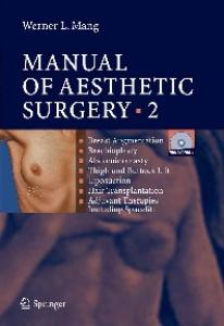
MANUAL OF AESTHETIC SURGERY
Second Edition
䊏 䊏 䊏 䊏 䊏 䊏
Rhinoplasty Rhytidectomy Eyelid Surgery Otoplasty Breast Surgery Brachioplasty
䊏 䊏 䊏 䊏 䊏
Abdominoplasty Thigh and Buttock Lift Liposuction Hair Transplantation Adjuvant Therapies, Including Spacelift
䊏
䊏
Photographic Documentation in Aesthetic and Plastic Surgery Informed Consent in Aesthetic and Plastic Surgery
Coauthors Frank Neidel · Marian Stefan Mackowski · Andrea Becker · Jan-Thorsten Schantz · Ulrike Then-Schlagau 337 Medical Illustrations by Hans Jörg Schütze 28 Plates of Surgical Instruments and 184 Photographs
ISBN 978-3-540-78794-5
eISBN 978-3-540-78795-2
DOI 10.1007/978-3-540-78795-2 Springer Heidelberg Dordrecht London New York Library of Congress Control Number 2010929103 ˇ Springer-Verlag Berlin Heidelberg 2010 This work is subject to copyright. All rights are reserved, whether the whole or part of the material is concerned, specifically the rights of translation, reprinting, reuse of illustrations, recitation, broadcasting, reproduction on microfilm or in any other way, and storage in data banks. Duplication of this publication or parts thereof is permitted only under the provisions of the German Copyright Law of September 9, 1965, in its current version, and permission for use must always be obtained from Springer-Verlag. Violations are liable for prosecution under the German Copyright Law. The use of general descriptive names, registered names, trademarks, etc. in this publication does not imply, even in the absence of a specific statement, that such names are exempt from the relevant protective laws and regulations and therefore free for general use. Product liability: The publishers cannot guarantee the accuracy of any information about dosage and application contained in this book. In every individual case the user must check such information by consulting the relevant literature. Cover-Design: eStudio Calamar, Figueres/Berlin DVD-Video: Video-Transfer, Groß-Umstadt Printed on acid-free paper Springer is a part of Springer Science + Business Media (www.springer.com)
Addresses Professor Dr. med. Dr. habil. Werner L. Mang Medical Director of the Bodenseeklinik, Lindau Clinic for Plastic and Aesthetic Surgery Graf-Lennart-Bernadotte-Straße 1 88131 Lindau / Germany Tel. +49 (0) 83 82 – 26 01 80 Fax +49 (0) 83 82 – 26 01 87 0 email:
[email protected] Internet: www.Bodenseeklinik.de APL-Professor at Klinikum rechts der Isar Munich Technical University Ismaninger Straße 22 81675 München / Germany Dr. med. Andrea Becker Plastic and Aesthetic Surgeon Bodenseeklinik Lindau Graf-Lennart-Bernadotte-Straße 1 88131 Lindau / Germany Dr. med. Marian Stefan Mackowski Plastic and Aesthetic Surgeon, General Surgeon Bodenseeklinik Lindau Graf-Lennart-Bernadotte-Straße 1 88131 Lindau / Germany Dr. med. Jan-Thorsten Schantz Bodenseeklinik Lindau Graf-Lennart-Bernadotte-Straße 1 88131 Lindau / Germany Dr. med. Ulrike Then-Schlagau Plastic and Aesthetic Surgeon, General Surgeon Bodenseeklinik Lindau Graf-Lennart-Bernadotte-Straße 1 88131 Lindau / Germany
This book is dedicated to my parents, Dr. Karl Mang (†) and Mrs Luise Mang
My thanks go to my wife, Sybille, and my children, Gloria and Thomas. Without my wife’s strength over the last 30 years, I would not have been able to achieve clinical and scientific prominence in the field of aesthetic and plastic surgery in Europe. Only her understanding for my work gave me the strength, in a 12-hour day with little holiday, to reach my goals.
Preface
Preface At the request of Springer-Verlag, Heidelberg, I have written with pride and pleasure the new edition of the Manual of Aesthetic Surgery. Volumes 1 (head and neck region) and 2 (body) have been integrated into one volume, which has been revised and extended with the addition of the topics “Cosmetic Aesthetic Surgery,” “Breast Surgery,” “Mini Lift,” “Mini Abdomen,” “Buttock Lift,” and “Tumescence Liposuction with the MicroAire® System.” The current trend is towards gentle surgical methods. The ‘Mang School’ has as its motto: Less is more! You should not see cosmetic surgery. Aesthetic surgery is feel-good surgery and not altering surgery. That should be the philosophy of this book. The first editions of both volumes of the Manual of Aesthetic Surgery had high print runs and were translated into many languages, including Spanish, Russian, and Chinese. The new edition bridges a few gaps, namely breast lifting and breast reduction. These operations are described in detail using illustrations and videos, in order to provide also plastic and aesthetic surgeons with standards. Standards are of crucial importance in aesthetic surgery. Results must be reproducible. Every aesthetic surgeon will then be able to build on this manual and refine his or her methods. A lot has been going on in recent years and my zest for action has not waned. As CEO of the Mang Medical One Clinic Group, I am responsible for 20 plastic surgeons performing expert aesthetic surgery in Germany every day. As a pioneer and visionary in this area in Germany, I see it as my life’s work to train many young aesthetic surgeons, to give lectures worldwide and to offer good aesthetic surgery in Europe at acceptable prices. I hope that the new edition of this book with its exciting additions meets with success and I wish happy reading to all of the interested doctors. The new handbook is a comprehensive work on aesthetic surgery and covers all procedures in the area of breast surgery, such as supramuscular or submuscular implantation, lifting, and reduction. Adjuvant treatments have also been brought up to date, even if not all dermatosurgical methods could be mentioned in their entirety. The book contains many new tips and tricks, all collected during my 20 years of experience as an aesthetic surgeon.
IX
Preface
This is therefore not just an entirely new edition of volumes 1 and 2, but it is an integrated volume with current and new surgical methods and standards regarding surgery and adjuvant treatments. Aesthetic surgery is an interdisciplinary specialist area. This book is intended for all specialist disciplines which are relevant to aesthetic surgery: ENT, head/neck surgery, oral surgery, dermatology, ophthalmology, plastic surgery, surgery, gynecology, etc. Therefore, it is an interdisciplinary handbook for every interested doctor, specialist, or medical student. Werner L. Mang
X
Foreword by M. P. Ceravolo
Foreword by M. P. Ceravolo Every plastic surgeon’s desk is invaded daily by leaflets illustrating new books, manuals, and atlases of plastic surgery. It is impossible to buy or read all of them. Therefore, it is important to understand which text is valuable and may enrich our knowledge and which may just represent an ornamental object on our bookshelf. There are at least three good reasons to have Professor Mang’s Manual of Aesthetic Surgery. The first is its author – a physician who has dedicated his life to studying the perfection and diffusion of aesthetic surgery. His culture, based on multidisciplinary experience, is enriched by a continuous innovative animus which has led him to create intermingling scientific relationships with the most experienced colleagues worldwide. This global vision has allowed Professor Mang to create an opus which goes beyond any frontier and represents a precious gem in the scientific world. The second reason is the up-to-date quality of his opus. Each subject of aesthetic surgery, from rhytidectomy to laser resurfacing, from mammaplasty to botulinum toxin, is approached following the most recent theories and advancements. And last but not least, the third reason is this book’s unique characteristic, its multimedia approach: text, illustrations, and video on DVD. The clarity of the descriptions combines with the precision of the drawings and the vivid explanatory imaging of the video. It is a book to read, to study, and to enjoy. Mario Pelle Ceravolo Professor of Plastic Surgery University of Rio de Janeiro, Brazil Medical College, New York, USA and Rome, Italy
XI
Foreword by D. L. Feinendegen
Foreword by D. L. Feinendegen Aesthetic surgery has experienced exceptionally rapid growth over the last few years. There has been a continual increase in the number of people requesting such operations and, alongside specialists in plastic surgery, more and more doctors from other specialist surgical fields are now working in this area. Until now, however, it has been possible to acquire sound training in aesthetic surgery within Europe in only a few large hospitals. Doctors interested in this field, therefore, often have to move abroad to obtain experience in aesthetic surgery. Professor Mang has been working to establish training in aesthetic surgery for many years. His greatest contribution has been to ensure interdisciplinary cooperation between plastic surgeons, ENT specialists, oral surgeons, and other surgeons active in the field of aesthetic surgery. Professor Mang has already made these ideas a reality in his new clinic. With this manual on aesthetic surgery, Professor Mang has created a foundation for training in aesthetic surgery. The first edition (originally published in two volumes) has already made a strong impression, with its clear structure and excellent, step-by-step diagrams that make even difficult surgical techniques easy to understand. The manual appeals particularly to young doctors who are having their first experience with aesthetic surgery. The additional option of audiovisual learning, provided by the integrated DVDs, underlines Professor Mang’s modern teaching concept. This new manual reflects Professor Mang’s tireless dedication to the task of continuing to establish the field of aesthetic surgery. I myself have come to value Professor Mang as a teacher and wish him continued success in making his ideas a reality. I hope that as many doctors as possible will be able to profit from these ideas, ultimately contributing to the welfare of patients. Dr. med. Dominik L. Feinendegen Plastic, Reconstructive, and Aesthetic Surgeon Zollikon, Switzerland
XII
Foreword by P. F. Fournier
Foreword by P. F. Fournier It is a great pleasure and a great honor for me to write a foreword to the second edition of Professor Werner L. Mang’s book. Professor Mang and I have been acquainted for many years and have attended many meetings together. He must be congratulated on having presented his great experience in aesthetic surgery in his book in a dynamic way with a video included in a DVD. All experienced aesthetic surgeons or surgeons learning aesthetic surgery who have not had the privilege to observe Professor Mang in his clinic at Lake Constance can be informed about the latest and best procedures used by Professor Mang. The text and illustrations are of exceptional quality and reading the various chapters is a real pleasure. All chapters have been written with great care and with the desire to be of the highest interest for the readers. There is no doubt that this second edition will have the same deserved success as the first edition. We are greatly indebted to Professor Mang for all the time he has spent in offering both seasoned and novice aesthetic surgeons eager to learn or refine a surgical procedure a true mine of precious and safe techniques. He is extending the horizons of our specialty by providing the readers with his contributions and the improvements he has brought to conventional techniques. He emphasizes details that continue to make our specialty creative and very practical at the same time. All such precise information is an incentive to read and learn more in order to achieve excellence in our daily work, in patient selection, in planning, and in performance. Again, we should be very grateful to Professor Mang for sharing his great knowledge, experience, creative mind, and insight. I have known Prof. Mang for more than 20 years. Following his surgical training, he gained an international reputation as a specialist in ENT and plastic surgery and, through the first edition of Manual of Aesthetic Surgery, he reached beyond the boundaries of Europe. In 1987, Prof. Mang founded the German Society for Aesthetic Medicine and was a pioneer in this field in Germany. I have frequently attended his wonderful conferences in Lindau on Lake Constance, listened to his excellent lectures, and become acquainted with interesting aesthetic surgeons from all over the world. My wife and I have been pleased to accept private invitations from the Mang family and these have given us the pleasure of meeting Professor Mang’s enchanting wife, Sibylle, and his children, Gloria and Thomas.
XIII
Foreword by P. F. Fournier
Prof. Mang’s clinical activity and his services to society are remarkable. He is a workaholic and pursues his goals in aesthetic surgery determinedly, properly, and with a lot of self-sacrifice. In many discussions with him, we wanted to find out what beauty really is. Cosmetic surgeons have heavy responsibilities and must be creative. For the Manual of Aesthetic Surgery, therefore, I have attempted to define the term beauty: Initially we are expected to believe that beauty has something to do with proportion, balance, and symmetry. Thus, I would like to attempt to explain beauty objectively by looking back to the starting point of the ancient Egyptians, Greeks, and Romans.
What Is Beauty? Beauty is a combination of form and proportion that brings us pleasure and that we can admire. The perception of beauty, however, varies between different cultures. Beauty is a balance between form and volume. Beauty produces in us an aesthetic feeling, an admiration, by pleasing the eye. Some people even claim that beauty is a visual phenomenon. Beauty is a combination of qualities, such as form, proportion, the color of the human face (or other objects) that charm the gaze. Over 200 years ago, David Hume (1711–1776), a Scottish philosopher, remarked, “Beauty is essentially a private and personal experience. Beauty is in the eye and mind of the beholder.” He also said, “Beauty is not a quality of the thing itself but that which exists in the mind of those who contemplate it.” Beauty is an individual emotion. A few philosophers have concluded, “Beauty is good, and what is good, is beautiful.” A long time ago, the philosopher Sapphie said, “That which is beautiful is good and he who is good will soon become beautiful.” Our early experiences influence how we judge now. Particularly because beauty does not captivate through detail but through the whole, which is greater than the sum of the individual parts, our parents, partners, ex-partners, wives, and friends remind us of experiences. In the same way, our current experiences will affect our feelings of tomorrow. The happy and unhappy phases of our lives leave behind traces that shape our inclinations. Faces that we loved during our youth, which gave us warmth and security, live on in our thoughts.
XIV
Foreword by P. F. Fournier
Beauty does not only have to do with the face, the voice, the body, or a charming appearance. A person is beautiful because of their character, their personality, their ability to feel joy and give pleasure to others, and their capacity for love. If we like a face, we like the mood which that person conveys. A person can be attractive in many ways. Beauty and charm are often confused. Cleopatra, George Sand, Louisa de la Valli`ere and Theodora were famous for their beauty. In truth, they were very beautiful but also had a lot of charm. Beauty is more an illusion than a reality. Beauty exists not only for the eye but also for the mind. A beautiful personality emphasizes the beauty of the face. There are numerous ways of defining beauty and it is often associated with charm. Charm, however, differs from beauty because it is permanent, whereas beauty fades. The English say, “Charm lasts! Beauty passes!” Ultimately, we can see that it is not only the eyes which judge whether someone is beautiful or not but the mind which plays a much greater role and judges the heart and inner beauty. According to the American sociologist Frumkin, a woman is judged in relation to her sexual charisma. Whether she is judged beautiful or not beautiful depends not only on the symmetry of her proportions but also on whether these attributes suggest potential sexual possibilities. The sensual emotion is then transformed into an aesthetic feeling. Following these classic explanations, we can conclude that the perception of beauty differs among cultures and individuals and that it is not only a question of form and symmetry. A person’s personality, charm, and inner beauty play an important part in giving a person a pleasing image. The eyes alone do not make a judgment, but the head and the heart as well. The mind is influenced by our past experiences, which affect our judgment, just as our current experiences influence the future. One of Buddha’s teachings tells us, “Today is the son of yesterday and the father of tomorrow.” Beauty is like an iceberg; only one small part is visible. Konrad Lorenz, Nobel Prize Winner for Medicine and Physiology, has made a special contribution to our understanding of the biology of behavior. This has helped us to understand human beauty.
XV
Foreword by P. F. Fournier
When someone feels drawn to a face, this is because the face has childlike features. Everyone instinctively feels attracted to a childlike face. The sight of a childlike face evokes an emotion that is automatically linked with a desire to protect. It is the same in both humans and animals. Konrad Lorenz explained this in the following terms: the desire to protect one’s offspring is prompted by something which the offspring sends out, a physical peculiarity, a sound, a smell. It is the same in man. There are signals which provoke protective instincts, sympathy, and tenderness. What are they, asks Konrad Lorenz? In infants, the signals come from the head. The roundness and fullness, the prominent forehead, the full cheeks, a small snub nose; all these infantile characteristics provoke a protective instinct. A child’s face is associated with purity, friendliness, honesty, and vulnerability. We know that women keep their curves, whereas men lose them. A good plastic surgeon should therefore ensure that, during surgery, he optimizes the characteristics which, as in a baby, provoke affection, tenderness, and a desire to protect. Softness and roundness = tenderness. Once again, to give the impression of beauty, it is of fundamental importance to be able to recognize childlike features in an adult’s face. Features, however, are not the sole cause of the protective reflex; expressions are also important. These at least have the advantage that they are within the reach of everyone. A few people know how helpful expressions can be in getting someone to do something or in pleasing someone. The emotions which were elicited by Brigit Bardot’s childlike features were helped particularly by her famous “spoilt child”-like pouting. Just as well known are the childlike expressions used, or abused, by Marilyn Monroe and Audrey Hepburn. It has even been rumored that Marilyn Monroe deliberately made herself up badly to give the impression that she was a small girl who still did not know how to get ready properly and, even after long sessions at the hairdressers, immediately rumpled her hair to restore the disheveled appearance of a small girl who had just come in from playing. Men have no desire to protect women who do not have a childlike appearance and want to dominate men, and feel reminded more of their mother than their wife. Women are more concerned with beauty than men and, consciously or subconsciously, display this childlike behavior. They are consciously or
XVI
Foreword by P. F. Fournier
subconsciously shy, fragile, weak, innocent, na¨ıve, ignorant, temperamental, admiring, inquisitive, etc. A few women even emphasize weaknesses to trigger the protective instinct. Have I already mentioned that apparent weaknesses in women are also their strengths? All this to strike a man directly in his heart. Napoleon said, “Women’s two weapons are make-up (the significance of this will be discussed later) and the tears of a small, helpless child.” It is easy to understand why childlike features in an adult can move someone, in the same way as freckles, full red cheeks, long eyelashes, blond curls, and well-defined and full lips. Among men, as we can see in a few of the great sex symbols, the side parting (Clark Gable, Gary Cooper), an untidy mane of hair (Leonardo de Caprio), and daily shaving can only be explained as the desire for a childlike appearance. It is not necessary, however, to have all these attributes; one is usually sufficient. Every individual can display childlike features at any time. As regards particular features, if someone does not have these, he or she can usually acquire them with the help of cosmetic surgery. Beauty is not merely a completely natural phenomenon; instead, it has been a cultural phenomenon for a long time and this is the case particularly in the present day. People try to improve themselves and women, to whom beauty is more important than it is to men (men tend to try to obtain power), try to improve their beauty and charm with make-up and accessories like spectacles, false eyelashes, earrings, hair styles, permanent make-up around the lips, eyelids and eyebrows, hats, necklaces, and the invisible accessory, perfume. A few modern accessories have been developed by beauty professionals to disguise beauty defects, e.g., wide spectacle side pieces hide crow’s feet, a high frame emphasizes the length of a nose that is too short and, conversely, a lower frame disguises a nose that is too long. All these strategies are discussed discreetly and in detail in women’s magazines. An old proverb describes this perfectly: “Thirty percent of beauty is natural, seventy percent is created by vanity.” The disadvantage of this resource is that it is not possible to look young and beautiful without it. Make-up has always been around and if a face is to be beautified, it should be made to look natural and the face should resemble a young face. Lipstick, for example, creates the intense color of young red lips, which is a sign of a more rapid metabolism. Blusher is a reminder of childlike red cheeks and powder gives the face the pale, velvety skin of youth. Desmond Morris called this “over-stimulation.” Long false eye-
XVII
Foreword by P. F. Fournier
lashes remind us of the long eyelashes of children. If applied badly, however, make-up can also ruin the beauty of a face. It can be both friend and foe. In ethnological books, we can read that in former times, witches improved the appearance of sick people so that the relatives were not shocked when they saw them. Childlike features and expressions are therefore important in provoking the protective instinct, but the voice should also be soft and pleasant, like a child’s. A hard, metallic voice, such as smokers have, is not reminiscent of that of a child. Clothing should be pleasing to the eye and mind and it should have a good cut. The mini skirt makes us think of the long legs of an adolescent. Colors remind us of childhood; light colors, like blue or pink, are always chosen by older women. Naturally, black should be avoided. In conclusion, all human senses should be stimulated: sight, hearing, smell (children do not have a smell – thus we use deodorant), and touch. The firmness of the skin is also important. Beauty institutes have understood this for a long time and apply it enthusiastically. Do we not read in women’s magazines: ladies have beautiful breasts, a flat stomach, and good legs, but are they also firm? The firmness and elasticity of tissue are fundamental qualities of a child’s skin and a part of their beauty. Beauty is costly. It is easy for wealthy people to get jewelry and beauty accessories, but these are more difficult to acquire without money. This is one explanation of the popularity of aesthetic medicine and surgery among the less well-off and among those who cannot please merely with their natural gifts or with the artificial resources of the wealthy. As they are only able to please with their body, the less wealthy will more pay readily for an operation to remove acquired or existing supposed defects so that they can continue to be admired. The idea of using the child formula is well known. The heart should be receptive to generosity, and this is used for reasons other than just noble ones. Thus, for example, a child’s face next to the product in an advertisement increases sales and turnover. Whether these are medications or other products, if the consumers are sensitive, sales will increase. Naturally, a way to the heart is sought but also, and predominantly, a way to the wallet. The child formula strategy is likewise used to direct public attention to countries in need, to collect donations, and to fight
XVIII
Foreword by P. F. Fournier
poverty and suffering. A begging child will always get more money than an adult. The Disney films, of which audiences are so fond, use ever smaller and ever more vulnerable animals; we always see the young mouse, the puppy or the fawn, never the fully grown animal. This also applies to toys. Usually, small animals and babies are used as dolls. As Saint Exup´ery said, “One can only see well with the heart.” It is also important to know that a physical defect also provokes a protective reflex. A few celebrated personalities and women involved in politics keep a slight squint, which could be easily corrected, to provoke this famous protective mechanism and thus strengthen their influence and power of persuasion. They do not want an operation. It is just as well known that if one part of the face is not perfect and other facial features are consciously highlighted, then the defect is less striking, as the eye is drawn to the emphasized features. If, for example, the eyes are beautiful and the nose is not, the eyes should be emphasized even more to disguise the unattractive nose. Make-up artists advise this even though they are not familiar with Konrad Lorenz’s theories and nevertheless know how to beautify a face. A scar can deflect attention from the beauty of a face. To conceal public embarrassment, Passot says, “Give him a medal and he will be taken for a hero.” Similarly, make-up artists do not know about the Muller-Lyer illusion of two lines of equal length with arrows pointing in different directions at either end. Nevertheless, they know how to give eyes the appearance of being closer together, by applying make-up to the inner corner or, conversely, increasing the distance between the eyes by applying makeup to the outer corner. The same applies to cheek bones in a face that is either too long or too short. Blusher is applied either further apart or closer together as appropriate. Why be beautiful? The reasons suggested are pride and a desire to be admired and seen positively by others. The cult of beauty is actually cultural. Humans are the only life form who do not accept their fate but try to improve it. Preserving beauty means improving the quality of life to beautify life. The progress of civilization in all areas has led to increased life expectancy. This does not seem to be sufficient; the quality of life must also be maintained and a life must be “beautiful” for longer. Some people say that medicine has given a few more years to life; aesthetic medicine and surgery have given life to these years.
XIX
Foreword by P. F. Fournier
Beauty and fashion are external signs of our inner need to express and redefine ourselves. Fashion is only a stylistic device in the work of art which is life. Beauty does not last forever but everyone knows that, at the same time, beauty does not have an age. It is possible to look good at 20 but it is also possible to remain irresistible for an entire lifetime, as Coco Chanel remarked. Madame de Pompadour said, “The first requirement of a woman is to please.” It is more and more difficult to fulfill this requirement with increasing age. This reminds me of an old woman who came to me and asked for a facelift. When I showed that I had little interest in performing this procedure because of her age, she said, in a quiet voice, “When one has ceased to please, one doesn’t have to displease for long.” Ultimately, the desire to be beautiful is not a desire to be admired but a desire to be loved. In addition, this desire for love is ultimately the only thing that the followers of the cult of beauty want to communicate. Konrad Lorenz acknowledges this, “Everyone loves children and wants to protect them, this is hereditary.” Can you resent someone for wanting to be like them in order to be loved more? There is no doubting this theory. We should remember that the plastic surgeon should reconstruct childlike features in his work, if this is possible and desired, in order to provoke positive feelings and admiration. We recognize the link between beauty and admiration and the intense fluctuations of the spirit and the mind. Theodore Gautier summarized this well, “To admire is to love with the mind, to love is to admire with the heart.” Konrad Lorenz’s theory is strengthened with details. To provoke a protective instinct, an adult’s face must resemble that of a child; this is considered to be beautiful. The perception of beauty is subjective; the personality and qualities of the individual play a part. In those who experience this, this perception is influenced by earlier experiences. Pierre F. Fournier, M.D. Honorary President of the French Society of the Aesthetic Surgery (National Society) Paris, France
XX
Foreword by R. Kaufmann
Foreword by R. Kaufmann In the past few decades, aesthetic surgery has witnessed outstanding progress. This has been mainly driven by a growing public demand for corrective surgical procedures together with an increasing awareness and the highest expectations for the best quality. Today’s understanding of aesthetic surgery, its technical and developmental stages, and the level of performance reflects the results of ongoing and combined inputs made by leading physicians and pioneers from diverse subspecialties working in this field, including plastic surgeons, dermatologists, ENT specialists, maxillofacial surgeons, ophthalmologists, and others. However, although many procedures in the face are routinely performed within these subspecialties, major aspects of the art of cosmetic surgery are usually not covered by subspecialty training alone. Professor Mang, one of the most experienced experts in aesthetic medicine and surgery today, has undertaken the challenge to provide colleagues interested in this fascinating area with a modern state-of-the-art manual, showing a step-by-step approach to aesthetic surgical techniques. Chapters 3–6 deal exclusively with facial procedures. The conceptual frame of the present manual promises the best and easiest access to this demanding field by combining explanatory texts with video sequences on DVD and stepwise illustrations. I am convinced that Professor Mang’s work will be of great value to colleagues from various subspecialties, and I hope that it will have the success that it deserves. Roland Kaufmann, MD Professor and Chair of Dermatology J.W. Goethe University Frankfurt/Main Germany
XXI
Foreword by S. Malakhov
Foreword by S. Malakhov I first became acquainted with Prof. Mang in 2002 when he performed surgery at the St. Petersburg Medical Academy at the invitation of Prof. Zapessotsky. Many plastic surgeons watched him doing this and were fascinated by his atraumatic operation technique. One of my assistants went to Prof. Mang’s clinic at Lake Constance for further study and reported back on the friendly and excellent training received in Prof. Mang’s clinic. On the occasion of his visit to St. Petersburg, Prof. Mang presented me with the first volume of the first edition of Manual of Aesthetic Surgery. I was impressed by the clear and concise way in which cosmetic operations were explained. This concept of audiovisual teaching was unknown at that time in Russia. All of my colleagues who have an interest in the subject of aesthetic and plastic surgery were also impressed with this manual. The first volume dealt with cosmetic operations in the head/neck area, while the second volume described cosmetic operations in the trunk area in a way that was equally clear and comprehensible. This second edition, which combines the two volumes into one book, is an ideal textbook for learning about aesthetic surgery; this type of operation has not previously been taught in this form, particularly in Russia. I witnessed Prof. Mang’s hospitality on the occasion of the International Conference for Aesthetic Surgery in Lindau in July 2003. I was impressed not only with the scientific part of the conference in which 600 people participated, but also with the social event on the occasion of the opening of Prof. Mang’s new clinic. There are plans to further strengthen and intensify this relationship between the clinic at Lake Constance and the Medical Academy in St. Petersburg. I wish Prof. Mang much success for the new Manual of Aesthetic Surgery. Finally, I would like to complete my foreword by providing a little information on the history of plastic surgery in Russia:
Plastic and Aesthetic Surgery in Russia – the Past and the Future The development of plastic surgery in Russia is closely associated with the name of the great Russian surgeon, N.I. Pirogov. It was he who first paid attention to the aesthetic results of surgery on open areas of the human body. In his famous book, Basis of General War Field Surgery,
XXII
Foreword by S. Malakhov
he touched on the topics repeatedly. One of his followers wrote the thesis on rhinoplasty. In 1865, another brilliant follower, Y.K. Shimanovskii, published the first manual in the world for practical surgeons, Human Body Surface Surgery, for which he was awarded the I. Bush prize. This unique book contains more than 150 drafts and schemes of plastic surgery procedures made with the author’s own hand, many of them still relevant. A little later in 1869, the young Russian surgeons, S.M. Yanovich-Chainski, A.S. Yatsenko, and P.Ya. Pyasetskii, took up the idea of J. Reverdin concerning free transplantation of autodermal microflaps for closing wound defects and implemented their experience in Russia. In 1898, K.P. Suslov worked out the original method for the elimination of nose defects by transplantation of free skin–cartilage transplants from the ear. Unfortunately, during the first decades of the twentieth century, there were three revolutions and the First World War, which did not favor the development of plastic surgery in Russia. Nevertheless, in 1916 the worldwide recognized work by V.P. Filatov was published. The work was dedicated to the results of using round fat-dermal flaps developed by V.P. Filatov. This method was the only possibility for tissue complex transplantation right up to the second half of the twentieth century when flaps with axial blood supply came into use. Other famous Russian surgeons who played an important role in plastic surgery development include: P.I. Diakonov, N.A. Bogoraz, A.A. Limberg, A.E. Rauer, B.I. Vozchek, B.S. Preobrazhenskii, I.M. Mikhelson, among many others. After World War II, a special system was organized for the treatment of burn patients; this played a particular role in the formation and development of plastic surgery in Russia. During this research and organization work, several generations of plastic surgeons grew up, who have a good knowledge of the most up-to-date methods of free and local skin plastic surgery procedures, including those using microsurgery. The methods of skin reconstruction by means of different variants of combined autoallodermaplastics were developed and implemented. The most significant names in this field were Y.Y. Dzhanelidze, T.Y. Ariev, M.I. Shraiber, N.I. Atiasov, B.S. Vihriev, among others. In 1930, in Moscow increasing interest in plastic surgery led to the creation of the Institute of Beauty, which is now called the Institute for Plastic Surgery and Cosmetology. In 1961, a similar clinic was opened in Leningrad. In the following decades this trend developed rapidly,
XXIII
Foreword by S. Malakhov
and there are now hundreds of centers working in the field of beauty surgery in Russia. At the end of the last century, many specialists understood that plastic surgery is an independent and complex specialty that requires the longterm education of individual surgeons. That is why, at the end of the 1980s and the beginning of the 1990s, the first structure for the training and retraining of plastic surgeons was included in the system of continuous medical education in Moscow. In 1997, at the Saint Petersburg Medical Academy of Postgraduate Studies, the first special department and clinic for plastic and aesthetic surgery was created. Specialists in this department have experience in all methods of plastic surgery and educational work. The department provides long-term programs for the basic training of young specialists (3–5 years) and short-term programs for continuing medical education. The intensive research work of the departmental staff allows training programs to be refreshed and ongoing improvement of the surgery procedures. All of the above means that the education of plastic surgeons is continuous and that their professional level is constantly renewed. The following events have favored the development of plastic surgery in Russia: creation of the All-Russian Society of Plastic, Reconstructive and Aesthetic surgeons; publishing of several periodicals; organization and holding of scientific conferences, seminars, and master-classes in different regions of Russia; constant contact with international societies of plastic surgeons. My colleagues and I believe that aesthetic surgery in Russia has a great future. Professor S. Malakhov Head of the Clinic for Plastic and Aesthetic Surgery Saint Petersburg Medical Academy of Postgraduate Studies, Russia
XXIV
Foreword by H. Massiha
Foreword by H. Massiha Aesthetic plastic surgery is perhaps the fastest growing area in the field of surgery. As more and more surgeons spend more time performing aesthetic operations, it becomes increasingly evident that authoritative instructions are needed to extend the competence of the surgeon. Although it looks simple, aesthetic surgery is very demanding technically and artistically. The task of becoming a good aesthetic surgeon could be greatly eased by observing and working with masters in the field. Since the option of working with these experts is not practical for most surgeons, reading their works and becoming familiar with their ideas becomes even more important. A major advantage of this book is not only the large number of elegant diagrams, but also the inclusion of a video presentation. Professor Mang has undertaken the monumental task of bringing together the most advanced and practical techniques of aesthetic surgeons in the Manual of Aesthetic Surgery. This book is the result of long years of research, observation, and hard work in the pursuit of excellence in aesthetic plastic surgery. In addition to his extensive education in the field of head and neck surgery, Professor Mang has traveled all over the world to visit, observe, and exchange ideas with the greatest aesthetic surgeons. On numerous occasions he has invited these authorities to his clinic to share his ideas and perfect his concepts and techniques through creative interactions and exchanges. The purpose of the book is not only to teach young aesthetic surgeons about basic operations and how to avoid pitfalls and complications, but also to emphasize what is currently the state of the art in aesthetic surgery. This book will satisfy all types of aesthetic surgeons and will help to improve their results with the ultimate beneficiary being “the allimportant patient”. I highly recommend this wonderful book of aesthetic surgery to all surgeons who seek the opportunity to improve their results. I congratulate Professor Mang in providing this vital service to the field of aesthetic surgery and to the young aesthetic surgeon. Hamid Massiha, MD, FACS Professor of Plastic Surgery New Orleans, Louisiana, USA
XXV
Foreword by D. Millesi
Foreword by D. Millesi Aesthetic surgery has developed with enormous velocity over the past decade. Owing to the growing number of patients undergoing aesthetic surgery, it is not only more and more accepted in a wider range of our population, but also has to fulfill the growing expectations of very critical patients. Many new techniques are at our disposal and the number is constantly growing. Apart from basic techniques, detailed technical points become more and more important for successful outcomes. It is nearly impossible to provide a complete survey of all techniques available today in a single textbook, not to mention the variety of technical details that are frequently not described. It is to W. L. Mang’s credit that he elected the forum of a manual instead of a large textbook to present his great personal experience. His techniques and tricks are presented in the form of very instructive sketches, and any surgeon who wants to enter the field of aesthetic surgery can do this easily following the impressive illustrations. Now the new edition of the Manual of Aesthetic Surgery is available. It covers liposuction, breast implants, hair transplantation, aesthetic surgery of the extremities, and abdominal plastic surgery. These chapters on surgical techniques are complemented by a chapter on adjuvant therapies, including lipotransfer. The new edition is designed according to the same principles as the first edition. Again, the main focus is on the illustrations, which are easy to follow and help the reader to understand the individual surgical steps. In addition to the excellent optical presentations, a DVD is included, providing audiovisual presentations. I personally have profited enormously from the brilliant images, and I am sure that this new edition will help beginners in this field in the same way. It would be advantageous if all prominent surgeons in aesthetic surgery would present their professional experience in a way similar to W.L. Mang. Prim. Dr. med. Dagmar Millesi Fachärztin für plastische und ästhetische Chirurgie Vienna, Austria
XXVI
Foreword by I. Pitanguy
Foreword by I. Pitanguy In his Manual of Aesthetic Surgery, Professor Mang provides a clearly written and comprehensible book that can be read by all physicians who may have an interest in the field of aesthetic plastic surgery. Prof. Mang shares with us his vast experience in aesthetic surgery and presents the techniques that have proven useful in his hands. Together with his team of collaborators at the clinic at Lake Constance, Prof. Mang describes operations clearly and explicitly. Especially for the younger surgeon, the book offers the opportunity to become acquainted with aspects of surgery of the abdominal and breast regions, as well as the upper and lower limbs. When Prof. Mang first visited me in Brazil in 1972, he impressed me as a particularly hard-working colleague, eager for knowledge. Through his numerous visits to my clinic in Rio, and during my own visits to Germany, I have grown to know Prof. Mang and his delightful family well and to value the friendship that we have developed, which is characterized by that special charm all Germans are capable of giving. With the completion of his clinic at Lake Constance, Prof. Mang fulfilled his life’s dream. I wish both his clinic and this book much success. Prof. Ivo Pitanguy, FACS, FICS Head Professor of the Post-Graduate Courses in Plastic Surgery of the Pontifical Catholic University of Rio de Janeiro and the Carlos Chagas Post-Graduate Medical Institute. Member of the Brazilian Society of Plastic Surgery, the Brazilian National Academy of Medicine, and the Brazilian Academy of Letters
XXVII
Foreword by R. Schmelzle
Foreword by R. Schmelzle The Manual of Aesthetic Surgery, written by Professor W.L. Mang from Lindau, presents aesthetic and plastic surgery from the perspective of one of the leading practitioners in the field. Although the manual is intended for young physicians and representatives of many different medical specialties, it is also an important reference work for members of the field of aesthetic surgery itself. The excellent illustrations provided by a graphic artist from SpringerVerlag are accompanied by brief explanatory texts. Together they allow the entire field of aesthetic surgery to unfold in a clear and easy-tounderstand manner. A point worthy of special mention is the audiovisual character of this textbook, which presents the most important operations in the head and neck area via text, illustrations, and video. This second edition will also serve as a guide in the area of “quality assurance,” a goal which is frequently cited today. I hope that the Manual of Aesthetic Surgery will reach a large audience. Dr. Dr. Rainer Schmelzle Professor of Maxillo-facial Plastic Surgery University of Hamburg Germany
XXVIII
Foreword by H. U. Steinau
Foreword by H. U. Steinau This new textbook of Werner L. Mang’s Manual of Aesthetic Surgery brings together his group of experienced plastic surgeons and specialized ENT and maxillofacial colleagues to share their profound personal knowledge in treating the most common aesthetic problem areas. Anatomical landmarks and potential pitfalls are clearly depicted and discussed. The publisher has provided coloured pictures of excellent quality with accompanying schematic drawings. The chapters explain the selection of optimal surgical strategies and details are given on their basic instrumental supplementation, surgical principles, and potential problems. Advanced planning and selection of safe solutions are followed by various anesthetic regimens including tumescence techniques, which are now used routinely in ambulatory surgery. Taken as a whole, this second edition represents a valuable contribution that will provide novices during residency with a broad-based training program. Its interesting case collections and methodologies afford experienced aesthetic surgeons the opportunity to critically compare their preferred treatment options with convincing “second opinions.” Professor Mang and his multidisciplinary team are to be commended for their continuous educational efforts and outstanding didactic accomplishments. Professor Dr. med. Hans U. Steinau Department of Plastic Surgery and Burns BG-University Hospital “Bergmannsheil” RU-Bochum, Germany
XXIX
Introduction
Introduction Aesthetic surgery is an interdisciplinary specialty. Its members are recruited from the fields of surgery, gynecology, orthopedics, otolaryngology (ENT), maxillofacial surgery, plastic surgery, ophthalmology, and dermatology. In most cases, they are specialists who have become interested in practicing aesthetic surgery after completing their specialist training. Aesthetic surgery is not synonymous with plastic surgery. During their training in aesthetic surgery, aspiring aesthetic surgeons have to learn special surgical techniques, which are unfortunately not adequately described in the postgraduate training catalogues. This manual has been prepared as an audiovisual medium. It presents the most important standard operations in the field of aesthetic surgery in a clearly understandable style. We hope that the manual will help young surgeons to learn the techniques of aesthetic surgery and – equally important – to avoid mistakes and complications. The manual is primarily aimed at specialists in aesthetic surgery. However, it is also suitable for physicians who become interested in aesthetic surgery after finishing medical school who would like to find out about the field, as well as for the natural target group of physicians who want to learn the techniques of aesthetic surgery after completing their specialist training. In line with the author’s didactic concept, the manual is accompanied by a DVD containing a video film of each operation and by a surgical atlas with 337 color illustrations. In the surgical atlas, the individual steps are shown again and explained in detail in the accompanying text. This structure allows the physician to reproduce each surgical step precisely and to master the associated techniques. There are obviously several variations of each surgical technique. The author has deliberately concentrated on the standard operations as his contribution toward “demystifying” the field of aesthetic surgery. This reflects his conviction that aesthetic surgery, like any other kind of surgery, is a reproducible discipline which can be learned. The manual is designed to serve two purposes: the education of young surgeons and quality assurance in the field of aesthetic surgery. It is the only work of its kind available internationally. The manual is intended to be a basic tool and is not for professionals and doctors who have been practicing in this specialty for a long time.
XXXI
Introduction
It is intended to be a textbook for doctors who are starting out in this field and want to learn about it. Naturally, it was not possible to mention all the tricks, subtleties, and latest operation methods, suture materials, implants, etc. in this book. Every aesthetic surgeon must learn these through further training and conferences. However, for every surgical technique in trauma or abdominal surgery, the basics of the operation must be standardized. This was achieved well with the first edition of the manual. It has been the most successful book of its kind for Springer-Verlag, Heidelberg, and has been published in Spanish, Russian, and Chinese because of the enormous interest it received. The same format and approach has been adopted in this second edition. I had never thought that this book would be so well accepted. It has become an interdisciplinary textbook for surgeons, ENT and dental surgeons, plastic surgeons, dermatologists, gynecologists, orthopedists, and urologists and has a place in many hospital libraries throughout the world. After the first edition was published, I received invitations to give lectures and surgical courses and take up chairmanships from almost everywhere in the world. I have been accredited as an honorary professor at foreign universities and see my life’s task in plastic and aesthetic surgery as being to bring together all specialties that teach and research the field of aesthetic surgery in order to ensure excellent quality assurance in relation to patient care. As a result of my lectures to the most varied specialist societies on every continent, I have discovered again and again that there is competition between ENT surgeons, dental surgeons, and plastic surgeons in almost every country, even though all three specialties perform extremely valuable work in the field of aesthetic surgery. The leading plastic surgeons of the past, Diefenbach, von Gräfe, Joseph, and Lexer were either ENT surgeons or general surgeons. We must never forget our history and the disciplines from which the specialty of aesthetic and plastic surgery has developed. Anyone who has had sound surgical training and has an interest in the field of aesthetic surgery will value this book as a benchmark. It can help in allowing the specialty of aesthetic surgery to be taught in an interdisciplinary way, so that the specialties concerned can mutually exchange knowledge and thus contribute to further progress in this field.
XXXII
Introduction
Aesthetic surgery can only achieve a serious basis in the long-term through constant training, the exchange of ideas, attendance at conferences, and the opening up of all specialties that perform valuable work in this field. Neither plastic surgery nor ENT and dental surgery can claim this specialty for themselves alone, as there is too much overlap both historically and in the specialist further-training guidelines. For this reason, work is carried out in an interdisciplinary way at the clinic at Lake Constance with the departments ENT and plastic surgery, plastic and reconstructive surgery, maxillofacial surgery, aesthetic dental surgery, dermatology, and venous, hair, and laser surgery. This is the only way a large clinic can cover the entire spectrum. The same applies to a well-trained aesthetic surgeon. He will always have main areas within his field of work and will be unable to cover all operations professionally alone. This is why the model of the clinic at Lake Constance will be successful in the long term, as in this clinic different groups of specialists are unified and offer interdisciplinary aesthetic surgery. This is the clinic of the future. Every year approximately 3,000 operations are carried out at the clinic at Lake Constance, which has five operating theaters and 50 beds. The Manual of Aesthetic Surgery should be seen as the symbiosis of my lifetime work with the Bodenseeklinik. It has been published to coincide with the building of the new clinic (completion 2003). All physicians with an interest in aesthetic surgery can build on this and refine their surgical techniques during the course of their life. The basic principles must be standardized, so that dangers and risks can be reduced. Rhinoplasty should not be performed differently in London and Rome, and liposuction techniques should be the same in New York and Tokyo. Just as in abdominal surgery, there are basic principles that must be observed so that the operations and results can be reproduced and serious treatment errors can be avoided. Naturally, there are variations in the operations, whether the procedure is rhinoplasty, otoplasty, breast implants, or liposuction. The same applies to operations on the appendix or tonsils. The basic surgical technique used, however, is always the same. The anatomy never lies. It is therefore essential that the basic operations are standardized, particularly in aesthetic surgery, which I consider to be the most difficult type of surgery, as the surgeon must not only be well trained, but must also be a psychologist and artist.
XXXIII
Introduction
This manual was written at the urgent request of many of the numerous physicians who come to the author’s clinic every day as observers. The objective of the manual is to give a large number of physicians a solid, broad, and interdisciplinary foundation in aesthetic surgery. This is evident at many points in the manual and especially in the words of introduction written by the following authors: – Mario Ceravolo, MD, University Professor, General Plastic Surgery, Rome, Italy – Dominik L. Feinendegen, Plastic, Reconstructive and Aesthetic Surgeon, Zollikon, Switzerland – Pierre F. Fournier, M.D., Honorary President of the French Society of the Aesthetic Surgery (National Society), Paris, France – Roland Kaufmann, MD, University Professor, Dermatology, Frankfurt, Germany – S. Malakhov, Professor, Head of the Clinic for Plastic and Aesthetic Surgery, Saint Petersburg Medical Academy of Postgraduate, Russia – Hamid Massiha, MD, University Professor, Plastic Surgery, New Orleans, Louisiana, USA – Dagmar Millesi, Prim. Dr. med., Plastic Surgery, Vienna, Austria – Ivo Pitanguy, MD, University Professor, Plastic Surgery, Rio de Janeiro, Brazil – Rainer Schmelzle, MD, University Professor, Maxillofacial Plastic Surgery, Hamburg, Germany – Hans U. Steinau, Professor Dr. med., Department of Plastic Surgery and Burns, BG-University Hosptial “Bergmannsheil”, RU-Bochum, Germany
XXXIV
A General Remark
A General Remark If I may be permitted another remark here: The author’s philosophy and the philosophy of the Bodenseeklinik is interdisciplinary cooperation, instruction and further training of young doctors, cooperation with all professional societies for the promotion of good patient care, and further development in the field of aesthetic surgery. The Manual of Aesthetic Surgery has thus come about through tireless work. My clinic at Lake Constance is the largest clinic of its kind in Europe, a training clinic with interdisciplinary cooperation between all specialties that provide a stimulus for aesthetic surgery. Doctors from the disciplines of plastic surgery, ENT and dental surgery, dermatology, aesthetic dentistry, and anti-aging medicine all work in the clinic at Lake Constance. There are also dietary assistants, specialist beauticians, hairstylists, color consultants, and psychologists. Long-term success can only be achieved when aesthetic surgery is seen to be holistic medicine and the correct indications are available. Many patients have serious psychological problems that cannot be solved even by the best cosmetic surgery. These patients are then dissatisfied with the surgeon and try to find a cure from other surgeons. If these surgeons do not then cooperate with the surgeon who carried out the previous operation, the patient will complain. Medicolegal problems have an important role in aesthetic surgery throughout the world. The specialty can only have a long-term future if doctors are welltrained, act as good colleagues towards one another, and do not want to make their name at the cost of others. I would therefore like to pass on this message to all the surgeons in the world: be considerate and fair to colleagues, regardless of their specialty. The Hippocratic Oath should apply to cosmetic surgeons, too. The philosophy of the Mang school is naturalness. Less is more. Health before beauty. Cosmetic surgery is not “alteration surgery” but rather “well-being surgery.” The aim of every operation, whether it is a facelift, rhinoplasty, or a breast implant, should be a natural result. The patient should feel good and the surgery should not be conspicuous. Faces that are perfectly smooth, unnaturally augmented lips, and huge breasts are no longer the trend of the twenty-first century. The manual therefore presents surgical techniques that provide natural and normal results.
XXXV
A General Remark
Aesthetic surgery is not beauty surgery. It is instead high-tech surgery with the highest surgical standards. As with every other surgical procedure, the risks mean that specialist surgical personnel, anesthesia, recovery rooms, and inpatient monitoring are essential. Surgery on a day-case basis is only advisable for minor procedures carried out under local anesthesia, such as eyelid corrections, spacelifts, hair transplantations, and laser operations. Otherwise, an inpatient stay is necessary, as most complications, e.g., severe bleeding, occur within the first 24 h after the operation. The current worldwide problem of medicolegal issues in cosmetic surgery procedures should be combated with extensive expert activity. In addition to providing accurate oral and written information in the presence of witnesses and photographic documentation, use of the correct surgical techniques and postoperative monitoring are extremely important in avoiding the possibility of becoming liable for compensation. More and more patients are happy to take legal action and this means that good training, quality assurance, good relationships between colleagues, and professional interaction with patients are even more essential.
The Standard Procedures The standard procedures are clear and can be easily and safely learned following good basic surgical training. Similar basic surgical rules apply to brachioplasty, abdominoplasty, thigh lift, and buttock lifts. In principle, these procedures entail cleanly lifting a cutaneous/fatty flap from the fascia and tightening the skin appropriately, using a large cutaneous resection and positioning the incisions in such a way that they are preferably not visible. The surgeon’s talent is estimating the correct cutaneous resection, so that not too much and not too little is removed, and accurate surgical planning of the incisions so that they will preferably be in a non-visible area. The intracutaneous suturing technique with Monocryl, a suture which is not removed, is now standard and provides the best results. In certain cases, the skin may also be adapted with overcast cutaneous suturing with thin nylon, following subcutaneous, tension-free skin closure. When these continuous sutures are removed in time, the cosmetic results of the suturing are no different than for intracutaneous suturing. For all operations associated with large scars, follow-up treatment is very important. A compression dressing should be worn for approxi-
XXXVI
A General Remark
mately 4 weeks and follow-up treatment for the scar should be carried out with a silicone plaster. Scars resulting from brachioplasty, abdominoplasty, and thigh and buttock lifts in particular are often unpredictable and must be discussed in detail with the patient when the procedure is explained so that there is no disagreement later. The standardized surgical procedures described in detail in the manual are presented in abridged form below.
Rhinoplasty About 70% of all functional-aesthetic rhinoplasties are performed to reshape long noses with a bump. For this reason, the manual presents a reproducible and simple technique for correcting this type of nose deformity. In addition, several variations are briefly described. During aesthetic nasal tip correction using the eversion method, a mucosal epithelial layer inevitably remains following the removal of large portions of the alar cartilage. The more the tip is reduced, the more excess skin there will be. This so-called Mang‘s triangle is resected following suturing in order to achieve nonirritable healing of the skin inside the nasal wings without step formation. Strictures and stenoses can be avoided by taking pains to leave the mucosa intact during the removal of alar cartilage. Furthermore, the removal of equal-sized triangles on both sides facilitates aesthetic shaping of the nasal wings.
Rhytidectomy Using the Tumescence Technique – Mini Lift The manual presents a standardized surgical facelift operation using the tumescence technique. This procedure involves simple and gentle dissection with transection of the osteodermal ligaments, as described by Hoefflin (extended supraplatysma plane facelift, referred to as ESP lift). Dissection of the superficial musculo-aponeurotic system (SMAS) is not necessary, since the sagging caused by the aging process is a problem of lipocutaneous tissue and – similar to breast ptosis – is not attributable to fascia and muscle layers at deeper levels. The method presented is standardized, easy to reproduce, and gentle to the tissue. In addition, it involves very little loss of blood and yields excellent longterm results. The tumescence facelift technique presented here for the first time facilitates dissection and is therefore an especially good approach for newcomers to aesthetic surgery. This method of face lifting produces the best long-term results.
XXXVII
A General Remark
Upper Eyelid Surgery Following the surgical steps shown in the manual, even an inexperienced aesthetic surgeon can perform upper eyelid blepharoplasty without any difficulty. The manual shows exactly how the excess skin is removed symmetrically on both sides after the surgical area has been marked. In addition, it demonstrates the pointwise medial and intermediate separation of the orbital septum in preparation for liposuction. Upper eyelid blepharoplasty is one of the most frequently performed operations in the field of aesthetic surgery; it is performed on an outpatient basis under local anesthesia. This procedure achieves a dramatic aesthetic effect with a modest investment of surgical effort; moreover, it enjoys a high degree of acceptance among patients.
Lower Eyelid Surgery Lower eyelid blepharoplasty requires substantial surgical skill and experience. Whereas skin can be removed in the upper eyelid region without any difficulty, the resection of excess skin in the lower eyelid area requires great circumspection and restraint. The surgical technique presented takes account of all the important steps, such as surgical planning, liposuction, and skin resection. In addition, it provides precise instructions on how to prevent complications so that even a beginner will not make any serious mistakes. The operation can be carried out under local anesthesia or with a larynx mask. The most important points to observe here are the exact liposuction (of “baggy eyes”), proper hemostasis, and gentle skin resection. An inexperienced surgeon should initially remove too little rather than too much skin.
Otoplasty Out of the large number of otoplasty (anthelix plasty) procedures described in the literature, the manual presents a surgical procedure developed by the author which successfully combines the converse and Stenström operations. This operation is carried out in easily understandable anatomical steps. The auricle is repositioned without any tension at a 30° angle; as a result of the removal of the concha, modest ear reduction is achieved.
XXXVIII
A General Remark
This operation is suitable for patients aged 6 or older; it can be performed on an outpatient basis under local anesthesia.
Breast Augmentation This procedure is requested very frequently. The incision line and access are decisive factors in the success of the operation. In the manual and video, we present the simplest and safest type of access. This involves making a small incision in the inframammary fold and, with supramuscular insertion, clean dissection between the fascia and the gland. With submuscular access, the implant is inserted below the pectoral muscle, after this has been carefully detached at the medial and caudal attachment. One video shows supramuscular access, as this is the easiest surgical technique for novices and provides an aesthetically pleasing result if the skin condition is good. The other video shows the submuscular access. In a clinical study on our patients, we were able to establish that there is no significant difference in the rate of fibrosis in submuscular and in supramuscular access. The rate of fibrosis among our patients was less than 4% for both these methods. The choice of implant is also important. Only licensed implants should be used. We would advise against using cheap implants and implants that have not undergone long-term testing. The concept of breast augmentation described in the manual can be used as a basis. Experience is very important, particularly in breast surgery, as regards the shape of the implant (round, low profile, high profile, anatomical, etc.) and the best position (sub- or suprapectoral). In addition to an access incision in the inframammary fold, naturally the incision can also be made above the nipple or via the axilla. This requires additional experience and practice. The wound is sutured intracutaneously with 4.0 Monocryl. The sutures are not tightened and the incision can be treated with a silicone plaster 4 weeks after the operation for 2 months. Usually, there is no residual visible scar. The procedure is performed under conventional anesthesia and with antibiotic cover. The patient should wear a specially fitted sports bra for 4 weeks after the operation.
XXXIX
A General Remark
Breast Lifting and Breast Reduction We have attempted to standardize and clearly represent breast lifting and breast reduction. There are already innumerable methods for mastopexy. The success of an operation lies in its systematization. The text, images and film have been arranged so clearly and logically that every interested doctor can learn these methods. That is the aim of this handbook, to produce extensive and accurate instructions in order that risks can be avoided and results are reproducible. Particularly in breast lifting and breast reduction surgery, preoperative planning and marking are very important. We present the caudal stalk technique according to Robbins, which ensures good blood supply to the nipple. The procedure can be learnt logically and in a clear manner. Of course, each aesthetic surgeon who masters these standard procedures can develop further with other methods; however, the basics of aesthetic surgery must be correct first. This can then be usefully built upon.
Brachioplasty An important factor in brachioplasty, as with all major tightening operations on the torso, is that there may be residual scars if the suturing technique and wound healing are poor. This must be made clear to the patient before the operation. An important preoperative stage in the operation is to mark the surplus skin to be resected precisely on the patient, who should be standing. The size of the resection is also a decisive factor in the successful outcome. If too little is resected, this will result in folds in the medial area of the upper arm. If too much is resected, hypertrophic scars may form. The surgical technique is simple. It basically consists of dissection of a cutaneous/fatty flap from the fascia of the upper arm, step-by-step resection, and wound closure in three layers. As the patient is often worried about a large caudal scar extending to the epicondyle of the upper arm, we have developed a modified technique: the “fish mouth” technique. With this technique, the tightening is not performed vertically and primarily on the upper arm, but horizontally and on the skin of the axilla. With this type of incision, the incision on the inside of the upper arm does not extend beyond the proximal third. Postoperative scar care is also important with this type of incision. The patient must be monitored for 24 h after the operation, and the special compression dressing can be removed after 8 days.
XL
A General Remark
Abdominoplasty – Mini Abdominoplasty The art of a good aesthetic surgeon is in choosing the right indication. He needs many years of experience to do this. It is possible to achieve good results without making large incisions with the new method of tumescence liposuction, particularly with collections of fat in the abdomen/hip area. If, however, there is a lot of surplus skin and the patient has lost more than 30 kg in weight, or pregnancies have severely stretched the upper abdomen and periumbilical region, abdominoplasty is indicated. If it is necessary to tighten only the lower abdomen, it may be possible to avoid moving the navel. However, it is usually necessary to make an incision around the navel and reimplant this in the correct position. In the video, the basic abdominoplasty technique is described clearly, concisely, and simply so that every experienced surgeon will be able to perform this procedure. As with all tightening operations on the body, the procedure consists of an operation on the thick cutaneous/fatty flap along the abdominal fascia. It is essential that the surgeon makes precise markings on the torso while the patient is standing and carefully plans the surgery prior to the operation. The level of the incision must be defined precisely so that it will always be possible to avoid a vertical incision. The more surplus skin there is, the more caudally the incision may be placed. During abdominoplasty it must also be taken into account that the mons pubis is usually included in the tightening. Dissection is carried out along the abdominal fascia as far as the costal arch. The entire cutaneous/fatty flap is then pulled downward and resected in stages, with the upper body slightly flexed, so that later neither too little skin (bulging) nor too much skin (risk of necrosis) is resected. A preoperative autologous blood donation is advisable for very obese patients. Ultrasound investigation for umbilical and abdominal wall hernias is also recommended. Intraoperative and postoperative thrombosis and infection prophylaxis is given for 10 days after the operation. The 4.0 Monocryl sutures must not be tight. A silicone plaster is applied after 4 weeks for 2 months. Care of the scar is essential. This is the mark of a good abdominoplasty. Similarly, the reconstruction of the navel must appear natural and there should be no “dog ears” at the sides in the caudal area of the incision. The procedure is performed under general anesthesia during an inpatient stay. A special girdle should be worn for 4 weeks after the operation.
XLI
A General Remark
Thigh and Buttock Lift – New Methods The technique for a thigh lift is similar to that for brachioplasty. Deep, subcutaneous dissection of the fascia and step-by-step resection of the skin, previously marked precisely, are performed. An operation on the medial side of the thigh is one of the most unsatisfactory operations an aesthetic and plastic surgeon can perform. The patient’s expectations of the procedure are usually too great and he/she is then disappointed by the result. The patient should therefore be given an extremely detailed explanation prior to the thigh and buttock lift. The indication should be considered carefully and if the patient expects too much, they should preferably be turned away. The extent of the resection should be defined carefully the day before the operation. If the skin on the inner side of the thigh is loose, the buttock region is usually also loose, so these operations can be combined well. The incision line in the buttock area should not extend beyond the lateral buttock fold, as otherwise there may be residual aesthetically displeasing scars, which often disturb patients more than hanging skin. With extremely slack skin in the area of the medial thigh, vertical tightening extending to the medial side of the knee can be performed in addition to horizontal tightening in the groin and buttock region. This allows extremely intense tightening of the entire medial thigh, but the residual scar should be drawn to the patient’s attention and explained. The video shows the most frequently requested operation for horizontal thigh and buttock lifting. In contrast to brachioplasty, it is important that the thick cutaneous/fatty flap be secured at two points to achieve a longer-lasting result and better scar formation, owing to gravity in the thigh area. The points for fixation are the periosteum of the pubic bone and the inguinal ligament. The extent of the resection is defined with key sutures, and the area is resected in stages so that not too much and not too little skin is removed. The operation is performed under general anesthesia on an inpatient basis. Thrombosis and infection prophylaxis is started. A special girdle must be worn for 3 weeks after the operation, followed by care of the scar with a silicone plaster.
XLII
A General Remark
Because of the many requests, hair transplantation, Prof. Mang’s spacelift, and a few brief descriptions of adjuvant therapies have also been included. Adjuvant therapies are being continually developed and newly published, mainly within the field of dermatology. For this reason, only the essential features of the adjuvant therapies are described very briefly in this manual, with no claim to completeness. The new edition retains the essential texts on biological implants (collagen), lipotransfer, botulinum toxin, dermabrasion, ultrapulse CO2 laser, erbium: YAG laser, coblation, and chemical peeling. In addition, we have filmed short videos on biological implants (collagen, hyaluronic acid), botulinum toxin, dermabrasion, erbium: YAG laser, and chemical peeling. For space reasons, these films have been kept very short and should show that adjuvant therapies should also be included in the repertoire of an experienced aesthetic surgeon. Two of these treatments have been described in detail in the video and the text.
Liposuction – Removal of Fat with the Tumescence Technique (Mang’s Solution) – MicroAire® System Liposuction is one of the most frequently performed operations in aesthetic and plastic surgery. In men, liposuction is primarily requested for the abdominal/hip area; in women, it is for the lateral and medial thigh, buttocks, and hips (“saddle area”). Dry suction under general anesthesia does not merely put a strain on the cardiovascular system with an increased risk of thrombosis and embolism, but also causes blood loss, including a drop in hemoglobin to under 8 g %, as well as destroying the infrastructural supporting tissue (IST). This infrastructural supporting tissue is maintained when the tumescence technique is used, so that there is no “chewing gum effect” following liposuction, i.e., the skin does not have depressions in it but instead is tightened. The tumescence technique was first published at the beginning of the 1990s by Jeff Klein. Lidocaine was used as local anesthesia. In view of the toxicity, we carried out a large study that showed that the aesthetic and plastic surgical tumescence technique with lower doses of prilo-
XLIII
A General Remark
caine solution (Mang’s solution) produces the same results with a lower incidence of complications: ) ) ) ) )
Mang’s solution = 0.9 % saline solution (NaCl) 3000 ml 1 % prilocaine 1500 mg (= 150 ml) Epinephrine 3 mg 8.4 % NaHCO3 30 mEq Triamcinolone acetonide 30 mg
As high doses of prilocaine may cause methemoglobinemia, no more than 6000 ml of this tumescence solution should be injected per session either manually or with a pump. The results are very good if the correct indications are given. There is no blood loss, the risk of thrombosis and embolism is significantly reduced, and there is protection from infection. The patient is mobile on the first day after the operation. The patient should be monitored for 24 h after the operation. He/she may leave the hospital with a special girdle, which must be worn for 3 weeks. The injection sites may be treated with scar ointment for 3 weeks after the operation. Then the region treated by liposuction should be exercised in a gym under supervision. In the video, the manual tumescence liposuction technique with Mang’s solution is presented as a basic technique. Auxiliary devices of whatever type (MicroAire®, ultrasound, reciprocator, etc.) may be useful and reduce the liposuction time, although the same results can be achieved perfectly with the manual technique. This technique is simple, can be performed without any large instruments, and there are no significant risks if it is carried out by a specialist. Similarly, the tumescence injection can be given manually or mechanically with a pump. The manual technique, however, is very time-consuming and it is necessary for the surgeon to have a lot of stamina, so at our clinic we apply the tumescence solution quickly (without too much pressure and over at least 45 min) and homogeneously with a six-cannula pump system. Following local tumescence anesthesia, you should wait 30 min and then begin liposuction. If performed by an experienced surgeon, manual liposuction may take up to 90 min and up to 45 min with the MicroAire® system. The patient must be prepared for the total liposuction procedure with tumescence to last approximately 2.5 h.
XLIV
A General Remark
Hair Transplantation Hair transplantation is a procedure frequently performed in men. We have an experienced transplantation team, managed by Dr. Frank Neidel. Depending on the indication, we work with both slit and micropunch techniques, manually or with laser assistance. Precise surgical planning, the correct technique, and the schedule of the hair transplantation team, which is made up of the surgeon and three assistants who prepare the hair roots, are all important factors. Approximately 3000 hair roots are transplanted per session. The procedure is performed on an outpatient basis under local anesthesia. The patient is then given antibiotic cover and hair hygiene is strict. The hair should be washed on the fourth day after the operation with a mild chamomile shampoo.
Spacelift The name spacelift was chosen by Prof. Mang and protected by patent (German Patent Office, Patent and Logogram No: 303 23891), as threedimensional fat droplets of 0.1–0.3 mm are injected via the purified autologous fat cells into the space between the cutaneous and adipose tissue of the face, virtually as if in a honeycomb. As these fat droplets are not injected in a bolus dose but by using microinjections, they do not die but retain a vascular association and are transformed into fibroblasts, or rather connective tissue cells, and thus stabilize the aging process. Fat cells are thus injected into the space between the cutaneous and adipose tissue, particularly at those sites where the collagen and elastin fibers break down with age, i.e., in the nasolabial, mouth, forehead, lateral eye, and cheek regions. The spacelift should be seen as a prophylaxis against aging after the 35th year of life. If there is surplus skin in the area of the neck/cheeks or eyelids, conventional tightening or lifting must be performed. A spacelift cannot replace a facelift. The procedure is carried out on an outpatient basis under local anesthesia. Cooling and lymph drainage are then necessary for 3 days, along with antibiotic cover.
Adjuvant Therapies Adjuvant forms of therapy administered during or after surgical procedures – or as a single form of therapy – play a very important role in
XLV
A General Remark
the area of aesthetic facial surgery. However, it is very important that the correct form of therapy should be selected for each type of skin aging. The manual presents a critical assessment of each individual method along with practical instructions enabling the surgeon to perform them independently. In the chapter on laser therapy and adjuvant therapies, a great deal of attention has been devoted to tricks, techniques, mistakes, and risks. As a result, even an inexperienced physician can quickly obtain an overview of the most important procedures in aesthetic surgery currently performed on an outpatient basis. This manual has been created to put aesthetic surgery on a serious professional foundation equal to that of the other surgical specialties and to offer standard techniques. Conscientious patient instruction, correct evaluation of the indication for surgery, and quality-oriented and standardized surgical techniques, together with good follow-up care, guarantee satisfied patients. Werner L. Mang
XLVI
Acknowledgements
Acknowledgements As aesthetic surgeons, we are often unable to define beauty, and we should not be swayed by fashion. What is said to be beautiful in the media today may be different again in a few years’ time. The aesthetic surgeon must therefore impart timeless beauty through his creative work. The patient must feel good. Less is often more and overly aggressive aesthetic surgery is not my style. During the 1980s and 1990s, I spent a lot of time at conferences in the USA and Brazil, but in the last few years I have been more active in Russia and China. I often receive invitations from these countries because of my Manual of Aesthetic Surgery as aesthetic surgery is only just being developed there, and any knowledge in this field is extremely welcome. I have become acquainted with many competent plastic surgeons who are very interested in the field of aesthetics, particularly in Russia. The demand is also increasing in these countries. I have a close relationship with the University of St. Petersburg through Prof. Malakhov, whose human qualities I admire just as much as his surgical skills. Within Europe, our task is also to share our knowledge in aesthetic surgery. In doing this, doctors will make a substantial contribution to international understanding. The same applies to China where there is a great demand for knowledge in aesthetic surgery. Young doctors from this country have demonstrated their technical skill in my clinic. I have already mentioned all of my medical colleagues with whom I have been working since 1975 and have had the privilege to learn from, as well as everyone who has helped me on the way. In addition to these, I would also like to mention my long friendship with Prof. Ivo Pitanguy. I first visited Prof. Pitanguy at his clinic in Rio de Janeiro in 1972. Since then, Prof. Ivo Pitanguy has often taken part in conferences in Lindau and is always a very welcome guest in our home. His professional competence, his charm, his gentlemanly nature, his warm-heartedness and his ability to get things done, as well as his pioneering spirit and his love for aesthetic and plastic surgery have perhaps encouraged me to continue resolutely in this specialty and to pass on my knowledge to young colleagues. This young team of enthusiastic aesthetic and plastic surgeons at my clinic has also helped me to complete the manual. For this, I would like to give particular thanks to my plastic surgery consultants, Dr. med. A Becker and Dr. med. Marian Stefan Mackowski for their assistance, Dr. med. Frank Neidel for compiling the hair trans-
XLVII
Acknowledgements
plantation chapter, Dr. J.T. Schantz for the photographic documentation and his marvelous care of patients on the ward. Particular thanks also go to Dr. med. Ulrike Then-Schlagau (plastic and general surgeon) for her tireless and enthusiastic work on this book and whose excellent qualifications have been a positive asset to this second edition. My sincere thanks for this. I would also like to thank Ms Annemarie Anzenbacher and Ms Karina Engelhardt for the clerical work and organization and my entire surgical and inpatient team who likewise gave up much time to compile the films and photographs. My particular thanks naturally go to Springer-Verlag and, in particular, Ms Gabriele Schröder, who has always been very patient with me and has not pressed me too much, despite my delays. I would like to thank Martha Berg and Joachim W. Schmidt for the wonderful production of the manual and, last but not least, Mr Klaus Peter Prieur, who recorded the films in the operating room and edited and set them in the studio with much patience, skill, and originality. The Manual of Aesthetic Surgery is brought to life by the excellent illustrations. Mr Hans Jörg Schütze created these in an ingenious way. He was present at the operations and drew every important stage. I offer him my warmest thanks for this. Werner L. Mang
XLVIII
History – Vita
History – Vita Whereas aesthetic surgery has enjoyed widespread acceptance in the USA and Brazil since the 1970s, it is only during the last 20 years that this field has become established in Germany and Europe. At the age of 14 – way back in 1964 – I already knew that I wanted to become a facial surgeon. I was absolutely fascinated by plastic surgery, and spent my free time making models of faces and noses. From the start, my father, Dr. Karl Mang, supported my wish to study medicine and to specialize in plastic surgery. During my first year of medical school, I traveled to Brazil to meet Prof. Ivo Pitanguy, my most important role model at that time. Starting in 1972, I made regular visits to Professor Pitanguy’s clinic during the summer and semester breaks. Over the years our mentorstudent relationship grew into a deep friendship, and we now see each other regularly at congresses all over the world. Without the generosity with which Professor Pitanguy shared his knowledge and experience, and the intellectual and professional fascination he exerted on me, I would perhaps not have had the strength to raise the field of aesthetic surgery to its present level in Germany. In 1987, I founded the German Society of Aesthetic Surgery, of which I was President for 12 years and I am now President of the International Society for Aesthetic Medicine (IGÄM e.V.). False modesty aside, I feel that it is fair to say that I have had a decisive influence on the development of aesthetic surgery in Germany. At congresses in Germany, surgeons with international reputations are often invited as guest speakers. When I accept such invitations, my objective is always to promote quality assurance and advances in aesthetic and plastic operations. After studying medicine, I underwent postgraduate training in surgery, with guest residencies in Australia, England, and the USA. I realized early on that training in otolaryngology (ENT medicine) is indispensable to a surgeon planning to work in the area of facial plastic surgery. During this part of my postgraduate training (1975–1980), I received enormous clinical and scientific support from Professor Volker Jahnke. After gaining this additional qualification as an ENT specialist and performing a number of plastic operations, I worked on the staff of the Klinikum rechts der Isar in Munich run by Professor Werner Schwab. Professor Schwab deserves a large amount of the credit for encouraging
XLIX
History – Vita
my scientific work and allowing me to pursue an independent course as an aesthetic surgeon. Without his tolerant support, I would perhaps never have founded the German Society of Aesthetic Surgery or completed the work required to achieve professorial status. I have always viewed aesthetic surgery as an interdisciplinary field of study. It is evident from the forewords in this manual, written by prominent representatives of several medical specialties, that the various medical specialties have a lot to learn from each other. Dermatology is one of the fields related to aesthetic surgery. Professor Roland Kaufmann is a dermatologist who has accompanied us along much of our journey. In the field of oromaxillofacial surgery, I would like to express my special gratitude to Professor Rainer Schmelzle, whose motto is “cooperation instead of confrontation.” Professor Schmelzle shares my view that medical specialties have a lot to learn from each other and that quality assurance and continued education are of central importance in the field of aesthetic surgery. During the 1990s, I was very involved in collaborating with aesthetic surgeons in the USA and in setting up international training programs. I have had a good relationship – a friendship in fact – with Dr. George Brennan for many years. I visited Dr. Brennan for the first time in 1983 and was immediately fascinated by his facelift technique. In 1998, I asked him if he would like to become vice president of the World Society of Aesthetic Surgery (WASS). The WASS would like to set up an annual exchange program in aesthetic surgery between Europe and the USA. We have already held highly successful winter meetings in St. Moritz and Aspen and summer meetings in Lindau and Newport Beach. I would like to take this opportunity, moreover, to thank Dr. Mario Ceravolo, who participated actively in the development of aesthetic surgery. This colleague has always encouraged and supported me in the view that physicians attending professional conferences should not concentrate exclusively on scientific topics but should seize the opportunity to make invaluable personal contacts offered by conferences, meetings, publications, and professional societies. Another person deserving of special mention is Dr. Bruce Connell, an outstanding aesthetic surgeon who has taught me a lot. During my visits to the USA – and his visits to Germany – we have spent many memorable times together. I have the highest respect for his work and am very pleased that he is taking such
L
History – Vita
an active role in the WASS – and now in the International Society of Aesthetic Medicine (ISAM e.V.). These acknowledgements would not be complete without a mention of Dr. Hamid Massiha from New Orleans, whose excellent blepharoplasty techniques and outstanding human qualities inspire my deep respect. Finally, I would like to thank all of the colleagues who agreed to write a foreword to the Manual of Aesthetic Surgery. These people are not just good plastic and aesthetic surgeons but I also count them as friends. Werner L. Mang
LI
Ten Rules
Ten Rules During the past 20 years, the author has performed more than 30,000 surgical procedures with the aim of improving patients’ appearance. During this time, he has developed a professional philosophy which can be summed up in ten rules: 1. Your conduct toward your colleagues should be characterized by fairness. Cooperation not confrontation is the most important thing. 2. Health takes precedence over beauty. Aesthetic surgeons, like all other physicians, are bound by the Hippocratic Oath. 3. Genuine beauty cannot be purchased. 4. Healthy nutrition, sports, and a positive lifestyle often do more for a person’s look than aesthetic surgery. 5. A patient should never incur debts for aesthetic surgery. 6. Patients should be given comprehensive information about the costs and risks of the operation. Cooperation with a trained cosmetician is a vital part of preoperative and postoperative treatment. 7. There are limits to what can be achieved by aesthetic surgery. Even the best aesthetic surgeon has dissatisfied patients. 8. Beware of poorly trained physicians, emotionally disturbed patients, and cranks. 9. Neither the doctor nor the patient stands to profit from longdrawn-out litigation; the only “winners” are the lawyers. 10. A patient should never make a rash decision to undergo aesthetic surgery. If he or she has any doubts at all, it is best to obtain a second opinion from another surgeon before going ahead with the surgery. The decisive factors for the success of aesthetic surgery are the conscientious instruction of patients, state-of-the-art surgical methods, and professional follow-up care. As a result of the large number of cases treated there over the years, the Lake Constance Clinic has an impressive photographic archive. The manual has been designed as an audiovisual medium and is accompanied by a surgical atlas. The surgical techniques are explained concisely, objectively, and vividly to a target audience, including physi-
LII
Ten Rules
cians from all medical specialties. The aim of the authors is to improve surgical results and prevent complications. It is intended less for specialists in aesthetic surgery than for physicians who want to take up aesthetic surgery. The manual is therefore also suitable for interested students, interns, and residents. The surgical methods described have been applied in a large number of cases (more than 1000 for each technique); over the years, a sizable body of clinical and scientific documentation has been amassed for each method. The methods have proven themselves from the point of view of surgical outcome, risk minimization, and long-term results.
LIII
Contents
Contents 1 Photographic Documentation in Aesthetic and Plastic Surgery 1 2 Informed Consent in Aesthetic and Plastic Surgery 7 3 Rhinoplasty 13 4 Rhytidectomy 65 5 Eyelid Surgery – Blepharoplasty 165 6 Otoplasty 227 7 Breast Surgery 263 8 Brachioplasty 349 9 Abdominoplasty 379 10 Thigh and Buttock Lift 433 11 Liposuction 475 12 Hair Transplantation 519 13 Adjuvant Therapies, Including Laser Surgery 541 14 Aesthetic Surgery: Quo Vadis? 629 15 Bibliography 631 16 Subject Index 649 17 List of Suppliers 656
LV
1 Photographic Documentation in Aesthetic and Plastic Surgery
1 Photographic Documentation in Aesthetic and Plastic Surgery Standardized photo documentation is of fundamental importance in aesthetic plastic surgery. Historically, photo documentation has its roots in the 18th century. Only with the development of the dry-plate technique by George Eastman, founder of the Kodak-Eastmann company, did photography become accessible to a wider public and, as a result, more popular. In this chapter, the most important aspects of photo documentation will be explained and presented. A fundamental aspect of high-quality standardized photography lies in the preoperative planning of the procedure. In this regard, imaging techniques allow procedures to be depicted and documented, and enable the patient to be given a detailed explanation of the surgical procedure. Furthermore, adequate documentation fulfils an important role in the medico-legal context. It should be noted that the patient’s informed consent is required prior to photographs being taken and published. Digital photo documentation has become an integral part of medical training and quality control. In particular, the increasing use of digital photography has set new standards and enables the production of digital visual patient records. For decades, photo documentation has had a firm place in academic practice. However, only with the advent of the digital technique did photo documentation become a widely used instrument. A large number of surgeons carry out this practice of documentation to a great extent independently. It is not rare that this results in non-standardized and incorrect documentation, which in turn can have a negative effect on the surgical result in the long-term. In addition, digital image processing techniques allow images to be manipulated and intentionally falsely depicted. Nevertheless, digital photography has some clear advantages. One very important aspect is the significantly lower costs compared with analog photography. The time aspect is also significant, given that developing and digitalizing processes are dispensed with, thus guaranteeing immediate availability of this medium. In particular, in an increasingly digitally networked society, audiovisual communication plays a central role.
1
1 Photographic Documentation in Aesthetic and Plastic Surgery
Scientifically sound photo documentation requires that the object to be documented is photographed from various angles and perspectives. In facial plastic surgery, for example, the so-called Frankfurt horizontal plane plays a particularly important role. It depicts a standardized procedure with frontal, lateral, and 45° images. Thus, an imaginary line between the upper edge of the tragus and the infraorbital edge is formed. In addition, camera equipment, film material, light sources, as well as background and picture detail should be standardized. Clothing or jewelry should not be worn. Of the innumerable reports in the specialist literature on medical photo documentation, there are only a few studies which have specifically investigated the influence of incorrect photo documentation on aesthetic results. Sommer and Mendelsohn investigated how varied positioning of the object can lead to an altered appearance and, therefore, to differing evaluations of findings. They concluded that the slightest variation in positioning, such as flexion and extension of the head, can cause pronounced changes in the image as a whole. To create internationally comparable standards, there is a consensus that nasal images, for example, should be taken frontally, laterally at a 90° angle on both sides, and at a 45° angle. Light quality, background, and camera settings should be consistent. An ingenious system of nasal documentation was described by Kuhnl and Wolf, whereby all planes were documented and standardized by means of one single image with the help of mirrors in the form of a semi-circle. In addition to the aforementioned aspects of photographic technique and instrumentation, an appropriate software program for data acquisition should be available. A good database enables images to be searched for and sorted by name, procedure, surgeon, as well as diagnosis. All image data should be stored on a server in duplicate in order to minimize loss of data. In addition, all access should be passwordprotected in order to avoid distribution of confidential data to nonauthorized third parties. The most recent development in the area of non-invasive imaging technology in plastic surgery is 3D laser scanning. Using this method, structures can be captured in three dimensions and depicted by means of a laser scanner. This opens up numerous possibilities in preoperative planning and the assessment of complex structures such as the female breast or the face, for example. Moreover, morphometric analyses and
2
1 Photographic Documentation in Aesthetic and Plastic Surgery
volume definition can be carried out. Software programs make it possible for images to be overlaid, thus enabling accurate preoperative and postoperative comparisons to be made. In combination with rapid prototyping procedures, such as the one demonstrated below, precise implants and models can be developed.
Surface scanner
Laser emitting window
Creation of curved surface to represent underlying chest wall
Overlaying of breast onto chest wall image
Calculation of breast volume with Rapidform software
I. Volume Symmetry: Calulation of breast volume with Rapidform software (Fig. 1.1)
Distance (mm)
● 120 ● 90 ● 60 ● 30 ●0
II. Contour Symmetry (Fig. 1.2)
Matched pair
3
1 Photographic Documentation in Aesthetic and Plastic Surgery
Examples in Photo Documentation 1. Breast Procedures Overview: Entire thorax Borders: Cranial: shoulder (upper part) Caudal: small breasts: lower part of rib cage large breasts: umbilical region
Fig. 1.3 a Frontal view b 45° Lateral c 90° Lateral d Lateral
4
1 Photographic Documentation in Aesthetic and Plastic Surgery
2. Facial Procedures Face and Neck Borders: Cranial: vertex Caudal: jugulum Bilateral: helix rim Position: horizontal
Fig. 1.4 a Frontal view b From distal direction: maximum elevation of the head c 3/4 Lateral: lateral border: lateral canthus d 90° Lateral: tip of the nose
5
1 Photographic Documentation in Aesthetic and Plastic Surgery
3. Body Contouring Procedures: Abdomen Overview: Entire abdomen Borders: Cranial: ⬃ 4 cm cranial to the xiphisternum Caudal: 15 cm caudal to the inguinal ligament Medial, lateral: lateral body contour
Fig. 1.5 a Frontal view b Posterior view c 45° Lateral d 90° Lateral
If necessary, functional views are indicated.
6
2 Informed Consent in Aesthetic and Plastic Surgery
2 Informed Consent in Aesthetic and Plastic Surgery Informed consent forms the legal and ethical framework of all medical practice. The roots of modern medical informed consent go back to 1914 when a New York judge stated: “Every human being of adult years and sound mind has a right to determine what shall be done with his body, and a surgeon who performs an operation without his patient’s consent commits an assault for which he is liable in damages.” Widespread access to medical information via television and, in particular, the internet has led to an increase in public awareness of aesthetic plastic surgery. Patients consider themselves not simply as consumers, but as partners and decision-makers during the course of treatment. Therefore, communication and availability of information regarding treatment, prognosis, and aftercare are significant aspects that need to be discussed and presented in the informed consent discussion. This is a prerequisite of all elective surgical procedures. Patients have a right to receive comprehensive information. Hence the following guiding principle: Only the patient who is fully informed can make an informed decision. Both the treatment and the informed consent should be sufficiently and understandably documented. Medical liability litigation relating to aesthetic plastic surgery procedures is increasing. Approximately 16% of all medical malpractice cases are indirectly or directly associated with the informed consent itself. It is assumed that this percentage is significantly higher for plastic surgery; however, there are no statistics available with regard to this. Therefore, the fewer reasons there are for a procedure to be carried out on purely medical grounds, the ‘more significant’ the aspect of informed consent and documentation becomes. The format and extent are country- and medical system-specific, and depend on the jurisdiction under which they fall. For this reason, regulations are constantly being amended. In Great Britain, O’Brien and his colleagues questioned 63 patients, who were undergoing elective plastic surgery procedures, in an open survey with regard to informed consent and what they would like to know prior to an operation. They found that patients particularly wanted information on the type and extent of the procedure and its potential benefits.
7
2 Informed Consent in Aesthetic and Plastic Surgery
Makdessian investigated the effectiveness of verbal informed consent during plastic aesthetic procedures as opposed to written informed consent. The study found that written informed consent is significantly better than verbal informed consent. In the German-language literature, Fengler published a study which examined the formal legal framework of informed consent. One interesting aspect of informed consent regards financial perspectives. Moreover, Fengler once again emphasizes the importance of photo documentation as a central pillar of informed consent. In summary, it is established that informed consent plays an increasingly important role in aesthetic plastic surgery. Also, with regard to the increasing number of medico-legal cases, it is apparent that surgeons who conduct rigorous informed consent practices can significantly reduce their risk of becoming involved in litigation relating to informed consent. At our institution, we follow a procedure of duplicate patient information whereby the patient receives an extensive explanation of the treatment procedure and associated risks during the first consultation. As a rule, the first photo documentation is compiled at this meeting. On patient admission, which is generally one day prior to surgery, the procedure is explained again in full and new photo documentation is compiled. Both explanations are documented in writing in a standardized manner. Postoperatively, a further detailed discussion between physician and patient takes place in which details relating to the operation as well as postoperative aftercare are discussed. The specific risks associated with common procedures are listed below: 1. Liposuction: dimpling, thrombosis and embolism, contour irregularities, small scars at liposuction insertion sites, allergic reactions resulting from the tumescence solution, hypoesthesia in the aspirated areas 2. Thigh and buttock lift: broadening scars, contour irregularities, side asymmetries, vascular and neural injuries 3. Abdominoplasty: preoperative ultrasound recommended to avoid internal abdominal organs, impaired wound healing, hematoma and seroma, malposition of the navel and umbilical necrosis, lateral “dog-ears”
8
2 Informed Consent in Aesthetic and Plastic Surgery
4. Breast augmentation: sensory changes in the nipple region, impairment in breast feeding, necrosis of skin, capsular fibrosis, displacement of prosthesis, asymmetry 5. Rhinoplasty: asymmetry, dimpling of nasal skin, injury to adjacent structures (teeth and dura at the roof), difficulties breathing, rhinitis sicca. For rhinoplasty in particular, it is mandatory to perform specific nasal examinations which should include: a) Rhinoscopic examination of the nose, including the nasopharynx, with an endoscopic system b) Rhinomanometry c) X-ray of the nose in two planes (occipitomental and lateral) d) Allergy tests e) Odor and taste perception tests f) Tube ventilation test g) Adequate external examination of the nose, e.g., measurement of the nasofacial and nasolabial angles h) Facial morphometry 6. Rhytidectomy: facial nerve injury, skin dehiscence and necrosis, asymmetry, visible scars, shift of hairline, alopecia areata, traction at the ear lobes, hypopigmentation and hyperpigmentation 7. Blepharoplasty: ectropion, asymmetry, blindness, keratitis sicca, visible scarring. It is imperative to apply ophthalmologic ointment into the eye prior to the procedure 8. Fillers: contour irregularities, scars, surgical removal of injected material, granuloma, allergic reactions, infections 9. Otoplasty: hematoma, shape asymmetry, nerve injuries, infections
Guiding Principles in the Informed Consent Discussion 1. Persons Obliged to Obtain Informed Consent The physician is obliged to obtain informed consent; this task may not be delegated to non-medical staff. The physician carrying out the procedure does not necessarily need to obtain the informed consent himself; however, he or she should ensure the suitability and specialist qualifications of the physician obtaining the informed consent. If several physicians are taking part in a procedure, each is obliged to obtain informed consent for their part.
9
2 Informed Consent in Aesthetic and Plastic Surgery
2. Informed Consent in Non-medically Indicated Procedures (Cosmetic Procedures) In the case of cosmetic surgery, the patient must be given the opportunity to carefully consider the advantages and disadvantages of the procedure. He or she must decide whether undergoing a risk-associated procedure is preferable to his or her current situation. 3. Timing of the Informed Consent Discussion The patient’s right to self-determination requires the informed consent discussion to take place at an appropriate time. This should ensure the patient freedom to decide without time pressure. In the case of outpatient surgery, informed consent on the day of the procedure is sufficient. For inpatients, informed consent should be given on the day prior to the procedure. 4. Informed Consent for Foreign-Language Speakers It is particularly important that the person in question is able to understand the doctor’s explanation. Where necessary, an interpreter should be made available. 5. Extent of the Informed Consent Discussion The physician must explain the main features of the treatment without, however, going into details. Therefore, the extent of the informed consent depends upon the urgency and severity of the procedure and its consequences, as well as the patient’s knowledge and level of education.
10
3 Rhinoplasty
3 Rhinoplasty Correction of Nasal Hump and/or “Long Nose” with the Endonasal Eversion Method (Mang Technique) 䊏
Introduction 15
䊏
Anatomical Overview 17
䊏
Instruments and Medication 18
䊏
Duplicate Patient Instruction 22
䊏
Nasal Examination 22
䊏
Photographic Documentation 23
䊏
Surgical Planning 23
䊏
Tumescence Injection Technique 26
䊏
Disinfection 28
䊏
Suction and Surgical Planning 28
䊏
Incision Line 30
䊏
D´ecollement 32
䊏
Correction of the Nasal Tip with the Eversion Method 34
䊏
Nasal Shortening 38
䊏
Bump Ablation 40
䊏
Reshaping of the Tip 46
䊏
Suturing of the Mucosal Incisions 48
䊏
Resection of the “Mang Triangle” 50
䊏
Osteotomies 52
䊏
External Dressing 56
䊏
Postoperative Medication and Precautions 58
䊏
Results 59
䊏
Tips and Tricks 62 The symbol
indicates parts of the procedures shown in the video
13
3 Rhinoplasty
Introduction Rhinoplasty procedures are among the most difficult and most controversial operations in the area of plastic and aesthetic facial surgery. To achieve functional and aesthetic unity, the surgeon requires in-depth knowledge of both internal and functional structures. Experienced surgeons specializing in rhinoplasty usually have no problem using an endonasal approach, but inexperienced surgeons and newcomers to aesthetic surgery may find that the extranasal approach initially gives them a better overview. However, this advantage can be rapidly negated by the unsatisfactory aesthetic results. In the operations performed using an endonasal approach, we distinguish between the vestibular margin incision, the intercartilaginous incision, and the intracartilaginous incision. For primary septorhinoplasty, the intercartilaginous and intracartilaginous approaches are generally adequate; the length of the nasal tip and the degree of nasal curvature desired by the patient are of decisive importance here. The vestibular margin incision is made only when the luxation technique is to be used. This technique is indicated for reoperations and for patients with broad nasal tips (ballooning phenomenon). When the operation is performed on patients with long noses, it is advisable to place the intracartilaginous incision as far as possible toward the vestibular margin in order to achieve an optimal lifting, shortening, and rotation of the nasal tip. If the surgeon has intimate knowledge of the internal nasal valves and the mucosa are left intact, large portions of the triangular and alar cartilage can be removed without compromising nasal function. The entire cartilaginous and bony nasal framework can be pushed off, dissected, and reshaped using the endonasal approach. Moreover, the angle of the glabella can be exposed and corrected using this endoscopic approach. Likewise, cartilage pieces of many different shapes and sizes can be reimplanted, incorporated by modeling to achieve a more aesthetic nasal tip and dorsum, and fixed in place with fibrin adhesive. In our opinion, the main problems associated with the extranasal method are the insufficient exposure of the supratip region, the heightened scar formation, and the distortions occurring in the nasal tip and soft tissue until wound healing is completed after about 6 months. Open rhinoplasty is necessary only in exceptional cases, e.g., pro-
15
3 Rhinoplasty
nounced cleft deformations, noses that have been largely destroyed by repeated unsuccessful operations, and extremely fleshy nasal tips. The majority (67%) of patients undergoing nose operations at our hospital are female patients between 18 and 40 years who have a long nose with a bump. Surgical correction of a “thin-skinned” long nose with a bump is one of the most rewarding aesthetic procedures, as the results are excellent. Patients usually feel much better in both their professional and their private life and in general have a more positive outlook. We have therefore described the individual steps required for this type of nasal correction in great detail in this chapter. The basic prerequisites for successful rhinoplasty are as follows: comprehensive and candid instruction of the patient, deflation of exaggerated expectations, qualified professional training, and the aesthetic imagination of the surgeon.
16
3 Rhinoplasty
23 1
22 21 20 19 18 17 16 15
2 3 4
5
14 13
6 7
12
8 9 10
11
Anatomical Overview (Fig. 3.1) 1. 2. 3. 4. 5. 6. 7. 8. 9. 10. 11. 12. 13.
Cartilage of nasal septum Lateral nasal cartilages Lateral crus of greater alar cartilage Medial crus of greater alar cartilage Vestibule of nose Cartilage of nasal septum Anterior nasal spine Dilator muscle of naris Upper lip Depressor muscle of nasal septum Infraorbital nerve Piriform aperture Levator muscle of upper lip and ala of nose
14. 15. 16. 17. 18. 19. 20. 21. 22.
Angular artery Compressor muscle of naris Accessory nasal cartilages Nasomaxillary suture Supratrochlear nerve Infratrochlear nerve Nasal bones Frontonasal suture External nasal branches of anterior ethmoidal nerve 23. Procerus muscle
17
3 Rhinoplasty
Instruments and Medication (Fig. 3.2–3.4) 1 2 3 4 5 6 7 )
8 9 10 11 12 13 14 15 16 17 18 19 20 21 22 23 )
24 25 ) ) ) ) ) ) ) ) ) ) ) ) ) )
18
Nasal speculum (short) Bayonet-shaped forceps Tweezers Adson-Brown Delicate surgical tweezers Columella clamp Scalpel handle Turned nose scissors Mang Scissors for suture material Dissecting scissors Wullstein Bone rongeur Luer Nasal scissors Heymann Raspatory sharp Dieter Raspatory sharp/blunt Freer Raspatory McKenty Delicate long single-pronged wound retractor Fine long two-pronged wound retractor Retractor blunt Fine wound retractor sharp Mallet Cottle Chisel 4 mm Chisel 10 mm Large bone file Aspirator Rongeur Weil-Blakesley Elevator Needle holder small Dissecting and ligature forcep (mosquito forcep) Optical System (0 and 30 degrees) Electrocoagulation forceps Suture material (4/0 PDS, 4/0 Prolene, 6/0 Prolene) Scandicaine 0.5% with epinephrine (mepivacaine hydrochloride) 1:200,000 Saline 0.9% Xylometazoline hydrochloride solution Compresses (10 × 10 cm) Nasal packing Swabs Brown steristrips Leukosilk adhesive tape (w = 1.25 cm) Plaster of Paris Skin cleaning kit 10 ml Omnifix syringe with a long needle
3 Rhinoplasty
1
2
3
4
5
6
7
8
Fig. 3.2
19
3 Rhinoplasty
9
10
11
12
13
14
15
16
17
Fig. 3.3
20
3 Rhinoplasty 18
19
20
21
22
23
24
25
Fig. 3.4
21
3 Rhinoplasty
Duplicate Patient Instruction During the patient’s first consultation at the clinic before admission, he or she is already given comprehensive instructions on the objectives and risks of the contemplated procedure. A written record is kept of this instruction. One day before the actual procedure, the patient is again given full information on two separate occasions: once by the surgeon and once by the surgical resident. All potential risks of the procedure are set down in writing at this time.
Nasal Examination The following examinations should be performed before any rhinoplasty procedure performed for either functional or aesthetic reasons:
22
䊏
History of the nasal mucosa and skin
䊏
Rhinoscopic examination of the anterior and posterior nasal regions
䊏
Endoscopic examination of the nasal and nasopharyngeal passages with the 0° and 30° optical system
䊏
Rhinomanometry with and without detumescence
䊏
X-ray of the nose in two planes (occipitomental and lateral)
䊏
Allergy tests (if not performed previously)
䊏
Odor and taste perception tests
䊏
Tube ventilation test
䊏
Examination of the external nose in three planes, e.g., measurement of the nasofacial and nasolabial angle and other tests in the area of facial morphometry
3 Rhinoplasty
Photographic Documentation Overview image: Whole head with neck Borders: Cranial: crown Caudal: jugulum Bilateral: edge of the helix Detailed images: Borders: Cranial: middle of the forehead Caudal: middle of the chin Bilateral: ear attachment – – – –
From the front ¾ Lateral (lateral canthus as the border) 90° Lateral (tip of the nose to the edge of the helix) From a distal direction with maximum elevation of the head (tip of the nose at the height of the eyebrows)
Surgical Planning The procedure is usually performed under endotracheal anesthesia. On the day before the operation, the surgeon holds a lengthy discussion with the patient in which the changes desired by the patient, and the methods the surgeon will use to accomplish these changes, are discussed in detail. Another question to be clarified at this time is whether the patient is to undergo a purely aesthetic rhinoplasty or functional surgery to remove obstructions in the nasal air passages. At this time, the patients are warned not to have unrealistic expectations and are given detailed instructions on the precautions to be taken after surgery.
23
3 Rhinoplasty
Fig. 3.5
Ideal dimensions and angles that should be incorporated into the planning of a rhinoplasty procedure: 1. Nasolabial angle: 100°–110° (women) 95°–100° (men) 2. Mang’s angle:
110°–120° (formed by the intersection of the nasal root-to-tip and nasal tip-to-chin lines)
3. Glabellar angle: 35°
24
3 Rhinoplasty Fig. 3.6
The facial proportions are an important factor to be considered during planning of the septorhinoplasty. The nose should not be too large or dominant; neither should it be too small or doll-like. The art of aesthetic surgery lies in the creation of natural proportions. To achieve this goal, it is useful to divide the face into zones; for this purpose there are three horizontal zones and five vertical zones.
25
3 Rhinoplasty
Tumescence Injection Technique
26
䊏
Following disinfection of the external nose and the nasal vestibule, 10–20 ml of a solution consisting of a 1:1 mixture of 0.5% Scandicaine with epinephrine (mepivacaine hydrochloride) 1:200,000 and 0.9% saline is infiltrated as follows: first, the membranous part of the septum of the nose is infiltrated; this detaches the mucosa from the anterior edge of the cartilaginous septum in a fan-shaped pattern starting at the anterior nasal spine. From this location, the floor of the nasal vestibule is infiltrated up to the alar cartilage. (Fig. 3.7)
䊏
The surgeon now inserts a needle between the lateral crus and the lateral nasal cartilage in order to detach the skin above the bony and cartilaginous structures of the nose as far as the frontonasal suture. This procedure is carried out from both sides. Finally, fluid is placed in front of the anterior nasal aperture on both sides. The above procedure provides anesthesia and also facilitates the subsequent dissection. (Fig. 3.8)
3 Rhinoplasty Fig. 3.7
Fig. 3.8
27
3 Rhinoplasty
䊏
Sterile swabs saturated with naphazoline nitrate are now inserted to reduce the swelling of the nasal mucosa. (Fig. 3.9)
Disinfection 䊏
The entire facial skin and the nasal vestibule are now disinfected with a 1% cetrimide solution.
Suction and Surgical Planning
28
䊏
After about 10 min, the swabs are removed with the bayonet forceps. The surgical site is suctioned, and the individual steps to be carried out during the operation are planned.
䊏
All of the hairs in the nasal vestibule are now removed with a Chadwick scissors. This step prevents infection and gives the surgical team a good view of the nasal vestibule area.
䊏
For this purpose, the nasal wing is pulled upwards with the flat twopronged hook held in the left hand; simultaneously the middle finger presses the alar cartilage downward. The internal surfaces of the nasal wing are now under tension and the hairs can be removed without injuring the mucosa.
䊏
Subsequently, the mucosa are cleaned again with a moist swab. (Fig. 3.10)
3 Rhinoplasty Fig. 3.9
Fig. 3.10
29
3 Rhinoplasty
Incision Line 䊏
The columella clamp is placed exactly at the level where the incision line will be later on; it is then fixed in place with the screw mechanism. The clamp is held in the left hand slightly under tension and positioned vertically toward the front.
䊏
Holding the number 15 scalpel in his or her right hand, the surgeon simultaneously makes the transfixation incision. This incision starts at the anterior nasal spine and proceeds upwards along the anterior edge of the columella clamp. This major incision ends at the anterior margin of the top edge of the septum. (Fig. 3.11)
䊏
The columella clamp is now removed. Using his or her left hand, the surgeon places the flat two-pronged hook on the margin of the nasal wing and pulls it cranially. During this step the extended middle finger of the surgeon’s left hand luxates the nasal wing in the direction of the orifice of the nasal vestibule. In this position, the transfixation incision can be extended laterally with little effort and extended to form an intracartilaginous incision.
䊏
The alar cartilage is separated by dissection without injuring the overlying nasal skin.
䊏
The same procedure is carried out on the contralateral side. The figure shows several variants of the incision line (i.e., vestibular border incision, intracartilaginous incision, intercartilaginous incision). The greater the extent of nose shortening desired, the farther to the front the transfixation and intracartilaginous incisions should be placed. (Fig. 3.12)
30
3 Rhinoplasty Fig. 3.11
1
3 2
Fig. 3.12
1 Vestibular border incision 2 Intracartilaginous incision 3 Intercartilaginous incision
31
3 Rhinoplasty
D´ecollement
32
䊏
The next step is the mobilization of the incisions and the detachment of the skin on the dorsum of the nose from the cartilage and bones up to the frontonasal suture. The surgeon’s left hand is placed on the dorsum of the nose; the thumb and index finger serve as guide rails as the surgeon, holding the Wullstein scissors in his or her right hand, detaches the skin with gentle, spreading movements. During this procedure, the tip of the scissors always has contact to the underlying cartilage or bone. The limits of this dissection should be about 1.5 cm from the medial canthus.
䊏
The detachment of the skin from the dorsum of the nose is carried out from both the right and left intracartilaginous incisions. Care must be taken to detach and remove all adhesions. In this manner complete mobilization of the skin on the dorsum of the nose can be achieved up to the glabella. (Fig. 3.13)
䊏
Subsequently, the periosteum of the nasal bone is pushed off with a sharp raspatory, bent on its blunt tip. Here again, the thumb and index finger of the surgeon’s left hand serve as guide rails. At this location as well, the limits of the detachment work should be 1.5 cm from the medial canthus. (Fig. 3.14)
3 Rhinoplasty Fig. 3.13
Fig. 3.14
33
3 Rhinoplasty
Correction of the Nasal Tip with the Eversion Method (Fig. 3.15–3.18) This is the least traumatic method and is sufficient for achieving good aesthetic results in most cases. In cases where the primary complaint is a nasal hump or “long nose,” luxation of the alar cartilage or even open rhinoplasty is rarely the procedure of choice. 䊏
Using his or her right hand, the assisting surgeon places the flat short two-pronged hook in the free margin of the right nasal wing and pulls it cranially. At the same time, he or she pulls the dorsal margin of the alar cartilage toward the front with the long single-pronged hook held in his or her left hand.
䊏
Holding the Adson-Brown tweezers in his or her left hand, the surgeon now grasps the loose margin of the alar cartilage and dissects the overlying mucosa with the sharp-pointed bent nasal scissors. Great care should be exercised here to leave the overlying skin and mucosa intact to prevent later contraction and stenosis. If the mucosa is kept intact, the posterior portion of the alar cartilage can be removed up to its attachment to the septal cartilage. If these precautions are followed, the patient will not experience any breathing difficulty or valvular stenosis postoperatively.
34
3 Rhinoplasty Fig. 3.15
Fig. 3.16
35
3 Rhinoplasty
䊏
This is followed by the dissection of the anterior portion of the alar cartilage. For this purpose, the surgeon again uses the middle finger of his or her left hand to luxate the right nasal wing and detaches the skin on the roof of the nasal vestibule from the alar cartilage with the small bent nasal scissors. Using cautious spreading movements, the surgeon totally mobilizes the anterior portion of the alar cartilage without damaging the mucosa.
䊏
The assisting surgeon can now place the long single-pronged hook in the part of the alar cartilage that has been separated by dissection and pull it toward the front. The surgeon now detaches it with the bent nasal scissors. A narrow anterior band of cartilage about 3–4 mm in width remains in place. This resection causes a narrowing of the nasal wing and an elevation and shortening of the nasal tip. By means of the technique described above, the entire tip is rotated upwards.
䊏
This procedure is now repeated – in mirror-image fashion – on the contralateral side. Care should be taken here that the parts of the alar cartilage which are resected are identical in size on both sides in order to achieve homogeneous results on the nasal tip. Novices are advised to initially take a conservative approach to cartilage removal. Radical resection should be attempted only by experienced surgeons. Stenosis can be prevented only if the mucosa remains intact. The band of cartilage left in place should be approx. 3–4 cm in width.
䊏
36
The short flat two-pronged hook is now inserted again in the free alar margin; the wing is then luxated to the front with the middle finger. The surgeon now has a good view of the surgical area and can remove pieces of connective tissue and excess mucosa from the dome of the nose with the Chadwick scissors. This should be carried out with extreme caution, taking care not to injure the overlying skin. The extent of thinning to be undertaken here depends on the thickness of the skin on the dorsum of the nose.
3 Rhinoplasty Fig. 3.17
Fig. 3.18
37
3 Rhinoplasty
Nasal Shortening (Fig. 3.19, 3.20) 䊏
Using his or her right hand, the assisting surgeon places the flat two-pronged hook on the free margin of the nasal wing and pulls it upwards. Holding the long two-pronged hook in his or her left hand, the assisting surgeon pulls the columella toward the front.
䊏
The surgeon now has a good view of the anterior edge of the septal cartilage; using the number 15 scalpel, he or she dissects the mucosa from the anterior edge of the septum, starting with the anterior nasal spine and continuing up to the nasal dome.
䊏
Subsequently, a correspondingly large segment of the anterior edge of the septal cartilage is resected from the nasal spine to the dome with the same scalpel. The size of this cartilage strip is naturally based on the desired degree of nasal shortening.
䊏
This is followed by careful resection of the detached – and now excess – mucosa. Care should be exercised here not to shorten the nose too radically in order to prevent distortions in the area around the tip of the nose.
䊏
The usual procedure here is to remove mirror-image mucosal strips having a width of about 2–3 mm each from the anterior and posterior margins of the transfixation incision, respectively, with the number 15 scalpel. The surgeons have now succeeded in shortening and narrowing the nose and in rotating the tip region. A relatively large excess mucosal flap is now visible in both nasal wing regions. Later on, this flap will be resected in the form of a Mang triangle.
䊏
38
If septoplasty is necessary, it should be performed at this point. The surgeon should be careful not to mobilize the upper one third of the septum as this could cause difficulties during the planned ablation of the nasal hump.
3 Rhinoplasty Fig. 3.19
Fig. 3.20
39
3 Rhinoplasty
Bump Ablation (Fig. 3.21–3.25) 䊏
With his or her left hand, the surgeon inserts the retractor underneath the totally mobilized skin flap on top of the bony and cartilaginous nasal framework and pulls it upwards. Any remaining cords of connective tissue are removed endoscopically. The bony-cartilaginous bump is now clearly visible during suctioning.
䊏
Particular care should be taken to cleanly detach the mucosa from both the anterior septal cartilage and the nasal dome. The surgeon now begins to ablate the cartilaginous bump. Using a number 11 scalpel, he or she makes a horizontal incision, starting at the nasal vestibule and continuing up to the nasal bone, to separate the cartilaginous roof made up of the medial surfaces of the lateral nasal cartilages and the upper edge of the septal cartilage. (Fig. 3.21)
With the retractor still held under tension in an upward position, the surgeon now places the 11-mm chisel into the incision lines. (Fig. 3.22)
40
3 Rhinoplasty Fig. 3.21
Fig. 3.22
41
3 Rhinoplasty
䊏
The retractor can now be removed. The thumb and index finger of the surgeon‘s left hand now serve simultaneously as guide rails for the chisel and as protection for the medial canthus. The assisting surgeon now applies uniform, sensitive hammer blows commensurate with the thickness and density of the bony structure. The extent of bump ablation is geared to the lowest point in the region of the root of the nose. The bump should be ablated totally in order to prevent later irregularities in the region of the dorsum of the nose. (Fig. 3.23)
42
Fig. 3.23
43 3 Rhinoplasty
3 Rhinoplasty
44
䊏
The ablated bump is now removed with the Blakesley forceps. The surgeon should be careful to remove a piece of bone in the glabella, which tapers to become the root of the nose in the interests of achieving an esthetically correct narrowing. (Fig. 3.24)
䊏
The sharp edges of the nasal bone are smoothed with a relatively coarse rasp. For this purpose, the rasp is inserted from both sides over the intercartilaginous incision and under the skin on the dorsum of the nose. To prevent the instrument from slipping, the thumb and index finger of the surgeon’s left hand again serve as guide rails. (Fig. 3.25)
3 Rhinoplasty Fig. 3.24
Fig. 3.25
45
3 Rhinoplasty
Reshaping of the Tip (Fig. 3.26, 3.27)
46
䊏
With his or her right hand, the assisting surgeon now inserts the flat two-pronged hook in the free margin of the nasal wing and pulls it upwards under tension. At the same time, he or she inserts the long two-pronged hook in the columella and pulls it forward under tension with the left hand. The surgeon can achieve excellent visualization of the anatomical structures in this manner.
䊏
Any excess cartilage on the top edge of the septum is now removed with the number 11 scalpel or the delicate bent nasal scissors. This procedure can be carried out endoscopically; the top edge of the septum, the shortened alar cartilage, and the open roof resulting from the osteotomy are all clearly visible.
䊏
Using the number 15 scalpel, the surgeon now detaches the mucosa from the newly created anterior edge of the septum without perforating it.
䊏
If necessary, a small strip of cartilage can be resected in order to create a small trough in the tip region. This is to ensure optimal shaping of the supratip region. Failure to free all the connective tissue structures in this region from cartilage can produce unaesthetic results – such as a “parrot beak” – at a later date.
䊏
The superfluous mucosa in the region of the front edge of the septum can now be resected.
䊏
If the patient has requested a turned-up nose, it is necessary to remove correspondingly larger portions of the cartilaginous and bony structures of the nose.
䊏
When reshaping the nasal tip, the trick is to leave the mucosa intact. Under this condition, a large part of the alar cartilage can be resected without any negative effects on the nasal breathing passages.
䊏
This is followed by hemostasis with the electrocoagulation forceps. Special attention should be paid to the branch of the facial artery which reaches the alar cartilage from the dorsal side.
䊏
Subsequently, all the cartilaginous edges, including the anterior edge of the septum, are trimmed again. The edges are carved with the scalpel. The amount of excess connective tissue removed depends on the extent of nasal thinning desired.
3 Rhinoplasty Fig. 3.26
Fig. 3.27
47
3 Rhinoplasty
Suturing of the Mucosal Incisions 䊏
The mucosal incisions are now closed with interrupted 4/0 PDS sutures. These sutures are absorbed and do not have to be removed later. The surgeon always commences suturing basally by placing two to three sutures at the columella and then continues suturing in the direction of the upper edge of the septum and the nasal wing. (Fig. 3.28)
䊏
48
At this location, 3–4 sutures are generally required. (Fig. 3.29)
3 Rhinoplasty Fig. 3.28
Fig. 3.29
49
3 Rhinoplasty
Resection of the “Mang Triangle” (Fig. 3.30) With this technique, an excess mucosal flap is created in the nasal wing region, owing to the removal of cartilage and reduction of the nasal wing. 䊏
This “Mang triangle” is resected after the sutures are completed. To accomplish this, the assisting surgeon pulls the mucosal flap slightly toward the front with the Adson-Brown tweezers. With the flat twopronged hook inserted, the surgeon can now resect the triangle easily with the bent small nasal scissors. No additional sutures are necessary.
䊏
The identical procedure is carried out on the contralateral side. If the surgical work has been meticulous, mucosal triangles of equal size remain to be bisected bilaterally. The more radical the nose reduction, the larger are the resulting “Mang triangles.” Mattress sutures are generally not required in the columella region and are made only in rare cases.
䊏
Any fine corrections that still have to be made, e.g., reimplantation of cartilage in the nasal tip, the alar or triangular cartilage region, or in the dorsum of the nose, should be made now, i.e., prior to the osteotomies. For this purpose, the pieces of cartilage which were previously removed and then placed in a saline solution are now cut to form, crushed with a sharp hook, reimplanted in the desired region, and secured with fibrin adhesive. If larger pieces of cartilage are required, they can be taken from the concha of the ear.
䊏
Reduction of the nasal wings, if necessary, is also carried out prior to the osteotomies. For this purpose, a wedge-shaped piece is excised above a nasal wing margin incision. The incision in the nasal wing is closed without any tension with interrupted 5/0 Prolene sutures. A variation of this procedure is shown in the video film.
䉴
As a novice you should take care when removing the alar cartilage. It is important that none of the mucosa becomes perforated when the alar cartilage is being removed.
䉴
We will now show you the resected pieces of cartilage and bone again (Fig. 3.31): 1 2 3 4
50
Anterior edge of the septum Part of the alar cartilage Part of the triangular cartilage Cartilaginous bump (consisting of the cartilaginous to edge of the septum and the triangular cartilage)
5 Bony bump (consisting of both upper portions of the nasal bone and the bony top edge of the septum) 6 Lower part of the glabella
3 Rhinoplasty Fig. 3.30
6 5
4
3
2
1
Fig. 3.31
51
3 Rhinoplasty
Osteotomies (Fig. 3.32) The basal and transversal osteotomies are not carried out until all the soft tissue work has been completed. The associated extensive swelling and hemorrhage make precise correction of the soft tissues impossible. The surgical dressing is applied immediately after the osteotomies. This prevents swelling, in particular in the medial canthus. The paramedian osteotomy was already performed during the ablation of the nasal hump. The bilateral basal osteotomies are extended at the medial canthus to form the transversal osteotomy. As a result, both nasal bones are now totally mobile. Only a total osteotomy on all sides guarantees that the nose can be optimally narrowed and the dorsum delicately redesigned. Osteotomies are indispensable in all rhinoplasty procedures performed to remove nasal humps and/or correct “long noses,” i.e., in about two thirds of all rhinoplasties. If the deformity consists only of an unsightly nasal tip, it can be corrected under local anesthesia without osteotomies by means of the eversion method shown here. 䊏
To perform the basal osteotomy the surgeon places the 4-mm chisel at the most basal point of the anterior nasal aperture with his or her right hand. It is not necessary to predissect the mucosa here. While performing the basal osteotomy on the right side, the surgeon guides the lateral edge of the chisel with his or her left hand. On the left side the chisel is guided with the index finger of the surgeon’s left hand. Attention must be paid to the basal course of the osteotomy to prevent step formation. The assisting surgeon must have a good feeling for bone thickness so that controlled hammer blows are applied, especially at the medial canthus and at the transition to the transversal osteotomy. We use the relatively narrow 4-mm chisel here to avoid injuring the periosteum.
52
Fig. 3.32
53 3 Rhinoplasty
3 Rhinoplasty
䊏
Following this, swabs saturated with naphazoline nitrate are applied for 2 min.
䊏
During this period, the surgeon fractures and mobilizes the nasal bones along the osteotomy lines. This is performed by grasping the dorsum of the nose with a moist compress and making sideward movements with the thumbs and index fingers of both hands. Care should be taken not to tear the endonasal mucosa in order to prevent heavy bleeding. The nasal bone and the nose framework should now be totally mobile; this is necessary to attain optimal aesthetic results.
54
䊏
The nose is then reshaped, using both hands and with the aid of two moist compresses.
䊏
The surgeon checks the dorsum of the nose and the nasal wing region again for irregularities. If necessary, small pieces of cartilage can be reimplanted above the already closed cartilaginous incision. (Fig. 3.33)
䊏
In addition, the nasofacial angle, the nasolabial angle, and “Mang’s angle” are inspected to determine whether they are anatomically correct. (Fig. 3.34)
3 Rhinoplasty Fig. 3.33
Fig. 3.34
55
3 Rhinoplasty
The nose now has its final new form. The dressing which is subsequently applied does not exert any shaping or corrective effect; it serves merely as protection. 䊏
The swabs saturated with naphazoline nitrate are now removed again with the bayonet tweezers and the entire endonasal system is suctioned.
䊏
Finally, loose nasal packing is inserted and left in place for 24 h. Any blood effusions are carefully squeezed out by repeated shaping of the nose with both hands. (Fig. 3.35)
External Dressing
56
䊏
First the skin of the nose and cheeks is daubed dry with a compress saturated with naphtha.
䊏
The surgeon now places precut Steri-Strips™ on the dorsum of the nose in a roof-tile pattern. He or she starts directly behind the nasal tip in the supratip region and continues placing the adhesive strips in an overlapping fashion up to the root of the nose. To keep the nasal tip, which has been rotated cranially in position, a strip is pulled around it like a bridle. At the same time, this anterior bridle causes compression of the tip and supratip region. It is extremely important, in particular, to prevent any hemorrhage in order to prevent swelling, adhesions, and an unsightly “parrot’s beak.”
䊏
The nasal plaster is now cut into shape. We do not use ready-made splints because of our conviction that a plaster cast offers the best protection. After wetting the plaster, it can be modeled to fit the individual nose. In addition, a thermoplastic pad is heated in a water bath and then applied to the nose; this pad is shaped with an ice compress and allowed to cool.
䊏
Finally, the plaster and pad are attached to the patient’s cheeks and forehead with nontraumatic adhesive tape designed for use on skin. (Fig. 3.36)
3 Rhinoplasty Fig. 3.35
Fig. 3.36
57
3 Rhinoplasty
Postoperative Medication and Precautions 䊏
Antibiotics are administered orally for 5 days postoperatively to continue the antibiotic treatment instituted during the operation. The patient should observe absolute rest for 8 days after the operation; during this period, he or she should refrain from chewing vigorously and even from laughing or grimacing. No movements should be made in the nose region. The patient should sleep on his or her back supported by several pillows. Twenty-four hours after the operation, the nasal packing is taken out, the nose is carefully suctioned, and any remaining crusts are removed. After this, nasal ointment is applied to the nasal mucosa three times a day and the wound is cleaned to remove wound secretions and crusts once or twice a day. During the first 3 weeks after the operation, the patient is not allowed to take hot baths, go to the sauna, go out in the sun, or engage in strenuous activity of any kind.
䊏
After 6–8 days, the surgeon changes the nasal plaster cast. The patient is then instructed on how to apply strips to his or her nose in an overlapping roof-tile pattern every night for an additional 4 weeks. The correct positioning of the first adhesive strip in the supratip region is of major importance here. It takes about 6 months after the operation before the nose is completely healed and as stable as before the operation. Any corrections which may be necessary should not be undertaken until ½–1 year after the operation.
58
3 Rhinoplasty
Results
a
b
Fig. 3.37 a Before: A 23-year-old patient suffering from a long nose with a bump b After: The same patient 12 months after a septorhinoplasty
a
b
Fig. 3.38 a Before: A 37-year-old patient with a long nose, nasal hump, and receding chin b After: The same patient 12 months later following rhinoplasty and autologous cartilage-bone transplant from the nose bridge to the chin (profile-plasty according to Mang)
59
3 Rhinoplasty
a
b
Fig. 3.39 a Before: A 28-year-old patient with a long nose and nasal hump b After: 12 months after septorhinoplasty
60
3 Rhinoplasty a
b
c
d
Fig. 3.40 a, c Before: A 26-year-old patient with a wide, long nose and nasal hump b, d After: The same patient 12 months after septorhinoplasty and cheek remodeling
61
3 Rhinoplasty
a
b
Fig. 3.41 a Before: An 18-year-old patient with a long nose b After: The same patient 12 months after a aesthetic rhinoplasty
a
b
Fig. 3.42 a Before: A 28-year-old patient with a nasal bump b After: The same patient 12 months after septorhinoplasty
62
3 Rhinoplasty
Tips and Tricks In the film (time code 6:31), maximum cartilage removal is shown, such as is carried out by Prof. Mang only in exceptional cases where there is thick nasal skin. As a rule, removal of nasal tip and lateral nasal cartilage should be carried out moderately, carefully, and according to the individual. As a novice you should take care here! Note Nasal mucosa and the nasal valve are always maintained intact by an experienced nasal surgeon. This is the most important point in the method for correction of the nasal tip according to Mang. – Basal and transversal osteotomies are only carried out once all of the work on the soft tissue of the nose has been concluded. If this is carried out too early, then there will be severe swelling which makes accurate correction of the soft tissue impossible. – Tension-free closure of the incisions is indispensable to prevent later malformations and scar formation: The extremely sensitive region of the nasal vestibule must be reproduced in an anatomically correct manner, the function of the nasal valve must be maintained, and a possible postoperative suction phenomenon must be prevented. – In patients with primarily long noses with bumps, we generally (> 90%) use an endonasal technique. – In patients with noses requiring revision and patients with a cleft lip and palate, the extranasal technique is used (statistically < 10%). – Changing the plaster is carried out after 6–8 days; the second plaster is removed by the patient themselves after a further 6 days. If appropriate, changing the plaster need not be carried out; the patient can then remove the plaster that has been applied after 10 days. – We use 4.0 Vicryl rapid as suture material. These sutures can, however do not have to, be removed.
63
4 Rhytidectomy (Cervicobuccal Plasty)
4 Rhytidectomy (Cervicobuccal Plasty) Standard Operation: Extended Supraplatysmal Plane Lifting (ESP) Using the Tumescence Technique Transection of all Osteodermal Ligaments and Dissection of a Cervicofacial Lipocutaneous Flap Following Tumescence 䊏
Introduction 66
䊏
Anatomical Overviews 68
䊏
Instruments and Medication 72
䊏
Duplicate Patient Instruction and Photographic Documentation 77
䊏
Surgical Planning 77 䊏 Premedication 77 䊏 Anesthesia with Hypotension 78
䊏
Tumescence of the Face and Neck 78
䊏
The Mang Method of Tumescence Rhytidectomy 82
䊏
Liposuction and Undermining with the Suction Instruments 84 䊏 Endoscopic Brow Lift 86 䊏 Stage 1 Rhytidectomy (30–40 Age Group) 88 䊏 Stage 2 Rhytidectomy (40–45 Age Group) 90 䊏 Stage 3 Rhytidectomy (45–50 Age Group) 92 䊏 Stage 4 Rhytidectomy (50–Plus Age Group) – Standard Facelift 96 Incision Lines 96 Dissection of the Lipocutaneous Flap 100 Dissection of the Cheeks and Neck 106 Deep Dissection and Exposure of the Platysma 110 Visualization of Osteodermal Ligaments 110 Wound Trimming and Wound Sealing with Fibrin Adhesive 114 Skin Tightening 116 Skin Incision and Placement of Key Sutures 118 Subcutaneous Wound Closure 128 Temporal Flap Resection and Sutures 130 Periauricular Wound Closure 134 Retroauricular Skin Resection, Redon Drain, and Wound Closure 136 Identical Approach on the Contralateral Side 140 Special Bandaging Technique 140 Postoperative Care and Precautions 141 Mini Lift 146 Tips and Tricks: Rhytidectomy 161 Tips and Tricks: M-Lifting 162 Results 143, 159
䊏 䊏 䊏 䊏 䊏
The symbol
indicates parts of the procedures shown in the video
65
4 Rhytidectomy (Cervicobuccal Plasty)
Introduction It is not known exactly when the first facelift was performed. This procedure has its origins in Europe in the nineteenth century. However, the first facelifts consisted of only a minimal resection in which strips of skin were removed in front of the ears. Over the years, the dissection performed during facelifts has ventured into ever deeper anatomical layers. To date, procedures involving five different anatomical levels of dissection have been described: 1. Superficial facelift: Only a cutaneous flap with a small percentage of fat is dissected. However, a large number of blood vessels – in particular in the subdermal plexus – are destroyed by this type of dissection. The fat responsible for drooping cheeks and deep nasolabial folds is left in place, however, leading to poor aesthetic results. 2. Midsubcutaneous facelift: A larger percentage of fat is left in the cutaneous flap, but a fatty layer also remains on the platysma as well. This dissection layer does not correspond to any natural anatomical layer either, which means that important vessels are also destroyed when this technique is used. 3. Subplatysmal facelift: Dissection is performed directly under the superficial musculo-aponeurotic system (SMAS). 4. Subperiosteal lift: Dissection is performed directly on the bone. 5. Supraplatysmal lift: Dissection is carried out directly on the SMAS. Whereas no fat is left on the platysma with this technique, a thick, wellvascularized lipocutaneous flap is formed. This technique permits the release of all five osteodermal supporting ligaments (i.e., malar, parotid, masseteric, inferior distal zygomatic, and mandibular) and the repositioning of the five facial fat compartments (malar, labial, double chin, cheek region, drooping cheeks). This facelift technique is based on the simple, but logical principle that facial aging is due to the sagging of skin and fat and not to loosening of the SMAS; we believe that the platysmal aponeurosis is too thin to make a significant contribution to facial ptosis. This is analogous to ptosis of the breast, which is not caused by sagging of the pectoral muscle but to the force of gravity and the decreased resiliency of the cutaneous and glandular tissue. This “tumescence facelift” offers great advantages. Following the application of about 250 ml of a saline solution to each side of the face
66
4 Rhytidectomy (Cervicobuccal Plasty)
(+epinephrine+Xylonest+triamcinolonhydrochloride) to achieve tumescence, the entire face is suctioned with mini-needles. The great advantage of this method is that the surgeon can locate the correct anatomical layer effortlessly. This is because the fine-suction needles which are inserted into the tissue without pressure gravitate automatically to the area with the lowest resistance, i.e., the fatty tissue. Whereas the infrastructural supporting tissue remains fully intact, the SMAS is stripped of all fat. The actual dissection work then takes place quickly and, most importantly, with virtually no loss of blood. We have been performing extended supraplatysmal plane (ESP) tumescence facelifts for 10 years now with outstanding results. We have standardized this method, and it is now easy to reproduce, involves practically no loss of blood, is gentle to the facial structures, and produces good long-term results. The tumescence technique introduced here makes dissection distinctly easier; this is a great advantage, especially for beginners. An important point to bear in mind is that the aim of every facelift procedure should be to attain a natural and not a mask-like appearance.
67
4 Rhytidectomy (Cervicobuccal Plasty)
Anatomical Overview (Fig. 4.1) 1. Superficial temporal artery and vein, frontal branch 2. Epicranial muscle, occipitofrontal muscle, highest nuchal line of occipital bone 3. Supraorbital artery 4. Superciliary depressor muscle 5. Supratrochlear artery 6. Aponeurotic structure of the scalp 7. Procerus muscle 8. Supratrochlear nerve 9. Superciliary corrugator muscle 10. Supraorbital nerve, medial and lateral branches 11. Nasal bone 12. Zygomaticofacial nerve 13. Zygomatic bone 14. Zygomatic branches of facial nerve 15. Infraorbital nerve 16. Parotid gland 17. Infraorbital nerve (anastomosis with facial nerve) 18. Levator muscle of angle of mouth 19. Masseter muscle, zygomatic process of maxilla and lower border of zygomatic arch 20. Buccinator muscle 21. Buccal branch of facial nerve 22. Orbicular muscle of mouth 23. Marginal mandibular branch of facial nerve 24. External jugular vein 25. Sternocleidomastoid muscle 26. Thyrohyoid membrane
68
27. 28. 29. 30. 31. 32. 33. 34. 35. 36. 37. 38. 39. 40. 41. 42. 43. 44. 45. 46. 47. 48. 49. 50. 51. 52. 53.
Transverse nerve of neck Thyroid gland Cricoid cartilage Thyroid cartilage Median thyrohyoid ligament Platysma Mentalis muscle Depressor muscle of lower lip Mental branch of inferior alveolar artery Depressor muscle of angle of mouth Risorius muscle Depressor muscle of septum Levator muscle of angle of mouth Levator muscle of upper lip and ala of nose Greater zygomatic muscle Lesser zygomatic muscle Levator muscle of upper lip Facial artery and vein, lateral nasal branch Nasal muscle Facial artery and vein Medial palpebral ligament Superior palpebral sulcus Orbicular muscle of eye, lateral canthus Angular artery and vein Orbicular muscle of eye, medial margin of orbit Superficial temporal artery and vein, parietal branch Temporal muscle
2
3
45 6 7 8 9
4 Rhytidectomy (Cervicobuccal Plasty)
1
10
53 52 51 50 49 48
11
47 46 45 44 43 42 41 40 39 38 37
12 13 14 15 16 17 18 19 20 21 22 23
36 35 34 33 32 31 30
24 25 26 27 28
29
Fig. 4.1
69
4 Rhytidectomy (Cervicobuccal Plasty)
Anatomical Overview (Fig. 4.2) 1. Superficial temporal artery and vein (frontal branch) 2. Masseter muscle, lower border and medial surface of zygomatic arch 3. Supraorbital nerve 4. Supraorbital nerve (lateral branch) 5. Temporal branch of facial nerve 6. Orbicular muscle of eye 7. Malar ligament (McGregor‘s patch) 8. Angular artery and vein 9. Zygomatic ligament 10. Greater zygomatic muscle 11. Masseter muscle, zygomatic process of maxilla and lower border of zygomatic arch 12. Buccal branches of facial nerve 13. Lesser zygomatic muscle 14. Buccinator muscle 15. Distal zygomatic ligament 16. Orbicular muscle of mouth 17. Risorius muscle 18. Masseteric ligament 19. Parotid ligament
70
20. 21. 22. 23. 24. 25. 26. 27. 28. 29. 30. 31. 32. 33. 34. 35. 36. 37. 38. 39. 40. 41. 42.
Depressor muscle of angle of mouth Mandibular ligament Facial artery and vein Submental ligament Marginal mandibular branch of facial nerve Thyrohyoid membrane Internal jugular vein Cervical branch of facial nerve Thyroid cartilage Retromandibular vein External jugular vein Platysma Transverse nerve of neck Great auricular nerve Parotid gland Transverse facial artery Lesser occipital nerve Articular capsule, lateral ligament Sternocleidomastoid muscle Zygomatic branches of facial nerve Superficial temporal artery and vein Temporal muscle Auriculotemporal nerve
4 Rhytidectomy (Cervicobuccal Plasty)
1
2
3 4
42 41
5 6
40
7 8 9 10 11 12 13 14 15 16 17 18 19 20 21 22 23 24 25 26 27 28 29 30
39 38 37 36 35 34
33 32 31
Fig. 4.2
71
4 Rhytidectomy (Cervicobuccal Plasty)
Instruments and Medication (Fig. 4.3–4.5) 1 2 3 4 5 6 7 8 9 10 11 12 13 14 15 16 17 18 19 ) ) ) ) ) ) ) ) ) ) ) ) ) ) ) ) ) )
72
Tumescence pump syringe Liposuction handle Special small liposuction canula Comb (aluminium) Scalpel handle Dissecting scissors Wullstein Sharp two-pronged roller hook Mang Large retractor Large surgical tweezers Dissecting scissors Mang Tweezers Adson-Brown Needle holder small Needle holder medium Needle holder large Sharp clamp Backhaus Dissecting and ligature forcep (mosquito forcep) Delicate long two-pronged wound retractor Dissecting and ligature forcep Overholt Rongeur Weil-Blakesley Raspartory Scissors for suture material 2 Redon drains 8 Ch Sterile marking pen Electrocoagulation forceps Small dissecting swab Suture material (3/0 Resolon, 5/0 and 6/0 Prolene, 3/0 Vicryl, 4/0 Monocryl) Triamincinolene hydrochloride 4:1 ml dissolved in 20 ml 0.9% saline Arnica Solution 1:5 diluted with NaCl 0.9% 500 ml 0.9% saline 20 ml Scandicaine with epinephrine (mepivacaine hydrochloride) 1:200 000 20 ml/50 ml Xylonest 1% Suprarenin 1:1000 (0.5 ml) Disposable hypodermics (2 × 10 ml; 1 × 20 ml) Ample 10 × 10 cm sterile gauze pads 1 sterile cotton bandage 2 sterile 6 cm × 5 cm elastic bandages Mesh stocking
4 Rhytidectomy (Cervicobuccal Plasty)
1
2
3
4
5
6
7
Fig. 4.3
73
4 Rhytidectomy (Cervicobuccal Plasty)
8
9
10
11
12
13
Fig. 4.4
74
4 Rhytidectomy (Cervicobuccal Plasty)
14
15
16
17
18
19
Fig. 4.5
75
4 Rhytidectomy (Cervicobuccal Plasty)
There are two possible ways to perform a rhytidectomy (cervicobuccal plasty): 1. With local anesthesia In principle, all 4 facelift stages can be performed with local anesthetic. However, we reserve this method for stage 1, 2 and 3 facelifts. The tumescence solution consists of: 500 ml 0.9% saline + 50 ml Xylonest 1% + 0.5 ml Suprarenin 1:1000 0.5ml of triamincinolene hydrochloride 40. In addition, 10 ml of Scandicaine with epinephrine is injected into each of the periauricular regions. Prior to the administration of the local anesthetic, 25–50 mg of a tranquilizer is given intravenously for general sedation. 2. With endotracheal anesthesia The tumescence solution consists of: 500 ml 0.9% saline + 20 ml Xylonest 1% + 0.5 ml Suprarenin 1:1000 0.5ml of triamincinolene hydrochloride 40. In addition, 10 ml of Scandicaine with epinephrine 1:200,000 is injected on each side.
76
4 Rhytidectomy (Cervicobuccal Plasty)
Duplicate Patient Instruction During the patient’s first consultation at the hospital before admission, he or she is already given comprehensive instructions on the objectives and risks of the contemplated procedure. This usually takes place around 2–8 months before the scheduled operation. A written record is kept of this instruction. One day before the actual procedure, the patient is again given full information on two separate occasions: once by the surgeon and once by the surgical resident. All potential risks of the procedure, including injury of the facial nerve and possible permanent damage, are set down in writing at this time.
Photographic Documentation Overview image: Whole head with neck Borders: Cranial: crown Caudal: jugulum Bilateral: edge of the helix Position: strictly horizontal – From the front – ¾ Lateral: lateral border: lateral canthus – 90° Lateral: tip of the nose to the occiput
Surgical Planning 䊏
Local or endotracheal anesthesia?
䊏
Has the patient undergone prior aesthetic surgery?
䊏
What health risks have to be taken into consideration? Premedication
䊏
On the evening before surgery: e.g., 25 mg Tranxilium (clorazepate dipotassium).
䊏
On the day of surgery: 25–50 mg of Tranxilium (clorazepate dipotassium) (according to the patient’s weight) and possibly one tablet of Catapressan 75 (clonidine HCl), depending on the patient’s blood pressure.
77
4 Rhytidectomy (Cervicobuccal Plasty)
Anesthesia with Hypotension
The systolic RR value should not exceed 130 mmHg intraoperatively and postoperatively. This value can be well controlled during the first 24 h after surgery with clonidine HCl. The patient’s face and neck are disinfected with a 1% cetrimide solution. The same solution is then used to shampoo the patient’s hair under sterile conditions. Finally, the patient is covered with sterile drapes.
Tumescence of the Face and Neck 䊏
Following the periauricular injection of 10 ml of Scandicaine with epinephrine (mepivacaine hydrochloride), the entire surgical area is subjected to a homogeneous “watering.” The above-mentioned saline solution is injected subcutaneously with the specially developed tumescence pump-syringe shown in the picture. (Fig. 4.6) With this procedure, the lipocutaneous flap is separated from the underlying muscles. Cautious preliminary dissection is now carried out in a fan-shaped pattern in the cervicofacial region, with care taken to preserve anatomical structures.
䊏
The surgeon starts in the preauricular region, in front of the tragus, as shown in the picture. The 10-cm long needle of the tumescence pump hypodermic is inserted at an angle of 30°. Pumping motions are made with the hypodermic to separate the lipocutaneous check flap from the underlying tissue in a fanlike pattern. Owing to the metered pumping motions, there is always a depot of saline solution in front of the needle. As a result, important anatomical structures are preserved. In particular, close attention must be paid to the branches of the facial nerve. The nasolabial fold and the bony orbital margins delineate the boundaries of the tumescence. In the forehead region, the cranial edge of the eyebrows (excluding the supraorbital foramen) constitutes the boundary of the tumescence. About 4 cm of the border of the hair-bearing scalp is rendered tumescent; later, this is undermined in an extremely bloodless manner with the suction device. Endoscopically controlled separation of the lipocutaneous flap is thus achieved effortlessly.
78
Fig. 4.6
79 4 Rhytidectomy (Cervicobuccal Plasty)
4 Rhytidectomy (Cervicobuccal Plasty)
䊏
After tumescence has been achieved in the forehead and cheeks, the lipocutaneous flap of the neck is separated in a fan-shaped pattern from the platysma. The neck represents the caudal boundary here. During this procedure, the assisting surgeon overextends the patient’s head to protect anatomical structures such as the thyroid cartilage, the thyroid gland, and the large vessels in the neck.
䊏
Laterally, tumescence is carried out up to the level of the anterior edge of the trapezoid muscle.
䊏
The picture shows the status following tumescence of the right half of the face; tumescence has not yet been achieved on the left side. (Fig. 4.7) The complications described by Ramirez following tumescence in the facial area do not occur under the following conditions: – e 180 ml is injected on each side of the face; – about 20 min after tumescence, the tumefied area is undermined and carefully suctioned with 1.5–1.8 mm facial cannulas; – smokers undergo stringent screening to determine their suitability for the procedure.
80
Fig. 4.7
81 4 Rhytidectomy (Cervicobuccal Plasty)
4 Rhytidectomy (Cervicobuccal Plasty)
The Mang Method of Tumescence Rhytidectomy A review of more than 1000 rhytidectomy procedures performed with the Mang method shows that this face-lifting procedure produces better results and fewer complications. Even the novice aesthetic surgeon has fewer problems finding the target layer for dissection following prior tumescence. For rhytidectomies in male patients, in particular, the tumescence technique offers enormous advantages; the tissue is loosened, dissection is made much easier, and the tightening effect is markedly enhanced. The use of the tumescence technique is therefore highly advantageous for the aesthetic surgeon learning how to perform rhytidectomies. Thanks to the “preliminary dissection” with the suction cannula, most of the skin can be detached bluntly with a dissection swab.
82
4 Rhytidectomy (Cervicobuccal Plasty)
The basic incision lines serve as guidelines; they should be modified in each individual case to take account of the patient’s age and the degree of flaccidity displayed by his or her skin. Since stage 4 rhytidectomy is the most frequently performed face-lifting procedure, this method is shown in detail in the video film. We have developed our own cannulas (a 1.2-mm straight cannula and a 1.8-mm bent cannula) for facial suction. Using these specially designed cannulas, we are able to carry out suction of the entire face after it has been “watered” with approx. 350 ml of tumescence solution. Nine 2-mm-wide microincisions are made in the face; tumescence is then carried out diagonal to these incisions and suction is performed selectively following a precise grid. What is decisive here is less the liposuction than the separation of the “entire facial skin from the underlying tissue and the selective, diagonal perforation of the subcutaneous tissue.”
83
4 Rhytidectomy (Cervicobuccal Plasty)
Liposuction and Undermining with the Suction Instruments (Fig. 4.8, 4.9) 䊏
䊏
䊏
䊏
䊏
䊏
84
Using a number 15 scalpel, the surgeon makes one to two incisions in the upper submental transverse fold. From this location, the surgeon predissects about 2 cm with the blunt dissecting scissors. The assisting surgeon overextends the patient’s head with one hand and smoothes the skin of the neck with the palm of the other hand. Using the blunt suction needle, the surgeon can now undermine, dissect, and remove excess fat. During this procedure, the surgeon’s left palm presses gently on the tissue to be suctioned; at the same time he or she undermines, dissects, and suctions with the right hand. These activities are carried out without force, as if one were guiding the bow of a violin. The decisive advantage of this method is that it makes the subsequent dissection work significantly easier, since the target layer has already been identified by the numerous perforations, which give it the appearance of Swiss cheese. As a result, dissection can be carried out in a substantially shorter period of time and with distinctly less blood loss. At the same time, the tightening effect exerted on the skin is intensified; the removal of fat cells results in fibrosis and, thus, tightening of the subcutaneous tissue. This is especially true in the forehead region, where – similar to the procedure followed during an endoscopic lift – the skin is detached with the suction needle following tumescence and can then be fixed with a screw. Following liposuction of the neck and double chin, the cheeks are undermined in a fan-shaped pattern via an incision in the skin fold in front of the tragus. The boundaries here are the nasolabial fold, the caudal bony orbital margins and 2 cm lateral to the corner of the mouth. The course of the mandibular branch of the facial nerve should be avoided. From the same incision, the lateral portion of the neck is undermined and liposuction performed up to the front edge of the trapezoid muscle. Suction dissection of the forehead is carried out following one lateral incision on the right and left side, respectively, and a medial incision about 2 cm behind the hairline. During this dissection, as during the tumescence, the supraorbital foramen is excluded. At the end of the suction procedure, the entire lipocutaneous flap is detached from the underlying structures; care should be taken to preserve the supporting tissue as well as blood vessels and nerves. If the excess facial skin is not too voluminous, which is usually the case in patients under 45 years of age, good aesthetic results can be achieved with tumescence dissection alone.
Fig. 4.8
Fig. 4.9
85 4 Rhytidectomy (Cervicobuccal Plasty)
4 Rhytidectomy (Cervicobuccal Plasty)
There are four different facelift stages; if desired, these procedures can be combined with an endoscopic brow lift. Endoscopic Brow Lift (Temporal Tightening) (Fig. 4.10)
The traditional rhytidectomy is frequently combined with an endoscopic brow lift. Here, again, prior tumescence of the tissue (approx. 100 ml) offers the following advantages: – Easy and fast dissection – Less bleeding and swelling Following tumescence five 3-mm-long incisions are made in the haircovered scalp (i.e., at 12 noon, 2 p.m., 3 p.m., 9 p.m., and 10 p.m.); the entire skin of the forehead (including the periosteum) is detached under endoscopic control. The detached skin is fixated again postoperatively with a taped bandage or two 5-mm screws. The holes for the screws can be drilled manually or with an electric drill. It is important to use a drill with a lock to avoid perforating the skull. The screws are taken out at the time of suture removal 10 days postoperatively.
86
4 Rhytidectomy (Cervicobuccal Plasty) Fig. 4.10
······· Tumescence margin – – – Suction margin
87
4 Rhytidectomy (Cervicobuccal Plasty)
Stage 1 Rhytidectomy (30 – 40 Age Group) (Fig. 4.11) 䊏
Seven incisions (3 mm) are made during stage 1 rhytidectomy performed with the Mang method. The dotted pattern shows the tumescence and/or suction carried out with the 1.2-mm or 1.8-mm facial cannula. The special feature of this technique is that, after the entire face has been tumefied, the surgeon can find the target layer (i.e., the layer offering the least resistance) with the blunt swab without using force; the swab is guided gently like the bow of a violin. The enormous advantage of this technique is that injuries to nerves and blood vessels are virtually ruled out. Endoscopic examination reveals that fat cells have been selectively removed while the infrastructural support tissue remains intact. This is of great importance for the fibrosation or tightening effect since it brings about an effective tightening of facial skin without skin resection. The decisive points to be observed here are cannula diameter (1.2 mm to max. 2 mm), correct technique (diagonal undermining), and the selection of the rhytidectomy stage appropriate for the particular patient. During a study carried out at our hospital, we observed that various incision techniques can be used, depending on the patient’s age and degree of skin flaccidity, to obtain optimal results. Moreover, all of the rhytidectomy procedures can be performed with local anesthesia.
88
4 Rhytidectomy (Cervicobuccal Plasty) Fig. 4.11
······· Tumescence and suction margin
89
4 Rhytidectomy (Cervicobuccal Plasty)
Stage 2 Rhytidectomy (40 – 45 Age Group) (Fig. 4.12)
This procedure is performed in younger patients aged 40 years and above, in whom only the nasolabial and cheek regions have to be raised. 䊏
This kind of facelift is carried out with a local anesthetic at an outpatient facility.
䊏
Following tumescence and undermining, an approx. 2-cm-long incision is made along the temporal hairline; it is continued in the preauricular area up to the lower margin of the tragus. Along this t-shaped incision line only about 3–5 cm of the lipocutaneous flap are detached in the preauricular region.
䊏
This small flap is subsequently pulled up almost vertically; it is then attached to the part of the scalp located cranially to the flap with a sharp Backhaus hook. The excess flap is removed along the temporal hairline. The excess preauricular skin is also removed with the Mang dissecting scissors. During any face-lifting procedure, it is important to place the patient’s head in the center line before tightening the skin in order to achieve symmetrical results.
90
4 Rhytidectomy (Cervicobuccal Plasty) Fig. 4.12
······· Tumescence and suction margin – – – Preparation margin –––– Incision line
91
4 Rhytidectomy (Cervicobuccal Plasty)
Stage 3 Rhytidectomy (45 – 50 Age Group) (Fig. 4.13–4.14)
This method is used in patients aged 45 years and older. During this procedure, the nasolabial fold and the sagging skin in the mental and submental regions, i.e., “drooping cheeks,” are tightened. In this age group, lateral facial skin is typically less tight. Drooping cheeks are a problem. The tightening effect attained with suction and undermining alone is no longer sufficient; the incision lines must be extended to achieve good results. The dotted area represents the facial area that is tumefied and undermined. The incision lines demarcating the detached area show that this is a minimally invasive procedure.
92
䊏
We do not make any incisions behind the ear or in the occipital region. This is very important for people of both sexes with short hair and for women with upswept hairdos.
䊏
An S-shaped incision is made in the temporal hair region; it is routed around the auricular lobe and then continued for 2 cm in a retroauricular direction.
䊏
A lipocutaneous flap with the approximate dimensions 4–8 cm is excised; a technique of sharp dissection followed by blunt dissection is used here.
䊏
Subsequently, the temporal vascular bundle is exposed and ligated at the upper pole of the ear. Afterwards, the surgeon goes into the layer between the two fascial sheets of the temporal muscle above the ligature. As a result, dissection is being performed one layer deeper here than in the preauricular region; this protects the hair roots.
䊏
Following wound edge trimming, precise hemostasis, flushing with triamincinolene hydrochloride 40 (1 ml diluted in 20 ml of 0.9% saline), and adjustment of the patient’s head to bring it into the center line, the lipocutaneous flap is pulled cranially in the direction of the ear line (!); at this location, the cranial end of the flap is attached to the stationary portion of the scalp with a sharp Backhaus hook.
4 Rhytidectomy (Cervicobuccal Plasty) Fig. 4.13
······· Tumescence and suction margin – – – Preparation margin –––– Incision line
93
4 Rhytidectomy (Cervicobuccal Plasty)
䊏
The “ear line” is a straight line connecting the lowest point of the auricular lobe with the highest point of the helix. This line shows the direction of rotation during the facelift procedure (craniofacial rotation at a 30° angle).
䊏
Afterwards, an incision is made in the excess skin above the tragus with the dissecting scissors; the skin is then attached with a 3/0 Resolon interrupted suture. Additional fixation sutures are placed on the upper and lower auricular poles. The Backhaus hook can now be removed and the remaining excess skin resected.
䊏
Following repeated hemostasis, wound closure is carried out in two layers. During every wound closure, care should be taken that the cutaneous sutures are not under tension. For this reason, the tension is distributed among the primary sutures. Only after the wound has been closed subcutaneously with 3–4/0 PDS sutures is the skin closed with intracutaneous or continuous sutures.
94
Fig. 4.14 The “ear line”
95 4 Rhytidectomy (Cervicobuccal Plasty)
4 Rhytidectomy (Cervicobuccal Plasty)
䊏
Stage 4 Rhytidectomy (50-Plus Age Group) – Standard Facelift (ESP Lift) (Fig. 4.15–4.53)
During the ESP tumescence rhytidectomy, the skin is detached from the entire face and neck following prior undermining with 1- to 2-mm cannulas and tumescence. In this traditional rhytidectomy, the incision lines depend on the hairline: the incision is made along the hairline in the temporo-occipital region in patients with a high hairline and in the hair-bearing scalp in other patients. A thick lipocutaneous flap is dissected directly above the mimicry muscles and the platysma; the skin is then tightened by craniofacial rotation (30°). A piece of skin up to 6 cm long is removed. When this technique is used, no tightening of the SMAS or platysma is necessary. A 5-year observational and comparison study conducted at our hospital showed that excellent results are achieved by using the tumescence-supported ESP technique as a routine measure.
The standard facelift procedure is as follows: 䊏
Following tumescence and undermining with 1- to 2-mm facial cannulas, disinfection, and suction, a metal comb is used to comb and part the patient’s hair in preparation for the incision (Fig. 4.16). No hair must be shaved or cut off. Incision Lines
䊏
These are first drawn with a sterile marking pen. An important point to bear in mind is that the incision line can – and, in fact, must – vary, depending on the patient’s individual hairline. We show here the incision lines made on a patient with a normal hairline.
96
4 Rhytidectomy (Cervicobuccal Plasty) ······· Tumescence and suction margin – – – Preparation margin –––– Incision line Fig. 4.15
Fig. 4.16
97
4 Rhytidectomy (Cervicobuccal Plasty)
98
䊏
Using a number 15 blade, the surgeon starts the incision in the preauricular region. While he pulls the patient’s ear in a dorsal fashion, the assisting surgeon stretches the patient’s facial skin slightly. Now the incision is continued temporally to the upper curve of the S in the hair region; the assisting surgeon gently pulls up the hair lying in front of the incision. The incision is then continued around the auricular lobule about 2 mm above the retroauricular fold cranially; from here it proceeds above the mastoid into the hair-covered portion of the neck in a zigzag pattern. (Fig. 4.17)
䊏
The assisting surgeon now inserts the long two-pronged hook in the retroauricular incision and pulls the auricle slightly to the front. Using the number 15 blade and then the surgical tweezers, the surgeon can now detach the skin flap over the mastoid. The tendon of the posterior auricular muscle and the insertion of the sternocleidomastoid muscle are exposed. Dissection is continued caudally along this important anatomical line until the great auricular nerve is reached.
䊏
Dissection must always be carried out under tension. (Fig. 4.18)
Fig. 4.17
Fig. 4.18
99 4 Rhytidectomy (Cervicobuccal Plasty)
4 Rhytidectomy (Cervicobuccal Plasty)
Dissection of the Lipocutaneous Flap (Fig. 4.19–4.28)
100
䊏
Subsequently, sharp dissection is continued in the preauricular region.
䊏
The assisting surgeon pulls the cavity of concha dorsally. The surgeon dissects a 2-cm broad lipocutaneous strip with the number 15 blade until the superficial temporal artery is reached. This artery and vein are then exposed together. (Fig. 4.19)
Fig. 4.19
101 4 Rhytidectomy (Cervicobuccal Plasty)
4 Rhytidectomy (Cervicobuccal Plasty)
102
䊏
Above this vascular bundle, the surgeon works between the two fascial sheets of the temporal muscle in the temporal region, i.e., dissection is continued one layer deeper here than in the preauricular region in order to protect the hair roots. (Fig. 4.20)
䊏
From this access, the entire forehead can be detached bluntly with the raspatory in an endoscopically controlled procedure. This is accomplished quickly and effortlessly, owing to the prior dissection via tumescence. (Fig. 4.21)
Fig. 4.20
Fig. 4.21
103 4 Rhytidectomy (Cervicobuccal Plasty)
4 Rhytidectomy (Cervicobuccal Plasty)
104
䊏
The temporal vascular bundle is now exposed by precise blunt and sharp dissection.
䊏
Blunt dissection is performed with a saline compress placed over the index finger. The skin is pushed up to the lateral orbital margin in this manner. (Fig. 4.22)
䊏
Sharp dissection is carried out cranially with the Wullstein scissors. The vascular bundle, consisting of the superficial temporal artery and vein, is explicitly exposed.
䊏
Following precise exposure, the temporal vascular bundle is ligated with 3/0 Vicryl suture material. (Fig. 4.23)
Fig. 4.22
Fig. 4.23
105 4 Rhytidectomy (Cervicobuccal Plasty)
4 Rhytidectomy (Cervicobuccal Plasty)
This vascular bundle is an important anatomical structure. It represents the cranial dissection boundary which must be respected to avoid injury to the branch of the facial nerve in the forehead. (Fig. 4.24)
Dissection of the Cheeks and Neck (Fig. 4.25, 4.26) 䊏
Afterwards, further dissection is carried out in the cheek region with the Mang dissecting scissors.
䊏
For this purpose, the surgeon inserts the roller hook in the lipocutaneous flap and pulls it up vertically with his or her thumb. The surgeon now has a good view of the dissecting layer. The parotid capsule serves as a guide structure. Following the perforations created by the tumescence dissection, the surgeon detaches the thick lipocutaneous flap. During the dissection in the direction of the orbit, a hard resilient cord is encountered. This is the ligament of the orbicularis oculi muscle. It is exposed and transected.
䊏
Creating constant tension by pulling upwards with his or her left thumb in the roller hook, the surgeon continues dissection up to the nasolabial fold. This fold constitutes the medial dissection boundary.
䊏
For the dissection of deeper lying areas, the roller hook is replaced by Langenbeck forceps. In place of the Mang scissors, a swab or a saline compress placed over the index finger can be very useful as a blunt dissection instrument. To ensure optimal lighting conditions, the novice is advised to use a battery-powered forehead lamp.
106
Fig. 4.24
Fig. 4.25
107 4 Rhytidectomy (Cervicobuccal Plasty)
4 Rhytidectomy (Cervicobuccal Plasty)
䊏
This is followed by medium-level and deep-level retroauricular dissection.
䊏
The assisting surgeon places the long two-pronged hook in the retroauricular fold and pulls the auricle toward the front. The surgeon inserts the two-pronged roller hook in the lipocutaneous flap that has already been formed and pulls it tautly toward the dorsal region. The dissection layer is now in clear view. Further dissection is carried out along the sternocleidomastoid muscle dorsally and caudally with the Mang dissection scissors. Care should be taken not to damage the great auricular nerve or the jugular vein. Dissection is now much easier to accomplish in the throat and neck area as a result of the perforations created by the prior tumescent liposuction procedure. (Fig. 4.26)
108
Fig. 4.26
109 4 Rhytidectomy (Cervicobuccal Plasty)
4 Rhytidectomy (Cervicobuccal Plasty)
Deep Dissection and Exposure of the Platysma (Fig. 4.27) 䊏
The platysma is identified following the complete exposure of the sternocleidomastoid muscle.
䊏
Subsequently, the lipocutaneous flap is detached above the platysma up to the lower edge of the thyroid cartilage. Ideally, this flap should be detached by blunt dissection with the swab. To provide a better view of the surgical area, Langenbeck forceps are used. At this location, as well, it is easy to push back the entire submental region, thanks to the tumescence procedure. Owing to the intact vascular structure, the surgical site now resembles a spider’s web. The infrastructural supportive tissue is easily exposed; it can be removed or coagulated if necessary. The risk of injury to the facial nerve is virtually ruled out with this dissection method, since blunt dissection methods are used in critical areas such as the mandibular angle, the lateral orbital region, and the nasolabial area.
Visualization of Osteodermal Ligaments (Fig. 4.28) At the transition to the submental dissection area, another important osteodermal ligament described by Hoefflin comes into view. This ligament begins at the caudal end of the nasolabial fold and radiates across the lower mandible into the cranial platysma.
110
䊏
The surgeon transects this ligament, taking care not to damage the facial nerve.
䊏
The novice can test nerve activity during dissection with a device for stimulating the facial nerve. Moreover, it is important to expose the branches of the platysma in the direction of the sternocleidomastoid muscle and the thyroid cartilage so that they can be transected.
Fig. 4.27
Fig. 4.28
111 4 Rhytidectomy (Cervicobuccal Plasty)
4 Rhytidectomy (Cervicobuccal Plasty)
䊏
After the lipocutaneous flap has been completely detached up to the submental center line, the wound edges are trimmed precisely. Hemostasis is performed with the electrocoagulation forceps. (Fig. 4.29)
䊏
Afterwards, the surgeon flushes the surgical site with a solution consisting of 1 ml of triamincinolene hydrochloride 40 mixed with 20 ml of 0.9% saline. This keeps postoperative pain and swelling to a minimum.
䊏
Following this flushing, the surgical field is carefully daubed with a moist flattened compress.
䊏
Following the application of saline compresses, the identical procedure is followed on the contralateral side (left).
䊏
Here again, all the above-mentioned ligaments must be transected. Taking care not to injure the nerves or blood vessels, the surgeon detaches the entire submental region – extending to the contralateral side – up to the center line. It is important to detach a lipocutaneous flap of sufficient thickness and to expose it on all sides in the entire cervicobuccal area. This is an important prerequisite for performing the subsequent skin tightening efficiently, naturally, and without any tension. (Fig. 4.30)
112
Fig. 4.29
Fig. 4.30
113 4 Rhytidectomy (Cervicobuccal Plasty)
4 Rhytidectomy (Cervicobuccal Plasty)
Wound Trimming and Wound Sealing with Fibrin Adhesive (Fig. 4.31) 䊏
After the left side has been dissected, precise hemostasis is performed again on the right side under controlled hypotension. The head is lowered to identify any sources of bleeding. Hemostasis is carried out with the following technique: with the aid of a battery-powered headlamp, the surgeon places the Langenbeck forceps in the lipocutaneous flap with his or her left hand and pulls it upwards at a 90°angle. Holding the electrocoagulation forceps in his or her right hand, the surgeon coagulates the blood source; a moist flattened saline compress is used as a pad.
䊏
Larger blood vessels can be ligated at this time if necessary. A large number of anatomical structures can now be identified in the surgical area that has been exposed underneath the lipocutaneous flap. These include: the temporal muscle, the capsule of the parotid gland, the orbicular muscle of the eye, the orbicular muscle of the mouth, the platysma, the sternocleidomastoid muscle, the thyroid cartilage, the great auricular nerve, the external jugular vein, and the upper pole of the thyroid gland capsule.
䊏
Finally, the wound area is flushed several times with triamincinolene hydrochloride 40 and then dried with a saline compress.
䊏
In older patients with arteriosclerosis, as well as in patients with a tendency toward hemorrhage, the wound is sealed with fibrin adhesive (Tissucol) prior to wound closure (see picture). This prevents extensive microhemorrhage and swelling postoperatively.
114
Fig. 4.31
115 4 Rhytidectomy (Cervicobuccal Plasty)
4 Rhytidectomy (Cervicobuccal Plasty)
Skin Tightening (Fig. 4.32, 4.33)
116
䊏
The patient’s head is adjusted so that it is in the central line. Subsequently, the two skin flaps are evaluated and pulled cranially exactly along the “ear line” described above. The fingers of both hands are employed for this purpose.
䊏
Using maximum pull, the surgeon now attaches the retroauricular lipocutaneous flap to the stationary occipital scalp with a sharp Backhaus hook.
䊏
Subsequently, the preauricular flap is pulled taut along the “ear line.” It is also attached to the temporal portion of the scalp with a Backhaus hook. Natural-looking skin tightening without creases is achieved only if the direction of rotation is cranial and not lateral.
䊏
This is followed by compression of the flap for 1 min with a smoothly applied saline compress.
Fig. 4.32
Fig. 4.33
117 4 Rhytidectomy (Cervicobuccal Plasty)
4 Rhytidectomy (Cervicobuccal Plasty)
Skin Incision and Placement of the Key Sutures (Fig. 4.34–4.43) 䊏
118
The first primary or key suture is made following the incision of the fold at the level of the tragus. The flap is attached immediately in front of the tragus with a 3/0 Resolon suture. (Fig. 4.34, 4.35)
Fig. 4.34
Fig. 4.35
119 4 Rhytidectomy (Cervicobuccal Plasty)
4 Rhytidectomy (Cervicobuccal Plasty)
120
䊏
The excess flap is now resected from the tragus to the upper auricular pole. (Fig. 4.36)
䊏
To make the second primary suture, the temporal hairline is rotated to the upper auricular pole, where it is fixed with a suture. (Fig. 4.37)
Fig. 4.36
Fig. 4.37
121 4 Rhytidectomy (Cervicobuccal Plasty)
4 Rhytidectomy (Cervicobuccal Plasty)
The following procedure is used to place the retroauricular primary suture:
122
䊏
The dorsal lipocutaneous flap is grasped with the surgical tweezers and pulled cranially. The excess portion of the flap is incised in the dorsal ear fold and attached to the skin on the dorsal portion of the ear. (Fig. 4.38)
䊏
The excess flap which is now stretched over the ear is incised up to the upper pole of the auricular lobe and the ear lobe is unrolled toward the outside with the surgical tweezers. (Fig. 4.39)
Fig. 4.38
Fig. 4.39
123 4 Rhytidectomy (Cervicobuccal Plasty)
4 Rhytidectomy (Cervicobuccal Plasty)
䊏
The assisting surgeon now rotates the preauricular lobe cranially with the tweezers so that the surgeon can place the primary suture on the lower pole of the auricular lobe. (Fig. 4.40)
䊏
This is followed by the preauricular resection of the flap. Care should be taken that this resection is carried out without any tension and in the form of an arch in order to prevent ugly scar formation.
䊏
124
Excess fatty and connective tissue is excised from the edges of the incision. (Fig. 4.41)
Fig. 4.40
Fig. 4.41
125 4 Rhytidectomy (Cervicobuccal Plasty)
4 Rhytidectomy (Cervicobuccal Plasty)
126
䊏
This is followed by removal of the excess skin flap in the retroauricular fold. The excess is usually not so pronounced at this location since rotation was strictly carried out cranially and not dorsally in accordance with the ear line. This resection is also carried out with the number 15 blade. To permit a better view of the surgical area, the assisting surgeon pulls the ear gently toward the front with the long two-pronged hook. (Fig. 4.42)
䊏
All of the primary sutures have now been made for the periauricular lobe resection. (Fig. 4.43)
Fig. 4.42
Fig. 4.43
127 4 Rhytidectomy (Cervicobuccal Plasty)
4 Rhytidectomy (Cervicobuccal Plasty)
Subcutaneous Wound Closure (Fig. 4.44, 4.45) 䊏
All areas under tension will be closed with subcutaneous sutures. These will be made with 4/0 PDS or Monocryl™* sutures both in front of and behind the auricle.
* Ethicon GmbH, Robert-Koch-Str. 1, 22851 Norderstedt, Germany
128
Fig. 4.44
Fig. 4.45
129 4 Rhytidectomy (Cervicobuccal Plasty)
4 Rhytidectomy (Cervicobuccal Plasty)
Temporal Flap Resection and Sutures (Fig. 4.46–4.47) 䊏
This is followed by flap resection in the temporal hair region. Following release of the Backhaus hook, the excess skin flap is excised without any tension and attached step by step in the cranial direction with 3/0 Resolon sutures. If there are areas of tension here, subcutaneous sutures must be made at this location as well. It is important to distribute this tension mainly among the sutures in the hair region to prevent scar formation in the visible areas in front of and behind the auricle.
130
Fig. 4.46
Fig. 4.47
131 4 Rhytidectomy (Cervicobuccal Plasty)
4 Rhytidectomy (Cervicobuccal Plasty)
If the hairline is too high at the cranial auricular pole or if the patient’s hair is too thin, we make a hairline cut. In this case, the incision does not go through the temporal hair region. Instead it runs from the upper auricular pole on an almost horizontal course along the caudal temporal hair boundary in the direction of the orbitae. (Fig. 4.48)
132
4 Rhytidectomy (Cervicobuccal Plasty) Fig. 4.48
······· Tumescence and suction margin – – – Preparation margin –––– Incision line
133
4 Rhytidectomy (Cervicobuccal Plasty)
䊏
Cutaneous wound closure is now performed in the retroauricular fold. This can be accomplished with continuous or interrupted sutures. We use 5/0 Prolene as suture material. Caution must be exercised in any case to position the sutures about 2 mm above the fold. This is the only way to achieve perfect aesthetic results. (Fig. 4.49)
Periauricular Wound Closure 䊏
Wound closure is now carried out in the visible preauricular region with continuous 6/0 Prolene sutures. This can be accomplished with no tension at all since several sutures have already been made subcutaneously and have taken all the tension out of the wound surface. Intracutaneous sutures are a possibility at this point. However, they do not offer any aesthetic advantages. Care should be taken to achieve exact anatomical positioning of the auricular lobe. The ear lobe should not be sutured to the head. (Fig. 4.50)
134
Fig. 4.49
Fig. 4.50
135 4 Rhytidectomy (Cervicobuccal Plasty)
4 Rhytidectomy (Cervicobuccal Plasty)
Retroauricular Skin Resection, Redon Drain, and Wound Closure (Fig. 4.51–4.53) 䊏
The excess lipocutaneous flap is now removed in the retroauricular and mastoid regions. The assisting surgeon holds the ear toward the front with the long two-pronged hook.
䊏
The excess lipocutaneous flap is resected with the number 10 blade.
䊏
Afterwards, the Backhaus hook is first removed from the surgical area together with the skin flap.
䊏
The surgeon shaves the area with a radius of 2 mm around the incision edges with a number 10 scalpel to ensure that the wound is clean. The shaved hair grows back quickly and covers the incision line completely in this case. If the hairline is too high in the occipital region, the incision lines described above are precluded. In this case, the hairline cut that runs along the occipital hairline must be used (see picture of the hairline cut, p. 133).
136
Fig. 4.51
137 4 Rhytidectomy (Cervicobuccal Plasty)
4 Rhytidectomy (Cervicobuccal Plasty)
䊏
Finally, the wound is closed layer by layer with 3/0 Resolon sutures, starting in the dorsal hair region. At this location as well, subcutaneous sutures have already been made with 4/0 PDS or Monocryl. (Fig. 4.52)
138
䊏
After about four sutures, a Redon drain is inserted. This drain must be pushed forward carefully with a long bent hook on both sides in order to achieve a good suction effect in the entire cervicobuccal area. The drain is attached behind the auricle and left in place for 24 h. It can be replaced if necessary.
䊏
Subcutaneous wound closure is now carried out in the visible retroauricular region, i.e., the region without hair.
䊏
This incision, which is about 3 cm long, is closed – like a Z-plasty – with 5/0 continuous Prolene sutures without any tension.
䊏
This is followed by connection of the Redon drain and checking of the suction effect. (Fig. 4.53)
Fig. 4.52
Fig. 4.53
139 4 Rhytidectomy (Cervicobuccal Plasty)
4 Rhytidectomy (Cervicobuccal Plasty)
Identical Approach on the Contralateral Side 䊏
The submental tumescence incisions are now sutured with 6/0 Prolene and the forehead incisions with 3/0 Resolon. Special Bandaging Technique Following repeated disinfection and cleaning of the wound, a notched Sofratyl gauze pad is placed on the periauricular wound and held in place with a 10 × 10 cm dry compress (also notched). A 6-cm wide soft Softban cotton bandage is dipped in an arnica solution diluted 1:5 with 0.9% saline, wrung out, and then wrapped around the wound areas with measured traction. To prevent the bandage from slipping, a gauze strip should be wrapped around the neck several times.
䊏
Now, two elastic bandages (6 × 5 cm) are wound relatively tightly like a spica bandage. This bandage should also be wound intermittently around the neck. Care should be taken to wind the bandage strips without creasing or folding them in order to achieve smooth healing of the lipocutaneous flap. This bandage is held in place with a mesh stocking pulled over the head; it is left in place for 24 h.
䊏
Controlled cooling of the surgical area with chilled saline solution compresses should be instituted immediately.
䊏
Hypotension is also maintained as the patient comes out of the anesthesia. The patient should refrain from any sort of pressing in order to prevent swelling and hematomas. The patient’s systolic blood pressure should not be allowed to rise above 130 mmHg.
140
4 Rhytidectomy (Cervicobuccal Plasty)
Postoperative Care and Precautions 䊏
Antibiotic protection was already instituted during the operation and is continued orally for 7 days postoperatively, starting in the evening of the day of surgery. In addition, we administer nonsteroid antiphlogistic agents to minimize swelling and inflammation.
䊏
The surgical wound should be cooled intermittently during the first 3 days postoperatively.
䊏
The patient is given strict instructions to restrict his or her activities drastically for 8 days. He or she is advised to sleep on his or her back, not to laugh or grimace, and to avoid strenuous activities.
䊏
The bandage and Redon drains are removed after 24 h. The patient can subsequently be discharged if arrangements have been made for aftercare at an outpatient facility.
䊏
The wounds are examined and cleaned daily by a physician. Using a cotton swab, the patient applies a thin layer of healing ointment to the sutured areas three times a day. In addition, he or she should wear a loosely wound silk scarf during the day to protect the wound against dirt and dust. At night the patient should wear a protective bandage to prevent injuries to the ear region. These precautions are to be followed for around 10 days. The patient is allowed to wash his or her hair under supervision on the third day after the operation. We also recommend that lymph drainage, electrotherapy, and professional cosmetic treatments be instituted on this day to promote wound healing. The patients should avoid exposure to solar radiation. Spectacle frames should not be place directly on the ear in order to prevent infection and pressure points. The sutures may be removed between day 7 and day 10. Sauna visits, sports, exposure to solar radiation, and hair dying should be avoided for 4 weeks. The patient will be able to return to work after 2 weeks. The patient should be advised that the results of aesthetic surgery are not visible for several weeks after the operation. Moreover, scars, swelling, and a loss of sensation around the ears can last for months. Finally, the patient should be advised that aesthetic surgery is not a solution to emotional problems.
141
4 Rhytidectomy (Cervicobuccal Plasty)
Results
a
b
Fig. 4.54 a Before: A 54-year-old patient with an aging face b After: The same patient 12 months after ESP-tumescence lifting Note the fresh natural appearance and intact facial mimicry
a
b
Fig. 4.55 a Before: A 65-year-old patient with an aging face b After: The same patient 12 months after ESP-tumescence lifting accompanied by a brow lift and upper eyelid blepharoplasty
143
4 Rhytidectomy (Cervicobuccal Plasty)
a
b
Fig. 4.56 a Before: A 68-year-old patient with an aging face b After: The same patient 12 months after brow lift (hairline cut), lower eyelid blepharoplasty, stage 2 cheek/neck lift and chemical peeling (TCA 35%)
a
b
Fig. 4.57 a Before: A 67-year-old patient with an aging face b After: The same patient 12 months after stage 3 cheek/neck lift, upper and lower eyelid blepharoplasty
144
4 Rhytidectomy (Cervicobuccal Plasty)
a
b
Fig. 4.58 a Before: A 76-year-old patient with cheek and neck (platysma) sagging b After: The same patient 12 months after stage 3 cheek and neck lift
a
b
Fig. 4.59 a Before: A 64-year-old patient with cheek and neck sagging, double chin b After: The same patient 12 months after face lift and submental platysma lift
145
4 Rhytidectomy (Cervicobuccal Plasty)
Mini Lift Disinfection The skin of the face and neck and the entire head, including the hair, are disinfected with Cetrimide solution 1%. Bepanthen creme is applied to the conjunctival gap of the eyes. Premedication One hour prior to the operation, the patient is sedated with Clorazepam 25 mg (up to 70 kg of body weight) and Clonidine 75 ` g (irrespective of the patient’s blood pressure). Anesthesia (Fig. 4.60) Intravenous sedation with analgesia consists of a mixture of Midazolam 5 mg and Piritramide 3.75 mg (up to 70 kg body weight). The incision lines are infiltrated with Scandicaine 0.5% with Adrenalin 1:250,000 (10–15 ml each side). The areas to be undermined in the temples, buccal regions, and in the retroauricular region will also be infiltrated with approximately 50 ml tumescence solution on each side (basic solution 500 ml 0.9% NaCl + 50 ml Xylonest 1%, + 0.5 mg Suprarenin 1 : 1000 + 0.5 ml Triamcinolone 40).
146
Fig. 4.60
147 4 Rhytidectomy (Cervicobuccal Plasty)
4 Rhytidectomy (Cervicobuccal Plasty)
Preliminary Marking of Incision Lines (Maximum Dissection) (Fig. 4.61) In a Mini Lift, the incision line runs in the preauricular region initially from the tip of the ear lobe along the insertion of the ear, continues along the posterior surface of the tragus and then further along the base of the ear, up to 5 cm above the tip of the helix in an arch cranially (here the incision corresponds to the usual cut when carrying out a traditional lift, p. 99). In the retroauricular region, the incision extends from near the attachment of the posterior auricular muscle immediately into the postauricular sulcus. This gives an S-shape with the cranial limb in the temporal hair region and with the caudal crus around the ear lobe in a dorsal direction.
148
Fig. 4.61
149 4 Rhytidectomy (Cervicobuccal Plasty)
4 Rhytidectomy (Cervicobuccal Plasty)
Incision (Fig. 4.62) Once the skin has blanched (adrenaline effect), the surgeon starts the incision in the preauricular or intra-auricular region. In the region of the temporal hair, the incision is made as far as the temporal fascia (and ligature of the superficial temporal vein and artery if required) in order to protect the hair roots. In the preauricular region, the incision is continued with a number 15 scalpel in the retrotragal region. During the retrotragal incision, it is recommended that this incision be kept very superficial in order not to damage the underlying cartilage and perichondrium. The postauricular incision should be made in the form of a Burow’s triangle.
150
Fig. 4.62
151 4 Rhytidectomy (Cervicobuccal Plasty)
4 Rhytidectomy (Cervicobuccal Plasty)
Dissection (Fig. 4.63, 4.64) The dissection should be carried out in the cranial section (blunt) on the temporal fascia. In the preauricular region this will be dissected with the Mang dissecting scissors. During this, the clear cutaneous flap should be dissected only as far as the start of the ligament (maximum of 3–4 cm from the tragus), which connects the SMAS with the parotid fascia and the overlying skin. Protection of these ligaments ensures tightening of the SMAS en bloc with the skin – this is the trick during a Mini Lift. (If these ligaments are transected, ligament connections to the skin in a further ventral direction will be lost and, as a result, the skin there can only be transplanted independently of the SMAS.) In the caudal region, the lobe is dissected to the point where the platysma-auricular fascia can be identified and divided.
152
Fig. 4.63
Fig. 4.64
153 4 Rhytidectomy (Cervicobuccal Plasty)
4 Rhytidectomy (Cervicobuccal Plasty)
Resection (Fig. 4.65) The ventral cutaneous lobe with the attached SMAS under maximum tension is rotated in a cranial direction by approximately 70° and temporarily fixed to the scalp with clamps. Due to the S-shaped design at both the cranial and caudal ends, there is Burow-like excess skin. The excess parts of skin are gradually incised, starting at the key points. The first key point is at the transition of the tragus to the helix. Following incision, the first key suture should be made with Polyamide 3/0 at this point. The second holding suture should be placed at the tip of the helix and the third and last holding suture should be in the region of the incisura antitragica. Between the key sutures, the skin is then be gradually resected corresponding to the base of the ear. In order to achieve a step-free transition, the skin is slightly tapered during resection. In the caudal region of the ear lobe, the skin is resected free of tension in order to avoid pulling the ear lobe in a caudal direction. The excess skin at the ear lobe is moved in a dorsal direction where it is redraped with very small creases. The cutaneous creases will smoothen out within 6 months postoperatively at the latest.
154
Fig. 4.65
155 4 Rhytidectomy (Cervicobuccal Plasty)
4 Rhytidectomy (Cervicobuccal Plasty)
Wound Revision, Hemostasis, Cutaneous Suturing (Fig. 4.66–4.68) Meticulous hemostasis with bipolar diathermia is carried out and the skin is closed without a drain being fitted. In the hair region, the skin is closed in one layer using Ethilon 3/0 with interrupted sutures, while in the periauricular region the key sutures are replaced with subcutaneous sutures using Polyglecaprone 25 4/0 Ethilon. Definitive cutaneous closure is carried out with spiraling sutures using Polyamide 6/0 (these preauricular sutures are removed on the seventh postoperative day). The wounds are covered with Steri-Strips™. There is no need for further dressings.
156
Fig. 4.66
Fig. 4.67
157 4 Rhytidectomy (Cervicobuccal Plasty)
Fig. 4.68
158
4 Rhytidectomy (Cervicobuccal Plasty)
4 Rhytidectomy (Cervicobuccal Plasty)
Results
a
b
c
d
Fig. 4.69 a, c Before: A 49-year-old patient with deep nasolabial folds and double chin b, d After: The same patient 12 months after minilift (M-lift) and submental liposuction
159
4 Rhytidectomy (Cervicobuccal Plasty)
a
b
Fig. 4.70 a Before: A 52-year-old patient with nasolabial folds b After: The same patient 12 months later after minilift (M-lift)
160
4 Rhytidectomy (Cervicobuccal Plasty)
Tips and Tricks: Rhytidectomy – Sutures in the SMAS are to be kept very flat and parallel to the course of the nerves in order not to damage the nerves. – If the repositioned fat seems too prominent, the lower surface of the lipocutaneous flap can be sculpted. – One of the most important steps during rhytidectomy is that, following extensive dissection of the lipocutaneous flap, rotation of the lipocutaneous flap is in the correct and thereby natural direction along the envisaged line from the earflap in a craniodorsal direction, and not in a lateral direction. Only in doing so can a natural result be achieved that is free of creases. – The temporal hairline can only be rotated slightly in a craniodorsal direction, as otherwise the stigma an incorrectly carried-out facelift will be seen in the long-term. – In the retroauricular region, step formation along the hairline is to be stringently avoided. – In patients with a senile hairline or if extensive cutaneous resection of more than 3 cm in the preauricular region and 4–5 cm in the postauricular region is expected, a cut should be made in the pretrichial region corresponding to a hairline cut. Postauricular step formation as well as extreme repositioning of the temporal hairline will be avoided by doing so. The hairline cut is also often necessary in repeat procedures. – If the chosen temporal dissection plane is too superficial, it can lead to destruction of the hair roots and thereby permanent hair loss. Therefore, the dissection should take place at the deep fascia with ligature of the anterior superficial temporal artery and vein. – For correction of the nasolabial and labiomental region, loosening of the inferior distal zygomatic ligament as well as of the mandibular ligament is important. (Note: facial nerve!) The novice should take care, feel the area, observe the anatomy, and prepare carefully. Then a facelift is a safe operation. – Any tension on the sutures should be avoided to prevent hypertrophic scars. – On the lower surface of the prepared cutaneous flap, at least 3 mm of fatty tissue should be left. Only in doing so can the lines and wrinkles be counteracted, which will become visible once the swelling has subsided. In addition, perfusion of the flap will be improved.
161
4 Rhytidectomy (Cervicobuccal Plasty)
Tips and Tricks: M-Lifting The M-Lift (known as the Mini Lift or Mang Lift) is simple, low-risk tightening of the buccal region. The SMAS is rotated en bloc with the cutaneous flap by 70° in a cranial direction with no dissection. Advantages: – No risk of injuring the nerves. – No disturbances to perfusion, as only minimal undermining is carried out. – Short operation time under local anesthesia. – Good long-term result in the nasolabial and buccal regions. (Marionette lines from the age of 40). – Due to anatomical studies, there is evidence that with minimal cutaneous dissection and protection of the ligaments, tightening of the SMAS en bloc with the skin is possible.
162
5 Eyelid Surgery – Blepharoplasty
5 Eyelid Surgery – Blepharoplasty Upper Eyelid Surgery – Blepharoplasty 䊏
Introduction 167
䊏
Anatomical Overview 169
䊏
Instruments and Medication 170
䊏
Duplicate Patient Instruction 172
䊏
Ophthalmological Status 172
䊏
Photographic Documentation 173
䊏
Surgical Planning 173
䊏
Preliminary Marking of Incision Lines 174
䊏
Local Anesthesia 174
䊏
Disinfection 174
䊏
Type of Incision 176
䊏
Skin Resection Under Tension 178
䊏
Medial and Intermediate Lipectomy 180
䊏
Removal of a Strip of Connective Tissue and Muscle 184
䊏
Cutaneous Sutures 186
䊏
Postoperative Treatment and Precautions 190
䊏
Results 191
165
5 Eyelid Surgery – Blepharoplasty
Lower Eyelid Surgery – Blepharoplasty 䊏
Introduction 192
䊏
Anatomical Overview 193
䊏
Instruments and Medication 194
䊏
Duplicate Patient Instruction 196
䊏
Ophthalmological Status 196
䊏
Surgical Planning 197
䊏
Preliminary Marking of Incision Lines 198
䊏
Local Anesthesia 198
䊏
Disinfection 198
䊏
Type of Incision 200
䊏
Excision of the Musculocutaneous Flap 204
䊏
Medial, Intermediate, and Lateral Lipectomy 208
䊏
Removal of a Muscular Strip From the Orbicular Muscle of the Eye 212
䊏
Skin Resection 214
䊏
Sutures and Dressing 216
䊏
Postoperative Treatment and Precautions 222
䊏
Results 223
䊏
Tips and Tricks 224 The symbol
166
indicates parts of the procedures shown in the video
5 Eyelid Surgery – Blepharoplasty
Upper Eyelid Surgery – Blepharoplasty Introduction The origins of corrective eyelid surgery go back to the tenth century. The word “blepharoplasty” was first mentioned in Europe by a surgeon named von Graefe in 1818. After this, a large number of surgeons began to develop new techniques for aesthetic lid surgery. Up to 1940, these procedures involved only the excision of skin. Most articles published after this date, however, mention the resectioning of fat around the eyes as an essential constituent of corrective lid surgery. In addition to otoplasty, blepharoplasty is the most highly refined – and also the most frequently requested – procedure in the area of aesthetic facial surgery. Although “baggy eyelids” can occur at any age, this condition is usually the result of aging. Corrective eyelid surgery is performed in patients aged 35 years and older. The results achieved by blepharoplasty over a 10-year period compare favorably with the positive results associated with a facelift. During upper eyelid blepharoplasty, the excess skin and fatty tissue has to be removed in order to achieve good aesthetic results and to prevent premature recurrences. The technique used for skin resectioning should not put the wound edges under tension, since this might impair normal lid-closing. At the inner ocular angle, particular care must be taken to achieve precise fat resectioning. In the following, we have presented a standardized blepharoplasty procedure step by step. Surgeons who have recently started performing eyelid surgery will find this approach especially useful.
167
5 Eyelid Surgery – Blepharoplasty
1 2 3 4
23 22 21 20 19 18
5 6 7 8 9
17
10 11 12 13 14
16 15
Anatomical Overview (Fig. 5.1) 1. 2. 3. 4. 5. 6. 7. 8. 9. 10. 11. 12.
Eyebrow Supraorbital incisure Supraorbital nerve (medial branches) Supraorbital margin of frontal bone Supraorbital nerve (lateral branches) Adipose body of orbit Bulbar conjunctiva Tarsal cartilages Supratrochlear nerve Upper lacrimal duct Fornix of lacrimal sac Medial palpebral ligament
13. 14. 15. 16. 17. 18. 19. 20. 21. 22. 23.
Medial angle of eye Upper lacrimal point Upper eyelid Lateral angle of eye Lacrimal nerve (palpebral branches) Orbicular muscle of the eye (palpebral part) Palpebral lacrimal gland Orbital lacrimal gland Levator muscle of upper eyelid Tarsal membrane Supraorbital nerve (lateral branches)
169
5 Eyelid Surgery – Blepharoplasty
Instruments and Medication (Fig. 5.2) 1 2 3 4 5 6 7 8 9 ) ) ) ) ) ) ) ) ) )
170
Scalpel handle Delicate eyelid scissors Tweezers Adson-Brown Delicate surgical tweezers Delicate long two-pronged wound retractor Delicate long two-pronged wound retractor Delicate long single-pronged wound retractor Needle holder small Dissecting and ligature forceps (mosquito forceps) Delicate electrocoagulation forceps Marking pen Disposable hypodermic syringe (10 ml) with a fine needle (20 gauge) Ethilon suture material, 6/0 or 7/0, with a small needle White Steri-Strip™ tape 10 ml 0.5% Scandicaine with epinephrine (mepivacaine hydrochloride) 1:200,000 Small sterile compresses NaCl 0.9% Skin cleaning kit Cetrimide solution (1%)
2
3
5 Eyelid Surgery – Blepharoplasty
1
4
5
6
7
8
9
Fig. 5.2
171
5 Eyelid Surgery – Blepharoplasty
Duplicate Patient Instruction During the patient’s first visit to the hospital before admission, he or she is already given detailed instructions on the objectives and risks of the contemplated procedure. A note of this is made in the patient’s file. One day before the actual procedure, the patient is again given comprehensive information on two separate occasions: once by the surgeon and once by the surgical resident. All potential risks of the procedure, including injury to the eyeball or the lacrimal gland, are set down in writing. An important point to make at this time is that eyelid surgery, called “blepharoplasty,” will have no effect on skin laxity in the latero-cranial region of the upper lid. An endoscopic brow lift is the method of choice here.
Ophthalmological Status Prior to the eyelid operation, the anatomy and function of the eye sockets have to be checked by an ophthalmologist. The following examinations are recommended:
172
䊏
Vision test to determine corrected and uncorrected visual acuity in the right and left eye. In patients over 40 years, visual acuity should be checked for both near and distance vision.
䊏
Examination to detect strabismus
䊏
Eyelid position
䊏
Internal ocular pressure
5 Eyelid Surgery – Blepharoplasty
Photographic Documentation Overview image: Whole face Borders: Cranial: forehead hairline Caudal: lower edge of the chin Right/left: ear attachment Detailed images: Borders: Cranial: middle of the forehead Caudal: middle of the nose Right/left: ear attachment – From the front – ¾ Lateral (lateral canthus as the border) – 90° Lateral (tip of the nose to the edge of the helix) Functional images: – Eyes fully closed – Eyes closed tightly – Looking upwards – Looking downwards
Surgical Planning This procedure is always carried out under local anesthesia. The patient is given 25–50 mg of Tranxilium (clorazepate dipotassium) by mouth to achieve mild sedation. The shape, size, and position of the eyes must be determined precisely, preoperatively. Frequently, there is a difference between the two sides.
173
5 Eyelid Surgery – Blepharoplasty
Preliminary Marking of Incision Lines (Fig. 5.3) 䊏
The incision lines are always marked while the patient is still conscious. This is because the individual anatomy and physiology can only be reproduced with the patient in a waking state.
䊏
Begin by marking the lower palpebral fold with dots. Then mark the upper resection boundary. This is clearly demarcated by the sharp boundary between the thicker facial skin cranially and the thinner skin of the upper lid.
䊏
To obtain a better overview, ask the patient to open and close his or her eyes several times.
䊏
The resection boundaries follow the anatomical lines of the upper eyelid. In most cases, it can be observed that the right and left eyelids are asymmetrical. Care should be taken to reproduce this asymmetry during the marking procedure so that it can be corrected later by adjusting the size of the resected area. The incision in the upper eyelid region should have a curve slightly upward at the lateral edge. This is to tighten the skin and prevent drooping.
Local Anesthesia (Fig. 5.4) 䊏
Starting laterally, slowly infiltrate the surgical area with Scandicaine with epinephrine (mepivacaine hydrochloride). Use a 20-gauge needle; keep the tip pointed upward to prevent injury to the eyeball. The thin skin of the eyelid can be easily detached in this manner. The solution diffuses toward the medial corner of the eyelid. A total of two to three injections and 4 ml of Scandicaine will usually be sufficient.
Disinfection 䊏
174
Disinfect the face with a 1% cetrimide solution. Insert ophthalmological ointment into the conjunctival sac to protect the conjunctivae. The surgical area is now covered with sterile drapes.
Fig. 5.3
Fig. 5.4
175 5 Eyelid Surgery – Blepharoplasty
5 Eyelid Surgery – Blepharoplasty
Type of Incision (Fig. 5.5)
176
䊏
As a rule, start with the right eye
䊏
The novice should check the incision boundaries and reapply the incision markings.
䊏
Care should be taken that the incision runs roughly parallel to the arch of the eyebrow, which serves as a guideline for the eyelid arch and, thus, the new palpebral fold. The local anesthetic causes the skin on the upper eyelid to swell and tighten. This is of great assistance to the surgeon making the incision.
䊏
The assisting surgeon now pulls the upper eyelid downward with his or her left index finger while the surgeon pulls the skin upward until it is taut with two fingers of his or her left hand. The lower incision boundary is now under tension and an incision can easily be made from the medial to lateral orbital margin with a number 15 blade. Now, the upper incision boundary is put under tension, and an incision is made – again from medial to lateral – along the incision marking.
Fig. 5.5
177 5 Eyelid Surgery – Blepharoplasty
5 Eyelid Surgery – Blepharoplasty
Skin Resection Under Tension (Fig. 5.6, 5.7)
178
䊏
The assisting surgeon now gently pulls the two cut edges apart, taking care not to crush the eyeball.
䊏
Using delicate surgical tweezers, the surgeon pulls the lateral end of the strip of skin upward at a 90° angle and dissects it – first with the number 15 blade and then with the delicate eyelid scissors – moving medially. Since the eyelid skin has already been detached by the infiltration of the local anesthetic, it is impossible to miss the correct tissue layer located directly above the muscle.
䊏
Hemostasis is now performed with an electrocoagulation forceps and a small, flattened moist compress.
Fig. 5.6
Fig. 5.7
179 5 Eyelid Surgery – Blepharoplasty
5 Eyelid Surgery – Blepharoplasty
Medial and Intermediate Lipectomy (Fig. 5.8–5.12) Lipectomy is never performed in the lateral angle of the upper eyelid because of the danger of injuring the lacrimal gland at this location.
180
䊏
The surgeon initially exerts light pressure on the eyeball with his or her index finger to determine the size and exact location of the fat deposits under the orbital septum.
䊏
Frequently, skin resection is sufficient here.
䊏
Using the above-mentioned delicate hook, the assisting surgeon pulls the skin taut in the surgical area. The surgeon now splits the orbital septum medially with the fine eyelid scissors and then bluntly dissects the excess fat; this can be squeezed out by applying light pressure. The tarsal muscle is visible but remains intact.
䊏
Hemostasis is again performed. (Fig. 5.9)
Fig. 5.8
Fig. 5.9
181 5 Eyelid Surgery – Blepharoplasty
5 Eyelid Surgery – Blepharoplasty
182
䊏
Using the delicate surgical tweezers, the surgeon extracts the fat pad with the delicate surgical tweezers and pulls it upward and out. The base of the fat pad is grasped with a mosquito hook. (Fig. 5.10)
䊏
With the bent mosquito hook left in place, the remainder of the excess fat is resected with the delicate eyelid scissors. (Fig. 5.11)
Fig. 5.10
Fig. 5.11
183 5 Eyelid Surgery – Blepharoplasty
5 Eyelid Surgery – Blepharoplasty
䊏
With the mosquito hook still left in place, the edges of the incision are carefully coagulated. This is to prevent the fat pad from whipping back and possibly causing bleeding of the eyeball.
䊏
The same procedure is followed to remove the intermediate fat body, if there is one. While resecting fat in this manner, the eyeball is the limiting factor. It is very hard to continue removing tissue as one gets closer to the eyeball. Care should be taken to split the orbital septum only pointwise. In the lateral part of the lid, in particular, it is important to leave the septum intact to prevent injury to the lacrimal gland. (Fig. 5.12)
Removal of a Strip of Connective Tissue and Muscle (Fig. 5.13) 䊏
184
In the lateral region of the upper eyelid, in particular, it is generally a good procedure to remove a strip of connective tissue and muscle to achieve the desired tightening effect. The new palpebral fold can later be reshaped and correctly positioned.
Fig. 5.12
Fig. 5.13
185 5 Eyelid Surgery – Blepharoplasty
5 Eyelid Surgery – Blepharoplasty
䊏
Repeated trimming of wound edges and bipolar coagulation. (Fig. 5.14)
Cutaneous Sutures (Fig. 5.15)
186
䊏
The wound is closed, without tension, with cutaneous sutures. A fine needle and 6/0 or 7/0 Ethilon suture material are used. Wound closure starts at the lateral orbital margin with simple interrupted sutures. These are followed by continuous subcuticular sutures. The wound edges should be slightly everted; care should be taken not to suture any subcutaneous tissue.
䊏
The assisting surgeon holds the suture material for the continuous sutures under mild tension. The medial end of the thread is left relatively long; no knot is made.
Fig. 5.14
Fig. 5.15
187 5 Eyelid Surgery – Blepharoplasty
5 Eyelid Surgery – Blepharoplasty
188
䊏
The surgeon now checks whether the eyelid slit is about 2–3 mm open and the sutures lie exactly in the palpebral fold.
䊏
After the same procedure has been carried out on the contralateral eye, the symmetry of both eyelids should be rechecked. (Fig. 5.16)
䊏
At the end of the operation, the wound is fixed with Steri-Strip tape, which is left in place for 24 h. (Fig. 5.17)
Fig. 5.16
Fig. 5.17
189 5 Eyelid Surgery – Blepharoplasty
5 Eyelid Surgery – Blepharoplasty
Postoperative Treatment and Precautions The patient can already leave the hospital 2–4 h after the operation. Strenuous activities should be avoided postoperatively. The proper position for sleeping during the postoperative period is on the back, with the upper body elevated. To protect the surgical wound and the conjunctivae, the patient should wear sunglasses for the first several days after the operation. The antibiotic treatment which was instituted intraoperatively should be continued for 5 days postoperatively. During the first 24 h, the wound should be intensively and intermittently cooled. The sutures can generally be removed by the patient’s own ophthalmologist 4 days after the operation. Patients with a tendency to form scar tissue should massage a scar-preventing ointment into the skin of the upper eyelid, starting on the 14th day postoperatively. If a tendency toward swelling or bruising is noted, medication should be administered to reduce swelling. Any small scars which remain can be “polished away” with a laser after ½–1 year. Two weeks after the operation, the patients can resume normal physical activities, including sports.
190
5 Eyelid Surgery – Blepharoplasty
Results
a1
a2
b1
b2
c1
Fig. 5.18 a1, b1, c1 Before: Eyelid ptosis with muscle weakness a2, b2, c2 After: The same patients 12 months after blepharoplasty
c2
191
5 Eyelid Surgery – Blepharoplasty
Lower Eyelid Surgery – Blepharoplasty Introduction Lower lid blepharoplasty is one of the most difficult operations in the facial area. To prevent eyelid eversion, great care must be exercised here not to cut away too much tissue or overly tighten the skin. If the surgeon resects too little skin, however, the patient will be dissatisfied. The two most important points to follow during lower eyelid blepharoplasty are the formation of a strong musculocutaneous flap and carefully dosed resectioning of fatty tissue – removing too little tissue results in an unhappy patient, while removing too much results in the phenomenon of “hollow eye sockets.” The extent of fatty tissue removed must be explained in detail to the patient before the operation. We use canthal sutures only in patients with severely drooping eyelids. If blepharoplasty is performed without an external incision, an ultrapulsed CO2 laser or an erbium:yttrium–aluminum–garnet (YAG) laser can be used in patients with only a small amount of excess cutaneous tissue. During this procedure, excess fat is removed via the conjunctiva and the lower eyelid is resurfaced. We believe that this is the only indication for using a laser during blepharoplasty; it does not offer any advantages on the upper eyelid. In the following, we have presented a standardized blepharoplasty procedure step by step. Surgeons who have recently started performing eyelid surgery will find this approach especially useful.
192
5 Eyelid Surgery – Blepharoplasty
1 2 3 4 5 6 24
7
23 22 21 20
8 9 10 11 12 13 14 15 16 17
19
18
Anatomical Overview (Fig. 5.19) 1. 2. 3. 4. 5. 6. 7. 8. 9. 10. 11. 12. 13.
Semilunar fold of conjunctiva Lower lacrimal point Lacrimal caruncle Medial angle of the eye Superior lacrimal canal Fornix of lacrimal sac Medial palpebral ligament Inferior lacrimal canal Lacrimal sac Angular artery Adipose body of orbit Nasolacrimal duct Orbital septum
14. 15. 16. 17. 18. 19. 20. 21. 22. 23. 24.
Frontal process of maxilla Supraorbital margin Zygomatic bone Inferior palpebral branches of infraorbital nerve Infraorbital nerve Orbicular muscle of the eye Lower tarsal cartilage Lower eyelid Eyelid edges Inferior conjunctival fornix Lateral angle of the eye
193
5 Eyelid Surgery – Blepharoplasty
Instruments and Medication (Fig. 5.20) 1 2 3 4 5 6 7 8 9 ) ) ) ) ) ) ) ) ) )
194
Scalpel handle Delicate eyelid scissors Tweezers Adson-Brown Delicate surgical tweezers Delicate long two-pronged wound retractor Delicate long two-pronged wound retractor Delicate long single-pronged wound retractor Needle holder small Dissecting and ligature forceps (mosquito forceps) Delicate electrocoagulation forceps Marking pen Disposable hypodermic syringe (10 ml) with a fine needle (20 gauge) Ethilon suture material, 6/0 and 7/0, with a small needle White Steri-Strip™ tape 10 ml 0.5% Scandicaine with epinephrine (mepivacaine hydrochloride) 1:200,000 Small sterile compresses NaCl 0.9% Skin cleaning kit Cetrimide solution (1%)
2
3
5 Eyelid Surgery – Blepharoplasty
1
4
5
6
7
8
9
Fig. 5.20
195
5 Eyelid Surgery – Blepharoplasty
Duplicate Patient Instruction During the patient’s first visit to the clinic before admission, he or she is already given detailed instructions on the objectives and risks of the contemplated procedure. A note of this is made in the patient’s file. One day before the actual procedure, the patient is again given comprehensive information on two separate occasions: once by the surgeon and once by the surgical resident. All potential risks of the procedure, including the development of an ectropion (eyelid eversion), are set down in writing at this time.
Ophthalmological Status Prior to the eyelid operation, the anatomy and function of the orbital region have to be checked by an ophthalmologist. The following examinations are recommended:
196
䊏
Vision test to determine corrected and uncorrected visual acuity in the right and left eye. In patients over 40 years, visual acuity should be checked both for near and distance vision.
䊏
Examination to detect strabismus (owing to the danger of double vision postoperatively).
䊏
Eyelid position (tendency to ectropion).
䊏
Internal ocular pressure in patients over 40 years because of the danger of an increase in pressure following the administration of corticoid medication.
䊏
Anterior segment and fundus.
5 Eyelid Surgery – Blepharoplasty
Surgical Planning This procedure is carried out under local anesthesia or under general anesthesia administered through a mask. Inexperienced surgeons, in particular, should be careful to excise only a narrow musculocutaneous strip to prevent the occurrence of an ectropion. This is the major difference between upper eyelid blepharoplasty and lower eyelid blepharoplasty; during the former procedure, a substantially more generous approach can be taken to skin removal. It should be borne in mind that lower eyelid blepharoplasty is one of the most difficult cosmetic operations undertaken in the facial region. This procedure demands a high degree of experience on the part of the surgeon. In this film, we are demonstrating the classic standardized procedure for lower eyelid tightening. Laser removal of orbicular fatty tissue is an alternative procedure.
197
5 Eyelid Surgery – Blepharoplasty
Preliminary Marking of Incision Lines (Fig. 5.21) 䊏
The incision is about 1–2 mm below the eyelash line. Start in the medial angle of the eye and continue laterally to form a natural crow‘s foot pattern. Care should be taken not to exceed the lateral margin of the eyebrow, which constitutes the lateral margin of the incision as well.
䊏
Like the skin of the upper eyelid, the skin of the lower eyelid is substantially thinner than the adjacent facial skin. Thus the boundaries of the planned undermining are readily determined. These boundaries are now marked (see film).
Local Anesthesia (Fig. 5.22) 䊏
About 5 ml of Scandicaine with epinephrine (mepivacaine hydrochloride) is infiltrated from a lateral to a medial direction on each side. During infiltration the tip of the needle is held constantly in an upward position to prevent injury to the eyeball. This procedure distributes the anesthetic evenly in the area which has been marked and is to be undermined. The boundaries of the planned dissection are thus already clearly visible.
Disinfection
198
䊏
The entire face is disinfected with a 1% cetrimide solution. Ophthalmological ointment is inserted in the conjunctival sac to protect the conjunctivae.
䊏
The surgical area is now covered with sterile drapes; perforated drapes are the optimal choice here.
Fig. 5.21
Fig. 5.22
199 5 Eyelid Surgery – Blepharoplasty
5 Eyelid Surgery – Blepharoplasty
Type of Incision (Fig. 5.23) 䊏
As a rule, start with the right eye.
䊏
The inexperienced surgeon should check the incision boundaries and redraw the incision lines. One should be aware that the anatomical unity of the lateral eyelid region must be kept intact to avoid creating unattractive eye shapes.
䊏
200
A number 15 blade is inserted at the lateral end of the marking. The initial incision is made from here to the outermost corner of the eye; the length of this incision should be 5 mm at the maximum. The assisting surgeon now gently pulls the skin of the lower eyelid in a downward direction while the surgeon tightens the skin cranially. The incision is now continued in a medial direction parallel to the anterior palpebral limbus with the number 15 blade; the distance between the incision and the eyelash line is always 1–2 mm.
Fig. 5.23
201 5 Eyelid Surgery – Blepharoplasty
5 Eyelid Surgery – Blepharoplasty
䊏
The small eyelid scissors are now inserted under the subciliary eyelid skin. (Fig. 5.24)
䊏
The incision can now be continued effortlessly parallel to the edge of the lower eyelid. It ends about 1 mm in front of the lacrimal point. When making the incision, the surgeon must be very careful not to come too close to the medial corner of the eyelid to prevent postoperative constriction of the palpebral fissure. During this procedure, the eyelid skin should always be under tension. (Fig. 5.25)
202
Fig. 5.24
Fig. 5.25
203 5 Eyelid Surgery – Blepharoplasty
5 Eyelid Surgery – Blepharoplasty
Excision of the Musculocutaneous Flap (Fig. 5.26)
204
䊏
The surgeon places the two-pronged hook on the upper edge of the incision and pulls it upward at a 90° angle. Meanwhile, the assisting surgeon continues to smooth the skin of the lower eyelid gently downward until it is taut.
䊏
The surgeon now dissects the flap consisting of the eyelid skin and the orbicular muscle along the markings. The fat deposits located below the orbital septum are soon visible. If the surgeon is working in the correct layer, this dissection proceeds with almost no loss of blood.
䊏
The musculocutaneous flap is detached up to the infraorbital margin, which can be easily palpated.
Fig. 5.26
205 5 Eyelid Surgery – Blepharoplasty
5 Eyelid Surgery – Blepharoplasty
206
䊏
To obtain an optimal overview of the surgical area, the surgeon places a 4/0 Prolene holding suture through the upper incision edge; this suture is then fastened to the hairline under tension with a mosquito hook. The assisting surgeon places a long two-pronged hook on the musculocutaneous flap in order to keep the edges of the area to be dissected far apart. (Fig. 5.27)
䊏
The surgeon can now expose the medial, intermediate, and lateral fat pads, which are the actual cause of the “baggy” eyelids, by bluntly dissecting them with the assistance of a moist flattened compress. (Fig. 5.28)
Fig. 5.27
Fig. 5.28
207 5 Eyelid Surgery – Blepharoplasty
5 Eyelid Surgery – Blepharoplasty
Medial, Intermediate, and Lateral Lipectomy (Fig. 5.29–5.32)
208
䊏
The surgeon first removes the medial fat deposits.
䊏
After carefully splitting the orbital septum pointwise, the surgeon exerts light pressure on the eyeball; the fat pad is thereby pushed outward and can now be dissected. (Fig. 5.30)
Fig. 5.29
Fig. 5.30
209 5 Eyelid Surgery – Blepharoplasty
5 Eyelid Surgery – Blepharoplasty
210
䊏
The base of the fat pad is grasped with a mosquito hook. With the hook left in place, the fat pad is excised with the delicate eyelid scissors. (Fig. 5.31)
䊏
With the hook still in place, the stump of the fat pad must be coagulated with the delicate electrocoagulation forceps to prevent hemorrhage into the eyeball.
䊏
For safety reasons, the fatty stump is now grasped below the level of the mosquito hook with delicate surgical tweezers guided with the left hand. The hook is now removed and the fatty stump is coagulated again above the surgical tweezers. The stump can now be released; it slips back immediately under the orbital septum. (Fig. 5.32)
Fig. 5.31
Fig. 5.32
211 5 Eyelid Surgery – Blepharoplasty
5 Eyelid Surgery – Blepharoplasty
䊏
Medial and lateral lipectomy is now performed with the same technique. It is important to split the orbital septum only pointwise. Care should be taken, moreover, that the caudal margin of the inferior tarsal muscle and the infraorbital margin constitute the cranial and caudal boundaries, respectively, of the lipectomy area.
䊏
Finally, the surgeon exerts slight pressure on the eyeball to make sure that the fat has been homogeneously removed. (Fig. 5.33)
Removal of a Muscular Strip From the Orbicular Muscle of the Eye
212
䊏
Using the delicate surgical tweezers, the surgeon now grasps the cranial margin of the orbicular muscle of the eye located beneath the lower edge of the cutaneous incision and resects a 3–5 mm wide muscular strip. At the same time, this step paves the way for the planned cutaneous resection by preventing unevenness and thickening on the incision surface. In addition, this muscle resection exerts a tightening effect.
䊏
This is followed by repeated trimming of the wound edges and hemostasis. The latter procedure is performed with the delicate electrocoagulation forceps and a small moist flattened compress. (Fig. 5.34)
Fig. 5.33
Fig. 5.34
213 5 Eyelid Surgery – Blepharoplasty
5 Eyelid Surgery – Blepharoplasty
Skin Resection
214
䊏
The holding suture is now removed and the excess skin in the lower eyelid is pressed out cranially and smoothed with a small moist compress.
䊏
If local anesthesia has been used, the patient should be asked to open and close his or her mouth several times. This is to ensure that only skin which can be smoothed over the eyeball without any tension is in fact resected. Tension of any kind is to be avoided.
䊏
The surgeon grasps the skin on the lateral corner of the eyelid with the small surgical tweezers. Exerting mild tension, he or she then resects the excess skin parallel to the lower eyelid. (Fig. 5.35)
䊏
Only after this procedure has been performed is skin resection carried out in the lateral ocular angle in a crow‘s foot pattern. (Fig. 5.36)
Fig. 5.35
Fig. 5.36
215 5 Eyelid Surgery – Blepharoplasty
5 Eyelid Surgery – Blepharoplasty
䊏
In general, canthal sutures are not necessary. These are made only in cases with thick skin and pronounced eyelid drooping. For this kind of suture, a small part of the muscle is attached to the periosteum of the lateral bony orbital margin with a 5/0 PDS suture to relieve the tension on the overlying skin. (Fig. 5.37)
Sutures and Dressing
216
䊏
Before cutaneous sutures are made, a small amount of fibrin adhesive is distributed in the wound to prevent swelling and bruising.
䊏
The skin flap is then modeled over the edges of the incision without any tension. (Fig. 5.38)
Fig. 5.37
Fig. 5.38
217 5 Eyelid Surgery – Blepharoplasty
5 Eyelid Surgery – Blepharoplasty
䊏
Suturing of the skin commences on the lateral edge. Two or three simple interrupted sutures are made between here and the lateral ocular angle. A small needle and 6/0 Ethilon suture material are used to prevent any bleeding caused by the stab incisions. (Fig. 5.39)
䊏
Subsequently, the wound is closed by continuous 7/0 subcuticular sutures. The assisting surgeon holds the suture material for the continuous sutures under constant mild tension.
䊏
The last suture in the medial ocular angle should be placed in a cranial to a caudal manner to prevent irritation of the conjunctivae during the recovery phase. The end of the thread is left relatively long; it is not knotted. Intracutaneous sutures are a possible alternative here. However, they do not offer any aesthetic advantages, and we prefer the illustrated method. (Fig. 5.40)
218
Fig. 5.39
Fig. 5.40
219 5 Eyelid Surgery – Blepharoplasty
5 Eyelid Surgery – Blepharoplasty
䊏
220
The wound is subsequently bandaged with two pieces of Steri-Strip tape. These are left in place for 24 h. The wound is cooled intermittently for 1 day. (Fig. 5.41)
Fig. 5.41
221 5 Eyelid Surgery – Blepharoplasty
5 Eyelid Surgery – Blepharoplasty
Postoperative Treatment and Precautions As a rule, the patient remains in the hospital overnight following the operation. The lower eyelid region is cooled intensively. If the operation was performed under local anesthesia, however, the patient can leave the hospital 4 h after the procedure. The antibiotic treatment instituted intraoperatively should be continued for 5 days postoperatively. The Steri-Strip tape and the sutures are removed on day 1 and day 4 postoperatively, respectively. The patient should wear sunglasses for at least 8 days after the operation to prevent irritation of the wound or conjunctivae by sunlight. Lower eyelid surgery is followed by a longer recovery period than upper eyelid surgery. Mild swelling and bruising can persist for up to 2 weeks. Lymph drainage can cause the swelling to subside more quickly. After the sutures have been removed, bruises can be covered with camouflage makeup. Any small scars which remain can be “polished away” with a laser after ½–1 year. This is necessary only in extremely rare cases, however.
222
5 Eyelid Surgery – Blepharoplasty
Results
a
Fig. 5.42
b
a Before: A 54-year-old man with upper eyelid ptosis and lacrimal sacks b After: The same patient 12 months later following upper and lower eyelid blepharoplasty with silk purse string suture
a
Fig. 5.43
b
a Before: Patient with pronounced baggy eyes and drooping eyelids. She experiences swelling (which is most noticeable in the morning) in the entire lower eyelid region b After: The same patient 12 months after upper and lower eyelid blepharoplasty
223
5 Eyelid Surgery – Blepharoplasty
Tips and Tricks Upper eyelid surgery is as easy to perform as lower eyelid surgery is difficult. Lower eyelid surgery is one of the most difficult operations in aesthetic facial surgery. The surgeon requires extensive skill and experience. Rather than the skin resection taking priority, the removal of the adipose tissue compartment (lateral, intermediary, and medial) and a strip of muscle are decisive. Novices should exercise great caution during skin resection, since even millimeters can make a difference. It is preferable to leave a little extra skin in order to avoid ectropion. During blepharoplasty of the upper lid, the orbital septum should only be divided in the medial region, as injury of the lacrimal gland in the lateral region of the lid can occur. In eyelid surgery, bleeding should be avoided under all circumstances. The principle errors include removing too little or too much adipose tissue (sunken eye), as well as excessive skin removal. Upper eyelid surgery is easy to perform. It is important that marking is carried out while the patient is awake and upright. Excess skin must be accurately marked. Following skin resection, removal of the medial adipose tissue compartment should not be forgotten. Exact hemostasis is very important (three-stage hemostasis). The inexperienced surgeon has a tendency towards excessive cutaneous and adipose tissue resection. It is not rare that this will lead to ectropion or a sunken eye. On the lower lid, “less is more.”
224
6 Otoplasty
6 Otoplasty Mang Procedure: Combination of the Stenström and Converse Procedures 䊏
Introduction 228
䊏
Anatomical Overview 229
䊏
Instruments and Medication 230
䊏
Photographic Documentation 232
䊏
Preliminary Examination of the Ear 232
䊏
Surgical Planning 233
䊏
Disinfection 233
䊏
Preliminary Marking of Incision Lines 234
䊏
Local Anesthesia 234
䊏
Incision 236
䊏
Skin Resection 238
䊏
Exposure of the Dorsal Surface of the Auricular Cartilage 240
䊏
Preparation of the Auricular Concha 242 1. Marking with Fine Needles 242 2. Marking of Incision Lines 242 3. Incision of Conchal Cartilage 244 4. Blunt Cartilage Dissection 244
䊏
Resection of the Concha 246
䊏
Reshaping the Anthelix 248
䊏
Cartilage Sutures 254
䊏
Wound Closure 256
䊏
Identical Approach on the Contralateral Side 256
䊏
Dressing 258
䊏
Postoperative Treatment and Precautions 258
䊏
Results 259
䊏
Tips and Tricks 261 The symbol
indicates parts of the procedures shown in the video
227
6 Otoplasty
Introduction Among the numerous anomalies which can occur in the head and neck region, protruding ears are certainly one of the most common. The two most frequent causes of this condition are a disproportionately shaped cavity of concha and an underdeveloped anthelical fold. The standard operations for otoplasty make use of the techniques developed by Pitanguy, Converse, Stenvers, Stenström, and Mustarde, among others. Our own procedure is a combination of the surgical procedures developed by Stenvers and Stenström. The aim of otoplasty is to reduce the angle between the ear and the head to 25°–30°. The incision is placed on the dorsal side of the auricle to ensure that the operation leaves no visible scars. By the end of the operation the auricle should have assumed its final desired position without any tension. The belief that the aesthetic outcome can be influenced by the application of corrective dressings is fallacious. The modified technique described in this chapter will substantially lower the recurrence rate. In the hands of a competent surgeon, otoplasty is an uncomplicated procedure within the field of aesthetic facial surgery. For patients with deformities of the ears (e.g., protruding ears), it can be recommended from ages 5–6 onward.
228
6 Otoplasty
1
23 22
2 3
21
4 5 6
20
7 8 9 10 11 12
19 18 17 16 15
13
14
Anatomical Overview (Fig. 6.1) 1. 2. 3. 4. 5. 6. 7. 8. 9. 10. 11. 12.
Temporoparietal muscle Crura of anthelix Greater muscle of helix Cymba Anterior incisure of the ear Lesser muscle of helix Crus of helix Bony external acoustic meatus Muscle of tragus Tragus Antitragus Intertragic incisure
13. 14. 15. 16. 17. 18. 19. 20. 21. 22. 23.
Antitragohelicine fissure Auricular lobe Helix Antitragus muscle Cavity of concha Posterior auricular muscle Concha of auricle Auricular tubercle Anthelix Scapha Triangular fossa
229
6 Otoplasty
Instruments and Medication (Fig. 6.2) 1 2 3 4 5 6 7 8 ) ) ) ) ) ) ) ) ) ) ) )
230
Delicate long single-pronged wound retractor Delicate long two-pronged wound retractor Sharp two-pronged roller hook Mang Dissecting scissors Wullstein Scalpel handle Tweezers Adson-Brown Large surgical tweezers Needle holder small 4 20-gauge needles Electrocoagulation forceps Marking pen Scandicaine with epinephrine (mepivacaine hydrochloride), 1:200,000 (approx. 30 ml) Cetrimide solution (1%) for hair and skin disinfection Antibiotic ointment Gauze strips (approx. 2 m × 1 cm) Compresses (10 × 10 cm) Prolene suture material (6/0) PDS suture material (4/0) Elastic bandage (6 cm × 5 m) Zinc oxide dressing
6 Otoplasty
1 2
3
4
5
6
7
8
Fig. 6.2
231
6 Otoplasty
Photographic Documentation Overview image: Whole head with neck Borders: Cranial: crown Caudal: jugulum Right/left: edge of the helix – From the front – From the back – 90° Laterally from right to left Detailed images: Whole ear Borders: Cranial: 2–4 cm cranially from the helix Caudal: mandibular angle Right/left: zygomatic process, mastoid process (ear in the centre)
Preliminary Examination of the Ear
232
䊏
Determination of the shape, size, and position of the auricle
䊏
Evaluation of the auricle position ventrally, cranially, dorsally, and in profile
䊏
Determination of the conchal-scapha angle (normally 90°)
䊏
Determination of the mastoid-scapha angle (normally 30°)
䊏
Evaluation of auricle shape (helix, anthelix, concha, lobule). The preliminary examination should include an inspection of the external ear canal and tympanic membrane.
6 Otoplasty
Surgical Planning Otoplasty can generally be performed in patients aged 5 years and older, since no significant growth of the auricle is expected after this age. In patients 10 years of age and older, the procedure can be carried out without any difficulty under local anesthesia. 䊏
Measurement of the distance between the helix and the lateral facial region
䊏
Evaluation of the shape of the anthelix fold
䊏
Evaluation of the cavity of concha
䊏
Evaluation of the lobule of the auricle In general, the objectives of this procedure are to reduce the size of the cavity of concha, reshape the anthelix, and reposition the smaller, reshaped auricle closer to the head.
Disinfection 䊏
As is done before facelift and brow lift surgery, the entire head, including the hair, is disinfected. Shaving is not necessary.
233
6 Otoplasty
Preliminary Marking of Incision Lines (Fig. 6.3) 䊏
Depending on the extent to which the auricle protrudes, a sickleshaped area of skin of appropriate dimensions is marked behind the auricle. Care should be taken to place the incision line at a safe distance from the helix (about 1 cm) to facilitate good scar healing. On the dorsal surface of the ear lobe, a triangular line is marked to allow for a good adaptation of the ear lobe after surgery.
Local Anesthesia (Fig. 6.4) 䊏
234
About 10 ml of Scandicaine with epinephrine (mepivacaine hydrochloride) 1:200,000 is infiltrated in a fan-shaped pattern on the dorsal surface of the auricle, starting at the retro-auricular fold. An additional 5 ml is infiltrated into the concha from the front. This approach results in a precise separation of skin and perichondrium and is part of the surgical preparation. The sensitive branches of the vagus and auriculotemporal nerves may be infiltrated, in addition, in front of the tragus.
6 Otoplasty Fig. 6.3
Fig. 6.4
235
6 Otoplasty
Incision (Fig. 6.5) 䊏
236
With the thumb and index finger of his or her left hand, the surgeon pulls the auricle vertically upwards. Carefully following the previously marked dots, he or she makes the incision with a number 15 scalpel. This incision starts on the dorsal surface of the lobule and continues far enough cranially that the crura of anthelix will be easily accessible during the later surgical dissection.
6 Otoplasty Fig. 6.5
237
6 Otoplasty
Skin Resection (Fig. 6.6–6.7)
238
䊏
The oval skin flap excised in this manner is now removed with a number 15 blade, taking care to preserve the perichondrium. During this procedure, the assisting surgeon pulls the edge of the auricle upward with the long two-pronged hook.
䊏
The remaining one third of the skin flap is now pulled off with the dissecting scissors.
6 Otoplasty Fig. 6.6
Fig. 6.7
239
6 Otoplasty
䊏
Hemostasis is now performed with the electrocoagulation forceps. (Fig. 6.8)
Exposure of the Dorsal Surface of the Auricular Cartilage (Fig. 6.9)
240
䊏
This is followed by complete epiperichondral mobilization of the skin on the dorsal side of the auricle. The edge of the helix and the periosteum of the mastoid bone constitute the ventral and dorsal boundaries, respectively.
䊏
The posterior auricular muscle is exposed and transected. The posterior auricular artery, which is encountered at this location, is coagulated or ligated. Care should be taken to achieve complete undermining of the dorsal surface of the auricle, i.e., all tissue bridges should be transected.
6 Otoplasty Fig. 6.8
Fig. 6.9
241
6 Otoplasty
Preparation of the Auricular Concha 䊏
䊏
䊏
242
1. Marking with Fine Needles (Fig. 6.10) Planning the resection of the auricular concha of auricle should begin on the ventral surface of the auricle. For this purpose, two or three fine 20-gauge hypodermic needles should be placed along the conchal-anthelix fold at an angle of exactly 90° to the auricular surface. The anterior crus and the antitragus serve as the cranial and caudal anatomical signposts, respectively. The middle needle is generally placed at the location of the greatest curvature of the concha.
2. Marking of Incision Lines (Fig. 6.11) The surgeon now inserts the short two-pronged hook with his or her left hand. Using a sterile marking pen, he or she makes mirror-image markings on the dorsal side of the area of the concha to be resected. After the incision boundaries have been marked, the hypodermic needles can be removed again.
6 Otoplasty Fig. 6.10
Fig. 6.11
243
6 Otoplasty
䊏
䊏
䊏
244
3. Incision of Conchal Cartilage (Fig. 6.12) With his or her left hand, the surgeon now reinserts the roller hook in the upper edge of the incision and pulls the auricle vertically upwards. When making the cartilage incision, he or she uses the middle finger of the same hand as an abutment; this finger is luxated from the ventral surface of the concha dorsally. This facilitates gentle incision of the cartilage and protects the surgeon from making too deep an incision into the skin on the ventral surface of the auricle. The cartilage can now be transected with one stroke of the number 15 blade, i.e., the conchal cartilage is excised along the incision boundary markings.
4. Blunt Cartilage Dissection (Fig. 6.13) Subsequently, the cartilage is bluntly dissected from the skin on the ventral surface of the concha with the dissecting scissors. Should the skin be accidentally perforated here, the perforation can easily be closed again with a 6/0 suture. The conchal cartilage must be mobilized up to the vicinity of the external ear canal and the crus of helix to achieve good cosmetic results and prevent unsightly skin layers and folds.
6 Otoplasty Fig. 6.12
Fig. 6.13
245
6 Otoplasty
Resection of the Concha (Fig. 6.14) 䊏
Following complete mobilization of the concha, the assisting surgeon takes over the roller hooks to let the surgeon get a firm broad-based hold on the conchal cartilage with the Adson-Brown tweezers and pull it dorsally. This cartilage is then severed at its base with the number 15 blade; all of the conchal cartilage is removed.
䊏
In addition, bridges of connective tissue and, if necessary, tissue from the posterior auricular muscle are removed so that the conchal pole can be rotated dorsally, without any tension.
䊏
Several fine corrections are to be made, i.e., the surgeon crushes any over-hanging cartilage with a delicate hook to prevent sharp transitions. A distinct lessening of tension is already visible in the auricular cartilage: the ear almost lies back against the mastoid of its own accord.
246
6 Otoplasty Fig. 6.14
247
6 Otoplasty
Reshaping the Anthelix (Fig. 6.15–6.22) 䊏
For this purpose, two or three fine hypodermic needles are again placed at the outer edge of the helix, starting on the ventral surface of the auricle; these needles stake out the line along which the auricle will be reshaped. The positions of the needles depend on the extent of anthelix curvature desired and the backward realignment of the helix. One needle is placed in the scapha in the upper third of the auricle; the second needle is placed caudal to the first in the medial third of the auricle.
䊏
248
The surgeon now pulls the roller hook upwards and marks the resection boundaries with a sterile marking pen. Using a number 15 blade, he or she then makes an arch-shaped incision in the cartilage along the marking lines without perforating the skin on the ventral surface. (Fig. 6.16)
6 Otoplasty Fig. 6.15
Fig. 6.16
249
6 Otoplasty
䊏
After the hypodermic needles have been removed, the anthelix roll which is to be reshaped is fully mobilized with the dissecting scissors and the skin of the auricle is completely detached. (Fig. 6.17)
䊏
The result is a totally movable cartilage flap making it possible to reshape the anthelix without any tension. Incision and dissection should be strictly subperichondral to prevent injury to the skin on the ventral surface of the auricle. (Fig. 6.18)
250
6 Otoplasty Fig. 6.17
Fig. 6.18
251
6 Otoplasty
252
䊏
To optimize the realignment of the anthelix without any tension, in addition, a cross-hatching pattern is cut into the front side of the cartilage with a number 15 blade. During this procedure, the assisting surgeon holds the auricle upwards with the roller hook. Grasping the anthelix with the Adson-Brown tweezers, the surgeon now makes many small cuts to form an arch-shaped and cross-hatched pattern. It is important to make only superficial cuts here to prevent the formation of visible cartilage edges. (Fig. 6.19)
䊏
In addition, both the frontal and dorsal sides of the anthelix can now be thinned and any connective tissue removed. (Fig. 6.20)
6 Otoplasty Fig. 6.19
Fig. 6.20
253
6 Otoplasty
䊏
Exact hemostasis and wound edge trimming.
Cartilage Sutures (Fig. 6.21, 6.22) If the reshaped anthelix cartilage is very thin and realigns itself without any tension, no cartilage sutures are required. A surgeon carrying out an otoplasty procedure should avoid cartilage sutures whenever possible, since every suture in ear cartilage is a potential source of infection. In thicker cartilage, however, cartilage sutures with 4/0 PDS sutures are needed. 䊏
The needle should be inserted perpendicular to the cartilage to prevent tearing. When placing the sutures, care should also be taken to achieve an aesthetically and anatomically correct anthelix shape. One or two sutures are made, depending on the degree of tension.
䊏
It is usually not necessary to attach the lower edge of the concha to the mastoid. In cases where sufficient realignment cannot be achieved in this region as a result of cartilage thinning, a fixation suture can be made with 4/0 PDS suture material. In this case, the remainder of the concha is sutured to the mastoid periosteum. To prevent stenosis of the external auditory tube, these sutures should not be placed too close to the mouth of the tube. The conchal-mastoid angle should be adjusted to 30°.
䊏
Fine corrections can now be made in the region of the tragus and antitragus.
䊏
This is followed by repeated hemostasis and trimming of wound edges. The operation site should be absolutely dry prior to wound closure to prevent the formation of an othematoma.
䊏
The entire auricle has now been realigned without any tension. The desired anthelix curvature has been attained. The conchal-mastoid angle is correct. If the auricular lobule still displays an aesthetically unacceptable protrusion, the ability of the cartilage to realign itself should be assessed. The two options available to the surgeon are to transect the cauda helices transversely or to make a YV-plasty as part of skin closure on the dorsal side of the ear lobe. This has the advantage of shortening the lobule at the same time.
254
6 Otoplasty Fig. 6.21
Fig. 6.22
255
6 Otoplasty
Wound Closure (Fig. 6.23, 6.24) 䊏
The surgical wound is generally closed in two layers subcutaneously with 4/0 PDS. Above this the skin is closed, without any tension whatsoever, with 5/0 Prolene interrupted sutures. Alternatively, the skin can be closed intracutaneously with continuous 5/0 PDS or Monocryl™ sutures. The advantage of this procedure, which is especially useful in pediatric patients, is that the sutures do not have to be pulled tight. There is no difference in the aesthetic results attained with the two techniques, provided that the sutures have been made totally without any tension. If the conchal-mastoid angle has been improperly aligned during dissection, this cannot be corrected later either by the cutaneous sutures or by a corrective dressing.
Identical Approach on the Contralateral Side Care should be taken to mark the incision lines behind the ear bilaterally shortly before the operation to make sure that the resected skin areas are of equal size. Moreover, the parts of the concha which are to be resected should be equal on both sides to ensure that the ears will be symmetrical in shape and size after the operation. However, the ears are usually asymmetrical before the operation, and this fact should be taken into account during the surgical planning. The head does not have to be shaved.
256
6 Otoplasty Fig. 6.23
Fig. 6.24
257
6 Otoplasty
Dressing 䊏
The dressing plays an important role in achieving fine modeling of the ear and ensuring recovery without complications. For this reason, a gauze strip impregnated with antibiotic ointment is packed exactly into the reshaped anatomical structures of the ear. No pressure of any kind may be exerted on the ear here; the soaked pad must be pressed uniformly. The external ear canal should be protected by a small ear plug.
䊏
In addition, the retroauricular incision area must be modeled to create an abutment which will support the contours on the ventral side of the auricle. The ear is then very gently squeezed with a moist compress and modeled again.
䊏
A notched ear compress is then put in place as cushioning and a second compress is placed lightly on top of it. This dressing is held in place with adhesive film to prevent it from slipping when the patient turns his or her head.
䊏
Finally, a zinc oxide head dressing is applied. This dressing does not exert any corrective effect but serves merely as protection against infection, blows, pulls, and tears.
Postoperative Treatment and Precautions The head dressing is left in place for 8 days. During this time the patient takes antibiotics orally as protection against possible infection. The procedure is generally carried out under local anesthesia on an outpatient basis. As a result, the patient can be sent home after an observation period of only a few hours. If the patient experiences pain or throbbing in his or her ear, the physician should be notified immediately. 䊏
After 8 days, the head dressing is taken off and the sutures are removed. After the sutures are removed, the patient should still wear a head band at night for 4 weeks and avoid possible trauma to the ear during the day. The patient should shampoo his or her hair very carefully, taking care not to bend the ear or otherwise tamper with the wound. The patient should be careful about sports for about 3 months after the operation. Complete healing takes about 6 months.
258
6 Otoplasty
Results
a
b
c
d
Fig. 6.25 a, c Before: A 41-year-old patient, bald by preference, with a pronounced otocleisis b, d After: 12 months after otoplasty (anthelix plasty)
259
6 Otoplasty
a
b
Fig. 6.26 a Before: A 7-year-old boy with protruding ears b After: The same patient 12 months following anthelixplasty
260
6 Otoplasty
Tips and Tricks – Attachment of the ear to the mastoid bone will be achieved by removal in toto of the conchal cartilage. – As a result, the conchal pole can be rotated in a dorsal direction free of tension; occasionally, resection of the posterior auricular muscle is necessary. – The skin on the dorsal side of the auricle should be completely undermined, i.e., all of the tissue bridges are to be divided, so that tension-free attachment of the ear is possible. – The new formation of the anthelix is made significantly easier by scarification of the anterior surface of the cartilage. It is important here to continue the incision across the cartilage edge. – In order to achieve tension-free adaption, optimal curvature of the anthelix, and a correct concha-mastoid angle, additional cartilage sutures may be necessary with thick cartilage. However, each cartilage suture carries a certain risk of infection; therefore, we advise that these sutures are not used with thin, tension-free cartilage. – Overcorrection, i.e., reduction of the angle between the auricle and the skull to less than 15°, should be avoided under all circumstances. – The ideal angle is between 20° and 30°. Therefore, it is important that the upper pole of the ear is well attached and that no unaesthetic ‘telephone ear’ develops.
261
7 Breast Surgery
7 Breast Surgery 䊏
7.1 Breast Augmentation 265
䊏
Introduction 265
䊏
Breast Implants 267
䊏
Anatomical Overview 269
䊏
Instruments 270
䊏
Duplicate Patient Information 276
䊏
Preliminary Examinations 276
䊏
Photographic Documentation 276
䊏
Surgical Planning 277
䊏
Incision Line in the Case of Inframammary Access 280
䊏
Definition of the Subsequent Breast or Implant Size by Establishing the Distance Between the Lower Margin of the Nipple and the Subsequent Inframammary Fold 281
䊏
Positioning of the Patient, of the Surgical Area 282
䊏
Tumescence 282
䊏
Inframammary Incision 284
䊏
7.1.1 Supramuscular Access 284 䊏 Dissection, Step 1 284 䊏 Dissection, Step 2: Precise Demonstration of the Caudal, Medial, and Lateral Borders of Pectoralis Major 286 Deep, Blunt Dissection 287
䊏 䊏
Wound Revision and Hemostasis Using the Illuminated Retractor and Bipolar Tweezers 288
䊏
Determining the Size and Shape of the Implant 288
䊏
Fitting the Final Implant 290
䊏
Exact Positioning of the Implant
䊏
Insertion of the Redon Drain (Size 10) 292
䊏
Deep Wound Closure 294
䊏
Two-Layer, Atraumatic Wound Closure Using 4.0 Monocryl 294
292
263
7 Breast Surgery
䊏
Dressing 296
䊏
Aftercare 296
䊏
7.1.2 Submuscular Access 298
䊏
Submuscular Implant 298 Incision and Dissection 298 Sizer and Final Implantation 302 Wound Closure 302
䊏
Dual Plane Dissection 305
䊏
Results 306
䊏
7.2 Breast Reduction/Breast Lifting 315
䊏
Preliminary Marking of Incision Lines 316
䊏
Tumescense Technique 322
䊏
Marking and Incision 324
䊏
Dissection 326
䊏
Trial Clamping/Repositioning of the Nipple 334
䊏
Wound Closure 338
䊏
Dressing 340
䊏
Results 344
䊏
Tips and Tricks 342, 346 The symbol
264
indicates parts of the procedures shown in the video
7 Breast Surgery
7.1 Breast Augmentation Introduction Since the beginning of humanity, the female breast has been synonymous with the idea of femininity. The “ideal” size, however, always depended on whatever was in vogue at the time, and any appropriate changes were made on illustrations. The first references to surgical interventions to increase the size of the female breast date back to the end of the nineteenth century. There are reports of treatments ranging from fat transplants to paraffin injections, from creams and various synthetic materials to silicone injections, and, as one can imagine, these had disastrous consequences. It was not until the 1960s that it became possible to develop usable silicone gel implants. The further development of these has continued until the present day and has given rise to a safe method of breast augmentation. This is due above all to the viscosity of silicone gel, which enables the implant to be as natural as possible. There are also saline-filled implants on the market, but these have inherent disadvantages. The saline can diffuse more easily through the outer silicone layer, which firstly may produce a loss and unevenness in size, and secondly may give rise to noises. Breast enlargement is a very frequently desired operation. This book presents the most simple, clear technique in order to ensure that the novice has a basic idea of how to introduce breast implants and to avoid risks. The simplest, safest access is by means of a 4-cm-long incision in the inframammary fold which, if made precisely, if an atraumatic suture technique is used, and if there is good postoperative treatment, is hardly visible after 3 months. The access described in the manual is very clear and easily understandable and also produces good aesthetic results. Of course, a breast implant may also be introduced via the nipple and via the axilla. This requires the person carrying out the operation to have appropriate experience. In some cases it will be indicated. Any breast implant, however, may be introduced without problems by means of the access described in the manual. It is then up to the young aesthetic surgeon to build on this knowledge. Once the question of access has been resolved, the second-most-important decision is whether the implant is going to be placed above or beneath the pectoralis major. Here, too, the manual gives clear and
265
7 Breast Surgery
easily understood instructions, namely, that, if there is good skin and gland coverage, the implant is positioned above the muscle, between muscle and gland. The operation is carried out macroscopically and the dissected pocket is monitored by means of an endoscope so that any bleeding is seen, all strands of connective tissue are cleanly cut through, and the implant pocket is prepared in an anatomically clean manner. In a clinical study of 1320 patients followed up at the Bodenseeklinik, the fibrosis rate was not significantly lower with submuscular access than with supramuscular access (90°) from several positions
507
11 Liposuction
Manual Liposuction 䊏
The injection into the adipose tissue can be done by hand, in which case the quantity of solution used must be constantly checked and attention must be paid to achieving an even distribution. This method takes about 1.5 h.
Mechanical Liposuction 䊏
The injections can also be carried out with an electric pump. When doing this, you must always ensure that the cannulas are in the correct position. The pump transfers the tumescence solution via a distribution system (3–6 connectors); it must always be ensured that the solution is injected evenly and not too rapidly. As the patient is responsive and mobile, tumescence/liposuction of any part of the body is possible. Tumescence can be discontinued when the areas to be treated show the so-called blanching effect, i.e., are white and elastic. A maximum of 6 l of solution should be injected in order to avoid cardiac or neurological irritation. The process lasts approximately 45 min. Tumescence and liposuction should be carried out with anesthesiology monitoring and stand-by.
Procedure
508
䊏
After thorough disinfection, again an incision is made with a size 11 scalpel. This incision is not sutured later and cannot be seen. This process is completely free of pain because of tumescence. The liposuction cannulas can be inserted without much pressure, and the openings should point towards the subcutis. At the beginning of the procedure, the cannula should not be more than 4.0 mm. At the end of the procedure, after the majority of the fat has been removed, a 2.0- to 3.0-mm cannula is used for delicate modeling. The tumescence technique allows the procedure to be carried out with almost no bleeding.
䊏
Tumescence allows the tissue to be tunneled without much effort. Novices should not initially use the assisted system, but should carry out liposuction manually in order to get a feel for the tissue. In order to achieve an even result, the same amount must be removed from all sides at angles of 90, 180, and 360°. The fat should be removed using smooth, constant, forward and backward movements, similar to the movement of a violin bow. The fat should always be removed from within the predetermined level and in a fan shape. Several incisions are necessary to reach the problem zones well, and one of these should
11 Liposuction
always be at the lowest point of the problem area to allow the tumescence to drain. As long as pure fat appears, the procedure can be continued without risk. When the fat becomes mixed with tumescence solution and then only tumescence solution appears, the procedure should be ended in order to avoid skeletonization and the formation of dimples. Ideally, a “fat film” should be left directly under the skin during liposuction. Liposuction should therefore always be carried out from the deepest layers to the upper ones. 䊏
Because the procedure is carried out under local anesthesia it is possible for the patient to roll over; therefore, all areas can be reached easily and evenly. This is a particular advantage for achieving homogeneous liposuction, as it brings about a tightening effect without the formation of dimples.
䊏
Because the patient is mobile, all problem areas on the face and the body can be treated. It should be ensured that suction is carried out evenly and in one plane in order to avoid contour irregularities. This is harder to even out than residual persistent deposits, which can be corrected without any problems.
䊏
To make sure the wound is well drained, an incision must be positioned at the lower pole of the area to be removed during liposuction. Contouring can also be carried out from here. If the patient experiences pain, a strong, fast-acting analgesic can be given via the venous cannula. Synthetic opioids, e.g., piritramide (Dipidolor®), have proved effective in these circumstances.
䊏
Liposuction should be carried out on an inpatient basis and requires a lot of experience. An experienced surgeon will preserve a thin layer of fat below the skin.
䊏
When using the aspirator, it is important that there is a constant vacuum of about 0.8 (Atmos Medizintechnik aspirator*).
Dressing 䊏
After liposuction, Steri-Strips are applied to the insertion sites. The wounds are not closed further because of the desired drainage effect. The Steri-Strips™ can be removed by the patient after 8 days. The dressing is applied with the patient standing up. Absorbent pads take up the fluid produced in the first few days after the procedure. A compression girdle is worn for a few weeks after the operation. Antibiotic cover and thrombosis prophylaxis should be given. * Atmos Medizin Technik GmbH&Co KG, Ludwig-Kegel-Str. 16, 79853 Lenzkirch, Germany
509
11 Liposuction
Aftercare 䊏
The patient is monitored for 24 h after the operation, during which time he or she should move about as much as possible (1 h lying down, 20 min walking up and down in the room so that the tumescent fluid drains).
䊏
On the 1st postoperative day, the entire dressing is changed and a compression girdle is fitted to counteract swelling and pain and to help adapt the skin to the changed contours of the body. This compression girdle also encourages the skin to tighten and should be worn for at least 4 weeks after the operation.
䊏
Two weeks after the operation, the skin can be treated with moisturizing body lotion, massaged gently on a daily basis into the areas of skin treated. Physical exertion, sport, and exposure to direct sunlight are permitted after 4 weeks. We recommend training in the gym after liposuction. A “top body” or “washboard stomach” can normally only be achieved by liposuction in combination with strenuous physical training, not by liposuction alone. With the help of liposuction, fat cells are permanently removed. Since the fat cells do not grow back, liposuction treatment produces a permanent effect. However, further changes to the shape of the body are possible. The results of the operation are dependent on the patient’s general health, the condition of the skin, the patient’s age and weight, and the hormonal content of the body, among other things. In particular, significant weight gain caused by nutrition will result in the layer of adipose tissue increasing again even in the treated area, as the remaining fat cells will fill out. Occasionally, wavelike unevenness or dimples become visible on the surface of the skin, but these usually reduce again within 6 months. As with all aesthetic procedures, corrective operations may be necessary if the results of the treatment do not meet the patient’s expectations or if an unsatisfactory aesthetic result is produced because of wound-healing disturbances, infections, postoperative bleeding, etc.
510
11 Liposuction
Results
a
Fig. 11.20
b
a Before: A 48-year-old patient with pronounced double chin b After: The same patient 12 months after tumescence liposuction and submental, horizontal compression
511
11 Liposuction
a
b
Fig. 11.21 a Before treatment b After treatment
Patient II: A major problem for men predominantly aged over 45 is deposits of fat in the chest area. Good, long-lasting results are obtained using the tumescence technique presented here. Side view 12 months later.
512
11 Liposuction a
b
c
d
Fig. 11.22 a, b Frontal and posterior view before the operation c, d Frontal and posterior view 12 months after the operation
Patient IV: A 39-year-old patient with collections of fat in the hip and abdominal areas, and 12 months after tumescent liposuction of abdomen, hips, and mons pubis.
513
11 Liposuction
a
b
c
d
Fig. 11.23 a, b Frontal and posterior view before the operation c, d Frontal and posterior view 12 months after the operation
Patient V: Patient with collections of fat around the hips, lateral and medial sides of the thighs, and the buttock region, and view after modeling of the abdomen, hips, and buttocks, 12 months after the operation.
514
11 Liposuction Fig. 11.24
MicroAire System The introduction of the MicroAire system represents a major step in the further development of tumescence lipectomy and is regularly used in the Mang school. With vibrations of up to 4,000 cycles per minute, there is a significant alleviation of moving the cannula in the tissue. The resulting tissue trauma is significantly smaller, with visibly improved results. No warmth is generated, so there is no danger of “burning”. A follow-up examination of patients (n = 350) showed that using vibration-assisted liposuction produces improved results and that, with an experienced and careful hand, there is no dimpling and a better tightening effect of the skin is achieved.
515
11 Liposuction
Tips and Tricks – Note: dimples or skeletization are difficult to correct. During liposuction, less is often more. – During each liposuction procedure, attention must be paid to symmetry. – Conventional, traumatizing techniques often lead to increased intraoperative and postoperative complications. These can be avoided through atraumatic, vibration-assisted liposuction under tumescent local anesthesia. – In order to prevent intra-abdominal injury, an abdominal wall hernia should be ruled out by means of ultrasound before the operation in cases of liposuction of the abdomen. – With liposuction, there is the risk of undulations in the upper abdomen and the risk of cutis laxa in the lower abdomen. – Liposuction in the thigh region can lead to the following problems: ) Lateral region: Excessive aspiration, dimples, disproportion ) Ventral und dorsal regions: Irregularities, disproportion, doubled or striated gluteal folds ) Medial/knee region: Cutis laxa in the proximal region, injury to the lymph nodes with the respective consequences – NOTE regarding suction in the gluteal region: If too much fat is removed, there will be skin that hangs down, dimpling, and a non-physiological flattening of the gluteal contour. – Caution is also advised in liposuction of the upper arms. Good results in this region can only be achieved with taut skin; otherwise, brachioplasty should be carried out. – During liposuction in the region of the calves and ankles, dimples, irregularities und long postoperative edema can occur. – Extreme liposuction procedures (> 4 liters) run the risk of a higher rate of complications. – A Body Mass Index > 30 is a contraindication for liposuction.
516
12 Hair Transplantation
12 Hair Transplantation 䊏
Introduction 520
䊏
Norwood Classification of Types of Hair Loss 522
䊏
Instruments 522 Basic Instrument Set (Sterilizable) 522 Instruments for Graft/Follicular Unit Preparation 524 Instruments for Micropunch Technique 526 Instruments for Microslit Technique 526
䊏
Preparation of the Patient, Hairline Design 528
䊏
Donor Area 528
䊏
Local Anesthesia, Tumescence 528
䊏
Donor Strip Harvesting 530
䊏
Skin Closure with Continuous Sutures 530
䊏
Follicular Unit Preparation (Minigrafts, Micrografts, Single Hairs) 532
䊏
Recipient Area, Holes, and Slits 532
䊏
Transplantation Channels: Micropunches, Microdrills, and Erbium:YAG Laser 534
䊏
Transplantation 534
䊏
Aftercare 536 Postoperative Precautions 536
䊏
Result 537
䊏
Tips and Tricks 539 The symbol
indicates parts of the procedures shown in the video
519
12 Hair Transplantation
Introduction Hair transplantation has been requested by many men since it is known that new methods (micropunch technique, slit technique, laserassisted) do not leave any visible scars. The hairs that are transplanted from the back of the head rarely fall out, and the procedure is atraumatic and virtually painless. A special team is necessary for hair transplantations. This is made up of a surgeon and at least two trained assistants who prepare the hair follicles. Besides precise preparation of the hair follicles, correct insertion of the hair follicles at the correct angle using either the micropunch or the slit technique is extremely important. This is the only way to achieve a natural result; it is the art of the hair transplant surgeon. For this reason, we have a dedicated hair transplantation team at the Bodenseeklinik who carry out only hair transplantations. The only way to produce good, lasting results is practice, experience, and the precise preparation and insertion of the hair follicles. In the hair transplantation chapter, a clear overview is given of what must be done to achieve successful hair transplantation. In addition to the precise harvesting of an appropriate donor strip from the back of the head with atraumatic closure, successful hair transplantation involves microscopic preparation of the hair follicles and insertion of the implants either by the micropunch technique or the slit technique, using either one or more follicles in either a manual or a laser technique. The precise insertion technique is determined individually for each patient and each area. Beauty ideals vary a great deal, but thick, shiny hair is desirable in all cultures because it is a symbol of health and youth. Even the ancient Egyptians saw it as a catastrophe if someone’s hair became thinner and thinner. In our society as well, where a youthful appearance plays a very important role, thick, healthy hair is a great advantage. In the Western world, roughly every second man is affected by hair loss. The most common form of hair loss is so-called androgenetic alopecia, masculine type hair loss (see Fig. 1 – Norwood classification of types of hair loss). The hormone dihydrotestosterone plays a key role in androgenetic alopecia. This hormone is formed from the male sex hormone testosterone under the influence of a particular enzyme. Dihydrotestosterone causes hair to become thinner and thinner in particular places such as the brow, temples, and the crown and finally to fall out.
520
12 Hair Transplantation
The decisive factor when it comes to hair transplantation is that hair on the back of the head (coronal hair), facial hair and body hair are immune to the hormone dihydrotestosterone. This explains why hairs taken from the back of the head and transplanted to bald patches do not then fall out. They continue growing and produce healthy hair, which can be washed, blow dried, and dyed normally. Transplantation of a patient’s own hair is a skillful redistribution of healthy hair follicles to bald patches and, with the new methods available, results in a natural appearance. Considerations before hair transplantation: The patient’s hair should be allowed to grow as long as possible so that the harvest area can be covered with the remaining hair and is not visible. The patient should not take any anticoagulants. The procedure is carried out under local anesthesia. After the operation, a loose-fitting hat (e.g., baseball cap) should be worn.
521
12 Hair Transplantation
Fig. 12.1 Norwood Classification of Types of Hair Loss
Instruments Basic Instrument Set (Sterilizable)* (Fig. 12.2)
1 2 3 4 5 6 7 8 9 10
Scissors, small Mosquito forceps, small Needle holder, small Dissecting forceps Delicate tissue forceps Tissue forceps, small Metal matrix for trichodensitometry (Neidel) Scalpel handle (blades available: sizes 10, 11, 15) Metal comb Syringe, Luer LOK 20 cc, for tumescence with saline 0.9%
* Aesculap AG&CoKG, Am Aesculap Platz, 78532 Tuttlingen, Germany
522
12 Hair Transplantation
1
2
3
4
5
6
7
8
9
10
Fig. 12.2
523
12 Hair Transplantation
Instruments for Graft/Follicular Unit Preparation (Fig. 12.3)
1 2 3 4 5 6
524
Petri dishes with saline 0.9% Scalpel handle (blades available: no. 10) Delicate tissue forceps Extremely delicate dissecting forceps Forceps for micro- and minigrafting (implantation) Wood for preparation
1
2
3
4
5
6
Fig. 12.3
525 12 Hair Transplantation
12 Hair Transplantation
Instruments for Micropunch Technique (Fig. 12.4)
1 Micropunch 0.8 mm diameter 2 Micropunch 1.0 mm diameter 3 Handpiece for micropunch, autoclavable (hand engine)
Instruments for Microslit Technique (Fig. 12.5)
1 Sharpoint (15°/22.5°/30°/45° pointed tip) 2 Handle
526
12 Hair Transplantation
1 2
3
Fig. 12.4
1
2
Fig. 12.5
527
12 Hair Transplantation
Preparation of the Patient, Hairline Design (Fig. 12.6) Donor Area 䊏
The donor area should not be more than 2 cm above an imaginary line connecting the tips of the patient’s ears behind the head. Be careful not to harvest an overly large skin strip so that you will not have to discard hair follicles later.
䊏
When determining the size of the donor area, keep the preparation capacity of your transplantation team in mind! Only shorten hairs whose follicles are to be dissected later. Leave the remaining hairs as long as possible so that they will cover the donor site after transplantation.
䊏
Measure follicle group density, i.e., follicular units per square centimeter.
䊏
With this figure, the number of follicular units to be transplanted can be calculated from the total area of the donor strip.
Local Anesthesia, Tumescence (Fig. 12.7)
528
䊏
Local anesthesia with articaine and adrenaline (e.g., B. Ultracaine DS-forte, Septanest with adrenaline 1/100,000) is administered in the form of a ring block below the harvest site, using an intradermal injection technique.
䊏
This is followed by intradermal infiltration anesthesia using 0.5% prilocaine with adrenaline.
䊏
Injection of a 0.9% saline solution is employed to achieve tumescence of the donor area.
䊏
Caution: Inject the tumescence solution intradermally and subdermally; subgaleal injection is contraindicated! This precaution prevents injury to major nerves and blood vessels during the subsequent skin incision.
Fig. 12.6
Fig. 12.7
529 12 Hair Transplantation
12 Hair Transplantation
Donor Strip Harvesting (Fig. 12.8) 䊏
Remove a trapezoidal donor strip with the base of the trapezoid in a caudal position!
䊏
Avoid transection of the hair follicles by making an incision at an angle of about 45° and cutting exactly parallel to the direction of hair growth.
䊏
The upper incision angle can vary. Use a magnifying device with a power of 2 ×. Multiple incisions at the same location cause transection, and thus destruction, of the hair follicle.
䊏
Cautiously excise the strips; pull gently to detach them below the hair roots in the fatty layer.
䊏
Do not injure the vascular-neural bundle. To avoid injuring the galea at all costs, the best policy is: hands off the galea!
䊏
Place the harvested strip into a sterile cooled 0.9% saline solution immediately.
䊏
No mobilization. No opening of the galea.
䊏
Hemostasis should be carried out on the galea only and not near the hair follicle.
䊏
Pull the edges of the wound together over the donor site using monofilament absorbable sutures (2 × 0 or 0), e.g., Monocryl™.
䊏
Insert the needle into the skin and out again below the hair roots; use a concealed knot. With this technique, the wound edges are already optimally adapted; smaller hemorrhages are automatically compressed.
Skin Closure with Continuous Sutures (Fig. 12.9)
530
䊏
Perform skin closure with running sutures; use non-absorbable monofilament sutures (e.g., Prolene or Resolon 4 × 0).
䊏
Make sure that the cutaneous sutures are not under tension and that the needle is inserted superficially. Inserting the needle too deeply may result in hair follicle necrosis and ultimately scar-tissue alopecia.
䊏
When harvesting, dissecting, or transplanting hair follicles, avoid doing anything that will result in trauma or reduced perfusion.
Fig. 12.8
Fig. 12.9
531 12 Hair Transplantation
12 Hair Transplantation
Follicular Unit Dissection (Minigrafts, Micrografts, Single Hairs) (Fig. 12.10) 䊏
The donor strip is placed on a non-slip sterile wooden board and sliced into small segments. Work with magnifying spectacles or a binocular microscope.
䊏
Avoid transection. Fix the skin firmly. Avoid multiple incisions.
䊏
The segments are divided further into strips; the follicular units are now arranged in a row on a piece of gauze.
䊏
As part of the dissection work, the units are separated and sorted into single-hair units or units containing 2–4 hairs.
䊏
For larger numbers of hair transplants, two to three trained surgical assistants are required for the dissection work.
䊏
Replace scalpel blades frequently. Do not crush the hair follicles!
䊏
The dissected follicular units are sorted into rows of 10 units each.
䊏
A total of 10 rows per gauze strip and Petri dish equals 100 follicular units or grafts. Cool the saline solution sufficiently before use. Keep the transplants moist at all times!
Recipient Area, Holes, and Slits (Fig. 12.11)
532
䊏
Ring blockade with articaine and adrenaline (e.g., Ultracaine DS-forte or Septanest with adrenaline 1/100,000). Be careful to use an intradermal injection technique and avoid subgaleal infiltration.
䊏
Infiltration with prilocaine 0.5% with adrenaline in the treatment area.
䊏
In addition, inject 0.9% saline solution to achieve intradermal and subdermal tumescence. Allow 10–15 min for the solution to take effect.
䊏
Be careful to work in the direction of hair growth. The use of a magnifying device with a power of 2–4 × is recommended. Following the hairline design, punch out 0.8-mm holes for transplants containing 1–2 hairs.
䊏
After punching between 5 and 10 holes, make a test transplant to determine whether the transplants can be inserted without any problems. For example, check whether the size and depth of the holes are sufficient.
䊏
Never transplant hair only along the marked line, as this results in an unsightly “pearl necklace effect.” A feathered hairline is the effect you want to achieve: “irregular regularity” is the key word here! Use the laser for bald areas; switch to cold steel methods in areas still covered by dense hair. Make continual test transplants to check the suitability of the holes. If necessary, change the laser setting. Use slender angled tweezers.
12 Hair Transplantation Fig. 12.10
Fig. 12.11
533
12 Hair Transplantation
Transplantation Channels (Fig. 12.12) 䊏
Use micropunches with a diameter of 0.8 mm, 1.0 mm or, in rare cases, 1.2 mm to avoid an unaesthetic tufted “doll’s head” effect.
䊏
Be sure to select micropunches that permit lateral skin ejection and have an internal ground surface.
䊏
The distance between hairs is increased by tumescence. The microholes are placed between healthy hair roots.
䊏
In patients with very dense remaining hair, employ a slit technique using chisel blades or 15°, 30°, or 45° Sharpoint blades.
䊏
Measure the number of holes or slits per square centimeter for the documentation.
䊏
The holes or slits must be counted consecutively to guarantee correspondence with the number of prepared follicular units.
䊏
Transplantation of follicular units with sharp angled microtweezers (e.g., Micro 2000 made by Medicon). Perform non-traumatic implantation with no crushing of hair roots. The follicular units are placed on moist gauze strips draped over the back of the surgeon’s left hand; they are picked up individually with the microtweezers and then transplanted.
䊏
Keep the follicular units moist!
Transplantation (Fig. 12.13) 䊏
Use swabs to keep the transplantation area clean and free of blood. Crusted dried blood prevents a clear overview of the surgical area. During hair transplantation, a systematic approach is vital!
䊏
When placing transplants in holes, the end of the transplant should be flush with the skin surface.
䊏
When placing transplants in slits, the transplants should project 0.5–1.0 mm above skin level. Never insert the transplants too deep since cysts are likely to form in 2–3 months in patients with deep transplants. Since the effect of adrenaline and tumescence wears off after 2–3 h, stay within the time limits for the transplantation procedure.
534
12 Hair Transplantation Fig. 12.12
a: Micropunches b: Microdrills c: Erbium:YAG Laser
Fig. 12.13
535
12 Hair Transplantation
Aftercare Postoperative Precautions No bandage is necessary with modern surgical methods. There is no permanent visible scarring. The same criteria apply, however, after a hair transplantation as after any other operation in the facial area. 䊏
Infection prophylaxis is given for 3 days after the operation. From the 3rd day, the patient can wash his or her hair with a mild chamomile shampoo. The hair can then be washed daily. The hair transplants are fixed securely and firmly.
䊏
After a maximum of 2 weeks, all crusts should have disintegrated with washing; crusts delay wound healing. Rough manipulation should be avoided, particularly in the 1st postoperative week, as there is a risk of postoperative bleeding. The patient can be professionally and socially active again 1 week after the operation. After 6 weeks, vasodilating hair lotion should be used. The crusts disintegrate quickly with regular washing.
536
12 Hair Transplantation
Results
a
b
c
d
e
f
Fig. 12.14 a, c, e Before: A 26-year-old patient with Morwood type IV hair loss b, d, f After: 12 months after hair transplantation with 3,250 follicular units
537
12 Hair Transplantation
a
b
c
d
Fig. 12.15 a, b Before the operation c, d After the operation
A 50-year-old patient with Norwood type V hair loss, and 12 months after the operation, following two procedures with a total of 3,120 follicular units.
538
12 Hair Transplantation
Tips and Tricks Novices should: – Only remove thin strips of skin (8 × 1 cm) and plan a working time of 8 h for this. – Initially, transplant no more than 800–1,000 hairs. – At first, plan several sessions of treatment. – Only work with magnifying glasses. – Take the direction of hair growth into consideration. – Use good tumescent solution to protect against neural and vascular injuries. – After making the implant channels (slit or hole), check immediately whether the transplantations can be transplanted without problems. If necessary, change the size of the holes or incisions.
539
13 Adjuvant Therapies, Including Laser Surgery
13 Adjuvant Therapies, Including Laser Surgery 䊏
Introduction 543
䊏
Local Anesthesia 544 䊏 Nerve Exit Points, Supraorbital Nerve, Intraorbital Nerve, Mental Nerve 544
䊏
1. Biological Implants 545 䊏 Report – Technique 547 Collagen and Hyaluronic Acid 547 Results 548 䊏 Application and Absorption of Collagen 550 䊏 Application Examples 554 䊏 Glabella Wrinkles 554 䊏 Eye Wrinkles 554 䊏 Nasolabial Folds 556 䊏 Lip Augmentation 558 Results 562 䊏 Acne Scars 563 䊏 Liquid Lifting 564 Results 562 Contouring Using the Mang Method 564 䊏 Liquid Lift According to Mang (Polylactic Acid + Juvederm) 566 䊏 Hyal System 568
䊏
2. Mang’s Spacelift (Autologous Fat Injection) 572 䊏 Introduction 572 䊏 History 572 䊏 Indications 573 䊏 Instruments 574 䊏 Technique 574 䊏 Injection Technique 574
䊏
3. Botulinum Toxin 586 䊏 Report – Technique 587 䊏 Indications 590 䊏 Anger Wrinkles 592 䊏 Horizontal Forehead Wrinkles 594 䊏 Neck 596 䊏 Upper Lip Wrinkles 596 䊏 Drooping Corner of the Mouth 596
541
13 Adjuvant Therapies, Including Laser Surgery
䊏
4. Dermabrasion 597 䊏 Indications 598 䊏 Technique 600 䊏 Dressing and Follow-Up Treatment 602 Results 603
䊏
5. Chemical Peeling 607 䊏 Stage 1 607 䊏 Stage 2 608 䊏 Stage 3 609 Results 611
䊏
6. Ultrapulse CO2 Laser Surgery 612 䊏 Indications 613 䊏 Pretreatment 613 䊏 Anesthesia 614 䊏 Surgical Steps 614 䊏 Follow-Up Treatment 619 Results 622
䊏
7. Erbium:YAG Laser 624 䊏 Introduction 624 䊏 Report – Technique 624 䊏 Tips and Tricks 626 The symbol
indicates parts of the procedures shown in the video.
Note: For almost 30 years, I have been observing the market of adjuvant treatments and fillers. I can only say that alloplastic fillers are to be avoided; they can result in irreparable damage, which can in turn keep the treating physician busy for a lifetime in a negative way. Therefore, in this book, only absorbable biological fillers have been represented, such as collagen, hyaluronic acid, polylactic acid, and autologous fat.
542
13 Adjuvant Therapies, Including Laser Surgery
Introduction Adjuvant therapies should be included in the repertoire of every aesthetic surgeon. It would exceed the scope of this manual to describe all adjuvant therapies in detail. Anyone who wishes to undertake further training in this field can find detailed information primarily in dermatological textbooks. Therefore, a few important adjuvant therapies will be dealt with only briefly in this volume. Dermabrasion, chemical peeling, and Erbium:YAG Laser treatment are examined methodically, but only very briefly to provide an understanding of the basic principles. Adjuvant therapies are very often combined with surgery, and an experienced aesthetic surgeon will choose appropriate treatments, depending on the types of wrinkles and skin type. We do not use injectable alloplastic materials, as damage may occur that is extremely difficult to correct and, in a few cases, even irreparable. The use of autologous fat injections (Mang’s spacelift) and biological implants, such as collagen, polylactic acid, and hyaluronic acid, is preferred. The decision to use botulinum toxin injections must be based on stringent criteria. The results for forehead wrinkles are good and the treatment can be repeated at intervals of 6 months. The euphoria generated by laser therapy in the early 1990s has not entirely satisfied expectations for the treatment of the “aging face.” The laser is not a “miracle weapon,” but has now attained an established place in the field of adjuvant therapies. We primarily use the ultrapulse CO2 laser for skin resurfacing. The surgeon must decide whether to perform dermabrasion, chemical peeling, or laser therapy for wrinkles in the perioral region on the basis of his/her experience and his/her own judgment. Dermabrasion with a diamond cylinder gives good long-term results with no scarring or abnormal pigmentation for moderately deep lip wrinkles in younger patients. Chemical peeling (e.g., trichloroacetic acid 35%) may be useful for older patients with deeper wrinkles. Erbium:YAG Laser provides the best results for wrinkles in the perioral region and particularly the area of the lower eyelids. The following adjuvant treatments are described in this book.
543
13 Adjuvant Therapies, Including Laser Surgery
Supraorbital nerve
Infraorbital nerve
Mental nerve
Fig. 13.1
Local Anesthesia Nerve Exit Points, Supraorbital Nerve, Infraorbital Nerve, Mental Nerve
If adjuvant therapies are not combined with operations (e.g., a facelift), they are performed under local anesthesia and as day-case treatment. Nerve block anesthesia with Ultracaine 1% (articaine) and additional adrenaline have proved to be successful. When treating the entire face by laser or chemical peeling, light sedation also can be induced with Dormicum (midazolam) with anesthesiology stand-by. No more than 30 ml 1% local anesthesia solution should be injected. Particularly sensitive areas (e.g., lips) can be infiltrated separately, in addition to nerve block anesthesia. With all operations carried out as day cases, a venous line and, if necessary, antibiotic prophylaxis are recommended.
544
13 Adjuvant Therapies, Including Laser Surgery
1. Biological Implants Since the beginnings of plastic and aesthetic surgery, research has been conducted to find injectable substances suitable for the correction of congenital or acquired deformities of the skin. These include acne, accident or operation scars, unsightly skin changes in the facial area (“aging face”), skin atrophy, and wrinkles caused by aging. In order to satisfy all the requirements in this area in terms of tolerability, durability, simple availability, and handling, many different substances were evaluated during extensive trials. Far from being overwhelmingly successful, these trials were associated with substantial adverse reactions and complications, especially during long-term follow-up. After conducting extensive studies ourselves on biological implants (e.g., collagen, hyaluronic acid and autologous fat) as well as on alloplastic materials, we have concluded that biologically degradable materials are superior to synthetic implants. Up until the early 1980s, silicone and paraffin oils were commonly injected to “plump up” wrinkles or to augment the lips or cheeks. Because of the enormous adverse reactions (ranging from lump formation, siliconomas, skin changes, and skin reddening to tumor development), we do not use these products. We also personally reject other alloplastic substances such as PMMA (polymethylmethacrylate in a collagen sheath) and HEMA (hydroethylene methacrylate enclosed in hyaluronic acid), since they can also cause severe and – more importantly – irreversible foreign body reactions accompanied by granuloma formation. Furthermore, there is a potential danger that these tiny plastic granules may migrate into the mimicry muscles, where they are next to impossible to remove. The development of a soft Teflon tube (expanded PTFE), which grows into the connective tissue and is thus held in position by this tissue, also failed to produce the desired breakthrough. This technique also resulted in foreign body reactions, slipping, and rejection. In comparison to the above substances, however, this type of implant can be easily removed at any time. The author recommends studying the benefits and complications associated with the use of alloplastic substances over a 5-year observation period before making a decision to use them. Under the author’s supervision, endogenous materials (e.g., bones, cartilage, fasciae, connective and fatty tissue, etc.) and biosynthetic materials (e.g., Zyderm and Zyplast or Restylane and Perlane) have been used exclusively for many years.
545
13 Adjuvant Therapies, Including Laser Surgery
1 2
3
4
5 6
Fig. 13.2 1. Glabellar wrinkles 2. Anger lines 3. Crow’s feet 4. Scars and other soft tissue defects 5. Nasolabial and perioral wrinkles 6. Lip augmentation Aging face (i.e , facial aging caused by exposure to weather)
Because of their biodegradability, biological injection materials offer the advantages that they are easily available, well tolerated, and can be applied easily and precisely without long-term adverse reactions. We were one of the first clinics in Europe to conduct scientific and clinical studies with collagen. Future research on injectable collagen must concentrate on improving absorption and immunological acceptance.
546
13 Adjuvant Therapies, Including Laser Surgery
䊏
The following prerequisites must be met to achieve success with collagen replacement treatment: – – – –
Meticulous patient selection Negative substrate reaction (prior to collagen injections) Careful evaluation of patient suitability Correct injection technique
Scarred or aged skin regenerates following collagen implantation; however, it is still subject to the normal aging process. The patients must be instructed that the results achieved with collagen will not last forever and that maintenance injections will become necessary to preserve the desired effect. Report – Technique 䊏
Collagen and Hyaluronic Acid The skin must be thoroughly disinfected prior to the injection. Nerve block anesthesia with 1% Ultracaine and additional adrenaline can be used in patients who are particularly sensitive to pain and when treating large areas. Surface anesthesia with the anesthesia ointment EMLA® (lidocaine-prilocaine cream) may be given at the patient’s request.
䊏
Hyaluronic acid and collagen are injected directly into the wrinkle using Mang’s serial point-by-point technique with overcorrection. The injection should be made at an angle of 30°. The patient should be lying down and the doctor carrying out the treatment should be sitting. Overcorrection can be up to 100%. If the injections are placed correctly, raising of the skin and a blanching effect will be visible immediately.
䊏
The injection should be as close as possible to the surface. The pointby-point technique is used to remove forehead wrinkles (glabella), nasolabial folds, and lip wrinkles. Fine eye wrinkles are treated with linear injections. The needle is inserted superficially along the eye wrinkle and pushed forwards; when it is retracted, the material flows like water into a riverbed. The wrinkle is then massaged immediately to prevent nodules forming. In principle, all wrinkles in the face and neck area can be treated with these two techniques. The skin is always pretensioned by applying mild traction.
䊏
Fine corrections can be made at the end of the procedure with the aid of a magnifying glass. All nodules and necklace-like structures should be smoothed and massaged. The injectable material should spread out, almost as if in a riverbed, if an optimal result is to be achieved.
547
13 Adjuvant Therapies, Including Laser Surgery
Results
Fig. 13.3 a This is a 38-year-old female patient with a deep nasolabial fold. Injection of 1 ml collagen on each side b Follow-up after four months with smoothed nasolabial fold
548
䊏
A high-concentration collagen is used to enlarge the lips. When carrying out lip augmentation for the first time, it is advisable to begin fillingin at the margin of the lip, i.e., at the transition of the lip from red to white. The needle should be inserted along the edge of the lip at an angle of 10–20°, starting at the corner of the mouth and working toward the center. Ideally, the material should be distributed along the vermilion border, thus redefining the contours. Up to 4 ml of collagen may be injected per session, depending on the extent of lip augmentation.
䊏
After the treatment is completed, dexpanthenol ointment should be applied evenly to the injection sites and the treated areas should be compressed under slight pressure for approx. 15 min. Avoid sun and alcohol for 24 h. Make-up can be worn again 1 day after the operation. The patient is also able to return to work 1 day after the operation.
13 Adjuvant Therapies, Including Laser Surgery
a
b
Fig. 13.4 a Before: A 38-year-old patient with nasolabial and perioral lines b After: The same patient 8 months after two treatments with hyaluronic acid, each with 2.0 ml Juvederm 3
a
b
Fig. 13.5 a Before: Patient with deep creases in the lower nasolabial region b After: The same patient 6 months after two collagen injections (2 × 2 ml of Zyplast at two-month intervals)
549
13 Adjuvant Therapies, Including Laser Surgery
Application and Absorption of Collagen
The injectable collagen is an ultrapurified dermal bovine collagen of type 1. The collagen is found in large amounts in the skin. Two concentrations (35 mg/ml, Zyderm I; 65 mg/ml, Zyderm II) are available; these come dissolved in physiological saline solution together with a local anesthetic (Novocain) in a ready-to-use ampoule. If the collagen is injected correctly intradermally, the treated skin area will become white (blanching effect) and raised. (Fig. 13.6, 13.7)
550
13 Adjuvant Therapies, Including Laser Surgery Fig. 13.6 Untreated wrinkle
Fig. 13.7 Intradermal administration of Zyderm with overcorrection
551
13 Adjuvant Therapies, Including Laser Surgery
䊏
Prior to the actual treatment, a test must be performed with 0.2 ml of collagen on the inside of the lower arm. The injection must be strictly intradermal. The patients must be observed carefully for 4 weeks to exclude individuals with positive test reactions. Another interesting advance in collagen treatment is the Zyplast implant. This form of injectable collagen is suitable for correcting contour defects of soft tissue. It also consists of a suspension of purified dermal bovine collagen; in contrast to the Zyderm implant, however, it is crosslinked to ensure that the implants will possess the necessary stability. Here again, periodic booster implants are required. (Fig. 13.8, 13.9) Hyaluronic acid (Juvederm) offers a good alternative to collagen. It is also a purely biological substance and is thus 100% biodegradable; however, it is not a protein like collagen, but a polysaccharide. Consequently, it does not possess the same allergenic potential and substrate testing is not necessary. The indications are roughly the same as for collagen.
552
13 Adjuvant Therapies, Including Laser Surgery Fig. 13.8 Deep wrinkle
Fig. 13.9 Intradermal administration of Zyplast without overcorrection
553
13 Adjuvant Therapies, Including Laser Surgery
Application Examples (Fig. 13.10)
Glabella Wrinkles 䊏
Zyderm is injected directly into the wrinkle at an angle of 10°–30° using the serial puncture technique with overcorrection developed by the author. Correct intradermal placement results in immediate blanching and raising of the skin. The point-by-point injection method prevents gaps or irregularities.
䊏
The collagen can emerge from large skin pores. If this occurs, turn the needle by 1/4 and continue with the injection. Eye Wrinkles
䊏
554
Zyderm should be injected as close to the surface as possible using the point-to-point serial technique until blanching occurs. Correction or slight overcorrection of the wrinkle is the aim here; however, excess overcorrection can be visible and should therefore be avoided. When treating eye wrinkles, stay outside the orbital margins.
Fig. 13.10
555 13 Adjuvant Therapies, Including Laser Surgery
13 Adjuvant Therapies, Including Laser Surgery
Nasolabial Folds (Fig. 13.11)
556
䊏
Depending on the extent of the fold, the use of Zyderm or Zyplast – or in certain cases Juvederm – is indicated. If the fold disappears when the skin is pulled taut (“stretch test”), you can probably achieve a complete correction.
䊏
When Zyplast is used, we recommend a correction of 100% and no overcorrection. Zyplast should be injected into the reticular dermis, via the point-to-point serial injection technique, at a 45° angle to the skin surface.
䊏
Zyderm is injected at an angle of 30°. Here, we recommend overcorrection of 50%–100%. If the collagen is placed correctly, you can observe immediate skin-raising and blanching. A layered technique using a combination of Zyplast and Zyderm is another option.
Fig. 13.11
557 13 Adjuvant Therapies, Including Laser Surgery
13 Adjuvant Therapies, Including Laser Surgery
Lip Augmentation (Fig. 13.12)
558
䊏
Depending on the individual patient’s pain sensitivity, we recommend performing a nerve block of n.V2. At the first lip augmentation session, it is advisable to start at the edge of the upper lip, i.e., at the transition between the lip vermillion and normal pigmented tissue.
䊏
Insert the needle along the lip edge from the corner of the mouth toward the middle of the lip at an angle of 10°–20°.
Fig. 13.12
Fig. 13.13
559 13 Adjuvant Therapies, Including Laser Surgery
13 Adjuvant Therapies, Including Laser Surgery
560
䊏
Inject Zyplast or Juvederm. Ideally, the injected substance should distribute itself along the vermillion border, thereby redefining the lip contours. If this does not happen, interrupt the treatment and massage the injected substance into the correct location.
䊏
The lower lip should be treated in the same manner. If the corner of the mouth droops, it should also be raised.
Fig. 13.14
Fig. 13.15
561 13 Adjuvant Therapies, Including Laser Surgery
13 Adjuvant Therapies, Including Laser Surgery
Results
a
Fig. 13.16
b
a Before: A 23-year-old patient b After: The same patient 8 months after lip augmentation using Juvederm Smile (2 ml)
562
13 Adjuvant Therapies, Including Laser Surgery
Acne Scars 䊏
Perform the stretch test to determine which scars respond to treatment with collagen or hyaluronic acid.
䊏
Outline the treatable scars with a marking pen.
䊏
Start in the beginning of the scar by injecting Zyplast at an angle of 45° to the reticular dermis. A delayed volatile blanching effect will occur in the area surrounding the scar.
䊏
Briefly massage the treated area. Several sessions are needed to achieve complete correction. Ice-pick scars are not suitable for collagen treatment or treatment with hyaluronic acid. Finally, we would like to reiterate our highly critical view of alloplastic substances which cannot be removed (e.g., silicone, PMMA, HEMA, and Vicryl) because of the unpredictable and, in some cases, irreparable adverse reactions they can cause. In extremely rare cases, allergies and hypersensitivity to hyaluronic acid have been reported; for this reason, the aesthetic surgeon should always make sure that the substrate reaction is negative before injecting collagen or hyaluronic acid.
563
13 Adjuvant Therapies, Including Laser Surgery
Liquid Lifting
Contouring Using the Mang Method This involves combined treatment with Sculptra®* for the deeper layers of skin (subcutaneous linear injection technique) and Juv´ederm®** for the superficial wrinkles (epidermal point-by-point injection technique). Our experience has shown that combined treatment with the lactic acid product Sculptra®* and the hyaluronic acid product Juv´ederm produces good results in the long term although neither material is alloplastic.
Fig. 13.17
* Sanofi Aventis Deutschland GmbH, Postfach 800860, 65908 Frankfurt/Main, Germany ** PharmAllergan, Pforzheimerstr. 160, 76275 Ettlingen, Germany
564
a
13 Adjuvant Therapies, Including Laser Surgery
Results
b
Fig. 13.18 a Before: A 42-year-old patient b After: 12 months after liquid lift. Two treatments each with polylactic acid (4 ml*) and hyaluronic acid (2 ml**) at an 8-week interval
a
b
Fig. 13.19 a Before: A 52-year-old patient with pronounced wrinkling in the facial area b After: The same patient after two treatments with polyactic acid (5 ml*) and hyaluronic acid (2.5 ml**) at an 8-week interval. Post-op image after 8 weeks.
* Sculptra, Fa. Sanofi-Aventis ** Juvederm 3, Fa. Pharm Allergan
565
13 Adjuvant Therapies, Including Laser Surgery
Liquid Lift According to Mang (Polylactic Acid + Juv´ederm)
For more than 30 years, we have been researching and experimenting in the area of fillers. Examinations of high, subcutaneous injections with polylactic acid show histologically an activation of elastin and collagen fibers. This polylactic acid also seems to cause a positive reaction in fibroblast activity, such that, with regular use, a possibly tightening of the subcutaneous tissue is achieved. Polylactic acid is not suitable for superficial, fine cutaneous folds in the nasolabial, forehead, or facial region. Here, collagen (Zyderm) or hyaluronic acid (Juv´ederm 3) is used. The intervals between injections should be six months. After results showed striking success, we named a combination method using polylactic acid and collagen/hyaluronic acid Liquid Lift (permanency > 1 year). During this, almost as in a honeycomb, the polylactic acid is injected into the superficial subcutaneous layer, distributed over the whole of the face, and collagen or hyaluronic acid injected selectively into the overlying folds and corresponding to the intracutaneous tissue. Sculptra®: Crystalline polylactic acid Polylactic acid is available as a lyophilisate that is dissolved with water for injections. In addition to microspheres, polylactic acid contains the products carboxymethylcellulose and mannitol. Poly-l-lactic acid is biocompatible, immunologically inactive, and biologically absorbable. Synthetic production is used; therefore, skin testing is not necessary.
566
䊏
Indication: deep folds, to provide contours and to build up volume, e.g., nasolabial folds, marionette folds, and cheeks, possibly chin, scars, and upper lip. Also to build up the cheeks in cases of lipoatrophy.
䊏
Mechanism: after Sculptra®* has been injected, the wrinkle is mechanically filled with the injected volumes. The water contained in the suspension is, however, absorbed by the body within a few days and the wrinkle returns. A gradual and natural build-up of volume is achieved only after this as a result of the formation of new collagen fibers. This provides a lasting effect which, in a good case, may last for more than 2 years. Induration may sometimes occur.
Contraindications: allergy to one of the components; acute or chronic skin diseases: injections in the vermilion of the lips.
䊏
Explanation of procedure: a written declaration of consent must be obtained from the patient regarding possible complications such as hematomas, swellings, reddening of the skin and formation of nodules.
13 Adjuvant Therapies, Including Laser Surgery
䊏
) Injection depth: deep dermis to the border to the subcutis ) Materials required: poly-l-lactic acid (Sculptra®)
Water for injections Possibly local anesthetic ) Storage: at room temperature (not above 30 °C) Usage 䊏
Reconstitute the lyophilisate with 5 ml water for injection (note: it can also be dissolved with 4 ml water and 1 ml local anesthetic). Add the water to the bottle slowly and allow to stand for at least 2 h so that the water can penetrate the lyophilisate.
䊏
Produce photographic documentation prior to treatment.
䊏
Possibly local anesthesia (cream or regional anesthesia).
䊏
disinfect skin.
䊏
Shake the bottle well until the suspension is homogeneous; immediately before use, shake again before opening the bottle in every case!
䊏
Use a 26-Ga needle for injection.
䊏
Check that the injection needle is unobstructed before every injection is given.
䊏
Linear injection technique: first insert the full length of the cannula, then inject with a slight punching pressure when withdrawing the needle. Inject only small quantities (0.1–0.2 ml per injection).
䊏
Then massage the area of the face treated (preferably with cream to reduce the friction) and cool if necessary to reduce the swelling.
䊏
Aftercare: cooling until the swelling has reduced. Massage the areas of the face treated for a few minutes over several days.
567
13 Adjuvant Therapies, Including Laser Surgery
Hyal System
Introduction The Hyal System* makes use of the fact that native hyaluronic acid has a high level of biointeractivity and can therefore increase fibroblast activity and the neosynthesis of endogenous hyaluronic acid, elastin, and collagen. Unlike highly cross-linked, chemically changed hyaluronic acids which are used exclusively as dermal fillers, the Hyal System injection technique attempts to create attractive tissue by building up the extracellular matrix of the skin areas in three dimensions using small droplets, a method similar to that used in a spacelift. The desired effect is achieved in 6 weeks at the latest. It is intended more as prophylaxis against aging skin and can also be used in the area of the neck, chest, and hands. In 1934, Karl Meyer and John Palmer isolated hyaluronic acid (a glucosaminoglycan) from the vitreous body of a cow’s eye. Hyaluronic acid, a linear polymer, is made up of the disaccharide units d-glucuronic acid and N-acetyl-glucosamine. It occurs naturally in human eyes, in joint surfaces, and in the skin. In the skin, it serves as a substrate of the cell structure and the extracellular matrix. In the dermis, it is associated with the elasticity and hydration of the skin. It also increases fibroblast activity and the neosynthesis of endogenous hyaluronic acid, elastin, and collagen. Until very recently, hyaluronic acid was considered to be only a space-filling substance with a purely mechanical function. We now know that hyaluronic acid specifically modulates biological processes in humans and animals via endogenous membrane receptors. Depending on the area of application, we can therefore regard hyaluronic acid both as a medication with a long-term pharmacological effect and as a medical product when only the viscoelastic properties of this macromolecule are used. In aesthetic medicine, it is used to reduce skin wrinkles, to increase regional volume, and to treat scarring, as well as to improve skin tautness and strength.
* Merz Pharmaceuticals GmbH, Eckenheimer Landstraße 100, 60318 Frankfurt/Main, Germany
568
13 Adjuvant Therapies, Including Laser Surgery
In general, it is possible to distinguish the two products. 䊏
1. Dermal filler ) Highly cross-linked, chemically modified hyaluronic acid ) Molecule inertia ) Static skin implant ) Mechanical increase in volume
䊏
2. Hyal System ) Native hyaluronic acid ) High level of biointeractivity ) Homogeneous distribution in the skin layers/surfaces ) Biorevitalization with long-term effect The Hyal System is available in 1.1-ml ready-to-use syringes. This is a natural, chemically unchanged hyaluronic acid polymer (polysaccharide). The solution is highly concentrated and has a low viscosity and therefore has good flow properties in comparison with dermal fillers. Report – Technique
䊏
Following surface or nerve-block anesthesia (supraorbital, infraorbital, mental) and disinfection, injection of the Hyal System into the papillary dermis via a 30-guage cannula is started.
䊏
The angle of insertion is normally 10°–15° and the cannula should then be advanced parallel to the surface of the skin. Blanching of the skin will be visible if the injection has been given correctly. A cross-link, tunnel, or fan injection technique is used depending on the anatomical region. A serial point-by-point injection technique can also be used with appropriate indications (nasolabial). In the cheek region, a cross-link injection technique is used, i.e., following an imaginary, diagonal framework; injections are given either at every, or at every second, horizontal and vertical point of intersection, and the entire area to be treated is thus undermined. The needle is inserted at an angle of 10°– 15° and then moved so that it is parallel to the surface of the skin. This ensures the correct injection level (the papillary dermis) is reached. The injections are first made in a horizontal direction and the area being treated is then briefly compressed. The injections are then continued in a vertical direction.
䊏
At the sides of the eyes, the tunnel technique is most suitable. Injections are made into the upper dermis in parallel lines.
569
13 Adjuvant Therapies, Including Laser Surgery
䊏
The cross-link technique is most suitable for treating the glabella and the upper area of the forehead because of the large area coverage. In modified form, this technique can be applied laterally.
䊏
To achieve a rejuvenating effect in the upper perioral region, the Hyal System is injected parallel to the upper lip in droplets. Similar injections are made parallel to the lower lip to complete this treatment.
䊏
The Hyal System can also be used to tighten larger areas of the neck, the d´ecollet´e region, and the hands, and a modified cross-link technique must be used in these areas, i.e., systematic, even, and fan-shaped injections must be given over the entire area to achieve a satisfactory result. The aim is to establish fibroblast activity and neogenesis of endogenous hyaluronic acid, elastin, and collagen.
䊏
Depending on the size of the areas to be treated, multiple syringes of 1.1 ml may be injected. We use 2 ampules per session.
䊏
In young patients who still have firm skin tone, three injections at intervals of 4 weeks will be necessary initially. Subsequent injections should be repeated every 4–8 months to maintain the result. In older patients with atonic skin and insufficient elasticity, three injections should be given at fortnightly intervals and boosters should then be given every 3–6 months. The cross-link technique, which lines the whole face with plain hyaluronic acid like a honeycomb, can trigger activation of the collagen and elastin fibres as with polylactic acid. Ultimately, residual connective tissue cells and fine scars remain after resorption. Follow-Up Treatment
䊏
570
Following treatment, the undermined area should be compressed for approx. 15 min. The Hyal System is an innovative method to restore better quality to the aging skin. In the future, it is certain that there will be many useful developments in aesthetic surgery.
Fig. 13.20
571 13 Adjuvant Therapies, Including Laser Surgery
13 Adjuvant Therapies, Including Laser Surgery
2. Mang’s Spacelift (Autologous Fat Injection) Introduction
The name spacelift was chosen by the author and protected by patent (no. 30323891) as appropriately purified and centrifugated, recycled fat droplets are injected into the entire face, as in a honeycomb, using microinjections. The fat particles break down but, as a result of the contact with vessels (because they are not injected in large quantities in a bolus dose), they are able to form their own fibroblasts and the catabolized fat cells are augmented with fibroblasts and elastin fibers. Virtually no scars are formed and the face stabilizes as a result of the procedure. Naturally, injections can be made beneath other wrinkles in the forehead and nasolabial area using a conventional fat injection technique. Lipotransfer is also recommended for lip augmentation. History
As early as 1893, Neuber reported that adipose tissue transplant material could survive only in the smallest particles. This is the most important condition for a successful fat transplant. In 1922, Lexer stated that if the adipose tissue is not damaged by bleeding either when it is removed or when it is implanted, it can survive for 3 years. In 1950, Peer announced that up to 50% of transplanted fat survives if excessive negative pressure is not exerted on the fat during extraction by suction and excessive positive pressure is not exerted on the fat during injection. Vascularization of the fat droplets takes place after 4 days and, until that time, survival is guaranteed as a result of diffusion. In 1986, Coleman reported that fat can only survive as a tissue compound and not as an individual cell. Oil, blood, and local anesthetics must be separated from the structural fat by gentle centrifugation. The individual particles of adipose tissue must be positioned close to the vessels to be fed to facilitate independent anchoring in the surrounding tissue. Thus, all the criteria for a stable transplant would be fulfilled.
572
13 Adjuvant Therapies, Including Laser Surgery
Indications ) To replace atrophied or wasted structures resulting from aging or the
sequelae of inflammatory skin diseases (e.g., acne) ) To strengthen existing structures ) To create harmonious and aesthetically pleasing facial features by replacing wasted tissue with fan-shaped, three-dimensional implantation of autologous fat particles ) Congenital or acquired deformities of the osseous and connective tissue structures (sequelae of burning, blunt soft-tissue injuries, facial fractures, cleft lips, midfacial hypoplasia, hemifacial atrophies, micrognathia) 䊏
The overall appearance of the face and the proportions can be improved by emphasizing specific facial structures (e.g., the chin appears smaller when the lips and the margins of the lower jaw are augmented).
䊏
The fat must be removed under sterile conditions in the operating room.
䊏
Sites for fat removal are those where contours can be achieved without creating hollows (e.g., double chin, lower abdomen, medial side of the thigh, knee).
䊏
Following tumescent anesthesia, the fat is removed using low-vacuum liposuction (–0.2 atm; this is approximately 20%–30% of the vacuum used with normal liposuction) with a blunt 2-ml suction cannula.
䊏
The diameter of the cannula openings should correspond to a LuerLock so that the fat particles can pass through the equipment without being damaged further during the later transplantation = gentle curettage of the tissue with minimal vacuum.
573
13 Adjuvant Therapies, Including Laser Surgery
Instruments (Fig. 13.21)
1 2 3 4
Tumescence pump syringe Handpiece Suction cannulas Sterile holder for centrifuge with syringe (Centrifuge, see video) 5 Syringes 10 ml and 1 ml with adapter Luer-Lock (Use, see video) Technique 䊏
If suction is performed using a conventional liposuction system, the fat is now transferred to 10-ml syringes under sterile conditions. The plungers are then removed from the syringes. The syringes are placed in a centrifuge and spun at 3,000 rpm for 4 min.
䊏
This separates the aspirate into three layers: ) The top layer consists of oil and ruptured fat cells; this is drained and
carefully dabbed away. ) The bottom layer consists of tumescence solution and blood; this is
drained off. ) The middle layer is made up of usable subcutaneous adipose tissue;
using an adapter, this is transferred into a 1-ml Luer-Lock syringe without traumatization. Injection Technique
574
䊏
The fat should be injected in a fan shape and in two to three layers. The face is built up and stabilized with fat droplets using a three-dimensional technique, as if in a honeycomb.
䊏
The supraorbital, infraorbital, and mental nerves can be blocked to provide anesthesia. The individual injection sites may also be treated with local anesthesia.
䊏
The fat removed using the tumescence technique is spun at 3,000 rpm for 4 min so that only vital, purified fat is used for the fat injection. The fat is transferred into 1-ml Luer-Lock syringes using a special adapter. The globules of fat can be positioned, as if they are a string of pearls, using a 20- or 23-Gg needle. This ensures surface contact with the surrounding capillaries and also allows the fat implants to become firmly anchored in the surrounding connective tissue.
Fig. 13.21
13 Adjuvant Therapies, Including Laser Surgery
4
1
5
2
3
575
13 Adjuvant Therapies, Including Laser Surgery
576
䊏
Three-dimensional implantation of fat globules is particularly effective. With this technique, several channels are placed on top of one another in a fan-shaped pattern at various levels within the subcutaneous tissue. It is best to begin with the deepest fan-shaped layer and then place the fan-shaped layers on top of one another. In addition, particularly pronounced mimicry wrinkles on the forehead and in the nasolabial area can be treated separately using the intracutaneous serial point-bypoint technique with a fine needle, in a similar way to the point-bypoint collagen technique.
䊏
The survival of the transplanted fat globules can only be guaranteed if the maximum distance to well-perfused host tissue is 1.5 mm. Otherwise, the fat transplant will die, it will be absorbed, or it will become calcified.
䊏
First, a tunnel is created at the tip using the cannula and without exerting any pressure. This is filled with purified fat when the cannula is pulled back, by exerting slight, uniform pressure on the plunger. A row of channels is then created with the cannula, and these are filled with fat when the cannula is pulled back.
䊏
Compression bandages are only necessary if there is concern about possible displacement of the implant. Areas with pronounced mimicry, e.g., the glabella, are immobilized with Steri-Strips™. Cooling for 2–3 days is advisable. Antibiotic cover is given.
13 Adjuvant Therapies, Including Laser Surgery
Possible Complications ) Edema indicating repair processes in the many small channels )
created; possible for up to 4 weeks. Hematomas (owing to incorrect technique). Overcorrection, undercorrection. The formation of palpable and visible nodules, even in the tissue surrounding the defect, can be avoided if a fan-shaped implantation technique is used. Fat necrosis occurs if too much fat has been implanted in a limited host area. Please note: the maximum distance permitted from the center of the fat droplet to the surrounding capillary tissue is 1.5 mm. Otherwise, fat necrosis and possibly calcification may occur. Migration of the fat implant is possible if the injection is made into muscle or firm connective tissue. Infection. Nerve and vascular damage is virtually ruled out if blunt cannulas are used. ) )
)
) ) )
Advantages of Lipotransfer ) The fat globules can be obtained easily using liposuction. ) The transplant is autologous. ) No immunological reactions/complications are to be expected. ) Fat can be injected below all wrinkles and depressions if the correct
technique is used. ) It is possible to repeat the treatment without any problems. ) The costs are comparatively low.
577
Fig. 13.22 Frontal injection sites
578
13 Adjuvant Therapies, Including Laser Surgery
Fig. 13.23 Lateral injection sites
579 13 Adjuvant Therapies, Including Laser Surgery
13 Adjuvant Therapies, Including Laser Surgery
Diagram of the Fat Injection (Fig. 13.24)
Ideally, the fat will be injected in drops into the infrastructural connective tissue (ICT) in a three-dimensional way. This ensures surface contact with the surrounding capillaries and allows the fat to become anchored in the surrounding connective tissue. It is transformed into separate scar and connective tissue as a result of fibroblast activity, which ensures the facial skin is stabilized and acts as a prophylaxis against aging. A spacelift is not recommended if there are hanging areas of skin. A spacelift can postpone the need for a facelift but is not a substitute for one.
Three-Dimensional Diagram of Fat Injection into Subcutaneous Tissue (Fig. 13.25)
The fat droplets lodge themselves in the subcutaneous tissue. If positioned correctly, and because they are not injected in a bolus dose, they become associated with the capillaries and consequently, following appropriate transformation, they help to stabilize the infrastructural connective tissue (ICT).
580
13 Adjuvant Therapies, Including Laser Surgery
Infrastructural connective tissue (ICT)
Fig. 13.24
Infrastructural connective tissue (ICT)
Injected vital fat cells
Fig. 13.25
581
13 Adjuvant Therapies, Including Laser Surgery
Increasing the Density of the Connective Tissue Following Breakdown of Fat Droplets
The loss of elastin and collagen fibers caused by aging can be partly offset with the breakdown/transformation of adipose tissue as a result of fibroblast activity. The absorption rate for fat is different for every patient, so even this procedure must be carefully explained. Even though this method does not offer eternal youth, the spacelift is a step forward towards the goal of biological anti-aging.
582
13 Adjuvant Therapies, Including Laser Surgery Fig. 13.26 Breakdown/transformation of adipose tissue as a result of fibroblast activity
583
13 Adjuvant Therapies, Including Laser Surgery
Results
a
b
Fig. 13.27 a Before: A 39-year-old female patient with drooping eyelids, nasolabial and lip wrinkles, and a tired facial expression. b After: Findings 6 months after the operation following fat injection
584
13 Adjuvant Therapies, Including Laser Surgery
a
b
Fig. 13.28 a Before: A 46-year-old patient b After: 12 weeks after spacelift
585
13 Adjuvant Therapies, Including Laser Surgery
3. Botulinum Toxin* More than 20 years have passed since the successful introduction of botulinum toxin A by the ophthalmologist Allen Scott for the treatment of blepharospasm. The neurotoxin produced by the anaerobic bacteria Clostrodium botulinum binds presynaptically to the motor end plates, thereby preventing the release of the neurotransmitter acetylcholine. This inhibition is initially irreversible; the muscle is selectively weakened. This effect lasts for 3–6 months, after which the muscle is reinnervated and regains its full range of activity. The potency of botulinum A toxin is calibrated by mouse assay; 1 unit (U) corresponds to LD50 for a defined mouse population. The LD50 for primates is approximately 40 U/kg body weight, i.e., a man weighing 70 kg would need approximately 3,000 U to experience serious symptoms of poisoning. After botulinum toxin A had been used successfully worldwide for the treatment of focal muscular hyperactivity such as blepharospasm, hemifacial spasm, torticollis, cerebral paresis, MS, SHT, and spastic symptoms following stroke, the serotype A revolutionized the treatment of facial wrinkles in the late 1980s. Since then, botulinum toxin A has become firmly established in the arsenal of treatment modalities for facial wrinkles. However, the indication for this treatment must be established on the basis of stringent criteria. Only wrinkles created by a hyperactivity of mimicry muscles are suitable for botulinum toxin treatment. Folds caused by a loss of collagen and elastic fiber underneath the dermis, pronounced weight loss, or gravitation cannot be influenced by Botox. Horizontal lines and glabella wrinkles are often difficult to remove surgically. The forehead is made up of numerous mimicry muscles that cannot be entirely smoothed-out even following a brow lift (endoscopic, coronal, or hairline cut). Botox is therefore an important resource for removing wrinkles in the forehead region. Patients are amazed at the results and even accept that the injections will have to be repeated after 4–6 months if they want to have a smooth forehead. Botulinum toxin must be injected by an experienced doctor under sterile conditions in the clinic, with the treatment carried out as day-case surgery. Otherwise, significant complications may occur, including paralysis of the eyes. The preoperative marking of the injection sites is * PharmAllergan, Pforzheimerstr. 160, 76275 Ettlingen, Germany
586
13 Adjuvant Therapies, Including Laser Surgery
particularly important if adverse side effects are to be avoided. The patient should frown so that it is possible to see the area of maximum muscle activity. Particular care should be taken in the supraorbital region and lateral to the pupillary boundary (illustrations). No more than 1.5 ml botulinum toxin, corresponding to 60 U of Botox, should be injected per session. Treatment should be repeated after 4 months at the earliest. It is safe to give three injections per year. As the ampules supplied by the company contain 2.5 ml botulinum toxin, which is dissolved in non-preserved saline solution, it is advisable to inject 1.2 ml per session. To avoid wasting of the material it is always advisable to treat two patients at the same time. It is possible to treat periorbital wrinkles, perioral wrinkles, a drooping corner of the mouth, and wrinkles in the chin and neck (platysma), as well as forehead wrinkles, with botulinum toxin. The platysma can extend over the thorax as far as beyond the second rib and is above the fascia here. Diagonal neck wrinkles can be treated via 6–12 injection sites. These should be positioned in the shape of an upside-down triangle and 4 U of Botox should be injected at each site, at intervals of 1 cm with the needle at an angle of 45°. This treatment can also be combined with a facelift, but we recommend that botulinum toxin is not to be given intraoperatively while the patient is under anesthesia. Botulinum toxin should not be given until the second day after the operation for medicolegal reasons. Report – Technique 䊏
Little material is required for botulinum toxin injections. The ampule contents are dissolved in 2.5 ml of a non-preserved saline solution. The suction of the syringe plunger is evidence of the vacuum inside the ampule.
䊏
For the injection, we use a conventional insulin syringe with appropriately fine graduations (4 U of botulinum toxin correspond to 0.1 ml). It is recommended that the novice use syringes with a volume of 0.3 ml so that the dosage of the injections can be even more accurate.
䊏
The injection is made directly into the center of the muscle with a 30-Ga cannula. In the forehead and glabella regions, it is recommended that the injection be made at an angle of 90°, vertical to the periosteum. The syringe is then pulled back slightly until the center of the muscle is reached. The material, usually 0.1 ml, is then injected. The injection
587
13 Adjuvant Therapies, Including Laser Surgery
Fig. 13.29 Botulinum toxin injection into the muscle
quantity is lower/fewer units are injected in the perioral and periorbital areas, i.e., 2–3 U.
588
䊏
The marking of the injection sites prior to the operation is particularly important if adverse side effects are to be avoided. The injection must be made under sterile conditions following prior careful disinfection.
䊏
In women with highly arched eyebrows, the muscles of the forehead are not highly developed. They have a lower mass and therefore only a small amount of Botox is required for paralysis. The fixed points for the injection in such cases are the midline between the two eyebrows, and on the vertical line from the inner canthus to the upper margin of the osseous orbit, as well as 1 cm cranial to this.
䊏
In women with more horizontal eyebrows, the muscles are more highly developed, and a slightly larger quantity of botulinum toxin is therefore required. Additional injections can be made 1 cm above the osseous margin of the orbit in a line running from the middle of the pupil in a cranial direction. There is a danger of ptosis if material is injected lower than this.
In patients with pronounced horizontal wrinkles caused by the activity of the frontalis muscle, the injections are made along an imaginary horizontal line between the eyebrows and hairline in the vertical line running from the pupil in a cranial direction. Further injections are made between these two points. Additional sites can be defined individually depending on muscle activity and the depth of the wrinkles in the forehead area.
䊏
Eyebrows that appear too straight and droop at the sides can be lifted with injections. In this procedure, botulinum toxin is injected into the upper lateral section of the orbicularis oculi muscle at a site close to the orbital margin, 1–2 cm above the lateral corner of the eyelid. Applying a counterpull to the frontalis muscle causes slight raising of the lateral eyebrow.
䊏
Crow’s feet are treated with one injection 1.5 cm lateral to the canthus and two injections cranial and caudal to this point. The osseous orbit serves as a point of orientation. Tensioning of the orbicularis muscle can sometimes create a tense or bitter facial expression. By injecting Botox into parts of the ring muscle, this can be modified to give the patient a more friendly facial expression. The injections are made directly below the edge of the lower eyelid in the mid-pupillary line. During the injections, the patient should have his or her eyes open and be looking upwards.
䊏
Depending on how vigorously the orbicularis oris muscle is contracted, 2–4 injection sites are distributed in a line along the lip margins, i.e., one point lateral to the philtrum on the left margin of the lips and one on the right, and one further point.
䊏
Furrows develop over the years as a result of the pull of the depressor anguli oris muscle and these run from the corner of the mouth in a caudal direction. The injection is made into the center of the muscle, which can be identified by palpation, approx. 1 cm lateral and 1 cm caudal to the corner of the mouth.
䊏
If the skin is highly elastotic, contraction of the mentalis muscle may result in the chin having a “cobblestone” appearance. Botulinum toxin (0.1 ml) is injected at two paramedial injection points, approx. 0.5–1 cm above the tip of the chin.
䊏
The platysma can be inactivated by botulinum toxin so that the neck appears smooth when tensioned. Treatment should be started with low doses. The experienced doctor can then extend the injections to the entire face with the following units:
13 Adjuvant Therapies, Including Laser Surgery
䊏
589
13 Adjuvant Therapies, Including Laser Surgery
䊏
Twenty units of botulinum toxin, injected into the procerus muscle and the middle of the corrugator supercilii muscle, divided into several individual doses, are sufficient to smooth “anger wrinkles.” To reduce the activity of the frontalis muscle, treatment should be with a total dose of around 16 units per session. The treated areas are compressed briefly after the injection. The patient must then keep his/her head upright.
䊏
Three units of botulinum toxin per injection site are used to smooth crow’s feet in the area of the eyes. In the perioral area, 1–2 U are injected per injection site with the needle at an oblique angle, inserted only slightly and pointing in a cranial direction.
䊏
To lift the corner of the mouth, 3–5 U are injected into the center of the depressor anguli oris muscle. The center is identified by palpation.
䊏
In the chin region, 3–5 U injected at two injection sites in the area of the mentalis muscle will be sufficient to achieve a smooth appearance. The injection should be vertical and in the direction of the periosteum.
䊏
When treating the submental region, the platysma should be contracted and held between the thumb and index finger (platysmal bands). Four units of botulinum toxin are injected subcutaneously, directly into the platysma at intervals of 1 cm with the needle at an angle of 45°.
䊏
There are many indications for the use of botulinum toxin and the aesthetic surgeon must gradually push the boundaries to be able to achieve good results without risks.
䊏
Mastery of the anatomy of the mimicry muscles, and the functional interactions between these muscles, is an indispensable prerequisite for the use of botulinum toxin injections. For these injections, we use a normal insulin syringe with a 30-gauge cannula. The injection is place directly into the muscle.
Indications The principal indications for the use of botulinum toxin are in the upper half of the face. In particular, crow’s feet, anger wrinkles, and horizontal lines in the forehead can be treated successfully. Many of the muscles in the lower half of the face participate in the normal functioning of the mouth and cheeks. Overly enthusiastic employment of botulinum toxin in the lower half of the face can produce vari-
590
13 Adjuvant Therapies, Including Laser Surgery
Results
Fig. 13.30 Botulinum toxin (Vistabel*) is used at the Bodenseeklinik mainly for facial forehead expression lines (80% of cases). It is used in the neck region in 20% of cases a, c, e Before b, d, f After
ous complications, such as an asymmetrical smile, an inability to eat normally, and impaired function of the muscular masticatory apparatus. Nevertheless, the injection of small doses into the incisive fossa of mandible muscle, nasal muscle, the levator muscle of the upper lip and ala of nose, the depressor muscle of the angle of mouth, or the platysma produces entirely satisfactory aesthetic results.
* Fa. Pharm Allergan
591
13 Adjuvant Therapies, Including Laser Surgery
Anger Wrinkles
592
䊏
In women with a high eyebrow curvature, the forehead muscles are not very well developed; since they have only a small mass, a small amount of botulinum toxin will suffice for paralysis. Only 20 U of Botox (divided into five single doses), injected into the procerus muscle and the medial part of the corrugator muscle of the eyebrow, suffice to smooth the anger wrinkles. The landmarks for the injection at this location are: the medial connecting line between the two eyebrows, a vertical line from the inner canthus to the upper margin of the bony orbits, as well as a line 1 cm cranial of this.
䊏
In women with eyebrows that are more horizontal, the muscles are more developed; as a result, a somewhat larger amount of the toxin is required. For this reason, we inject in a line running from the middle of the pupil in a cranial direction and inject an additional 3 U of Botox 1 cm above the bony orbital margin. Deeper injections are associated with the risk of ptosis.
䊏
In men, the muscles are usually more developed. For this reason, we inject 1 U more at each of the seven points listed above; this corresponds to a total dose of 35 U.
䊏
If certain precautions are observed, ptosis is virtually ruled out with this technique.
䊏
In the lateral orbital region, treatment with botulinum toxin produces extremely satisfactory results. We inject 3 U of botulinum toxin 1.5 cm lateral to the canthus or 1 cm lateral to the bony orbital margin. This is followed by two additional injections about 1 cm cranial and caudal to this point. As a result, the total dose per side amounts to 9 U.
䊏
Pointing the cannula slightly laterally prevents the formation of small hematomas which can easily occur in this region. Other possible complications, e.g., diplopia or ectropium, can be prevented by remaining 1 cm lateral to the bony orbits, by not injecting medial to a vertical line through the lateral canthus, and by staying away from the zygomatic muscle located caudally.
13 Adjuvant Therapies, Including Laser Surgery Fig. 13.31
No injection lateral to this line
Diagram of the injection sites in the forehead and peri-orbital region Please note: No injections should be made lateral to the midpupillary line owing to the risk of disorders in the eyebrow and upper lid region.
593
13 Adjuvant Therapies, Including Laser Surgery
Horizontal Forehead Wrinkles (Fig. 13.32)
594
䊏
In younger patients with pronounced horizontal wrinkles caused by vigorous activity of the frontal muscle, we inject a total dose of about 16 U per session. Along an imaginary horizontal line stretching between the eyebrows and the hairline, 4 U is injected bilaterally into the vertical line extending from the pupil in a cranial direction. An additional two injections are made between these two points.
䊏
In patients with low-lying eyebrows, injection of the frontal muscle with botulinum toxin may be contraindicated or should be combined with treatment of the depressor muscle. This is especially recommended in patients above the age of 50. During the first session, we avoid placing a botulinum toxin injection lateral to the vertical pupillary line. This can be corrected with small doses at further treatment sessions following exact instructions to the patient. The eyebrows can be raised, especially in the medial area, by an isolated injection into the depressor muscle. The eye then appears to be more wide open.
Fig. 13.32
595 13 Adjuvant Therapies, Including Laser Surgery
13 Adjuvant Therapies, Including Laser Surgery
Neck
Some people have a tendency to use their neck muscles in an expressive manner. In these individuals, treatment of the platysma ligaments and horizontal neck folds with 30–60 U Botox has proved to be extremely satisfactory. For this purpose, the patient must contract the platysma so that the surgeon can grasp the correct ligament and make three injections of 5 U each. No more than two to four ligaments should be treated per session. Horizontal folds are treated with a total dose of 20–30 U injected along the fold at a distance of 2–3 cm. No complications are to be expected at the above doses. At doses of 80 U and above, however, there is a risk of weakening the neck flexors or of causing dysphagia. Upper Lip Wrinkles
In patients with two to three deep upper lip wrinkles that do not respond sufficiently to fillers or dermabrasion, an extremely positive effect can be achieved by injecting 1–2 U into each wrinkle. The injections should be very superficial to avoid endangering the symmetry of the oral region. Drooping Corner of the Mouth
If fillers do not produce the desired effect here, 2 U Botox can be injected directly into the depressor muscle of the angle of mouth. This leads to a weakening of this muscle and thus a predominance of the levator muscle. The dangers of this injection are obvious; extreme caution is indicated. For the treatment of nasolabial folds, injection into the levator muscle of the upper lid and ala of nose is not the treatment of choice, either. We can state in summary that treatment with botulinum toxin represents a safe and important adjuvant technique for the treatment of patients with pronounced mimicry facial wrinkles. It lends itself, in particular, to combination with the endoscopic forehead lift.
596
13 Adjuvant Therapies, Including Laser Surgery
4. Dermabrasion The technique of dermabrasion allows the physician to carry out a controlled surgical scraping of the upper skin layers. In 1905, Kromayer introduced a method for treating unsightly skin disorders by mechanical abrading of the skin. Initially, converted dentists’ instruments were used to abrade the skin; the disadvantage of this technique was that it could be achieved only at slow speeds. Subsequently, electrical dermabrasion devices were developed; these were also so slow, however, that the skin had to be hardened by freezing before the treatment in order to achieve a uniform abraded surface. Further improvements made in the dermabrasion equipment included the development of better abrasion heads, e.g., rough wire brushes and diamond-impregnated burrs. The credit for developing a high-speed burr goes to Schreus; this new device was capable of about 30,000–35,000 rpms and made pretreatment with ice unnecessary. The rotation speed of the burr, which was refined by the Schuhmann Company in the following years, can be adjusted with a foot switch. This means that the speed and intensity of dermabrasion can be changed at any time during the abrading procedure. The guidance of the burr, especially at high speeds, requires training and practice to minimize the risks and dangers of this treatment. Employment of dermabrasion in the facial area, in particular, requires substantial concentration and skill.
597
13 Adjuvant Therapies, Including Laser Surgery
Indications
The most important indications are acne, accident scars, superficial moles, foreign body inclusions (e.g., tattoos), enlarged sebaceous glands, rhinophyma (“brandy nose”) and wrinkles caused by aging, especially in the perioral region. During surgical planning, the surgeon should bear in mind that it is usually better to repeat skin abrasion several times in order to attain the desired effect than to achieve a more drastic correction by sanding into deeper layers and thus triggering new scar formation. If this method is preceded by careful patient selection and precise evaluation of the indication, excellent results can be achieved when dermabrasion is used to treat the above conditions. The results can be improved even further by a combination with other treatments, e.g., punch biopsy of deeper scars or injection treatment with collagen. Dermabrasion is usually carried out on an outpatient basis under local anesthesia. A short anesthesia and hospitalization are recommended only in cases where large areas are to be abraded to remove accident or acne scars.
䉲
Stages of dermabrasion: overly deep sanding (i.e., stage 3 and above) carries with it the risk of new scar formation. Good results are achieved by stage 2 dermabrasion.
Epidermis
Papillary layer Reticular layer
Fig. 13.33
598
13 Adjuvant Therapies, Including Laser Surgery 1 2
5
3
4
Fig. 13.34
1. Acne scars 2. Rhinophyma 3. Accident scars
4. Wrinkles caused by aging 5. Superficial moles
599
13 Adjuvant Therapies, Including Laser Surgery
Technique (Fig. 13.35, 13.36) 䊏
We have developed our own abrasion heads coated by fine diamond dust, which is indispensable for optimal cosmetic results.
䊏
In addition to careful disinfection and draping of the surgical field, exact tightening of the skin by an experienced assisting surgeon is required. This skin stretching usually creates straight or level skin areas; this facilitates the abrasion and permits the administration of exact doses at deep levels. Skin tension must be maintained during the entire abrasion procedure.
䊏
A basic mistake made during high-speed dermabrasion is the false guidance of the abrasion head. The abrasion head must always be guided perpendicular to the plane of rotation. The diamond-impregnated burr must not be guided in the direction of rotation under any circumstances; instead, it must be held at an angle of 90° against the rotation of the abrasion head and guided over the skin surface with a light pressure. Care should be exercised, moreover, never to use the burr unsupported; the surgeon must always support the handle of the abrasion head on the skin with his or her thumb. If these points are observed, dermabrasion can be carried out quickly and over large surfaces; the creation of grooves by the exertion of varying pressure on the burr must be strictly avoided since these grooves cannot be corrected later. Dermabrasion should never be performed below the epidermis or dermis; in most cases, unsightly scars are inevitable when abrasion is carried out at these levels. The occurrence of point-shaped superficial bleeding is the most reliable sign that the dermabrasion procedure has reached the maximum depth. Not only does dermabrasion abrade the normal skin down to the level of the scar; it also superficially “freshens up” the scar and thus creates a stimulus for the creation of a new epithelium. Dermabrasion is completed when a uniform wound surface with fine point-shaped or diffuse bleeding is exposed.
600
Fig. 13.35
Fig. 13.36
601 13 Adjuvant Therapies, Including Laser Surgery
13 Adjuvant Therapies, Including Laser Surgery
Dressing and Follow-Up Treatment
602
䊏
Finally, gauze saturated with antibiotic ointment is placed on the wound. This dressing is removed after 24 h; after this, the wound is treated with chamomile compresses and a healing ointment until a dry crust forms.
䊏
All the crusts fall off after about 5–7 days. After this, makeup can be applied. Direct exposure to solar radiation should be avoided for 8–12 weeks.
䊏
The treatment can be repeated after 6 months, if necessary.
13 Adjuvant Therapies, Including Laser Surgery
Results
a
Fig. 13.37
b
a Before: Aging mouth region with thin lips and wrinkles in the perioral and nasolabial region a After: The same patient 8 months after dermabrasion and the injection of hyaluronic acid
603
13 Adjuvant Therapies, Including Laser Surgery
a
b
Fig. 13.38 a Before: A 49-year-old patient with perioral lines b After: 12 months after dermabrasion and minilift
604
13 Adjuvant Therapies, Including Laser Surgery
a
Fig. 13.39
b
a Before: A 64-year-old patient with pronounced perioral lines b After: 12 months after neck and cheek lift (stage 3) and dermabrasion
In conclusion, it is worth pointing out that dermabrasion has not become less important for the above indications since the introduction of the CO2 and erbium: YAG laser. When carried out properly by an experienced surgeon, it is superior to the laser in certain cases.
605
13 Adjuvant Therapies, Including Laser Surgery
Fig. 13.40 a Before: A 54-year-old patient with wrinkles in the perioral and upper lip areas b After: Findings 12 months after dermabrasion
606
13 Adjuvant Therapies, Including Laser Surgery
5. Chemical Peeling Chemical peeling is not – and has never been – a “do-it-yourself” beauty treatment. This peeling technique belongs in the hands of specially trained physicians (e.g., dermatologic or aesthetic surgeons). Stage 1
Method A fruit acid is applied to the skin at a concentration between 10% and 70%. This acid application removes the top layer of skin containing dead cells. For this purpose, we use either glycolic acid (hydroxyacetic acid) derived from sugar-cane juice or unripe grapes or the so-called AHAs (alpha hydroxy acids) found in milk, citrus fruit, apples, pineapple, and almonds. Alternatively, the treatment can be carried out in a single session with a stronger acid or over a period of 4 weeks with gradually increasing concentrations of acid. Effect The skin appears to have a smoother surface. It looks fresher – as if the patient had just returned from a vacation. Superficial pigmentation spots and freckles disappear. Preparation Skin preparation is started 4 weeks before the procedure. Products containing Vitamin A acid or a weak fruit acid are applied daily. These products already institute a mild peeling. The skin tolerates the acid better – and the final result is more satisfactory – if it is given a chance to become gradually accustomed to the acid. Procedure A chemical peel is a fast and painless procedure. The physician applies an even coating of the acid to the patient‘s face with a brush or cotton pads. The patient experiences a mild burning or tingling sensation during this application. Painkillers are not necessary, however, since the acid does not penetrate into deeper skin layers. The peeling causes a mild skin reddening which may persist for several days. Follow-Up Treatment During the first period at home, the patient should care for his or her skin with special moisturizers. Exposure to the sun and visits to the
607
13 Adjuvant Therapies, Including Laser Surgery
solarium are taboo during the treatment and for 14 days afterwards. Failure to observe this precaution may result in abnormal pigmentation. The effect of a stage 1 peel lasts for about 6–12 months. Stage 2
Method We use 20%–40% trichloroacetic acid (TCA). TCA removes the entire upper skin layer down to the reticular dermis. If the procedure is carried out properly, the skin regenerates without scarring. One variation of the classic TCA peel is the “blue peel,” in which blue dye is added to the trichloroacetic acid solution. The advantage of adding dye is that you can see exactly to what depth the “skin etching” has proceeded. The disadvantage is that the patient has to walk around for 2 days with a blue face before the peeling process sets in. Effect Deeper-seated pigmentation spots, superficial wrinkles, and mild acne scars are eliminated. Skin with unsightly large pores becomes noticeably finer. Preparation Thorough pretreatment (as for a stage 1 peeling) is indispensable for the medium peel. Two days before the peel, the patient additionally starts oral herpes prophylaxis which is continued for at least 5 days postoperatively. Procedure Since this procedure is fairly painful, we administer oral analgesia preoperatively. In addition, we apply a topical anesthetic via an EMLA occlusion bandage. Directly before the procedure, the skin is sprayed with a special solution of liquid nitrogen or acetylsalicylic acid to remove superficial skin scales. This allows the acid which is applied subsequently to penetrate better into the skin. About 10–15 min after the treatment, a pronounced reddening is seen. Follow-Up Treatment After 2–3 days, the skin begins to peel. Vaseline and medical moisturizers are applied to alleviate the feeling of tautness. The patient should stay at home for 1 week after this kind of peel. Evidence of the procedure is visible for 2 weeks after the peel. Patients should avoid exposure to the sun and visits to the solarium for between 3 months and 1 year after the procedure; in addition, they should use sunblockers with SPF 30.
608
13 Adjuvant Therapies, Including Laser Surgery
Risks Skin reddening frequently persists for more than 14 days. If the acid is not applied uniformly, the skin may have an uneven surface or develop scars and abnormal pigmentation. Effect The effect of a stage 2 peel lasts for 2 years on average – and even longer in patients who rigorously avoid nicotine and exposure to UV radiation. Stage 3
Method We use the phenol peel only to treat selected areas of the face, e.g., in patients with extremely severe wrinkles in the upper lip region. If a phenol peel is applied frequently and over larger areas, the result is the “Michael Jackson effect”, i.e., permanently lightened smooth skin with a waxlike appearance. Because this kind of peel is painful, it is performed under anesthesia administered with a laryngeal mask. Immediately after the procedure, i.e., as long as the peeled skin is exposed, there is a high risk of infection. Therefore, oral antibiotic treatment and herpes prophylaxis are instituted preoperatively in this group of patients – similar to the procedure followed for patients undergoing a full-face CO2 laser treatment. In addition, pretreatment of the facial skin for a 4-week period prior to the procedure is vital for good cosmetic results. During the surgical planning, it should be taken into account that this type of peel is suitable only for patients with light skin. In patients with darker skin, there is a risk of abnormal pigmentation. Prior to each treatment, a freshly made 60% solution is obtained from the pharmacy. We use the following formulation: Liquid phenol 30.0 ml Redistilled water 20.0 ml Septisol (hexachlorophene) 5.0 ml Oleum crotonis10 drops in 100-ml pipette bottles The phenol solution must be applied to the facial skin in a uniform thin layer with a cotton swab.
609
13 Adjuvant Therapies, Including Laser Surgery
Phenol also constitutes the basis for the Exoderm peeling. During the first 4 days after the phenol peel, the skin is covered with an antiseptic powder mask to lessen the risk of infection. The healing process takes at least 3 weeks and frequently as long as 6 months. Effect The Exoderm peel removes larger scars and smoothes deeper wrinkles. The skin tautening results in a face-lifting effect. The effect of a stage 3 peel lasts for 5–8 years. If the procedure is carried out incorrectly, it may cause scars. Scar formation is a danger, moreover, if the patients do not follow the necessary precautions at home. Patients should be screened carefully for this type of peel to prevent permanent abnormal pigmentation. After the procedure, the patients should avoid sun exposure for at least 6 months and ideally for the rest of their lives; the use of sunblockers with SPF 30 is also an absolute must. Two good alternatives to the phenol peel are the already described methods of dermabrasion and skin resurfacing with the CO2 ultrapulse laser.
610
a
13 Adjuvant Therapies, Including Laser Surgery
Results
b
Fig. 13.41 a Before, b 6 months after treatment
a
b
Fig. 13.42 a Before, b 6 months after treatment
Chemical peeling using trichloroacetic acid requires much experience on the part of the physician and much patience and the part of the patient. The chemical must be applied evenly. To avoid healing and pigment disorders, close follow-up is essential.
611
13 Adjuvant Therapies, Including Laser Surgery
6. Ultrapulse CO2 Laser Surgery* In the early 1990s, the employment of the ultrapulse CO2 laser became increasingly important as a therapeutic modality for facial rejuvenation and scar treatment. To prevent complications, the patients to be treated with this method should be screened carefully. Histological studies performed on tissue treated with the ultrapulse CO2 laser showed a qualitative and quantitative increase in fiber condensation by both the elastic and collagenous fibers in the dermis. Hence, a substantially deeper penetration (shrinking effect) is achieved by this method than by dermabrasion. The ultrapulse CO2 laser is suitable for treating superficial scars, giving aged skin a more youthful appearance, and smoothing both superficial and moderately deep wrinkles (i.e., wrinkles with a maximum depth of 1 mm). Laser treatment cannot replace a facelift. Nevertheless, it can improve the overall results achieved by a face-lifting procedure by smoothing the small lines and wrinkles remaining in the eye and mouth region. Pretreatment and after-treatment of the skin are crucial here. Conventional CO2 lasers frequently caused tissue injury by inflicting thermal damage on healthy tissue adjacent to the skin area being treated. The ultrapulse CO2 laser, in contrast, delivers a very high energy and a very brief computer-controlled ultrapulse form of radiation with a predefined penetration depth. As a result, thermal damage of adjacent tissue is reduced to a minimum. The CO2 laser beam has a wavelength of 10,600 nm and is absorbed best by water. The penetration depth is dependent on the degree of vascularization and pigmentation of the skin. The laser energy destroys tissue by rapidly heating and vaporizing the intracellular water. The ultrapulse CO2 laser delivers 300 mJ of energy during each brief light pulse lasting for less than 1/1000th of a second. When very high laser energy of this kind is applied over large skin areas with the aid of a scanner, thermal diffusion into the tissue is reduced to a minimum. At the same time, most of the energy is consumed by the vaporization process taking place in the epidermis and a very precise ablation effect is achieved. * Lumenis, Heinrich Hertz Str. 3, 63303 Dreieich, Germany
612
13 Adjuvant Therapies, Including Laser Surgery
Owing to the extremely short time of exposure, which is shorter than the regeneration time of the skin (695 ` sec), thermal damage is prevented. As a result, vaporization of the epidermis is carried out very precisely and without burning the underlying tissue. Indications ) Tired looking skin and multiple fine wrinkle formation ) ) ) ) ) )
(fullface application) Crow’s feet around the eyes Forehead wrinkles Perioral wrinkles Beginning of skin aging starting at age 38 Acne-scarred skin Scars
Furthermore, the laser can be used to treat benign skin lesions (e.g., pigment spots, small cutaneous granulomas, small cutaneous cysts, epidermal nevi, actinic keratoses, warts, etc.), as well as changes in the oral mucosa and lips (e.g., actinic cheilitis, leukoplakia, gingival tumors, papillomatosis, etc.) and angeomatous tumors. Extreme care should be exercised when treating patients with dark skin, patients with very pale skin with a pronounced tendency toward freckles, and patients with a tendency toward hyperpigmentation. During the extensive preoperative skin analysis described above, special attention must be devoted to the patient’s skin structure, skin color, type of pigmentation, wrinkle depth, skin thickness, skin blemishes, and skin infections (e.g., herpes). Pretreatment 䊏
Four weeks before the procedure, the patients routinely start a skin preparation program designed for their individual skin type to ensure that their skin will be clean, elastic and resilient, and free of fats or oils at the time of surgery. Products such as Terproline cream (Synchroline) are suitable for this purpose.
䊏
Furthermore, the patients must avoid exposure to UV radiation. They must abstain strictly from alcohol and nicotine and refrain from taking Marcumar (phenprocoumon) or aspirin.
䊏
Peelings and other invasive measures are prohibited during the week before the operation. One day before the operation, the patient should
613
13 Adjuvant Therapies, Including Laser Surgery
take 250 mg (2 × 1 tablet) of Ciprobay (ciprofloxacin) and 4 × 200 mg (4 × 1 tablet) of acyclovir; in patients with a history of herpes infections, acyclovir should be administered for 3 days preoperatively. 䊏
Prior to every laser procedure, an exact photographic documentation is prepared. Anesthesia
䊏
Circumscribed districts or “zones” are treated with local anesthetic administered in the form of superficial or infiltration anesthesia. In addition, 1 h before the procedure, a lidocaine/prilocaine cream (EMLA) is applied and covered with an air-tight foil. Subsequently, conduction anesthesia or infiltration anesthesia is administered with prilocaine cum adrenalin 1:200,000.
䊏
In patients receiving a fullface treatment, anesthesia is administered via a laryngeal mask. Surgical Steps
614
䊏
The patient’s eyes are protected with laser-protection glasses or eye shields. Subsequently, the skin is disinfected and the treatment parameters are set on the laser unit.
䉴
These are guide values set down on the basis of experience; they have to be adapted to take account of the skin consistency determined in the individual patient.
13 Adjuvant Therapies, Including Laser Surgery
Temporal Region Pass 1: 300 D6 Pass 2: 250 D4 Pass 3: 250 D4
Periorbital Region Pass 1: 200 D4 Pass 2: 175 D3 Nasobuccal Region Pass 1: 300 D6 Pass 2: 250 D4 Pass 3: 250 D4
Perioral Region Pass 1: 300 D6 Pass 2: 250 D4 Pass 3: 250 D4
Transition to Neck ”Flattening Zone”
Fig. 13.43
615
13 Adjuvant Therapies, Including Laser Surgery
The laser has a memory feature allowing the user to store standardized data such as application patterns, energy, frequency, density, and delays and to call them up at any time.
616
䊏
Depending on the zone to be treated, two to three passes are required to achieve the desired shrinking effect. In patients with extremely thick skin being treated with a small pattern, a fourth pass may be necessary. The decision to make a fourth pass should be undertaken only by an experienced laser surgeon.
䊏
When starting out, surgeons should not set a delay between applications or spots of less than 1 s. The treatment duration is approximately 20 min per region and approximately 1 h for a fullface procedure.
䊏
Selection of pattern no. 3 for the 1st pass of the laser over the large cheek area. Prior to this, the individual zones were circumscribed with pattern no. 5. (Fig. 13.44)
䊏
A smaller pattern is used in the eye area; the laser is kept at a distance of about 0.5 cm from the palpebral fissure to prevent the occurrence of an ectropium. (Fig. 13.45)
Fig. 13.44
Fig. 13.45
617 13 Adjuvant Therapies, Including Laser Surgery
13 Adjuvant Therapies, Including Laser Surgery
Fig. 13.46
618
䊏
Between each pass, the skin is cleaned with pads moistened with saline solution and then patted dry. (Fig. 13.46)
䊏
Following the last pass, silicone foil (TSR-Silon Folie, BESS Medizintechnik, Berlin) is applied over the treated area and subsequently coated with Vaseline to protect the exposed dermis against infection, reduce pain and burning, and prevent crust formation, thus shortening healing time. The foil is fixed in place with a spray bandage, adhesive strips, and possibly a head dressing. (Fig. 13.47)
13 Adjuvant Therapies, Including Laser Surgery Fig. 13.47
Follow-Up Treatment 䊏
The foil is left in place until day 5 after the operation; a thin layer of white Vaseline is applied twice daily.
䊏
After the foil is removed, the skin is cleaned twice daily with distilled water or a 1% acetic acid solution. After each cleaning, Vaseline is reapplied.
䊏
Antibiotic treatment with Ciprobay and virustatic treatment with acyclovir is continued until day 7. Exposure to the sun is absolutely prohibited.
619
13 Adjuvant Therapies, Including Laser Surgery
䊏
Starting on day 10 after the operation, the patient is to apply a sunscreen with LF 30 and take 500 mg of Vitamin C every morning.
䊏
Starting in the third week postoperatively, we recommend further follow-up skin treatment with Terproline (Synchroline), a skin regeneration ointment. Hyperpigmentation can be treated by applying a depigmentation cream twice daily (retinoic acid 0.05; hydrocortisone acetate 1.0; hydroquinone 4.0; cold cream ad 100). To prevent hyper- or hypopigmentation, the patient should avoid sun exposure for at least 3 months. If the ultrapulse CO2 laser is used correctly and carefully, good results can be achieved in patients with the skin disorders listed above. Following the onset of the shrinking effect, additional passes bring with them the danger of burning or scar formation. Yellow skin discoloration is another sign that the treatment limit has been reached. To attain a harmonious skin appearance, a transition zone must be created; this is especially important in the neck.
620
䉴
“Flattening” is carried out at an angle of 45 degrees to achieve a uniform junction between the facial skin and the neck skin. (Fig. 13.48)
䊏
Any complications must be recognized promptly and treated without delay.
Fig. 13.48
621 13 Adjuvant Therapies, Including Laser Surgery
13 Adjuvant Therapies, Including Laser Surgery
The ultrapulse CO2 laser has established itself as an adjuvant therapeutic modality in the field of aesthetic facial surgery. Like any other treatment, however, it cannot perform miracles. We still prefer dermabrasion, for example, to correct deep perioral creases. Two indisputable disadvantages of this method are the long healing period and the limited duration of the results.
Results
a
b
Fig. 13.49 a Before: A 62-year-old patient with pronounced light dermatosis b After: 6 months after full face therapy with the ultrapulse CO2 laser
622
13 Adjuvant Therapies, Including Laser Surgery
a
b
Fig. 13.50 a Before: A 54-year-old patient with elastin fiber degeneration in sun damaged skin b After: 6 months after ultrapulse CO2 laser treatment
623
13 Adjuvant Therapies, Including Laser Surgery
7. Erbium:YAG Laser Introduction
The euphoria generated by laser therapy in the 1990s has dissipated somewhat, as the long-term results of treatment for the aging face did not live up to all the expectations. Laser surgery will develop further in the future and the repertoire of the aesthetic surgeon is unimaginable without it, but its use must be considered very carefully. Skin resurfacing with pulsed CO2 laser treatment has been explained in detail above, so only pulsed erbium: YAG laser treatment will be described briefly here. The advantage of using erbium: YAG laser treatment instead of CO2 laser treatment is that there is less necrosis and the treated area heals more rapidly because of the lower thermal impact on deeper tissue layers. The lack of a coagulation effect, however, limits the treatment of wrinkles as it is presumed that the collagen structure will not change because the ablation is virtually non-thermal. In principle, pulsed CO2 laser treatment can be used in all cases where erbium: YAG laser treatment is recommended, so an aesthetic surgeon should only purchase an erbium: YAG laser if his work focuses on antiaging surgery of the face. Report – Technique 䊏
624
Crow’s feet in the lower-lid area can be treated well with the erbiumYAG laser. The advantage of this non-invasive procedure, which can be carried out on an outpatient basis, is the rapid healing of the treated sites. Following disinfection and anesthesia of the operation site (e.g., blocking of the infraorbital nerve), the boundaries of the section to be treated by laser are first defined. The laser is then guided evenly, section by section, over the area to be ablated. Slight overlapping will not be harmful. The use of the erbium: YAG laser as an additional resource during plastic and aesthetic procedures, e.g., facelift or blepharoplasty, is an elegant, non-invasive way of treating wrinkles and creases in aged and sun-damaged facial skin quickly, especially around the mouth and eyes and on the forehead and cheeks. When used correctly, possible risks such as abnormal pigmentation and scarring are virtually ruled out. The effect achieved is good when smoothing superficial and medium-depth skin wrinkles. When treating deeper skin wrinkles, there is definitely an improvement in the overall appearance, but the wrinkles cannot be completely removed. In this case, CO2 laser treatment is more effective. Wound discharge and crust formation are less
13 Adjuvant Therapies, Including Laser Surgery
Results
Fig. 13.51 a Before: A 39-year-old female patient with lower-lid creases b After: 6 weeks after erbium: YAG laser treatment
pronounced following erbium:YAG laser treatment and do not persist for as long as with CO2 laser treatment. There is also less postoperative skin reddening and this reduces more quickly. 䊏
Use of the erbium:YAG laser for patients who have aesthetically disturbing skin changes in the facial area that are not yet too severe can therefore enhance the spectrum of practical work performed by surgeons with an interest in skin surgery.
625
13 Adjuvant Therapies, Including Laser Surgery
Tips and Tricks
I have been working in the field of injectable anti-aging medication for 30 years and oversee over 20,000 procedures involving fillers at the Bodenseeklinik. The main conclusion I have reached is: do not use alloplastic fillers – only use biological fillers such as: collagen, hyaluronic acid, autologous fat and polylactic acid. These fillers do not cause any irreversible damage. Allergic reactions can occur; however, these are temporary. My favourite filler for wrinkles is collagen (Zyderm I, Zyderm II, Zyplast). If an allergy to collagen occurs with a positive allergy test, then we use hyaluronic acid (Juvederm II, III and IV). For deeper defects and fillings, we combine polylactic acid and hyaluronic acid or collagen according to a special technique. Patients often request autologous fat transfer. The procedures outlined in this book can have very good results. The following can be said about the injection technique: Zyderm is optimal in terms of its galenic properties. This is injected selectively at a 30° angle with overcorrection and a blanching effect. There is no nodular formation whatsoever. In the case of hyaluronic acid, it is important that attention be paid to overcorrection. It is best that this is injected according to a plan, as otherwise a “pearl necklace” and nodular formations can occur. Take care when injecting collagen and hyaluronic acid around the eyes! Polylactic acid is injected into the upper layer of the subcutis. It is extremely important that the injection is carried out drop-wise, in order to avoid the development of a nodular formation. All fillers have to be massaged out, in particular polylactic acid, and this should take place immediately after the injection. Injections of fat are more suitable for older people. These are not meant for small wrinkles but for deeper wrinkles or tissue defects. The fat should be injected two to three times at intervals of approximately six months, in order to maintain a plumped-up effect.
626
13 Adjuvant Therapies, Including Laser Surgery
The fat removed is frozen at –18 °C zero and must be thawed at least 3 h prior to injection. Please only thaw once; therefore, 2 ampoules of 10 ml should always be frozen. Botox should not be injected more frequently than once or twice a year. It is particularly suitable for the forehead and the wrinkles around the edges of the eyes. The treating physician must have a great deal of experience in the case of injections in other areas of the face, in order to ensure that paralysis and other side effects do not occur. Botox is often underestimated, which is when complications can arise. The motto throughout the field of aesthetic medicine and surgery is: Take care with cosmetic surgery! Health before beauty!
627
14 Aesthetic Surgery: Quo Vadis?
14 Aesthetic Surgery: Quo Vadis? Prospects There has been a boom in aesthetic surgery all over the world and the rate of growth has doubled. The age of patients ranges from 14 to 80 years and every fifth cosmetic operation is now requested by a man. Research into new materials, implants, instruments, and equipment, even robot-controlled operation modules, is important for the further development of aesthetic surgery, but these can never replace the skill of the aesthetic surgeon. A first-rate aesthetic surgeon must not only be well-trained; he must also be a psychologist and an artist if he wants good results. The fundamental requirement, however, is correct training. Aesthetic surgery is high-tech surgery. It has a fixed position in society and must establish itself as an independent, interdisciplinary specialty. Aesthetic surgery must no longer be taught as an appendage to the specialties of surgery, plastic surgery, ENT surgery, or maxillofacial surgery, but must be taught over a 3-year advanced training period following high-quality surgical or plastic surgery training and acknowledged as a specialty with a recognized title. This is my hope for the future, as only this will make it possible for us to achieve worldwide quality assurance and make aesthetic surgery a recognized specialist surgical discipline. Aesthetic surgeons should work together with specialists in all disciplines from whom they could learn, and with whom they should exchange their knowledge at conferences throughout the world, never forgetting the Hippocratic oath. Aesthetic surgery should not be “alteration surgery” but rather “well-being surgery.” We have understood our profession correctly if we are able to make patients feel good. As president of the International Society of Aesthetic Medicine (IGÄM*), in the future I would like to give all young colleagues with an interest in this field an opportunity to become members and make the specialty of aesthetic surgery accessible in a yearly “exchange of ideas.” Only when the range of treatments is improved and developed further internationally, and there is a spirit of cooperation among surgeons, will we be able to gain better recognition within society for this field. I hope that this manual can play a part in this and my dream one day, of standardized training leading to the title “aesthetic surgeon,” will become a reality. * International Society of Aesthetic Medicine, Feldstraße 80, D-40479 Düsseldorf e-Mail:
[email protected], www.igaem-online.de
629
14 Aesthetic Surgery: Quo Vadis?
Aesthetic surgery is a specialty of the future. Young doctors are extremely interested in this field. Doctors from all over the world visit Prof. Mang’s clinic every day (www.bodenseeklinik.de and www.mangklinik.ch). The Manual of Aesthetic Surgery forms the basis for comprehensive training in the field of aesthetic surgery. The Bodenseeklinik offers an opportunity for interested doctors to apply the knowledge described in the manual in practical aesthetic surgery. A hospitality fee of 250 US dollars per day is charged for this. This money will be used by the Prof. Mang Foundation charity to help needy children. So, what is the future for cosmetic surgery? People are getting increasingly older and more full-of-life. I am now 59 and have the energy of a forty-year-old. If my health allows it, I would also like to continue operating for the next 20 years, just like my friend, Professor Ivo Pitanguy. My school was not and is not excessive cosmetic surgery, as is unfortunately shown by many negative examples from the USA. Hollywood is a yardstick for aesthetic surgery and obsession with beauty is exaggerated there. Injected, big lips, smoothed faces with no facial movements, huge breasts, etc. are no longer requested. Natural cosmetic surgery is in. This is something that I see more and more in my lectures in Russia, China, and the USA. Reputable and standardized aesthetic surgery is demanded with as few risks as possible and a rapid recovery phase. Good aesthetic plastic surgeons will have lots of work in the future, merely in anti-aging surgery alone. For as long as it takes until the aging gene in DNA is decoded, we will continue to train more surgeons and develop new methods. Working together with research and industry, better materials will be produced in the future, which will also affect implants and laser surgery. These are fascinating and interesting tasks for the future of aesthetic surgery, which for me is the most stimulating and fascinating area of medicine. Prof. W. L. Mang
630
15 Bibliography
15 Bibliography Further Reading Dieffenbach JE (1845) Die operative Chirurgie. FA Brockhaus, Leipzig Gillies HR, Millard Jr. (1957) The principles and art of plastic surgery, vol I. Butterworth, London Miller CC (1924) Cosmetic Surgery. FA Davis, Philadelphia, pp 30–32 Phoog T (1974) Plastic Surgery. Almquist & Wiksell int., Stockholm Rogers BO (1984) History of the development of aesthetic surgery. In: Regnault P, Daniel RX (eds) Aesthetic plastic surgery. Little, Brown & Co., Boston Wilkinson TS (1994) Practical Procedures in Aesthetic Plastic Surgery. Springer, New York
Chapter 1: Photodocumentation in Aesthetic Plastic Surgery Becker DG, Tardy ME Jr. Standardized photography in facial plastic surgery: pearls and pitfalls. Facial Plast Surg. 1999;15(2):93–9 Claman L, Patton D, Rashid R. Standardized portrait photography for dental patients. Am J Orthod Dentofacial Orthop. 1990 Sep;98(3):197–205 Farkas LG, Bryson W, Klotz J. Is photogrammetry of the face reliable? Plast Reconstr Surg. 1980 Sep;66(3):346–55. Honrado CP, Larrabee WF Jr. Update in three-dimensional imaging in facial plastic surgery. Curr Opin Otolaryngol Head Neck Surg. 2004 Aug;12(4):327–31 Kokoska MS, Currens JW, Hollenbeak CS, Thomas JR, Stack BC Jr. Digital vs 35-mm photography. To convert or not to convert? Arch Facial Plast Surg. 1999 Oct-Dec;1(4):276–81 Kuhnel T, Wolf S. Mirror system for photodocumentation in plastic and aesthetic surgery. Br J Plast Surg. 2005 Sep;58(6):830–2 Larrabee WF Jr, Maupin G, Sutton D. Profile analysis in facial plastic surgery. Arch Otolaryngol. 1985 Oct;111(10):682–7 Photographic standards. Issued by the American Society of Plastic Surgeons Schwartz MS, Tardy ME Jr. Standardized photodocumentation in facial plastic surgery. Facial Plast Surg. 1990;7(1):1–12 Shentilkumar Naidu, Schantz JT, Ong SH, Ong FR, Lim J. Image guided breast reconstruction. 3D surface scans in the creation of patient specific breasts molds and morphovolumetric assessment. IPRAS. Berlin 2007 Sommer DD, Mendelsohn M. Pitfalls of nonstandardized photography in facial plastic surgery patients. Plast Reconstr Surg. 2004 Jul;114(1):10–4 Standards in der Plastischen Photographie. Vereinigung der Östereichischen Plastischen Chirurgen Wall S, Kazahaya K, Becker SS, Becker. Thirty-five millimeter versus digital photography: comparison of photographic quality and clinical evaluation. Facial Plast Surg. 1999;15(2):101–9. Zarem H (1984) Standards of photography. Plast Reconstr Surg 74:137
Chapter 2: Informed Consent in Aesthetic Plastic Surgery Armstrong A.P., Cole A.A., Page R.E. Informed consent: are we doing enough. Br J Plast Surg. 1997.50.637–40 Fengler H. Patientenaufklärung in der ästhetischen chirurgie. Handchir.Mikrochir Plast Chir. 2006.38.64–67 Gorney M. Claims prevention for the aesthetic surgeon: preparing for the less than-perfect outcome. Facial Plast Surg. 2002.18.135–42 Makdessian A.S., Ellis D.A. Irish J.C. Informed Consent in Facial Plastic Surgery. Arch. Facial Plast Surg. 2004. 6.26–30 Mavroforou A., Giannoukas A., Michalodimitrakis E. Medical ligitation in cosmetic plastic surgery. Med. Law. 2004. 23.479–488 O’Brien C.M , Thorburn T.G., Sibbel-Linz A., Mc Gregor A.D. Consent for plastic surgical procedures. J. Plast. Recon. Aesth. Surgery. 2006. 59.983–989 Shiffman M.A. Medical liability issues in cosmetic and plastic surgery. Med. Law. 2005.24:211–232
Chapter 3: Rhinoplasty Altman JI, Oeltjen JC. Nasal deformities associated with orthognathic surgery: analysis, prevention, and correction. J Craniofac Surg. 2007 Jul;18(4):734–9
631
15 Bibliography
Amodeo CA. The central role of the nose in the face and the psyche: review of the nose and the psyche. Aesthetic Plast Surg. 2007 Jul-Aug;31(4):406–10 Anderson J (1960) On the selection of patients for rhinoplasty. Otolaryngol Clin North. Am 8:685 Anderson JR (1988) The future of open rhinoplasty. Fac Plast Surg 5:189 Ardenas JC, Carvajal J. Refinement of rhinoplasty with lipoinjection. Aesthetic Plast Surg. 2007 Sep-Oct;31(5):501–5 Aufricht G (1958) A few hints and surgical details in rhinoplasty. Laryngoscope 53:317 Beekhuis GJ (1975) Surgical correction of saddle nose deformity. Trans Am Acad Ophthalmol Otolaryngol 80:596 Bernstein LA (1975) Basic technique for surgery of the nasal lobule. Otolaryngol Clin North Am 8:599 Boccieri A, Marano A. The conchal cartilage graft in nasal reconstruction. J Plast Reconstr Aesthet Surg. 2007;60(2):188–94. Epub 2006 May 26 Brent B (1979) The versatile cartilage autograft: Current trends in clinical transplantation. Clin Plast Surg 6:163 Brent B (1980) The correction of microtia with autogenous cartilage grafts. Plast Reconstr Surg 66:1 Brown JB, McDowell F (1965) Plastic Surgery of the Nose. CV Mosby, St. Louis Byrd HS, Meade RA, Gonyon DL Jr. Using the autospreader flap in primary rhinoplasty. Plast Reconstr Surg. 2007 May;119(6):1897–902 Chatrath P, De Cordova J, Nouraei SA, Ahmed J, Saleh HA. Objective assessment of facial asymmetry in rhinoplasty patients. Arch Facial Plast Surg. 2007 May-Jun;9(3):184–7 Chung BJ, Batra PS, Citardi MJ, Lanza DC. Endoscopic septoplasty: revisitation of the technique, indications, and outcomes. Am J Rhinol. 2007 May-Jun;21(3):307–11 Cochran CS, Ducic Y, DeFatta RJ. Restorative rhinoplasty in the aging patient. Laryngoscope. 2007 May;117(5):803–7 Conley J (1985) Intranasal composite grafts for dorsal support. Arch Otolaryngol 111:241 Constantian MB (1985) Grafting the projecting nasal tip. Ann Plast Surg 14:391 Converse JM (1955) The cartilaginous structures of the nose. Ann Otol Rhinol Laryngol 64:220 Converse JM (1964) Deformities of the nose. (Reconstructive plastic surgery, vol 2). WB Saunders, Philadelphia London, p 695 Cottle MH, Loring, RM (1948) Surgery of the nasal septum: new operative procedures and indications. Ann Otolaryngol 57:707 Courtiss EH, Gargan TJ, Courtiss GB (1984) Nasal physiology. Ann Plast Surg 13:214 Daniel RK. Rhinoplasty: septal saddle nose deformity and composite reconstruction. Plast Reconstr Surg. 2007 Mar;119(3):1029–43 Daniel RX, Lessard ML (1984) Rhinoplasty: a graded aesthetic–anatomical approach. Ann Plast Surg 13:4361 Dayan SH. Evolving techniques in rhinoplasty. Facial Plast Surg. 2007 Feb;23(1):62–9; discussion 70–9 Eisenberg I (1982) A history of rhinoplasty. S Afr Med J 62:286 Emsen IM. New and Detailed Classification of Saddle Nose Deformities: Step-by-Step Surgical Approach Using the Current Techniques for Each Group. Aesthetic Plast Surg. 2007 Sep 21 Falces E, Gorney M (1972) Use of ear cartilage grafts for nasal tip reconstruction. Plast Reconstr Surg 50:147 Flowers RS (1977) The surgical correction of the non-caucasian nose. Clin Plast Surg 4:69 Flowers RS (1993) Rhinoplasty in Oriental patients: repair of the East Asian nose. In: Daniel R (ed) Aesthetic rhinoplasty. CV Mosby, St. Louis Fredericks S (1972) Tripod resection for „Pinocchio“ nose deformity. Plast Reconstr Surg 53:531 Gendeh BS, Tan VE. Open septorhinoplasty: operative technique and grafts. Med J Malaysia. 2007 Mar;62(1):13–8 Gerarchi P, Mendelsohn M. The wide nasal dorsum: Evaluation and management. Otolaryngol Head Neck Surg. 2007 Apr;136(4S):S32-S40 Giammance OF (1985) Aesthetics and aesthetic surgery of the nasal base: subtle indications. Am J Cosmetic Surg 2:16 Gilbert PM, Taghizadeh R. Re-grafting in secondary rhinoplasty. J Plast Reconstr Aesthet Surg. 2007;60(8):970–1
632
15 Bibliography
Goin MK (1977) Psychological understanding and management of rhinoplasty patients. Clin Plast Surg 43:3 Goldman IB (1967) Principles in rhinoplasty. Minn Med 50:833 Gonzales-Ulloa M (1984) The fat nose. Aesthetic Plast Surg 8:135 von Graefe CF (1818) Die Rhinoplastie. Dietrich Reimer, Berlin, p 13 Guerrero-Santos J (1984) Temporoparietal free fascia grafts in rhinoplasty. Plast Reconstr Surg 74:465 Guyuron B (1988) Precision rhinoplasty, part I: The role of lifesize photographs and soft-tissue cephalometric analysis. Plast Reconstr Surg 81:489 Hinds EC, Kent IN (1969) Genioplasty: The versatility of the horizontal osteotomy. Oral Surg 27:690 Hoefflin SM (1988) Cartilage crusher. Plast Reconstr Surg 81:1 Hoefflin SM (1988) Decadron and Benadryl, in Technical Forum: Bulletin of the International Society of Clinical Plastic Surgeons Hoefflin SM (1997) Ethnic Rhinoplasty. Springer, New York Joseph J (1931) Nasenplastik und sonstige Gesichtsplastik: nebst einem Anhang über Mammaplastik. Curt Kabitzsch, Leipzig, pp 507–509 Juri J, Juri C, Belmont JA et al. (1985) Neighboring flaps and cartilage grafts for correction of serious secondary nasal deformities. Plast Reconstr Surg 76:876 Kamer FM, Cohen A (1985) Median horizontal split tip. Otolaryngol Head Neck Surg 93:35 Lo S, Rowe-Jones J. Suture techniques in nasal tip sculpture: current concepts. J Laryngol Otol. 2007 Aug;121(8):e10. Mang WL (1980). Techniken und Methoden der modernen Medizin. Steinkopf Verlag Mang WL (1983) , Funktionell-Ästhetische Nasenplastik Ärztl. Prax. 35:1206–1208 Mang WL (1984) Bioimplantate in der HNO-Chirurgie. HNO-Nachr 58:58 Mang WL (1984) Modifikationen bei der Anthelixplastik und der Nasenkorrektur sowie Möglichkeiten der Kollagen-Injektion. Laryng Rhinol Otol 63:323–329 Mang WL (1987) Aktuelle Bemerkungen zur funktionell-ästhetischen Rhinochirurgie. HNO 35:274–278 Mang WL (1993) Fehler und Gefahren bei ästhetischen Nasenkorrekturen. In: Neumann HJ (ed) Ästhetische und plastisch-rekonstruktive Gesichtschirurgie. Einhorn-Presse Verlag, Reinbek, pp 109–118 Mang WL (1994) Funktionell ästhetische Septorhinoplastiken. In: Rahmanzadeh R, Scheller EE (eds) Alloplastische Verfahren und mikrochirurgische Maßnahmen. Einhorn-Presse Verlag, Reinbek, pp 556–560 McCollough EG, English JL (1985) A new twist in nasal tip surgery: an alternative to the Goldman tips for the wide or bulbous lobule. Arch Otolaryngol 111:524 McCurdy JA Jr. (1977) Surgery of the nasal tip: current concepts. Ear Nose Throat J 56:238 McKinney P (1984) Teaching model for rhinoplasty. Plast Reconstr Surg 74:846 McKinney PW, Mossie RD, Bailey MH (1988) Calibrated alar base excision: A 20-year experience. Aesthetic Plast Surg 12:71 Millard DR (1976) Secondary rhinoplasty surgery. Symposium on corrective rhinoplasty. CV Mosby, St. Louis Millard DR Jr. (1979) Three very short noses and how they were lengthened. Plast Reconstr Surg 65:10 Millard DR (1980) Alar cinch in the flat, flaring nose. Plast Reconstr Surg 65:669 Millard DR Jr. (1960) External excisions in rhinoplasty. Br J Plast Surg 12:340 Monasterio F, Michelena J (1988) The use of augmentational rhinoplasty techniques for the correction of the non-Caucasian nose. Clin Plast Surg p 57 Murrell GL. The nasal tripod revisited. Arch Facial Plast Surg. 2007 Mar-Apr;9(2):141–2; author reply 142–3 Nicolle FV (1986) Secondary rhinoplasty of the nasal tip and columella. Scand J Plast Rec Surg 20:67 Ortiz-Monasterio F (1972) Rhinoplasty in the thick skin nose. Abstract book first in the National Congress ISAPS, Rio de Janeiro, p 14 Ortiz-Monasterio F, Olmedo A, Oscoy L (1981) The use of cartilage grafts in primary aesthetic rhinoplasty. Plast Reconstr Surg 67:597 Palacin JM, Bravo FG, Zeky R, Schwarze H. Controlling Nasal Length with Extended Spreader Grafts: A Reliable Technique in Primary Rhinoplasty. Aesthetic Plast Surg. 2007 Sep 10
633
15 Bibliography
Pearson BW, Goodman WS (1973) SMR, septoplasty and the surgical relief of nasal obstruction. Can J Otolaryngol 2:238 Peck GC (1976) Rhinoplasty surgery. In: Millard DR Jr. (ed) Symposium on corrective rhinoplasty. CV Mosby, St. Louis Peck GC (1977) The difficult nasal tip. Clin Plast Surg 4:1 Peck GC (1984) Techniques in aesthetic rhinoplasty. Gower Medical Publishing Ltd., New York Peck GC (1986) Nasenplastik. Thieme, Stuttgart Peterson RA (1976) Open-flap rhinoplasty. In: Millard DR Jr. (ed) Symposium on corrective rhinoplasty. CV Mosby, St. Louis Pitanguy I (1965) Surgical importance of a dermocartilaginous ligament in bulbous noses. Plast Reconstr Surg 36:247 Pitanguy I (1994) Revision rhinoplasty. Am J Cosmet Surg 11:183–187 Pitanguy I, Ceravolo M (1982) Secondary rhinoplasty. Aesthetic Plast Surg 6:47–54 Rees TD (1973) Secondary rhinoplasty: symposium on aesthetic surgery of the nose, ears and chin. CV Mosby, St. Louis Rees TD (1980) Secondary rhinoplasty in aesthetic plastic surgery. WB Saunders, Philadelphia, p 388 Rettinger G, Kirsche H. Complications in septoplasty. Facial Plast Surg. 2006 Nov;22(4):289–97 Ribeiro JS, da Silva GS. Technical advances in the correction of septal perforation associated with closed rhinoplasty. Arch Facial Plast Surg. 2007 Sep-Oct;9(5):321–7 Rogers BO (1972) Rhinoplasty. In: Goldwyn RM (ed) The unfavorable result in plastic surgery. Little, Brown & Co., Boston Rozsasi A, Leiacker R, Kuhnemann S, Lindemann J, Kappe T, Rettinger G, Keck T. The impact of septorhinoplasty and anterior turbinoplasty on nasal conditioning. Am J Rhinol. 2007 May-Jun;21(3):302–6 Saflan LS (1984) Cosmetic rhinoplasty: radiological features. Head Neck Surg 7:139 Schwab W, Mang WL et al. (1984) Was gibt es Neues in der plastischen Chirurgie im Kopf-Halsbereich für den praktizierenden HNO-Arzt? Laryngol Rhinol Otol 63:323– 343 Sheen JH (1975) Achieving more nasal tip projection by the use of a small autogenous vomer or septal cartilage grafts. Plast Reconstr Surg 56:35 Sheen JH (1976) Finesse in rhinoplasty. In: Millard DR Jr. (ed) Symposium on corrective rhinoplasty. CV Mosby, St. Louis Sheen JH (1978) Aesthetic rhinoplasty. CV Mosby, St. Louis Sheen JH, Constantian MB (1991) Primary rhinoplasty. In: Aston S, Smith JN, Grabb WC (eds) Plastic surgery: a concise guide to clinical practice, 4th edn. Little, Brown & Co., Boston Skoog T (1966) A method of hump reduction in rhinoplasty. Arch Otolaryngol 83:283 Skoog T (1974) Plastic surgery: new methods and refinements. WB Saunders, Philadelphia Skoog T (1975) Plastic surgery. WB Saunders, Philadelphia Stevens MH (1977) General anesthesia in nasal septal surgery. Ear Nose Throat J 56:22 Swartout B, Toriumi DM. Rhinoplasty. Curr Opin Otolaryngol Head Neck Surg. 2007 Aug;15(4):219–27 Tardy ME Jr, Brown RJ (1990) Surgical Anatomy of the Nose. Raven Press, New York Tobin HA, Webster RC (1986) The less-than-satisfactory rhinoplasty: comparison of patient and surgeon satisfaction. Otolaryngol Head Neck Surg 94:86 Tollefson TT, Sykes JM. Computer imaging software for profile photograph analysis. Arch Facial Plast Surg. 2007 Mar-Apr;9(2):113–9 Walter C (1969) Composite grafts in nasal surgery. Arch Otolaryngol 90:622 Walter C (1997) Plastisch-chirurgische Eingriffe im Kopf-Hals-Bereich. Thieme, Stuttgart Webster RC, Smith RC (1980) Rhinoplasty. In: Goldwyn RM (ed) Long-term results in plastic and reconstructive surgery. Little, Brown & Co., Boston Whitaker IS, Karoo RO, Spyrou G, Fenton OM. The birth of plastic surgery: the story of nasal reconstruction from the Edwin Smith Papyrus to the twenty-first century. Plast Reconstr Surg. 2007 Jul;120(1):327–36 Willemot J (1969) Correction of old nasal deviation. Plast Reconstr Surg 43:430 Wright ST, Calhoun KH, Decherd M, Quinn FB. Conchal cartilage harvest: donor site morbidities, patient satisfaction, and cosmetic outcomes. Arch Facial Plast Surg. 2007 Jul-Aug;9(4):298–9 Zojaji R, Javanbakht M, Ghanadan A, Hosien H, Sadeghi H. High prevalence of personality abnormalities in patients seeking rhinoplasty. Otolaryngol Head Neck Surg. 2007 Jul;137(1):83–7
634
15 Bibliography
Chapter 4: Rhytidectomy Aston SJ (1983) Platysma – SMAS cervicofascial rhytidoplasty. Clin Plast Surg 40:507Atkins D, Frodel J. Skin rejuvenation in facial surgery. Facial Plast Surg. 2006 May;22(2):129–39 Bakamjian VY (1972) The deltopectoral skin flap in head and neck surgery. In Conley J, Dickinson JT (eds) Plastic and reconstructive surgery of the face an neck. Thieme, Stuttgart Baker TJ (1978) Patient selection and psychological evaluation. Plast Surg 5:3 Baker TJ, Gordon HL (1986) Surgical Rejuvenation of the Face. Mosby, St. Louis Baker TJ, Gordon HL, Mesienko P (1977) Rhytidectomy. Plast Reconstr Surg 59:24 Brennan HG (1991) Aesthetic Facial Surgery. Raven Press, New York Castanares S (1974) Facial nerve paralysis coincident with, or subsequent to, rhytidectomy. Plast Reconstr Surg 54:637 Carniol PJ, Ganc DT. Is there an ideal facelift procedure? Curr Opin Otolaryngol Head Neck Surg. 2007 Aug;15(4):244–52. Review Coleman JR Jr. Short incision, short flap face-lift surgery versus deep plane face-lift surgery. Facial Plast Surg. 2007 Feb;23(1):45–8; discussion 49 Conley J (1970) Concepts in head and neck surgery. Thieme, Stuttgart Connell BF (1978) Contouring the neck in rhytidectomy by lipectomy and a muscle sling. Plast Reconstr Surg 61:376 Connell BF (1978) Eyebrow, face and neck lifts for males. Clin Plast Surg 5:15 Conway H (1970) The surgical face lift-rhytidectomy. Plast Reconstr Surg 45:124 Fredericks S (1974) The lower rhytidectomy. Plast Reconstr Surg 54:537 Gasparotti M, Lewis CM, Toledo LS (1993) Superficial Liposculpture. Manual of Technique. Springer, New York Friedman O. Facelift surgery. Facial Plast Surg. 2006 May;22(2):120–8 Gillies HR, Millard Jr. (1957) The principles and art of plastic surgery, vol I. Butterworth, London Gold MH. Fractional technology: a review and clinical approaches. J Drugs Dermatol. 2007 Aug;6(8):849–52. Review Goldwyn RM (1980) Long-term results in plastic and reconstructive surgery. Little, Brown & Co., Boston Gonzales-Ulloa M (1980) The history of rhytidectomy. Aesthetic Plast Surg 4:1 Gonzales-Ulloa M, Stevens E (1968) Role of chin correction in profileplasty. Plast Reconstr Surg 41:477 Guerrero-Santos J (1978) The role of the platysma muscle in rhytidoplasty. Clin Plast Surg 5:29 Hamilton JM (1993) Submental lipectomy with skin excision. Plast Reconstr Surg 92:443–447 Hochman M. Midface barbed suture lift. Facial Plast Surg Clin North Am. 2007 May;15(2):201–7, vi Johnson JB, Hadley RC (1964) The aging face. In: Converse JM (ed): Reconstructive Plastic Surgery. WB Saunders, Philadelphia, pp 1306–1342 Joseph J (1921) Plastic operation on protruding cheek. Dtsch Med Wochenschr 47:287 Joseph J (1928) Verbesserung meiner Hängewangenplastik (Melomioplastik). Dtsch Med Wochenschr 54:567 Kalbermatten DF, Erba P, Wettstein R, Pierer G. Hemiface Rhytidectomy. Aesthetic Plast Surg. 2007 Aug 25 Kamer FM, Nguyen DB. Experience with fibrin glue in rhytidectomy. Plast Reconstr Surg. 2007 Sep 15;120(4):1045–51; discussion 1052 Kazanjian VH, Converse JM (1972) The surgical treatment of facial injuries. Williams & Wilkins Co., Baltimore Lexer E (1910) Zur Gesichtsplastik. Arch Klin Chir 92:749 MacGregor FC (1953) Some psychological hazards of plastic surgery of the face. Plast Reconstr Surg 12:123 MacGregor FC (1970) Selection of cosmetic surgery patients: social and psychological considerations. Surg Clin North Am 51:289 Mang WL (1992) Ästhetische Gesichtschirurgie. Steinkopf Verlag Neumann HJ (ed) Ästhetische und plastisch-rekonstruktive Gesichtschirurgie. Einhorn-Presse Verlag, Reinbek, pp 109–118 Mang WL, Walter C (1984) The value of fibrin glue in the facial surgery: a five years study. In: Eur Acad of Facial Surgery, Paris, May 17–20 Mang WL, Walter C (1984) Arteficial bone, a new implant in facial plastic surgery. In: Plastic and reconstructive surgery of the head and neck, Vol 2. Mosby, St. Louis pp1144–1149
635
15 Bibliography
Mart´en E. The „pac-man“ earlobe operation. Plast Reconstr Surg. 2006 Aug;118(2):559 Millard R (1971) Augmentation mentoplasty. Surg Clin North Am 51:333 Millard DR Jr., Garst WP, Beck RL (1972) Submental and submandibular lipectomy in conjunction with a face lift, in the male or female. Plast Reconstr Surg 49:385 Miller CC (1924) Cosmetic Surgery. FA Davis, Philadelphia, pp 30–32 Mitz V, Peyronie M (1976) The superficial musculo-aponeurotic system (SMAS) in the parotid and cheek area. Plast Reconstr Surg 58:80 Morrison C, Zins J. An alternative approach to brow lift fixation: temporoparietal fascia, galeal, and periosteal imbrication. Plast Reconstr Surg. 2007 Oct;120(5):1433–4; author reply 1434–5 Nassif PS. Evolution in techniques for endoscopic brow lift with deep temporal fixation only and lower blepharoplasty-transconjunctival fat repositioning. Facial Plast Surg. 2007 Feb;23(1):27–42; discussion 43–4 Naumann HH, Helms J, Heberhold C, Jahnsdoerfer ER, Kastenbauer ER, Panje WR, Tardy ME Jr (1998) Kopf- und Hals-Chirurgie (2. Aufl). Thieme, Stuttgart Ortiz-Montasterio F, Barrera G, Olmedo A (1978) The coronal incision in rhytidectomy – the brow lift. Clin Plast Surg 5:167 Pangman WJ II, Wallace RM (1961) Cosmetic surgery of the face and neck. Plast Reconstr Surg 27:544 Panje WR, Moran WJ (1989) Free Flap Reconstruction of the Head and Neck. Thieme, New York Patroc´ınio LG, Patroc´ınio JA, Couto HG, Muniz de Souza H, Carvalho PM. Subperiosteal facelift: a 5-year experience. Rev Bras Otorrinolaringol (Engl Ed). 2006 Sep-Oct;72(5):592–7 Perlyn CA, Lowe JB 3rd. The S.O.F.T. Cheek method: an approach to lower eyelid and midface rejuvenation. Mo Med. 2006 May-Jun;103(3):275–81 Pitanguy I (1968) Augmentation mentoplasty. Plast Reconstr Surg 42:460–464 Pitanguy I (1978) Ancillary procedures in face lifting. Clin Plast Surg 5:51 Pitanguy I (1979) The aging face. In: Carlsen L, Slatt B (eds) The naked face. General Publishing, Ontario, p 27 Pitanguy I (1980) Upper facial nerve anatomy and forehead lift. Symposium on problems and complications in aesthetic plastic surgery of the face. Monterrey, California, p 45 Pitanguy I, Ceravolo M (1981) Hematoma postrhytidectomy: how we treat it. Plast Reconstr Surg 67:526–528 Pitanguy I, Ceravolo M (1983) Our experience with combined procedures in aesthetic plastic surgery. Plast Reconstr Surg 71:56–63 Pitanguy I, Ramos A (1966) The frontal branch of the facial nerve: the importance of its variations in the face-lifting. Plast Reconstr Surgery 38:352–356 Pitanguy I et al. (1980) Nerve injuries during rhytidectomy: considerations after 3,203 cases. Aesthetic Plast Surgery 4:257–265 Pitanguy I et al. (1987) Rhytidoplastik: Perioperative Richtlinien. Laryng Rhinol Otologie 66:586–590 Pitanguy I et al. (1995) Submental Liposuction as an ancillary procedure in face-lifting. FACE, vol 4 (1): 1–13 Pontius AT, Williams EF 3rd. The evolution of midface rejuvenation: combining the midface-lift and fat transfer. Arch Facial Plast Surg. 2006 Sep-Oct;8(5):300–5 Rees TD (1973) Cosmetic facial surgery. WB Saunders, Philadelphia Rees TD (1980) Aesthetic plastic surgery. WB Saunders, Philadelphia Rees TD, Aston SJ (1978) Complications of rhytidectomy. Clin Plast Surg 5:1 Rees TD, Wood-Smith D (1973) Cosmetic facial surgery. WB Saunders, Philadelphia Rettinger G (1988) Plastisch-rekonstruktive Gesichtschirurgie. In: Theissing, J (ed) Mund-, Hals- und Nasenoperationen, 2 Aufl. Thieme, Stuttgart Rogers BO (1974) The role of physical anthropology in plastic surgery today. Clin Plast Surg 1: 439 Schwab W (1994) Atlas der Kopf-Hals-Chirurgie. Kohlhammer Schwarcz RM. Techniques in midface-lifting. Facial Plast Surg. 2007 Aug;23(3):174–80 Schwenzer N (1989) Profilplastik. In: Schwenzer N, Pfeiffer G (eds) Fortschritte der Kiefer- und Gesichts-Chirurgie, Bd. XXXIV. Thieme, Stuttgart Sclafani AP. Comprehensive periorbital rejuvenation with resorbable endotine implants for translid brow and midface elevation. Facial Plast Surg Clin North Am. 2007 May;15(2):255–64, viii Spira M, Gerow FJ, Hardy SB (1967) Cervicofacial rhytidectomy. Plast Reconstr Surg 40:551
636
15 Bibliography
Stark GB, Bannasch H. The „golden thread lift“: radiologic findings. Aesthetic Plast Surg. 2007 Mar-Apr;31(2):206–8 Stark RB (1977) A rhytidectomy series. Plast Reconstr Surg 59:373 Stucker FJ Jr. (1979) Profile contouring including cheiloplasty. Arch Otolaryngol 105:680 Thomson DP, Ashley FL (1978) Face-lift complications. Plast Reconstr Surg 61:40 Tollefson TT, Sykes JM. Computer imaging software for profile photograph analysis. Arch Facial Plast Surg. 2007 Mar-Apr;9(2):113–9 Uchida JI (1965) A method of frontal rhytidectomy. Plast Reconstr Surg 35:218 Verpaele A, Tonnard P, Gaia S, Guerao FP, Pirayesh A. The third suture in MACS-lifting: making midface-lifting simple and safe. J Plast Reconstr Aesthet Surg. 2007;60(12):1287–95 Webster GV (1972) The ischemic face lift. Plast Reconstr Surg 50:560 Wirth GA, Paul MD. Reconstructive rhytidectomy? Aesthetic Plast Surg. 2007 Jan-Feb;31(1):1–5
Chapter 5: Eylid Surgery Baker TJ, Gordon HL, Mesienko P (1977) Upper lid blepharoplasty. Plast Reconstr Surg 60:692 Collin JRO (1991) Lidchirurgie. Thieme Stuttgart Elschnig A (1930) Fetthernien, sog. „Tränensäcke“ der Unterlider. Klin Mbl Augenheilk 84:763 Flowers RS (1987) Advanced blepharoplasty principles of precision. In: Gonzales-Ulloa, Meyer, Smith et al. (eds) Aesthetic plastic surgery, vol 2. Piccin, Padova Gonzales-Ulloa M (1962) Facial wrinkles. Plast Reconstr Surg 29:658 Hugo NE, Stone E (1974) Anatomy for a blepharoplasty. Plast Reconstr Surg 53:381 Levis GK (1954) Surgical treatment of wrinkles. Arch Otolaryngol 60:334 Lewis JB Jr. (1969) The Z-blepharoplasty. Plast Reconstr Surg 44:331 Mang WL, Walter C (1983) Flaps and grafts in lid and tearduct reconstruction. In: 4th Int Sympos on Plastic and Reconstruct. Surgery of the Head and Neck. Los Angeles McKinney P, Zurowski ML, Mossie R (1991) The fourth option: a novel approach to lower lid blepharoplasty. Aesthetic Plast Surg 15:293–296 Pitanguy I (1971) Blefaroplastia. Rev Bras Cirurg 61:193 Pitanguy I et al. (1985) Blepharoplasty: personal experience with 4,564 consecutive cases. Ophthalmic Plastic Reconstr Surgery 1:9–22 Rees TD (1990) Blepharoplasty. In: McCarty JG (ed) Plastic surgery. WB Saunders, Philadelphia, p 2345 Rees TD, Dupuis C (1970) Cosmetic blepharoplasty in the older age group. Ophthalmic Surg 1:30 Sheen JH (1974) Supratarsal fixation in upper blepharoplasty. Plast Reconstr Surg 45:424 Sheen JH (1974) Tarsal fixation in lower blepharoplasty. Plast Reconstr Surg 54:424 Sheen JH (1978) Tarsal fixation in lower blepharoplasty. Plast Reconstr Surg 62:24 Spira M (1977) Lower blepharoplasty – a clinical study. Plast Reconstr Surg 59:35 Trelles MA, Sanchez J, Sala P et al. (1992) Surgical removal of lower eyelid fat using the carbon dioxide laser. Am J Cosm Surg 9:149–152 Trepsat F (1989) Anthopexie externe et blepharoplastie. Ann Chir Plast Esthet 34:255
Chapter 6: Otoplasty Converse JM, Nigro A, Wilson FA, Johnson N (1956) A technique for surgical correction of lop ears. Trans Am Acad Ophthalmol Otolaryngol 59:551 Converse JM, Wood-Smith D (1963) Technical details in the surgical correction of the lop ear deformity. Plast Reconstr Surg 31:118 Keen WW (1890) New method of operation for relief of deformity of prominent ears. Ann Surg 11:49 Mustardd JC (1963) The correction of prominent ears using simple mattress sutures. Br J Plast Surg 16:170 Pitanguy I, Flemming I (1976) Plastische Eingriffe an der Ohrmuschel. In: Naumann HH (ed) Kopf und Halschirurgie, Bd III. Thieme, Stuttgart Stenström SEJ (1963) A „natural“ technique for correction of congenitally prominent ears. Plast Reconstr Surg 32:509 Tanzer RC (1962) The correction of prominent ears. Plast Reconstr Surg 30:236 Theissing J (1996) HNO-Operationslehre (3. Aufl.) Thieme, Stuttgart Weerda H (1988) Reconstructive surgery of the auricle. Fac Plast Surg 5:399
637
15 Bibliography
Chapter 7: Breast Surgery Adams WP Jr, Spear SL. Augmentation mammaplasty. Plast Reconstr Surg. 2006 Dec;118(7 Suppl):5S-6S Al-Sabounchi S, De Mey AM, Eder H. Textured saline-filled breast implants for augmentation mammaplasty: does overfilling prevent deflation? A long-term follow-up. Plast Reconstr Surg. 2006 Jul;118(1):215–22; discussion 223 Altman AM, Alt EU. Cell-Assisted Lipotransfer for Cosmetic Breast Augmentation: Supportive Use of Adipose-Derived Stem/Stromal Cells. Aesthetic Plast Surg. 2007 Aug 7 Araco A, Gravante G, Araco F, Delogu D, Cervelli V, Walgenbach K. A retrospective analysis of 3,000 primary aesthetic breast augmentations: postoperative complications and associated factors. Aesthetic Plast Surg. 2007 Sep-Oct;31(5):532–9 Araco A, Gravante G, Araco F, Delogu D, Cervelli V, Walgenbach K. Infections of breast implants in aesthetic breast augmentations: a single-center review of 3,002 patients. Aesthetic Plast Surg. 2007 Jul-Aug;31(4):325–9 Bosch G, Jacobo O (2001) Aesthetic breast augmentation: the double implant. Aesthetic Plast Surg 25:353–356 Bosch G, Jacobo O (2002) The double pocket technique: aesthetic breast augmentation. Aesthetic Plast Surg 26:461–464 Burden WR (2001) Skin-sparing mastectomy with staged tissue expander reconstruction using a silicone gel prosthesis and contralateral endoscopic breast augmentation. Ann Plast Surg 46:234–236; discussion 236–237 Caleel RT (2000) Transumbilical endoscopic breast augmentation: submammary and subpectoral. Plast Reconstr Surg 106:1177–1182; discussion 1183–1174 Camirand A, Doucet J, Harris J (1999) Breast augmentation: compression – a very important factor in preventing capsular contracture. Plast Reconstr Surg 104:529–538; discussion 539– 541 Candiani P, Campiglio GL (1997) Augmentation mammoplasty: personal evolution of the concepts looking for an ideal technique. Aesthetic Plast Surg 21:417–423 Castello JR, Barros J, Vazquez R (1999) Giant liponecrotic pseudocyst after breast augmentation by fat injection. Plast Reconstr Surg 103:291–293 Cheung YC, Su MY, Ng SH, et al (2002) Lumpy silicone-injected breasts. Enhanced MRI and microscopic correlation. Clin Imaging 26:397–404 Cho BC, Lee JH, Ramasastry SS, et al (1997) Free latissimus dorsi muscle transfer using an endoscopic technique. Ann Plast Surg 38:586–593 Coleman SR, Saboeiro AP. Fat grafting to the breast revisited: safety and efficacy. Plast Reconstr Surg. 2007 Mar;119(3):775–85; discussion 786–7 Collis N, Platt AJ, Batchelor AG (2001) Pectoralis major „trapdoor“ flap for silicone breast implant medial knuckle deformities. Plast Reconstr Surg 108:2133–2135; discussion 2136 Coln D, Gunning T, Ramsay M, et al (2002) Early experience with the Nuss minimally invasive correction of pectus excavatum in adults. World J Surg 26:1217–
[email protected]: Cothier-Savey I, Tamtawi B, Dohnt F, et al (2001) Immediate breast reconstruction using a laparoscopically harvested omental flap. Plast Reconstr Surg 107:1156–1163; discussion 1164– 1155 Diekmann S, Diekmann F, Hauschild M, et al (2002) Digital full field mammography after breast augmentation (in German). Radiologe 42:275–279 Dowden RV (2000) Dispelling the myths and misconceptions about transumbilical breast augmentation. Plast Reconstr Surg 106:190–194; discussion 195–196 Elliott LF (2001) Breast augmentation with round smooth saline or gel implants: the pros and cons. Clin Plast Surg 28:523–529 Elliott LF (2002) Circumareolar mastopexy with augmentation. Clin Plast Surg 29:337–347 Fernandez CP, Lopez FM, Burrieza PI (1999) Our experience with the triple-V transareolar incision for augmentation mammaplasty. Aesthetic Plast Surg 23:428–432 Frey M (1999) A new technique of reduction mammaplasty: dermis suspension and elimination of medial scars. Br J Plast Surg 52:45–51 Giacalone PL, Bricout N, Dantas MJ, et al (2002) Achieving symmetry in unilateral breast reconstruction:17 years experience with 683 patients. Aesthetic Plast Surg 26:299–302 Gordon JB, Barot LR, Fahey AL, et al (2001) The Internet as a source of information on breast augmentation. Plast Reconstr Surg 107:171–176
638
15 Bibliography
Graf RM, Bernardes A, Rippel R, et al (2003) Subfascial breast implant: a new procedure. Plast Reconstr Surg 111:904–908 Gutierrez L, Montes A (2002) Siliconomas: a case report. Rev Med Child 130:793–797 Hakelius L, Ohlsen L (1997) Tendency to capsular contracture around smooth and textured gelfilled silicone mammary implants: a five-year follow-up. Plast Reconstr Surg 100:1566–1569 Hall-Findlay EJ (2002) Pedicles in vertical breast reduction and mastopexy. Clin Plast Surg 29:379–391 Hed´en P (2003) Plastic Surgery and You. Silander & Fromholtz Förlags AB Hed´en P, Jernbeck J, Hober M (2001) Breast augmentation with anatomical cohesive gel implants: the world’s largest current experience. Clin Plast Surg 28:531–552 Herborn CU, Marincek B, Erfmann D, et al (2002) Breast augmentation and reconstructive surgery: MR imaging of implant rupture and malignancy. Eur Radiol 12:2198–2206 Heden P, Nava MB, van Tetering JP, Magalon G, Fourie le R, Brenner RJ, Lindsey LE, Murphy DK, Walker PS. Prevalence of rupture in inamed silicone breast implants. Plast Reconstr Surg. 2006 Aug;118(2):303–8; discussion 309–12 Hidalgo DA (2000) Breast augmentation: choosing the optimal incision implant and pocket plane. Plast Reconstr Surg 105:2202–2216; discussion 2217–2208 Hinderer UT (2001) Circumareolar dermo-glandular plication: a new concept for correction of breast ptosis. Aesthetic Plast Surg 25:404–420 Ho WS, Ying SY, Chan AC (2002) Endoscopic-assisted subcutaneous mastectomy and axillary dissection with immediate mammary prosthesis reconstruction for early breast cancer. Surg Endosc 16:302–306 Hoch J, Stahlenbrecher A. Bottoming out in augmentation mammaplasty--correction and prevention. Handchir Mikrochir Plast Chir. 2006 Aug;38(4):233–9. German Klein SM, Bergh A, Steele SM, et al (2000) Thoracic paravertebral block for breast surgery. Anesth Analg 90:1402–1405 Lai YL, Weng CJ, Chen YR, et al (2001) Circumnipple-incision longitudinal-breast dissection augmentation mammaplasty. Aesthetic Plast Surg 25:194–197 Mang, WL (1996) Ästhetische Chirurgie Bd. I. Einhorn Presse Verlag Marck KW, van der Biezen JJ, Dol JA (1996) Internal mammary artery and vein supercharge in TRAM flap breast reconstruction. Microsurgery 17:371–374 Massiha H (2000) Augmentation in ptotic and densely glandular breasts: prevention treatment and classification of double-bubble deformity. Ann Plast Surg 44:143–146 Massiha H (2002) Scar tissue flaps for the correction of postimplant breast rippling. Ann Plast Surg 48:505–507 McCarthy CM, Pusic AL, Disa JJ, Cordeiro PG, Cody HS 3rd, Mehrara B. Breast cancer in the previously augmented breast. Plast Reconstr Surg. 2007 Jan;119(1):49–58. Review Niechajev I (2001) Mammary augmentation by cohesive silicone gel implants with anatomic shape: technical considerations. Aesthetic Plast Surg 25:397–403 Niechajev I, Jurell G, Lohjelm L. Prospective Study Comparing Two Brands of Cohesive Gel Breast Implants with Anatomic Shape: 5-Year Follow-Up Evaluation. Aesthetic Plast Surg. 2007 Jul 25 Pardo Mateu L, Chamorro Hernandez JJ (1998) Partial myotomy of the pectoralis major in submuscular breast implants. Aesthetic Plast Surg 22:228–230 Pechter EA (1998) A new method for determining bra size and predicting postaugmentation breast size. Plast Reconstr Surg 102:1259–1265 Pereira JJ (1997) Aesthetic breast surgery with inverted-T scar placed above the inframammary sulcus. Aesthetic Plast Surg 21:16–22 Pitanguy, J (1981) Aesthetic Plastic Surgery of Head and Body. Springer New York Berlin Planas J, Migliano E, Wagenfuhr J Jr, et al (1997) External ultrasonic treatment of capsular contractures in breast implants. Aesthetic Plast Surg 21:395–397 Pound EC 3rd, Pound EC Jr (2001) Transumbilical breast augmentation (TUBA): patient selection technique and clinical experience. Clin Plast Surg 28:597–605 Prantl L, Schreml S, Fichtner-Feigl S, Poppl N, Eisenmann-Klein M, Schwarze H, Fuchtmeier B. Clinical and morphological conditions in capsular contracture formed around silicone breast implants. Plast Reconstr Surg. 2007 Jul;120(1):275–84 Rohrich RJ, Adams WP Jr, Potter JK. A review of psychological outcomes and suicide in aesthetic breast augmentation. Plast Reconstr Surg. 2007 Jan;119(1):401–8. Review.
639
15 Bibliography
Serletti JM, Moran SL (1998) The combined use of the TRAM and expanders/implants in breast reconstruction. Ann Plast Surg 40:510–514 Songcharoen S (2002) Endoscopic transumbilical subglandular augmentation mamma-plasty. Clin Plast Surg 29:1–13 Spear SL, Bulan EJ, Venturi ML. Breast augmentation. Plast Reconstr Surg. 2006 Dec;118(7 Suppl):188S-196S; discussion 197S-198S Spear SL, Heden P. Allergan’s silicone gel breast implants. Expert Rev Med Devices. 2007 Sep;4(5):699–708 Sudarsky L (2001) Experience with transumbilical breast augmentation. Ann Plast Surg 46:467– 472; discussion 472–463 Tebbetts JB (2001) A surgical perspective from two decades of breast augmentation: toward state of the art in 2001. Clin Plast Surg 28:425–434 Tebbetts JB (2002) Achieving a predictable 24-hour return to normal activities after breast augmentation. 2. Patient preparation refined surgical techniques and instrumentation. Plast Reconstr Surg 109:293–305; discussion 306–297 Tebbetts JB. Axillary endoscopic breast augmentation: processes derived from a 28-year experience to optimize outcomes. Plast Reconstr Surg. 2006 Dec;118(7 Suppl):53S-80S Tebbetts JB, Adams WP. Five critical decisions in breast augmentation using five measurements in 5 minutes: the high five decision support process. Plast Reconstr Surg. 2006 Dec;118(7 Suppl):35S-45S Unterweger M, Meuli-Simmen C, Caduff R, et al (2000) Breast imaging after augmentation with homologous adipose tissue implant (in German). Schweiz Rundsch Med Prax 89:894–896 Vila-Rovira R (1999) Breast augmentation by an umbilical approach. Aesthetic Plast Surg 23:323–330 Villafane O, Garcia-Tutor E, Taggart I (2000) Endoscopic transaxillary subglandular breast augmentation using silicone gel textured implants. Aesthetic Plast Surg 24:212–215 Werner A (1998) B-technique after augmentation mammaplasty to correct dislocation of nipple and areola (in German). Zentralbl Gynekol 120:559–561 Wyatt JP. Preparing for breast augmentation: informed consent. Plast Surg Nurs. 2005 OctDec;25(4):196–8. No abstract available Yanaga H, Tai Y, Kiyokawa K, et al (1999) An ipsilateral superdrainaged transverse rectus abdominis myocutaneous flap for breast reconstruction. Plast Reconstr Surg 103:465–472 Yang D, Wang X, Xia S (1997) Augmentation mammaplasty through the external oblique muscle route (in Chinese). Zhonghua Zheng Xing Shao Shang Wai Ke Za Zhi 13:188– 190 Youn ES. Importance of the new position of the nipple-areola complex in breast augmentation surgery. Plast Reconstr Surg. 2006 Dec;118(7 Suppl):18S-31S; discussion 32S-34S
Chapter 8: Brachioplasty Abramson DL. Minibrachioplasty: minimizing scars while maximizing results. Plast Reconstr Surg. 2004 Nov;114(6):1631–4; discussion 1635–7 Aly A, Soliman S, Cram A. Brachioplasty in the massive weight loss patient. Clin Plast Surg. 2008 Jan;35(1):141–7 Baroudi R (1984) Body sculpturing. Clin Plast Surg 11:419–443 de Souza Pinto EB, Erazo PJ, Matsuda CA, et al (2000) Brachioplasty technique with the use of molds. Plast Reconstr Surg 105:1854–1860; discussion 1861–1855 Borges AF, Alexander JE (1962) Relaxed skin tension lines. Z-plasties on scars, and fusiform excision of lesions. Brit J Plast Surg 15:242 Cannistra C, Valero R, Benelli C, Marmuse JP. Brachioplasty after massive weight loss: a simple algorithm for surgical plane. Aesthetic Plast Surg. 2007 Jan-Feb;31(1):6–9; discussion 10–1 Chandawarkar RY, Lewis JM. Fish-incision’ brachioplasty. J Plast Reconstr Aesthet Surg. 2006;59(5):521–5 Gilliland MD, Lyos AT (1997) CAST liposuction:an alternative to brachioplasty. Aesthetic Plast Surg 21:398–402 Gilliland MD, Lyos AT (1997) CAST liposuction of the arm improves aesthetic results. Aesthetic Plast Surg 21:225–229 Goddio AS (1989) A new technique for brachioplasty. Plast Reconstr Surg 84:85–91 Goddio AS (1990) Brachioplasty: new technique. Ann Chir Plast Esthet 35:201–208 Hallock GG, Altobelli JA (1985) Simultaneous brachioplasty, thoracoplasty and mammoplasty. Aesthetic Plast Surg 9:233–235
640
15 Bibliography
Hauben DJ, Benmeir P, Charuzi I (1988) One-stage body contouring. Ann Plast Surg 21:472–479 Heddens CJ. An update on brachioplasty. Plast Surg Nurs. 2006 Apr-Jun;26(2):68–72 Hurwitz DJ, Neavin T. Body contouring of the arms and brachioplasty. Handchir Mikrochir Plast Chir. 2007 Jun;39(3):168–72 Hurwitz DJ, Neavin T. L brachioplasty correction of excess tissue of the upper arm, axilla, and lateral chest. Clin Plast Surg. 2008 Jan;35(1):131–40 Lillis PJ (1999) Liposuction of the arms. Dermatol Clin 17:783–797 Lockwood T (1995) Brachioplasty with superficial fascial system suspension. Plast Reconstr Surg 96:912–920 Mang WL (1996) Ästhetische Chirurgie Bd. I. Einhorn Presse Verlag Mang WL (1996) Ästhetische Chirurgie Bd. II. Einhorn Presse Verlag Nielsen S, Levine J, Clay R, et al (2001) Adipose tissue metabolism in benign symmetric lipomatosis. J Clin Endocrinol Metab 86:2717–2720 Pitanguy J (1981) Aesthetic Plastic Surgery of Head and Body. Springer New York Berlin Rees TD (1980) Aesthetic Plastic Surgery. WB Saunders, Philadelphia Regnault P (1983) Brachioplasty axilloplasty and pre-axilloplasty. Aesthetic Plast Surg 7:31–36 Soliman S, Rotemberg SC, Pace D, Bark A, Mansur A, Cram A, Aly A. Upper body lift. Clin Plast Surg. 2008 Jan;35(1):107–14 Teimourian B, Malekzadeh S (1998) Rejuventation of the upper arm. Plast Reconstr Surg 102:545–551; dicussion 552–543
Chapter 9: Abdominoplasty Abramson DL (1998) Tumescent abdominoplasty: an ambulatory office procedure. Aesthetic Plast Surg 22: 404–407 Aly AS. Cram AE, Chao M, et al (2003) Belt lipectomy for circumferential truncal excess: the University of Iowa experience. Plast Reconstr Surg 111: 398–413 Andrades P, Prado A, Danilla S, Guerra C, Benitez S, Sepulveda S, Sciarraffia C, De Carolis V. Progressive tension sutures in the prevention of postabdominoplasty seroma: a prospective, randomized, double-blind clinical trial. Plast Reconstr Surg. 2007 Sep 15;120(4):935–46; discussion 947–51 Bang RL, Behbehani AI (1997) Repair of large, multiple, and recurrent ventral hernias: an analysis of 124 cases. Eur J Surg 163:107–114 Bezerra FF, Moura RM. A simple template to improve preoperative marking in abdominoplasty. Plast Reconstr Surg. 2007 Mar;119(3):1142 Borud LJ, Warren AG. Modified vertical abdominoplasty in the massive weight loss patient. Plast Reconstr Surg. 2007 May;119(6):1911–21; discussion 1922–3 Bukowski TP, Smith CA (2000) Monfort abdominoplasty with neoumbilical modification. J Urol 164:1711–1713 Byun MY, Fine NA, Lee JY, et al (1999) The clinical outcome of abdominoplasty performed under conscious sedation: increased use of fentanyl correlated with longer stay in outpatient unit. Plast Reconstr Surg 103:1260–1266 Cao H, Zheng Z, Song W (2000) Abdominoplasty with a combined technique of lipectomy and liposuction(in Chinese). Zhonghua Zheng Xing Wai Ke Za Zhi 16:348–350 Cardenas-Camarena L, Gonzalez LE (1998) Large-volume liposuction and extensive abdominoplasty: a feasible alternative for improving body shape. Plast Reconstr Surg 102:1698–1707 Carwell GR, Horton C E Sr (1997) Circumferential torsoplasty. Ann Plast Surg 38:213–216 Castillo PF, Sepulveda CA, Prado AC, Troncoso AL, Villaman JJ. Umbilical reinsertion in abdominoplasty: technique using deepithelialized skin flaps. Aesthetic Plast Surg. 2007 SepOct;31(5):519–20 Dini GM. A new position to hide the abdominoplasty scar. Plast Reconstr Surg. 2007 Apr 1; 119(4):1391–2 Fenn CH, Butler PE (2001) Abdominoplasty wound-healing complications: assisted closure using foam suction dressing. Br J Plast Surg 54:348–351 Ferraro GA, Rossano F, Miccoli A, Contaldo L, D’Andrea F. Modified Mini-Abdominoplasty: Navel Transposition and Horizontal Residual Scar. Aesthetic Plast Surg. 2007 Aug 24 Flageul G, Elbaz JS, Karcenty B (1999) Complications of plastic surgery of the abdomen. Ann Chir Plast Esthet 44:497–505 Forlini W, Manjarrez A. A helpful trick for the abdominoplasty scar. J Plast Reconstr Aesthet Surg. 2007;60(5):574–5
641
15 Bibliography
Franco T, Franco D (1999) Neoomphaloplasty: an old and new technique. Aesthetic Plast Surg 23:151–154 Furness PD 3rd, Cheng EY, Franco I, et al (1998) The prune-belly syndrome: a new and simplified technique of abdominal wall reconstruction. J Urol 160:1195–1197; discussion 1216 Grolleau JL, Lavigne B, Chavoin JP, et al (1998) A predetermined design for easier aesthetic abdominoplasty. Plast Reconstr Surg 101:215–221 Hage JJ, Karim RB (1998) Abdominoplastic secondary full-thickness skin graft vaginoplasty for male-to-female transsexuals. Plast Reconstr Surg 101:1512–1515 Heddens CJ (2001) Belt lipectomy: procedure and outcomes. Plast Surg Nurs 21:185–189, 199; quiz 191 Honig JF, Hecker JW (1998) Indications and technique of the M-U abdominal incision and initial clinical results of modified abdominoplasty (in German). Zentralbl Gynakol 120:262–268 Iglesias M, Bravo L, Chavez-Munoz C, Barajas-Olivas A. Endoscopic abdominoplasty: an alternative approach. Ann Plast Surg. 2006 Nov;57(5):489–94 Karnes J, Salisbury M, Schaeferle M, et al (2002) Hip lift. Aesthetic Plast Surg 26:126–129 Khan UD. Risk of Seroma with Simultaneous Liposuction and Abdominoplasty and the Role of Progressive Tension Sutures. Aesthetic Plast Surg. 2007 Sep 12 Kim J, Stevenson TR. Abdominoplasty, liposuction of the flanks, and obesity: analyzing risk factors for seroma formation. Plast Reconstr Surg. 2006 Mar;117(3):773–9; discussion 780–1 Kurul S, Uzunismail A (1997) A simple technique to determine the future location of the umbilicus in abdominoplasty. Plast Reconstr Surg 100:753–754 Lampe H, Wolters M (2001) Possibilities and limits of ambulatory surgery: demands and reality in plastic surgery (in German). Kongressbd Dtsch Ges Chir Kongr 118:642–646 Le Louarn C, Pascal JF (2000) High superior tension abdominoplasty. Aesthetic Plast Surg 24:375–381 Lee MJ, Mustoe TA (2002) Simplified technique for creating a youthful umbilicus in abdominoplasty. Plast Reconstr Surg 109:2136–2140 Malic CC, Spyrou GE, Hough M, Fourie L. Patient satisfaction with two different methods of umbilicoplasty. Plast Reconstr Surg. 2007 Jan;119(1):357–61 Mallucci P, Pacifico MD, Waterhouse N, Sabbagh W. The differential fascial glide: a technical refinement in abdominoplasty. J Plast Reconstr Aesthet Surg. 2007;60(8):929–33 Mang WL (1980) Techniken und Methoden der modernen Medizin. Steinkopff Verlag Mang WL (1996) Ästhetische Chirurgie Bd. II. Einhorn Presse Verlag Massiha H, Montegut W, Phillips R (1997) A method of reconstructing a natural-looking umbilicus in abdominoplasty. Ann Plast Surg 38:228–231 Massiha H (2002) Superior positioning of the ptotic umbilicus in abdominoplasties and TRAM flaps. Ann Plast Surg 48:508–510 Micheau P, Grolleau JL (1999) Incisional hernia. Patient management. Approach to the future operated patients (in French). Ann Chir Plast Esthet 44:325–338 Nguyen TT, Kim KA, Young RB (1997) Tumescent mini abdominoplasty. Ann Plast Surg 38:209–212 Ohana J, Karcenty B, Mekouar R, et al (1999) „Modern“ combined abdominoplasty (in French). Ann Chir Plast Esthet 44:463–479 Pollock H, Pollock T (2000) Progressive tension sutures: a technique to reduce local complications in abdominoplasty. Plast Reconstr Surg 105:2583–2586; discussion 2587–2588 Pitanguy I (1981) Aesthetic plastic surgery of head and body. Springer, Heidelberg New York Berlin Pravecek EJ, Worland RG (1998) Tumescent abdominoplasty: full abdominoplasty under local anesthesia with i.v. sedation in an ambulatory surgical facility. Plast Surg Nurs 18:38–43 Rogliani M, Silvi E, Labardi L, Maggiulli F, Cervelli V. Obese and nonobese patients: complications of abdominoplasty. Ann Plast Surg. 2006 Sep;57(3):336–8 Rozen SM, Redett R. The two-dermal-flap umbilical transposition: a natural and aesthetic umbilicus after abdominoplasty. Plast Reconstr Surg. 2007 Jun;119(7):2255–62 Schoeller T, Wechselberger G, Otto A, et al (1998) New technique for scarless umbilical reinsertion in abdominoplasty procedures. Plast Reconstr Surg 102:1720–1723 Sensoz O, Arifoglu K, Kocer U, et al (1997) A new approach for the treatment of recurrent large abdominal hernias: the overlap flap. Plast Reconstr Surg 99:2074–2078 Sevin A, Senen D, Sevin K, Erdogan B, Orhan E. Antibiotic use in abdominoplasty: prospective analysis of 207 cases. J Plast Reconstr Aesthet Surg. 2007;60(4):379–82
642
15 Bibliography
Shestak KC (2000) Progressive tension sutures: a technique to reduce local complications in abdominoplasty. Plast Reconstr Surg 105:2587–2588 Sozer SO, Agullo FJ, Santillan AA, Wolf C. Decision making in abdominoplasty. Aesthetic Plast Surg. 2007 Mar-Apr;31(2):117–27 Spiegelman JI, Levine RH. Abdominoplasty: a comparison of outpatient and inpatient procedures shows that it is a safe and effective procedure for outpatients in an office-based surgery clinic. Plast Reconstr Surg. 2006 Aug;118(2):517–22; discussion 523–4 Stewart KJ, Stewart DA, Coghlan B, Harrison DH, Jones BM, Waterhouse N. Complications of 278 consecutive abdominoplasties. J Plast Reconstr Aesthet Surg. 2006;59(11):1152–5 Zukowski ML, Ash K, Spencer D, et al (1998) Endoscopic intracorporal abdominoplasty: a review of 85 cases. Plast Reconstr Surg 102:516–527
Chapter 10: Thigh and Buttock Lift Buttock Lift Collantes E; Veroz R, Escudero A, et al (2000) Can some cases of ‘possible’ spondyloarthropathy be classified as ‘definite’ or ‘undifferentiated’ spondyloarthropathy? Value of criteria for spondyloarthropathies Spanish Spondyloarthropathy Study Group. Joint Bone Spine 67:516–520 Ersek RA, Salisbury AV (1995) The saddle lift for tight thighs. Aesthetic Plast Surg 19:341–343 Goddio AS (1991) Skin retraction following suction lipectomy by treatment site: a study of 500 procedures in 458 selected subjects. Plast Reconstr Surg 87:66–75 Greis PE; Kuhn JE, Schultheis J, et al (1996) Validation of the lift-off test and analysis of subscapularis activity during maximal internal rotation. Am J Sports Med 24:589–593 Heddens CJ (2001) Belt lipectomy: procedure and outcomes. Plast Surg Nurs 21:185–189, 199; quiz 191 Lavigne P, Loriot de Rouvray TH (1994) The superior gluteal nerve. Anatomical study of its extrapelvic portion and surgical resolution by trans-gluteal approach (in French). Rev Chir Orthop Reparatrice Appar Mot 80:188–195 Lewis JR Jr (1980) Body contouring. South Med J 73:1006–1011 Lockwood TE (1991) Transverse flank-thigh-buttock lift with superficial fascial suspension. Plast Reconstr Surg 87:1019–1027 Lockwood T (1993) Lower body lift with superficial fascial system suspension. Plast Reconstr Surg 92:1112–1122; discussion 1123–1115 Pascal JF, Le Louarn C (2002) Remodeling bodylift with high lateral tension. Aesthetic Plast Surg 26:223–230 Pitanguy I (1981) Aesthetic Plastic Surgery of Head and Body. Springer Heidelberg New York Berlin Pitanguy I, Ceravolo M (1983) Our experience with combined procedures in aesthetic plastic surgery. Plast. Reconst. Surg 71:56–63 Schwetlick G, Weber U, Klingmuller V, et al (1990) The vascularized pedicled flap of superior gluteal artery to hip bone chip – a new concept in the revascularization of hip necrosis in adults (in German). Unfallchirurgie 16:75–79 Teimourian B, Adham MN (1982) Anterior periosteal dermal suspension with suction curettage for lateral thigh lipectomy. Aesthetic Plast Surg 6:207–209 Thigh Lift Candiani P, Campiglio GL, Signorini M (1995) Fascio-fascial suspension technique in medial thigh lifts. Aesthetic Plast Surg 19:137–140 Ersek RA, Salisbury AV (1995) The saddle lift for tight thighs. Aesthetic Plast Surg 19:341–343 Lockwood TE (1988) Fascial anchoring technique in medial thigh lifts. Plast Reconstr Surg 82:299–304 Lockwood T (1993) Lower body lift with superficial fascial system suspension. Plast Reconstr Surg 92:1112–1122; discussion 1123–1115 Pitanguy I (1981) Aesthetic Plastic Surgery of Head and Body. Springer Heidelberg New York Berlin Pratt CA, Buford JA, Smith JL (1996) Adaptive control for backward quadrupedal walking V. Mutable activation of bifunctional thigh muscles. J Neurophysiol 75:832–842 Schoeller T, Meirer R, Otto-Schoeller A, Wechselberger G, Piza-Katzer H (2002) Medial thigh lift free flap for autologous breast augmentation after bariatric surgery. Obes Surg 12:831–834
643
15 Bibliography
Schultz RC, Feinberg LA (1979) Medial thigh lift. Ann Plast Surg 2:404–410 Spirito D (1998) Medial thigh lift and DE.C.LI.VE. Aesthetic Plast Surg 22:298–300 Stadelmann WK (2002) Intraoperative lymphatic mapping to treat groin lymphorrhea complicating an elective medial thigh lift. Ann Plast Surg 48:205–208 Teimourian B, Adham MN (1982) Anterior periosteal dermal suspension with suction curettage for lateral thigh lipectomy. Aesthetic Plast Surg 6:207–209
Chapter 11: Liposuction Adanali G, Erdogan B, Turegun M, et al (2002) A new T-shaped adaptor for easy quick and efficient fat harvesting during liposuction. Aesthetic Plast Surg 26:340–344 Al-basti HA, El-Khatib HA (2002) The use of suction-assisted surgical extraction of moderate and large lipomas: long-term follow-up. Aesthetic Plast Surg 26:114–117 Al-Shareef Z, Hamour OA, Al-Shlash S, et al (2002) Laparoscopic treatment of hepatic hydatid cysts with a liposuction device. JSLS 6:327–330 Augustin M, Zschocke I, Sommer B, et al (1999) Sociodemographic profile and satisfaction with treatment of patients undergoing liposuction in tumescent local anesthesia. Dermatol Surg 25:480–483 Augustin M, Vanscheidt W, Sattler G, et al (1999) Tumescent technique for local anesthesia. Use and prospectives of aa new anesthetic method (in German). Fortschr Med 117:40–42 Badin AZ, Moraes LM, Gondek L, et al (2002) Laser lipolysis: flaccidity under control. Aesthetic Plast Surg 26: 335–339 Bauer T, Gruber Sund Todoroff B(2001) Peri-areolar approach in pronounced gynecomastia with focus-plasty and liposuction (in German). Chirurg 72:433–436 Beck-Schimmer Bund Pasch T (2002) Tumescent technique for local anesthesia (in German). Anasthesiol Intensivmed Notfallmed Schmerzther 37:84–88 Bruno G, Abbiati Gund Amadei F (2000) Lipoplastics of legs: our experience with a new cannula compared with classical technique. Aesthetic Plast Surg 24:401–405 Cao H, Zheng Zund Song W(2000) Abdominoplasty with a combined technique of lipectomy and liposuction (in Chinese). Zhonghua Zheng Xing Wai Ke Za Zhi 16:348–350 Cardenas Restrepo JC, Munoz Ahmed JA (2002) New technique of plication for miniabdominoplasty. Plast Reconstr Surg 109:1170–1177; discussion 1178–1190 Cardenas-Camarena L, Cardenas A, Fajardo-Barajas D (2001) Clinical and histopathological analysis of tissue retraction in tumescent liposuction assisted by external ultrasound. Ann Plast Surg 46:287–292 Cardenas-Camarena L, Andino-Ulloa R, Mora RC, et al (2002) Laboratory and histopathologic comparative study of internal ultrasound- assisted lipoplasty and tumescent lipoplasty. Plast Reconstr Surg 110:1158–1164; discussion 1165–1156 Commons GW, Halperin B, Chang CC (2001) Large-volume liposuction: a review of 631 consecutive cases over 12 years. Plast Reconstr Surg 108:1753–1763; discussion 1764–1757 Goes JC, Landecker A (2002) Ultrasound-assisted lipoplasty (UAL) in breast surgery. Aesthetic Plast Surg 26:1–9 Goyen MR (2002) L@ref reference:ifestyle outcomes of tumescent liposuction surgery. Dermatol Surg 28:459–462 Grotting JC, Beckenstein MS (2001) Cervicofacial rejuvenation using ultrasound-assisted lipectomy. Plast Reconstr Surg 107:847–855 Gupta SC, Khiabani KT, Stephenson LL, et al (2002) Effect of liposuction on skin perfusion. Plast Reconstr Surg 110:1748–1751 Hasche E, Hagedorn M, Sattler G (1997) Subcutaneous sweat gland suction curettage in tumescent local anesthesia in hyperhidrosis axillaris (in German). Hautarzt 48:817–819 Heddens CJ (2001) Belt lipectomy: procedure and outcomes. Plast Surg Nurs 21:185–189, 199; quiz 191 Herr J, Hofheinz H, Hertl C, et al (2003) Is there evidence for excessive free radical production in vivo during ultrasound-assisted liposuction? Plast Reconstr Surg 111:425–429 Horn LC, Fischer U, Hockel M (2001) Occult tumor cells in surgical specimens from cases of early cervical cancer treated by liposuction-assisted nerve-sparing radical hysterectomy. Int J Gynecol Cancer 11:159–163 Huang JI, Beanes SR, Zhu M, et al (2002) Rat extramedullary adipose tissue as a source of osteochondrogenic progenitor cells. Plast Reconstr Surg 109:1033–1041; discussion 1042–1033
644
15 Bibliography
Ilhan H, Tokar B (2002) Liposuction of a pediatric giant superficial lipoma. J Pediatr Surg 37:796–798 Iyer SG, Lim J, Lim TC (2002) Aspiration for gross cystic disease of breast: a technique using liposuction apparatus. Plast Reconstr Surg 110:1810–1811 Karmo FR, Milan MF, Silbergleit A (2001) Blood loss in major liposuction procedures: a comparison study using suction-assisted versus ultrasonically assisted lipoplasty. Plast Reconstr Surg 108:241–247; discussion 248–249 Klein JA (1987) The tumescent technique for liposuction surgery. J Am Acad Cosmetic Surg 4:263–267 Klein JA (1993) Tumescent technique for local anesthesia improves safety in large volume liposuction. Plast Reconstr Surg 92:1085–1098 Klein JA (1998) Intravenous fluids and bupivacaine are contraindicated in tumescent liposuction (letter), Plastic Reconstr Surg 102:2516–2518 Lampe H, Wolters M (2001) Possibilities and limits of ambulatory surgery: demands and reality in plastic surgery (in German). Kongressbd Dtsch Ges Chir Kongr 118:642–646 Maiolo C, Cervelli V, De Fede MC, et al (2002) Soft tissue composition in upper leg lipodystrophy: application of dual energy x-ray absorptiometry. Aesthetic Plast Surg 26:345–347 Mang WL (1999) Tumescence local anesthesia. Interview with Prof Dr Werner Mang on the developmental state of this new local anesthesia method. Interview by Werner Rossling/Hinrich Kuster (in German). Urologe A 38:615–616 Mang WL (2002) Manual of Aesthetic Surgery 1. Springer Berlin Heidelberg New York Mang WL, Sawatzki K, Materak J (1999) Tumescent technique in aesthetic plastic surgery with low doses of prolocain solution. Am J Cosmet Surg 16 Mang WL, Materak J, Kuntz S (1999) Liposuktion in Tumeszenzlokalanästhesie – Grenzen der Prilocaindosierung. Z Hautkrankheiten 93(74):157–161 Markey AC (2001) Liposuction in cosmetic dermatology. Clin Exp Dermatol 26:3–5 Matarasso A (2002) Suction mammaplasty: the use of suction lipectomy alone to reduce large breasts. Clin Plast Surg 29:433–443 May JW Jr, Silverman RP, Kaufman JA (1999) Flap perfusion mapping: TRAM flap after abdominal suction-assisted lipectomy. Plast Reconstr Surg 104:2278–2281 Morrison W, Salisbury M, Beckham P, et al (2001) The minimal facelift: liposuction of the neck and jowls. Aesthetic Plast Surg 25:94–99 Navarro-Viana F (2001) Rhytidectomy assisted with ultrasound techniques: the ultra-lipo-lift technique. Aesthetic Plast Surg 25:175–180 Price MF, Massey B, Rumbolo PM, et al (2001) Liposuction as an adjunct procredure in reduction mammaplasty. Ann Plast Surg 47:115–118 Regidor PA, Schmidt M, Walz KA, et al (2001) Liposuction for „body contouring“ in gynecology (in German). Zentralbl Gynakol 123:153–157 Saray A, Ocal K, Berberoglu M (2001) Endoscopic balloon dissection for removal of lipomas via transaxillary route. Aesthetic Plast Surg 25:463–467 Sattler G, Sommer B, Bergfeld D, et al (1999) Tumescent liposuction in Germany: history and new trends and techniques. Dermatol Surg 25:221–223 Sattler G, Sommer B (2000) iporecycling: a technique for facial rejuvenation and body contouring. Dermatol Surg 26:1140–1144 Schwarz M (2001) Pitfalls in liposuction (in German). Kongressbd Dtsch Ges Chir Kongr 118:669–670 Scuderi N, Paolini G, Grippaudo FR, et al (2000) Comparative evaluation of traditional ultrasonic and pneumatic assisted lipoplasty: analysis of local and systemic effects efficacy and costs of these methods. Aesthetic Plast Surg 24:395–400 Senen D, Adanali G, Ayhan M, et al (2002) Contribution of vitamin C administration for increasing lipolysis. Aesthetic Plast Surg 26:123–125 Sommer B, Sattler G (1998) Tumescence local anesthesiaImprovement of local anesthesia methods for surgical dermatology (in German). Hautarzt 49:351–360 Sommer B, Sattler G (2000) Current concepts of fat graft survival: histology of aspirated adipose tissue and review of the literature. Dermatol Surg 26:1159–1166 Thomas J (2001) Adjunctive tumescent technique in massive resections. Aesthetic Plast Surg 25:343–346 Tung TC (2001) Endoscopic shaver with liposuction for treatment of axillary osmidrosis. Ann Plast Surg 46:400–404
645
15 Bibliography
Valeriani M, Mezzana P, Madonna Terracina FS (2001) Liposculpture and lipofilling of the gluteal-trochanteric region: anatomical analysis and technique. Acta Chir Plast 43:95–98 Voigt M, Walgenbach KJ, Andree C, et al (2001) Minimally invasive surgical therapy of gynecomastia: liposuction and exeresis technique (in German). Chirurg 72:1190–1195 Walgenbach KJ, Riabikhin AW, Galla TJ, et al (2001) Effect of ultrasonic assisted lipectomy (UAL) on breast tissue: histological findings. Aesthetic Plast Surg 25:85–88 Ziccardi VB (2000) Adjunctive cervicofacial liposuction. Atlas Oral Maxillofac Surg Clin North Am 8:81–97 Zuk PA, Zhu M, Mizuno H, et al (2001) Multilineage cells from human adipose tissue: implications for cell-based therapies. Tissue Eng 7:211–228
Chapter 12: Hair Transplantation Benett R (1988) Fundamentals of cutaneous surgery. Mosby, St Louis, pp 709–719 Bernstein RM, Rassmann WR, Seager D, Shapiro R, Cooley JE, Norwood OT, Stough DB, Beehner M, Arnold J, Limmer BL, Avram MR, McClellan RE, Rose PT, Blugerman G, Gandelman M, Cotterill PC, Haber R, Jones R, Vogel JE, Moy RL, Unger WP (1998) Standardizing the classification and description of follicular unit transplantation and mini-micrografting techniques. Dermatol Surg 24:957–963 Devine JW, Howard PS (1985) Classification of donor hair in male pattern baldness and operations for each type. Facial Plast Surg 2:189–190 Farber GA (1982) The punch scalp graft. Clin Plast Surg 9:207–220 Hill TG (1984) Enhancing and survival of full-thickness grafts. J Dermatol Surg Oncol 10:639–642 Limmer B (1997) The density issue in hair transplantation. Dermatol Surg 23:747–750 Lucas MWG (1988) The use of minigrafts in hair transplantation surgery. J Dermatol Surg Oncol 14:1389–1392 Lucas MWG (1990) Micro and mini hair grafting. J Dermatol Surg Oncol 16:69–70 Lucas MWG (1991) Large vs small grafts, slits vs holes, vol 2, no 1. Hair Transplant Forum, pp 1–3 Lucas MWG (1994) Planning a hair transplantation: the artist’s touch. Am J Cosmet Surg 11:315–319 Marritt E (1984) Single-hair transplantation for hairline refinement: a practical solution. J Dermatol Surg Oncol 10:962–963 Montagna W (1992) Atlas of normal human skin. Springer, New York, p 314 Morrison ID (1981) An improved method of suturing the donor site in hair transplant surgery. Plast Reconstr Surg 67:378–380 Neidel FG (1995) Zur Technik der Eigenhaarverpflanzung mit Mini- und Mikrografts. Dermatol Bild 10:9–19 Neidel FG (1997) Haartransplantationen. In: Schulz H, Altmeyer P, Stücker M, Hoffmann K (eds) Ambulante Operationen in der Dermatologie. Hippokrates, Stuttgart, pp 112–113 Neidel FG, El-Gammal S (1994) Non-invasive evaluation of growth rates of mini- and micrografts in hair transplantation. 5th Congress of the International Society for Aesthetic Surgery, Berlin, April 22–24, 1994 Neidel FG, Altmeyer P, Finkel B (1998) Laser-assistierte autologe Haarfollikeltransplantation – LAAHT. In: Mang WL, Bull HG (eds) Ästhetische Chirurgie. Einhorn, Reinbek, pp 118–121 Neidel FG, Fuchs M, Krahl D (1999) Laser-assisted autologous hair transplantation with the Er:YAG laser. J Cutan Laser Ther 1:229–231 Nordström REA (1971) Hair transplantation. The use of hair-bearing compound grafts for correction of alopecia due to chronic discoid lupus erythematosus, traumatic alopecia and male pattern baldness [Suppl 1]. Scand J Plast Reconstr Surg (Thesis and academic dissertation) Nordström REA (1980) Hair growth in subcutaneously buried composite hair-bearing skin grafts. Scand J Plast Reconstr Surg 16:91–93 Nordström REA (1981) Micrografts“ for improvement of the frontal hairline after hair transplantation. Aesthetic Plast Surg 5:97 Nordstöm REA (1981) Reconstruction of the temporal hairline. Aesthetic Plast Surg 5:103–106 Nordström REA (1985) Punch hair grafting methods. Facial Plast Surg 2:205 Nordström REA (1985) Special techniques in surgical hair replacement. Facial Plast Surg 2:207 Norwood OT, Shiell RC (1984) Hair transplant surgery, 2nd edn. Charles C Thomas, Springfield, Ill Okuda S (1939) The study of clinical experiments of hair transplantation. Jpn J Dermatol Urol 46:135
646
15 Bibliography
Orentreich N (1959) Autografts in alopecias and other selected dermatological conditions. Ann NY Acad Sci 83:463 Pinski JB (1984) How to obtain the „perfect“ plug. J Dermatol Surg Oncol 10:953–956 Unger WP, Shapiro R (2004) Hair transplantation. Dekker, New York, Basel
Chapter 13: Adjuvant Therapies Including Laser Surgery Ali RS, Garrido A, Ramakrishnan V (2002) Stacked free hemi-DIEP flaps: a method of autologous breast reconstruction in a patient with midline abdominal scarring. Br J Plast Surg 55:351–353 Al-Shraim M, Jaragh M, Geddie W. Granulomatous reaction to injectable hyaluronic acid (Restylane) diagnosed by fine needle biopsy. J Clin Pathol. 2007 Sep;60(9):1060–1061 Alt TH (1991) Dermabrasion. In: Krause CJ, Mangat DS, Pastorek N (eds) Aesthetic facial surgery. Lippincott, Philadelphia, pp 623–641 Aspacio RA, Wheeland RG (1992) Use of lasers in cosmetic surgery. Am J Cosm Surg 9:131–140 Baker TJ (1962) Chemical face peeling and rhytidectoma. A combined approach for facial rejuvenation. Plast Reconstr Surg 29:199–207 Baker TJ (1963) Chemical face peeling: An adjunct to surgical face lifting. South Med J 56:412 Cardenas-Camarena Lund Guerrero MT (2002) Improving nasal tip projection and definition using interdomal sutures and open approach without transcolumellar incision. Aesthetic Plast Surg 26:161–166 Cho BC, Lee J H, Ramasastry SS, et al (1997) Free latissimus dorsi muscle transfer using an endoscopic technique. Ann Plast Surg 38:586–593 Ersek RA (1991) Transplantations of purified autologous fat: a three year follow up is disappointing. Plast Reconstr Surg 878:219 Felmerer G, Muehlberger T, Berens von Rautenfeld D, et al (2002) The lymphatic system of the deep inferior epigastric artery perforator flap: an anatomical study. Br J Plast Surg 55:335–339 Gausas RE(1999) Technique for combined blepharoplasty and ptosis correction. Facial Plast Surg 15:193–201 Germain MA, Barreau-Pouhaer L, Missana MC (1997) Breast reconstruction using free rectus abdominis myocutaneous flap (in German). Chirurgie 122:364–368 Germain MA, Barreau-Pouhaer L, Missana MC, et al (1997) Rectus abdominis myocutaneous flaps (in German). Chirurgie 122:360–362 Glavas IP, Purewal BK. Noninvasive techniques in periorbital rejuvenation. Facial Plast Surg. 2007 Aug;23(3):162–7 Hirsch RJ, Cohen JL. Soft tissue augmentation. Cutis. 2006 Sep;78(3):165–72. Review Huss FR, Kratz G (2002) Adipose tissue processed for lipoinjection shows increased cellular survival in vitro when tissue engineering principles are applied. Scand J Plast Reconstr Surg Hand Surg 36:166–171 Illouz YG (1999) Liposuction of the abdomen. Ann Chir Plast Esthet 44:481–495 Jacob CI, Kaminer MS (2002) The corset platysma repair: a technique revisited. Dermatol Surg 28:257–262 Klotz DA, Howard J, Hengerer AS, et al (2001) Lipoinjection augmentation of the soft palate for velopharyngeal stress incompetence. Laryngoscope 111:2157–2161 Lanzl I, Mert´e RL. Treatment of wrinkles with botulinum toxin. Ophthalmologe. 2007 Sep;104(9):777–82 Le Louarn C, Buthiau D, Buis J. Structural aging: the facial recurve concept. Aesthetic Plast Surg. 2007 May-Jun;31(3):213–8 Lemperle G, Gauthier-Hazan N, Wolters M. Complications after dermal fillers and their treatment. Handchir Mikrochir Plast Chir. 2006 Dec;38(6):354–69 Lew D (1996) The use of autogenous fat grafts in the correction of facial asymmetries. Atlas Oral Maxillofac Surg Clin North Am 4:67–81 Litton C (1962) Chemical face lifting. Plast Reconstr Surg 29:371 Malet T (2000) Reinjection of autologous fat in moderately deep upper lid sulci of anophthalmic sockets. Orbit 19:139–151 Malur S, Bechler J, Schneider A (2001) Endoscopic axillary lymphadenectomy without prior liposuction in 100 patients with invasive breast cancer. Surg Laparosc Endosc Percutan Tech 11:38–41; discussion 42 Mang WL, Sawatzki K, Plinkert K, Rieger D (1999) Critical analysis of the „skin resurfacing“ using the ultrapuls CO2 laser. Prog Biomed Opt Imaging 1
647
15 Bibliography
Noodleman FR, Harris DR (2002) The laser-assisted neck lift: modifications in technique and postoperative care to improve results. Dermatol Surg 28:453–458 Park MC, Lee JH, Chung J, et al (2003) Use of internal mammary vessel perforator as a recipient vessel for free TRAM breast reconstruction. Ann Plast Surg 50:132–137 Perkins SW (2000) Achieving the „natural look“ in rhytidectomy. Facial Plast Surg 16:269–282 Schoeller T, Bauer T, Gurunluoglu R, et al (2003) Modified free paraumbilical perforator flap: the next logical step in breast reconstruction. Plast Reconstr Surg 111:1093–1098 Tzikas TL. Autologous fat grafting for midface rejuvenation. Facial Plast Surg Clin North Am. 2006 Aug;14(3):229–40 Wise JB, Greco T. Injectable treatments for the aging face. Facial Plast Surg. 2006 May;22(2):140–6
648
16 Subject Index
16 Subject Index abdominal belt 414, 415 abdominal wall tightening 381 abdominoplasty 379 – anatomical overview 382 – closure of the navel 408 – complete mobilization of the umbilical stalk 400 – defining of the resection boundaries 404 – disinfection 394 – dissection – of the lower abdomen 396 – of the upper abdomen 400 – dressing 414 – fitting the abdominal belt 414 – fixation of the surplus sections 410 – incision 394 – lines 392 – around the navel 396 – instruments 384 – mobilization and dissection of the navel 398 – patient information 388 – photographic documentation 388 – positioning 394 – postoperative treatment 390 – preliminary examinations 388 – Redon drains 412 – repositioning of the navel 405 – resection of the skin 411 – results 416 – specific risks 8 – surgical planning 389 – tumescence 394 – wound closure 413 acetylcholin 586 Achilles tendon 498 acne 598 – scars 563 adjuvant therapy 541 – local anesthesia 544 – nerve block anesthesia 544 – results 548 adrenaline effect 150 Adson-Brown tweezer 34, 50, 246, 249 aesthetic rhinoplasty 62 aesthetic surgery – future perspectives 629 – informed consent 7 – photographic documentation 1 aging face 545 alar cartilage 36 – removal 50 alloplastic fillers 542, 626 alopecia 520 alpha hydroxy acid (AHA) 607
androgenetic alopecia 520 anger wrinkles 590, 592 anthelix – plasty 259, 260 – reshaping 248 anti-tragus 254 areola ring 322 arteriosclerosis 114 auricular cartilage 240 auricular concha 242 – resection 242 autologous fat injection 572, 580 – complications 577 – indications 573 – injection technique 574 – instruments 574 – results 584 – technique 574 Backhaus clamp 364 Backhaus hook 90, 92, 116 baggy eyelid 167, 206, 223 ballooning phenomenon 15 basal osteotomy 63 bilateral basal osteotomy 52 biological implant 545 biological injection material 546 bipolar diathermia 156 Blakesle forceps 44 blanching effect 550, 563 blepharoplasty 143, 165 – lower eyelid 192 – anatomical overview 193 – disinfection 198 – instruments and medication 194 – local anesthesia 198 – marking of incision lines 198 – ophthalmological status 196 – patient instruction 196 – postoperative treatment 222 – precautions 222 – results 223 – skin resection 214 – surgical planning 197 – sutures and dressing 216 – type of incision 200 – specific risks 9 – tips and tricks 224 – upper eyelid 167 – anatomical overview 169 – cutaneous suture 186 – disinfection 174 – incision lines 174 – instruments and medication 170 – local anesthesia 174 – ophthalmological status 172
649
16 Subject Index
– patient instruction 172 – photographic documentation 173 – postoperative treatment 190 – precautions 190 – results 191 – skin resection under tension 178 – surgical planning 173 – type of incision 176 blepharospasm 586 blue peel 608 body contouring 484 – photographic documentation 6 botulinum toxin 586 – indications 590 – injection into the muscle 588 – results 591 – technique 587 brachioplasty 349 – aftercare 370 – anatomical overview 353 – cutaneous suture 368 – deep dissection 364 – disinfection 360 – dissected dermofat flaps 364 – dressing 370 – hemostasis 364 – incision 362 – lines 358 – instruments 354 – patient information 358 – photographic documentation 358 – positioning 360 – preliminary examinations 358 – resection in stages 366 – skin closure 368 – superficial dissection 362 – surgical planning 358 – tips and tricks 377 – tumescence 360 brandy nose 598 breast – augmentation 265, 346 – aftercare 296 – anatomical overview 269 – deep blunt dissection 287 – deep wound closure 294 – disinfection 282 – dressing 296 – dual plane dissection 304 – exact positioning of the implant 292 – inframammary access 280 – inframammary incision 284 – insertion of the Redon drain 292 – instruments 270 – patient information 276 – photographic documentation 276 – positioning of the patient 282 – preliminary examinations 276
650
– Redon drain 296 – results 306 – specific risks 9 – submuscular access 266, 298 – submuscular implant 298 – supramuscular access 284 – surgical planning 277 – tumescence 282 – enlargement 265 – hypertrophy 345 – hypoplasia 311 – implant 267 – exact positioning 292 – shape 288 – size 288 – tear shape 267 – lifting 315 – results 344 – tips and tricks 342 – parenchyma 315 – photographic documentation 4 – ptosis 304, 312, 314 – reduction 315 – results 344 – tips and tricks 342, 346 – surgery 263 bump ablation 40 Burow-like excess skin 154 Burow’s triangle 150 buttock crease 456 buttock lift 433 – anatomical overview 437 – de-epithelializing technique 472 – deep dissection 448 – deep wound closure 452 – definition of resection boundaries 448 – disinfection 444, 456 – dissection 446 – dressing 454, 462 – hemostasis 448, 460 – incision of the skin 446 – instruments 438 – patient information 442 – photographic documentation 442 – positioning 444, 456 – postoperative treatment 464 – preliminary examinations 442 – Redon drain 452, 460 – results 465 – skin closure 454 – skin resection 450 – specific risks 8 – surgical planning 443 – tips and tricks 472 – tumescence 444, 456 – with reshaping of the gluteal fold 466 – wound closure 460, 462 Caesarian section 389
data acquisition 2 delicate eyelid scissors 210 depressor anguli oris muscle 589, 590 dermabrasion 543, 597 – dressing 602 – follow-up treatment 602 – high-speed 600 – indications 598 – results 603 – technique 600 dermal filler 569 dermofat flap 364 – vertical splitting 398 dermolipectomy 458 diathermia 156 digital photo documentation 1 dihydrotestosterone 520 diplopia 592 dog ears 368, 382, 410 double chin 511 drooping – cheeks 92 – corner of the mouth 596 – eyelid 192, 223, 584 ear line 94, 116 ectropion 196, 197, 592 elastin 568 – fiber 572, 582 – degeneration 623 electrical dermabrasion 597
16 Subject Index
capsular fibrosis 311, 346 cellulite 389, 390, 434 cerebral paresis 586 Chadwick scissors 28, 36 cheek – dissection 106 – lift 144 – remodelling 61 – sagging 145 chemical peeling 144, 543, 607 – results 611 chewing gum effect 504 Clostrodium botulinum 586 cobblestone appearance of the chin 589 collagen 547, 566, 568, 626 – absorption 550 – application 550 – fibers 582 columella clamp 30 concha resection 246 conchal cartilage incision 244 conchal-mastoid angle 254 coronal hair 521 corrugator muscle 592 corrugator supercilii muscle 590 crow’s feet 214, 589, 590, 624
electrocoagulation 178, 332 – forceps 46, 112, 114, 212, 240 endoscopic brow lift 86 Erbium:YAG laser treatment 543, 624 – results 625 – technique 624 eversion method 34 Exoderm peeling 610 extended supreaplatysmal plane (ESP) tumescence – facelift 67, 143 – rhytidectomy 96 eye wrinkles 554 eyeball bleeding 184 eyelash line 198, 200 eyelid – eversion 192, 196 – ptosis 191 – scissors 178, 180, 202 – surgery, see blepharoplasty face and neck – photographic documentation 5 – tumescence 78 – complications 80 facelift 66 – midsubcutaneous 66 – subplatysmal 66 – superficial 66 facial proportions 25 fat cells 510 fat film 504, 509 fibrin adhesive 114 fibroblast 572 – activity 566, 568 filler, specific risks 9 fish-mouth incision 351, 374 Frankfurt horizontal plane 2 frontalis muscle 589 glabella wrinkles 554 glycolic acid 607 gynecomastia 492 hair follicle 520, 528 hair loss – Norwood classification 520, 522 – type IV 537 – type V 538 hair roots 534 hair transplantation 519 – aftercare 536 – donor area 528 – donor strip harvesting 530 – follicular unit dissection 532 – hairline design 528 – instruments 522 – for micropunch technique 526
651
16 Subject Index
– for microslit technique 526 – micrografts 532 – minigrafts 532 – preparation of the patient 528 – results 537 – single hairs 532 – skin closure 530 – tips and tricks 539 – transplantation channels 534 – tumescence 528 hairline design 528 HEMA 563 hemifacial spasm 586 hemostasis 114 herniation 430 hollow eye sockets 192 horizontal forehead muscle 594 hyal system 568, 569 – follow-up treatment 570 hyaluronic acid 545, 547, 549, 552, 563, 566, 568, 626 hydroethylene methacrylate 545 hydroxyacetic acid 607 hyperpigmentation 620
652
– – – – – – – – – –
ice-pick scars 563 ICT, see infrastructural connective tissue informed consent 7 – extent 10 – for foreign language speakers 10 – guiding principles 9 – in cosmetic procedures 10 – timing 10 inframammary fold 280, 281, 306 infrastructural connective tissue (ICT) 500, 502, 580 inguinal ligament 450 – fixation suture 452 internal ocular pressure 172
aftercare 510 cross-section 502 dressing 509 in the abdominal area 381 instruments 478 location of the incision site 498 Mang’s tumescent technique 500 manual 508 mechanical 508 mechanical and manual tumescence 496 – MicroAire system 515 – of a double skin 494 – of the abdomen, hips, thighs 482, 483 – of the axilla, chest, hips, lateral side 486, 487 – of the breast, axilla, upper arms 492 – of the calf and ankles 490 – of the hips, back, thighs, buttocks 484, 485 – of the lower extremities 490 – of the medial/lateral side of the thighs, knee 488, 489 – patient information 481 – preliminary examination 481 – procedure 508 – results 511 – rhytidectomy 84 – specific risks 8 – surgical planning 482 – tips and tricks 516 lipotransfer 572, 577 liquid lifting 564 – Mang’s method 564, 566 – results 565 low-lying eyebrows 594 lower eyelid surgery 192, 224 Luer-Lock syringe 574 lymph drainage 141
lacrimal sac 223 Langenbeck – forceps 106, 110, 114 – retractor 289, 290 laser – scanning 2 – surgery 541, 612 light dermatosis 622 lip – augmentation 558, 562, 572 – results 562 – wrinkles 584 lipectomy 180, 208, 212 lipoaspiration 418, 424 lipoatrophy 566 lipocutaneous flap 12, 92, 96, 110, 136 – dissection 100 liposuction 345, 475
M-lift, see mini lift macromastia 315 mammography 277 mammotome 334 Mang dissection scissors 106, 152 Mang triangle 38 – resection 50 Mang’s fish-mouth technique 360 Mang’s spacelift 543, 572 – indications 573 Mang’s tumescence solution 496 manual liposuction 508 manual tumescence 496 mastoid periosteum 254 mastopexy 344 mechanical liposuction 508 mechanical tumescence 496 mentalis muscle 589, 590
open rhinoplasty 15 operation scar 545 orbicular muscle 212 orbicularis oculi muscle 589 osteodermal ligament 110 osteotomy 46, 63 – bilateral basal 52 – paramedian 52 – total 52 othematoma 254 otocleisis 259 otoplasty 167, 227 – anatomical overview 229 – cartilage suture 254 – disinfection 233 – dressing 258 – incision 236 – instruments and medication 230 – local anesthesia 234 – marking of incision lines 234 – photographic documentation 232
– – – – – – – – –
16 Subject Index
meticulous hemostasis 156 Metzenbaum dissecting scissors 284, 288, 448 MicroAire system 515 microhemorrhage 114 mini abdominoplasty 418 – herniation 430 – results 431 – tips and tricks 430 – with lipoaspiration 418 monopolar electrocoagulation 326 mosquito hook 182, 206, 210 mucosal flap 50 musculocutaneous flap excision 204 nasal shortening 38 nasal tip – correction 63 – with the eversion method 34 – reshaping 46 nasofacial angle 54 nasolabial angle 54 nasolabial fold 548, 556 nasolabial wrinkles 584 navel – closure 408 – reconstruction 382 – repositioning 405 neck – dissection 106 – fold 596 – lift 144 – muscle 596 – sagging 145 nerve block anesthesia 544, 547 nipple-areola complex (NAC) 300, 315 Novocain 550
postoperative treatment 258 precautions 258 preliminary examination of the ear 232 results 259 skin resection 238 specific risks 9 surgical planning 233 tips and tricks 261 wound closure 256
paraffin oil 545 paramedian osteotomy 52 parrot peak 46, 56 pearl necklace effect 532 pectoral fascia 302 pectoralis major muscle 266, 282, 286, 347 phenol peel 609 photographic documentation 1 – body contouring procedures 6 – breast procedures 4 – facial procedures 5 Pitek test 342 plastic surgery – informed consent 7 – photographic documentation 1 platysma – deep dissection 110 – exposure 110 – lift 145 PMMA, see polymethylmethacrylate polylactic acid 566, 626 polymethylmethacrylate (PMMA) 545, 563 polysacccharide 552 preauricular flap 116 pregnancy 381 procerus muscle 590 profile-plasty 59 protruding ears 260 pubic bone 450 – fixation suture 450 rectus abdominis fascia doubling 402 Redon drain 136, 138, 370, 412 retroauricular skin resection 136 rhinomanometry 22 rhinoplasty 13–63 – anatomical overview 17 – d´ecollement 32 – disinfection 28 – endonasal approach 15 – external dressing 56 – extranasal approach 15 – incision line 30 – instruments and medication 18 – nasal examination 22 – open procedure 15 – osteotomy 52 – patient instruction 22
653
16 Subject Index
– photographic documentation 23 – postoperative medication 58 – precautions 58 – results 59 – specific risks 9 – stenosis 36 – suction 28 – surgical planning 23, 28 – tumescence injection technique 26 rhytidectomy 65–162 – anesthesia with hypotension 78 – anatomical overview 68, 70 – electrotherapy 141 – endoscopic brow lift 86 – instruments and medication 72 – liposuction 84 – lymph drainage 141 – Mang method 88 – mini lift 146, 152 – cutaneous suturing 156 – hemostasis 156 – results 159 – tips and tricks 162 – wound revision 156 – patient instruction 77 – photographic documentation 77 – placement of key sutures 118 – postoperative care 141 – precautions 141 – premedication 77 – Redon drain 136, 138 – results 143 – skin incision 118 – skin tightening 116 – special bandaging technique 140 – specific risks 9 – stage 1 88 – stage 2 90 – stage 3 92 – stage 4 96 – subcutaneous wound closure 128 – surgical planning 77 – temporal flap resection 130 – tips and tricks 161 – tumescence – Mang method 82 – of the face and neck 78 – undermining with suction instruments 84 – watering 78 – with endotracheal anesthesia 76 – with local anesthesia 76 – wound closure 134, 136 – periauricular 134 – wound sealing 114 – wound trimming 114 roller hook 248, 249 Rox retractor 286
654
saddle area 484 septoplasty 38 septorhinoplasty 15, 25, 59, 60, 62 shrinking effect 612 silicone 267, 545, 563 – foil 618 – gel implant 265 skin – aging 351 – atrophy 545 – etching 608 SMAS, see superficial musculo-aponeurotic system sound photo documentation 2 spacelift 585 standard facelift 96 steal phenomenon 430 sternocleidomastoid muscle 98, 108 strabismus 172, 196 stretch test 556 stroke 586 Stuttgart belt 296 submental liposuction 159 subperiosteal lift 66 sulcus bicipitalis medialis 359, 362 sunken eye 224 superficial musculo-aponeurotic system (SMAS) 66, 96 supraplatysmal lift 66 synthetic opioids 509 tattoo 598 Teflon tube 545 telephone ear 261 thigh lift 433 – anatomical overview 436 – deep dissection 448 – deep wound closure 452 – definition of resection boundaries 448 – disinfection 444 – dissection 446 – dressing 454 – hemostasis 448, 460 – incision of the skin 446 – instruments 438 – patient information 442 – photographic documentation 442 – positioning 444 – postoperative treatment 464 – preliminary examinations 442 – Redon drain 452, 460 – results 465 – skin closure 454 – skin resection 450 – specific risks 8 – surgical planning 443 – tips and tricks 472 – tumescence 444
ultrapulse CO2 laser surgery 612 – anesthesia 614 – indications 613 – pretreatment 613 – results 622 – surgical steps 614 umbilical stalk, complete mobilization 400 upper arm ptosis 376
16 Subject Index
– wound closure 460 thyroid cartilage 110 TLA, see tumescent local anesthesia torticollis 586 total osteotomy 52 tragus 254 transversal osteotomy 63 trichloroacetic acid (TCA) 608, 611 tubular breast 313 tumescence – facelift 66 – liposuction 477 – rhytidectomy 82 – Mang method 82 tumescent local anesthesia (TLA) 500
upper eyelid – ptosis 223 – surgery 167, 224 upper lip wrinkles 596 upper-arm tightening 351 vascular bundle 106 verbal informed consent 8 Vicryl 563 vision test 172, 196 visual acuity 172 Vitamin A acid 607 wedge-shaped dermolipectomy 458 weight loss 351 Wullstein scissors 32, 104 YAG laser 192 zero point of the fascia 434 zinc oxide head dressing 258 Zyderm 550, 554, 556, 566 Zyplast 549, 556, 563 – implant 552
655
17 List of Suppliers
17 List of Suppliers Aesculap AG&CoKG Am Aesculap Platz 78532 Tuttlingen Germany
Mentor Deutschland GmbH Ludwigstr. 45 85399 Hallbergmoss Germany
Asclepion Laser-Technologies GmbH Im Semmicht 1 a 07751 Jena Germany
Merz Pharmaceuticals GmbH Eckenheimer Landstraße 100 60318 Frankfurt/Main Germany
Atmos Medizin Technik GmbH & Co. KG Ludwig-Kegel-Str. 16 79853 Lenzkirch Germany Baxter Deutschland GmbH Edisonstraße 4 85716 Unterschleißheim Ethicon GmbH Robert-Koch-Str. 1 22851 Norderstedt Germany Johnson & Johnson Principal Office 1 Johnson & Johnson Plaza New Brunswick New Jersey 08933 USA Lumenis Heinrich Hertz Str. 3 63303 Dreieich Germany Med Art Ludendorfer Weg 39 96188 Stettfeld Germany
Pharm Allergan Pforzheimerstr. 160 76275 Ettlingen Germany Q-Med GmbH Berliner Ring 89 64625 Bensheim Robumed Stephansfelderstr. 6 78532 Tuttlingen Germany Sanofi-Aventis Deutschland GmbH Postfach 800860 65908 Frankfurt/Main Germany Storz GmbH Mittelstr. 8 78532 Tuttlingen Germany Tap Med Gutshof 15 – 17 34270 Schauenburg – Hoof Germany