PHARMACOCHEMISTRY LIBRARY- VOLUME 30 THE HISTAMINE H 3 RECEPTOR
A target for New Drugs
PHARMACOCHEMISTRY LIBRARY, edited by H. -13mmerman Other titles in this series Volume 18 Trends in Receptor Research. Proceedings of the 8th Noordwijkerhout-Camerino Symposium, Camerino, Italy, 8-12 September, 1991 edited by P. Angeli, U. Gulini and W. Quaglia Volume 19 Small Peptides. Chemistry, Biology and Clinical Studies edited by A.S. Dutta Volume 20 Trends in Drug Research. Proceedings of the 9th Noordwijkerhout-Camerino Symposium, Noordwijkerhout (The Netherlands), 23-27 May, 1993 edited by V. Claassen Volume 21 Medicinal Chemistry of the Renin-Angiotensin System edited by P.B.M.W.M. Timmermans and R.R. Wexler Volume 22 The Chemistry and Pharmacology of Taxol| and its Derivatives edited by V. Farina Volume 23 Qsar and Drug Design: New Developments and Applications edited by T. Fujita Volume 24 Perspectives in Receptor Research edited by D. Giardina, A. Piergentili and M. Pigini. Volume 25 Approaches to Design and Synthesis of Antiparasitic Drugs edited by Nitya Anand Volume 26 Stable Isotopes in Pharmaceutical Research edited by Thomas R. Browne Volume 27 Serotonin Receptors and their Ligands edited by B.Olivier et al. Volume 28 Proceedings XIVth International Symposium on Medicinal Chemistry edited by F. Awouters Volume 29 Trends in Drug Research II. Proceedings of the 11th Noordwijkerhout-Camerino Symposium, NoordwijkerhoJJt (The Netherlands), 11-15 May,1997 edited by H. van der Goot
PHARMACOCHEMISTRY Editor:
Volume
LIBRARY
H. T i m m e r m a n
30
THE HISTAMINE H3 RECEPTOR A Target for New Drugs
Edited by"
ROB LEURS and HENK TIMMERMAN Department of Pharmacochemistry, Free University Amsterdam, The Netherlands
1998 ELSEVIER Amsterdam
- Lausanne - New York-
Oxford - Shannon
- Singapore
- Tokyo
ELSEVIER SCIENCE B.V. Sara Burgerhartstraat 25 P.O. Box 211, 1000 AE Amsterdam, The Netherlands 91998 Elsevier Science B.V. All rights reserved. This work and the individual contributions contained in it are protected under copyright by Elsevier Science B.V., and the following terms and conditions apply to its use: Photocopying Single photocopies of single chapters may be made for personal use as allowed by national copyright laws. Permission of the publisher and payment of a fee is required for all other photocopying, including multiple or systematic copying, copying for advertising or promotional purposes, resale, and all forms of document delivery. Special rates are available for educational institutions that wish to make photocopies for non-profit educational classroom use. Permissions may be sought directly from Elsevier Science Rights & Permissions Department, PO Box 800, Oxford OX5 1DX, UK; phone: (+44) 1865 843830, fax: (+44) 1865 853333, e-mail:
[email protected]. You may also contact Rights & Permissions directly through Elsevier's home page (http://www.elsevier.nl), selecting first 'Customer Support', then 'General Information', then 'Permissions Query Form'. In the USA, users may clear permissions and make payments through the Copyright Clearance Center, Inc., 222 Rosewood Drive, Danvers, MA 01923, USA; phone: (978) 7508400, fax: (978) 7504744, and in the UK through the Copyright Licensing Agency Rapid Clearance Service (CLARCS), 90 Tottenham Court Road, London WlP 0LP, UK; phone: (+44) 171 436 5931; fax: (+44) 171 436 3986. Other countries may have a local reprographic rights agency for payments. Derivative Works Subscribers may reproduce tables of contents for internal circulation within their institutions. Permission of the publisher is required for resale or distribution of such material outside the institution. Permission of the publisher is required for all other derivative works, including compilations and translations. Electronic Storage or Usage Permission of the publisher is required to store or use electronically any material contained in this work, including any chapter or part of a chapter. Contact the publisher at the address indicated. Except as outlined above, no part of this work may be reproduced, stored in a retrieval system or transmitted in any form or by any means, electronic, mechanical, photocopying, recording or otherwise, without prior written permission of the publisher. Address permissions requests to: Elsevier Science Rights & Permissions Department, at the mail, fax and e-mail addresses noted above. Notice No responsibility is assumed by the Publisher for any injury and/or damage to persons or property as a matter of products liability, negligence or otherwise, or from any use or operation of any methods, products, instructions or ideas contained in the material herein. Because of rapid advances in the medical sciences, in particular, independent verification of diagnoses and drug dosages should be made. First edition 1998 Library of Congress Cataloging in Publication Data A catalog record from the Library of Congress has been applied for. ISBN: 0-444-82936-9 ~ ) The paper used in this publication meets the requirements of ANSI/NISO Z39.48-1992 (Permanence of Paper). Printed in The Netherlands.
PHARMACOCHEMISTRY LIBRARY ADVISORY BOARD T. Fujita
Department of Agricultural Chemistry, Kyoto University, Kyoto, Japan
E. Mutschler
Department of Pharmacology, University of Frankfurt, Frankfurt, Germany
N.J. de Souza Research Centre, Wockhardt Centre, Bombay, India F.J. Zeelen
Heesch,The Netherlands
This Page Intentionally Left Blank
To the memory of our friend and colleague, Giulio Bertaccini
This Page Intentionally Left Blank
PREFACE It took about fifteen years after the role of histamine in allergic diseases had been established before the first clinically useful antihistamine was available in the late thirties. When the H2 receptor had been defined, it took less time until the H2 antagonist cimetidine was ready for clinical use. So, when in the early eighties the H3 receptor was identified, many thought that soon an H3 ligand, an agonist or an antagonist, would become available as a therapeutic agent. Such has not happened, however. One might wonder why. One factor is without doubt the fact that many investigators do consider histamine mainly, if not only, as a mediator present in e.g. mast cells, being released during allergic events. Histamine is, as has become very clear, an important neurotransmitter, though. Its role in the nervous system, especially in the central part of it, is rather extensive. The H3 receptor is mainly found as a presynaptic one, both on histaminergic neurons (the auto-type) and on other neuronal systems (the hetero-type). Both the H3 agonist and the H3 antagonist cause important pharmacological effects. Several ligands have become available by now, induding radiolabelled analogues. In this book the current state of affairs with regard to the medicinal chemistry and pharmacology of the H3 receptor and the several ligands available are presented by a number of experts in the field. The book presents an extended review of what has happened since the first H3 paper appeared. We hope that this work will lead to an increase of the interest, of both academia and industry, for the H3 receptor, especially as a target for drug development. During the preparation of the book we were shocked by the sudden death of Professor Giulio Bertaccini. Prof. Bertaccini had just finished, with his associates, his important contribution to the present work. We have dedicated this book to the memory of Giulio, an excellent scientist, an extremely good friend and a superb human being. H. Timmerman R. Leurs
This Page Intentionally Left Blank
CONTENTS Preface . . . . . . . . . . . . . . . . . . . . . . . . . . . . . . . . . . . . . . . . . . . . . . . . . . . . . . . . . . . . . . . . . . . . . . . . . . . . . . . . . . . . . . . . . . . . . . . . Subclassification of histamine receptors, H3-receptor subtypes? Localization of H3 receptors in the brain J.M. Arrang, S. Morisset, C. Pillot and J.-C. Schwartz
......................................
ix
1
Modulation of in vitro neurotransmission in the CNS and in the retina via H3 heteroreceptors E. Schlicker and M. Kathmann . . . . . . . . . . . . . . . . . . . . . . . . . . . . . . . . . . . . . . . . . . . . . . . . . . . . . . . . . . . . . . . . . . . H3 receptor modulation of the release of neurotransmitters in vivo P. Blandina, L. Bacciottini, M.G. Giovannini and P.E Mannaioni . . . . . . . . . . . . . . . . . . . . . . . . .
13 27
H3 receptor modulation of neuroendocrine responses to histamine and stress U Knigge, A. Kjcer, H. Jorgensen and J. Warberg . . . . . . . . . . . . . . . . . . . . . . . . . . . . . . . . . . . . . . . . . . . .
41
Functional role of histamine H3 receptors in peripheral tissues G. Bertaccini, G. Coruzzi and E. Poli
..........................................................
Biochemical properties of the histamine H3 receptor M. Hoffmann, H. Timmerman and R. Leurs . . . . . . . . . . . . . . . . . . . . . . . . . . . . . . . . . . . . . . . . . . . . . . . . . . . Radioligands for the histamine H3 receptor and their use in pharmacology EP. Jansen, R. Leurs and H. Timmerman . . . . . . . . . . . . . . . . . . . . . . . . . . . . . . . . . . . . . . . . . . . . . . . . . . . . . . Substituted imidazoles, the key to histaminergic receptors W.M.P.B. Menge and H. Timmerman . . . . . . . . . . . . . . . . . . . . . . . . . . . . . . . . . . . . . . . . . . . . . . . . . . . . . . . . . . . . Synthesis of radioligands for the histamine H3 receptor A.D. Windhorst, R. Leurs, W.M.P.B. Menge, H. Timmerman and J.D.M. Herscheid
....
Medicinal chemistry of histamine H3 receptor agonists M. Krause, H. Stark and W. Schunack . . . . . . . . . . . . . . . . . . . . . . . . . . . . . . . . . . . . . . . . . . . . . . . . . . . . . . . . . . Medicinal chemistry of histamine H3 receptor antagonists J.G. Phillips and S.M. Ali . . . . . . . . . . . . . . . . . . . . . . . . . . . . . . . . . . . . . . . . . . . . . . . . . . . . . . . . . . . . . . . . . . . . . . . . . Molecular modelling studies of histamine H3 receptor ligands LJ.P. de Esch, P.H.J. Nederkoorn and H. Timmerman . . . . . . . . . . . . . . . . . . . . . . . . . . . . . . . . . . . . . . . .
59 113 127 145 159 175 197 223
Brain histamine in pathophysiological conditions and brain diseases P. Panula, T. Sallmen, O. Anichtchik, K. Kuokkanen, M. Lintunen, J.O. Rinne, M. Miitt6, J. Kaslin, K.S. Eriksson and K. Karlstedt . . . . . . . . . . . . . . . . . . . . . . . . . . . . . . . . . . . . . . . . . . . . . . . . . . . . . .
243
Histamine H3 antagonists as potential therapeutics in the CNS K. Onodera and T. Watanabe . . . . . . . . . . . . . . . . . . . . . . . . . . . . . . . . . . . . . . . . . . . . . . . . . . . . . . . . . . . . . . . . . . . . .
255
Clinical application of HA H3 receptor antagonists in learning and memory disorders C.E. Tedford . . . . . . . . . . . . . . . . . . . . . . . . . . . . . . . . . . . . . . . . . . . . . . . . . . . . . . . . . . . . . . . . . . . . . . . . . . . . . . . . . . . . . . . . .
269
Author index . . . . . . . . . . . . . . . . . . . . . . . . . . . . . . . . . . . . . . . . . . . . . . . . . . . . . . . . . . . . . . . . . . . . . . . . . . . . . . . . . . . . . . . . . Subject index . . . . . . . . . . . . . . . . . . . . . . . . . . . . . . . . . . . . . . . . . . . . . . . . . . . . . . . . . . . . . . . . . . . . . . . . . . . . . . . . . . . . . . . . .
287 289
This Page Intentionally Left Blank
R. Leurs and H. Timmerman (Editors) The Histamine H 3 Receptor 9 1998 Elsevier Science B.V. All rights reserved.
Subclassification of histamine receptors, Localization of H3 receptors in the brain
H3-receptor
subtypes ?
J-M. Arrang a, S. Morisset a, C. Pillot b and J-C. Schwartz a aunit6 de Neurobiologie et Pharmacologie Mol6culaire (U. 109), Centre Paul Broca de I'INSERM, 2ter rue d'Al~sia, 75014 Paris, France bLaboratoire de Physiologie, Facult~ des Sciences Pharmaceutiques et Biologiques, 4 Avenue de I'Observatoire, 75006 Paris, France Histamine is released in the brain from neurons projecting in a diffuse manner to widely divergent cerebral areas and arising from the tuberomammillary nucleus of the posterior hypothalamus. It affects target cells via activation of three receptor subtypes termed H~, H2 and H3 [1-3]. Understanding the roles of a central neurotransmitter requires the identification of the neuronal populations expressing its various receptor subtypes. Detailed mappings of H~, H2 and H3 receptors have been established in rodent brain [4]. Recently, the cloning of H~ and H2-receptor subtypes has allowed to further study via in situ hybridization the phenotype of neurons expressing these receptors. The H3 receptor was evidenced as an autoreceptor. Its cloning is still awaited and should clarify its molecular pharmacology, the putative existence of H3 subtypes, as well as the phenotype of neurons expressing its gene transcripts. 1. SUBCLASSIFICATION OF HA RECEPTORS
The H1, H2 and H3-histamine receptor subtypes have been characterized by means of functional assays and design of selective agonists and antagonists [1,2]. The recent cloning of cDNAs encoding H~ and H2 receptors has provided further information about the molecular pharmacology of these two receptors. Although the H3 receptor has not yet been isolated, all three receptors seem to belong to the superfamily of receptors coupled to G proteins. Their main properties are summarized in Table 1. 1.1. The histamine H1 receptor The H1 receptor was initially defined in functional assays (e.g. smooth muscle contraction) and the design of potent antagonists, the so-called 90
- 75
20 - 40
80
6 - 43
Sensitivity to guanine nucleotides
+
+
+
Functional potency (ECs0-value, nM)
4
15
62
Species characterized
rat guinea-pig primate
rat guinea-pig mouse
rat bovine
References used
[ 1-5]
[6-14]
[8,9]
[1] Arranget al., 1987, [2] Arrang et al., 1990, [3] West et al., 1990a, [4] Kilpatrick & Michel, 1991, [5] Yanai et al., 1994, [6] Korte et al., 1990, [7] West et al., 1990b, [8] Cumming et al., 1991, [9] Zweig et al., 1992, [10] Clark et al., 1993, [11] Kathmann et al., 1993, [12] West et al., 1994, [13] Clark & Hill, 1995, [14] Brown et al., 1996. Data are derived from receptor binding studies in the species indicated, using brain membranes preparations (except for Cumming, 1991; autoradiographic study), aValues correspond to experiments using rat whole brain or rat cerebral cortex. The specific binding is related to radioligand concentrations around or below the KD-value. ECs0-Values correspond to the inhibition of the potassium induced [3H]histamine release from rat cerebral cortex slices, as taken from Leurs et al. (1992). Abbreviations: [3H](R)txMeHA, [3H](R)tx-methylhistamine;[3H]Na-MeHA, [3H]Na-methylhistamine.
high affinity binding state of the receptor.
However, for the H3-receptor the underlying
mechanism for the effect of guanine nucleotides on radioligand receptor binding seems to be more complex. The reduction of [3H]Na-methylhistamine binding induced by GTP),S might result from both a reduced Bma x or from a reduced affinity of the ligand (West et al., 1990b; Clark et al., 1993). It has also been suggested that guanine nucleotides affect a subpopulation of [3H]NtX-methylhistamine binding sites (West e t al., 1990b). In one report, using [3H](R)~methylhistamine, the guanine nucleotide induced reduction of agonist binding has been described to be dependent on the presence of calcium in the incubation buffer (Arrang e t al., 1990). It is obvious that the different radiolabelled H3-agonists do not display a straightforward binding profile with respect to the G-protein c o u p l i n g which may partly explain the inconsistencies found in literature. A remarkable discrepancy exists between the receptor binding affinities and the functional potencies of H3-agonists (see also Table 1). In general, the affinity of agonists observed in binding studies exceeds their functional potency by about 10-fold (Arrang e t al., 1990;
130 Schwartz et al., 1990; Leurs et al., 1995b). In contrast, for H3-antagonists, a good correlation between receptor binding affinity and functional potencies is generally obtained (Kathmann et al., 1993; Jansen et al., 1994; Schlicker et al., 1994; Leurs et al., 1995b; see also next section). The discrepancy between binding affinities and functional potencies as observed with agonists could result from the involvement of G-protein coupling in agonist binding, i.e. the radiolabelled agonists may bind to the high affinity receptor state predominantly. This phenomenon might also explain the relatively low H3-receptor densities observed using tritiated agonists (30 to 190 fmol/mg of protein, Table 1) as compared to the densities observed for most radiolabelled antagonists (70 to 400 fmol/mg of protein, Table 2). The involvement of Gprotein coupling in agonist binding can be regarded as a general drawback of the use of radiolabelled agonists as tools for receptor binding, complicating the interpretation of the binding data. Nevertheless, H3-receptor agonists proved to be valuable H3-receptor binding tools.
HNx,,,~N
H HN,,,~N
iodophenpropit
iodoproxyfan
s
.SCH3
H HNx,,~N
S-methylthioperamide
HN,,,~N
thioperamide
NHL) H HNx,,,~N
GR168320
Figure 2. The chemical stuctures of H3-antagonists used a radioligands.
131
3. R A D I O L A B E L L E D
H3-ANTAGONISTS
The complexity of the radiolabelled agonists binding profiles stressed the need to develop r a d i o l a b e l l e d antagonists for H3-receptor binding studies. In the last five years two radioiodinated and three tritiated antagonists have been reported. The main characteristics of these ligands are summarized in Table 2. Binding of the antagonists to rat brain show a high affinity, and is saturable and reversible. Like the agonists, also the radiolabelled antagonists described so far are imidazoles, differing in their side chain (see Figure 2). The introduction of [125I]-labelled antagonists has led to probes with a high specific activity, yielding a higher sensitivity as compared to tritiated compounds. The high sensitivity of the radioiodinated probes has been especially beneficial with respect to the exposure time required to obtain receptor binding autoradiograms (i.e. several hours versus several months, using conventional Hyperfilm).
It may be noted though that the exposure times of tritiated
radioligands could be significantly reduced to several days using the recently developed storage
Table 2. Characteristics of radiolabelled antagonists used to study histamine H3-receptors. [125I]Ippa
[125I]IPFb [3H]SMTC [3H]thioperamidea [3H]GR168320a
Dissociation constant (KD, nM)
0.3 - 0.6
0.1
2.1
0.8
0.1
Receptor density (Bmax, fmol/mg) Specific binding (% of total binding)
250 - 350
78
550
73
412
45 - 55d
50 - 60
70- 80
50 - 60
>90
Specific activity (Ci/mmol)
2000
2000
50
6
5
Sensitivity to guanine nucleotides
-
+
N.D.
N.D.
N.D.
Functional potency (KB-value, nM)
0.3
1- 5
N.D.
4
0.2
Species characterized
rat mouse
rat
rat
rat
rat
References used
[ 1,2]
[3]
[4]
[5]
[6]
[1] Jansen et al., 1992, [2] Jansen et al., 1994, [3] Ligneau et al., 1994, [4] Yanai et al., 1994, [5] AlvesRodrigues et al., 1996, [6] Brown et al., 1996. Data are derived from receptor binding studies using brain membranes preparations, and values correspond to experiments using arat cerebral cortex, brat striatum or Crat forebrain, d-75% as determined in mouse whole brain. The specific binding is related to radioligand concentrations around or below the KD-value. Functional potencies correspond to the inhibition of the potassium induced [3H]histamine release from rat cerebral cortex slices or to H3-receptor mediated inhibition of the electrically contracted guinea-pig jejunum. Abbreviations: [125I]IPP, [125I]iodophenpropit; [125I]IPF, [125I]iodoproxyfan; [3H]SMT, [3H]S-methylthioperamide;N.D., not described.
132 phosphor imaging method, which has been applied to study the autoradiographic distribution of [3H](R)t~-methylhistamine and of [3H]S-methylthioperamide (Yanai et al., 1992; Yanai et al., 1994).
120 I
,
I
t
I
i
I
i
I
,
I
,
I
,
I
o~.,~
=
100 4 + 1 l.tM GTP~S
9 ,,,,i
o
80-
cD
600 0 ~ ,,,,i
control
"
40.
~
20
~:~
~
x~l
Ki,H= 4 nM Ki,L = 0.2 l.tM
t-,,l
-
0 - 11
- 10
-9
-8
-7
-6
-5
-4
log[immepip]
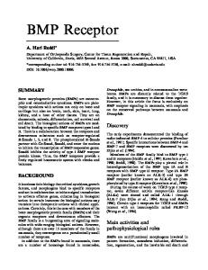
Figure 3. Effect GTPTS on the displacement of []25I]iodophenpropit from rat cerebral cortex membranes by immepip.
Consistent with conventional models of antagonist receptor binding, saturation binding curves of [125I]iodophenpropit were not affected by guanine nucleotides. Displacement of [125I]iodophenpropit by H3-agonists was usually biphasic in nature and displacement curves were shifted to the right (towards a monophasic curve) after inclusion of G T P ~ in the incubation medium, consistent with an involvement of G-proteins in the binding of the agonists (Jansen et al., 1994; Leurs et al., 1996: see also Figure 3). In contrast to []25I]iodophenpropit, the binding of [125I]iodoproxyfan to rat striatal membranes was shown to be partly sensitive to guanine nucleotides (Ligneau et al., 1994). This observation might be related to the partial agonistic nature of iodoproxyfan recently described (Schlicker et al., 1996). Also for [125I]iodoproxyfan biphasic agonist displacement binding curves were reported. Histamine competition binding curves were shifted to the right by guanine nucleotides (Ligneau et al., 1994).
133 The effect of guanine nucleotides on the binding curves of the tritiated antagonists has not been described so far. Using tritiated antagonists, agonist competition binding curves were found to be biphasic. Competition binding curves of (R)~-methylhistamine were not affected by GTPTS, using [3H]thioperamide as a radioligand (Alves-Rodrigues et al., 1996). This rather unexpected finding might result from a changed G-protein coupling at the relatively low temperature (i.e. 4~ used in the study (Alves-Rodrigues et al., 1996). Altogether, the radiolabelled antagonists provided further evidence for an involvement of G-proteins in H 3receptor mediated signal transduction. Not all the antagonists display a 'classical' behaviour regarding the sensitivity towards guanine nucleotides however. Using radiolabelled antagonists, the affinities of different unlabelled antagonists generally correlate well with their functional potencies (PA2-values). For [3H]S-methylthioperamide a detailed binding analysis including competition binding curves of different ligands has not been presented however. Considering competition binding curves of agonists, with [125I]iodophenpropit, [3H]thioperamide and [3H]GR168320, a good correlation has been described between the high affinity binding site of agonists and their EC50-values found in functional studies ( J a n s e n et al., 1994; Alves-Rodrigues et al., 1996; Brown et al., 1996). For [125I]iodoproxyfan, agonist affinities obtained from competition binding experiments were 3 to 10-fold higher as compared to their functional potencies. In this respect, [125I]iodoproxyfan displays characteristics comparable to radiolabelled agonists (see previous section). A remarkable feature of the radiolabelled H3-antagonists is that their nonspecific binding generally appeared to be high, except for [3H]GR168320. The definition of the nonspecific binding of the radiolabelled antagonists needs critical consideration. For several antagonists, radioligand binding displaced by antagonists was found to exceed radioligand binding displaced by agonists. An obvious interpretation of these observations is that the radiolabelled antagonists bind to 'non-H3-receptor components' from which they are readily displaced by H 3antagonists, and not by H3-agonists. The phenomenon has been observed for [ 125I]iodophenpropit, [125I]iodoproxyfan, [3H]thioperamide and for [3H]S-methylthioperamide (Jansen, 1997; Ligneau et al., 1994; Alves-Rodrigues et al., 1996; Yanai et al., 1994). The magnitude of the different displacement seems to be different for each ligand and is largely dependent on the tissue and species used. For [125I]iodophenpropit, the difference between agonist and antagonist displacement largely varies between different rat brain regions. In cortical brain areas and in the basal ganglia a difference of 10 to 20% was found between [125I]iodophenpropit binding displaced by (R)o~-methylhistamine and by the H3-receptor antagonist thioperamide (Jansen, 1997). Yet, the non-H3-receptor component could amount 3040% in regions with lower H3-receptor densities, like the thalamus and the hippocampus (Alves-Rodrigues, 1996). In contrast, (R)o~-methylhistamine and thioperamide displaced the same fraction of [125I]iodophenpropit binding in mouse brain (Jansen, 1997). A difference of about 40% between binding displaced between agonists and antagonists has been observed for [125I]iodoproxyfan, in rat striatal tissue (Ligneau et al., 1994). Comparable differences were
134 found for [3H]thioperamide binding to rat cerebral cortex membranes (Alves-Rodrigues et al., 1996). From these results it may be concluded that, in general, H3-agonists can be regarded as more reliable tools to define the nonspecific binding of the radiolabelled antagonists. At present, the origin of the non-H3-receptor binding component(s) of H3-antagonists is largely unknown. Recently, iodophenpropit and thioperamide have been screened on about forty different receptor assays (Leurs et al., 1995a). The screening did not yield a possible candidate for the nonspecific binding of both radioligands. Both iodophenpropit and thioperamide display a relatively high affinity for 5HT3-receptors (Ki-values of 11 nM and 120 nM, respectively). At the experimental conditions used, 5HT3-receptor binding does not interfere with the the assay for both radioligands however. For [3H]thioperamide, binding to cytochrome P450 isoenzymes may be involved (Alves-Rodrigues et al., 1996). The same has been suggested for [3H]Smethylthioperamide (Yanai et al., 1994). Interestingly, [125I]iodoproxyfan was recently reported to bind with high affinity to a histamine transporter present in murine hematopoietic progenitor cells (Corbel et al., 1997). Also thioperamide displayed a high affinity to these binding sites, in contrast to H3-agonists. Whether this interaction could contribute to high nonspecific binding of [ 125I]iodoproxyfan in the striatum remains to be determined. The origin of the nonspecific binding may of course differ between the radiolabelled antagonists. Illustrative for this rationale is the observation that the thioperamide related compound [3H]GR168320 does not seem to exhibit the phenomenon of a differential displacement by agonists and antagonists, at least in rat cerebral cortex membranes (Brown et al., 1996). The relatively high nonspecific binding of most radiolabelled antagonists can be regarded as a less favourable feature. In this respect, [3H]GR168320 may be the most appropriate radioligand. However, due to its low specific activity of 4.8 Ci/mmol, a relatively high amount of protein is required in the receptor binding assay, especially when tissues containing lower H3-receptor densities are assessed. The low specific activity of [3H]GR168320 seems to be inadequate for the generation of autoradiographic images. Therefore, a [125I]-labelled radioligand with properties similar to [3H]GR168320 would be desirable.
4. LOCALIZATION OF H3-RECEPTOR BINDING SITES IN THE CNS
The autoradiographic distribution of H3-receptor binding sites has been studied in rat brain using different radioligands, i.e. [3H](R)~-methylhistamine (Arrang et al., 1987; Yanai et al., 1992; Pollard et al., 1993; Ryu et al., 1995), [3H]N~ (Cumming et al., 1991; Cumming et al., 1994) and more recently with [125I]iodophenpropit (Jansen et al., 1994), [125I]-iodoproxyfan (Ligneau et al., 1994) and [3H]S-methylthioperamide (Yanai et al., 1994). In contrast to the differences in binding characteristics between the H3-receptor
135
Table 3. Comparison of [125I]iodophenpropit and [3H](R)t~-methylhistamine binding sites in rat brain. radioligand binding (% of total cerebral cortex)
[ 125i] iodophenpropit 1) (auto radiog raphy)
[3H](R)ct_methylhistamine 2) (membrane preparations)
anterior cerebral cortex
97 + 8
108 + 2
medial cerebral cortex
96 + 11
100 + 2
posterior cerebral cortex
110 + 13
85 + 6
olfactory tubercle hippocampus
121 + 13 53 + 8
103 + 8 48 + 4
caudate putamen
127 + 10
108 + 11
nucleus accumbens
133 + 7
126 + 12
septum
77 + 4
N.D.
hypothalamus (anterior)
67 + 5
70 + 2
hypothalamus (posterior)
61 + 7
54 + 8
hypothalamus (lateral) hypothalamus (VMH)
68 + 4 78 + 13
N.D. N.D.
thalamus anterior amygdaloid area amygdala (posterior)*
54 + 16 98 + 6 69 + 10
N.D. N.D. N.D.
substantia nigra pons
141 + 21 33 + 14
97 + 7 28 + 5
cerebellum
8+ 5
7+ 3
l~Specific binding was determined using 1 ktM (R)t~-methylhistamine. 2)Values reported by Pollard et al., 1993. *Including: amygdalohippocampal area (AHi) and posteromedial cortical amygdaloid nucleus (PMCo). N.D.: not described. The density of [125I]-iodophenpropit binding sites in the cortex is 268 fmol/mg of protein (Jansen et al., 1994). Randomized brain sections of three to five rats were used. Values are expressed as mean + SD of three to five separate determinations of which each was performed at least in triplicate.
radioligands found using brain membrane preparations, a consistent overlap is observed so far with respect to the autoradiographic distribution of the radioligand binding sites. In Table 3 a comparison of the distribution of [3H](R)o~-methylhistamine and [125I]iodophenpropit binding sites in rat brain is given. A comprehensive description of the distribution of [3H](R)t~-methylhistamine binding sites in the rat CNS has been given by Pollard et al. (Pollard et al., 1993). In brief, highest densities are observed in the cerebral cortex, the olfactory tubercles, the caudate putamen, the nucleus accumbens and the substantia nigra. Moderate densities are found in the hippocampus, the
136 globus pallidus, the thalamus and the hypothalamus, including the histaminergic perikarya in the posterior area. (For a more detailed overview of the H3-receptor binding sites in the CNS see Chapter 1). H3-Receptor binding sites in the CNS display a distribution pattern distinct from the localization of the histaminergic varicosities, which may in part be explained by the existence of H 3heteroreceptors. A presynaptic localization of H3-receptors on noradrenergic (Schlicker et al., 1989), and on serotonergic (Fink et al., 1990; Alves-Rodrigues et al., 1995) nerve terminals in the cerebral cortex has been indicated from functional studies. Receptor binding studies provided evidence for a presynaptic localization of H3-receptors on GABA neurons in the substantia nigra (Cumrning et al., 1991" Ryu et al., 1994). The presynaptic localization of H 3receptors in the substantia nigra has recently been confirmed with superfused rat brain slices, demonstrating an inhibition by H3-agonists of dopamine Dl-receptor stimulated GABA release (Garcia et al., 1997). In addition to presynaptic receptors, autoradiographic studies have indicated the presence of postsynaptic H3-receptor binding sites as well. Chemical destruction of postsynaptic structures in the striatum using quinolinic and kainic acid resulted in a marked decrease of striatal H 3receptor binding sites (Cumming et al., 1991; Pollard et al., 1993; Ryu et al., 1994). Consequently, a major part of the striatal H3-receptors may be located on striatal GABA neurons, representing more than 85% of the striatal efferents (Kita & Kitai, 1988).
5. H3-RECEPTOR BINDING STUDIES IN PERIPHERAL TISSUES The densities of H3-receptors in the periphery appeared to be much lower as compared to densities in the CNS. This makes peripheral H3-receptors less accessible for receptor binding studies, and consequently explains that only a limited number of studies on H3-receptor binding in peripheral tissues has been described so far. H3-Receptors in the periphery of the guinea-pig have been characterized with [3H]N amethylhistamine (Korte et al., 1990). In most tissues H3-receptor densities were below 1 fmol/mg of protein. Highest densities (between 4 and 8 fmol/mg protein) were found in the large intestine, the ileum, the pancreas and the pituitary. A full pharmacological characterization of the [3H]Na-methylhistamine binding sites in the peripheral tissues was not presented (Korte et al., 1990).
H3-Receptors were also detected in the human gastric mucosa (Courillon-Mallet et al., 1995). [3H]Na-Methylhistamine saturation binding to mucosal H3-receptors yielded a receptor density of 10 fmol/mg of protein. H3-Receptor binding was reduced in Heliobacter p y l o r i infected patients (Courillon-Mallet et al., 1995). Gastric H3-receptors have also been characterized using a human fundic tumor cell line (HGT-1). Binding of [3H]Na-methylhistamine to these cells was sensitive to GTP),S and to both cholera and pertussis toxin, again indicating the
137 coupling of the gastric H3-receptors to G-proteins (Cherifi et al., 1992). Similar results have been obtained in the murine pituitary tumor cell line AtT-20 (Clark et al., 1993; West et al., 1994). In guinea-pig lung the distribution of H3-receptors has been visualized by receptor autoradiography (Schwartz et al., 1990). [3H](R)o~-Methylhistamine binding was scattered in the parenchyma. A more dense labelling was observed in the bronchioles (Schwartz et al., 1990). Except for [3H]S-methylthioperamide, receptor binding studies to peripheral tissues have not been described for radiolabelled antagonists. [3H]S-Methylthioperamide showed a considerably high amount of nonspecific binding, which interfered with the accurate determination of H 3receptors in peripheral tissues (Yanai et al., 1994). Based on the relatively high amount of nonspecific binding observed with most radiolabelled H3-antagonists, similar limitations may evolve for other radiolabelled antagonists.
6. HETEROGENEITY OF RADIOLIGANG BINDING SITES 6.1. Radiolabelled H3-agonist binding sites In 1990, West et al. reported that thioperamide and burimamide discriminated [3H]N~methylhistamine binding to rat brain membranes into high and low affinity binding sites (West et al., 1990b). [3H]NC~-Methylhistamine binding was partly decreased by the GTP analogue GTPyS. In the presence of GTPyS, thioperamide and burimamide yielded monophasic competition binding curves, with affinities corresponding to their high affinity binding sites. From these results the existence of subtypes of H3-receptors i.e. H3A- and H3B-receptors was proposed, the latter being sensitive towards guanine nucleotides (West et al., 1990b). [3H]N ~Methylhistamine itself did not discriminate between the proposed H3A- and H3B-receptors. In a study by Arrang and co-workers, using the agonist [3H](R)o~-methylhistamine as the radioligand, biphasic competition binding curves in rat cerebral cortex membranes were obtained for burimamide, but not for thioperamide (Arranget al., 1990). A guanine nucleotide sensitivity of the burimamide binding sites was not reported in this study. In contrast to [3H]N~-methylhistamine (West et al., 1990b), in the standard incubation medium, binding of [3H](R)o~-methylhistamine was not sensitive to the GTP analogue Gpp(NH)p. However, when calcium was added to the incubation buffer, two binding sites were found for [3H](R)o~methylhistamine, the low affinity site being abolished by Gpp(NH)p (Arrang et al., 1990). From these observations it may be suggested that the possible heterogeneity of burimamide binding sites and of [3H](R)~-methylhistamine binding sites are unrelated phenomena. The heterogeneity of [3H](R)a-methylhistamine binding sites was suggested to result from the conversion of a subpopulation of the receptors into low-affinity binding sites, triggered by
138 calcium (Arrang et al., 1990). A heterogeneity of [3H](R)~-methylhistamine binding sites has also been found in kinetic studies, using buffer without calcium (West et al., 1990a). In this study a homogeneous population of [3H](R)~-methylhistamine binding sites was observed at equilibrium conditions (i.e. saturation binding analysis) however. Thioperamide and burimamide yielded monophasic competition binding curves in this report (West et al., 1990a). The three reports cited illustrate the complexity of the receptor binding data obtained with the radiolabelled agonists and the controversies in literature with respect to a heterogeneity of H 3receptor binding sites. Biphasic competition binding curves for burimamide have been described in several reports using [3H](R)~-methylhistamine (Arrang et al., 1990) and [3H]Na-methylhistamine (West et al., 1990b; Kathmann et al., 1993; Cumming & Gjedde, 1994; Brown et al., 1996). Accordingly, different studies reported a heterogeneous displacement of [3H]Na-methylhistamine by thioperamide (West et al., 1990b; Cumming & Gjedde, 1994; Clark & Hill, 1995; Brown et al., 1996). Controversially, other studies did not confirm the presence of two distinct binding sites for burimamide (West et al., 1990a; Kilpatrick & Michel, 1991; Clark & Hill, 1995) and for thioperamide (Arrang et al., 1990; West et al., 1990a; Kilpatrick & Michel, 1991; Kathmann et al., 1993). One explanation for the different observations concerning heterogeneity of thioperamide and burimamide binding sites is the relatively small difference in affinity between the two separate binding sites, making it difficult to discriminate them statistically. In addition, the controversies concerning the heterogeneity of binding sites may arise from different experimental conditions used, like the choice of buffer (Tris-HC1, phosphate, HEPES), the ionic composition of the buffer (mono- and divalent cations) and the tissue preparation used (cerebral cortex versus whole brain). For example, it has been reported that the affinity of thioperamide for [3H](R)~methylhistamine binding sites was 10-fold higher in phosphate buffer as compared to Tris-HC1 buffer (West et al., 1990a). In contrast the affinity of the agonists histamine, (R)~methylhistamine and Na-methylhistamine were not substantially different when phosphate and Tris-HC1 buffer are compared (Arrang et al., 1987; West et al., 1990a). The ionic composition of the buffer has been indicated to differentially affect binding characteristics of ligands. As previously cited, guanine nucleotide sensitivity of [3H](R)~methylhistamine (but not of [3H]Na-methylhistamine; West et al., 1990b) was dependent on the presence of calcium in the buffer (Arrang et al., 1990). Sodium ions were shown to abolish the low affinity binding site of thioperamide, whereas the binding affinities of clobenpropit and Na-methylhistamine were not affected (Clark & Hill, 1995). From these results, it was suggested that the H3-receptor exists in different conformations, for each of which thioperamide has a different affinity (Clark & Hill, 1995). Hence, contribution of differential allosteric effects dependent of the buffer composition may relate to the observed heterogeneity of binding sites and to the controversy in literature in this respect. A differential allosteric action of sodium
139 has also been reported for other receptor systems including the binding of Hi-receptor antagonists (Treherne et al., 1991; Gibson et al., 1994). The allosteric effect may also be related to the involvement of G-proteins in the binding of agonists to the receptor, further complicating the interpretation of the binding data. Altogether, the complexity of the binding profile of radiolabelled agonists does not provide a sound basis for the definition of H3-receptor subtypes. An important criterion for the identification of receptor subtypes is that they are related to distinct functional responses. Based on the functional potencies of thioperamide and of tiotidine, H3A- and H3B-receptors were suggested to be linked to H3-receptor mediated inhibition of histamine release and synthesis, respectively (West et al., 1990b). At present, not much additional evidence for this suggestion has been presented. Histamine H3-receptors inhibiting noradrenaline release in mouse brain cortex slices have been suggested to represent the H3A-receptor subtype (Schlicker et al., 1992; Schlicker et al., 1994). To our knowledge, functional responses in brain tissue related to the H3B-receptor have never been observed however.
6.2. Radiolabelled H3-antagonist binding sites [ 125I]Iodophenpropit was biphasically displaced from rat cortex membranes by the antagonists burimamide and dimaprit (Jansen et al., 1994). In contrast to agonist binding, antagonist binding was not affected by GTPTS. Hence, biphasic competition binding curves of burimamide and dimaprit were likely not related to the G-protein coupling of the [125I]iodophenpropit binding sites. For the other radiolabelled antagonists, biphasic competition binding curves of antagonists have so far not been demonstrated. Remarkably, thioperamide and burimamide yielded steep competition binding curves (Hill-coefficients of 1.7 and 1.9, respectively) in rat striatal membranes using the [125I]-iodoproxyfan assay (Ligneau et al., 1994). A heterogeneous distribution of putative H3-receptor subtypes has not been demonstrated so far. Using a receptor autoradiographic approach, we have recently found that [125I]iodophenpropit binding to ten different rat brain areas was not discriminated by a chemically heterogeneous group of H3-receptor antagonists (Jansen, 1997). As mentioned before, for all radiolabelled antagonists, biphasic competition binding curves were reported for H3-agonists (Jansen et al., 1994; Yanai et al., 1994; Ligneau et al., 1994; Alves-Rodrigues et al., 1996; Brown et al., 1996). In the [125I]iodophenpropit and [125I]iodoproxyfan binding assays, agonist competition binding curves were sensitive to guanine nucleotides (Jansen et al., 1994; Ligneau et al., 1994). The apparent heterogeneity of agonist binding may therefore be attributed to the involvement of G-proteins in the agonist receptor binding rather than to a receptor heterogeneity. For the tritiated antagonists, the sensitivity to guanine nucleotides was not studied or could not be demonstrated at the experimental conditions
140 used (Alves-Rodrigues et al., 1996). Yet, it can be concluded that receptor binding studies with both, radiolabelled agonists and with radiolabelled antagonists did not reveal exclusive evidence for H3-receptor heterogeneity. In general, the exploration of H3-receptor subtypes requires the availability of ligands with a higher selectivity towards one of these putative subtypes.
7. R E S U M P T I O N A N D C O N C L U D I N G R E M A R K S
Studies performed with H3-receptor radioligands have substantially contributed to the current knowledge of the characteristics, distribution and function of the histamine H3-receptor. Tritiated agonists were successfully used to study H3-receptors in rodent and primate CNS. Binding studies with radiolabelled agonists provided evidence for a role of G-proteins in H 3receptor mediated signal transduction. The apparent involvement of G-protein coupling in the binding of the radiolabelled agonists may underlie two less favourable features of the radioligands however. At first, an overestimation of H3-agonists potencies is obtained in competition binding studies. Secondly, the complexity of the radiolabelled agonists binding dynamics makes it difficult to distinguish binding phenomena related to G-protein coupling, allosteric interactions, and receptor heterogeneity in terms of H3-receptor subtypes. Radiolabelled agonists are advantageous with respect to their low nonspecific binding in the rat CNS. [3H](R)o~-Methylhistamine and [3H]NC~-methylhistamine were both shown to be very useful studying the distribution of H3-receptors by autoradiography. The introduction of radiolabelled H3-receptor antagonists yielded improved tools for H 3receptor binding studies. With the use of these ligands additional evidence was provided for the interaction of H3-receptors with G-proteins. As compared to radiolabelled agonists, [125I]iodophenpropit, [3H]GR168320 and [3H]thioperamide exhibit the advantage of a good correlation between agonist binding affinities and their functional potencies. So far, studies with radiolabelled H3-antagonists did not provide significant progress in the search for H3-receptor heterogeneity. Ligands which more clearly discriminate between putative subtypes are still awaited, and a link between binding heterogeneity and functional receptor responses will be indispensable. Not all radiolabelled antagonists display a straightforward binding profile, which may in part be due to the relatively high amount of nonspecific binding, to be considered as a disadvantage. In this respect [3H]GR168320 is a promising ligand, displaying a negligible amount of nonspecific binding, allowing a unambiguous interpretation of receptor binding data.
141
REFERENCES Alves-Rodrigues, A. (1996). Doctoral thesis: The histamine H3-receptor in the rat brain: pharmacological and (patho)physiological aspects. Vrije Universiteit, Amsterdam, The Netherlands. Alves-Rodrigues, A., Jansen, F.P., Leurs, R., Timmerman, H. and Prell, G.D. (1995). Interaction of clozapine with the histamine H3-receptor in rat brain. Br. J. Pharmacol. 114, 1523-1524. Alves-Rodrigues, A., Leurs, R., Wu, T.S.W., Prell, G.D., Foged, C. and Timmerman, H. (1996). [3H]-Thioperamide as a radioligand for the histamine H3-receptor in rat cerebral cortex. Br. J. Pharmacol. 118, 2045-2052. Arrang, J.M., Garbarg, M., Lancelot, J.-C., Lecomte, J.-M., Pollard, H., Robba, M., Schunack, W. and Schwartz, J.C. (1987). Highly potent and selective ligands for histamine H3-receptors. Nature 327,117-125. Arrang, J.M., Roy, J., Morgat, J.-L., Schunack, W. and Schwartz, J.C. (1990). Histamine H3-receptor binding sites in rat brain membranes: modulations by guanine nucleotides and divalent cations. Eur. J. Pharmacol. 188, 219-227. Barbin, (3., Palacios, J.M., Rodergas, E., Schwartz, J.C. and Garbarg, M. (1980). Characterization of the high-affinity binding sites of [3H]histamine in rat brain. Mol. Pharmacol. 18, 1-10. Brown, J.D., Oshaughnessy, C.T., Kilpatrick, G.J., Scopes, D.I.C., Beswick, P., Clitherow, J.W. and Barnes, J.C. (1996). Characterisation of the specific binding of the histamine H3-receptor antagonist radioligand [3H]GR168320. Eur J Pharmacol 311, 305310. Cherifi, Y., Pigeon, C., Leromancer, M., Bado, A., Reyldesmars, F. and Lewin, M.J.M. (1992). Purification of a histamine H3-receptor negatively coupled to phosphoinositide turnover in the human gastric cell line Hgtl. J. Biol. Chem. 267, 25315-25320. Clark, E.A. and Hill, S.J. (1995). Differential effect of sodium ions and guanine nucleotides on the binding of thioperamide and clobenpropit to histamine H3-receptors in rat cerebral cortical membranes. Br. J. Pharmacol. 114, 357-362. Clark, M.A., Korte, A. and Egan, R.W. (1993). Guanine nucleotides and pertussis toxin reduce the affinity of histamine H3-receptors on AtT-20 cells. Agents and Actions 40, 129134. Corbel, S., Traiffort, E., Stark, H., Schunack, W. and Dy, M. (1997). Binding of histamine H3-receptor antagonists to hematopoetic progenitor cells. Febs Lett. 404, 289-293. Courillon-Mallet, A., Launay, J.M., Roucayrol, A.M., Callebert, J., Emond, J.P., Tabuteau, F. and Cattan, D. (1995). Helicobacter pylori infection: Physiopathologic implication of NC~methylhistamine. Gastroenterology 108, 959-966. Cumming, P. and Gjedde, A. (1994). Subclasses of histamine H3-antagonist binding sites in
142 rat brain. Brain Res. 641, 203-207. Cumming, P., Gjedde, A. and Vincent, S. (1994). Histamine H3 binding sites in rat brain localization in the nucleus of the solitary tract. Brain Res. 641, 198-202. Cumming, P., Shaw, C. and Vincent, S.R. (1991). High affinity histamine binding site is the H3-receptor - characterization and autoradiographic localization in rat brain. Synapse 8, 144-151. Fink, K., Schlicker, E., Neise, A. and G6thert, M. (1990). Involvement of presynaptic H3receptors in the inhibitory effect of histamine on serotonin release in the rat brain cortex. Naunyn-Schmied. Arch. Pharmacol. 342, 513-519. Fujimoto, K., Mizuguchi, H., Fukui, H. and Wada, H. (1991). Presynaptic localization of histamine H3-receptors in rat brain. Biochem. Biophys. Res. Commun. 177, 907-912. Garcia, M., Floran, B., Arias-Montano, J.A., Young, J.M. and Aceves, J. (1997). Histamine H3-receptor activation selectively inhibits dopamine D 1-dependent [3H]-qt-aminobutyric acid release from depolarisation-stimulated slices of rat substantia nigra pars reticulata. Neuroscience, in press. Gibson, W.J., Roques, T.W. and Young, J.M. (1994). Modulation of antagonist binding to histamine H 1-receptors by sodium ions and by 2-amino-2-hydroxymethyl-propan-1,3-diol HC1. Br. J. Pharmacol. 111, 1262-1268. Herbert T.E., Moffett, S., Morello, J.P., Loisel, T.P., Bichet, D.G., Barret, C. and Bouvier, M. (1996). A peptide derived from a beta2-adrenergic receptor transmembrane domain inhibits both receptor dimerization and activation. J. Biol. Chem. 271, 16384-16392. Jansen, F.P. (1997). Doctoral thesis: The histamine H3-receptor in the rat brain: pharmacological and (patho)physiological aspects. Vrije Universiteit, Amsterdam, The Netherlands. Jansen, F.P., Rademaker, B., Bast, A. and Timmerman, H. (1992). The first radiolabelled histamine H3-receptor antagonist, [125I]iodophenpropit: saturable and reversible binding to rat cortex membranes. Eur. J. Pharmacol. 217, 203-205. Jansen, F.P., Wu, T.S., Voss, H.P., Steinbusch, H.W.M., Vollinga, R.C., Rademaker, B., Bast, A. and Timmerman, H. (1994). Characterization of the binding of the first selective radiolabelled histamine H3-receptor antagonist, [ 125I]-iodophenpropit, to rat brain. Br. J. Pharmacol. 113, 355-362. Kathmann, M., Schlicker, E., Detzner, M. and Timmerman, H. (1993). Nordimaprit, homodimaprit, clobenpropit and imetit - affinities for H3 binding sites and potencies in a functional H3-receptor model. Naunyn Schmied. Arch. Pharmacol. 348, 498-503. Kilpatrick, G.J. and Michel, A.D. (1991) Characterisation of the binding of the histamine H3receptor agonist [3H](R)a-methylhistamine to homogenates of rat and guinea-pig cortex. In: New perspectives in histamine research (Timmerman, H. & Van der Goot, H., Eds) pp 6975, Birkhauser Verlag, Basel.
143 Kita, H. and Kitai, S.T. (1988). Glutamate decarboxylase immunoreactive neurons in rat neostriatum: their morphological types and populations. Brain Res. 447, 346-352. Korte, A., Myers, J., Shih, N.-Y., Egan, R.W. and Clarck, M.A. (1990). Characterization and tissue distribution of H3-histamine receptors in guinea-pigs by N~-methylhistamine. Biochem. Biophys. Res. Commun. 168, 979-986. Leurs, R., Kathmann, M., Vollinga, R.C., Menge, W.M.P.B., Schlicker, E. and Timmerman, H. (1996). Histamine homologues discriminating between two functional H3-receptor assays. Evidence for H3-receptor heterogeneity? J. Pharmacol. Exp. Ther. 276, 10091015. Leurs, R. and Timmerman, H. (1992). The histamine H3-receptor: a target for developing new drugs. Prog. Drug Res. 39, 127-165. Leurs, R., Tulp, M.T.M., Menge, W.M.B.P., Adolfs, M.J.P., Zuiderveld, O.P. and Timmerman, H. (1995a). Evaluation of the receptor selectivity of the H3-receptor antagonists, iodophenpropit and thioperamide: an interaction with the 5-HT3- receptor revealed. Br. J. Pharmacol. 116, 2315-2321. Leurs, R., Vollinga, R.C. and Timmerman, H. (1995b). The medicinal chemistry and therapeutic potentials of ligands of the histamine H3-receptor. Prog. Drug Res. 45, 107165. Ligneau, X., Garbarg, M., Vizuete, M.L., Diaz, J., Purand, K., Stark, H., Schunack, W. and Schwartz, J.C. (1994). [125I]Iodoproxyfan, a new antagonist to label and visualize cerebral histamine H3-receptors. J. Pharmacol. Exp. Ther. 271, 452-459. Martinez-Mir, M.I., Pollard, H., Moreau, J., Arrang, J.M., Ruat, M., Traiffort, E., Schwartz, J.C. and Palacios, J.M. (1990). Three histamine receptors (H1, H2 and H3) visualized in the brain of human and non-human primates. Brain Res. 526, 322-327. Ng, G.Y.K., O'Dowd, B.F., Lee, S.P., Chung, H.T., Brann, M.R., Seeman, P. and George, S.R. (1996). Dopamine D 2 receptor dimers and receptor blocking peptides, B iochem. B iophys Res, Commun. 227, 200-204. Oishi, R., Adachi, N. and Saeki, K. (1993). NC~-Methylhistamine inhibits intestinal transit in mice by central histamine HI-receptor activation. Eur. J. Pharmacol. 237, 155-159. Pollard, H., Moreau, J., Arrang, J.M. and Schwartz, J.C. (1993). A detailed autoradiographic mapping of histamine H3-receptors in rat brain areas. Neuroscience 52, 169-189. Ryu, J.H., Yanai, K., Sakurai, E., Kim, C.Y. and Watanabe, T. (1995). Ontogenetic development of histamine receptor subtypes in rat brain demonstrated by quantitative autoradiography. Developmental Brain Res. 87, 101-110. Ryu, J.H., Yanai, K. and Watanabe, T. (1994). Marked increase in histamine H3-receptors in the striatum and substantia nigra after 6-hydroxydopamine-induced denervation of dopaminergic neurons: an autoradiographic study. Neurosci. Lett. 178, 19-22. Schlicker, E., Behling, A., Ltimmen, G. and G6thert, M. (1992). Histamine H3A receptor-
144 mediated inhibition of noradrenaline release in the mouse brain cortex. Naunyn-Schmied Arch Pharmaco1345, 489-493.
Schlicker, E., Kathmann, M., Bitschnau, H., Marr, I., Reidemeister, S., Stark, H. and Schunack, W. (1996). Potencies of antagonists chemically related to iodoproxyfan at histamine H3-receptors in mouse brain cortex and guinea-pig ileum: evidence for H3-receptor heterogeneity? Naunyn-Schmied Arch Pharmaco1353, 482-488. Schlicker, E., Kathmann, M., Reidemeister, S., Stark, H. and Schunack, W. (1994). Novel histamine H3-receptor antagonists: affinities in an H3-receptor binding assay and potencies in two functional H3-receptor models. Br. J. Pharmacol. 112, 1043-1048. Schwartz, J.C., Arrang, J.M., Garbarg, M. and Pollard, H. (1990). A third histamine receptor subtype: characterisation, localisation and functions of the H3-receptor. Agents and Actions 30, 13-23. Sinkins, W.G., Kandel, M., Kandel, S.I., Schunack, W. and Wells, J.W. (1993). Proteinlinked receptors labeled by [3H]histamine in guinea-pig cerebral cortex. 1. Pharmacological characterization. Mol. Pharmacol. 43, 569-582. Sinkins, W.G. and Wells, J.W. (1993). Protein-linked receptors labeled by [3H]histamine in guinea-pig cerebral cortex. 2. Mechanistic basis for multiple states of affinity. Mol. Pharmacol. 43, 583-594. Treherne, J.M., Stern, J.S., Flack, W.J. and Young, J.M. (1991). Inhibition by cations of antagonist binding to histamine HI-receptors: differential effect of sodium ions on the binding of two radioligands. Br. J. Pharmacol. 103, 1745-1751. West, R.E., Myers, J., Zweig, A., Siegel, M.I., Egan, R.W. and Clark, M.A. (1994). Steroid-sensitivity of agonist binding to pituitary cell line histamine H3-receptors. Eur. J. Pharmacol. 267, 343-348. West, R.E., Zweig, A., Granzow, R.T., Siegel, M.I. and Egan, R.W. (1990a). Biexponential kinetics of (R)cx-[3H]methylhistamine binding to the rat brain histamine H3-receptor. J. Neurochem. 55, 1612-1616. West, R.E.J., Zweig, A., Shih, N.-Y., Siegel, M.I., Egan, R.W. and Clarck, M.A. (1990b). Identification of two H3-histamine receptor subtypes. Mol. Pharmacol. 38, 610-613. Yanai, K., Ryu, J.H., Sakai, N., Takahashi, T., Iwata, R., Ido, T., Murakami, K. and Watanabe, T. (1994). Binding characteristics of a histamine H3-receptor antagonist, [3H]Smethylthioperamide: Comparison with [3H](R)ot-methylhistamine binding to rat tissues.Jap. J. Pharmacol. 65, 107-112. Yanai, K., Ryu, J.H., Watanabe, T., Iwata, R. and Ido, T. (1992). Receptor autoradiography with [11C] and [3H]-labelled ligands visualized by imaging plates. Neuroreport 3, 961-964. Zweig, A., Siegel, M.I., Egan, R.W., Clark, M.A., Shorr, R.G.L. and West, R.E. (1992). Characterization of a digitonin-solubilized bovine brain H3-histamine receptor coupled to a guanine nucleotide-binding protein. J. Neurochem. 59, 1661-1666.
R. Leurs and H. Timmerman (Editors) The Histamine H 3 Receptor (~ 1998 Elsevier Science B.V. All rights reserved.
145
Substituted imidazoles, the key to histaminergic receptors W. M. P. B. Menge, H. Timmerman Division of Medicinal Chemistry, Leiden/Amsterdam Center for Drug Research, Vrije Universiteit, De Boelelaan 1083, 1081 HV, Amsterdam, The Netherlands
1. I N T R O D U C T I O N Substituted imidazoles are an example of a pharmaceutically important class of heterocyclic compounds, several of which have been incorporated in drugs that have reached the market, e.g. cimetidine, ondansetron and losartan (figure 1).
CH3~~f~-
S ~
H
N
.y."
CI
NCN
0
N~
,,, ,~,,,,,,j B
N""
%,
H O.__/
\
~
~
=(/__k NH
#
H Figure 1: Drugs containing an imidazole ring, cimetidine, ondansetron and losartan Also in the development of specific ligands for the histamine receptors numerous substituted imidazoles have been synthesised during the last decades. This is not surprising since the natural ligand, histamine, is also a substituted imidazole. Via a change in the substitution pattern of the imidazole nucleus or a modification of the existing substituent many histamine analogues and imidazole derivatives, with potent and selective agonistic or antagonistic activity, have been prepared (figure 2). These compounds and their structure-
146 activity relationships have played an important role in the classification of the histamine receptors into the three, currently known, subtypesl.
]~
,,..,..,~ NH2 H H CH3 ~ s ~ N y N ' c H N~NH
a
NCN
"CF3 Figure 2: Examples of potent and selective histaminergic ligands Continued investigations of the structure-activity relationships of ligands for these receptors have also led to the development of potent non-imidazolic ligands for the histamine H 1- and H2-receptor subtypes. However, the state of the art on the medicinal chemistry of the histamine H3-receptor indicates that this most recently discovered receptor subtype has a remarkable preference for substituted imidazoles 2 as ligands. Replacement of the imidazole ring in known potent H3-1igands by other heterocycles leads consistently to a complete loss or, at the very least, a drastic decrease in affinity 3. Therefore, the research on the structureactivity relationships for the histamine H3-receptor still depends on the availability of new substituted imidazoles as ligands. Also, for the two other histamine receptor subtypes there is a continuing interest in the synthesis of new substituted imidazoles and their pharmacological evaluation as histaminergic ligands. For example; the recent discovery of the inverse agonism of several compounds formerly classified as histamine H2-antagonists 4, the systematic studies on the binding interactions of the agonistic and antagonistic ligands with specific amino acid residues of the histamine H1-5 and H2-receptor 6 or the study on the molecular mechanism of activation 6,7 of these receptors. And last but not least, the continuous refinement of pharmacological and molecular modelling techniques demand a (re-) investigation of old and new compounds with the imidazole pharmacophore. The synthesis of imidazoles and the evaluation of their pharmacological activities can therefore be considered to be the key to a better understanding of the histamine receptors. Further new developments such as combinatorial technologies and the uprise of molecular biology will certainly play an important role in the need for and the synthesis and evaluation of new substituted imidazoles in the coming years.
147 2. SYNTHESES OF IMIDAZOLES A comprehensive review on imidazole chemistry by Grimmett has appeared recently 8. Therefore, the next paragraphs will focus on the most recent developments in the synthesis of substituted imidazoles used as ligands for the histamine receptors. Special attention will be pay to methods with perspective for the synthesis of histaminergic ligands. 2.1. Modification of imidazole precursors A simple and direct approach to new imidazole containing ligands is the modification of commercially available imidazoles. Unfortunately, only a limited number of substituted imidazoles is available and most of them are not ideally substituted for further modification because they have only plain alkyl or phenyl substituents. An example of this approach is the synthesis of 3-aminopropylimidazole 9 and 3-hydroxy-propylimidazole 10 from urocanic acid (figure 3).
/CO2H
OH 5 steps N~,,,, NH
~ 58% yield
~ N~,~,,,, NH
F
NH2
5 steps 56% yield ~
N~,,, NH
Figure 3: Urocanic acid as a source for fragments of histaminergic ligands These compounds are valuable intermediates for the synthesis of histamine H3- and H2ligands. 2.1. Condensation approaches Most substituted imidazoles are obtained via the condensation of non-cyclic fragments to form the desired imidazole. The majority of these condensation reactions has recently been reviewed by Grimmett 8. The largest number of imidazoles obtained via condensation approaches is only of limited use for the development of histaminergic ligands as they are mainly polysubstituted imidazoles. For the histamine receptors proper substitution of the imidazole ring is usually limited to the 4(5)-position, with the exception of the histamine HIreceptor, where a broader range of substituents on the 2-position is tolerated. The most frequently applied condensation approach for histaminergic ligands is the condensation of an o~-substituted carbonyl compound, C4-C5 backbone, with an amidine or activated amide, C2-precursor (figure 4). The Bredereck 11 method, in which an o~-bromo
148 carbonyl compound is condensed with an amidine or another activated precursor to form the heterocyclic ring 12,13,14, is a popular example of such an approach.
@ a4
X
R5
C2-precursor
R4 N ' ] ~
"-
O
R5
N~[/NH Re
X = halogen, amine, ketone or hydroxy group
Figure 4. The synthesis of imidazoles using conventional condensation aproaches. The isolated yields of the Bredereck method are satifactory for the synthesis of di-substituted compounds (figure 5) but are generally lower for 4(5)-mono-substituted imidazoles. o
o
NH 2
o Y
-CF 3
CF 3
Figure 5. Synthesis of histamine Hl-agonists via the Bredereck method Other important methods involve the condensation of an aminoketone 15,16 (figure 6) or a diketone 17 with an imidate or other activated amide. In general, the methods are of a broad scope and have been used to synthesize a variety of imidazole derivatives. N
NH
N
HC- NH 2 H2N
O
several steps
N ~ NH
N
N~
)
NH
Figure 6: The synthesis of thioperamide Major drawbacks of the condensation approaches 8 are the difficult syntheses of the starting materials, the low yields of the end products and the lack of flexibility in the approach. Each new target compound requires a different precursur and thus a completely new synthesis route including all the difficulties associated with the isolation of the product.
149 In conclusion, although satisfactory condensation reactions are available to prepare most of the desired substituted imidazoles, there is a continuing interest in new, less elaborate and more flexible, synthesis routes.
2.2. The synthon approach An alternative way to synthesize imidazoles in a more flexible manner is to use a synthon. An example of such a synthon approach, is the synthesis of imidazoles using tosylmethyl isocyanide (TOSMIC). The original procedure using this synthon gave only moderate overall yields of the substituted imidazoles 18, mainly due to the poor yield in the first step (figure 7). Tos
R
RCHO
Tos- CH 2- NC
R
NH3, MeOH_
t-BuOK, DME
N ~,~,,/O
"-
/ ~ N ~,~,i NH
Figure 7. The synthesis of imidazoles using TosMIC. However, recently the yield of the first step of this imidazole synthesis was improved dramatically by replacement of the basic catalyst (tBuOK) by a milder basic catalyst, sodium cyanide 19. Due to these milder reaction conditions a greater variety of aldehydes, one of the starting materials for the synthesis of imidazoles, can be used increasing the flexibility of the method 2~ even more. This synthon approach can also be used to prepare the bioisesteric substituted thiazole analogs 21. Other recent improvements of the TOSMIC synthon approach include the use of different precursors for the C5-N 1 part of the imidazole ring. For example by the use of silyl imines 22. R R Tos-CH2 . N C
+
Y
t N~/
,.~ "-
N ~/,N
H
Y = silyl, p-tosyl or dimethylsulfamoyl group
Figure 8. Improved synthesis of imidazoles using TosMIC. These silyl imines are generated in situ from the corresponding aldehydes resulting in a reduction of the number of reaction steps and simplifies the work-up procedures (figure 8). Another option is to convert the aldehyde into a N-tosyl or a N,N-dimethylsulfamoyl imine 23.
150 In this latter approach the product of the reaction is the NH protected imidazole. The isolation the imidazole in the protected form can be advantageous in the further derivatisation of the compound.
2.3. Solid Phase Synthesis approaches Conventional liquid phase synthesis suffers from the limitation that each product or intermediate has to be separated from the other components of the reaction mixture. An elegant answer to this problem is to use a solid phase synthesis (SPS) approach. In such an approach the compounds are synthesized on a solid support and simple washing steps replace the laborious work up and isolation procedures. At the end of the synthesis the product is released from the solid support. The SPS of oligomers of amino acids or nucleotides is well estabilished and task chemists are facing now is the development of SPS routes for small organic molecules. In the field of imidazole chemistry the first example of a SPS approach to synthesize imidazoles has allready appeared. The group of Mjalli 24 reported a SPS of imidazoles on the basis of the Ugi, four component condensation reaction 25 (figure 9).
O
O
O
e-o R2COOH,R1NH2, ArC(=O)CHO ...._
~'~O ~ R
Ar
1) NH4OAc~ 2) TFA
RI~ N" ~ Ar > N R2
Figure 9. A Solid Phase Synthesis route to substituted imidazoles. Allthough this SPS route averted some of the problems inherent to the synthesis of substituted imidazoles via condensation approaches, the value of the synthesized libraries of compounds is of a limited interest for the histamine receptor research field. First, there is only a limited number of glyoxals, one of the four reaction components, available as precursor. Secondly, only tri- and tetra-substituted imidazoles can be prepared via this method. And finally, the linker (HO-C(=O)-(CH2)2-), a pharmacophore not common to histaminergic ligands, remains present in the final product. These drawbacks ask for further development of new solid phase synthesis methods to prepare imidazole libraries for the discovery of compounds active at histaminergic receptors.
151 3. S Y N T H E S E S OF I M I D A Z O L E S VIA D I R E C T F U N C T I O N A L I S A T I O N OF T H E I M I D A Z O L E RING A method to avoid the lack of flexibility inherent to the condensation approach is to functionalise an imidazole ring in a direct manner. Although organo-lithium chemistry is widely used in organic chemistry its use in the substitution of the heterocyclic ring remains limited to a few examples 8. Indeed for imidazoles, the most popular method of preparation seems still to be the condensation approach. This condensation approach works well when only one or a few specific target structures are aimed for. However, if a series of imidazoles with a range of substituents is desired the organometallic approach has clear advantages as far as flexibility and diversity is concerned. The synthesis of imidazoles with the aid of organometallic reagents has evolved into three different approaches; -
the deprotonation approach, in combination with use of protective groups,
-
a metal-halogen exchange approach, also making use of protective groups,
-
and recently the scope of the former methods has been broadened even more through the use of transition metal catalysed transformations.
In the following paragraphs the general strategies used in organometallic transformations of imidazoles to prepared histaminergic ligands will be reviewed.
3.1. The deprotonation-functionalisation approach Since the imidazole nucleus is prone to react with various reagents under all kinds of reaction conditions, quite early strategies were developed to tame this unruly heterocyclic ring. This led to the development of protective groups for the imidazole nitrogen. Among the first are acyl and urethane based protective groups 26, which are commonly used in peptide chemistry. However, these protective groups are labile under the more drastic deprotonation reaction conditions and were replaced by the more stable benzyl-27, 28, trity1-29 and methoxymethyl-protective groups 27. The use of these specific NH-protective groups allows deprotonation of the C2 position and eventually also deprotonation of the C5 position of the imidazole ring. 1) NH Protection 2) n-BuLi, E 1
N~,,, NH
"-3) n-BuLi, E2 4) Deprotection
E2 ] ~ N.,,,.. NH "~ /
E1
Figure 10. Functionalisation of imidazoles via a deprotonation approach.
152 The reaction of these anions with electrophiles presents a general synthesis route to 2-, 5monosubstituted or 2,5-disubstituted imidazoles (figure 10). These and other protective groups have been reviewed by the groups of Iddon 30 and Chadwick 31 and evaluated for their suitability in the C2 deprotonation of imidazoles (see table 1). The benzyl-group, for example, proved to be a stable and easily removable protective group but led to side reactions in the deprotonation approach in a number of cases32, 33. The more stable alkoxymethyl-groups work well in the lithiation step but the removal of this protective group can not always be accomplised 27 easily. Another candidate, the tosyl-group is effective in the protection of the NH function but the intermediate anion is not reactive enough towards most commonly used electrophiles. The trityl-group proved to be the first reliable NH protective group for use in the C2 deprotonation of imidazoles 29, although deprotonation was slow. Table 1 Protective groups for the NH function Protective group
lithiation at C2, temperature,time -60, 1 hr
methyliSenzf;i"
........................................ "~/~'"~'O"m~'n
deprotection conditions
reference
none reported
34
....................... iq"~"fi3""8'r'
............................................................
"2"~'3"E~3
..............
H2/Pd(C) '~'ri'~;'i'" ............................................. ~Ti"~"~rs "(i:i:/~e'i'fi'o'x~;i:i:/ei'fff,'J'-'"
............................. ft'e'i"conc'?'~Ti"'rT"i:/rs
............. "iSO;":i.3"'mi'n
....................... ~'~'Oh-~'i~i"~i3nc~"
................................... "2"9'3'8' ..................... .............................................. "2"737
.....................
reflux, 8 hrs "'i':i~ei~i:/ox~;')e~i~;i"" .................. :~'t)~'"~'0"m~'n" ....................... i3~"~'i~"h"~'i~'~'O'~"
........................................... "% .............................
reflux, 4 hrs '~'e'~'ffox~'me'~i~;'i"
.................. '-'~07i3"ml'n
....................... alrrii~r?i~?i~ew~/i
................................... ~~ .............................
"~ilme'~fi'~;'iami'n'o'" ................... "'%~'"i"iqr ............................... ~iq'" ~'ig'i'Si~'i"i~'w'"mfnuie
s "....................... '~'~)".............................
methyl~"~ri'm"eiii~'i'~'i~fiy
.............. "'~J~7~'O"mi'n ....................... ~'iq-~er~{iS~ire'~u'xiiF/r
ethoxymethyl"Benzenesui'i~on~;i:
..................... 4"0" .............................
n-Bu4NF/THF, reflux, 2 hrs ................ :~i3~'"~"~ir ............................... 'i'~q-" ~aOh"SffT'i'"
dimethylsulfamoyl-
-65, 15 min
i'"ffr" ...................................... "~'i'i:~'~" ......................
30% HBr, reflux, 7 hrs
31,42,43,44
2% KOH, reflux, 12 hrs To facilitate the deprotonation conditions the diethoxymethyl- group, an acid labile protective group, was introduced. This group not only protected the NH function but also stabilised the organolithium intermediate.
153 Currently, a wide range of protective groups is available for protection of the NH function during the functionalisation of the C2 position. Yet, large differences in protection, deprotonation and deprotection conditions exist, leaving the task to the chemist to evaluate those prior to the introduction of the protective group. In general, the diethoxymethyl-, dimethylaminomethyl- and trityl-groups are preferred because of their ease of introduction and deprotection. For the functionalisation of the C5 position of the imidazole nucleus much stronger basic conditions are needed than for the functionalisaation of the C2 position. The hydrogen atom on the C5 position has a much weaker acidity than the hydrogen atom on the C2 position. Therefore, functionalisation of the C5 proved to be much more difficult and requires the use of an additional protective group for the C2 position if 4(5) mono-substituted imidazoles are desired (table 2). Table 2 Protective groups for the C2-position C2 protective.group
NH protective lithiation conditions deprotection reference group temperature, time Phenylthio(m)ethoxy-70, 2hrs AI(Hg) water, 30,37 ............................................................methyl: .....................................................................................R.T.,...1..3....h..r..s............................................... trimethylsilyltrimethylsilyl-78, 30 min water, RT, 1 hr 40,45 ethoxyethyl'~ri'eii~is~:
............................ ~i'm'e~ii~;]: .................... : ~ ' g ; " ~ ' t ~ " ~ n
sulfamoyl~"~u~ii~ime~i~
silyl-
.................. ~ i ' m e ~ ' "
.............................. " ~ ' i q " ~ ; " ~ ; "
.................... : ~ ' ~ ' ? ' ~ ' i S " ' ~ n .............................. " ~ N " ~ ; " ~ ; "
sulfamoyl-
............ ~f~ ......................
30 min ............ ~ 2 f ~ ' ~ ..............
30 min
Functionalisation of the C5 position also puts a larger strain on the stability of the NH protective group 46. A large number of protective groups cannot cope with this demand and deprotonation at other sites is found, for example in case of the benzyl group 47. Besides deprotonation at other sides, also nucleophilic cleavage of the protective group occurs, as is observed for the diethoxymethyl-, 1-ethoxyethyl- and benzenesulfonyl-groups. Another aspect, which is unfortunately neglected by some authors, is the last step of the reaction sequence, the deprotection step. It is not obvious that removal of the protective groups is equally easy for the (poly-)substituted compounds as for the unsubstituted imidazoles 37,43,46. Some cases have been reported in which removal of the protective group could only be achieved under very harsh conditions 31. The trityl-, dimethylsulfamoyl- and the SEM-groups work best for NH protection in the C5 functionalisation of the imidazoles. Protective groups for the C2 position such as the
154 triethylsilyl and t-butyl-dimethylsilyl groups have proven their effectiveness both in ease of use and in their stability during the lithiation step. This sequential functionalisation of the 2- and 5-position can be performed in an one-pot procedure 48 (figure 11).
1) Me2NSO2CI,Et3N
~
(CH2)nCI 1) Gabrielsynthesis
~
(CH2)nNH2
I._
N~,, I NH 2) n-BuLi,TBDMS-CI 3) n-Buki, I-(CH2)n-GI
Ny
NSO2NMe2 2) Hydrolysis
N~,,,, NH
mSim
Figure 11. Synthesis of homologs of histamine Since the electrophile is introduced adjacent to the NH protective group, substantial steric hindrance may be encountered in the following reaction steps. In case of the dimethylsulfamoyl protected imidazole-5-carboxaldehyde, a rapid isomerisation to the 4substituted product can be induced catalytically by traces of triethylamine or by mere standing at RT for several days 42. The effect of steric hindrance by the protective group was also observed in the reduction of ethyl dimethylsulfamoyl-imidazolecarboxylate with DIBAH. The 5-isomer could not be reduced, whereas the 4-isomer is reduced easily to the imidazole carboxaldehyde under the standard conditions 49. In conclusion, the deprotonation approach to functionalise imidazoles has proved to be feasable and constitutes a new flexible method for the preparation of especially 4(5)-monosubstituted imidazoles in a straight forward manner (figure 12).
1) Me2NSO2Cl,Et3N 2) n-BuLi,TMS-CI "-
N~,,,, NH 3)
~ OH 1) Ac20, pyridine N y NSO2NMe2 2) Hydrogenolysis
CHO
3) Hydrolysis
--Si m
I Figure 12: Synthesis of Immepip
t
.~f~/NH
N ~ NH
155
3.2. The metal-halogen exchange approach The metal-halogen approach emerged in 1981 when brominated imidazoles were treated with t-butyl lithium to give the corresponding lithium anions 8. The reaction works best when a halogen was exchanged on the 2-position. If exchange was attempted at the 4(5)-position a rapid equilibration of the intermediate anion on the 4(5)-position to the more stable anion on the 2-position 5~ occured. However, in a proper reaction sequence (C2, C5 and C4) all three positions on the imidazole ring can be substituted via a metal-halogen exchange. Alternatively, the C2 position can be protected followed by another metal-halogen exchange step to give the 5-mono-substituted product 51. An recent improvement of the metal-halogen exchange is the use of a Grignard reagent instead of a alkyl-lithium reagent. The magnesium anion on the C4 atom is stable and reacts with a series of electrophiles 52,53 without isomerisation. This approach has been applied succesfully in the synthesis of new histamine H3 receptor agonists 54 (figure 13) and in an improved synthesis of thioperamide 55.
N~I/
""
EtMgBr~ NO2
I
Tr
~N'N'~
NO2 1, . . AI(Hg, _HCl 2 , "--
~ ~N ~
NH2
H
I
Tr
Figure 13. Synthesis of histamine H3-agonists.
3.3. Substitutions with the aid of transition metals Starting from the moment that lithiated imidazoles where used in the synthesis of substituted imidazoles attempts have been made to transmetallate them into organozinc 53, organocopper and organopalladium 56 species. These transmetallations are a further extension of the scope of the organometallic methods (figure 14).
1)EtMgBr,DCM,r.t. 2)Zngr2,ed(eeh3)4 Ph3C-N~ N
3) B r ~
~'~ Ph3C-N~ N
Figure 14: Arylation of imidazoles in a direct fashion.
156 For example, via cross-coupling reactions arylated imidazoles, which are otherwise difficult to prepare from imidazoles, can be prepared in a direct reaction. However, only a few examples of this approach have been reported sofar. 4. CONCLUSIONS The field of imidazole chemistry is still full of new and exciting developments. Both in the field of condensation approaches as in the direct functionalisation with the aid of organometallic reagents many new methods and approaches have emerged in the last decade. The use of a solid phase synthesis approach proved to be possible and it is exciting see what the future might bring in this respect. These developments will help the medicinal chemists in their search for new and selective ligands for the characterisation of histamergic receptors and in the development of ligands for other biological targets. REFERENCES
1.
R. Leurs, M.J. Smit and H. Timmerman, Pharmac. Ther., 66 (1995) 413
2.
R. Leurs, R.C. Vollinga and H. Timmerman, In Progress in Drug Research., J. Jucker (Ed.), Birkh~iuser Verlag, Basel, 45 (1995) 107
3.
K. Kiec-Kononowics, X. Ligneau, J-C. Schwartz, W. Schunack. Arch. Pharm. 328 (1995) 445,469
.
M.J. Smit, R. Leurs, A.E. Alewijnse, J. Blauw, G.P. Van Nieuw Amerongen, Y. Van de Vrede, E. Roovers and H. Timmerman, Proc. Natl. Acad. Sci. USA, 93 (1996) 6802
5.
A.M. Ter Laak, J. Venhorst, G.M. Donn6-op den Kelder and H. Timmerman, J. Med. Chem., 38 (1996) 3351
.
P.H.J. Nederkoorn, J. Van Lenthe, H. Van der Goot, G.M. Donn6-Op den Kelder and H. Timmerman, J. Comp.-Aided Mol. Design, 10 (1996) 461
.
P.H.J. Nederkoorn, E.M. Van Gelder, G.M. Donn6-Op den Kelder and H. Timmerman, J. Comp.-Aided Mol. Design, 10 (1996) 479
.
M.R. Grimmett, Imidazole and Benzimidazole Synthesis, Academic Press, San Diego, (1997)
.
H. Stark, K. Piunand, X. Ligneau, A. Rouleau, J-M. Arrang, J-C. Schwartz, W. Schunack. J. Med. Chem. 39 (1996) 1220
10. C. Sellier, A. Buschauer, S. Elz, W. Schunack. Liebigs Ann. Chem. (1992) 317 11. H. Bredereck, R. Gompper, H.G.V. Shuh and W. Theilig, Angew. Chem., 71 (1959) 753
157 H. Bredereck, R. Gompper and D. Hayer, Chem. Ber., 92 (1959) 338 12. P. Dziuron and W. Schunack, Arch. Pharm., 307 (1974) 89 13. S. Elz and W. Schunack, Z. Naturforsch., 42 (1987) 238 14. H. Detert, A. Hageltiken, R. Seifert and W. Schunack, Eur. J. Med. Chem., 30 (1995) 271 15. W. Markwald, Ber. Dtsch. Chem. Ges., 52 (1892) 2354 16. W. Schunack, Arch. Pharm., 306 (1973) 934 17. B. Radziszewski, Chem. Ber., 15 (1882) 2706 18. A.M. van Leusen, F.J. Schaart and D. van Leusen, Recl. Trav. Chim. Pays-Bas, 98 (1979) 258 A.M. van Leusen, J. Wildeman and O.H. Oldenziel, J. Org. Chem., 42 (1977) 1153 19. D.A. Home, K. Yakushijin and G. Btichi, Heterocycles, 39 (1994) 139 20. I.J.P. de Esch, The synthesis of cyclopropylhistamine, a new rigid and selective Histamine H3-agonist. Submitted for publication. 21. D.E. Bergstrom, P. Zhang and J. Zhou, J. Chem. Soc. Perkin Trans., (1994) 3029 D.E. Bergstrom and P. Zhang, Tetrahedron Lett., 32 (1991) 6485 22. N.-Y. Shih, Tetrahedron Lett., 34 (1993) 595 23. R. ten Have, M. Huisman, A. Meetsma and A.M. van Leusen, Tetrahedron, 53 (1997) 11355 24. C. Zhang, E.J. Moran, T.F. Woiwode, K.M. Short and A.M.M. Mjalli, Tetrahedron Lett., 37 (1996) 751 25. S. Sarshar, D. Siev and A.M.M. Mjalli, Tetrahedron Lett., 37 (1996) 835 26. E. Muller (Ed), Houben-Weyls Methoden der organischen Chemie, Peptides I. Georg Thieme Verlag, Stuttgart, 1980 27. A.M. Roe, J. Chem. Soc., (1963) 2195 28. D.A. Shirley and P.W. Alley, J. Am. Chem. Soc., (1957) 4922 29. K.L. Kirk, J. Org. Chem., 43 (1978) 4381 30. B. Iddon, Heterocycles, 23 (1985) 417 31. D.J. Chadwick and R.I. Ngochindo, J. Chem. Soc. Perkin Trans. I, (1984) 481 32. R. Breslow, J.T. Hunt, R. Smiley and T. Tarnowski, J. Am. Chem. Soc., 105 (1983) 5337 33. N.J. Curtis and R.S. Brown, J. Org. Chem., 45 (1980) 4038 34. M. Begtmp and P. Larsen, Acta. Chem. Scand., 44 (1990) 1050 35. D.J. Chadwick and S.T. Hodgson, J. Chem. Soc. Perkin Trans. I, (1982) 1833 36. D.P. Davis, K.L. Kirk and L.A. Cohen, J. Heterocyclic Chem., 19 (1982) 253 37. C.C. Tang, D. Davalian,P. Huang and R. Breslow, J. Am. Chem. Soc., 100 (1978) 3918 38. T.S. Manoharan and R.S. Brown, J. Org. Chem., 53 (1988) 1107 39. A.R. Katritzki. G.W. Newcastle and W.-Q. Fan, J. Org. Chem., 53 (1988) 5685
158 40. B.H. Lipshutz, B. Huff and W. Hagen, Tetrahedron Lett., 29 (1988) 3411 41 R.J. Sundberg, J. Heterocyclic Chem., 14 (1977) 517 42. J.-W. Kim, S.M. Abdelaal, L. Bauer and N.E. Heimer, J. Heterocyclic Chem., 32 (1995) 611 43. A.J. Carpenter and D.J. Chadwick, Tetrahedron, 42 (1986) 2351 44. R.C. Vollinga, W.M.P.B. Menge, R. Leurs and H. Timmerman, J. Med. Chem., 38 (1995) 266 45. J.P. Whitten, D.P. Matthews and J.R. McCarthy, J. Org. Chem., 51 (1986) 1891 46. R.I. Ngochindo, J. Chem. Soc. Perkin Trans. I, (1990) 1645 47. H. Ogura and H. Takahashi, J. Org. Chem., 39 (1974) 1374 48. R.C. Vollinga, W.M.P.B. Menge and H. Timmerman, Recl. Trav. Chim. Pays-Bas. 112 (1993) 123 49. R.C. Vollinga, PhD thesis Amsterdam (1995) 50. M.P. Groziak and L. Wei, J. Org. Chem., 56 (1991) 4296 51. M.P. Groziak and L. Wei, J Org. Chem., 57 (1992) 3776 52. D.S. Carver, S.D. Lindell and E.A. Savillestones, Tetrahedron, 63 (1997) 1448 53. R.M. Turner, S.V. Lewy and S.D. Lindell, Synlett., (1993) 748 54. N.-Y. Shih, A.T. Lupo Jr, R. Aslanian, S. Orlando, J.H. Piwinskii, M.J. Green, A.K. Ganguly, M.A. Clark, S. Tozzi, W. Kreutner and J.A. Hey, J. Med. Chem., 38 (1995) 1593 55. J.H.M. Lange, H.C. Wals, A. van den Hoogenband, A. van de Kuilen and J.A.J. den Hartog, Tetrahedron, 51 (1995) 13447 56. A.S. Bell, D.A. Roberts and K.S. Ruddock, Tetrahedron Lett., 29 (1988) 5013
R. Leurs and H. Timmerman (Editors) The Histamine H 3 Receptor O 1998 Elsevier Science B.V. All rights reserved.
159
Synthesis of radioligands for the histamine H3 receptor
Albert D. Windhorst ~"), Rob Leurs ~b), Wiro M.P.B. Menge 24 h for BP 2.94 (36) as compared to 3 min for non-derivatized (R)-ec-methylhistamine (12). The bioavailability in healthy human volunteers expressed as area under the curve (AUC) of (R)-c~-methylhistamine (12) was about 100-fold increased after oral application of BP 2.94 (36) at a dose of 0.5 mg/kg (= 35 mg or 0.1 mmol). Even 24 h after application of this single dose the plasma level of (R)-c~-methylhistamine (12) was observed to be 30 nM, which is approximately ten times higher than the ECs0 value at H3 receptors [6]. At present, BP 2.94 (36) is under clinical development in Phase II trials for the treatment of asthma, pneumoallergic diseases, and others. Preclinical studies in rodents clearly displayed anti-inflammatory as well as antinociceptive activity of BP 2.94 (36) given orally at low doses. These effects are mediated by inhibitory H3 receptors located on sensory C-fibres in several different tissues. In particular, capsaicin-induced plasma protein extxavasation was dose-dependently inhibited in airways, digestive tract, skin, conjunctiva, urinmT bladder, nasal mucosa, and dura mater of the rat. In the p-phenylbenzoquinone-induced writhing test in mice, BP 2.94 (36) had a pronounced antinociceptive activity similar to that of acetylsalicylic acid. This effect was significantly abolished by the H3 receptor antagonist thioperamide but not by naloxone. Furthexxnore, BP 2.94 (36) reduced zymosan-induced edema. This antiinflammatolT effect was also abolished by thioperamide [6]. The outcome of preclinical studies in rodents furthermore suggests that BP 2.94 (36) may also be used as an antiulcer agent, since it was found to prevent gastric mucosal lesions induced by either ethanol or non-steroidal anti-inflammatory drugs, i. e., acetylsalicylic acid and indomethacin [47-49]. Although BP 2.94 (36) shows pronounced peripheral activity, it enters the brain only to an insufficient extent. However, the CNS delivery of (R)-c~-methylhistamine (12) at sufficient levels after oral application is a prerequisite for possible central indications like depression or sleep disorders. To optimize the pharmacokinetic profile
190 Table 8 Pharmacokinetic Data of Diaw1 Azomethine Prodrugs of (R)-tx-Methylhistamine (12)
o_ x
H
H
Compound
AUCplasma(12) a
AUCcNs (12) a
(nl~ x h/ml)
(n$ x h/[~)
Ref.
186
-
[44]
No.
X
Z
36
H
Phenyl BP 2.94/FUB 94
37
F
Phenyl
FUB 275
1182
9
[44]
38
F
4-F-Phenyl
FUB 303
296
32
[44]
39
F
4-CI-Phenyl
FUB 246
387
55
[44]
40
C1
4-F-Phenyl
FUB 337
230
33
[44]
41
CI
4-C1-Phenyl
FUB 338
361
80
[44]
42
F
2-Pyn'olyl
FUB 307
247
97
[45]
43
F
2-Furanyl
FUB 274
481
83
[45]
44
F
3-Furanyl
FUB 353
569
94
[45]
45
F
2-Thienyl
FUB 258
726
56
[45]
46
F
3-Thienyl
FUB 306
649
24
[45]
a Free (R)-cz-methylhistamine (12) following p.o. application of a defined dose of the respective prodrug equivalent to 24 lamol (12)/kg or 3 mg (12)/kg.
substitution pattern of the pro-moiety was systematically altered. The obtained novel azomethine prodrugs were orally administered to mice to determine their in vivo phannacokinetic parameters by means of the above-mentioned RIA, which allowed the quantitative detellnination of (R)-ct-methylhistamine (12) in both plasma and CNS.
191 In the course of the prodnJg development lipophilicity-enhancing and electronwithdrawing halogen substituents were intToduced into the benzophenone pro-moiety of BP 2.94 (37-41). This derivatization resulted in strikingly increased absorption and CNS delivelT of (R)-~z-methylhistamine (12) (Table 8). As expected from the estimated electTonic parameters the halogenated prodrugs 34-38 displayed higher levels of liberated (R)-c~-methylhistamine (12) in plasma and CNS compared to the parent prodrug BP 2.94 (36). Although BP 2.94 (36) reached high plasma levels of (R)-c~-methylhistamine (12), no CNS penetration was detectable by the specific RIA. However, halogenation led to a significantly increased CNS potency, and the data presented in Table 8 illustrate the progressive increase in (R)-et-methylhistamine (12) levels in the CNS achieved by systematic fluorination and chlorination of the promoiety. Finally, the dichloro derivative FUB 338 (41) proved to be most effective for central delivery, which is mainly attributed to well balanced stability and lipophilicity. Surprisingly, the monofluorinated prodrug FUB 275 (37) reached by far the highest plasma levels of (R)-c~-methylhistamine (12), although its brain penetration was observed to be relatively low. Accordingly, FUB 275 (37) may be suitable for peripheral indications like BP 2.94 (36), because it was also found to prevent gastric mucosal lesions induced by either ethanol or non-steroidal anti-inflammatory drugs [47,48]. On the basis of these observations, a series of heteroaryl phenyl azomethine prodrugs were developed in which the fluorinated phenolic residue of FUB 275 (37) was retained, whereas the second phenyl ring was substituted by several heteroaromatic cycles. Significant differences of the in vivo pharmacokinetics were observed between the electron-withdrawing six-membered heterocycles and the electrondonating five-membered heterocycles (42-46), the latter proving to be most effective [45]. In contrast to the former which were found to be far too unstable for oral administration, the five-membered heteroaryl derivatives 42-46 appeared to penetrate easily into the brain and displayed high CNS potency, comparable to that of the halogenated benzophenone imines. Nevel~heless, striking differences were observed depending on the heteroaromatic residue and its position of substitution (Table 8). With regard to the CNS delivelT of (R)-cz-methylhistamine (12), it is clearly indicated that the pylxolyl derivative FUB 307 (42) as well as the isomeric furanyl imines FUB 274 (43) and FUB 353 (44) showed the highest CNS levels. Although the 2-thienyl prodrug FUB 258 (45) delivered slightly lower CNS levels of (R)-et-methylhistamine (12) than the furanyl analogues 43 and 44, it was still more than twice as effective as compared to its 3-thienyl isomer FUB 306 (46) [45]. All azomethines showed the same phannacokinetic profile with a tmax value of 0.5 h which &'ops off to zero within 3 h. However, in contrast to any other azomethine the oral application of the pyn'olyl imine FUB 307 (42) led to an incomparably prolonged CNS delivelT of (R)-c~-methylhistamine (12) with a moderate Cmax value.
192 I
Thus, FUB 307 (42) may be regarded as a kind of 'retard prodrug' and represents the first reported compound of the azomethine prodrug type and particularly the first histamine H3 receptor agonist to possess such prolonged in vivo phannacokinetics [45]. The ratio of CNS and plasma values of liberated (R)-ct-methylhistamine (12) underlines the suitability of these azomethines to realize the above-mentioned prodrug concept (Figure 3). In conclusion, the phannacokinetic properties of the prodrugs can be shifted to the desired direction by varying the substitution pattern of the pro-moiety. As the pharmacokinetic properties of the azomethine prodrugs of (R)-ct-methylhistamine (12) were found to strikingly depend on their physico-chemical properties, it was attempted to quantify the relationship between lipophilicity and CNS penetration of the benzophenone derived imines [50]. The main obstacle to the experimental measurement of the azomethines log P values was the competing hydrolysis of the
Figure 3: Ratio of CNS and plasma levels of liberated (R)-ot-methylhistamine (12) defined as AUCc~s(12)/AUCpln~ma(12) after p.o. application of the respective azomethine prodrugs.
193 imine double bond. Therefore, the Rf values were determined by means of reversed phase TLC and subsequently conve~ed to Rm0 values which reflect the theoretical values in pure water. These data con'elated well with calculated log P values according to Rekker's revised f-system. A computer assisted calculation of log P led to comparable results thereby proving the excellent correlation of experimentally and theoretically obtained data. It was previously suggested that the relationship between lipid solubility and brain uptake might be a sigmoidal one, and this was also observed with the more hydrophobic compounds lying on the more linear portion of the curve. The final linear regression analysis of the logarithm of the brain penetration indices, which were defined as the ratio of proch~g in the CNS to the AUC of prodrug in the plasma, versus the calculated log P values resulted in a significant correlation. These results clearly point out the relationship between lipophilicity and brain uptake of diphenyl azomethine prodrugs of (R)-a-methylhistamine (12), and they furthermore suggest that these prodnags enter the brain mainly by passive diffusion [50]. 4. SUMMARY To date there is a number of structurally differem histamine H3 receptor agonists available which display high potency as well as distinct selectivity. Depending on the nature of the phalInacological experiment either (R)-cz-methylhistamine (12) or imetit (24) are used as agonists of choice, although in some tests immepip (30) or other H3 receptor agonists may have some advantages. At present we know only very little about the molecular interaction of histamine with its H3 receptor. However, when the amino acid sequence will be known in the future, the small number of molecular modelling studies to clarify the mechanism of receptor activation will presumably increase, pm~ticularly if we take into account the results of nonaminergic partial agonists. Among the various agonists described so far, the most promising approach to achieve therapeutic application seems to be the orally active prodrugs of (R)-ocmethylhistamine (12), of which one, BP 2.94 (36), is already under investigation in clinical phase II tlials. The outcome of these studies will thus offer a clear perspective of possible therapeutic indications and the potemial market of a H3 receptor agonist development.
REFERENCES
1. J.-M. An'ang, M. Garbarg and J.-C. Schwartz, Nature (London), 102 (1983) 832. 2. J.-M. An'ang, M. Garbarg and J.-C. Schwartz, Neuroscience, 23 (1987) 149. 3. J. L. Bm'gaud and N. Oudart, J. Pharm. Phannacol., 45 (1993) 955.
194
E. Schlicker, B. Malinowska, M. Kathmann and M. G6thert, Fundam. Clin. Pharmacol., 8 (1994) 128. J.-C. Schwartz et al., in The Histamine H3 Receptor as Target for New Drugs, R. Leurs and H. Timmerman (eds.), Elsevier Science Publishers B. V., Amsterdam, 1998. A. Rouleau, M. Garbarg, X. Ligneau, C. Mantion, P. Lavie, C. Advenier, J.-M. Lecomte, M. Krause, H. Stark, W. Schunack and J.-C. Schwartz, J. Pharmacol. Exp. Ther., 281 (1997) 1085. R. Leurs, R. C. Vollinga and H. Timmerman in Progress in Drug Research, B. Jucker (ed.), Birkh~iuser Verlag, Basel, 1992, pp 127-165. R. Lipp, H. Stark and W. Schunack in The Histamine Receptor, Receptor Biochemistry and Methodology, J.-C. Schwartz and H. L. Haas (eds.), Wiley Liss, Inc., New York, 1992, pp 57-72. J.-M. Arrang, M. Garbarg, T. T. Quach, M. Dam Trung Tuong, E. Yeramian and J.-C. Schwartz, Eur. J. Pharmacol., 111 (1985) 73. 10. M. Kathmann, E. Schlicker and M. GOthert, Psychopharmacology, 116 (1994) 464. 11. S. Gobbi, W. Menge and H. Timmerman, Poster presented at the 1 l th Noordwijkerhout-Camerino Symposium 1997, Noordwijkerhout, The Netherlands. 12. C. R. Ganellin, personal communication. 13. D. G. Cooper, R. C. Young, G. J. Durant, and C. R. Ganellin in Comprehensive Medicinal Chemistry: the Rational Design, Mechanistic Study & Therapeutic Application of Chemical Compounds, C. Hansch (ed.), Pergamon Press, Oxford, U.K., 1990, pp 323--421. 14. J.-C. Schwartz, J.-M. Arrang, M. Garbarg and W. Schunack in Innovative Approaches in Drug Research, A. F. Harms (ed.), Elsevier Science Publishers B. V., Amsterdam, 1986, pp 73-89. 15. J.-M. Arrang, M. Garbarg, J.-C. Lancelot, J.-M. Lecomte, H. Pollard, M. Robba, W. Schunack and J.-C. Schwartz, Nature (London), 327 (1987) 117. 16. G. Gerhard and W. Schunack, Arch. Pharm. (Weinheim), 313 (1980) 709. 17. R. Lipp, J.-M. Arrang, M. Garbarg, P. Luger, J.-C. Schwartz and W. Schunack, J. Med. Chem., 35 (1992)4434. 18. R. J. Friary, P. Mangiaracina, M. Nafissi, S. O. Orlando, S. Rosenhouse, V. A. Seidl and N.-Y. Shih, Tetrahedron, 49 (1993) 1993. 19. J.-M. Arrang, J.-C. Schwartz and W. Schunack, Eur. J. Med. Pharmacol., 117 (1985) 109. 20. R. Lipp, H. Stark, J.-M. Arrang, M. Garbarg, J.-C. Schwartz and W. Schunack, Eur. J. Med. Chem., 30 (1995) 219. .
.
.
o
.
195
21. J.-M. An'ang, M. Garbarg, W. Schunack, J.-C. Schwartz and R. O. Lipp, European Patent Application 0 214 058 (1987). 22. N.-Y. Shih, A. T. Lupo, R. Aslanian, S. Orlando, J. J. Piwinski, M. J. Green, A. K. Ganguly, M. A. Clark, S. Tozzi, W. Kxeutner and J. A. Hey, J. Med. Chem., 38 (1995) 1593. 23. N.-Y. Shih, R. Aslanian, A. T. Lupo, L. Duguma, S. Orlando, J. J. Piwinski, M. J. Green, A. K. Ganguly, M. Clark, S. Tozzi, W. Kreutner and J. A. Hey, Poster presented at New Perspectives in Histamine Research 1994, Winnipeg, Manitoba, Canada. 24. I. J. P. de Esch, W. M. P. B. Menge, P. H. J. Nederkom and H. Timmerman, Poster presented at the l lth Noordwijkerhout-Camerino Symposium 1997, Noordwijkerhout, The Netherlands. 25. M. A. Khan, S. L. Yates, C. E. Tedford, K. Kirschbaum and J. G. Phillips, submitted. 26. L. B. Hough, J. K. Khandelwal and T. W. Mittag, Agents Actions, 11 (1981) 425. 27. H. van der Goot, M. J. P. Schepers, G. J. Sterk and H. Timmerman, Eur. J. Med. Chem., 27 (1992) 511. 28. W. Howson, M. E. Parsons, P. Raval and G. T. G. Swayne, Bioorg. Med. Chem. Lett., 2 (1992) 77. 29. C. R. Ganellin, B. Bang-Andersen, Y. S. Khalaf, W. Tertiuk, J.-M. Arrang, M. Garbarg, X. Ligneau, A. Rouleau and J.-C. Schwartz, Bioorg. Med. Chem. Lett., 2 (1992) 1231. 30. R. C. Vollinga, W. M. P. B. Menge, R. Leurs and H. Timmerman, J. Med. Chem., 38 (1995) 266. 31. N.-Y. Shih, R. Aslanian, A. Lupo, J. J. Piwinski, M. J. Green and A. K. Ganguly, Imidazolyl or Imidazolylalkyl Substituted with a Four or Five Membered Nitrogen Containing Heterocyclic Ring, PCT Int. Patent Appl. WO 93/12108 (1993). 32. N.-Y. Shih and M. J. Green, Imidazolylalkyl Substituted with a Six Membered Nitrogen Containing Heterocyclic Ring, PCT Int. Patent Appl. WO 93/12107 (1993). 33. R. C. Vollinga, J. P. de Koning, F. P. Jansen, R. Leurs, W. M. P. B. Menge and H. Timmerman, J. Med. Chem., 37 (1994) 332. 34. N.-Y. Shih, Imidazolyl-Alkyl-Piperazine and-Diazepine Derivatives as Histamine H3 Agonists/Antagonists, PCT Int. Patent Appl. WO 93/12093 (1993). 35. W. Sippl, H. Stark, H.-D. Hrltje, Quant. Stmct.-Act. Relat., 14 (1995) 121. 36. R. Leurs, M. Kathmann, R. C. Vollinga, W. M. P. B. Menge, ' E. Schlicker and H. Timmerman, J. Phalxnacol. Exp. Ther., 276 (1996) 1009. 37. E. Schlicker, M. Kathmann, H. Bitschnau, I. Marr, S. Reidemeister, H. Stark and W. Schunack, Naunyn-Schmiedeberg's Arch. Pharmacol., 353 (1996) 482.
196
38. G. F. Watt, D. A. Sykes, S. P. Roberts, N. P. Shankley and J. W. Black, Poster presented at the Meeting of the British Pharmacological Society 1997, Edinburgh, UK. 39. J.-C. Schwartz, J.-M. Arrang, M. Garbarg, A. Quemener, J.-M. Lecomte, X. Ligneau, W. G. Schunack, H. Stark, K. Purand, A. Hills, S. Reidemeister, S. Athmani, C. R. Ganellin, N. Pelloux-Leon and W. Tertiuk, FR Patent FR 2 732 017- A1 (1995). 40. J.-M. A~/ang, M. Garbarg, J.-C. Schwartz, R. Lipp, H. Stark, W. Schunack and J.-M. Lecomte, Agents Actions, Suppl., 33 (1991)55. 41. X. Ligneau, M. Garbarg, M. L. Vizuete, J. Diaz, K. Purand, H. Stark, W. Schunack and J.-C. Schwal~tZ, J. Pharmacol. Exp. Ther., 271 (1994) 452. 42. S. Yamazaki, E. Sakurai, N. Hikichi, N. Sakai, K. Maeyama and T. Watanabe, J. Pharm. Pharmacol., 46 (1994) 371. 43. J.-C. Schwartz, H. Pollard, S. Bischoff, M. C. Rehault and M. Verdi&e-Sahuque, Eur. J. Pharmacol., 16 (1971) 326. 44. M. Krause, A. Rouleau, H. Stark, P. Luger, R. Lipp, M. Garbarg, J.-C. Schwartz and W. Schunack, J. Med. Chem., 38 (1995) 4070. 45. M. Krause, A. Rouleau, H. Stark, P. Luger, M. Garbarg, J.-C. Schwartz and W. Schunack, Arch. Phann. Phann. Med. Chem., 329 (1996) 209. 46. M. Krause, A. Rouleau, H. Stark, M. Garbarg, J.-C. Schwartz and W. Schunack, Pharmazie, 51 (1996) 720. 47. G. Morini, D. Grandi and G. Bei~accini, Digestion, 56 (1995) 145. 48. G. Morini, D. Grandi, M. Krause and W. Schunack, Inflamm. Res., 46 Suppl. 1 (1997) S101. 49. M. Belcheva, K. Marazova, V. Lozeva and W. Schunack, Inflamm. Res., 46 Suppl. 1 (1997) Sl13. 50. M. Kxause, A. Rouleau, H. Stark, M. Garbarg, J.-C. Schwartz and W. Schunack, Sci. Pharm., 64 (1996) 503.
R. Leurs and H. Timmerman (Editors) The Histamine H 3 Receptor 1998 Elsevier Science B.V. All rights reserved.
197
M e d i c i n a l C h e m i s t r y of H i s t a m i n e H3 R e c e p t o r A n t a g o n i s t s J a m e s G. Phillips and Syed M. Ali Gliatech Inc., 23420 Commerce Park Road, Cleveland, Ohio 44122 1. I N T R O D U C T I O N Since the disclosure of thioperamide 1 (Ki = 4 nM) as a potent and selective histamine H3 receptor antagonist [1], there has been a large n u m b e r of 4(5)substituted imidazole derivatives prepared and evaluated for their H3 receptor affinity. Most of the efforts directed towards the design of new H3 antagonists have
S) I
1
H been summarized recently by Leurs et al. [2] and S t a r k et al. [3]. Prominent H3 receptor antagonists t h a t have emerged from these studies are clobenpropit 2 (Ki = 0.5 nM) [4], iodoproxyfan 3 (Ki = 5 nM) [5], GR 175737 4 (Ki = 6.3 nM) [6], and GT2016 5 (Ki = 40 nM) [7] (Figure 1). Despite all of the efforts to date, no H3 NH
O__N
H
4
O
C1
H
Figure 1. Prominent histamine H3 receptor antagonists antagonist has reached clinical trial evaluation. However, several improved clinical candidates have emerged and are progressing towards h u m a n clinical trials. The design of clinically useful H3 antagonists must reckon with the limitations of earlier
198 compounds. Some of the more potent H3 receptor antagonists did not possess adequate ability to cross the blood-brain barrier [8]. Other compounds with good H3 affinity displayed H2 activity as well [9]. Comparison of pharmacological data of new compounds must also take into consideration t h a t the previous classification of some known H3 ligands as pure antagonists has come under closer scrutiny. For example, the radiolabeled ligand iodoproxyfan 3, originally reported as a H3 antagonist, has very recently been disclosed as behaving as a partial agonist in mouse brain cortex and guinea-pig ileum [10]. It will be the intent of this review to briefly summarize the medicinal chemistry efforts prior to 1995, and provide an update on the recent progress being made. 2. E A R L Y M E D I C I N A L C H E M I S T R Y O F H3 A N T A G O N I S T S Several strategies have been employed in carrying out SAR studies leading to the development of selective and potent H3 receptor antagonists. The discovery of thioperamide 1 comes from synthetic efforts t h a t were initiated with the knowledge t h a t some of the known H2 receptor ligands (impromidine 6 (Ki = 63 nM) and burimamide 7 (Ki = 63 nM)) displayed significant H3 antagonist activity (Figure 2). In seeking a specific antagonist of histamine at cerebral H3 receptors able to cross the blood-brain barrier, SAR studies leading to thioperamide were designed to incorporate some of the structural features of 6 and 7, yet reduce their poor CNS penetration and eliminate H2 activity. Thus, 4-[4(5)-imidazolyl]piperidine 8 was
N jI v _
s
NH
A
CH3
A
v 7
H
H
_N.
N~
c.3
! o
H
H Figure 2. H2 Receptor ligands with significant H3 receptor affinity employed as an integral building block and lead optimization studies resulted in selection of N-cyclohexylcarbothioamide substitution [11] (Figure 3). SAR efforts were subsequently directed toward the development of new H3 receptor antagonists using 8 as a synthetic intermediate and thioperamide as a template. The emphasis of these studies has been on exploring the viability of replacing the thiourea moiety of thioperamide and obtaining good blood-brain barrier penetration. It has been reported that some thiourea containing compounds, for example, metiamide, have been associated with toxic side effects [12]. 4-[4(5)-Imidazolyl]piperidine-amide derivatives 9 (Figure 4) prepared by the Schwartz group [13] and our group [14] were the focus of early studies to circumvent the limitations of thioperamide for drug development. GT-2016 5 is the most well characterized prototype amide
199
S N_~'~IN--H
I H
N"
1
8
Figure 3. Early SAR studies of H3 antagonists leading to discovery of thioperamide analog developed, and exhibits high affinity and selectivity for the histamine H3 receptor, as well as excellent blood-brain barrier penetration, and drug induced increases in histamine release in the cerebral cortex [7]. 4-[4(5)Imidazolyl]piperidine containing urea, guanidine, amidine, and carbamate analogs have been mentioned in the patent literature, but very little information other than their receptor binding affinities is available [2,3,11,13]. The tritium labeled S0
SCH3
.r
}
I 9 I 10 H H Figure 4. Early derivatives prepared to circumvent the limitations of thioperamide
methylated derivative of thioperamide 10 (KD = 2.1 nM) [15] has been described as an H3 antagonist, but has been shown to have high affinity for non-H3 receptors in peripheral tissues (Figure 4). Many of the early SAR efforts concerning the preparation of new H3 antagonists have made use of the readily available 4(5)-substituted imidazole containing scaffolds: histamine, homohistamine, or their isothiourea or alcohol analogs. These synthetic intermediates have provided additional opportunities for the preparation of structurally diverse H3 antagonists. SAR studies with these intermediates have focused their attention on optimizing receptor affinity by studying the influence of chain length between the imidazole ring and a polar spacer group as well as the chain length between the spacer group and a lipophilic ring. In this regard, Lipp et al. [16] prepared amides and amines of histamine. This strategy was continued using homohistamine or homologs of histamine as the scaffold, but studied the influence of amides, guanidines, thioamides, and carbamates as polar spacer groups. Some of the potent H3 antagonists made using this approach are the
200 disubstituted guanidine 11 ( K i - 0.74 nM) [17] and the homohistamine amide 12 (Ki = 5 nM) [18] (Figure 5). In similar fashion, 4-(2-hydroxyethyl)imidazole and 4-(3hydroxypropyl)imidazole have been used to prepare potent carbamate containing Ha antagonists. A representative of this series is carbamate 13 (Ki - 4 nM) which has good CNS penetration [19] (Figure 5). Van der Goot et al. [4] have made productive
I
11
H
12
I
H
nI
13
Figure 5. Ha antagonists using histamine, homohistamine or analogous alcohol scaffolds and polar spacer groups use of the scaffold represented by the known Ha agonist, imetit 14. Their studies examined the influence of chain length between the imidazole ring and an isothiourea or guanidine spacer as well as the chain length between those spacer groups and various lipophilic rings. These efforts led to the discovery of a new series of antagonists exemplified by clobenpropit 2 and the ,25I radiolabeled ligand, iodophenpropit 15 [20] (figure 6).
I125
~
8\ ~\/I' S
S
~
I
NH2 NH
14
H
15
Figure 6. H3 antagonists from derivatization of the H3 agonist, imetit In 1993 our group at Gliatech initiated SAR studies directed towards the development of new H3 antagonists using 4-[4(5)-imidazolyl]piperidine as a scaffold, but used GT-2016 as a template [21]. These efforts were an attempt to further delineate some of the structural features of this antagonist t h a t are important for potent H3 receptor binding activity. Several prior studies had emphasized the
201 influence of chain length between the imidazole ring and a polar spacer as well as the chain length between those spacer groups and various lipophilic rings. Our studies d e m o n s t r a t e d t h a t several factors besides the distances of the imidazole head group to the polar spacer and polar spacer to lipophilic tail group (cyclohexyl in these cases) are involved in d e t e r m i n i n g the level of b i n d i n g activity. Table 1 shows the b i n d i n g data and distances for several derivatives of GT-2016. o
N
H
GT-2107, Ki = 887 nM
H
G T - 2 1 5 8 , Ki = 147 n M
N
O
II N
I H
GT-2100, Ki = > 1000 nM
H
GT-2016, Ki = 40 nM
N
Table 1. H i s t a m i n e H3 binding d a t a for GT-2016 derivatives Figure 7 shows an overlay of the energy minimized s t r u c t u r e s for each of the compounds in Table 1. GT-2016, 2107, and 2158 all show good overlay of their
202 energy minimized structures. They also exhibit comparable distances between the imidazole head group and the cyclohexyl tail, as well as comparable distances between the head group and spacer, and spacer and tail. Yet, the binding activities for GT-2016 and 2158 are distinctly better t h a n GT-2107. Apparently, the possibility of a strong hydrogen bonding interaction between the hydroxy group on the piperidine ring of GT-2107 and the nitrogen of its imidazole head group is detrimental to ligand-receptor interaction. Moreover, the p l a n a r amide functionality of GT-2016 provides higher affinity t h a n the more flexible tertiary
Figure 7. Overlay of energy minimized structures of GT-2016, 2100, 2107 and 2158 amine of GT-2158. The sulphonamide GT-2100 shows poorer overlap with the other 3 congeners and much weaker affinity. However, the differences of distances between head and spacer, and spacer and tail are not enough to explain differences in binding affinity. There is a bit of difference between imidazole head and lipophilic tail for GT-2100 (13.6 ang.) vs the other 3 analogs (14.3-14.4 ang.). It is important to distinguish t h a t the sulfur atom of the polar sulphonamide functionality in GT-2100 is hybridized trigonal bipyramidal, whereas the carbonyl of the amide group of GT-2016 and 2107 is hybridized sp 2 and the carbon of the amine of GT-2158 is sp 3. Two different research groups have investigated the replacement of the Ncyclohexylcarbothioamide portion of thioperamide with an aromatic nitrogen containing heterocycle. Successful use of this synthetic strategy in which an NH-R group (R = aromatic nitrogen containing heterocycle) served as a thiourea or urea equivalent had been employed in the design and development of brain-penetrating H2 receptor antagonists [22]. Noteworthy contributions to the H3 receptor field from this perspective are UCL 1283 16 (Ki = 42 nM) [23] and the benzothiazole derivative 17 [24] (Figure 8).
203
y ~~
CF3
16
/~
~
17
H H Figure 8. H3 antagonists with the NH-R group "urea equivalent" Ganellin et al. [23] have combined histamine or its sulfur analog with nitrogen containing heterocycles ("urea equivalent" strategy) to prepare a unique series of potent H3 antagonists related to UCL 1283 16. Examples of compounds from these efforts are the pyridine 18 (Ki = 17 nM) and UCL 1199 19 (Ki = 4.8 nM) (Figure 9).
N,,,,//N.
H
S~N.
18
H
19
Figure 9. H3 antagonists derived from histamine-like scaffolds and substituted nitrogen containing aromatic heterocycles. Shih and coworkers have disclosed a new series of H3 antagonists in which they have replaced the -CH2CH2S- linker of clobenpropit 2 with a para-substituted phenyl ring. Only patent information has appeared regarding this series [25]. An example of this series is methanimidamide 20 (Ki = 7.2 nM) (Figure 10).
20
C1
Figure 10. H3 antagonists with a 4-(methylphenyl)imidazole scaffold Glaxo scientists have reported on a new series of H3 antagonists t h a t contain a 1,2,4-oxadiazole ring as a bioisostere equivalent of the isothiourea functionality of
204 clobenpropit. The most publicized representative of this series, GR 17537 4 (Ki - 7.9 nM) [6] (figure 2), shows significantly better CNS penetrability t h a n clobenpropit. Timmerman's group has reported t h a t certain homologs of histamine behave as potent and selective H3 antagonists [26]. The 4(5)-(c0-aminoalkyl)-lH-imidazole derivative, impentamine 21 (pA2 = 8.4) with a 5-carbon chain between the imidazole head group and the terminal primary amino group was the most potent compound of the series (Figure 11). However, it has been subsequently shown t h a t impentamine behaves as a pure antagonist in the guinea-pig jejunum, but as a partial agonist in mouse brain cortex [27].
H
21
H
22
Figure 11. Amines and isothioureas t h a t are H3 antagonists Ganellin and co-workers have also used the imetit template to prepare H3 antagonists. The di-N-methyl derivative 22 exhibits a Ki = 51 nM (Figure 11) [28]. 3. R E C E N T M E D I C I N A L C H E M I S T R Y O F H3 A N T A G O N I S T S Much of the more recent medicinal chemistry efforts devoted towards the development of new H3 antagonists provide improvements over earlier studies. The availability of 1~I radiolabeled ligand analogs of iodoproxyfan and iodophenpropit, and advances in H3 pharmacology, have served to intensify efforts directed towards selection of an H3 antagonist clinical candidate. For example, Ganellin and coworkers have recently disclosed a new series of phenoxyalkyl imidazoles that are ether analogs of UCL 1199 19 and UCL 1283 16. These derivatives are prepared from the readily synthesized imidazole scaffolds, 4-(2-hydroxyethyl)imidazole or 4(3-hydroxypropyl)imidazole. Potent examples of this series [29] are UCL 1390 23 (Ki = 12 nM) and UCL 1409 24 (Ki = 14 nM) (Figure 12). These derivatives are reported to possess ED.~0 values that are sub 1 mg/kg. At the same time, a series of (phenylalkyloxy)propyl imidazoles were disclosed as potent H3 antagonists [5]. The most important compound of this class is iodoproxyfan 3. These ether derivatives are reported to possess excellent oral bioavailability, as well as preferred pharmacodynamic and pharmacokinetic profiles. However, recently Schlicker et al. [10], as well as Black's group [30], have suggested that iodoproxyfan elicits an
205
o~ H
~
CN
CF3
4
23
Figure 12. Phenoxyalkyl imidazole H3 antagonists agonist response in a selective H3 receptor bioassay of guinea pig ileum. Other substituted aromatic derivatives of iodoproxyfan elicited agonist responses of varying magnitude that appeared to be related to the nature of the substituent in the 4-position of the aromatic ring. Conformational analysis of these compounds led to the proposal that the gradual loss of agonism through the series was associated with an increased preference for adopting folded conformations because of a possible n-stacking interaction between the imidazole ring and the remote aromatic ring. Other non aromatic ether derivatives of this series behave as full antagonists. Mor et al. [31] have reported a QSAR study on a series of p a r a - and m e t a substituted 4(5)-phenyl-2-[[2-[4(5)-imidazolyl]ethyl]thio]imidazoles in which the carbothioamide fragment of thioperamide 1 has been replaced by a substituted imidazole ring, and the piperidine ring has been replaced by a -CH2CH2S- linker. Potent representatives of these studies are the thiolimidazoles 25 (Ki - 10 nM) and 26 (Ki = 3.1 nM) shown in figure 13. The objective of these studies was to obtain information for optimizing the pharmocokinetic properties such as protein binding and CNS penetration of polar group containing H3 antagonists.
~
OC3H7
O,, O
25 Figure 13. New thiolimidazole H3 receptor antagonists
26
206 Other scaffolds and spacer groups continue to be used effectively for the synthesis of new H3 antagonists. In vitro and in vivo data on a series of oxygen-containing H3 antagonists t h a t includes straight chain esters, ketones, and alcohols, as well as a series of straight chain amine containing compounds that includes amides, thioamides, ureas, and thioureas has been described [32-33]. The ketone 27 (Ki = 23 nM) which exhibits an oral EDs0 = 3.5 mg/kg and the urea 28 (Ki = 8 nM) (Figure 14) are of particular interest (Figure 14).
D 27
H
28
Figure 14. Representative acyclic oxygen and amine containing H3 receptor antagonists Studies directed towards the design and development of new and potent H3 receptor antagonists have revealed three essential features required for good binding affinity: imidazole head group, spacer, and hydrophobic tail group. A variety of polar spacer groups such as amide, thioamide, guanidine, urea, thiourea, ester, and carbamate had been investigated in synthesizing potent H3 antagonists. These studies have in large part focused their efforts by m a k i n g structural modifications of the reference antagonist 1. Recent studies at Gliatech directed towards the development of new H3 antagonist ligands began with the recognition that verongamine 29 (figure 15) possesses a r a t h e r unique template. Verongamine isolated from the marine sponge, Verongula gigantea, has been reported as an histamine H3 receptor antagonist with an ICs0 of 0.5 pM [34]. Viewing the structural features of 29 from the perspective of previous studies, it was easy to recognize that it contains an imidazole head group, a polar and p l a n a r amide-oxime spacer group, and an aromatic hydrophobic tail. However, consideration of these features from a conformational and perhaps a stereochemical viewpoint provided us with the impetus to e m b a r k on medicinal chemistry SAR studies that we felt would add some new insights to the H3 receptor field [35]. It had already been demonstrated t h a t potent and selective histamine H3 receptor agonists possess distinct stereochemical features [36-37]. None of the reported H3 receptor antagonists exhibit any stereochemical presentations. Furthermore, no studies concerning the effect of a stereochemical feature on antagonist binding had been reported. First, we considered that 29 contains a 2carbon straight chain between a 4(5)-substituted imidazole head group and the
207 amide-oxime spacer (figure 15). We envisioned that replacement of these first two carbons of the 2-carbon straight chain of 29 with a t r a n s cyclopropane ring directly attached to the imidazole ring at the 4(5)-position would produce conformationally restricted analogs. At the same time, this cyclopropane incorporation would introduce the chiral element of cyclopropyl ring orientation. Next, we contemplated that verongamine is most likely derived biosynthetically from the coupling of histamine with tyrosine. The histamine-tyrosine amide 30 (figure 16) derived Imidazole Head t~
..OH
Spacer
Hydrophobic tail
.
N , ~ O C H 3 H ~N ~ H
~
"" "~XBr
29
Figure 15. Verongamine, the only natural product disclosed as an H3 antagonist from this premise has a chiral amino substituent. We anticipated that the amideamine moiety of 30 or an olefin-amine isostere as shown in 31 (figure 16) could serve as functional equivalents of the amide-oxime array of 29. It is well established from efforts in the preparation of peptide mimetics that a t r a n s olefin functional group can serve as an isostere of an amide bond. We felt that the chiral
HI
H
NH2
30
f~"~
'OH
NH2 [ ~
H
'OH
31
Figure 16. Hypothetical chiral analogs of Verongamine amino substituent might provide additional conformational alignment with the H3 receptor through its potential ability to interact with the receptor via a hydrogen bonding or an ionic interaction. Certainly, we also considered that these analogs could be prepared with diligence from available amino acids. For completeness, we also decided to investigate the functional equivalence of the t r a n s and cis olefin spacers, as well as the acetylene (Figure 17).
208
Figure 17. New spacers considered for use in synthesis of H3 antagonists Thus, our studies examined the effect of these incorporations on Ha receptor affinity by making ligands t h a t could be envisioned synthetically by modifying the two structural features A and B of 29 illustrated in figure 18: A: 2-carbon straight chain vs t r a n s cyclopropyl ring and B: amide-oxime a r r a y vs the other spacers depicted in figure 17. For the purpose of these SAR studies, we used the cyclohexyl or phenyl tail which were available from commercially available (S)-N-Boccyclohexylalanine or (S)-N-Boc-phenylalanine. B
A
.o.N I
H Figure 18. SAR studies of new H3 antagonists using Verongamine template The implementation of our strategies to prepare new and potent H3 receptor antagonists first examined the effect of the chiral amino substituent and olefin incorporation (feature B) on binding affinity. Table 2 shows the binding data for several of these amino containing derivatives. The compounds containing the n a t u r a l configuration of the chiral amino substituent were all more potent t h a n verongamine. GT-2231 which contains both the chiral (S) amino substituent and the t r a n s olefin isostere was approximately 500 times more potent t h a n the natural product. The reduced affinity of GT-2136 (D-amino derivative) demonstrated the importance of the amino functionality in the ligand-receptor interaction.
209
N,H2
~
A
.Bfl-
H GT #
R
Chirality of Amino Group
A---B
K i (nM)
2130
Phenyl
S
N---C(O)
104+ 14.0
2136
Phenyl
R
N--C(O)
2418+112
2140
Cyclohexyl
S
N--C(O)
30.8 + 2
2231
Cyclohexyl
S
Trans C : C
1 + 0.1
Table 2. New H3 antagonists containing a chiral amino substituent The cyclopropyl analogs of interest to us were prepared from either the acids 32 and 33 or the cyclopropylamines 34 and 35 [38]. The n-butyl esters of 32 and 33 were separated by chiral column technology. The absolute configuration of the H
O
H
O
H
H
|
l?r
Tr
32
Tr
33
Tr
34
35
Figure 19. Key intermediates for preparation of cyclopropyl H3 antagonists. cyclopropyl ring of these intermediates was established from X-ray crystallographic studies of a derivative prepared by a new diastereoselective cyclopropanation method [39]. An ORTEP diagram of the derivative 36 is shown in figure 20.
210
H
0
H [ /A..- CH3
"- o S,.o I
Tr 36 Figure 20. ORTEP diagram of derivative 36 used to establish cyclopropane ring stereochemistry of new Ha antagonists The cyclopropyl analogs of GT-2140 (Table 1), GT-2163 37 and GT-2164 38 (figure 21), were prepared by coupling 34 and 35 with (S)-N-BOC-cyclohexylalanine followed by deprotection. The binding affinities obtained for these derivatives H
H
H
37
NH2~"I
H
H
H
NH2~'~
38
Figure 21. Cyclopropylamide-amino H3 antagonists established that there is a preference for the (1R,2R) configuration of the cyclopropane ring. Furthermore, there was an order of magnitude greater activity for the (1R,2R) cyclopropane compound in comparison to its straight chain congener (Table 3).
211
O H GT#
Cyclopropane configuration
Ki (nM)
2140
None
30.8
2163
1R, 2R
1.85 + 0.5
2164
IS, 2S
21.5 + 1.8
+ 2
Table 3. Demonstration of stereo preference of cyclopropylamide-amino Ha antagonists Cyclopropane compounds containing the olefin isostere replacement for the amide bond were prepared using Julia olefination chemistry. Aldehydes 39 and 40 were obtained by LiAIH4 reduction of the chiral n-butyl esters of 32 and 33, respectively, followed by swern oxidation of the corresponding alcohols (Figure 22). Condensation of the (S)-N-BOC-cyclohexylalanine sulfone 41 with aldehyde 39 gave after treatment with 2% Na(Hg) and deprotection, the t r a n s and cis olefin-amines H
0
H
0
H 'i'r
H Tr
39
40
Figure 22. Intermediates for synthesis of cyclopropyl-olefin-amine Ha antagonists 42 and 43 in a ratio of 7:3, respectively (Scheme 1). GT-2232 42 with the (1R, 2R) configuration of the cyclopropane ring, the t r a n s olefin orientation, and the (S) configuration of the primary amino substituent is one of the most potent Ha antagonists ever prepared. Replacement of the amide bond with the t r a n s olefin
212 isostere in this series increased Ha binding affinity by almost an order of magnitude (Table 4). H
NH2 ~ " ~
N H
0
H
+
Tr
39
~"
4
~
42 H
20~ N"
H
N~I~2
43 ~
Scheme 1. Synthesis of cyclopropyl-olefin-amine Ha antagonists
N_H2
H GT #
Cyclopropane configuration
A---B
Ki (nM)
Amide
1.85 + 0.5
2163
1R, 2R
2232
1R, 2R
Trans olefin
0.37 + 0.2
2252
1R, 2R
Cis olefin
97.7+ 28
Table 4. Cyclopropane-chiral amino Ha antagonists The synthesis of cyclopropyl compounds containing the olefin replacement for the amide bond but without the additional primary chiral amino substituent were prepared by modified Julia olefination procedures. Addition of the benzothiazole sulfone 44 to aldehyde 39 gave trityl protected olefins in a 1:1 ratio. These
213 derivatives were separated by HPLC and subsequently deprotected to provide cyclopropyl olefins 45 (GT-2208) and 46 (GT-2209) (scheme 2). In similar fashion, H
I
H
0
H
O0
45
S~V
H
I
Tr
39
44 H
46 Scheme 2. Synthesis of cyclopropyl-olefin H3 antagonists with the (1R,2R) configuration aldehyde 40 was converted to cyclopropyl olefins 47 (GT-2207) and 48 (GT-2210) (figure 23). Table 5 shows the binding data for these four analogs. The derivatives with the (1R, 2R) cyclopropyl configuration are an order of magnitude more potent than their (1S, 2S) analogs. However, derivatives 45 and 46 are yet an order of magnitude less potent than GT-2232 which contains the additional structural feature of a chiral amino group which is five bonds away from the imidazole functionality.
H
N
:
~" H
H 47
48
Figure 23. Cyclopropyl-olefin H3 antagonists with the (1S, 2S) configuration
214
A.B H GT #
Cyclopropane configuration
A---B
Ki (nM)
2208
1R, 2R
Trans olefin
2.4 + 0.2
2209
1R, 2R
Cis olefin
6.2 + 0.8
2207
1S, 2S
Trans olefin
56 + 9
2210
1S, 2S
Cis olefin
108+ 3.9 n
Table 5. Binding data for cyclopropyl-olefin H3 antagonists The straight chain congeners of GT-2208 and GT-2209 were synthesized from aldehyde 49 which is readily available from urocanic acid. Wittig chemistry provides the cis olefin 50 from 49 (Figure 24). The t r a n s olefin 51 is obtained from Na (lig. NH3) reduction of acetylene 52 (Figure 24).
Tr
49
H
52
51
H
53
Figure 24. Straight chain scaffold that provides olefin and acetylene H3 antagonists The binding activities exhibited for these straight chain compounds are not substantially different than their (1R, 2R) cyclopropyl analogs (Table 6). It appears that the conformational restriction imparted by the (1R, 2R) cyclopropyl ring
215 provides about a factor of 2 increase in binding activity when the chiral amino substituent is not included. Most importantly, it is the particular combination of three features: (1R, 2R) cyclopropyl ring configuration, trans olefin geometrical orientation, and (S) stereochemistry of an additional primary amino substituent
~
~
A'B
H GT #
A--- B
Ki (nM)
2228
Trans olefin
15.2 + 2.4
2227
Cis olefin
4.2 + 0.6
2260
Acetylene
2.9+ 0.2
m
Table 6. Binding data for straight chain olefin or acetylene H3 antagonists that affords the best ligand-receptor interaction (GT-2232). Figure 25 shows the overlay of the energy minimized conformers of GT-2130, 2140, 2163, and 2232. The distances from the imidazole ring to the lipophilic tail of all four compounds is not significantly different (11.438 ang- 11.507 ang.) and suggests that subtle conformational effects are important in the ligand-receptor interaction.
Figure 25. Overlay of the H3 receptor antagonists: GT-2130, 2140, 2163, and 2232.
216 The binding data obtained for acetylene 52 encouraged us to pursue a more detailed study of acetylene-based H3 antagonists [40]. Molecular modeling studies performed with 29 and the acetylene congener 53 (figure 24) show a good overlay of their energy minimized conformations (Figure 26). The best overlay of these two structures was obtained when a 2-carbon linker between the acetylene moiety and the aromatic hydrophobic tail was employed for 53. Employing a Topliss operational scheme for aliphatic side chain substitution [41], we evaluated ligands having the
Figure 26. Overlay of energy minimized conformations of 29 and 53. general structure 54 which were prepared using the acetylene derivative 55 as the base compound (Figure 27).
/CH3
H
54
55
H
Figure 27. Studies of acetylenes as H3 antagonists / R
2000 nM
H
Vl Kib > 2000 nM
a For convenience, only one of the corresponding enantiomers is indicated. b K i Value for H 3 receptor binding, determined as described by Korte et al. [21 ].
Contrary to the finding of Shih and co-workers, Mazurek et al. [22] claimed on basis of ab initio molecular orbital calculations that the conformation of (R)ot-methylhistamine (2) that is recognized by the H 3 receptor has an intramolecular hydrogen bond between the cationic side chain amine and the basic N n atom of the imidazole. This conformation corresponds to one of the gauche-conformers depicted in table 2. To test this hypothesis of Mazurek and co-workers, we used (S)o~,(S)~-trans-cyclopropylhistamine (VUF 5297; compound (7) in Figure 7) which exhibits H 3 agonistic activity [23]. This rigid cyclopropylcontaining histamine analogue (7) is unable to form an intramolecular hydrogen bond and reveals that internal hydrogen bond formation is not essential for H 3 activity. This conclusion is in line with the aforementioned model by Low and co-workers as intramolecular hydrogen bonding would hamper a tautomeric shift of the imidazole which is suggested to be essential for histamine H 3 receptor activity. The development of rigid histamine analogues is important for the determination of the H 3 receptor pharmacophore as the conformations of these compounds only allow restricted spatial orientation of the imidazole ring with respect to the basic nitrogen in the side
228 chain of the ligands. Sippl and co-workers [24] were the first to develop such a pharmacophore model for histamine H 3 agonists. Superimposing all relevant conformations of a series of selected agonists (1-10) (Figure 3) and using (R)ot,(S)13-dimethylhistamine (3) as a template, two pharmacophores evolved in which all imidazole rings and all protonated sidechain nitrogens could be perfectly superimposed. Only one pharmacophore revealed a good overlap of the hyrophobic part of the sidechains (Figure 4) and was therefore selected for further investigation by Sippl et al. Histamine reveals a gauche-trans conformation in this model (cf. Figure 2, "c1 and x2). The methyl group of Sch 49648 (5) occupies the same region of space as the (R)o~-methyl group of R(o0,S(13)-dimethylhistamine (3) while the pyrrolidine ring overlaps with the (S)]3-methyl group. The relatively longer side-chains of imetit (8) and SKF 91606 (9) adopt folded conformations, thereby occupying the same region of space as the (S)13-methyl group of (R)~,(S)~-dimethylhistamine (3). It was noted that of the different stereoisomers of cyclopropylhistamine (7), the (S)c~,(S)13-enantiomer fits best in the derived pharmacophore. Recent studies in our laboratories indeed established (S)o~,(S)~cyclopropylhistamine (VUF 5297) as the eutomer of this small and rigid compound [23]. Using the H 3 receptor pharmacophore as a template, H61tje and Sippl [25] built a pseudoreceptor model for the H 3 receptor agonist binding site. The pseudoreceptor concept seeks to construct a model receptor that mimics the essential ligand-macromolecule interactions of the true biological receptor. Starting point for the construction of a psuedoreceptor using the receptor-mapping program YAK [26-28] are the superimposed ligands in their bioactive conformation. In this model, potential binding sites (anchor points) are defined for each of the ligand molecules of the pharmacophore. For this purpose the program YAK uses an extensive database that holds information about ligand-receptor interactions. Suitable binding partners (e.g. amino acids, metal ions and solvent) result from this process and are positioned in three-dimensional space. The collection of binding partners constitutes a pseudoreceptor for the ligands used as the original template.
229
~
+NH3 /
H3
+NH3
H-N,,,,,~N
H-N,,~N
(1)
(2)
(R)a ,(S) 13-dimethylHA PD2=8.5b
Histamine (HA)
pD2=7.4a
+f~~.N,IH H H-N,,,~N (s)
SCH 49648 PD2=7.1a H-N,,,,~N
+
NH2
(8)
Imetit
pD2=8.1a
H- N,,,,~.N
H-N,,~N
(4)
NCC-methylHA pD2=7.8b
(R)oc-methylHA 9 PD2=8.4b
+,H ~ _ _ / N ~H ..-H-N,,~.N
H-N,,,~N
(6)
SCH 50971 PD2=7.5a H-N~,,~N
_C
NH2
+
(9)
+
~/NH3
SKF 91606 PD2=9.0a
H-N,,,,~N (lo)
Immepip
pD2=8.0 a
Figure 3. H 3 receptor agonists investigated by Sippl et al. [24]. It is noted that the reported biological data have been obtained using different pharmacological systems. a Agonistic activity determined as the inhibition of K+-stimulated [3H]-histamine release on rat cortex [29]. b Agonistic activity determined as the inhibition of electrically evoked, cholinergic contractions of guinea pig intestine preparations [30]. c The authors had no information about the H 3 activity of the distinct stereoisomers. See text for details.
H61tje and Sippl selected a representative "training set" of twelve superimposed structures. The aforementioned YAK process resulted in a pseudoreceptor that consists of six amino acid residues (Figure 5). In this pseudoreceptor model, the imidazole ring of the ligands is bound to a tyrosine residue that donates a proton to the imidazole and an asparagine residue that accepts a proton from the imidazole. Hence, two distinct H-bond have been found, which explain the strict binding mode of the imidazole. It has to be noted that the position of these receptor residues relative towards the imidazole is a consequence of
230
Figure 4. The pharmacophore model for H 3 receptor agonists by Sippl et al. [24].
Figure 5. Pseudoreceptor model for histamine H 3 agonists by H61tje and Sippl [25]. Four ligands are shown inside the binding region. Figure 4 and Figure 5 were kindly provided by Prof. Dr. H61tje.
231 considering only the "c-tautomer of the ligands while constructing the pharmacophore model. H~31tje and Sippl gave no rationale for the selection of the "c tautomer. In addition to the hydrogen-bonding interactions, the imidazole heterocycle has an hydrophobic interaction with a phenylalanine of the pseudoreceptor. Around the hydrophobic part of the side-chains a leucine and a isoleucine fragment are located. The positively charged side chain nitrogens interact with a negatively charged aspartate of the pseudoeceptor. The YAK program can be used to estimate relative free energies of binding between ligands and pseudoreceptor. H61tje and Sippl calculated for the training set, used to construct the pseudoreceptor, that the correlation coefficient for experimental [31,32] versus calculated free energies of binding equals 0.99 and the RMS deviation is 0.21 Kcal/mol. Subsequently, H61tje and Sippl tested the pseudoreceptor model by predicting biological binding data for a test set of four additional agonists not included in the construction of the pseudoreceptor training set. The test set revealed a RMS deviation of 0.66 Kcal/mol, indicative of the accuracy of the model. It has to be recognised however, that structural diversity between the compounds used to construct the model and the test set is rather limited. We noticed that more severe testing of the predictive power of the pseudoreceptor with structurally more diverse agonists like Immepip (10) has not been reported.
5. A qualitative model for histamine H 3 receptor agonists and antagonists Recently, our group developed a qualitative H 3 ligand binding model for agonists and antagonists [33]. It has already been outlined that the imidazole ring is present in all potent H 3 ligands, agonists and antagonists. Moreover, all H 3 ligands have an identical substitution behaviour concerning the heterocycle (recall our earlier command on the fact that ring alternations are not allowed for H 3 activity). This crucial role of the imidazole ring in both agonists and antagonists strongly suggests that this part of all ligands (both agonists and antagonists) bind to the same receptor site and that this interaction is very strict. Therefore, we suggest two simultaneous H-bonds between the imidazole and the receptor binding site for both agonists and antagonists. Since at present no (experimental) evidence for either the I: nor the ~ form exists, we aselectively picked the "c form for the sake of choosing one out of these two possible tautomers of the imidazole moiety. Another substructure that is present in all ag0nists and many antagonists is a basic nitrogen in the imidazole side-chain. Considering the endogenous agonist histamine and the suggestion that the histamine H 3 receptor is most likely a G-protein coupled receptor (GPCR) [3,4], it can be classified as an aminergic GPCR. Receptors of this type all share a highly conserved aspartic acid residue (Asp) in transmembrane domain three (TM III) [34]. This Asp is involved in binding the positively charged nitrogen atom of the aminergic ligands and can therefore be seen as the main anchoring point for agonist binding [34, 35]. In addition to the
232 predominant role of binding agonists, for some aminergic receptors this highly conserved Asp has been claimed to be involved in binding of antagonists as well [35-37]. Assuming that the imidazole of agonists and antagonists binds in the same manner to the receptor, it was speculated that the Asp that is expected to interact with the amino-group of the agonists might as well be available for binding antagonists having a basic nitrogen atom. In our model, a carboxylate was selected to mimic the interaction of the Asp of the receptor with the basic nitrogen of the ligands. The Co~ and C13 atoms of this carboxylate are fixed with respect to the rigid protein backbone (Figure 6). Within the protein, rotation around the C~-CI3 bond (and around the CI3 - Cy bond) is allowed, therefore this degree of freedom was integrated in our model. We used the aforementioned model derived by Sippl et al. [24] to position the Asp with respect to histamine in the predicted bioactive conformation. This procedure results in a pharmacophore-pharmacon complex that can be defined by the relative position of the imidazole ring of the ligands with respect to the Ca and C~ atoms of the carboxylate. Because we suggest that the positions of these atoms is identical for all ligandcarboxylate complexes (due to the aforementioned restrictions in the binding mode), the relative positions of these atoms were fixed for all subsequent calculations. The positions of all other atoms of the complex were optimized by applying a density functional approach. To this end, we used the Amsterdam Density Functional (ADF) program package [38, 39] adapted to parallel computers.
~
o
/
protein backbone
o + /
"ze.."~_
H
~
NH3
"Hx~..~[.~"
Figure 6. Schematic representation of the pharmacophore-pharmacon complex. (See text for more details). The interaction modes of a series of characteristic histamine H 3 receptor agonists (110) (figure 7) and antagonists (11-18) (Figure 8) with the flexible receptor residue was investigated. The resulting optimized complexes were superimposed. In this procedure the imidazoles and the Co~ and C13 atoms obviously show a perfect overlap as no variance in their relative position was tolerated during the geometry optimizations.
233
~
+NH3 H-N,,,,~.N
H- N,,,,~.N
H
(5)
SCH 49648 PD2=7.1a /_~,,/~,/S,,~I/N H2
H-N,,~N
(3)
(R)o~,(S) 13-dimethylHA pD2=8.5b
H-N,,~N
NH2
+
(8) Imetit pD2=8.1a
H-N,,.~.N
H-N,,,~N
(2)
(1) Histamine (HA) pD2=7.4a
H H-.~I+__
+NH3
H3
"H
.. H-N,,,~N
(6)
N
H-N.,.~N
H
_ ~
4-
.NH3
H-N,,~N
(7)
SCH 50971 PD2=7.5a ~
~) N~-methylHA PD2=7.8b
(R)o~-methylHA pD2=8.4b
VUF 5297 pD2=7.1b
2
NH2
+
(9) SKF 91606 pD2=9.0a
H-N,,,~N
(10) Immepip PD2=8.0a
Figure 7. The H 3 agonists (1-10) studied by De Esch et al. [33]. Biological data have been obtained using different pharmacological systems and direct quantitative comparison is therefore hampered. Agonistic activity determined as the inhibition of K+-stimulated [3H]-histamine release on rat cortex [29]. b Agonistic activity determined as the inhibition of electrically evoked, cholinergic contractions of guinea pig intestine preparations [30]. a
234
O
+NH2 H-N,,,~N
H (11) Clobenpropit pA2=9.9a
c
I
H-N,,~ N
H~
Cl
/~"A'"~ H- N,,,~N
(12)
~
(13)
VUF 5202 pA2=9.0a
Ki=12 nM b +H CI
H-N,,,,~N
-
(15) Ki=0.4 nMb
v
(14) Ki=7 nMb
N---/ H
/
~'-H
H- N,,,,~N
(16) VUF 4929 pA2=8.4a
|
N~N H-N,,,~N
H H 117) VUF 4613 pA2=8.0a
H-N,,,~N 118) Thioperamide pA2=8.9a
Figure 8. The H 3 antagonists (11-18) studied by De Esch et al. [33]. Biological data have been obtained using different pharmacological systems and direct quantitative comparison is therefore hampered. Histamine H 3 receptor activity determined as the effect on K+-stimulated [3H]histamine release on rat cortex [29]. b Histamine H 3 receptor activity determined as the effect on electrically evoked, cholinergic contractions of guinea pig intestine preparations [30]. a
Superimposing the agonist-carboxylate complexes (Figure 9) revealed that all agonists interact with the carboxylate in its all trans conformation. It is not necessary to superimpose all basic nitrogens in the side-chain of the agonists in the same position in 3D space. A certain degree of positional freedom of this substructure of the ligands is introduced due to flexibility of the interacting carboxylate. An additional advantage of this approach is that the directionality of the hydrogen bond between the basic nitrogen of the ligands and the receptor site point is automatically taken into account and allows for additional spatial flexibility for the nitrogen in 3D space. The conformations of the agonistic structures are similar to those in
235 the aforementioned model by Sippl e t al. [24] who however did not use any interacting group from the receptor. Comparing Sippl and co-workers' model with our model (cf. Figure 4 and Figure 9), the nitrogen position may be too restricted in the former model. Thus, we were able to reproduce Sippl and co-workers' model [24] by applying a totally different technique. Furthermore, in our model it was shown that proper hydrogenbonds between the agonists and a flexible receptor interaction site can be formed. To validate the model, especially with respect to the position of the receptor site-point and the stereoselectivity of the receptor towards the ligands, both enantiomers of the small and rigid compound trans-cyclopropylhistamine were synthesized and the distinct stereoisomers were tested for their H 3 activity [23]. Both enantiomers, that differ significantly in pharmacological activity, give a proper interaction with the carboxylate in the all trans conformation (validating the position of the receptor site-point). Only the configuration of the active stereoisomer (S)o~,(S)13-cyclopropylhistamine (VUF 5297) (7) is in good sterical agreement with the pharmacophore (validating the stereoselectivity of the receptor). Again, this is in agreement with the findings reported by Sippl e t a/.[24]. Having incorporated a flexible receptor residue in the model, the binding mode of antagonists was studied as well. The selected antagonists all have a protonated or neutral sidechain nitrogen (cf. Figure 8). The lipophilic tail that is attached to the polar group in the sidechain of these antagonists, had to be truncated to a small methyl-group to keep the geometry optimizations using ADF manageable. The methyl groups can of course only give an indication about the position and direction of the lipophilic tail of the antagonists. Antagonists that lack a basic nitrogen in the side-chain (see chapter 11 of this book) probably bind with their imidazole moiety at the imidazole binding-site and reach with their lipophilic tail into a lipophilic pocket that is available for antagonists binding. Because these molecules cannot participate in a monopole-monopole interaction with the aspartic acid residue, these structures were not considered in this study. Assuming a similar imidazole binding for all H 3 ligands (both agonists and antagonists), it was revealed that the investigated antagonists can all have an interaction with the carboxylate that also binds to the agonists. Superimposing all optimized complexes of agonists and antagonists with the carboxylate revealed that the position of the aspartic acid is clearly different when binding to agonists or antagonists (Figure 10). We therefore propose from this model that the molecular determinant accounting for agonistic vs. antagonistic activity is the conformation of the carboxylate. These results suggest an important role of the Asp in receptor stimulation. Similar findings (for the histamine H 1 receptor) have been reported by Ter Laak and co-workers [40].
Figure 9. The ten superimposed agonist-carboxylate complexes. All imidazole rings of the ligands (upper right), all Ctx and all C~ of the Asp (bottom) occupy the same positions in 3D space for all complexes.
Figure 1t). The superimposed agonist- and antagonistcarboxylate complexes. The imidazole (upper right) of all ligands occupy the same positions in 3D space and the Ct~ and C~ atoms of the carboxylates (bottom) are perfectly superimposed as well. The cluster of carboxylates pointing towards the upper fight bind to agonists and the cluster of carboxylates pointing towards the upper left bind to antagonists.
Figure 11. The model developed in our laboratory suggests two distinct lipophilic pockets available for antagonists binding (indicated by 1 and 2). (See text for more details).
Figure 12. Superposition of the three iodine-substituted benzyl derivatives (cf. Table 3) illustrates the difference in dihedral angle (q~). (See text for more details). 1',9
238 With regard to the antagonistic structures in the model two distinct orientations were found for the methyl groups that represent the lipophilic tails (Figure 11). The methyl groups of VUF 4713 (17) and thioperamide (18) have a very different position and orientation (these two compounds reach into pocket no. 2, cf. Figure 11) suggesting the existence of two lipophilic pockets which are available for binding antagonists. The presence of two different pockets may explain the differences in SAR observed for the lipophilic moiety of antagonists (for a detailed discussion see literature [41, 42]). To gain more insight into the binding properties of one of these lipophilic pockets (i.e. pocket no. 2, cf. Figure 11), a series of thioperamide analogues was synthesized to determine the SAR of the lipophilic tail of this antagonist [41]. As indicated by the results presented in Table 4, the activity of halogen substituted benzyl analogues depends on the position of substitution and on the specific halogen. Substitution at the ortho position favours the H 3 antagonistic activity, except for fluorine.
Table 3 pA 2 values a of several halogen-substituted benzyl analogues of thioperamide as determined by Windhorst and co-workers [41 ] S
- N/~N/~
-
N-N,,,~,N X
ortho
meta
para
H F C1 Br
a
7.4 7.4 7.4 6.0 6.4 6.2 8.2 7.8 7.2 7.8 7.6 6.8 I 8.2 7.6 6.7 Antagonistic activity determined as the influence on electrically evoked, cholinergic contractions of guinea pig intestine preparations [30].
An additional modelling study was undertaken to quantify these findings [41 ]. To this end, the already geometry optimized complex of carboxylate and truncated thioperamide (18) was used as a template. This fixed template was used to construct the different benzyl analogues shown in Table 4, by attachment of the distinct lipophilic tails. In this additional modelling study, only the geometry of these lipophilic tails were optimized using the
239 aforementioned ADF program [38, 39]. These calculations reveal that the dihedral angle (q0) between the thiourea moiety and the phenyl group differs depending on the position and nature of substitution of the benzyl ring. This effect is illustrated in Figure 12 by superimposing the three iodine analogues (cf. Table 3). An excellent correlation (eq. 1) was found between the antagonistic activity (pA2) and the dihedral angle q0 and the charge 5 on the substituted carbon atom of the benzyl group (n= 13, r=0.93, F=31.57): pA 2 = -0.02 q0 - 0.933 5 + 4.72
(eq. 1)
The results of this QSAR study support the accuracy of the geometry of the ligandcarboxylate complexes in our qualitative H 3 ligand binding model depicted in Figure 9-11. Additional molecular modelling studies will be undertaken to gain more insight into the requirements for binding of the lipophilic part of the other antagonists to the other lipophilic site no. 1 (cf. Figure 11). These molecular modelling studies will also include the binding mode of antagonists that lack a basic nitrogen in the side chain (omitted in the model presented in Figure 9-11). At present, we are developing new antagonists that have two lipophilic tails. These ligands are aimed to have interaction with both putative lipophilic pockets no. 1 and no. 2. This new class of compounds may have improved pharmacological and/or pharmacokinetic profiles.
6. Conclusions
The histamine H 3 receptor has not been cloned yet and hence, virtually nothing is known about the receptor topography. However, ligand based molecular modelling studies have contributed to the understanding of the molecular features involved in ligand-receptor interaction. All potent H 3 ligands posses an imidazole ring. In it's neutral form, the imidazole can exist in two tautomeric forms (N rt and N ~, respectively). At present, it is not clear which tautomeric form is recognised by the receptor. Several molecular modelling studies and the development of rigid histamine analogues have revealed that the bioactive conformation of the endogenous agonist is a gauche-trans form. Pharmacophore models for H 3 agonists have been developed that give an excellent indication about the sterical requirements of H 3 agonists and can account for the observed stereoselectivity of the receptor. The pharmacophore model for agonists described by Sippl et al. and the model developed our group using different theoretical approaches indicates the position of the aspartic acid residue of the receptor that is expected to interact with the cationic amino group of the aminergic ligands. We investigated the binding mode of the
240 flexible aspartic acid residue with both agonists and antagonists. In this model (Figure 9-11), the molecular determinant for agonistic versus antagonistic activity seems to be dependent on the conformation of the aforementioned aspartic acid histamine H 3 receptor residue. This study reveals two lipophilic pockets available for antagonist binding.
References
.
.
5. 6.
10. 11. 12. 13. 14. 15. 16. 17. 18. 19. 20. 21.
Leurs, R.; Timmerman, H. The histamine H 3 receptor: a target for developing new drugs. In Progress in Drug Research; Jucker, E. Ed.; Birkh~iuser Verlag: Basel, 1992; Vol. 39. pp. 127-165. Schwartz, J.-C.; Arrang, J.-M.; Garbarg, M.; Gulat-Marnay, C.; Pollard, H. Modulation of histamine synthesis and release in brain via presynaptic autoreceptors and heteroreceptors. In Presynaptic receptors and the question of autoregulation of neurotransmitter release; Kalsner, S.,Westfall T.C. Eds.; Annals of the New York Academy of Sciences: New York, 1991; Vol. 604; pp. 40-54. Cherifi, Y.; Pigeon, C.; Le Romancer, M.; Bado, A.; Reyl-Desmars, F.; Lewin, M. J. M. J. Biol. Chem. 1992, 267, 25315. Nozaki, M.; Sperelakis, N. Am. J. Physiol. 1989, 256, H455. Clark, M. A.; Korte, A.; Egan, R. W. Agents & Actions 1993, 40, 129. Sakai, N.; Onodera, K.; Maeyama, K.; Yanai, K.; Watanabe, T. Life Sci. 1991, 48, 2397. Oohara, A.; Yoshimatsu, H.; Kurokawa, M.; Oishi, R.; Saeki, K.; Sakata, T. J. Neurochem. 1994, 63, 677. Miyazaki, S.; Onodera, K.; Imaizumi, M.; Timmerman, H. Life Sci. 1997, 61,355. Garbarg, M.; Trung Tuong, M. D.; Gros, C.; Schwartz, J.-C. Eur. J. Pharmacol. 1989, 164,1. Alves-Rodrigues, A.; Leurs, R.; Wu, T. S.; Prell, G. D.; Foged, C.; Timmerman, H. Br. J. of Pharmacol. 1996, 118, 2045. Ganellin, C. R. Characterization and distribution of histamine receptors. In The chemical regulation of biological mechanisms; Creighton, A. M.; Turner, S. Eds.; Royal Society of Chemistry: London, U.K., 1982; pp. 1-15. Eriks, J. C.; Goot, H. v. d.; Sterk, G. J.; Timmerman, H. J. Med. Chem. 1992, 35, 3239. Eriks, J. C.; Goot, H. v. d.; Timmerman, H. Mol. Pharmacol. 1993, 44, 886. Nederkoorn, P. H. J.; Vernooijs, P.; Donne-Op den Kelder, G. M.; Baerends, E. J.; Timmerman, H. J. Mol. Graphics 1994, 12, 242. Nagy, P. I.; Durant, G. J.; Hoss, W. P.; Smith, D. A. J. Am. Chem. Soc. 1994, 116, 4898. Low, C. M. R.; Vinter, J. G.; Kalindjian, S. B.; Pether, M. J.; Black, J. W. Abstracts P 151 at the joint meeting of the British Pharmacological Society and the Soci6t6 Franqoise de Pharmacologie, Edinburgh, UK, 1997. Topiol, S.; Weinstein, H.; Osman, R. J. Med. Chem. 1984, 27, 1531. Richards, W. G.; Wallis, G.; Ganellin, C. R. Eur. J. Med. Chem. 1979, 14, 9. Smeyers, Y. G.; Romero-Sanchez, F. J.; Hernandez-Laguna, A. J. Mol. Struct. (THEOCHEM) 1985, 123, 431. Shih, N.-Y.; Lupo, A. T. J.; Aslanian, R.; Orlando, S.; Piwinski, J. J.; Green, M. J.; Ganguly, A. K.; Clark, M. A.; Tozzi, S.; Kreutner, W.; Hey, J. A. J. Med. Chem. 1995, 38, 1593. Korte, A.; Meyers, J.; Shih, N.-Y.; Egan, R. W.; Clark, M. A. Biochem. Biophys. Res. Commun. 1990, 168, 979.
241 22. 23. 24. 25.
26. 27. 28. 29. 30. 31. 32. 33. 34. 35. 36. 37. 38. 39. 40. 41. 42.
Mazurek, A. P.; Karpinska, G. Z. Naturforsch. 1994, 49 c, 471. De Esch, I. J. P. et al. Submitted for publication Sippl, W.; Stark, H.; H61tje, H.-D. Quant. Struct.-Act. Relat. 1995, 14, 121. H61tje, H.-D.; Sippl, W. Molecular modelling studies on histamine H 2_ and H3receptor agonists. In Proceedings, XIVth International Symposium on Medicinal Chemistry; Awouters, F. Ed.; Elsevier Science B.V.: Amsterdam, 1997; Vol. 28; pp. 137-148. Vedani, A.; Zbinden, P.; Snyder, J. P. J. Receptor Res. 1993, 13, 163. Snyder, J. P.; Rao, S. N.; Koehler, K. F.; Vedani, A. Pseudoreceptors. In 3D QSAR in drug design; Kubinyi, H. Ed.; ESCOM Science Publisher B.V.: Leiden, 1993; pp. 336-354. Vedani, A. ALTEX 1994, 11, 11. Arrang, J. M.; Garbarg, M.; Schwartz, J. C. Nature 1983, 302, 832. Vollinga, R. C.; Zuiderveld, O. P.; Scheerens, H.; Bast, A.; Timmerman, T. Meth. Find. Exp. Clin. Pharmacol. 1992, 14, 747. Ligneau, X.; Garbarg, M.; Vizuete, M. L.; Diaz, J.; Purand, K.; Stark, H.; Schunack, W.; Schwartz, J. C. J. Pharmacol. Exp. Ther. 1994, 271,452. Lipp, R.; Stark, H.; Schunack, W. Pharmacochemistry of H 3 Receptors: The Histamine Receptor. In Receptor Biochemistry and Methodology; Schwartz, J.-C.; Haas, H. L. Eds.; Wiley-Liss, Inc., 1992; Vol. 16; pp. 57-72. De Esch, I. J. P.; Timmerman, H.; Nederkoorn, P. H. J. Submitted for publication Oliveira, L.; Paiva, A. C. M.; Vriend, G. J. Comp.-Aided Mol.Design 1993, 7, 649. Strader, C. D.; Sigal, I. S.; Candelore, M. R.; Rands, E.; Hill, W. S.; Dixon, R. A. F. J. Biol. Chem. 1988, 263, 10267. Ter Laak, A. M." Venhorst, J.; Donn6-op den Kelder, G. M.; Timmerman, H. J. Med. Chem. 1995, 38, 3351. Leurs, R.; Smit, M. J.; Menge, W. M. B. P.; Timmerman, H. Biochem. Biophys. Res. Com. 1994, 201,295. Baerends, E. J.; Ellis, D. E.; Ros, P. Chem. Phys. 1973, 2, 41. te Velde, G. Numerical integration and other methodological aspects of bandstructure calculations, Vrije Universiteit 1990. Ter Laak, A. M.; Timmerman, H.; Leurs, R.; Nederkoorn, P. H. J.; Smit, P. H. J.; Donn6-Op den Kelder, G. M. J. Comp. -Aided Mol. Design 1995, 9, 319. Windhorst, A. et al. Submitted for publication. Vollinga, R. C.; Menge, W. M. P. B.; Leurs, R.; Timmerman, H. J. Med. Chem. 1995, 38, 2244.
This Page Intentionally Left Blank
R. Leurs and H. Timmerman (Editors) The Histamine H 3 Receptor 9 1998 Elsevier Science B.V. All rights reserved.
Brain histamine diseases
in pathophysiological
243
conditions
and brain
P. P a n u l a a, T. Sallmen a, O. Anichtchik ~, K. Kuokkanen ~, M. L i n t u n e n a, J.O. Rinne b, M. M~itt5 b , J. Kaslin ~ , K.S. Eriksson ~ and K. Karlstedt a aDepartment of Biology,/kbo Akademi University, Tykist5katu 6, 20520 T u r k u bDepartment of Neurology, University of Turku, 20520 Turku, Finland
I. INTRODUCTION Histamine is widely distributed in the h u m a n brain 24. The histaminergic neurons lie in the posterior hypothalamus, where about 64,000 cells form a dispersed group referred to as nucleus tuberomammillaris I (Fig. 1). Histaminecontaining neurons have not been found in other areas of the h u m a n brain 4~ During fetal development another t r a n s i e n t histamine system is present in at least r a t brain, where raphe neurons contain histamine 59. The general organization of the histaminergic system is similar in all m a t u r e vertebrates: cell bodies located in the tuberomammillary nucleus 43 provide almost all parts of the CNS with varicose fibers containing histamine. In h u m a n brain, histaminecontaining projections have so far been shown to extend to various parts of the cerebral cortex 4~and cerebellar cortex 44. Histamine m a y regulate higher brain functions by at least two mechanisms. It enhances hippocampal N-methyl-D-aspartate (NMDA)-mediated synaptic currents in cultured hippocampal neurons 4'61, an effect mediated t h r o u g h the polyamine-binding site on the NMDA receptor complex 61. The significance of this mechanism in vivo is not yet fully understood. Histamine also switches thalamic neuronal activity from rhythmic burst firing to single-spike activity through histamine H 1 and H 2 receptors, thus promoting accurate transmission of thalamocortical relay neurons and processing of sensory inputs and cognition 27. Histamine m a y thus mediate its effects through specific histamine HI, H 2 and H 3 receptors (for a recent review, see 22,52)and through modulatory actions on other receptors.
2. BLOOD BRAIN BARRIER AND BRAIN TRAUMA Histamine displays potent effects on the brain vessels in vivo and in vitro. It increases pinocytosis and synthesis of prostaglandins in brain capillaries TM, and promotes t r y p a n blue-albumin diffusion through endothelial cells 4~. High levels of h i s t a m i n e have been detected in microvessel-enriched fractions isolated from
244
Figure 1. Histamine-containing tuberomammillary neurons and nerve fibers in normal h u m a n posterior hypothalamus. guinea pig or bovine brain 48.,7. However, direct evidence of histamine synthesis in brain capillary endothelial cells is still lacking. In an immortalized brain endothelial cell line, RBE4 TM, no L-histidine decarboxylase (HDC) mRNA was detected by PCR or in situ hybridization, and histamine was undetectable by HPLC and immunocytochemistry 2~ However, both H 1 and H 2 receptor mRNA:s were present, and the expression appeared to be downregulated by dexamethasone. Given t h a t steroids have beneficial effects on cerebral ischemia and edema, and histamine receptors may mediate increased permeability, this downregulation of histamine receptors may contribute to the effects of steroids on vascular permeability. 3. S E I Z U R E S
Some H I receptor antagonists induce seizures in h u m a n s 62'53'3~ and increase epileptic discharges in patients suffering from epilepsy 5I'63. H 1 ligand uptake is 9 16 also increased in PET-images of epileptic foci from patients with seizures . In the r a t brain, both autoradiographic binding studies a9 and in situ hybridization studies with specific oligonucleotides for H 1 receptor mRNA 23 suggest t h a t H 1 receptors are a b u n d a n t in areas involved in seizure activity (Fig. 2). Moreover,
245
Figure 2. A) Expression of H 1 receptor mRNA in normal adult r a t brain as revealed by in situ hybridization with an oligonucleotide probe. High expression is evident in the dentate gyrus and hippocampus, moderate expression in the reticular thalamic nucleus, striatum, zona incerta and cerebral cortex. B) No signal can be seen in a consecutive section hybridized with a control probe. activation of the central histaminergic system by t r e a t m e n t s t h a t increase brain h i s t a m i n e levels reduces convulsions in epileptic animal models, and suppression of the histaminergic system increases seizure duration and/or sensitivity 5s'57'5~ The existing evidence thus suggests t h a t histamine contributes to the modulation of seizure activity through H 1 receptors. 4. DEGENERATIVE DISEASES 4.1. Alzheimer's disease
Previous studies on histamine in Alzheimer's disease report conflicting results. In one study, significant increases in histamine concentrations were found in almost all brain areas except for the corpus callosum and globus pallidus 6, whereas another study reported decreases in the frontal, temporal and occipital cortices and in the caudate nucleus 26. In a recent study, histamine concentrations were significantly lower in the hypothalamus, hippocampus and temporal cortex of Alzheimer brains t h a n in control brains 42. The differences in the prefrontal cortex, occipital cortex, putamen, pars compacta and pars reticulata of the s u b s t a n t i a nigra were not significant, although a tendency to reduced levels was seen. No difference in the concentrations was seen in the caudate nucleus. In the same study, the distribution of histamine-containing nerve fibers was examined in normal and Alzheimer brains. In normal brains all areas of the temporal lobe contained histamine-immunoreactive nerve fibers (Fig. 3). There was no obvious difference in the morphology or distribution of nerve fibers between normal brains and those of patients suffering from Alzheimer's disease Figs. 4 and 5). Long varicose fibers immunoreactive for h i s t a m i n e entered the
246
A 2 -?. . . . . . . . . : - - --":." ~ . ' ~ ,,-
,*"
,,..
9
---+,. ~X.-k_
: '
_"..,"
"
"
,'~
"
~
z. l
~.'-,.~-.
,
~ ";
",
-,
"/-"
"-I r~"
,,-,_r~ " "I-'
"~ .... ~
S
'-
? r~
J
,~. J ~
J ,,
Figure human 3; GD, Panula
PHG
-
3. Distribution of histamine-immunoreactive nerve fibers in normal hippocampal structures. F, fimbriae; CAI-CA3, cornu ammonis, areas 1gyrus dentatus; S, subilucum; PHG, parahippocampal gyrus. From et a l . 42
hippocampus both via the fimbriae and the perforant pathway. The density of these fibers in the fimbriae and subiculum was higher than in other areas of the hippocampus. Scattered fibers were seen in hippocampal fields CAI-CA3 and in the dentate gyrus. Moderately dense fibers innervated all layers of the parahippocampal gyrus and entorhinal cortex. No histamine-immunoreactive m a s t cells or capillary endothelial cells were seen in these brain areas. Other areas of the h u m a n brain have not been studied in detail. In Alzheimer's disease, neurofibrillary tangles are found to colocalize with histamine in the tuberomammillary areas in the posterior hypothalamus 1, and significantly reduced neuron numbers of the tuberomammillary nucleus in Alzheimer's brains 33have been reported. Based on current knowledge, nerve fibers are the primary storage site of histamine in the h u m a n hippocampus and associated temporal lobe structures. It appears that the hypothalamic histaminergic neurons project to the hippocampus both through the subiculum and fimbriae, which is in agreement with previous findings in the rat and with a b u n d a n t fiber bundles originating from the hypothalamic histamine-containing neurons in the h u m a n brain 1. Lack of histamine-immunoreactive mast cells suggests t h a t the observed changes occur in the neuronal pool. Obviously, the postmortem time and storage
247
Figure 4. H i s t a m i n e - i m m u n o r e a c t i v e varicose nerve fibers in the subiculum of an Alzheimer brain. Modified from P a n u l a et al. 42.
Figure 5. H i s t a m i n e - i m m u n o r e a c t i v e varicose fibers in the alveus a n d subiculum of a n o r m a l h u m a n brain. Modified from P a n u l a et al. 42. conditions are essential factors t h a t contribute to previous conflicting results, as a significant increase in b r a i n h i s t a m i n e concentration occurs w i t h increasing p o s t m o r t e m time.
248 The cholinergic system is commonly considered to be the one most severely affected in Alzheimer's disease, but loss of dopamine, serotonin and noradrenaline have also been reported in a n u m b e r of studies (for review, see e.g.14). It is interesting to note that tetrahydroaminoacridine (THA), a nonspecific cholinesterase inhibitor that affects cholinergic functions 35 and is found beneficial in Alzheimer's disease ~6, also increases the action potential duration of histaminergic neurons 47 and inhibits histamine N-methyltransferase 8, and may thus affect histaminergic transmission in the brain. Decreased histaminergic input m a y also affect cholinergic activation of cortical and hippocampal neurons, as h i s t a m i n e excites cholinergic nucleus basalis neurons 21 and stimulation of the t u b e r o m a m m i l l a r y histaminergic neurons increases hippocampal acetylcholine release in rats, an effect inhibited by an H 1 receptor antagonist, pyrilamine 29. The presence of a widespread histaminergic neuronal system in the temporal lobe, as shown here and in other parts of the cerebral cortex 4~ may also have implications in other disorders involving cortical functions. T a k e n together, significant reductions, up to 55% in the hippocampus, are found in brain neuronal histamine content in Alzheimer's disease. Lack of brain h i s t a m i n e may contribute to the cognitive decline in Alzheimer's disease. However, activation of different histamine receptors may exert different modulatory effects on other systems. For example, extended use of H 2 blocking agents has been reported to delay the onset of Alzheimer's disease among siblings at high risk 5.
4.2. Parkinson's disease Histamine is a known cataleptogen (for a review, see3V), and the mechanism appears to be related to H 1 receptor r a t h e r t h a n H 2 receptor function 36. 1-Methyl4-phenyl-l,2,3,6-tetrahydropyridine (MPTP) induces degeneration of the dopaminergic neurons of the substantia nigra 1~ The oxidative conversion of MPTP to MPP+, which is a substrate for dopamine transporters, requires monoamine oxidase 25. Inhibitors of MAO B, but not of MAO A, prevent neurotoxicity of MPTP 55, which renders MAO B a potentially important enzyme in the MPTP-induced destruction of dopaminergic neurons. Interestingly, the sites of M P T P oxidation in Wistar rats were limited to a few systems, namely the histaminergic t u b e r o m a m m i l l a r y neurons, serotonin neurons of the raphe nuclei, and the noradrenergic medullary neurons 34. The t u b e r o m a m m i l l a r y histamine neurons are also known to contain MAO B in several species, and these neurons innervate the substantia nigra 41'2'3. These findings suggest that the MAO B activity responsible for the oxidation of MPTP to MPP+ m a y reside in the nigral projection fibers and/or cell bodies of the tuberomammillary histamine neurons. HDC activity in postmortem parkinsonian brains has been reported to be normal 13, and histamine levels in the neocortex, hippocampus and h y p o t h a l a m u s of MPTP-treated mice were unaffected 8, which suggests t h a t the histamine neurons do not undergo degeneration after MPTP treatment. However, they may still be responsible for the toxic effect, provided t h a t the levels of MPP+ in
249 histaminergic neurons do not reach toxic levels. If astrocytes are the predominant site of MPP+ formation as has been suggested, it remains to be explained how MPP+, a polar compound, gets from astrocytes to the extracellular space. In histaminergic neurons, it could be packaged in secretory vesicles. A fairly high density of histaminergic fibers is characteristic of the substantia nigra of many mammals, including humans. The histamine levels in postmortem parkinsonian brains are considerably higher than those of control brains, whereas no differences can be seen in multiple system atrophy 31. Although this may in part be due to medication, the possibility for specific pathology of the histaminergic system and associated changes in histamine levels and metabolism merit further investigation. 5. S C H I Z O P H R E N I A
Schizophrenia is currently viewed as a complex, at least in part neurodevelomental disorder of unknown etiology. A number of structural changes in schizophrenic brains have been described, and changes in dopaminergic functions are associated with many other neurotransmitter and receptor changes 54'6~ Reduced Bma~ of histamine H I receptor binding in postmortem schizophrenic frontal cortex 32, and a 2.6-fold increase in the cerebrospinal fluid concentration of tele-methylhistamine, the only known proximal histamine metabolite in the brain 46, suggest that the histamine turnover may be increased and followed by downregulation of H 1 binding. Famotidine, a histamine H 2 receptor antagonist, has reduced negative symptoms in some patients 19, a finding supported by an open-labeled trial ll. An allelic variant, H2R649G, is 1.8 times more frequent in schizophrenics than in normal controls 3s. Histaminergic nerve fibers are present in all layers of the h u m a n neocortex 4~ but autoradigraphic or in situ hybridization studies on regional differences in histamine receptors in normal or diseased brains have not yet been published. 6. CONCLUSIONS
Distinct changes in the histaminergic system of the brain have been reported in pathophysiological conditions and human brain diseases, including major degenerative disorders. However, in none of these disorders is there as yet evidence of primary involvement of specific pathology of the tuberomammillary histamine neurons. Recent cloning of the two histamine receptors enables studies on receptor expression and mutations. Studies on these will cast further light on the significance of the changes in the histamine system of the brain.
250 ACKNOWLEDGEMENTS
The authors' original research has been supported by the Medical Research Council of the Academy of Finland and Sigrid Juselius Foundation, CIMO and the Signal Transduction Program of/kbo Akademi University. REFERENCES
1. Airaksinen M.S., Paetau A., Palj~irvi L., Reinikainen K., Riekkinen P.J., Suomalainen R. and Panula P. (1991), Neuroscience 44: 465-481. 2. Airaksinen M.S. and Panula P. (1988), J. Comp. Neurol. 273: 163-186. 3. Airaksinen M.S., Flugge G., Fuchs E. and Panula P. (1989), J. Comp. Neurol. 2 8 6 : 289-310. 4. Bekkers J.M. (1992), Science 261: 104-106. 5. Breitner J.C., Welsh K.A., Helms M.J., Gaskell P.C., Gau B.A., Roses A.D., Perick-Vance M.A. and Saunders A.M. (1995), Neurobiol. Aging 16: 523-530. 6. Cacabelos R., Yamatodani A., Niigawa H., Hariguchi S., Tada K., Nishimura T., Wada H., Brendeis L. and Pearson J. (1989), Meth. Find. Exp. Clin. Pharmacol. 11: 353-360. 7. Carpenter W.T. and Buchanan R.W. (1994), New Engl. J. Med. 330: 681-690. 8. Cumming P., Jakubovic A. and Vincent S.R. (1989), Eur. J. Pharmacol. 16{}: 299-301. 9. Cumming P., Reiner P.B. and Vincent S.R. (1990), Biochem. Pharmacol. 40: 1345-1350. 10.Davis G.C., Williams A.C., Markey S.P., Ebert M.H., Caine E.D., Reichert C.M. and Kopin I.S. (1979), Psychiatry Res. 1: 249-254. 11.Deutsch S.I., Rosse R.B., Kendrick K.A., Fay-McCarthy M., Collins J.P. Jr. and Wyatt R.J. (1993), Clin. Neuropharmacol. 16: 518-524. 12.Durieu-Trautman O., F~d~rici C., Creminon C., Foignant-Chaverot N., Roux F., Claire M. and Strosberg A.D. (1993), J. Cellular Physiol. 155: 104-111. 13.Garbarg M., Javoy-Agid F., Schwartz J.C. and Agid Y. (1983), Lancet 1: 7475.
251 14.Hardy J., Adolfsson R., Alafuzoff I., Bucht G., Marcusson J., Nyberg P., Perdahl E., Wester P. and Winblad B. (1985), Neurochem. Int. 7: 545-563. 15.Heikkila R.E., Hess A. and Duvoisin R.C. (1983), Science 224: 1451-1453. 16.Iinuma K., Yokoyama H., Otsuki T., Yanai K., Watanabe T., Ido T. and Itoh M. (1993), Lancet 341: 238. 17.Jarrott B., Hjelle J.T. and Spector S. (1979), Brain Res. 168: 323-330. 18.Joo F., Dux E., Karnushina I.L., Hal~sz N., Gecse/k., Ottlecz A. and Mezei Zs. (1981), Agents Actions 11: 129-134. 19.Kaminsky R., Moriarty T.M., Bocine J., Wolf D.E. and Davidson M. (1990), Lancet 335: 1351-1352. 20.Karlstedt K., Sallmen T., Eriksson K.S., Lintunen M., Wasowicz K., Fukui H., Jo6 F. and Panula P. (1996), Soc. Neurosci. Abstr. Vol. 22, Part 1, p53. 21.Khateb A., Fort P., Pegna A., Jones B.E. and Muhlethaler M. (1995), Neuroscience 69: 495-506. 22.Leurs R., Smit M. and Timmerman H. (1995), Pharmac. Ther. 66: 413-463. 23.Lintunen M., Karlstedt K., Fukui H. and Panula P. (1996), Eur J. Neurosci. Suppl. 9, p75. 24.Lipinski J.F., Schaumburg H.H. and Baldessarini R.J. (1973), Brain Research 52: 403-408. 25.Markey S.P., Johannessen J.N., Chiueh C.C., Burns R.S. and Herkenheim MA (1984), Nature 311: 464-467. 26.Mazurkiewicz-Kwilecki I.M. and Nsonwah S. (1989), Pharmacol. 67: 75-78.
Can. J. Physiol.
27.McCormick D.A. and Williamson A. (1991), J. Neurosci. 11: 3188-3199. 28.Mirra S.S., Heyman A., McKel D., Sumi S.M., Crain B.J., Brownlee L.M., Vogel F.S., Hughes J.P., van Helle G., Berg L. and participating CERAD neuropathologists (1991), Neurology 41: 479-486. 29.Mochizuki T., Yamatodani A., Okakura K., Horii A., Yamamoto Y. and Yamatodani A. (1994), J. Neurochem. 62: 2275-2282.
252 30.Mueller S. M. (1983), New Engl. J. Med. 308: 653. 31.Mfitt5 M., Panula P., Relja M., Eriksson K.S., Kalimo H., RSyttfi M., and Rinne J. (1997), Movement Disord. 12: 829. 32.Nakai T., Kitamura N., Hashimoto T., Kajimoto Y., Nishino N., Mita T., Tanaka C. (1991), Biol. Psychiat. 30: 349-356. 33.Nakamura S., Takemura M., Ohnishi K., Suenaga T., Nishimura M., Akiguchi I., Kimura J. and Kimura T (1993), Neurosci. Lett. 151: 196-199. 34.Nakamura S. and Vincent S.R. (1986), Neurosci. Lett. 65: 321-325. 35.Nordberg A., Nilsson-Hgtkanson L., Adem A., Lai Z. and Winblad B. (1989), Progr. Clin. Biol. Res. 317: 1169-1178. 36.Onodera K. (1991), Meth. Find. Exp. Clin. Pharmac. 13: 397-403. 37.Onodera K., Yamatodani A., Watanabe T. and Wada H. (1994), Progr. Neurobiol. 42: 685-702. 38.Orange P.R., Heath P.R., Wright S.R., Ramchand C.N., Kolkeiwicz L. and Pearson R.C. (1996), Mol. Psychiat. 1: 466-469. 39.Palacios J.M., Warmsley J.K. and Kuhar M.J. (1981), Neuroscience 6: 15.37. 40.Panula P., Airaksinen M.S., Neuroscience 34: 1127-1132.
Pirvola
U.
and
Kotilainen
E.
(1990),
41.Panula P., Pirvola U., Auvinen S. and Airaksinen M.S. (1989), Neuroscience 28: 585-610. 42.Panula P., Rinne J., Kuokkanen K., Eriksson K.S., Sallmen T., Kalimo H. and Relja M. (1998), Neuroscience In Press. 43.Panula P., Yang H.-Y.T. and Costa E. (1984), Proc. Natl. Acad. USA 81: 2572-2576. 44.Panula P., Takagi H., Inagaki N., Yamatodani A., Tohyama M., Wada H. and Kotilainen E. (1993), Neurosci. Lett. 160: 53-56. 45.Panula P., Hfipp61fi O., Airaksinen M.S., Auvinen S. and Virkamfiki A. (1988), J. Histochem. Cytochem. 36: 259-269.
253 46.Prell G.D., Green J.P., Kaufmann C.A., Khandelwal J.K., Morrishow A.M., Kirch D.G., Linnoila M. and Wyatt R.J. (1995), Schizophr. Res. 14: 93-104. 47.Reiner P.B. and McGeer E.G. (1988), Eur. J. Pharmacol. 155: 265-270. 48.Robinson-White A. and Beaven M.A. (1982), J. Pharmacol. Exp. Ther. 223: 440-445. 49.Rotrosen D. and Gallin J.I. (1986), J. Cell Biol. 103: 2379-2387. 50.Scherkl R., Hashem A. and Frey H.H. (1991), Epilepsy Res. 10: 111-118. 51.Shimoda Y., Koizumi A. and Tanaka K. (1960), Yonago Acta Med. 4: 99-102. 52.Schwartz J.C., Arrang J.M., Garbarg M., Pollard H. and Ruat M. (1991), Physiol. Rev. 71: 1-51. 53.Schwartz J.C. and Patterson J.H. (1978), Am J. Dis. Child. 132: 37-39. 54.Seeman P (1993), Arch. Neurol. 50: 1093-1095. 55.Sonsalla P.K. and Nicklas W.J. (1992), In: Handbook of parkinson's disease, WC Koller Ed., Marcel Dekker, New York, 1992, pp. 319-340. 56.Summers W.K., Majowski V., March G.M., Tackiki K. and Kling A. (1986), N. Engl. J. Med. 315: 1241-1245. 57.Tuomisto L. and Tacke U. (1986), Neuropharmacology 25: 955-958. 58.Tuomisto L., Tacke U. and Willamn A. (1987), Agents Actions 14, 558-560. 59.Vanhala A., Yamatodani A. and Panula P. (1994), J. Comp Neurol. 347: 101114. 60.Williamson P.C. (1993), Arch. Neurol. 50: 1096-1097. 61.Vorobjev V.S., Sharonova I.N., Walsh I.B. and Haas H. (1993), Neuron 837-844.
11:
62.Wyngaarden J.B. and Seevers M.H. (1951), J. Am. Med. Assoc. 145: 277-283. 63.Yokoyama H., Onodera K., Maeyama K., Yani H., Inuma K., Tuomisto L. and Watanabe T. (1992), Naunyn-Schmiedbergs Arch. Pharmacol. 346: 40-45.
This Page Intentionally Left Blank
R. Leurs and H. Timmerman (Editors) The Histamine n 3 Receptor 1998 Elsevier Science B.V.
Histamine
H 3 Antagonists
255
as Potential
Therapeutics
in the CNS
Kenji O n o d e r a a a n d Takehiko W a t a n a b e b a D e p a r t m e n t of P h a r m a c o l o g y , T o h o k u U n i v e r s i t y School S e i r y o - m a c h i 4-1, Aoba-ku, S e n d a l 980-77, J a p a n
of D e n t i s t r y ,
b D e p a r t m e n t of P h a r m a c o l o g y I, Tohoku University School S e i r y o - m a c h i 2-1, Aoba-ku, S e n d a i 980-77, J a p a n
of Medicine,
1. INTRODUCTION In 1983, S c h w a r t z a n d his coworkers d e m o n s t r a t e d the presence of a novel p r e s y n a p t i c a u t o r e c e p t o r d e s i g n a t e d as the H 3 receptor [1,2]. A few years later, t h e e x i s t e n c e of the h i s t a m i n e H3-autoreceptor was f u r t h e r c o n f i r m e d by the i n t r o d u c t i o n of the first selective l i g a n d s s u c h as (R)-ctm e t h y l h i s t a m i n e as a n a g o n i s t a n d t h i o p e r a m i d e as a n a n t a g o n i s t , w h i c h allowed p h a r m a c o l o g i c a l s t u d i e s on f u n c t i o n of h i s t a m i n e H3-receptors [3]. Nowadays, more specific H 3 - a n t a g o n i s t s have become available s u c h as clobenpropit, GT-2016, AQ0145, a n d FUB 181 [4-7], w h i c h will be d e s c r i b e d in o t h e r c h a p t e r s in details. O n t h e o t h e r h a n d , recent n e u r o p h a r m a c01ogical s t u d i e s of t h e c e n t r a l h i s t a m i n e r g i c s y s t e m have s h o w n a v a r i e t y of physiological roles of h i s t a m i n e in l e a r n i n g a n d memory, c o n v u l s i o n , thermo regulation, circadian rhythm, locomotion, neuroendocrine regulation, a n d o t h e r s (8,9). In a d d i t i o n , the clinical i m p o r t a n c e of h i s t a m i n e r g i c drugs in t r e a t m e n t s of P a r k i n s o n ' s disease, Alzheimer disease, epilepsy, m o t i o n s i c k n e s s , etc. h a s been i n d i c a t e d (10,11). In t h i s c h a p t e r , we d e s c r i b e d s t u d i e s on c e r t a i n physiological roles of b r a i n h i s t a m i n e e l u c i d a t e d by t h e a p p l i c a t i o n of h i s t a m i n e Ha-receptor a n t a g o n i s t s a n d d i s c u s s e d t h e i r p o s s i b i l i t i e s a s t h e r a p e u t i c s to CNS diseases, p a r t i c u l a r l y to d i s o r d e r s of learning and memory and convulsions.
2. LEARNING AND MEMORY Recent s t u d i e s s h o w e d t h a t the c e n t r a l h i s t a m l n e r g i c s y s t e m plays a n i m p o r t a n t role in l e a r n i n g a n d m e m o r y in r o d e n t s . In brief, a c t i v a t i o n of t h e h i s t a m i n e r g i c s y s t e m by i n t r a c e r e b r o v e n t r i c u l a r (i.c.v.) a d m i n i s t r a t i o n of h i s t a m i n e or i n t r a p e r i t o n e a l (i.p.) a d m i n i s t r a t i o n of L-histidine, a p r e c u r s o r of h i s t a m i n e , leads to improve l e a r n i n g a n d m e m o r y in r o d e n t s [12-14]. These effects were a n t a g o n i z e d by h i s t a m i n e Hi-receptor a n t a g o n i s t s . Moreover, i n h i b i t i o n of the c e n t r a l h i s t a m i n e r g i c s y s t e m by b l o c k i n g t h e H I r e c e p t o r s or h i s t a m i n e s y n t h e s i s r e s u l t e d in d i s t u r b a n c e of l e a r n i n g a n d m e m o r y [13]. However, little is k n o w n a b o u t the effects of h i s t a m i n e H a a n t a g o n i s t s on v a r i o u s physiological events, especially on l e a r n i n g a n d
256 m e m o r y , e x c e p t t h a t M e g u r o et al. s h o w e d t h a t t h i o p e r a m i d e i m p r o v e d l e a r n i n g / m e m o r y i n p a s s i v e a v o i d a n c e t e s t of P / 8 s e n e s c e n c e a c c e l e r a t e d (SAM) m i c e [15]. In t h i s s e c t i o n , we s h o w e d t h e e f f e c t s of t h l o p e r a m l d e a n d clobenpropit on a scopolamine-induced l e a r n i n g deficit u s i n g a n s t e p t h r o u g h - p a s s i v e a v o i d a n c e t e s t in mice.
2.1. E f f e c t s of t h i o p e r a m i d e and c l o b e n p r o p i t on t h e s c o p o l a m i n e i n d u c e d l e a r n i n g d e f i c i t in t h e s t e p - t h r o u g h p a s s i v e a v o i d a n c e t e s t in mice. T r e a t m e n t w i t h s c o p o l a m i n e (I m g / k g ) s i g n i f i c a n t l y s h o r t e n e d s t e p t h r o u g h l a t e n c y in t h e r e t e n t i o n t r i a l c o m p a r e d w i t h t h e v e h i c l e - t r e a t e d c o n t r o l g r o u p i n all c a s e s . T h i o p e r a m i d e , c l o b e n p r o p i t or in c o m b i n a t i o n w i t h z o l a n t i d i n e d i d n o t affect t h e s t e p - t h r o u g h l a t e n c y a t t h e d o s e t e s t e d in t h e r e t e n t i o n t r i a l c o m p a r e d w i t h t h e v e h i c l e - t r e a t e d c o n t r o l g r o u p [16,17]. T h i o p e r a m i d e (20 m g / k g ) a l o n e [16] or c l o b e n p r o p i t (10 a n d 20 m g / k g ) a l o n e s h o w e d a t e n d e n c y to r e v e r s e t h e s c o p o l a m i n e - i n d u c e d s h o r t e n i n g of s t e p t h r o u g h l a t e n c y in t h e r e t e n t i o n trial ( F i g u r e I) [17]. N o t a b l y , in c o m b i n a t i o n w i t h z o l a n t i d i n e (20 m g / k g , i.p.), t h i o p e r a m i d e (20 m g / k g ) o r
Figure 1 Effect of clobenpropit on s c o p o l a m i n e - i n d u c e d shortening of the stept h r o u g h l a t e n c y in the p a s s i v e avoidance test. Male ICRmice (CleaJapan, Inc., Tokyo, J a p a n ) , aged 6 w e e k s and weighing 30-35 g were h o u s e d u n d e r s t a n d a r d conditions (23_+ I~ light-dark cycle with the light on from 7:00 to 19:00) with free access to water a n d food in their home cage. b e t w e e n 13:00 and 17:00. The s t e p - t h r o u g h p a s s i v e avoidance test was p e r f o r m e d b e t w e e n 13:00 and 17:00 as d e s c r i b e d previously (16). Briefly, an acquisition trial was p e r f o r m e d as follows: the m i c e w e r e placed in alight c o m p a r t m e n t facing a w a y f r o m a d a r k c o m p a r t m e n t . W h e n the mice e n t e r e d the dark c o m p a r t m e n t , an electrical foot shock (constant voltage: 75 V)was delivered to the grid. Twenty-four hours later, a retention trial was p e r f o r m e d in the same m a n n e r as an acquisition trial, and the latency for entering the dark c o m p a r t m e n t ( s t e p - t h r o u g h latency) was recorded. If the mice did not enter the dark c o m p a r t m e n t within 3 0 0 sec in the retention trial, the test was s t o p p e d and the s t e p - t h r o u g h l a t e n c y was r e c o r d e d as 300 sec. Clobenpropit (CLB; 5, 10, and 20 m g / k g ) a n d scopolamine (SCO, 1 m g / k g ) were a d m i n i s t e r e d i.p. 60 and 15 rain, respectively, before the acquisition trial. Physiological saline was injected into the r e f e r e n c e group. E a c h column and bar r e p r e s e n t the s t e p - t h r o u g h latency in the r e t e n t i o n trial as the m e a n _+ S.E. of 10 mice. Significant difference: #P < 0.05 vs. vehicle-treated control group.
257
Figure 2 Effect of clobenpropit plus zolantidine on scopolamine-induced shortening of the step-through latency and the antagonism of (R)- a-methylhistamine (A) and pyrflamine (B)in the passive avoidance test. (R)- a-Methylhistamine (MHA, 20 mg/kg) or pyrflamine (PYR, 20 mg/kg), zolantidine (ZOL, 20 mg/kg), clobenpropit (CLB, 10 mg/kg), and scopolamine (SCO, 1 mg/kg) were administered i.p. 80, 70, 60, and 15 min, respectively, before the acquisition trial. Physiological saline was injected into the reference groups. Each coltunn and bar represent the step-through latency m the retention trial as the mean • S.E. of 15 mice. Significant difference: *P < 0.05. c l o b e n p r o p i t (10 mg/kg) significantly improved the s c o p o l a m i n e - i n d u c e d s h o r t e n i n g of s t e p - t h r o u g h l a t e n c y in the r e t e n t i o n trial, a n d t h e s e amelio r a t i n g effects were a n t a g o n i z e d by (R)- a - m e t h y l h i s t a m i n e (20 mg/kg) a n d p y r i l a m i n e (20 mg/kg) (Figure 2) [16,171. ( R ) - a - M e t h y l h i s t a m i n e or p y r i l a m i n e alone did n o t affect the s t e p - t h r o u g h l a t e n c y at the dose t e s t e d in t h e r e t e n t i o n trial c o m p a r e d with the v e h i c l e - t r e a t e d c o n t r o l group. Z o l a n t i d i n e (20 mg/kg) alone affected n e i t h e r the s t e p - t h r o u g h l a t e n c y in the retentionl6,17]. Thus, t h i o p e r a m i d e or clobenpropit in c o m b i n a t i o n with z o l a n t idine s i g n i f i c a n t l y i m p r o v e d the l e a r n i n g deficit p r o d u c e d by scopolamine. This is c o n s i s t e n t with o u r previous f i n d i n g t h a t t h i o p e r a m i d e plus z o l a n t i d i n e a m e l i o r a t e d a s c o p o l a m i n e - i n d u c e d l e a r n i n g deficit u s i n g the elevated plusmaze test in mice [181. These d a t a s h o w t h a t the p o t e n c y of c l o b e n p r o p i t a g a i n s t a s c o p o l a m i n e - i n d u c e d l e a r n i n g deficit is r o u g h l y 2-fold higher t h a n t h a t of t h i o p e r a m i d e . Since t h e a m e l i o r a t i n g effect of H 3 a n t a g o n i s t s in c o m b i n a t i o n w i t h z o l a n t i d i n e was a n t a g o n i z e d by (R)- a - m e t h y l h i s t a m i n e , the a m e l i o r a t i n g effect of H 3 a n t a g o n i s t s a n d z o l a n t i d i n e is p r o b a b l y due to the i n c r e a s e d release of e n d o g e n o u s h i s t a m i n e via a u t o r e c e p t o r s on h i s t a m i n e r g i c n e u r o n s (Figure 3) [191. This is s u p p o r t e d by our d a t a in w h i c h c l o b e n p r o p i t d o s e - d e p e n d e n t l y decreased h i s t a m i n e levels a n d i n c r e a s e d h i s t i d i n e decarboxylase activity in the m o u s e b r a i n (Table 1) [1,2,201 a n d the b l o c k a d e of h i s t a m i n e H 3 r e c e p t o r s leads to e n h a n c e n e u r o n a l h i s t a m i n e release, r e s u l t i n g in lower h i s t a m i n e levels in t i s s u e h o m o g e n a t e s [1, 2,201.
258
--- 9 300
Control
.-..0-- Thioperamide
El. 0
200
..Q 0
g
100
0
-~iO
injection .
r
.
i
o
.
-
9
.
i
60
.
9
-
.
!
12o
9
,
,
.
9
!
18o
!
240
,
,
.
a;o
aso~i.
Figure 3 Effect of thioperamide on histamine release m e a s u r e d by in v l v o microdialysis in the anterior hypothalamus. *P< 0.05 v s . control group. The basal output is defined as an average of the first 3 fractions before injection of saline or thiope ramide, and s u b s e q u e n t fractions are e x p r e s s e d as percentages of it (mean+_S.E.). Data from Mochizuki et al. (1991) [19].
Table I E f f e c t s of c l o b e n p r o p i t o n h i s t a m i n e level a n d h i s t i d i n e d e c a r b o x y l a s e (HDC) a c t i v i t y of m o u s e b r a i n Dose
H i s t a m i n e levels
HDC actlvity
( m g / k g , 1.p.)
(pmol/g)
0
312.1•
( p m o l / m l n / m g protein)
0.3
273.5•
0.193•
1.0
262.0• 15.8 a
0.230•
a
3.0
231.7•
0.304•
a
0.190=0. 008
b
The mice (n=6) were killed 60 min after clobenproplt. "P< 0.05 and bp< 0.01 vs. the saline-treated group. Moreover, t h e a m e l i o r a t i n g effect w a s a n t a g o n i z e d by p r e t r e a t m e n t w i t h p y r i l a m i n e , a h i s t a m i n e H i - r e c e p t o r a n t a g o n i s t , in t h i s study. This r e s u l t s u g g e s t e d t h a t t h e a m e l i o r a t i n g effect of H 3 a n t a g o n i s t s p l u s z o l a n t i d i n e is a l s o m e d i a t e d t h r o u g h p o s t s y n a p t i c h i s t a m i n e H1 receptors. However, it is n o t a b l e t h a t c l o b e n p r o p i t or t h i o p e r a m i d e a l o n e could n o t s i g n i f i c a n t l y i m p r o v e t h e s c o p o l a m i n e - i n d u c e d l e a r n i n g deficit. The H 3 a n t a g o n i s t s b l o c k h i s t a m i n e H 3 recept ors, a n d t h e n i n c r e a s e n e u r o n a l h i s t a m i n e , w h i c h in t u r n stimulates both postsynaptic histamine H 1 and H2 receptors. 2M e t h y l h i s t a m l n e , a h i s t a m i n e H~-receptor a g o n i s t , f a c i l i t a t e d m e m o r y , a n d 4 - m e t h y l h i s t a m l n e , a h i s t a m i n e H2-receptor a g o n i s t , w o r s e n e d m e m o r y (21). T h e r e f o r e , it is c o n c e i v a b l e t h a t s t i m u l a t i o n of h i s t a m i n e H~ r e c e p t o r s m a y
259 improve t h e l e a r n i n g deficit, b u t t h a t of h i s t a m i n e H 2 receptors m a y have the o p p o s i t e effect. A l t h o u g h we have the d a t a t h a t even t h l o p e r a m i d e a l o n e improved l e a r n i n g deficit in SAM-mice in the passive a v o i d a n c e test [151, we have no d a t a to e x p l a i n w h y t h i o p e r a m i d e alone showed opposite effects in the s a m e m e t h o d , except the differences in s t r a i n a n d m a n i p u l a t i o n of l e a r n i n g deflcl t. In s u m m a r y , h i s t a m i n e H a - a n t a g o n i s t s s u c h as t h i o p e r a m i d e a n d c l o b e n p r o p i t in c o m b i n a t i o n with zolantidine, a h i s t a m i n e H2-receptor a n t a g o n i s t , amelio r a t e d the s c o p o l a m i n e - i n d u c e d effect. This a m e l i o r a t i n g effect was m e d l a t ed t h r o u g h h i s t a m i n e H a receptors a n d / o r h i s t a m i n e H receptors. 2.2 I n v o l v e m e n t of h i s t a m i n e learning and memory
H3 receptors
as
heteroreceptors
in
H i s t a m i n e H3-receptors have been reported to regulate n o t only t h e release a n d t u r n o v e r of h i s t a m i n e via a u t o r e c e p t o r s on h i s t a m l n e r g l c nerve e n d i n g s [1-31, b u t also the releases of n o r a d r e n a l i n e , d o p a m l n e , s e r o t o n i n , a n d acetyl choline via hetero receptors on n o n - h i s t a m l n e r g l c a x o n terrnin als [22261. T h i o p e r a m i d e i n c r e a s e d the release of these n e u r o t r a n s m i t t e r s , while h i s t a m i n e a n d ( R ) - o ~ - m e t h y l h l s t a m i n e decreased t h e m via h i s t a m i n e H 3 h e t e r o r e c e p t o r s in vitro [22-261. 2 . 2 . 1 The r e l a t i o n s h i p b e t w e e n h i s t a m i n e r g i c a n d c h o l i n e r g i c s y s t e m s : E f f e c t of H 3 a n t a g o n i s t s on brain a c e t y l c h o l i n e or c h o l i n e l e v e l s in m i c e The c e n t r a l cholinergic s y s t e m is also k n o w n to play a n i m p o r t a n t role in l e a r n i n g a n d m e m o r y [27, 28], a n d t h u s it is highly possible t h a t there is a close r e l a t i o n s h i p b e t w e e n the cholinergic a n d h i s t a m i n e r g i c n e u r o n s y s t e m s . For example, the m e m o r y f a c i l i t a t i n g effect of 2 - m e t h y l h i s t a m i n e was a t t e n u a t e d by a m u s c a r i n i c a n t a g o n i s t , pirenzepine [211. Conversely, a c t i v a t i o n of the c e n t r a l h i s t a m i n e r g i c s y s t e m could a n t a g o n i z e the s c o p o l a m i n e - i n d u c e d l e a r n i n g deficit, as s h o w n here [14,16-18]. I n vitro a n d in vivo s t u d i e s s h o w e d t h a t t h i o p e r a m i d e - i n d u c e d i n c r e a s e of a c e t y l c h o l i n e release was m e d i a t e d by h i s t a m i n e H 3 h e t e r o r e c e p t o r s [25,26,291. Therefore, we e x a m i n e d the p o s s i b i l i t y t h a t H 3 a n t a g o n i s t s , in a d d i t i o n to a c t i n g on the h i s t a m i n e r g i c system, acted via the cholinergic s y s t e m in p r o d u c i n g its a m e l i o r a t i n g effect. T h i o p e r a m i d e (20 mg/kg) in c o m b i n a t i o n with z o l a n t i d i n e (20 mg/kg) s i g n i f i c a n t l y i n c r e a s e d c o n t e n t s of c h o l i n e in the brain. However, in case of clobenpropit, we could n o t detect a n y c h a n g e s in acetylcholine or c h o l i n e levels. Therefore, the c h a n g e s of c h o l i n e i n d u c e d by t h i o p e r a m i d e m a y n o t be due to h i s t a m i n e H 3 h e t e r o r e c e p t o r s or p r o b a b l y h e t e r o r e c e p t o r s played a m i n o r role in t h e m o d u l a t i o n of acetylcholine release. These d a t a s u g g e s t t h a t the c o n t r i b u t i o n of the cholinergic s y s t e m to the a m e l i o r a t i n g effect i n d u c e d by h i s t a m i n e H3-receptor a n t a g o n i s t s is n o t m e d i a t e d by h i s t a m i n e H 3 hetero receptors, b u t by p o s t s y n a p t i c h i s t a m i n e H~ receptors. This is s u p p o r t e d by previous r e p o r t s t h a t h i s t a m i n e excites cholinergic n e u r o n s t h r o u g h h i s t a m i n e H I receptors [30, 31].
260 2.2.2 The relationship between s y s t e m s : E f f e c t of H 3 a n t a g o n i s t s t h e i r m e t a b o l i t e s in m i c e
histaminergic and monoaminergic o n b r a i n l e v e l s of m o n o a m i n e s a n d
T h i o p e r a m i d e is s u g g e s t e d to e n h a n c e 3H-overflow after i n c u b a t i o n of c o r t i c a l slice w i t h 3 H - n o r a d r e n a l i n e or 3 H - s e r o t o n i n , a n d H 3 a n t a g o n i s t i n h i b i t e d t h e r e l e a s e s in s i t u (22,24,26). C o m p t o n et al. r e p o r t e d t h a t b i l a t e r a l l e s i o n s of t h e l o c u s c o e r u l e u s , a n o r a d r e n e r g i c n u c l e u s , i m p a i r e d l e a r n i n g a n d m e m o r y (32). Thus, we c o n s i d e r e d t h e p o s s i b i l i t y t h a t H 3 a n t a g o n i s t s , in a d d i t i o n to t h e h l s t a m i n e r g l c s y s t e m , a c t e d via t h e m o n o a m i n e r g l c s y s t e m in p r o d u c i n g its a m e l i o r a t i n g effect. T h i o p e r a m l d e h a s no i n f l u e n c e o n t h e m o n o a m l n e r g i c s y s t e m in mice (33). O i s h i e t al., (34) r e p o r t e d t h a t t h l o p e r a m i d e did n o t induce a n y s i g n i f i c a n t c h a n g e s in t h e c o n t e n t s of m o n o a m i n e s a n d t h e i r m e t a b o l i t e s , b u t a c t u a l l y e n h a n c e d t h e h i s t a m i n e t u r n o v e r r a t e in mice a n d rats. C l o b e n p r o p i t a l o n e or in c o m b i n a t i o n w i t h z o l a n t i d i n e did n o t affect the ratio of ( H V A + D O P A C ) / d o p a m i n e or 5-HIAA/5-HT at d o s e s t e s t e d in a n y r e g i o n s e x a m i n e d . C l o b e n p r o p i t (10 a n d 20 mg/kg) a l o n e or in c o m b i n a t i o n w i t h z o l a n t i d i n e s i g n i f i c a n t l y I n c r e a s e d t h e M H P G / n o r a d r e n a l i n e r a t i o in t h e m i d b r a i n a n d / o r p o n s a n d m e d u l l a o b l o n g a t a , i n d i c a t i n g a n i n c r e a s e in n o r a d r e n a l i n e t u r n o v e r in t h e s e r e g i o n s [171. In r e l a t i o n to t h e m o n o a m i n e r g l c s y s t e m s we observed t h a t c l o b e n p r o p i t i n c r e a s e d t u r n o v e r r a t e of n o r a d r e n a l l n e only in some b r a i n r e g i o n s [171, a l t h o u g h h i s t a m i n e H 3 h e t e r o r e c e p t o r s m o d u l a t e the r e l e a s e s of n o r a d r e n a l i n e , d o p a m i n e , a n d s e r o t o n i n [23-261. Thus, it a p p e a r s t h a t t h e c o n t r i b u t i o n of h i s t a m i n e H 3 h e t e r o r e c e p t o r s o n the m o d u l a t i o n of m o n o a m i n e r g i c n e u r o t r a n s m i t t e r s m a y be m i n o r , j u s t being s i m i l a r to t h e c h o l i n ergic s y s t e m .
3. C O N V U L S I O N S S e v e r a l lines of evidence have i n d i c a t e d t h a t t h e c e n t r a l h i s t a m i n e r g i c s y s t e m plays a n i m p o r t a n t role in i n h i b i t i o n of c o n v u l s i o n s [10,20,35-401. T u o m i s t o a n d Tacke [35] s h o w e d t h a t m e t o p r i n e , a n i n h i b i t o r of h i s t a m i n e N - m e t h y l t r a n s e f e r a s e t h a t i n c r e a s e s in h i s t a m i n e levels after its s y s t e m i c a d m i n i s t r a t i o n , i n h i b i t e d h i n d l i m b e x t e n s i o n after m a x i m a l e l e c t r o s h o c k in rats. In mice, a c t i v a t i o n of the h i s t a m i n e r g i c s y s t e m by i.p. a d m i n i s t r a t i o n of L - h i s t i d i n e s h o w e d the a n t i c o n v u l s i v e effects o n t h e d u r a t i o n of t h e clonic a n d c o n v u l s i v e c o m a p h a s e s in mice [361. These effects were f o u n d to d e p e n d o n t h e b r a i n levels of h i s t a m i n e (Figure 4) a n d to be m e d i a t e d t h r o u g h c e n t r a l h i s t a m i n e H~-receptors [36,37]. Moreover, i n h i b i t i o n of h i s t a m i n e s y n t h e s i s leads to e n h a n c e t h e clonic a n d c o n v u l s i v e c o m a p h a s e s in y o u n g e r mice [361. C o n c e r n i n g h i s t a m i n e H3-1igands a n d seizures, S c h e r k l et al. r e p o r t e d t h a t n e i t h e r a n a g o n l s t n o r a n a n t a g o n i s t i n f l u e n c e d t h e seizure t h r e s h o l d for e l e c t r i c a l l y - i n d u c e d c o n v u l s i o n s [411.
261 lOO
y FMH ~
75
> 0
~9 0 cO Q
control " ~ , , , ~
50
=
25
=
0
induced by met0prine
0 oF,,q
!
i
:
:
J
400 800 1200 1600(pmoV~) llistamine in diencephalon
Figure 4 Correlation between the duration of chronic convulsion and the histamine level in the diencephalon in mice. 3.1. Effects of thioperamide c o n v u l s i o n s in m i c e .
and clobenpropit
on electrically-induced
T h i o p e r a m i d e (3.75-15 mg/kg) or clobenpropit (0.3-3 mg/kg) s h o w e d a d o s e - d e p e n d e n t i n h i b i t i o n of c o n v u l s i o n s (Figure 5) [20,40]. The time course of s u p p r e s s i v e effects of t h i o p e r a m i d e (7.5 mg/kg) on tonic, clonic a n d convulsive c o m a p h a s e s in mice. The m a x i m u m effects of t h i o p e r a m i d e (7.5 mg/kg) or c l o b e n p r o p i t (1 mg/kg) on c o n v u l s i o n s were observed one h o u r after the a d m i n i s t r a t i o n (Figure 6): C l o b e n p r o p i t was a p p r o x i m a t e l y 10 times m o r e ' p o t e n t than thioperamide against electrically-induced c o n v u l s i o n s . A n t i c o n v u l s i v e effects by t h i o p e r a m i d e or clobenpropit were a n t a g o nized by p y r i l a m i n e a n d ( R ) - a - m e t h y l h i s t a m i n e . In s u m m a r y , a n t i c o n v u l s i v e effects by t h i o p e r a m i d e or clobenpropit were a n t a g o n i z e d by p y r i l a m i n e a n d ( R ) - a - m e t h y l h i s t a m i n e , i n d i c a t i n g t h a t h i s t a m i n e is r e l e a s e d from h i s t a m i n e r g i c nerve t e r m i n a l s t h r o u g h h i s t a m i n e H 3 r e c e p t o r s a n d i n t e r a c t s with h i s t a m i n e H~ receptors on p o s t s y n a p t i c n e u r o n s [20,40]. These findings s u p p o r t the h y p o t h e s i s t h a t the c e n t r a l h i s t a m i n e r g i c n e u r o n s y s t e m is involved in the i n h i b i t i o n of seizures. In o t h e r words, h i s t a m i n e is a n e n d o g e n o u s a n t i c o n v u l s a n t , which is the b a s i s of possib ilty of H3 a n t a g o n i s t s as a n t i c o n v u l s a n t s . 3.2.
E f f e c t s o f AQ 1 4 5 o n e l e c t r i c a l l y - i n d u c e d
c o n v u l s i o n s in m i c e
A new H 3 a n t a g o n i s t AQ 145 (N- l - a d a m a n t y l - N ' N " [ 1,5-(3-4,(5)- IH i m i d a z o l y l ) - p e t a n e d i y l ] f o r m a m i d i n e dihydrochloride) was s y n t h e s i z e d by Green Cross P h a r m a c . Co, Osaka, J a p a n . AQ 145 h a s 5-fold higher a f f i n i t y to H 3 r e c e p t o r s t h a n t h i o p e r a m i d e in in vitro b i n d i n g assay. The effect of t h i s c o m p o u n d o n e l e c t r i c a l l y - i n d u c e d c o n v u l s i o n s in mice was exami:ned as s i m i l a r l y as described above. The d u r a t i o n s of tonic, clonic a n d c o n v u l s i v e come p h a s e s were s i g n i f i c a n t l y d e c r e a s e d by i.p. a d m i n i s t r a t i o n of 30 m g / k g dose [6].
262
20
Tonic**
10 ]
Clonic
80
15 i
Convulsive coma
60
10
5
40
5
20
0
0 0
0.3
1
0
3
0
0.3
1
3
0
0.3
1
3
Clobenpropit (mg/kg) Figure 5 E f f e c t s of c l o b e n p r o p i t on the d u r a t i o n of tonic, clonic a n d convulsive c o m a p h a s e s of electrically i n d u c e d c o n v u l s i o n s in mice (n=8). Six-week-old male ddY mice ( F u n a b a s h i F a r m Co., F u n a b a s h i , J a p a n ) w e i g h i n g 2 3 - 2 6 g w e r e u s e d . The a n i m a l s w e r e h o u s e d u n d e r s t a n d a r d conditions (22+2~C, light-dark cycle with the light on f r o m 7 : 0 0 tO 19:00) with free a c c e s s to w a t e r a n d food in their h o m e cages. E x p e r i m e n t s w e r e p e r f o r m e d b e t w e e n 13:00 a n d 16:00. Convulsions w e r e induce d as d e s c r i b e d e a r l i e r [20,40]. Briefly, e l e c t r o c o n v u l s i v e s h o c k w a s i n d u c e d b y a p p l y i n g a n electric c u r r e n t (110-Hz s q u a r e w a v e s of 30 mA for 0.1 s) t h r o u g h ear-clip e l e c t r o l o d e s attached with e l e c t r o e n c e p h a l o g r a p h paste. Seizure s u s c e p t i b i l i t y w a s e v a l u a t e d as the d u r a t i o n of the v a r i o u s p h a s e s of convulsions. The tonic p h a s e w a s r e g a r d e d as the period b e t w e e n the o n s e t o f h i n d l i m b s e x t e n s i o n a n d the s t a r t of myoclonic j e r k s , the clonic p h a s e as that during m y o c l o n i c j e r k s , a n d the c o n v u l s i v e c o m a p h a s e as that b e t w e e n the e n d of myoclonic j e r k s a n d r e c o v e r y of the r i g h t i n g reflex. The mice w e r e subjec ted to e l e c t r o s h o c k 60 min after the i.p. a d m i n i s t r a t i o n of c l o b e n p r o p i t . *P < 0 . 0 5 and ** P < 0.01 vs. the s a l i n e - t r e a t e d group.
100 L 9O 80 . . . . . . . . . . . . . . . . . . . . . . . . . . ,,... 100 L0 9O E 0 80
1,4
L) ~ 7o 10o
,~
80
'(CONVULSIVE) COMA
j . [
I
[ ~ '
70 I
0
i
30
i
60
i
120
Time (min)
l
240
Figure 6 Time c o u r s e of effect of t h i o p e r a m i d e on electricaUy-induced c o n v u l s i o n s in mice. *P < 0 . 0 5 a n d ** P < 0.01 vs. control group.
263 4. MISCELLANEOUS
AND PERSPECTIVES
In t h i s c h a p t e r , we can clearly d e m o n s t r a t e t h a t the h i s t a m i n e H 3 a n t a g o n i s t s are effective in e x p e r i m e n t a l models of d e m e n t i a a n d epilepsy. The p o t e n c y of c l o b e n p r o p i t a g a i n s t a s c o p o l a m i n e - i n d u c e d l e a r n i n g deficit is r o u g h l y 2-fold higher t h a n t h a t of thioperamide, while clobenpropit was a p p r o x i m a t e l y 10 t i m e s more p o t e n t t h a n t h i o p e r a m i d e a g a i n s t electricallyi n d u c e d c o n v u l s i o n s . The p h a r m a c o l o g i c a l a c t i o n s of h i s t a m i n e H 3 a n t a g o n i s t s are a l m o s t the s a m e as t h o s e by i.c.v, a d m i n i s t r a t i o n of h i s t a m i n e or its a g o n i s t s in a n i m a l s [44,45]. S a k u r a i e t al. r e p o r t e d t h a t t h i o p e r a m i d e does n o t p e n e t r a t e the b r a i n easily by their p h a r m a c o k i n e t i c s t u d y [42]. Recently, M o c h i z u k i e t al. s t u d i e d the p e n e t r a t i o n of h i s t a m i n e H3-1igands by e x vivo e x p e r i m e n t s , s h o w i n g t h a t clobenpropit is worse in p e n e t r a t i o n t h r o u g h the b l o o d - b r a i n barrier t h a n t h i o p e r a m i d e does [43]. Nevertheless, the r e s u l t s of the p h a r m a c o k i n e t i c s t u d i e s does n o t always agree with the r e s u l t s from p h a r m a c o l o g i c a l s t u d i e s . In fact, we o b t a i n e d positive effects of h i s t a m i n e H 3 - a n t a g o n i s t s s u c h as t h i o p e r a m i d e a n d c l o b e n p r o p i t on e l e c t r i c a l l y - i n d u c e d c o n v u l s i o n s in mice. The i.c.v. a d m i n i s t r a t i o n of h i s t a m i n e directly s t i m u l a t e s t h e p o s t s y n a p t i c h i s t a m i n e r e c e p t o r s [44], w h e r e a s h i s t a m i n e H 3 a n t a g o n i s t s increase the release of n e u r o n a l h i s t a m i n e , a n d the released h i s t a m i n e s t i m u l a t e s the p o s t s y n a p t i c h i s t a m i n e r e c e p t o r s (Table 2) [1,2,45]. The i.c.v, a d m i n i s t r a t i o n of h i s t a m i n e p r o d u c e s b e h a v i o r a l a r o u s a l in c o n s c i o u s s t a t e a n d decreases the d u r a t i o n of n a r c o s i s of p e n t o b a r b i t a l - t r e a t e d a n i m a l s [10]. However, in h u m a n s , it is i m p o s s i b l e to a d m i n i s t e r h i s t a m i n e t h r o u g h the brain. Therefore, h i s t a m i n e H 3 a n t a g o n i s t s provide a good tool to a c t i v a t e the c e n t r a l h i s t a m i n e r g i c n e u r o n system. Previously, we suggested the clinical i m p o r t a n c e of c e n t r a l h i s t a m i n e r g i c system in epilepsy, Alzheimer's disease, P a r k i n s o n ' s disease, m o t i o n sickness, s c h i z o p h r e n i a , n a l c o l e p s y a n d d e p r e s s i o n [I0]. Since classical antiepileptics s u c h as p h e n y t o i n a n d p h e n o b a r b i t a l have s e r i o u s side effects s u c h as drowsiness, s e d a t i o n a n d d e p r e s s i o n [46], h i s t a m i n e H 3 a n t a g o n i s t s will be a c a n d i d a t e of t h e r a p y of epilepsy w i t h o u t t h e s e side effects. That is b e c a u s e i n c r e a s e d release of e n d o g e n o u s h i s t a m i n e i n d u c e d by t h i o p e r a m i d e p r o d u c e d wakefulness, a n d the effect was d i m i n i s h e d by p r e t r e a t m e n t with a n h i s t a m i n e H ~ - a n t a g o n i s t [47,48]. Moreover, t h i s wakeful a c t i o n m a y also be useful to treat the p a t i e n t s of nalcol epsy. In a d d i t i o n , K a t h m a n n et al. [49] a n d Rodriges et al. [50] p o i n t e d o u t the i m p o r t a n t role of h i s t a m i n e H3-receptors in the atypical profile of clozapine, w h i c h h a s a n efficacy to the n e u r o l p e t i c - n o n r e s p o n s i v e s c h i z o p h r e n i a p a t i e n t s . C l o z a p i n e a c t s as a n a n t a g o n i s t with a p p a r e n t K Bvalue of 79.5 nM in a s t u d y on H3-mediated i n h i b i t i o n of (3H)-5-hydroxytryptamine release from rat b r a i n cortex slices [50]. Ryu e t at. s u g g e s t e d t h a t h i s t a m i n e H 3 r e c e p t o r s were closely r e l a t e d to the dopaminergic n e u r o n system, especially d o p a m i n e Da receptors, d e m o n s t r a t i n g the h e t e r o g e n e o u s d i s t r i b u t i o n of h i s t a m i n e H3- a n d d o p a m i n e D I- a n d D2-receptors in the e x t r a p y r a m i d a l s y s t e m of rat [51, 52]. The i n c r e a s e s of the h i s t a m i n e H3-, d o p a m i n e D~a n d D2-receptorS b i n d i n g sites were reported in t r e a t m e n t with q u i n o l i n i c acid and 6-hydroxydopamine. The u p r e g u l a t i o n of h i s t a m i n e H3-receptors o c c u r r e d in the s u b s t a n t i a nigra a n d s t r i a t u m after 6 - h y d r o x y d o p a m i n e i n j e c t i o n into the rat b r a i n [51]. The t r e a t m e n t o f q u i n o l i n i c acid result ed in
264 s i m i l a r i n c r e a s e s in h i s t a m i n e H 3- a n d d o p a m i n e D~-receptor b i n d i n g sites in t h e s t r i a t u m a n d i p s i l a t e r a l s u b s t a n t i a nigra. D o p a m i n e D2-receptor b i n d i n g sites were relatively well conserved, w h e r e a s H3-receptors i n c r e a s e d c o n s i d e r a b l y [52]. These d a t a s u g g e s t t h a t h i s t a m i n e H 3- a n d d o p a m i n e D l - r e c e p t o r b i n d i n g sites are localized on t h e s t r i a t o n i g r a l p r o j e c t i o n n e u r o n s w h i c h are t o g e t h e r s e n s i t i v e to q u i n o l i n i c acid, a n d t h a t t h e d i s t r i b u t i o n a l c o m p a r t m e n t of d o p a m i n e D2-receptor b i n d i n g sites is quite different from t h o s e of h i s t a m i n e H 3- a n d d o p a m i n e D~-receptors [51-53]. Further a p p r o a c h e s in c l i n i c s will be needed to clarify t h e effect of h i s t a m i n e H 3a n t a g o n i s t s on d i s o r d e r s r e l a t e d to c e n t r a l D 1-receptors. In c o n c l u s i o n , from t h e f i n d i n g s of v a r i o u s basic r e s e a r c h e s , h i s t a m i n e H 3 a n t a g o n i s t s m a y be useful for t h e t h e r a p i e s of A l t z h e i m e r ' s disease, n a r c o l e p s y , s c h i z o p h r e n i a , a n d d e p r e s s i o n in a d d i t i o n to d e m e n t i a a n d epilepsy.
Table 2 F u n c t i o n a l c h a n g e s by s t i m u l a t i o n of p o s t s y n a p t i c h i s t a m i n e r e c e p t o r s with h i s t a m i n e r e l e a s e i n d u c e d by t h i o p e r a m i d e or c l o b e n p r o p i t Involved p o s t s y n a p t i c r e c e p t o r s HI-receptors Amerio l a t i n g effect s on scopol a m i n e - i n d u ced l e a r n i n g defici ts
References
H2-receptors
Potentiation
>>
Suppression
14,16, 17
E lectr ical ly- i n d u c e d convulsions
Suppression
No c h a n g e
F e e d i n g behavi or
Suppression
No c h a n g e
54
Arousal pattern in E E G
Increase
No c h a n g e
47
Morphine-induced a n t i n o ciception
Suppression
Potentiation
55
Bold characters show the effects of histamine
>>
H 3
20,40
antagonists finally.
REFERENCES I. J.C. Schwartz, J.M. Arrang,
M. Garbarg,
a n d H. Pollard, Hist-
aminergic Neurons: Morphology and Function. T. W a t a n a b e and H . W a d a eds. C R C Press: Boca R a t o n . ( 199 I) 85. 2. J.M. Arrang, M. G a r b a r g a n d J.C. S c h w a r t z , N a t u r e 302 (1983)832
265 3. J.M. Arrang, M. G a r b a r g a n d J.C. Schwartz, Neurosclence 15 (1985) 553. 4.
H. Van Der Goot, M.J.P. Schepers, G.J. Sterk, a n d H. T i m m e r m a n ,
Eur. J.Med. Chem. 27 (1992) 511. 5. C.E. Tedford, S.L. Yates, G.P. Paulowski, J.W. Nalwalk, L.B. Hough, M.A. Khan, G.J. D u r a n t a n d R.E.A. Frederickson, J. P h a r m a c o l . Exp. Ther. 275 (1995) 598. 6. K. M u r a k a m i ,
H. Y o k o y a m a ,
K. Onodera,
K. I i n u m a ,
a n d T.
W a t a n a b e , Meth. Find. Exp. Clin. P h a r m a c o l . 1 7 ( S u p l . C ) ( 1 9 9 5 ) 7 0 . 7. W. S c h u n a c k , H. Stark, K. P u r a n d , A. H/ils, X. Ligneau, J.M.Arrang, J.C. S c h w a r t z , A b s t r a c t of XXVI E u r o p e a n H i s t a m i n e R e s e a r c h S o c i e t y (Seville) (1998). 8. J.C. S c h w a r t z , J.M.
Arrang,
M.
Garbarg
and
E.
Traiffort.
P s y c h o p h a r m a c o l o g y ; the f o u r t h G e n e r a t i o n of Progress. Bloom, F.E. a n d Kupfer, D.J. MA, (eds). Raven Press, New York, (1995) 397. 9. H. Wada, N. Inagaki, A. Y a m a t o d a n i , a n d T. W a t a n a b e , Trends Neurosci. 14 ( 1991) 415 10. K, O n o d e r a , A, Y a m a t o d a n i , T .
W a t a n a b e a n d H. Wada, Prog.
Neurobiol. 42 (1994) 685 11. R . L e u r s a n d H. T i m m e r m a n , Prog. D r u g Res. 39 (1992) 127. 12. M.A.M.R. De Almeida, a n d I. Izquierdo, A r c h . I n t . P h a r m a c o d y n . T h e r . 291 (1988) 202 13. K. T a s a k a , New
Advances in H i s t a m i n e Research. K. T a s a k a (ed),
Springer-Verlag, Tokyo (1994) 27. 14. S. Miyazaki, M. I m a i z u m i a n d K. Onodera, Life Sci. 57 (1995) 1563. 15. K. Meguro, K. Yanai, H. Y o k o y a m a , A. S a k u r a i , K. Maeyama, T. W a t a n a b e a n d T. M a t s u z a w a , 8 (1992) 299. 16. S. Miyazaki, M. I m a i z u m i a n d K. Onodera, Meth. Find. Exp. Clin. P h a r m a c o l . 17 (1995) 653. 17. S. Miyazaki, K. O n o d e r a , K. M. I m a i z u m i a n d H. T i m m e r m a n ,Life Sci. 61 (1997) 355. 18. S. Miyazaki, M. I m a i z u m i a n d K. Onodera, Life Sci. 57 (1995)2137. 19. T. M o c h i z u k i , A. Y a m a t o d a n i , K. O k a k u r a , M. T a k e m u r a , N. I n a g a k i a n d H. Wada, N a u n y n - S c h m i e d . A r c h . P h a r m a c o l . 343 (1991) 190. 20. H. Y o k o y a m a , K. O n o d e r a , K. Maeyama, E. S a k u r a i , K. l i n u m a , R. Leurs,
H.
Timmerman
and
T. W a t a n a b e ,
Eur.
J.
Pharmacol.
255 260{ 19 94) 23 21. S.K. B h a t t a c h a r y a ,
Eur. J. Pharmacol. 183 (1990) 925.
22. E. Schllcker, A. Behling, G L u m m e n , a n d M. GSthert, N a u n y n Schmied. A r c h . P h a r m a c o l . 345 (1992) 489. 23. E. Schlicker, K. Fink, M. Detzner, a n d M. G S t h e r t , J . N e u r a l Transm. 93 ( 1993} 1 24. E. Schlicker, R. Betz a n d P h a r m a c o l . 337 {1988} 588
M. Gt~thert,
25. J. C l a p h a m a n d G.J. Kllpatrick,
N a u n y n - S c h m i e d . Arch.
B r . J . P h a r m a c o l . 107 {1992} 919
26. A. Philippu, A n n a l s of Psychiatry: Basic a n d Clinical Neuroscience 3 (1992) 79. 27. E. Giacobini, Prog. B r a i n Res. 98 (1993) 447. 28. A.G. K a r c z m a r , Prog. N e u r o - P s y c h o p h a r m a c o l . Biol. Psychiat. 19 (1995) 187. 29. T. Mochizuki, K. O k a k u r a - M o c h i z u k i , A. Horii, Y. Y a m a m o t o a n d A . Y a m a t o dani, J N e u r o c h e m . 62 {1994) 2275, 30. A. K h a t e b , P. Fort, A. Pegna, B.E. J o n e s a n d M. Mtihlethaler, N e u r o s c i e n c e 69, (1995) 495. 31. N. G o r e l o v a a n d P.B. Reiner, J. Neurophysiol. 75, (1996) 707. 32. D.M.
C0mpton,
K.M.
Dietrich,
J.S.
Smith
and
K.
Davis,
N e u r o R e p o r t 7 {1995) 177. 33. M. I m a i z u m i a n d K. O n o d e r a , Life Sci. 53 {1993) 1675. 34. R. Oishi, M. Nishibori, Y. Itoh, S. Shishido a n d K. Saeki, Eur. J. P h a r m a c o l . 184 {1990) 135. 35. L. T u o m i s t o , a n d U. Tacke, N e u r o p h a r m a c o l o g y , 25 (1986)955. 36. H. Y o k o y a m a , K. O n o d e r a , K. Maeymna, K. Yanai, K. I i n u m a , L, T u o m i s t o , a n d T. W a t a n a b e , N a u n y n - S c h m i e d . Arch. Pharmacol. 346 {1992)40. 37. H. Y o k o y a m a , K. O n o d e r a , K. l i n u m a , a n d T. W a t a n a b e , Psychop h a r m a c o l o g y . 112 (1993) 199. 38. H. Y o k o y a m a , K. O n o d e r a , K. l i n u m a , a n d T. W a t a n a b e , Pharmacol. Biochem. Behav. 47 (1994)503. 39. H. Yokoyama, K. l i n u m a K. Yanai, T. W a t a n a b e , E. Sakurai, K. O n o d e r a , Meth. Find. Exp. Clin. Pharmacol. 15 (1993)183. 40. H. Y o k o y a m a , K. O n o d e r a , K. linuma, a n d T. W a t a n a b e , Eur. J. P h a r m a c o l . 234 (I 993 ) 129.
267 4 I. R. S c h e r k l , A. H a s h e m a n d H.H. Frey, E p i l e p s y Res. I 0 (I 99 I) I I I. 42. E. S a k u r a i ,
E. G u n j i , Y. Iizuka, N. Hikichi, K. M a e y m n a a n d T.
W a t a n a b e , J. P h a r m a c o l . P h a r m a c . 46(1994)209. 43. T. M o c h i z u k i , F.P. J a n s s e n , R. Leurs, A.D. W i n d h o r s t , A. Y a m a t o d a n i , K. M a e y a m a a n d H. T i m m e r m a n .
B r a i n Res. 743(1997) 178.
44. K. O n o d e r a a n d Y. O g u r a A d v a n c e s in t h e B i o s c i e n c e s . Vol. 33. B. Uvntis a n d K. T a s a k a (eds.), P e r g a m o n Press, Oxford,127( 1982).39 45, K. O n o d e r a a n d T. W a t a n a b e . J p n . J. P s y c h o p h a r m a c o l . , 15 (1995)87. 46.
T.W.
Roll
and
L.S.
Schleifer,
The
Pharmacological
Basis
of
T h e r a p e u t i c s (6th Ed.),. A. F. G i l m a n , L. S. G o o d m a n a n d A. G i l m a n (Eds.). M a c m i l l a n , New York. (1975) 448. 47. J . S . Lin, K. S a k a i a n d M. J o u v e t , N e u r o p h a r m a c o l o g y , 27 (1988) 11 I. 48. J . S . Lin, K. S a k a i , G. V a n n i - M e r c i e r , J.M. Arrang, M. G a r b a r g , J.C. S c h w a r t z , a n d M. J o u v e t ,
B r a i n Res. 523(1990)325.
49. M. K a t h m a n , E. S c h l i k e r a n d M. G 6 t h e r t . P s y c h o p h a r m a c o l o g y , 116 (1994) 464. 50. A.A. R o d r i g u e s , F.P. J a n s e n , R. Leurs, H. T i m m e r m a n a n d G.D. Prell. Br J P h a r m a c o l 114 (1995)1523. 51. J . H . Ryu, K. Y a n a i a n d T. W a t a n a b e . Neurosci. Lett. 178:19, (1994). 52. J . H , Ryu, K. Y a n a i , R, Iwata, T, Ido a n d T. W a t a n a b e , N e u r o R e p o r t , 5 (1994)621. 53. J . H . Ryu, K. Yanai, X.L. Z h a o a n d T. W a t a n a b e . Br J P h a r m a c o l , 118 (1996) 585. 54. T . S a k a t a , H i s t a m i n e r g i c N e u r o n s : M o r p h o l o g y a n d F u n c t i o n . Eds. T. W a t a n a b e a n d H. W a d a (eds). C R C Press: B o c a R a t o n . (199 I) 297. 55. T. S u z u k i , K. T a k a m o r i , Y. T a k a h a s h i , M. Narita, M. M i s a w a a n d K. O n o d e r a Life Sci. 54 (I 994) 203.
This Page Intentionally Left Blank
R. Leurs and H. Timmerman (Editors) The Histamine H 3 Receptor 9 1998 Elsevier Science B.V. All rights reserved.
269
Clinical application of HA Ha receptor a n t a g o n i s t s in l e a r n i n g and m e m o r y disorders C. E. Tedford Gliatech Inc., 23420 Commerce Park Road, Cleveland, Ohio 44122 1. I N T R O D U C T I O N Histamine (HA) is known to influence many homeostatic processes including arousal (sleep/wakefulness), eating and drinking, attentional or learning and memory processes, neuroendocrine, locomotor as well as other CNS behaviors. 1, 2 To date, several lines of evidence strongly suggest a role of neuronal HA in cognitive processes a-9 and more recently the use of H3 antagonists in learning and memory disorders has been suggested. 8-12 This chapter will attempt to summarize previous findings and describe some recent results in context with HA's influence on whole brain activity with emphasis on cognitive processes. More specifically, the potential use of HA Ha receptor antagonists in cognitive disorders will be discussed. In addition, several reviews on the neuronal mechanisms of learning and memory exist and are beyond the scope of this chapter. 13-16 For example, Sarter reviewed the complex and critical components of early stage information processing and its relationship to known clinic disorders involving higher cognitive function. 13 As described, the initial selective processing of information in which relevant information is captured and non-relevant information is discarded is essential for appropriate cognitive function. Several CNS disorders can be attributed to impairments in information processing and the role of HA and the potential use of the H3 antagonist will be discussed in those terms. 2. HISTAMINE'S MODULATORY ROLE IN WHOLE BRAIN ACTIVITY Immunohistochemical, in situ hybridization, and retrograde tracer studies have clearly established the localization of histaminergic cell bodies in the tuberomammillary nuclei of the posterior hypothalamus with widespread projections to most regions of the diencephalon and telencephalon. 17-24 The widespread distribution of these histaminergic projections resembles that of other biogenic amines. However, in contrast to the well-defined monoamine terminals, an overlapping topography is seen at the level of the terminal fields from the projections originating from the E l-E5 histaminergic nuclei. This has been suggested to indicate that the HA neuronal system may provide a single unified modulatory effect on whole brain activity. 25-28 The influence of HA may also be neuromodulatory in nature as suggested by the paucity of terminal dendrite-dendrite synapses.
270 Indirectly, evidence of HA's role in cognitive processes is also seen by the temporal nature of histaminergic neuronal activity. The activity of histaminergic neurons is maximal during periods of wakefulness and reduced during sleep, suggesting a circadian rhythm for the histaminergic neurons. 5, 29-30 The activity of the histaminergic neurons and HA release is greater in rats during the night or dark period corresponding to their most active or aroused state. 31 In contrast, in the diurnal Rhesus monkey, HA transmission paralleled waking, being higher during the daytime.5, a2 In humans, CNS penetrating HI receptor blockers will lead to marked sedation or impairment in cognitive performance during the day. ~ 5 These findings suggest that the HA system is particularly involved in maintaining wakefulness and may promote enhanced attention or vigilance during the wake state, a6-a8 Conversely, impairment in histaminergic tone during the wake state leads to performance impairment and decreased vigilance. 3. HISTAMINE PROCESSES
Ha
RECEPTOR-INVOLVEMENT
IN
COGNITIVE
3.1 Ha R e c e p t o r s and CNS Localization As reviewed in this book, early studies demonstrated the existence of a third subtype of HA receptor, the Ha receptor. 2, 39-41 Receptor distribution studies further demonstrated that the H3 receptor is found in the highest levels in the brain and in very low levels in the periphery. The regional distribution of Ha receptors in the brain parallels the areas known to receive histaminergic innervation. 21, 42 The cerebral cortex being one of the areas in which high levels of Ha receptors were found. A rostrocaudal gradient of I-Iz cortical receptors was seen, with the frontal cortex receiving the highest density, and within the cortex, deep layers (IV-VI) having the greatest density. 42 Limbic regions of the basal forebrain including the caudate nucleus, globus paUidus, olfactory tubercle and nucleus accumbens also contained high levels of Ha receptors as well as the reticular part of the substantia nigra. 42 The hypothalamus, where the densest network of histaminergic fibers are found, contained a moderate density of Ha receptors. 42 The HA ~ receptor is further localized on the histaminergic nerve terminals in the brains of rats. 4~ 43-46 Of the three subclasses of HA receptor that have been identified in the brain, the H1 and H2 receptors are thought to be exclusively postsynaptic. 46-49 The Ha receptor found on the histaminergic nerve terminal was defined as an autoreceptor and is thus a HA receptor subtype that is uniquely positioned to regulate the amount of HA synthesized and released from the HA neurons. 45, 50 Moreover, the presence of Ha receptors on several non-histaminergic nerve terminals (heteroreceptors) has now been further established. Modulation of neurotransmitter
271 release has been shown by the I-I3 receptor for several systems (i.e., norepinephrme, dopamine, serotonin, acetylcholine, GABA, etc.) as reviewed earlier in this book 51~5. Recent studies investigating the ontogenic development of the H~ and I-I3 receptors have also established marked mismatches and independent developmental patterns as well as possible modulation of the various attentional and motor monoamine systems by the Ha receptors. ~s The high levels of Hz receptors found in well conserved limbic regions suggest involvement in arousal, emotion, motor and cognitive functions. H3 receptors found in the cortex and hippocampus, also imply a role in higher learning function. Together, the neuroanatomical localization of the I-I3 receptor and its modulatory influence on neurotransmitter release strongly suggests an involvement in higher learning processes.
3.2 H3 Receptors and Cortical Activation Numerous lines of evidence support a role of neuronal HA in arousal/vigilance or sleep/wake mechanisms. 27, 37, ~7~0 Administration of intracere-broventricular (icv) HA induces the appearance of a cortical arousal EEG pattern in rabbits sl and increases spontaneous locomotor activity, grooming and exploratory behavior in both saline and pentobarbital-treated rats. 57 The cortical EEG dysynchronization pattern seen after administration of HA is similar to the vigilant pattern acetylcholine produces after local administration. An interaction between cholinergic and histaminergic neurons to facilitate cortical activation is supported by the demonstration that HA excites nucleus basalis cholinergic neurons and increases tonic firing of the cortical projecting cholinergic neurons, s2 Histamine has also been shown to modulate the threshold for induction of LTP in hippocampal slices by enhancing NMDA-gated currents. 7 The most recent advances in understanding the role of HA in arousal mechanisms have been gained through the use of selective HA I-I3 receptor ligands. 44, e~4 (R)-a-methylhistamine (RAMHA) was the first potent selective agonist for the HA I-I3 receptor. Oral administration of RAMHA caused a significant increase in deep slow wave sleep in the catr consistent with a reduction in HA release and diminution in histaminergic tone. Conversely, thioperamide, a prototypical selective H3 antagonist enhances wakefulness in a dose-dependent fashion in the cat 65 and in the rat. 6~ The arousal properties of thioperamide are prevented by pretreatment with mepyramine, a H1 receptor antagonist, suggesting that the increase in HA release caused by thioperamide was acting on post-synaptic H1 receptors. 65 These findings suggest that I-I3 receptor blockade might provide enhanced neuronal firing and provide improvements in vigilance or cognitive processing.
272
3.3 Ha Receptors Antagonists and Animal Models of L e a r n i n g and Memory Several studies initially indicated that HA was involved in higher learning and memory functions. Histamine given immediately post-training has been shown to increase recall in a step-down inhibitory avoidance task. 3 Likewise, pre-testing administration of HA enhances retention performance of rats in an active avoidance task while H1 antagonists impair retention. 66-68 Both HA and acetylcholine can also prevent the impairing effects of H1 antagonists on memory retrieval. 66~8 These early findings clearly implicate neuronal HA in higher learning processes. In 1996, Prast 9 further showed that histidine and HA (icv) facilitated social memory in rats. However, bilateral lesions of the tubermammilary nucleus appear to produce improvements in learning suggesting that removal of the inhibitory HA tone is facilitatory for cognitive processes. 69 These results are intriguing and somewhat counter to the previous results on HA direct effects on EEG activation and improvements in learning and animal models. However, the extent and selectivity of HA neuronal cell loss should be established for the TM lesions. Acute and chronic lesion effects on HA receptor subtypes should also be assessed in light of the distinct and opposing modulatory effects HA receptor subtypes have on non-HA neurotransmitter systems. The role in learning and memory processes of neuronal versus mast cellderived HA has also been investigated. Histamine, administered icv immediately post-training have been shown to increase memory in a step-down inhibitory avoidance task. 3 However, 48/80, a mast cell HA releaser, was ineffective in producing cognition enhancement when administered icv.4 These findings indicate that neuronaUy derived HA is responsible for the improvements in learning and memory tasks. Recently, the role of the Ha receptor in cognition has been reported. Thioperamide, the prototypical ~ antagonist and now other improved I-I3 antagonists has been shown to enhance recall in a variety of studies. Glaxo has reported that two selective Ha antagonists, clobenpropit and thioperamide, produced significant improvements in a delayed non-matching to position task in rats, a model of short term memory, vo Thioperamide also attenuated the learning deficit induced by scopolamine in an elevated plus-maze in mice alone, and in combination with the H2 antagonist, zolantidine, s These effects were blocked by pretreatment with H1 antagonists. More recently, thioperamide was shown to improve learning and memory in a senescence-accelerated genetic mouse strain in a step-through passive avoidance response (PAR).11 Thioperamide increased histidine decarboxylase (HDC) activity and improved response latency. In those studies, the senescence-accelerated control mice had reduced forebrain levels of HDC activity suggesting that improvement in HA neuronal activity could be useful in age-related memory decline.
273 Several other studies have demonstrated an involvement of HA-acetylcholine (ACh) interactions in areas of higher learning which further support the role of HA in cognitive processing. The cholinergic hypothesis of learning and memory has been proposed for several years to support the loss of memory seen in normal aging as well as diseases such as Alzheimer's disease (AD).71 This model has been supported by a wealth of studies describing the importance of basal forebrain cholinergic afferents to hippocampal and cortical areas in cognitive processes as well as the effects of pharmacological m_~nipulations with muscarinic agonists, antagonists and AChE inhibitors on learning and memory tasks. This hypothesis appears to be finally somewhat validated with the recent clinical approval and demonstrated improvements in AD patients with the AChE inhibitors, Cognex~ and Aricept | Stimulation of Ha receptors with RAMHA and imetit has been shown to decrease ACh from the frontal cortex and impair cognitive performance in both object recognition and PAR. 12 In contrast, RAMHA was shown to improve recall in a water maze, suggesting opposite effects in a hippocampal-driven spatial learning paradigm. 72 These findings suggest differential influences by HA H3 receptor activation or blockade in either short term memory paradigms such as the object recognition and PAR versus spatial learning tasks such as the Morris water maze. The involvement of the frontal cortex in short term/working memory as well as the developmental role of the cholinergic amygdaloid system in "short term memory" of the passive avoidance learning in the rat has been described and both areas contain high to moderate densities of I-Ia receptors and would support their role in short term memory tasks. 12,73-75 Together, the recent neuroanatomical, biochemical and behavioral data emerging support the role of neuronal HA and I-Iz receptor antagonists in cognition modulation. Studies at Gliatech have focused on the development on non-thiourea Ha receptor antagonists. 10 GT-2016 was demonstrated to be a selective Ha receptor antagonist which penetrated the CNS very effectively and increased cortical HA release (Fig. 1).
I
I
HN X,.~, N
~k
I
N--C--
(CH2)4
Fig. 1 S t r u c t u r e of GT-2016. We were further interested in the potential EEG cortical activation and cognitive properties of GT-2016. EEG studies confirmed the unique wake-promoting or vigilant properties of GT-2016. 76 Subsequently, we conducted studies in collaboration with Dr. James McGaugh at the Univ. of California, Irvine to establish
274 the memory-enhancing properties of GT-2016 in mice. Four double-blind studies were conducted in normal and amnesiac mice, using both an inhibitory avoidance response and a Y-Maze reversal paradigm for assessment of cognitive performance. Cognitive deficits were induced by pretraining administration of either scopolamine or diazepam. GT-2016 was administered post-training in all studies. Post-training administration of GT-2016 (10 and 30 mg/kg, i.p.) increased recall in normal mice in the PAR (Fig. 2).
Fig. 2 Effect of GT-2016 on S c o p o l a m i n e - o r D i a z e p a m - I n d u c e d A m n e s i a in the PAR in Mice. Effect of GT-2016 on 48 hr retention in the inhibitory avoidance task. A total of six groups of mice were tested per experiment (n = 12-18 mice per group). Left: All groups received either scopolamine (1.0 mg/kg, ip) or saline thirty rain prior to training. Right: All groups received either diazepam (2.0 mg/kg, ip) or saline thirty rain prior to training. Additionally, GT-2016 (10 and 30 mg/kg, ip) or vehicle was administered immediately following a single-trial training session. Retention latency was measured in a single trial 48 hrs following training. * indicates significant difference from saline-treated control group, p < 0.05. ** indicates significant difference from diazepam-treated control group, p < 0.05. GT-2016 also attenuated the PAR deficit induced by pretraining administration of either diazepam or scopolamine. Likewise, memory-enhancing properties were
275 indicated in the Y-Maze reversal paradigm with GT-2016, (data not shown). These findings demonstrate the memory-enhancing properties of GT-2016 in normal adult mice as well as attenuation of cognitive deficits in established drug-induced amnesiac models. To further establish the effects of the I~ antagonists on various cognitive processes, we established a juvenile pup model at Gliatech to evaluate the attentional aspects of the GT-2016 on cognitive processing in immature animals. Breese 77 in an excellent early study, examined the biochemical time course development of the monoamine systems in rats from 6 days prenatal to 80 days postnatal. Their studies indicated that the norepinephrine (NE) and dopamine (DA) systems completed maturation by about day 60. The greatest rise per unit time occurred between days 7 and 18, from this time forward the rise was relatively constant. The % of adult NE and DA content raised from approximately 20 to 60% during days 7-18. This represents a substantial maturation of the monoamine systems in a brief period, and coincides with the development of normal exploratory behavior and habituation profiles as well as cognitive function. Attention deficit disorders with hyperactivity can be produced in various juvenile rat models. 78"8~ One model utilizes selective lesioning of the dopaminergic neurons 5 days postnatally. This results in increases in motor activity and impairments in habituation in t-maze and shuttle box paradigms by 3 weeks postnatal.78 The hyperactive profile is specific to the dopamine system as selective lesioning of the norepinephrine system leads to habituation of the hyperactivity. 81 Cognitive deficits in the passive avoidance response can also be seen in 6-OH-dopa treated juvenile rats and suggests the involvement of monoamines in the modulation of learning and memory dttring development. 79 Moreover, the amygdaloid complex contains a moderate density of ~ receptors and the developmental role of the cholinergic amygdaloid system in passive avoidance learning in the rat has been described to occur during this same time period. 73"75 We initially saw deficits in a 48 hr recall of a single trial PAR test in the rat pups up to day 17 postnatally (Fig. 3). Rats (17-35 days) displayed maximal retention in a one-trial PAR. These findings are in agreement with others who have described the cognitive impairment of developing rat pups/4 Subsequently, the rate of learning was examined in a single-day 10 trial PAR to evaluate cognitive capabilities ~ig. 4).
276
Fig. 3 D e v e l o p m e n t of Cognitive Function in a S i n g l e - T r i a l P A R in J u v e n i l e R a t s . Rats of various ages were tested for retention of a single trial PAR, 48 hr after acquisition. Median latencies (sec) are shown 48 hr following the training trial. A 180 sec m a x i m u m cutoff was utilized in these experiments (n = 8-12/group).
Fig. 4 A c q u i s i t i o n of t h e M u l t i p l e - T r i M PAR in D a y 15-16 R a t P u p s . Juvenile r a t pups from two litters were tested for acquisition of a multi-trial PAR. L i t t e r m a t e s were equally divided into two groups in which intertrial time was either 1 or 3 rain. Animals were returned to their home cage with their l i t t e r m a t e s for the intertrial time period (n = 4-6/group). Acquisition of the multiple-trial PAR was complete by trial 10, however, s u s t a i n e d learning deficits were clearly apparent in the 15-16 day old rat pups.
277 Minimal recall of the first trial exposure was seen even with a 1 rain retest period (second trial). Intertrial time was then varied in littermates and results indicated t h a t the acquisition rate in the juvenile pups was further reduced at longer intertrial time periods (Fig. 4). Our results suggest that juvenile pups can learn, but extinction is rapid for cues important in learning and recalling new tasks. Finally, fitters (n _> 12 pups/treatment group) were equally divided into vehicle-or GT-2016-treated pups and tested for acquisition in the multi-trial PAR. GT-2016 was administered 30 rain prior to training (1 min inter-trial period). GT2016 was tested at doses of 5, 7.5, 10, 20 and 30 mg/kg, ip, (n=12-16/group), (Fig. 5). These doses would produce significant cortical Ha receptor occupancy (~15-90%) and thereby enhance HA release. 10
E
. m
--@-- GT-2016 (30 m~kg) --~-- GT-2016 (20 m~kg) --V-- GT-2016 (10 m~k~) --~-- GT-2016 (7.5 m~l~g) --0-- GT-2016 ( 5 mg/kg) - ~ - - Vehicle
Trial Number Fig. 5 E f f e c t of GT-2016 on A c q u i s i t i o n of the PAR in j u v e n i l e rat pups. Juvenile r a t pups (day 15-16) were tested for acquisition of a multi-trial PAR. L i t t e r m a t e s were equally divided into vehicle or drug t r e a t m e n t groups. GT-2016 salt was given ip at a dose of 5-30 mg/kg (base), 30 rains prior to training. Animals were r e t u r n e d to their home cage with their littermates for the intertrial time period (n = 12-18/group). * indicates statistically significant differences between drugt r e a t m e n t group and vehicle-treatment group at the specific trial #. Non-parametric statistical analysis (Kruskal-Wallis test) was conducted on median latencies (sec). Mean + SEM entry latencies (sec) are presented. No significant enhancement in acquisition rates were seen at the 5 mg&g dose, slight improvements were seen at 7.5 mg/kg dose and statistically significant increases in the acquisition rate were seen by trial two after the 10, 20 and 30 mg/kg dose of GT-2016. No significant differences in entry times were seen at trial 1
278 indicating no effect of GT-2016 on motor performance. Additionally, GT-2016t r e a t e d pups tended to maintain maximal latencies after initial acquisition of the t a s k (trials 5-10). These data demonstrate that H3 antagonists, like GT-2016 can improve acquisition of a novel task in the immature juvenile rats. Further, these findings are the first to correlate I-h receptor occupancy with a behavioral outcome. The utility of this model was further evaluated by establishing the procognitive effects of established attention deficit hyperactivity disorder (ADHD) agents in the juvenile rat pups. s2~ Methylphenidate (Ritalin| w a s tested in the juvenile pup model to assess its effects on acquisition in the PAR. Methylphenidate (3 mg/kg, ip) was chosen as an intermediate dose, which in adult rats provided clear evidence of psychostimulant activity. P r e t r e a t m e n t (20 rain) of the pups with methylphenidate produced a significant improvement in the acquisition of the PAR (Fig. 6).
Fig. 6 Effect of m e t h y l p h e n i d a t e on Acquisition of the PAR in juvenile r a t p u p s . Juvenile rat pups (day 15-16) were tested for acquisition of a multi-trial PAR. L i t t e r m a t e s were equally divided into vehicle or drug t r e a t m e n t groups. Methylphenidate salt was given ip at a dose of 3 mg/kg (base), 30 rains prior to training. Animals were returned to their home cage with their littermates for the intertrial time period. * indicates statistically significant differences between drugt r e a t m e n t group and vehicle-treatment group at the specific trial #. Non-parametric statistical analysis (Kruskal-Wallis test) was conducted on median latencies (sec). Mean + SEM entry latencies (sec) are presented (n - 12-18/group).
279 No significant differences were seen in the entry time for trial 1, suggesting, no impairment in motor or behavioral properties. Latencies to enter the dark room were significantly increased after the initial exposure to the footshock, similar to adult levels. Furthermore, no extinction was seen throughout the ten-trial testing period. These findings demonstrate that methylphenidate improves acquisition in the learning impaired juvenile rat pups analogous to the I-Iz antagonists. Recently, Smith and Coffin 84 described a variety of CNS agents on a single trial PAR in juvenile rats. Only established cognition enhancing agents showed any improvement (i.e. E2020, tacrine, arecoline, etc.) but other CNS agents (pargyline, diazepam, piracetam, NMDA, etc.) were devoid of procognitive activity, indicating the selective influence of these agents on learning and memory processes in the developing animals. Neither methylphenidate nor any I-Iz antagonists were evaluated.
4. H3 Receptor Antagonists and Clinical Utility: The data presented from several independent laboratories suggest t h a t blockade of the H3 receptor would lead to increased neuronal firing and EEG activation or arousal of higher learning centers. Moreover, several studies in various animal models have indicated improvements in acquisition as well as recall following use of a selective Hz antagonist. Modulatory effects on ACh and monoamine systems intimately involved in higher learning centers are also seen following blockade of the ~ receptor. Together, these findings strongly support the involvement of HA in learning and memory processes. Several models of h u m a n information-processing systems have been proposed and these models all recognize that several important variables in cognitive processing are involved. 13-16 One important variable is the attentional processes that allow the initial selective sorting of critical information. Critical information will achieve a higher level of processing and can then enter into additional stages of cognitive processing. The body of literature decribing the CNS modulatory effects of the histaminergic system support a role for neuronal HA in these early attentional processes and H3 antagonists could be envisioned to be useful in clinical disorders that exhibit dysfunction in attentional processes. CNS diseases have been identified with imbalances or the inability to attend to the initial stimuli and correctly process critical stimuli. 16 For example, schizophrenia has been proposed to be reflective of a hyperattentive state in which the occurrence of psychotic symptoms may be due to the inability to limit or selectively process critical stimuli versus non-critical stimuli. Conversely, agerelated memory decline and progressive dementia as seen in diseases such as Alzheimer's would clearly be associated with a hypoattentive state. 16, 85 Whether
280 through the natural aging process or in disease states where progressive loss in neuronal function occurs inevitably severe cognitive disorders would result. Additionally, diseases such as ADHD where potential asymmetry in higher learning centers disrupt the normal selective processing of stimuli and lead to reduced attentional regulation, s6-88 4.1 Alzheimer's D i s e a s e and Age-related m e m o r y disorders Alzheimer's disease is a neurodegenerative brain disorder which typically leads to progressive memory loss, dementia and, ultimately, death. 89 AD was first described in 1907 by Dr. Alois Alzheimer, a German psychiatrist, who discovered large numbers of unusual microscopic deposits in the brain of a demented patient upon autopsy. These deposits, now called senile neuritic plaques, contain highly insoluble beta-amyloid protein aggregates that form in particular regions of the brains of AD patients, including those involved with memory and cognition. Affected brain regions demonstrate significant loss of specific populations of neurons. 89 This neurotoxicity, characterized by the loss of neurons and the impaired function of surviving neurons, is a likely key contributor to the dementia that characterizes Alzheimer's disease. As described, the cholinergic hypothesis of memory decline has been proposed as a primary explanation for the loss in cognitive function as the disease progresses. However, while the cholinergic system is clearly compromised in AD, loss of other neuronal components clearly contribute to the decline in cognitive processes. Postmortem studies with brains from Alzheimer patients have indicated a significant decrease in HA levels, which indicates that alterations in the central histaminergic system may be one of the contributing factors to the impairments in cognition. 90 In terms of early stage losses in cognitive processes in AD, it has been clearly established that attentional abilities in AD patients are compromised. 8~, 9,-94 This loss in the AD patient's ability to properly process stimuli may result in the initial "forgetfulness" signs of AD and gradually worsen as the disease progresses until significant losses in several information processes are apparent. The palliative approaches of restoring or improving cognitive function using AChE inhibitors and selective M, muscarinic agonists are the main focus of the newer AD drugs. However, agents like the I-I3 antagonist may provide additional benefit in restoring cognitive function in AD patients by virtue of the modulatory effect HA has on whole brain activity. In essence, they may be considered as agents that would increase neuronal firing and improve the gain/noise ratio allowing appropriate selection of critical stimuli and further cognitive processing. In addition, disruptions in sleep/wake cycles and attentional aspects are particularly evident in the elderly and can negatively impact on daily function. This may reflect the normal neuronal loss associated with aging and similar utility could be seen for the use of I-I3 antagonists
281 in cognitive impairments associated with aging. The use of an H3 antagonist might provide an alternative approach to a wide range of age-related impairments. 4.2 A t t e n t i o n Deficit H y p e r a c t i v i t y D i s o r d e r s ADHD is a complex developmental disorder with underlying emotional, attentional and learning disabilities. ADHD occurs in 3-6% of the school age population. 95-96A common myth is that all ADHD children outgrow the disorder. It is estimated however, that over 50% of those children diagnosed with ADHD will continue to experience attentional problems as an adult. 97-98 The disorder is characterized by a delay in the age-appropriate control of behavior and the characteristic traits include deficits in sustained attention/vigilance, impulse control, rule-governed behavior and the regulation of activity in accordance with situational demands. ADHD is believed to be the result of neurotransmitter abnormalities, particularly the monoamines. 96,99.101 The primary drug therapies are psychostimulants which are indicated for both emotional based sleep disorders (i.e., narcolepsy) as well as ADHD. The drugs of choice are Ritalin| (methylphenidate), dextroamphetamine or Cylert| (pemoline), all CNS stimulants that effect the monoamine systems. The current therapies provide symptomatic relief but the current medications are not without side effects, including abuse potential, cardiovascular effects, insomnia, appetite suppression, head and stomach aches, crying and nervous mannerisms. The resemblance of ADHD to patients with lesions of the prefrontal cortex (PFC) has also been noted. 86,88 Poor attention regulation, disorganized behavior and impulsivity can be demonstrated in animals and humans with PFC lesions. More recently, PET studies in ADHD children have indicated an asymmetry in the prefrontal cortex and caudate regions of the brain consistent with the attentional deficits and hyperactivity experienced by the patients. 87"8s, 102 The right PFC has been shown to be smaller in patients with ADHD and ADHD patients also show impairments in test of prefrontal lobe function but not parietal attentional abilities. Imaging studies have further suggested that decreased activi W in the PFC and striatum can be restored by methylphenidate. 83 Thus, the ADHD patient may experience an imbalance in the initial processing of stimuli and treatment with agents that increase DA availability may normalize this imbalance. As described, the presence of high levels of H3 receptors in these regions suggest involvement in both attentional and motor systems. The abiliW of the HA system to unify neurotransmitter release and improve vigilant or attentional processes would further suggest the utility of Ha therapeutics in ADHD and other ~ attentional disorders. Together, with the recent evidence of arousal and cognitive enhancing properties by several laboratories after administration of H3 antagonists, the use of H3 antagonists in the treatment of ADHD seems plausible.
282
5. Concluding remarks In conclusion, the role of HA in modulating states of CNS arousal and vigilance is quite apparent. However, the complexity of human informationprocessing systems precludes the expectation that a single neurotransmitter system is responsible for the many aspects involved in cognitive processes. Therefore, the development of useful agents for the treatment of various cognitive disorders will require the continual assessment of disease-specific symptomology and possible intervention at a multitude of levels and stages. The discovery of the ~ receptor with its unique brain localization and function provides an opportunity to develop a new class of therapies. The need for novel agents for the treatment of debilitating sleep disorders, ADHD and cognitive disorders including Alzheimer's disease is clear. The profile of the I-I3 antagonists offered to date support the potential use of these agents in treating these diseases. REFERENCES 1. 2. 3. 4. 5. 6. 7. 8. 9. 10.
11. 12. 13. 14.
Timmerman H (1990) J. Med. Chem. 33: 4-11. Schwartz JC Arrang JM, Garbarg M, Pollard H and Ruat M (1991) Physiol. Rev. 71: 1-51. de Almeida MA and Izquierdo I (1986) Arch. Int. Pharmacodyn. Ther. 283: 193-198. de Almeida MA and Izquierdo I (1988) Arch. Int. Pharmacodyn. Ther. 291" 202-207. Onodera K, Yamatodani A, Watanabe T and Wada H (1994) Progress in Neurobiology 42: 685-702. Alvarez EO and Banzan AM (1995) J N e u r a l Trans. 101" 201-211. Brown RE, Fedorov NB, Haas HL and Reymann KG (1995) Neuropharmacol. 34:181-190. Miyazald S, Imaizumi M and Onodera K (1995) Life Sci 57: 2137-2144. Prast H, Argyriou A and Philippu A (1996) Brain Research 734:316-318. Tedford CE, Yates SL, Pawlowski GP, Nalwalk JW, Hough LB, Khan MA, Phillips JG, Durant GJ and Frederickson RCA (1995). J. Pharmacol. Exp. Ther. 275: 598-604. Meguro K-I, Yanai K, Sakai N, Sakurai E, Maeyama K, Saski H and Watanabe T, (1995) Pharmacol. Biochem. and Behavior, 50:321-325. Blandina P, Giorgetti M, Bartolini MC, Timmerman H, Leurs R, Pepeu G and Giovannini MG (1996) Br. J. Pharmacol. 119" 1656-1664. Nisson MJ, and Bullemer P (1987) Cogn Psychol. 19: 1-32. Cowen N (1988) Psychol Bull. 104: 163-191.
283 15. 16. 17. 18.
19. 20. 21. 22. 23. 24. 25. 26. 27. 28. 29. 30. 31. 32. 33. 34. 35. 36.
37. 38. 39. 40.
Mesulam MM, (1990) Ann Neurol. 28: 597-613. Sarter M, (1994) Psychopharmacol. 114: 539-550. Garbarg M, Barbin G and Schwartz JC (1974) Science 186: 833-835. Garbarg M, Barbin G, Liorens C, Palacios JM, Pollard H and Schwartz JC (1980) In: Neurotransmitters, receptors and Drug Action. (ed., Essman, W.B.) 179-202; Spectrum. Panula P, Yang HYT and Costa E (1984) PNAS 8._~1:2572-76. Watanabe T, Taguchi Y, Shiosaka S, Tanaka J, Kubota H, Terano Y, Tohyama J and Wada H (1984) Brain Res. 295: 13-25. Pollard H, Pachot I and Schwartz JC (1985) Neurosci. Lett. 5_.44:53-58. Bayliss DA, Wang YM, Zahnow CA, Joseph DR and Milhorn DE (1990) Mole. Cell. Neurosci. 1" 3-9. Castren E and Panula P (1990) Neurosci. Lett. 120:113-116. Erickson H, Blomqvist A and Kohler C (1991) J. Comp. Neurol. 311" 45-64. Kohler C, Swanson LW, Haglund L and Wu Y (1985) Neurosci. 1._66:85-110. Inagaki N, Yamaatodini A, Ando-Yamatodani M, Tohyama M, Watanabe T, and Wada H, (1988) J.Comp. Neurol. 273: 283-300. Wada H, Inagaki N, Yamatodani A and Watanabe T (1991) TINS 1._44:415418. Wada H, Inagaki N, Itowi N, and Yamatodani A (1991) Brain Research Bulletin 27: 367-370. Orr EL and Quay WB (1975) Endocrinology 9_..66:941-945. Vanni-Mercier G, Sakai K and Jouvet M (1984) C. r. Acad. Sci. 298" 195-200. Mochizuki T, Yamatodani A, Okakura K, Takemura M, Inagaki N and Wada H (1991) Naunyn Schmiedebergs Arch. Pharmacol. 343" 190-195. Onoe H, Yamatodani A, Watanabe Y, Ono K, Mochizuki T, Wada H and Hayaishi O (1992) Abstr. Eur. Soc. for Sleep Res. (Helsinki). Nicholson AN, Pascoe PA and Stone BM (1985) Neuropharmacol. 2_~4: 245250. Nicholson AN and Stone BM (1986) Eur. J. Clin. Pharmac. 3_.00:27-32. Nicholson AN, Pascoe PA, Tttrner C, Ganellin CR, Greengrass PM, Casy AF and Mercer AD (1991) Br. J. Pharmac. 104: 270-276. Monti JM, Pellejero T, Jantos H and Pazos S (1985) In: Sleep: Neurotransmitters and Neuromodulators. Wauguier A, Gaillard JM, Monti JM, Radulovacki M(eds), Raven Press, New York, 197-209. Monti JM (1993) Life Sci. 5._33:1331-1338. Pascoe, PA (1994) Pharmac. Ther. 6__!1:227-236. Schwartz J, Arrang J and Garbarg M (1986) TIPS 8" 24-28. Schwartz JC, Arrang JM, Garbarg M and Pollard H (1990) Agents and Action 30: 13-23. ...---.
284 41. Bertacccini G, Coruzzi G, Adami M, Pozzoli C, and Gambarelli E (1991) Ital. J. Gastroenterol. 23: 378-385. 42. Pollard H, Moreau J, Arrang JM, and Schwartz JC (1993) Neurosci. 5..22"169189. 43. Arrang JM, Garbarg M and Schwartz JC (1983) Nature 302: 832-837. 44. Arrang JM, Garbarg M, Lancelot JC, Lecomte J-M, Pollard H, Robba M, Schunack W and Schwartz JC (1987) Nature 327: 117-123. 45. Arrang JM, Garbarg M and Schwartz JC (1987) Neurosci. 2..33: 149-157. 46. Schwartz J, Garbarg M and Pollard H (1986) In: Handbook of Physiology, The Nervous System. Vol. IV. (eds. Bloom, F.E. et al.) American Physiological Society, Bethesda, 257-316. 47. Ash, A. and Schild, H.(1966) Br.J. Pharmac. Chemo. 27: 427-439. 48. Black JW, Duncan WAM, Durant GJ, Ganellin CR and Parsons ME (1972) Nature 236: 385-390. 49. Prell GD and Green JP (1986) Rev. Neurosci. 9" 209-254. 50. Arrang JM, Devaux B, Chodkiewicz JP and Schwartz JC (1988) J. Neurochem. 51: 105-108. 51. Fink K, Schlicker E, Neise A and GSthert M (1990) N-S. Arch. Pharmacol. 342:513-519. 52. Fink K, Schlicker E and Gothert M (1994) N-S Arch of Pharmacol. 349: 113-7. 53. Schlicker E, Behling A, Lummen G and Gothert M (1992) N-S Archives of Pharmacology 345(4): 489-493. 54. Schlicker E, Behling A, Lummen G, Malinowska B and Gothert M (1992) N-S Archives of Pharacology, 345: 639-646. 55. Schlicker E, Fink K, Detzner M and Gothert M (1993) J. Neural Trans. General Section, 93: 1-10. 56. Ryu JH, Yanai K, Sakurai E, Kim C-Y and Watanabe T (1995) Developmental Brain Research 8 7:101-110. 57. Kalivas P (1982) J.Pharmacol. Exp. Ther. 222: 37-42. 58. Lin JS, Sakai K and Jouvet M (1988) Neuropharmacology 2_Z7:111-122. 59. Sakai K, Mansari ME, Lin JS, Zhang JG, and Vanni-Mercier G (1990) In: The Diencephlon and Sleep. (eds. Mancia M and Marini G) Raven Press, Ltd. New York. 171-198. 60. Monti JM, Jantos H, Boussard M, Altier H, Orellana C and Olivera S (1991) Eur. J. Pharmacol. 205" 283-287. 61. Monnier M, Sauer R and Hatt AM (1970) Int. Rev. Neurobiol. 1..22"265-305. 62. Khateb A, Fort P, Pegna A, Jones BE, and Muhlethaler M (1995) Neurosci 6_99: 495-506. 63. Arrang JM, Garbarg M, Schwartz J-C, Lipp R, Stark H, Schunack W and Lecomte JM (1991) In: New Perspectives in Histamine Research. 55-67. 64. Hill SJ (1992) Biochem Soc. Trans. 20: 122-125.
285 65. Lin JS, Sakai K, Vanni-Mercier G, Arrang JM, Garbarg M, Schwartz JC and Jouvet M (1990) Brain Research 523: 325-30. 66. Kamei C, Chung YH and Tasaka K (1990) Psychopharmacol., 102: 312-318. 67. Kamei C and Tasaka K (1991) Japan J. Pharmacol., 5_.~7:473-482. 68. Kamei C, Okumura Y and Tasaka K (1993) Psychopharmacol., 111: 376-382. 69. Klapdor K, Hasenohrl RU and Huston JP (1994) Behav. Brain Res. 6._!1"113116. 70. Barnes JC, Brown JD, Clarke NP, Clapham J, Evans DJ, and O'Shaughnessy CT (1993) Eur. J. Pharmacol. 250: 147-152. 71. Fibiger HC, (1991) Trends Neurosci. 1_.44:220-223. 72. Smith CPS, Hunter AJ and Bennet GW (1994) Psychopharmacology 114:651656. 73. Dumery V, and Blozovski D (1987) Exp. Brain Res. 6._Z7:61-69. 74. Dumery V, Derer P and Blozovski D (1988) Dev. Psychobiol. 2__!1:553-565. 75. Blozovski D and Dumery V (1984) Behav Brain Res. 1_.33:97-106. 76. Tedford CE and Edgar D (1997) In: Neurobiology of Histamine. Humana Press, Totowa, NJ eds. Prell G, in press. 77. Breese GR and Traylor TD (1972) Br. J. Pharmacol. 44: 210-222. 78. Shaywitz BA, Teicher MH, Cohen DJ, Anderson GM, Young JG and Levitt P (1984) Psychopharmacol. 82: 73-77. 79. Cornwen-Jones CA, Decker MW, Chang JW, Cole B, Goltz KM, Tran, T and McGaugh JL (1989) Brain Res. 493" 258-268. 80. Sagvolden T, Metzger MA, Schiorbeck HK, Rugland AL, Spinnangr I and Sagvolden G (1992) Behav. Neural. Biol. 5._88:103-112. 81. Raskin LA, Shaywitz BA, Anderson GM, Cohen DJ, Teicher MH and Linakis J (1983) Pharmacol. Biochem. Behav. 19" 743-749. 82. Callaway E (1984) Biol. Psychiatry 1_.99:649-662. 83. Matochik JA, Liebenauer LL, King CA, Szymanski HV, Cohen RM and Zametkin AJ (1994) Am J Psychiatry 151: 658-664. 84. Smith RD and Coffin VL (1995) Soc. for Neurosci. 21" 158. 85. Filoteo JV, Dells DC, Massman PJ, Demadura T, Butters N, Salmon DP, (1992) J. Clin. Exp. Neuropsychol. 1_.44:871-883. 86. Barkley RA, Grodzinsky G, and DuPaul GJ (1992) J. Abnorm Child Psychol. 20" 163-188. 87. Hynd GW, Hern KL, Novey ES, Eliopulos D, Marshall R, Gonzalez J J and Voeller KK, (1993) J Child Neurol. 8" 339-347. 88. Arnsten FT, Steere JC, and Hunt RD (1996) Arch. Gen. Psychiatry 5..33:448455. 89. Selkoe, DJ (1994) In: Basic Neurochemistry: Molecular, Cellular and Medical Aspects. 5th Ed., Raven Press, LTD. NY, 919-933.
286 90. Mazurkiewicz-Kwilecki IM and Nsonah S (1988) Can. J. Physiol. Pharmacol., 6 7: 75-78. 91. Freed DM, Corkin S, Growden JH and Nissen MJ (1988) Neuropsychology 26: 895-902. 92. Grady CL, Grimes AM, Patronas NP, Sunderland T, Foster NL and Rapaport SI (1989) Arch. Neurol. 46:317-320. 93. Della Salla S, Laiacona M, Spinnler H and Ubezio C (1992) Psychol. Med. 22: 895-901. 94. Foldi NS, Jutagir R, DavidoffD and Gould T (1992) J. Gerontol. 47: 146-153. 95. MeUer WLK (1987) Prim. Care 14: 745-759. 96. Oades, RD (1987) Prog. Neurobiol. 29" 365-391. 97. Bellak L and Black RB (1992) Clinical Therapeutics 14:138-147 98. Levin GM (1995) American Pharmacy NS35: 8-20. 99. Swanson JM, Cantwell D, Lerner M, McBurnett K and Hanna G (1991) J. Learning Disabilities 24:219-230. 100. Wilens TE and Biederman J (1992) Pediatric Psychopharmacol. 15" 191-223. 101. Shenker A (1992) In: Advances in Pediatrics. Vol. 39; Mosby-Year Book, Inc., 337-383. 102. Lou HC, Henriksen L and Bruhn P (1990) Lancet 335: 8-11. ACKNOWLEDGEMENTS We would like to thank Drs. James McGaugh and Ines Collison for their work on GT-2016 and Dr. McGaugh for his early enthusiasm and support for the project.
287
A U T H O R INDEX Ali, S.M., 197 Anichtchik, O., 243 Arrang, J.M., 1
Leurs, R., 113, 127, 159 Lintunen, M., 243
Bacciottini, L., 27 Bertaccini, G., 59 Blandina, P., 27
Mannaioni, P.E, 27 M~itt6, M., 243 Menge, W.M.P.B., 145, 159 Morisset, S., 1
Coruzzi, G., 59
Nederkoorn, P.H.J., 223
de Esch, I.J.P., 223 Eriksson, K.S., 243 Giovannini, M.G., 27 Herscheid, J.D.M., 159 Hoffmann, M., 113 Jansen, EP., 127 Jorgensen, H., 41 Karlstedt, K., 243 Kaslin, J., 243 Kathmann, M., 13 Kja~r, A., 41 Knigge, U., 41 Krause, M., 175 Kuokkanen, K., 243
Onodera, K., 255 Panula, P., 243 Phillips, J.G., 197 Pillot, C., 1 Poli, E., 59 Rinne, J.O., 243 Sallmen, T., 243 Schlicker, E., 13 Schunack, W., 175 Schwartz, J.-C., 1 Stark, H., 175 Tedford, C.E., 269 Timmerman, H., 113, 127, 145, 159, 223 Warberg, J., 41 Watanabe, T., 255 Windhorst, A.D., 159
This Page Intentionally Left Blank
289
SUBJECT INDEX acetylcholine, 27, 255 acetylcholine arousal, 13 acetylene spacer, 197 adrenaline, 255 adrenergic nerve, 59 adrenocorticotropin, 41 airways, 59 Alzheimer's disease, 243, 255, 269 0t-melanocyte stimulating hormone, 41 aminergic receptors, 113 anti-appetite drugs, 13 arousal, 269 Attention Deficit Hyperactive Disorders (ADHD), 269 autoradiography, 1, 127 azomethine, 175 basophils, 59 13-endorphin, 41 bicarbonate secretion, 59 biochemistry H3 receptor, 113 blood-brain barrier, 243 blood pressure, 59 blood vessels, 59 BP 2.94, 41, 175 Bredereck methods, 145 cardioprotection, 59 cardiovascular system, 59 carrier mediated noradrenaline release, 59 caudate nucleus, 243 cerebellum, 243 cerebral cortex, 13, 27, 243 C-fibre, 59, 175 chemistry, 145, 197, 223 cholinergic neurotransmission, 59 chronotropic activity, 59 circadian rhythm, 255 clobenpropit, 13, 197, 255 clozapine, 197, 255 CNS, 1, 13, 27, 243, 255, 269 cognition, 13, 27, 269 condensations, 145 conformation, 223
conformationally restricted, 197 convulsion, 243, 255 corticotropin-releasing hormone, 41 [llc]UCL 1829, 159 dentate gyrus, 243 depression, 255 digestive system, 59 direct functionalisation, 145 dopamine, 13, 255 drug design, 223 endothelium, 59, 243 endotoxin, 41 enterochromaffin-like cell, 59 epilepsia, 13, 243, 255 [18F]FUB 272, 159 fluorination, 159 FUB 94, 175 FUB 258, 175 FUB 274, 175 FUB 307, 175 FUB 316, 175 FUB 338, 175 FUB 353, 175 FUB 373, 175 [lSF]VUF 5000, 159 GABA, 13, 27 gall bladder, 59 gastric acid secretion, 59 gastric damage, 59 gastric mucosa, 59 gastrin, 59 gastroprotection, 59 genito-urinary system, 59 glutamate, 13 gonadotropins, 41 GPCR database, 113 G-protein, 1, 113, 127, 197 Grignard reagent, 145 growth hormone, 41 guinea-pig ileum, 13
290 HA H3 receptor, 269 H3 antagonists, 269 H3-autoreceptor, 1, 27, 41 heart, 59 helicobacter pylori, 59 [3H]GR168320, 127, 159 H3 heteroreceptor, 13, 255 [3H]histamine, 127 hippocampus, 243 histamine homologues, 13, 197 histamine-N-methyltransferase, 175, 243, 255 histamine release, 1, 59 histamine synthesis, 41 histamine turnover, 41 histaminergic innervation, 1 [3H]Na_methylhistamine, 127 [3H]N(X-methylhistamine binding, 13 homohistamine, 197 [3H](R)_ct_methylhistamine, 127 H~ receptor, 1, 41, 113 H2 receptor, 1, 41, 113 [3H] S-methylthioperamide, 127, 159 [3H]thioperamide, 127, 159 hyperosmolality, 41 hypothalamus, 41 [123I]GR 190028a, 159 [123I]iodophenpropit, 159 [123I]iodoproxyfan, 159 [ 125I]iodophenpropit, 127 [125I]iodoproxyfan, 127 imetit, 13, 41, 175, 197 imidazole, 145, 197 lmmepip, 113, 175 lmmepyr, 175 immune system, 59 inflammation, 59, 175 motropic activity, 59 m situ hybridization, 1, 243 intestinal electrolyte transport, 59 intestinal motility, 59 intestinal peristalsis, 59 iodonation, 159 iodophenpropit, 197 iodoproxyfan, 13, 197 learning, 269 lipophilic side chain, 197 mast cell, 59, 243
memory, 27, 255, 269 metal/halogen exchange, 145 4(5)-methylhistamine, 175 metoprine, 255 microdialysis, 27 microvascular leakage, 59 molecular modelling, 223 monoamine oxidase, 243 monoamines, 13 motion sickness, 255 mucus secretion, 59 myenteric neurons, 59 myocardial ischemia, 59 Na-methylhistamine, 113, 175 N(X,Na-dimethylhistamine, 175 NANC neurotransmission, 59 narcolepsy, 255 N-cyclohexylcarbothioamide, 197 neurendocrine regulation, 255 neurotransmitter release, 13, 27, 255 nitric oxide, 59 NMDA receptor, 243 nociception, 175 noradrenaline, 13, 27 noradrenaline exocytosis (release), 59 NX-methylhistamine, 175 lesions, 1 olefin linker, 197 organolithium reagents, 145 1,2,4-oxadiazole, 197 oxytocin, 41 pancreas, 59 paraventricular nucleus, 41 Parkinson's disease, 243, 255 partial agonist, 175 pathophysiology, 243 PCR, 113 pertussis toxin, 113 PET, 159 pharmacophore, 223 phylogenetic tree, 113 pinocytosis, 243 pirenzepine, 255 pithed rat, 59 pituitary gland, 41 planar linker, 197 postsynaptic H3 receptor, 27, 59
291 presynaptic H3 receptors, 59 prodrug, 59, 175 prolactin, 41 protective groups, 145 pseudoreceptor, 223 purification H3 receptor, 113, 127 QSAR, 223 quinolinic acid, 255 radiolabelling methods, 159 radioligand binding, 1, 113, 127, 159 (R)-a-methylhistamine, 13, 41, 113, 175, 255 (R)-a,NX-dimethylhistamine, 175 (R)-0t,(S)-[3-dimethylhistamine, 175 (1R,2R)-cyclopropane linker, 197 receptor activation mechanism, 223 receptor cloning, 1, 113 receptor heterogeneity, 1, 113, 127 receptor interactions, 13 receptor localization, 1 reporter-gene assay, 113 respiratory system, 59 retina, 13 (S)-0t-methylhistamine, 13 (S)0t,(S)[3-cyclopropylhistamine, 223 SCH 49648, 175 SCH 50971, 175 schizophrenia, 243, 255 scopolamine, 255 seizure, 243 senescence accelerated mice, 255 serotonin, 13, 27 signal transduction, 1, 113 silyl imines, 145 SK&F 91606, 175 smooth muscle contractility, 59 solid phase synthesis, 145
solubilization H3 receptor, 113, 127 somatostatin, 59 species differences, 59 SPECT, 159 stereoselectivity, 223 stress, 41 striatum, 243 substantia nigra, 243, 255 substitution reactions, 145 suckling, 41 superfusion experiments, 13 supraoptic nucleus, 41 synaptosomes, 13 Synthon approach, 145 tautomerism, 223 tetrahydroaminoacridine, 243 therapeutic potential, 59 thermoregulation, 255 thioperamide, 13, 41, 113, 197 thiourea, 197 thyrotropin, 41 TosMIC, 145 tr a n s cyclopropane, 197 tritiation, 159 tuberomammilary nucleus, 243 UCL 1470, 1 Ugi condensation, 145 urea equivalent analogue, 197 uterus, 59 vagus nerve, 59 vas deferens, 59 vasopressin, 41 ventricular arrhythmias, 59 verongamine, 197 VUF 8328, 175 zolantidine, 255