The four hemopoietic colony-stimulating factors (CSFs) are the major regulators in the body of the production and activi...
7 downloads
477 Views
9MB Size
Report
This content was uploaded by our users and we assume good faith they have the permission to share this book. If you own the copyright to this book and it is wrongfully on our website, we offer a simple DMCA procedure to remove your content from our site. Start by pressing the button below!
Report copyright / DMCA form
The four hemopoietic colony-stimulating factors (CSFs) are the major regulators in the body of the production and activity of two types of white blood cells - granulocytes and macrophages. Since these cells are primarily responsible for innate resistance to infections, the CSFs are now being used clinically in the treatment of patients with damaged blood-forming systems as a result of disease or cancer chemotherapy. This book provides a detailed and up-to-date account of the discovery of the CSFs, their structure, molecular biology, and cellular receptors, the biology of the CSFs in vivo and in vitro, and their present and possible future clinical applications. Written by two of the pioneers in the discovery of the CSFs, it is a clear and well-illustrated survey of the history, current knowledge, and future directions of this exciting field of investigation, and also serves as a guide to the more general areas of growth factor and cytokine research. It will prove an invaluable review for cell biologists interested in how growth factors act on the body, as well as for clinicians applying the fruits of modern biotechnology to improved patient care.
THE HEMOPOIETIC COLONY-STIMULATING FACTORS
THE HEMOPOIETIC COLONY-STIMULATING FACTORS From biology to clinical applications DONALD METCALF The Walter and Eliza Hall Institute of Medical Research, Melbourne NICOS ANTHONY NICOLA The Walter and Eliza Hall Institute of Medical Research, Melbourne
CAMBRIDGE UNIVERSITY PRESS
CAMBRIDGE UNIVERSITY PRESS Cambridge, New York, Melbourne, Madrid, Cape Town, Singapore, Sao Paulo Cambridge University Press The Edinburgh Building, Cambridge CB2 2RU, UK Published in the United States of America by Cambridge University Press, New York www.cambridge.org Information on this title: www.cambridge.org/9780521461580 © Cambridge University Press 1995 This publication is in copyright. Subject to statutory exception and to the provisions of relevant collective licensing agreements, no reproduction of any part may take place without the written permission of Cambridge University Press. First published 1995 This digitally printed first paperback version 2006 A catalogue record for this publication is available from the British Library Library of Congress Cataloguing in Publication data Metcalf, Donald. The hemopoietic colony-stimulating factors : from biology to clinical applications / Donald Metcalf, Nicos Anthony Nicola. p. cm. Includes bibliographical references and index. ISBN 0-521-46158-8 (he) 1. Colony-stimulating factors (Physiology) I. Nicola, Nicos, 1950. II. Title. [DNLM: 1. Colony-Stimulating Factors. 2. Hematopoiesis. 3. Receptors, Colony-Stimulating Factor. WH 140 M588c 1995] QP92.M47 1995 612.1'12-dc20 DNLM/DLC for Library of Congress 94-23237 CIP ISBN-13 978-0-521-46158-0 hardback ISBN-10 0-521-46158-8 hardback ISBN-13 978-0-521-03481-4 paperback ISBN-10 0-521-03481-7 paperback
To Josephine and Alexandra
Contents
Preface
page xi
1
Historical introduction
2
General introduction to hemopoiesis Stem cells Progenitor cells Dividing and maturing populations Requirements for sustained hemopoietic cell formation General regulation of hemopoiesis Stromal control of hemopoiesis Hemopoietic regulators Unresolved problems concerning hemopoiesis
10 12 14 14 16 16 19 20 28
3
Key techniques in analyzing hemopoiesis Identification and enumeration of stem cells Progenitor cell assays Liquid-/solid-state cultures Long-term marrow cultures Continuous cell lines Serum-free versus serum-containing cultures Significance and limitations of bioassays for stem and progenitor cells Assays for hemopoietic growth factors Tests for direct action of a regulator Purification of factors
33 33 35 36 36 37 37
Biochemistry of the colony-stimulating factors GM-CSF G-CSF
44 44 50
4
1
vn
38 39 42 43
viii
5
6
7
8
Contents M-CSF (CSF-1) Multi-CSF(IL-3) Conclusions
55 61 64
Biochemistry of the colony-stimulating factor receptors M-CSF receptor (c-fms) GM-CSF receptors Multi-CSF receptors G-CSF receptor Binding sites in the extracellular domains of the GM-CSF and Multi-CSF receptors Biological signaling by Multi-CSF and GM-CSF receptors Conclusions
65 65 74 77 82
Molecular genetics of the colony-stimulating factors and their receptors GM-CSF Multi-CSF (IL-3) G-CSF M-CSF GM-CSF and Multi-CSF receptor a-chains GM-CSF and Multi-CSF receptor /3-chains G-CSF receptors M-CSF receptor (c-fms) Conclusions Biological actions of the colony-stimulating factors in vitro Proliferative stimulation Proliferative actions of specific CSFs Actions of other regulators in relation to CSF-stimulated cell proliferation Differentiation commitment Maturation induction Membrane transport and viability Functional activation General discussion The biology of colony-stimulating factor production, degradation, and clearance Serum CSF levels Estimates of CSF concentrations available to act on hemopoietic cells
85 87 90 91 91 95 97 101 102 103 104 107 108 109 109 132 144 149 154 157 158 162 166 168 172
Contents
9
10
11 12
ix
Tissue and cellular sources of the CSFs Inductive signaling of CSF production Serum half-life of the CSFs Clearance and degradation of the CSFs Urine CSF levels Comment
175 180 184 184 185 187
Actions of the colony-stimulating factors in vivo Routes of injection Type of CSF for injection In vivo effects of injected CSF Synergistic actions between CSFs and other regulators Comment
188 189 189 190 200 202
Role of the colony-stimulating factors in basal hemopoiesis In vivo effects of CSF suppression Relative importance of the CSFs as regulators of granulocytes and macrophages
205 206
Actions of the colony-stimulating factors in resistance to infections Role of the colony-stimulating factors in other disease states Consequences of sustained excess levels of CSFs Actions of CSFs in immune responses and inflammatory states Disease states associated with abnormal CSF levels
209 211 215 215 221 224
13 The colony-stimulating factors and myeloid leukemia Growth of myeloid leukemic cells in vitro CSF levels in leukemia Autocrine production of CSF in myeloid leukemia Suppression of myeloid leukemias by CSFs Comment
227 228 231 232 237 240
14 Clinical uses of the colony-stimulating factors Adverse responses Cancer patients receiving chemotherapy Chemotherapy plus bone marrow transplantation Peripheral blood stem cell transplantation Congenital neutropenia (Kostmann's syndrome) Cyclic neutropenia Myelodysplasia
242 242 246 248 250 252 252 255
x
15
Contents Aplastic anemia Myeloid leukemia AIDS Use of the CSFs in radiation accidents Comment
256 256 257 258 258
Conclusions
262
References Index
269 329
Preface
Our objective in writing this book was to prepare an up-to-date account of the colony-stimulating factors - hemopoietic regulators that have now entered clinical use. We assumed that this could be accomplished with sufficient documentation in a book of moderate length with perhaps some 600 references. However, we were discomfited to find that, in the past decade alone, there have been more than 12,000 publications on the colonystimulating factors, and we were forced to exercise a higher level of selectivity than originally anticipated. We attempted to acknowledge the work of others by broad referencing, but inevitably we are probably guilty of overreferencing our own studies, which have lodged more firmly in our memories. For this we beg the indulgence of our colleagues. On one matter we have exercised deliberate bias. It happens that most of what we know about the biochemistry and biology of the colonystimulating factors was established first with murine systems and was only subsequently confirmed with human systems. We have noted a growing unawareness, particularly among our clinical colleagues, that this was so and have therefore written the account in a manner that makes the real sequence of discovery evident. The biology of mice and humans can differ significantly, but in most respects this is not the case for the colonystimulating factors, and any pretensions to be dismissive of earlier mouse studies must be firmly checked. Progress has resulted from a fruitful interaction between both groups of investigators, and we are therefore happy to dedicate this account to all of our colleagues and competitors in acknowledgment of their achievements. The authors are indebted to Richard Mahony and Peter Maltezos for preparing the text figures and to Elizabeth Hodgson for her meticulous preparation of the manuscript. We thank Louise Wardrop, Kerrie Ayberk, Josephine Marshall, and Helen Ried for their assistance in compiling the references. xi
1 Historical introduction
Erythropoietin was the first humoral regulator of hemopoietic cells to be recognized and was discovered in the serum in 1906 as a consequence of some simple in vivo experiments on rabbits made anemic by bleeding (Carnot and Deflandre, 1906). While the purification of erythropoietin from anemic human urine - was not completed until 70 years later (Miyake et al., 1977), extensive studies using impure preparations of erythropoietin were performed in animals during the 1950s and 1960s. It was assumed that erythropoietin would be the prototype of comparable regulators for hemopoietic cells in other lineages. However, despite extensive in vivo studies using manipulated animals and tissue extracts, in the 60-year period until the mid-1960s, no convincing evidence was produced for the existence of comparable regulators. The 30 years since that time have seen a major change in this situation, because at least 20 hemopoietic regulators have now been characterized. This has been achieved by the use of tissue-cultured hemopoietic cells to provide initial bioassay systems. For each of these regulators, only subsequently was it possible to undertake experiments in animals to verify the biological actions of the regulator. Initially during this period, the discovery process required the purification by separative biochemistry of the active factors from medium conditioned by tissues or cell lines. More recently, this method of discovery has been progressively supplanted by the initial detection of new regulators by direct expression screening of cDNA pools, using cell lines as bioassays. Furthermore, the biochemical characterization of the active molecules has been made from recombinant protein, which has then been used to establish the actions of the regulator on normal cells. The colony-stimulating factors (CSFs) were the first regulators to be discovered after erythropoietin, and the history of their development (Table 1
2
The hemopoietic colony-stimulating factors
1.1) bridges the transition between discovery by characterization of active native molecules and the subsequent discovery process using recombinant factors. For this reason, there was already extensive knowledge of the in vitro biological actions of the native CSFs before the first recombinant CSFs became available to permit in vivo studies in animals. The CSFs were discovered as a consequence of the development of methods for growing granulocytic and/or macrophage colonies from bone marrow cells cultured in semisolid medium (Figure 1.1) (Bradley and Metcalf, 1966; Ichikawa et al., 1966). In such cultures, spontaneous proliferation of granulocytic and macrophage cells was not observed, and the formation of colonies required the addition of feeder cells, tissue fragments (Bradley and Metcalf, 1966), or medium conditioned by various tissues (Pluznik and Sachs, 1966). There was an obvious relationship between the amounts of such material added and the number and size of colonies developing, which offered a method for quantifying the stimulus being added. The operational term "colony-stimulating factor" was applied to the active factor that it was necessary to introduce into the cultures by these maneuvers. It was felt to be a reasonable possibility that CSF might actually be a genuine regulator of granulocytic and macrophage cells, analogous to the known erythropoietic regulator erythropoietin. Initial studies showed that a variety of organ tissues could stimulate granulocytic and macrophage colonies to develop when fragments of such tissues were co-cultured with marrow cells. Furthermore, comparable activity was demonstrable in medium (conditioned media) harvested from cultures of such organ fragments or from a variety of cell lines. Colonystimulating activity, usually resulting in the formation mainly of macrophage colonies, was also observed when mouse or human sera or human urine was added to murine bone marrow cultures (Robinson et al., 1967; Foster et al., 1968b; Robinson et al., 1969). Supporting the possibility that CSF might be a significant regulator of granulocyte and macrophage formation in vivo were observations that levels of colony-stimulating activity could be elevated in the serum or urine of mice or patients with infections and some types of leukemia situations in which perturbations in these populations might be expected. Beginning in the late 1960s, attempts were commenced in various laboratories to characterize the nature of CSF, also termed macrophagegranulocyte inducer (MGI). These indicated its likely protein nature, and initially the most extensively studied sources were human urine and medium conditioned by either mouse embryonic cells or mouse fibroblasts (Stanley and Metcalf, 1969; Landau and Sachs, 1971; Stanley et al., 1971).
Table 1.1. Chronology of CSF publications Murine
Human
Event
GM-CSF
G-CSF
M-CSF
Multi-CSF (IL-3)
First description of partially characterized native molecule Purification of native molecule Cloning of cDNA First in vivo testing in mice First clinical trials using recombinant CSF
1973 1977 1984 1987
1980 1983 1986 1986"
1971 1977 1987 1988°
1974 1982 1984 1986
a
In vivo testing in mice performed using cross-reactive human G-CSF or M-CSF.
GM-CSF
G-CSF
M-CSF
Multi-CSF (IL-3)
1979 1984 1985 — 1987
1979 1985 1986 — 1988
1969 1975 1985 — 1992
— — 1986 — 1990
The hemopoietic colony-stimulating factors
-
B
!
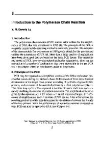
Figure 1.1. (A) Portion of an unstained culture of mouse bone marrow cells stimulated by a mixture of GM-CSF, G-CSF, and Multi-CSF showing the general appearance of the granulocyte-macrophage colonies developing after 7 days of incubation. (B) Granulocyte colony from such a culture after staining. Less mature cells are concentrated in the central region, and more mature cells migrate to form a corona around the colony.
This work led in 1975 to the purification of CSF from human urine, and this molecule appeared to be a glycoprotein of molecular weight (MW) 45,000 (Stanley et al., 1975). Oddly, this CSF appeared virtually unable to stimulate colony formation by human marrow cells in conventional agar cultures (Metcalf, 1974a) but was an effective stumulus for colony formation by mouse bone marrow cells. Unlike the original cultures that developed both granulocytic and macrophage colonies, the murine colony
Historical introduction
5
formation stimulated by human urinary CSF was predominantly macrophage in type, and this macrophage colony-stimulating factor was designated as M-CSF (alternatively, CSF-1). While this work was in progress, it was observed that extracts of all mouse tissues contained detectable CSF (Sheridan and Stanley, 1971), as did medium conditioned by minced fragments of such tissues. Lung-conditioned medium appeared of special interest not only because its level of activity was higher than that from other tissues, but because the medium stimulated both granulocytic and macrophage colony formation, and the apparent molecular weight of the active factor was only 23,000 (Sheridan and Metcalf, 1973a). Because lung tissue from mice preinjected with endotoxin contained and produced higher levels of CSF than did control lung tissue (Sheridan and Metcalf, 1972), a program was commenced to purify the CSF in medium conditioned by lung tissue from mice preinjected with endotoxin. Purification of this lung-produced form of CSF, given the name granulocyte-macrophage colony-stimulating factor (GM-CSF), was achieved in 1977 (Burgess et al., 1977) at the same time as parallel work achieved purification of murine M-CSF (CSF-1) from medium conditioned by mouse L-cells (Stanley and Heard, 1977). The murine M-CSF again appeared to be a markedly larger molecule (MW 70,000) than GM-CSF. Subsequent studies showed that M-CSF exists as a homodimer and that the monomeric polypeptide has a molecular weight of 26,000, more similar to that of native GM-CSF. The period 1971-1978 saw the development of modified culture procedures that allowed the formation of erythroid, megakaryocytic, and multipotential hemopoietic colonies (Metcalf, 1984). Erythroid colony formation, as anticipated, required addition of erythropoietin (Stephenson et al., 1971), but erythroid colony formation from less mature precursors could be strongly enhanced by the addition of a variety of cell- or tissueconditioned media, suggesting that other factors could stimulate the proliferation of earlier erythroid precursors. Eosinophil colony formation was recognized as occurring in cultures stimulated by many of the same types of crude conditioned media, including in particular mitogen-stimulated, lymphocyte-conditioned medium (Johnson and Metcalf, 1980). Part of this latter activity was subsequently shown to be due to the presence of a separate eosinophil regulator produced by T-lymphocytes - interleukin-5 (IL-5) (Sanderson, 1992). The formation of what were probably megakaryocytic colonies had been noted in cultures of mouse marrow cells stimulated by medium conditioned by the murine myelomonocytic leukemic cell line
6
The hemopoietic colony-stimulating factors
WEHI-3B, and cells of the same tumor stimulated the formation in marrow cultures of curious dispersed colonies of novel morphology (Metcalf et al., 1969). However, the most efficient method for stimulating megakaryocytic colony formation was later found to be the use of mitogenstimulated mouse lymphocyte-conditioned medium (Metcalf et al., 1975), an agent that also stimulated the formation of multipotential colonies (Metcalf etal., 1979). These miscellaneous observations prompted attempts to establish the nature of CSF in both pokeweed mitogen-stimulated spleen lymphocyteconditioned medium (SCM) and WEHI-3B-conditioned medium. In this laboratory, the assumption was made that distinct factors were likely to be responsible for stimulating these various colony types to develop, and initial results suggested that some separation of the biological activities seemed possible. However, other purification studies suggested that the various colony-stimulating activities in WEHI-3B-conditioned medium might be ascribed to a single-factor (Bazill et al., 1983), a conclusion eventually confirmed as purification studies were continued. The name "multipotential CSF" (Multi-CSF) was coined in this laboratory for the factor of MW 23,000-28,000 that stimulated colony formation by granulocytic, macrophage, eosinophil, megakaryocytic, blast, erythroid, mast, and multipotential cells (Cutler et al., 1985). These conclusions were preempted by the successful completion of an apparently unrelated study investigating the active factor in WEHI-3Bconditioned medium that induced the enzyme 20a-hydroxysteroid dehydrogenase in cultures of spleen cells or certain hemopoietic cell lines, thought at the time to be T-lymphoid in nature. This study, involving a rapid single bioassay, was completed promptly, and the name "interleukin-3" (IL-3) was coined for the active factor (Ihle et al., 1982). Subsequent analysis of the properties of this factor indicated that they were identical to those emerging for Multi-CSF (Ihle et al., 1983). A somewhat comparable analysis of the active factor in WEHI-3B-conditioned medium was based on a bioassay in which normal mast cells were stimulated to proliferate, and these studies identified an active factor, termed P-cellstimulating factor. Subsequent amino acid analysis of this factor revealed it also to be IL-3 (Multi-CSF) (Clark-Lewis et al., 1984). During an analysis of the CSF present in human placenta-conditioned medium, it was noted that a distinct form was present (termed CSF/3) that was highly hydrophobic and selectively stimulated granulocytic colony formation (Nicola et al., 1979b). In parallel studies on the nature of the elevated levels of CSF in the serum of mice preinjected with endotoxin,
Historical introduction
7
the major form of CSF appeared to be M-CSF, but on chromatography, highly hydrophobic fractions were observed that again stimulated the formation almost exclusively of mature granulocytic colonies (Burgess and Metcalf, 1980). Both observations suggested the existence of a further type of CSF. Neither placenta-conditioned medium nor serum was a particularly suitable starting material for fractionation, but a survey of organconditioned media from mice preinjected with endotoxin revealed the presence of active material with the similar dual properties of high hydrophobicity and selective granulocytic colony-stimulating activity (Nicola and Metcalf, 1981). It was also noted that this candidate CSF had the capacity to induce differentiation in colonies of WEHI-3B leukemic cells. Using the twin bioassays of granulocytic colony formation and induction of differentiation in WEHI-3B colonies, this CSF was purified to homogeneity from mouse lung-conditioned medium as a glycoprotein of MW 25,000 and given the name "granulocyte colony-stimulating factor" (G-CSF) (Nicola et al., 1983). Thus, by 1983, four distinct CSFs had been purified in small amounts from murine sources and had become available to the laboratories concerned for limited analyses of their in vitro biological actions on hemopoietic cells. With the prototype example of human M-CSF, it was assumed that comparable human CSFs existed for the other three murine CSFs. In human placenta-conditioned medium, it was shown that two forms of CSF existed, termed initially CSFce and CSF/3, which had biochemical and biological properties comparable with murine GM-CSF and G-CSF, respectively (Nicola et al., 1979b), and receptor competition studies confirmed that human CSF/3 must be closely similar to murine G-CSF (Nicola etal., 1985). The successful purification of human GM-CSF from medium conditioned by the Mo T-leukemic cell line was accomplished in 1984 (Gasson et al., 1984). In simultaneous studies, human G-CSF was purified from medium conditioned by a bladder cancer cell line 5637, initially under the name of "pluripoietin" (Welte et al., 1985), and by a squamous carcinoma cell line (Nomura et al., 1986). The successful purification of native human Multi-CSF (IL-3) (Zenke et al., 1991) was achieved only subsequent to the characterization of recombinant human Multi-CSF. The period during which human GM-CSF and G-CSF were being purified overlapped the period during which cDNAs encoding the murine CSFs were being cloned. Two groups independently cloned cDNAs encoding murine IL-3 by direct expression screening of cDNA pools using
8
The hemopoietic colony-stimulating factors
continuous murine cell lines as the proliferation bioassay (Fung et al., 1984; Yokota et al., 1984). This was followed by the cloning of a cDNA for murine GM-CSF using amino acid sequence-based nucleotide probes and cDNA libraries from lung and a T-lymphocyte cell line (Gough et al., 1984). Cloning of cDNAs for human GM-CSF was accomplished by groups either using direct expression screening of cDNA pools from the human leukemic Mo cell line (Wong et al., 1985) or using murine cDNA probes on a human cDNA library (Cantrell et al., 1985). This was followed in 1986 by the cloning of cDNAs for human G-CSF using nucleotide probes based on amino acid sequence data from G-CSF (Nagata et al., 1986a; Souzaet al., 1986). A genomic clone encoding human M-CSF was isolated using a sequencebased cloning strategy (Kawasaki et al., 1985), and subsequently a cDNA encoding murine M-CSF was obtained (De Lamarter et al., 1987). The successful cloning of a cDNA for human IL-3 was a more difficult feat, since murine-based probes were unsuccessful because of species divergence between the two molecules. It was eventually accomplished by direct expression screening of a primate cDNA library and the use of primate clones to probe a human cDNA library (Yang et al., 1986). All four murine and human CSF cDNAs were expressed with various degrees of difficulty in bacterial, yeast, or mammalian expression systems, with the production of biologically active recombinant CSF. These studies indicated that the carbohydrate content of the native CSF glycoproteins was not necessary for biological activity in vitro. Some early in vivo studies had been performed in mice using crude embryo-conditioned media containing high CSF levels (Bradley et al., 1969) or semipurined human urinary M-CSF (Metcalf and Stanley, 1971). These suggested that granulocytic and progenitor cell levels could be elevated by the injection of CSF. However, the ability to produce recombinant CSFs in relatively large amounts permitted the first effective studies to be performed on the action of fully purified CSFs on hemopoiesis in animals. The first such in vivo studies in mice were reported in 1986 for IL-3 (Multi-CSF) (Kindler et al., 1986; Metcalf et al., 1986a) and G-CSF (Fujisawa et al., 1986), and these were followed by comparable studies using GM-CSF and M-CSF. This work led to studies in primates of the actions of human GM-CSF, G-CSF, and Multi-CSF. The effectiveness of these CSFs in elevating white cell levels without obvious toxic effects in normal primates or primates with drug- or radiation-induced aplasia led to the commencement of human trials using purified recombinant CSF.
Historical introduction
9
The first Phase I clinical study on injected CSFs analyzed the responses of AIDS patients to GM-CSF (Groopman et al., 1987), and this was followed by two Phase I studies on the responses of cancer patients to injected G-CSF (Gabrilove et al., 1988; Morstyn et al., 1988). The obvious capacity of these two CSFs to increase white cell levels with minimal adverse responses led rapidly to Phase II studies of both in marrow-transplanted patients and in cancer patients following chemotherapy. A general extension of such trials led to the first approval for clinical use of G-CSF and GM-CSF in 1992. Clinical trials of Multi-CSF and M-CSF were initiated during this same period, although to date neither has yet been licensed for medical use. Since 1977 there has been a progressive recognition of the curiously pleiotropic actions of the CSFs on hemopoietic cells in vitro. However, the present clinical uses of CSFs are based largely on their actions as mandatory proliferative stimuli for granulocyte and monocyte-macrophage formation, and the present knowledge of the broader biological actions of CSFs on these populations has yet to be fully exploited in clinical applications. During this same period, at least 15 other hemopoietic regulators were characterized, with increasing awareness of the existence and probable importance of regulator networks and the likelihood that these regulators were designed to function most effectively when acting in combination (Metcalf, 1993b). This knowledge has begun to reach the clinical trial stage with the first tentative use of regulator combinations. However, the critical assessment of the opportunities for therapy now available is a formidable problem. It cannot really be accomplished until laboratory studies provide a compelling case for the use of certain defined combinations for particular clinical problems. In short, the discovery and availability of recombinant hemopoietic growth factors have grossly outstripped the capacity for clinical assessment of these agents, and it is currently proving to be a logistical nightmare for pharmaceutical companies attempting their introduction into clinical medicine. This is not helped by the regulatory requirement that each first be demonstrated to be effective as a single agent when the biology of hemopoietic regulators indicates that they are most effective when used in combination.
2 General introduction to hemopoiesis
To appreciate more clearly the roles played by the colony-stimulating factors in hemopoiesis, it is necessary to outline some aspects of the origin and organization of hemopoietic populations, the major cellular events occurring during hemopoiesis, and the general methods by which these events are regulated. The first hemopoietic cells arise from nonhemopoietic precursors as a transient finite event in the blood islands of the yolk sac (Metcalf and Moore, 1971). Many of these cells then migrate to the fetal liver, where they build up to a large population (Figure 2.1) (Moore and Metcalf, 1970; Metcalf and Moore, 1971). The major recognizable cells in such populations are embryonic erythroid cells and macrophages, although populations of stem cells and progenitor cells are also present. At least in chickens, and presumably also in mammals, this yolk sac-derived hemopoietic population becomes replaced by a second population of hemopoietic cells arising de novo from nonhemopoietic precursors in the para-aortic region of the fetus (Dieterlen-Lievre and Martin, 1981). These fetus-derived hemopoietic populations replace the existing yolk sac-derived cells in the liver and then populate the developing spleen and bone marrow. It is believed that no further de novo formation of hemopoietic cells occurs. As a consequence, throughout subsequent adult life, the hemopoietic populations are required to be self-sustaining as well as to generate continuously the large number of maturing cells in the eight major hemopoietic lineages that are needed to replace the relatively short-lived cells in the peripheral blood. Soon after birth, hemopoietic populations decline in the liver, and the relative importance of splenic hemopoiesis also progressively declines so that, in the adult, most (>95%) hemopoietic tissue is located in the bone marrow in scattered locations throughout the body (Metcalf and Moore, 10
General introduction to hemopoiesis Para-aortic region
11
Yolk sac blood island
Fetal liver
Fetal spleen
Fetal bone marrow
Figure 2.1. Schematic diagram of the early events in the development of the hemopoietic organs. Hemopoiesis begins in the yolk sac, from which cells migrate to the fetal liver. The fetal liver hemopoietic population is probably replaced during fetal development by a second wave of de novo hemopoiesis originating in the para-aortic region. The hemopoietic populations in the marrow and spleen are derived from migrants from the fetal liver.
1971). Particularly in the mouse, the spleen retains a capacity to expand rapidly its content of hemopoietic cells in situations requiring increased hemopoiesis, and if demand is extreme or the bone marrow is diseased the liver can also become reactivated as a hemopoietic organ. While maturing cells in hemopoietic tissue are readily identifiable by their characteristic morphology and can be arranged into likely maturation sequences, these maturing populations are not organized in architecturally stratified layers, as is the situation in tissues such as the skin or gut. Instead, hemopoietic regions of the marrow and spleen appear to contain an almost random admixture of cells of different lineages and maturation stages. This is not true in all species, and in the chicken there is obvious segregation of granulocytic from erythroid populations in the marrow. Data from an analysis of the location of stem cells in mouse marrow have also suggested that there is some segregation of these cells, particularly to regions adjacent to the bone cortex (Lord and Wright, 1984). Despite these data, the overwhelming impression is that hemopoietic cells are mixed in a virtually random manner in marrow tissue. Hemopoietic tissues also possess a network of specialized stromal cells. Although the precise identity and architectural arrangement of these cells
12
The hemopoietic colony-stimulating factors Stem cells Committed progenitor cells
Dividing maturing cells
Mature cells to
blood
Figure 2.2. Three basic compartments in hemopoietic tissues. The cells in each succeeding compartment are the progeny of, and are more numerous than, the cells in the preceding compartment. have yet to be characterized, they play a crucial role in permitting hemopoiesis and in regulating the cellular events that occur. Identification of the nature and number of the more ancestral hemopoietic cells and their interrelationship has required the application of a number of clonal-analytical procedures. These analyses have indicated that hemopoietic cells in each lineage can be stratified into three major sequential cell populations, each of progressively larger size: stem cells, progenitor cells, and dividing maturing populations (Figure 2.2).
Stem cells Stem cells are a minor subpopulation of hemopoietic cells, numbering approximately 1 per 105 marrow cells. When purified by complex cellsorting protocols, stem cells have been shown to have the morphology of small to medium mononuclear (lymphocyte-like) cells. Most have a large nuclear to cytoplasmic ratio, and the nucleus often has prominent nucleoli. The cytoplasm is not basophilic and contains no granules. Under normal conditions, most of these cells are not in cell cycle, but the cells have an extensive potential capacity for self-generation and for the production of a large number of committed progenitor cells. It is now recognized that stem cells are a very heterogeneous group of cells, the heterogeneity probably being based on hierarchical parent-progeny relationships (Figure 2.3). The most ancestral stem cells (termed repopulating cells) are capable of extensive self-renewal and are defined either by their capacity to repopulate hemopoietic and lymphoid organs on a long-term basis in irradiated animals or by their capacity to sustain long-term hemopoiesis in vitro.
General introduction to hemopoiesis
13
Repopulating cells In vitro long-term repopulating cells HPPC, CFU-A, CFU-S D14
Blast colony-forming cells CFU-S D7
Figure 2.3. Hemopoietic stem cells are highly heterogeneous but are probably stratified in a hierarchical sequence of cells of increasing maturity with more restricted proliferative potential. HPPC denotes high proliferative potential cells.
In turn, their progeny stem cells (spleen colony-forming units, CFU-S) can be identified as being able (a) to form large hemopoietic colonies in the spleen of irradiated recipients (demonstrable at day 14 or, if more mature, persisting only until day 7) and (b) to form blast colonies or very large colonies of cells in multiple lineages in clonal in vitro cultures. With highly enriched populations of murine stem cells (Kit+ Scal+ Rhl23l° Lin~ cells), 30 cells can permanently repopulate a lethally irradiated mouse, confirming the undoubted presence of repopulating cells in such preparations, yet all such cells can form blast colonies in appropriate agar cultures. As will be discussed later, the various stem cell assays must often detect the same stem cells, despite unequivocal evidence of hierarchical stratification within the stem cell population. The least-mature subsets of stem cells certainly can form hemopoietic progeny in all eight major hemopoietic lineages, but the most mature of the hemopoietic stem cells may no longer generate T- or B-lymphocytes. This remains a somewhat unclear question, as does the possible existence of subsets of these mature stem cells committed exclusively to the formation of only T- and/or B-lymphocytes. It is not possible to indicate how many progenitor cells an individual stem cell can generate. This depends on the stimuli applied to the stem cell and how much self-generation results. At one extreme, a stem cell might on occasion generate only two progenitor cells, while at the other extreme, a single stem cell can repopulate the hemopoietic tissues of an entire animal with evidence that such cells can then entirely repopulate secondary and tertiary recipients. Under in vitro culture conditions with
14
The hemopoietic colony-stimulating factors
appropriate stimuli and no evidence of self-renewal, individual stem cells can generate clones containing several thousand progenitor cells (Metcalf, 1991b), so their potential generating capacity can be exceedingly large. Progenitor cells Progenitor cells are usually in cell cycle, have the morphology of medium to large mononuclear (blast) cells with agranular basophilic cytoplasm, and comprise up to 1% of hemopoietic cells. These are a transit population formed by stem cells and expending themselves in the production of maturing progeny. From analyses performed in vitro, these cells appear to have a minimal or no capacity for self-generation. Progenitor cells are characterized by their capacity to form colonies of maturing progeny in semisolid cultures in response to stimulation by hemopoietic growth factors. The number of cells in such colonies can vary from five or ten thousand down to the lower limit for defining clones as colonies (usually 50 cells for most colony types). The variable size of these colonies is one of the parameters indicating the heterogeneity of progenitor cells even within one differentiation lineage. Cells forming colonies of small size are most likely to be the progeny of cells forming large colonies, a relationship most clearly evident in the erythroid lineage, where the progenitor cells are divided into two major classes - BFU-E, forming large, often multicentric (burst) colonies, and CFU-E, forming small colonies that are the equivalent of one of the subclones of a multicentric colony formed by BFU-E (Figure 2.4). Progenitor cells appear to differ from stem cells in being committed, usually to a single lineage of differentiation. Bi- and trilineage-committed progenitor cells do exist but are less frequent than single-lineage progenitors and are likely to be the immediate ancestors of the latter cells. Bipotential granulocyte-macrophage progenitor cells are somewhat different, at least in the mouse, in being a major subset of progenitor cells in this closely related pair of lineages. Most evidence indicates that differentiation commitment is irreversible, which means that progenitor cells cannot switch to another lineage or revert to multipotential stem cells. Dividing and maturing populations These cells comprise the vast majority of cells present in hemopoietic tissues. They are the progeny of progenitor cells in a particular lineage and
General introduction to hemopoiesis BFU-E
Maturing
15
non-
dividing cells •ORBC
Figure 2.4. Progenitor cells in the erythroid lineage (BFU-E and CFU-E) can be identified by their ability to form colonies in semisolid cultures. Granulocyte progenitors (G) can be similarly detected. With increasing maturation, a progressive reduction occurs in the number of progeny generated by individual progenitor cells. RBC denotes red blood cell.
form a morphologically recognizable sequence of cells within each lineage - for example, myeloblasts -• promyelocytes -• myelocytes -* metamyelocytes -» polymorphonuclear neutrophils (the last cell type is often referred to throughout the text simply as "granulocytes"). The least mature of these cells have a considerable capacity for proliferation and can produce clones of subcolony size in semisolid cultures. Their capacity for proliferation is probably not strictly fixed, as proposed in earlier studies, but it is dependent on the level of stimulation exerted by relevant growth factors. Nevertheless, their proliferative capacity is limited in most lineages, although there are obvious exceptions in the Tand B-lymphoid lineages. With progressive maturation, a progressive restriction is evident in the capacity of the cells for further proliferation, and typically the cells eventually mature to a postmitotic stage (e.g., metamyelocytes), after which no further cell division is possible. It is less clear whether macrophages are necessarily postmitotic cells, and under some circumstances apparently mature eosinophils may also exhibit a capacity for some cell division.
16
The hemopoietic colony-stimulating factors Requirements for sustained hemopoietic cell formation
With the exception of at least some stem cells, the remaining cells that can form cells in the granulocyte-macrophage lineage are transit cells, which become expended by their proliferation. For example, myelocytes cannot self-renew and therefore expend themselves in generating metamyelocytes and polymorphs. Similarly, committed granulocytic progenitor cells cannot self-renew and will expend themselves by forming myeloblasts and myelocytes. Any capacity of the system to produce granulocytes and macrophages in a sustained manner therefore requires a continuous replenishment of lineage-committed progenitor cells by cells from the most ancestral, stem cell compartment. The consequences of this arrangement are that when an agent like a CSF is used to stimulate granulocytic and/or macrophage formation by progenitor cells, the resulting increased cell production will terminate after a few days unless replacement progenitor cells are generated. The regulatory control of these different steps need not be identical; nevertheless, most regulatory factors do have actions at multiple levels. It becomes necessary, therefore, in the case of the CSFs to consider not only their actions on committed granulocyte-macrophage progenitor cells and their progeny, but also whether the CSFs have observable actions in stimulating progenitor cell formation by stem cells and possibly even stem cell self-renewal. As will be seen, the CSFs do have proliferative actions on cells in all three of the major hemopoietic compartments, although the magnitude of this action varies at the different stages, as can the nature of other regulatory factors interacting with the CSFs to control cells in the different stages.
General regulation of hemopoiesis The events involved in the formation and function of blood cells are highly complex, since they include (as shown in Figure 2.5) precisely coordinated new cell formation occurring in the widely separate populations of hemopoietic cells scattered throughout the body, correct lineage commitment, the initiation and completion of maturation with fidelity, controlled release of mature cells, maintenance of circulating blood cell levels, selective exit of some but not all cell types to the tissues, functional activation of such cells when necessary, coupled finally with a system for detecting and eliminating effete cells.
General introduction to hemopoiesis
17
MULTIPOTENTIAL STEM CELLS (repopulating cells, CFU-S, blast colony-forming cells)
COMMITTED ] PROGENITOR CELLS
etc.
IMMATURE PROLIFERATING CELLS Promyelocyte
Metamyelocyte
Release t o
blood
Macrophage
Exit to tissues
Marginated pools
DEATH
Figure 2.5. Schematic diagram of the major stages by which mature granulocytes and monocytes are generated. After release of mature cells from the bone marrow to the blood, the cells enter the tisues and, in the case of monocytes, undergo major changes in phenotype. E denotes erythroid progenitor cells; GM, granulocyte-macrophage progenitor cells; Eo, eosinophil progenitor cells.
18
The hemopoietic colony-stimulating factors
The regulation of such a complex series of events must be arranged so that basal levels of cell production and function can be controlled with precision, yet massive increases in cell production can be achieved rapidly in response to emergencies such as bleeding or infections. The complexity and apparent redundancy of the control systems that have been documented by recent studies may well be necessary to accomplish these two quite different roles for hemopoietic tissues. In principle, the control system requires not only a series of positive signals to initiate these various events, but also a balancing series of negative feedback signals to prevent major cyclic fluctuations in cell production and possibly to terminate amplified responses following restitution of normal conditions. Both types of control system could be activated by an inductive system based on sensing levels of mature cells, their products, or breakdown products or, alternatively, by an inductive system activated by tissue demands (e.g., oxygen deprivation or the presence of microorganisms). A feedback system based on monitoring of cell number or cell products would be elegant, but the weight of evidence suggests that the most likely induction systems that alter regulatory mechanisms are demand-generated (Metcalf, 1988a). It was anticipated that quite different regulators would be necessary to control such different events as new cell production, release of mature cells, or functional activation of mature cells in the tissues. It may well be that there are exclusive regulatory mechanisms for some of these events; nevertheless, it has become evident that individual regulators can have actions on a wide range of these events, making the biology of such regulators extremely complex. Regulation of cell production is achieved by what superficially appears to be a dual control system: (a) local control by microenvironmental cells located in the actual hemopoietic tissues, often involving intimate cell contact with hemopoietic cells, and (b) control by a series of humoral regulatory molecules, typically glycoproteins which can be produced by multiple cell types throughout the body. The two systems are not as separate as might first appear because the stromal cells have the capacity to produce many of the humoral regulators concerned and, under conditions of basal hemopoiesis, may be the major sources of these molecules. Furthermore, for at least some of the regulators, the stromal cells have an ability to produce them in a form held on the cell membrane, providing in part the molecular basis for the observed need for cell contact between stromal cells and responding hemopoietic cells if hemopoiesis is to be optimal. Although none have so far been discovered, it remains likely that
General introduction to hemopoiesis
19
stromal cells may produce unique regulatory molecules not produced elsewhere in the body. Alternatively, a regulator displayed on a stromal cell membrane might elicit responses in an adherent hemopoietic cell that are quite different from those elicited by the secreted form of the same regulator acting on nonadherent hemopoietic cells. Stromal control of hemopoiesis The evidence supporting the role of stromal cells in hemopoiesis comes from two separate groups of observations. After discovery of the phenomenon of hemopoietic colony formation in the spleen or marrow of irradiated mice injected with bone marrow cells (Till and McCulloch, 1961), it was shown that such colonies were clones and that these colonies were generated by ancestral hemopoietic cells now regarded as more mature stem cells. Spleen colonies, when examined early (7 days), often appear to be composed of cells of only a single lineage, for example, erythroid, granulocytic, or megakaryocytic. Analysis of spleen colony populations indicated that the cell initiating such colonies, the CFU-S, was capable of self-renewal, and progeny CFU-S could form colonies of multiple types, indicating that at least some were multipotential. A consistent difference was noted between the relative frequency of erythroid versus myeloid colonies developing in the spleen or bone marrow - spleen colonies being mainly erythroid, whereas marrow colonies were granulocytic. The conclusions from these and related observations were that hemopoietic stromal cells could somehow support, by what seemed to be a cell contact process, the self-generation of stem cells and the formation by them of maturing progeny. It was proposed that heterogeneity existed between such stromal cells, as evidenced by the differing cellular composition of the colonies formed, and that different stromal cells could commit stem cells to selective cell formation in a particular lineage (Trentin, 1989). Subsequent observations on the progenitor cell content of spleen colonies have complicated the conclusions drawn from this phenomenon of spleen colony formation. All colonies, regardless of the morphological type of their dominant cell population, contain committed progenitor cells of multiple lineages, and there is no significant difference between the lineage commitment range of progenitors in erythroid versus granulocytic colonies (Johnson and Metcalf, 1979). While it seems likely that stromal cells can support stem cell proliferation and progenitor cell formation in vivo, the morphological differences between colonies appear to be based more on a subsequent amplification
20
The hemopoietic colony-stimulating factors
and maturation of progeny formed by particular progenitor cells in any one colony. Thus, the difference between an erythroid stromal cell and a granulocytic stromal cell may relate more to their differing capacities to stimulate or permit the selective proliferation of maturing cells in the erythroid or granulocytic lineages. The general conclusions reached from these in vivo studies have been strongly supported by studies on long-term survival and proliferation of hemopoietic cells in cultures of bone marrow cells (Dexter et al., 1984). In such cultures an adherent layer of mixed stromal cells develops. Not only are these adherent cells crucial for the sustained formation of stem and progenitor cells, but cell formation is clearly concentrated in islands of cells firmly bound to the adherent layers. The long-term cultures have not proved sufficient to define fully the mechanisms involved. However, some information is beginning to emerge. For example, recent studies have shown that expression by the stromal cells of membrane-bound stem cell factor (SCF), as distinct from secretion by the cells of SCF into the medium, is essential for the sustained production of a large number of stem and progenitor cells (Toksoz et al., 1992). The role played by hemopoietic stromal cells is therefore crucial in hemopoiesis. While the precise mechanisms remain to be determined, at the very least the stromal cells provide a permissive microenvironment in which hemopoietic cells can be stimulated to proliferate and mature, and more likely the cells also provide a local source of many of the required specific regulatory molecules. In contrast to these observations on the importance of stromal cell contact for the behavior of stem cells, the proliferation of progenitor cells can be readily stimulated in dispersed semisolid cultures to form colonies of maturing cells under conditions in which no cell contact is present. Based on these two sets of observations, it is often stated that cell contact with microenvironmental cells is more important in controlling stem cell behavior and their formation of progenitor cells, while humoral regulators are the major regulators of events from the progenitor stage onward. This may be so, but the data do not really allow such a broad distinction to be made, and it is probably better to regard both control systems as potentially influencing all stages of hemopoiesis. Hemopoietic regulators To date, at least 20 hemopoietic regulators have been molecularly denned, and the implications raised by this large number require some discussion.
General introduction to hemopoiesis
21
Table 2.1. Hemopoietic growth factors grouped according to their known target cells Target population Stem cells Granulocytes Monocyte-macrophages Eosinophils Megakaryocytes Mast cells Erythroid cells T-Lymphocytes B-Lymphocytes Dendritic cells
Hemopoietic growth factors SCF, IL-1, IL-6, Multi-CSF (IL-3), GM-CSF, G-CSF, M-CSF, LIF, IL-12, flk-ligand G-CSF, GM-CSF, Multi-CSF, IL-6, SCF, IL-11 M-CSF, GM-CSF, Multi-CSF, O-CSF Multi-CSF, GM-CSF, IL-5 SCF, Multi-CSF, GM-CSF, IL-6, LIF, mpl-ligand Multi-CSF, SCF Early: SCF, Multi-CSF, GM-CSF; Late: Epo IL-1, IL-2, IL-4, IL-10, IL-7 SCF, IL-7, IL-4, IL-6, IL-2 GM-CSF
Abbreviations: SCF, stem cell factor; IL-1 through IL-12, interleukins-1 through -12; GMCSF, granulocyte-macrophage colony-stimulating factor; G-CSF, granulocyte colonystimulating factor; Multi-CSF, multipotential colony-stimulating factor; LIF, leukemia inhibitory factor; O-CSF, osteoclast colony-stimulating factor; Epo, erythropoietin.
The most relevant of these regulators for the present discussion are listed in Table 2.1 together with the hemopoietic cell types identified as responding to them in one manner or another. As evident from this table, multiple regulators have been identified as having actions on cells in any one lineage (Figure 2.6). Typically, at leastfiveto six regulators are known to be active on cells in any one lineage. The quantitative importance of a particular regulator in controlling cell production in a lineage can vary according to the particular stage the cells are at in the lineage and the presence or absence of other regulators that interact with the regulator in question. Furthermore, it is evident from Table 2.1 and Figure 2.6 that all of the hemopoietic regulators are active on cells of more than one lineage, and there are no known examples of a regulator with actions restricted to cells of a single lineage.
Regulator redundancy This arrangement has resulted in a commonly expressed view that hemopoietic regulators exhibit extreme redundancy - that there are far more regulators with appropriate actions than are really required to achieve the production of any one hemopoietic cell type. If this is correct, it should follow that deletion of any one regulator would have no observable effect
22
The hemopoietic colony-stimulating factors
STEM CELLS
IL-1,
IL-6, G-CSF, Multi-CSF
SCF,
IL-12
GM-CSF, M-CSF
PROGENITOR CELLS
MATURING CELLS IL-9,
SCF, Multi-CSF CM-CSF, IL-9,
I
SCF, Multi-CSF CM-CSF, IL-6 IL-11, Meg-CSF
I
G-CSF, GM-CSF Multi-CSF, SCF IL-6, M-CSF
J
G-CSF, GM-CSF Multi-CSF, SCF IL-6, M-CSF
Megakaryocyte Figure 2.6. Schematic diagram showing the regulators involved in the formation of neutrophils and megakaryocytes. Note that, at each stage, multiple regulators can be shown to have proliferative actions. (Reproduced with permission from Metcalf, 1993b.)
on cell production, since its loss could readily be compensated for by the overlapping actions of other regulators. However, there are alternatives to the redundancy hypothesis (Metcalf, 1993b). One possibility is that a hierarchy of regulators may exist - some with much greater importance or quantitative impact than others. Reasons will be given later why the CSFs are probably the most important regulators of granulocyte and macrophage formation even though, for example, SCF and IL-6 have some capacity to stimulate the formation of granulocytes.
General introduction to hemopoiesis
23
A variant of this theme is that some regulators control basal levels of cell production, while others become important only in emergencies that require greatly increased cell production. This possibility is not fully resolved by existing data and has in particular led to the necessity for a careful examination of the effects of deleting CSFs, because CSF levels tend normally to be very low and become readily detectable only during emergencies. To preempt the discussion of these particular data, the conclusion to date is that there is no clear evidence for the existence of distinct "basal" versus "emergency" factors. An important general concept to grasp is that hemopoietic regulatory factors are not exactly hormone-like in their biology. Unlike classical hormones, they do not have a single cell type as their source, and while they can often be detected in the circulation as would be expected of classical hormones, much of the production and action of hemopoietic regulators can occur locally. Thus, a particular regulator may be produced locally in the marrow at the site of responding immature hemopoietic populations while simultaneously, elsewhere in the body, a local inflammatory focus may be independently producing a high local concentration of the same regulator to achieve the local accumulation and activation of mature cells. Neither event will necessarily be detected by monitoring blood levels of that regulator. The general technical inability to assess local concentrations of regulators in these types of tissue continues to make it difficult to define the precise role played by a particular regulator in these various processes. Assessment of the situation is not helped by the capacity of some regulators to bind to the glycocalyx of some cells in a loose bonding that can potentially serve as a local, unmeasurable reservoir of a particular regulator (Gordon et al., 1987). Given this biology of regulator production, it is somewhat naive to attempt to classify regulators as greater or lesser in importance either in basal or emergency circumstances by simple assessment of circulating regulator concentrations, although this is commonly implied in many publications. Because regulator production can occur at multiple local sites, including in particular hemopoietic tissues, circulating regulator concentrations can provide only a very incomplete view of actual regulator concentrations in various tissues. A somewhat more sophisticated interpretation of the situation is to propose that certain regulators tend to act on early cells in a particular lineage, while others have their most important action on cells later in the sequence - the sequential notion of regulator control. This proposal has a certain elegance, and there are one or two cases where this interpretation
24
The hemopoietic colony-stimulating factors
may be reasonable. For example, in erythropoiesis it is evident that SCF and Multi-CSF have strong actions in stimulating the formation and initial cell divisions of the most ancestral committed erythroid progenitor cells (BFU-E), while, at least in a species like the mouse, erythropoietin has little or no action on such cells. Later in erythropoiesis, from the CFU-E stage onward, erythropoietin becomes the dominant regulator of erythroid cell proliferation. Unfortunately, this convincing example of sequential action is not typical of the situation in other lineages where individual regulators have readily demonstrable effects on cells at multiple stages in the lineage. On this basis, sequential action does not appear to be an adequate explanation for the existence of multiple regulators. Furthermore, whether or not a regulator appears to be active on cells of a particular lineage may be strongly influenced by what other regulators are acting on these cells. For example, in the megakaryocytic lineage, both SCF and IL-6, when acting alone, appear to be limited in their action to relatively mature precursors. Yet when either is combined with Multi-CSF, it becomes clear that each can exhibit significant proliferative actions on more ancestral cells in the sequence. This introduces an important aspect of the biology of hemopoietic regulators, namely, the distinctive difference between the responsiveness of stem cells and most progenitor cells to stimulation by regulators. In vitro studies using purified populations of stem cells have consistently revealed that no single regulator can stimulate the proliferation of these cells (Li and Johnson, 1992). No combination of known regulators has yet been reported to stimulate stem cell self-renewal, but if the outcome of stimulation is assessed by production of progenitor cells and more mature progeny, stem cell proliferation requires combined stimulation by two or more regulators. This is in sharp contrast to the behavior of typical progenitor cells, whose proliferation can readily be stimulated by the action of single regulators. From these facts, it becomes apparent that multiple regulators would be required if sustained cell production were to be achieved in any one lineage. Moreover, some progenitor cells have now been observed whose proliferation also appears to require multiple signaling by two or more regulators, extending the situations in which multiple regulators are necessary rather than redundant. In vitro analysis has also revealed, as shall be discussed later for the CSFs, that the combination of two regulators can achieve greater levels of cell production by typical progenitor cells than can be achieved by the use of double the concentration of either regulator. Furthermore, where
General introduction to hemopoiesis
25
progenitors are bipotential or multipotential, the use of regulator combinations can achieve a broadening of the mature cell types being generated and, as in vivo studies have shown, a broadening of the distribution of the mature cells produced. There is accumulating evidence, therefore, to support the view that the existence of a relatively large number of regulators with overlapping actions is not merely an example of biological redundancy; rather, the arrangement permits a subtlety and efficiency in cell production that can meet widely diverging requirements in health and emergencies (Metcalf, 1993b). Despite this conclusion, it should not be imagined that the injection of a single regulator in vivo would not achieve any observable effects on hemopoiesis. It is now common experimental and clinical experience that the injection of a single regulator can achieve remarkable biological responses. What the preceding discussion does make likely, however, is that the observed effects may not be due solely to the action of the injected regulator alone but may depend significantly on interactions with other regulators already present in the body. The current controversies over the possible redundancy of hemopoietic regulators have made it of wide interest to determine the effects on hemopoietic populations of removing or suppressing each regulator. Two general approaches are possible: (a) the administration of neutralizing antibody to the regulator in question or (b) the identification or creation of animals in which the gene encoding the regulator has been inactivated in every cell. This is an active area of experimental investigation, particularly using the powerful approach in which the gene is inactivated in embryonic stem cells that are then used to generate chimeric animals. If chimerism involves either the sperms or ova, some of the progeny of such animals will have inactivation of one copy of the regulator gene, and by appropriate matings mice can be generated with homozygous inactivation of both genes. Some of the regulators investigated by either approach are listed in Table 2.2. Certain comments can be made regarding the data obtained. In every case so far studied, suppression or deletion of a regulator has been observed to have some identifiable consequence, so no genuinely redundant regulators have been documented. In all cases, deletion of a single regulator has not resulted in a complete inability to produce cells in any one lineage. This result does indicate a degree of redundancy in hemopoietic regulators, but not complete redundancy. The effects of deletion can be quite subtle and appear to be compatible with a normal-enough life, at least for mice maintained under the protective conditions of pathogen-
26
The hemopoietic colony-stimulating factors Table 2.2. Regulators examined by suppression or gene inactivation studies Regulator
Consequences
Erythropoietin GM-CSF G-CSF M-CSF IL-2
Decreased erythropoiesis Defective resistance to lung infections Neutropenia Osteopetrosis, decreased macrophage populations Elevated IgGl, IgE, and IgA reduced T-cell responses, autoantibody formation, inflammatory bowel disease No IgE production, perturbed balance of Ig production, reduced TH2 cells Increased rate of bone turnover, reduced T-cells, impaired resistance to infections Elevated numbers of T- and B-lymphocytes and mast cells, inflammatory colitis Decreased erythropoiesis
IL-4 IL-6 IL-10 SCF
free housing. While in each case distinctive deficiencies are identifiable, there is at least one instance (with M-CSF) where an eventual correction of the deficiency is achieved by the body. Given the complexity of cellular processes required to activate hemopoiesis, it is not perhaps surprising that the control systems used should show evidence of both subtlety and fail-safe control systems. Regulator networks The existence of, and interactions between, multiple hemopoietic regulators constitute a network of regulatory control, but this network is potentially far more complex. Typically, each regulator can be produced by a wide variety of cell types - the most commonly involved being stromal cells, endothelial cells, fibroblasts, macrophages, mast cells, and lymphocytes. Curiously, individual cells, when induced, tend not to produce a single type of regulator but commonly to produce multiple types of regulators simultaneously. It is unclear why this should be a more efficient system, but it must be presumed that this arrangement has advantages. What is potentially possible with such an arrangement is that interactions between regulators, either competitive or enhancing, may also occur during the process of their production in addition to any interactions they may exhibit on responding cells.
General introduction to hemopoiesis
27
There is also considerable potential complexity in the system controlling the production of hemopoietic regulators. For most regulators, the level of regulator production by cells under basal conditions is very low. The characteristic feature of regulator-producing cells is, however, that they can greatly amplify the transcription and production of a regulator when induced by appropriate signals and that this amplification can occur with great rapidity. The lability of the regulator-producing system is clearly of advantage in achieving rapid responses to emergencies but does require a system for producing appropriate inductive signals. Figure 2.7 diagrams a very simple signaling network of the type that can be constructed by assembling data from individual in vitro experiments. In the diagram, an extrinsic signal (e.g., endotoxin) enters the body and either directly stimulates a cell (B) to increase transcription and production of a hemopoietic regulator or first stimulates another cell type (A) to produce an inducing signal for the regulator-producing cell. This Externally Derived Stimulus
@
/
Hemopoietic Regulator
0
Target Hemopoietic Cell
Figure 2.7. A simple regulator network showing some of the likely interactions among various cells. An externally derived stimulus such as endotoxin can initiate the production of a hemopoietic regulator (4) by cell (B) either by direct action (1) or byfirststimulating (2) another cell (A) to produce an inducing signal (3) such as IL-1. The regulator-producing cell may simultaneously produce other factors (5) that have direct actions on the hemopoietic target cell or that influence (6) another cell (D) to produce factors with such actions (8). The hemopoietic regulator may also act directly (7) on such a cell to stimulate the production (8) of such factors.
28
The hemopoietic colony-stimulating factors
regulator-producing cell will often simultaneously produce other factors that act directly on the hemopoietic target cell (C) to modulate the action of the hemopoietic regulator. Alternatively, these additional cell products may induce a further cell type (D) to produce factors influencing the response of the hemopoietic cell. The hemopoietic regulator itself may have a similar side action on such cells. The scheme outlined is a relatively simple example of signal networking involving hemopoietic growth factors, and far more complex schemes can be devised, again based on observed actions of regulators on individual cell types in vitro. While all the individual events in such network diagrams may have been documented from isolated in vitro studies, they are necessarily theoretical models, and it is unknown whether the events actually occur in vivo and how important they are in the regulation of either basal hemopoiesis or enhanced hemopoiesis following emergencies. The diagrams might again lead to the prediction that the injection of any single agent would be unlikely to override the dampening effects of the network or produce a measurable response in vivo. However, as noted earlier, the injection of a single agent can achieve reproducible responses, although networking may result in the occurrence of side effects of an injected agent on cells not themselves expressing receptors for the injected factor.
Unresolved problems concerning hemopoiesis It is relevant to the following discussion on the CSFs to comment briefly on certain unresolved general questions concerning hemopoiesis.
Is lineage commitment random or can it be influenced by extrinsic regulators? This is a central issue in hemopoiesis because it addresses the question of whether increased cell production in one lineage can be accomplished efficiently by the selective formation of the appropriate progenitor cells, or whether the process is an inherently wasteful one in which irrelevant progenitor cells are also formed that are not needed for the particular response. When multipotential stem cells are stimulated to generate committed progenitor cells, analysis has shown that there is no fixed sequence in which committed progenitor cells are generated, because the initial daughter or granddaughter cells of multipotential cells can be a mixture of cells of
General introduction to hemopoiesis
29
virtually any lineage or lineage combination (Nakahata et al., 1982; Ogawa et al., 1983). This provides one answer to an old general question in cell biology, namely, whether cells generate like or unlike progeny at cell division. The same data have led to the conclusion that progenitor cell commitment is a stochastic event. However, the random nature of a process does not rule out the possibility that extrinsic regulators can influence the probability with which certain of these random events occur. Four lines of evidence indicate that commitment can be influenced by extrinsic stimuli: (a) Apparently purified mouse hemopoietic stem cells can produce different types of progeny depending on whether they are injected intrathymically or intravenously or cultured in vitro. If injected intrathymically, T-lymphocytes are generated, whereas if they are injected intravenously, cells of all hemopoietic lineages are generated; if cultured in vitro, they generate progeny whose type depends on the growth factor combination used (Spangrude et al., 1988; Heimfield et al., 1991). (b) Various combinations of factors can induce the formation by stem cells of altered proportions of committed progenitor cells of various types (Metcalf, 1991b). (c) CSFs, when acting on bipotential granulocyte-macrophage progenitor cells, can bias subsequent cell formation into either the macrophage or granulocytic lineage (Metcalf and Burgess, 1982). (d) CSFs can induce specific differentiation commitment in certain multipotential cell lines (Heyworth et al., 1990). While, in total, the evidence makes a persuasive case, in each system it is difficult to account for every cell involved, leaving alternative possibilities that the effects may have been due to selection or amplification of a precommitted subpopulation. It is evident in vivo that a puzzling wastefulness does occur in certain responses that might be expected to require changes in the cells of only a single lineage. For example, in response to bleeding, increases also occur in nonerythroid progenitor cells in the spleen (Metcalf, 1969), and as will be discussed later, in response to the injection of G-CSF, increases occur in progenitor cells in the blood that include lineages not stimulated by G-CSF - an apparently purposeless occurrence (Duhrsen et al., 1988). This may indicate that a certain level of random commitment is inevitable during hemopoiesis, often meaning an unusable level of progenitor cell production.
Are an excessive number of progenitor cells formed? It is possible to use clonal cultures to estimate the total body progenitor cell number in an animal such as the mouse. The total number of progeny
30
The hemopoietic colony-stimulating factors
produced by such cells can also be established after the use of various types of stimuli in culture. The actual level of stimulation occurring in vivo can only be guessed at, so the actual number of progeny produced in vivo by these progenitor cells can only be approximated. However, conservative calculations indicate that, in the mouse, the number of progenitor cells available could produce approximately the required number of maturing progeny (Metcalf, 1984). These calculations appear to obviate the need to propose some radically different means of mature cell production in vivo. However, progenitor cells are a transitory population and estimates of the number of progenitor cells present at any one time cannot provide estimates of the rate of turnover of this population. While most progenitor cells appear to be in cell cycle in the adult, it cannot be ruled out that a significant number of them may die without producing maturing progeny. Animal models have recently been encountered in which puzzlingly high levels of mature cells are generated from what appear to be an unremarkable number of progenitor cells. It remains possible, therefore, that a certain level of abortive progenitor cell formation may normally occur, and if so, these cells may possibly be used to amplify hemopoiesis in emergency situations, reducing the need to generate additional progenitor cells.
What controls release of mature cells to the blood? In early studies on hemopoiesis, it was a popular notion that a leukocytosis was achieved simply by inducing the release of mature cells from the bone marrow, but a concerted effort to discover leukocyte-releasing factors was unsuccessful. Such factors with a simple releasing action may exist, but they have not yet been characterized. On the basis of present knowledge of the polyfunctional actions of most regulators, most researchers now feel that if an agent can accelerate the release of preexisting cells, it is also likely to stimulate new cell formation, and as will be discussed, CSFs have such dual actions. The process of mature cell release is probably much more sophisticated than realized. Most researchers probably have a vague notion that mature cells may be relatively motile and actively penetrate a sinusoidal endothelial barrier during their exit from the marrow. This may be true for neutrophilic granulocytes but clearly not for enucleated red cells. Furthermore, the behavior of hemopoietic cells in culture indicates that certain cells such as immature erythroid or megakaryocytic precursors have a surprising capacity for extensive migration. This is the cellular basis for
General introduction to hemopoiesis
31
the formation of multicentric erythroid or granulocytic colonies and the typical widely dispersed nature of megakaryocytic colonies. Cell retention in the marrow may in fact be an active process. Selective expression of adhesion molecules may be a mechanism for retaining immature hemopoietic cells in the marrow, but this area of hemopoietic biology requires extensive investigation. What controls the levels of cells in the peripheral blood? In normal health, the absolute numbers of various cells in the peripheral blood vary over only a relatively small range. Is there any regulatory system that monitors and maintains such levels, or do they simply passively reflect the level of new cell production in the bone marrow? The answer to this question probably varies from lineage to lineage. There is a strong presumption that red cell levels and/or their oxygen-carrying capacity are directly monitored by sensing cells that themselves produce, or signal the production of, erythropoietin. Such a surveillance system is not as easy to identify for cells of other lineages, and if animals are kept pathogen-free, abnormally low white cell levels do not appear to activate the transcription and production of relevant growth factors. Growth factor production increases promptly in a conventional animal subjected to a procedure that induces leukopenia. This leads to the conclusion that the response is triggered not by low leukocyte levels but by secondary infections and therefore represents a demand-generated response rather than one monitoring the number of cells. Conversely, there is no evidence so far for an overriding mechanism that responds to abnormally high white cell levels by inducing a reduction in progenitor cells or hemopoiesis. The situation with platelets may be intermediate. Like excess red cell levels, the presence of excess platelets may well be life-threatening, and some mechanism may have developed to prevent such excess levels. At present, therefore, outside the erythroid system there is no substantive evidence for the existence of regulatory mechanisms to monitor blood cell levels and to initiate corrections in hemopoiesis where necessary. Can mature hemopoietic cells exhibit any further capacity for proliferation in the tissues? In the erythroid, granulocytic, and megakaryocytic lineages, the mature cells produced clearly enter an irreversible postmitotic state. Conversely, some T- and B-lymphocytes can exhibit both an extensive capacity for
32
The hemopoietic colony-stimulating factors
reactivation and mitotic activity in peripheral lymphoid tissues. Three mature cells remain in an ambiguous situation - mast cells, macrophages, and eosinophils. While these have a mature morphology, under certain circumstances some can be stimulated in vitro to exhibit continued proliferative activity. Does this ever occur in vivo when such cells seed in nonhemopoietic tissues? The answer in general seems to be no, except for a limited degree of macrophage proliferation that sustains the number of cells in peritoneal and pleural cavities. In abnormal situations such as inflammatory foci, some activation of local proliferative activity seems likely. However, the overall impression is that most of these cells behave as typical end cells, possibly because the environment in nonhemopoietic tissues lacks the special characteristics of the hemopoietic microenvironment. The same general answer would appear to be the reason why circulating stem or progenitor cells do not initiate foci of hemopoiesis after lodging by accident in tissues such as the kidney or muscle. Such locations must be accessible to many of the necessary regulatory factors, but presumably what is again lacking is a conducive microenvironment. This conclusion is reinforced by the ready ability to graft hemopoietic tissue to almost any location in the body - but only if key stromal elements are also transferred.
3 Key techniques in analyzing hemopoiesis
It is useful at this stage to comment on some of the major techniques used in analyzing cellular hemopoiesis, because an understanding of what each documents and what its limitations are is necessary to assess the importance or relevance of much that will follow in the discussion of the colony-stimulating factors.
Identification and enumeration of stem cells Repopulating cells The most ancestral stem cells can be identified and defined only by their capacity to repopulate for a sustained period all hemopoietic and lymphoid tissues of an irradiated recipient (Hodgson and Bradley, 1979; Harrison, 1980). To identify progeny of the injected repopulating cells, such assays require markers that, ideally, can detect cells in all major lineages, whether dividing or not. Thus, the use of markers such as Lyl.l and Lyl.2 is ideal, permitting work with otherwise syngeneic donor and recipient mice. Sex markers and, to a lesser degree, marker chromosomes such as the T6 are also useful. With purified stem cells, as few as 30 cells can permanently repopulate an animal, often with evidence that the repopulating tissue is derived from a single cell. However, many such animals are chimeras because surviving host cells also contribute to hemopoiesis. The major disadvantage of this in vivo repopulation assay is that quantification is virtually impossible, and the assay cannot distinguish even two- to fivefold changes in the number of repopulating cells.
33
34
The hemopoietic colony-stimulating factors In vitro long-term culture-initiating cells
These stem cells characteristically form cobblestone areas of primitive hemopoietic cells on appropriate supporting underlayers. Quantification of such cells is possible by limit dilution assay (Ploemacher et al., 1991), and the technique can be applied to both human and mouse cells. However, the cobblestone areas themselves cannot be assumed to be individual clones, since movement of cells outside the areas can result in the formation of secondary areas. Some hierarchical stratification of the cells forming cobblestone areas is possible, based on how long such areas can be sustained and on their continuing capacity to generate stem and progenitor cell progeny (Ploemacher et al., 1991). This technique currently appears to be the most suitable method for identifying in vitro relatively ancestral stem cells, particularly when analyzing human populations.
Blast colony-forming
cell assays
Blast colony-forming cell assays are performed in semisolid cultures, and the blast colonies generated can therefore be enumerated as genuine clones (Suda et al, 1987; Metcalf, 1991b). All stem cells can form such colonies, and initially these are composed entirely of blast cells that on analysis prove to be progenitor cells. However, apparently similar colonies can be generated by some of the least-mature progenitor cells, so some overlap may exist between these two compartments. Such colonies lend themselves to further analysis of their content of progenitor cells by secondary reculture. At least with murine cells, blast colony-forming cells have never been detected in the common blast colonies grown from normal marrow, and it is possible that the stimuli used so far to initiate blast colony formation may suppress any capacity these cells might have had for selfgeneration.
Spleen colony formation The use of this technique has been largely restricted to murine populations (Till and McCulloch, 1961; Trentin, 1989). Two types of stem cell are detectable in irradiated recipient mice - one forming transient colonies enumerated after 7 days (CFU-S D7), the other forming colonies that persist for 14 days and are more often composed of multilineage
Key techniques in analyzing hemopoiesis
35
populations (CFU-S D14). Both types of colony contain some cells that can form similar spleen colonies in secondary irradiated recipients. The CFU-S D14 cells are more ancestral than the CFU-S D7 cells, and both are relatively mature cells of the stem cell compartment.
High proliferative potential cells/colony-forming unit-agar assays According to the in vitro culture method used, stem cells or early progenitor cells can generate very large colonies (> 2 mm in diameter) composed either of cells of multiple lineages or sometimes solely of macrophages (Bradley et al., 1980; Eckmann et al., 1988; Lorimore et al., 1990). Such assays are usually scored after at least 14 days of incubation, and the colonies concerned have almost certainly passed through an earlier phase of being composed wholly of blast cells. Thus, this assay probably detects the same types of stem cell detected in blast colony assays.
Comments There remains some confusion regarding the interrelationship between these various clonal assays with claims and counterclaims for the superiority of one over another for monitoring repopulating cells. It is possible that none of these assays satisfactorily monitors the small subset of genuine repopulating cells. Some of the apparent differences between the techniques probably originate from the fact that the technique itself commits the multipotential cells concerned into a restricted pattern of behavior with a differing end result (e.g., regarding self-generation or multipotentiality).
Progenitor cell assays Progenitor cell assays are based on the use of selective semisolid cultures using colony formation to identify the number, proliferative capacity, and lineage potential of the cells (Metcalf, 1984). The cultures, when performed properly, have a high detection efficiency, but each carries the risk of underestimating proliferative and lineage potentiality unless optimal culture conditions and combinations of appropriate stimuli are used. Even with optimal stimulation, it is evident that wide heterogeneity exists between progenitor cells in any one lineage. In general, cells forming very large colonies are less numerous than those forming smaller clones
36
The hemopoietic colony-stimulating factors
and are presumably more ancestral to such cells. There is also clear heterogeneity in quantitative responsiveness to stimulation by any one growth factor, even between apparently similar progenitor cells. It is usual to require clones to contain at least 40-50 cells before classifying them as colonies. Cells forming clones of smaller size (clusterforming cells) are more numerous than colony-forming cells and presumably represent the early progeny of such cells. They are commonly not enumerated or described in publications. Nevertheless, they exist in all cultures and form part of the precursor pool in any one lineage. Many of these cluster-forming cells are already identifiable morphologically as cells such as myeloblasts or promyelocytes, which simply emphasizes the fact that the complete sequence of hemopoiesis can be reproduced in appropriate in vitro cultures. It is only in the erythroid lineage that such clusterforming cells are separately identified and enumerated as the CFU-E. Thus, CFU-Es are the progeny of the erythroid progenitor cells, the BFU-E. In the megakaryocytic lineage, a similar sequence occurs. The most ancestral progenitor cells form multipotential colonies containing a range of megakaryocytes and cells of other lineages. More mature progenitors form colonies of variable size but containing megakaryocytes at all stages of maturation, from small diploid precursors to polyploid mature cells. The most mature megakaryocytic progenitor cells form small clones of 2-32 cells, all of which are large polyploid cells.
Liquid-solid-state cultures It is possible to culture stem cells in suspension in liquid culture and at subsequent times to monitor progenitor cell levels by clonal assays (Moore and Warren, 1987). This is in principle similar to a two-step solid-state culture in which blast colony cells are recultured. These two-step cultures are sometimes referred to as Delta assays.
Long-term marrow cultures Whole marrow suspensions are cultured in bottles for up to 6 months using special conditions and sera (Dexter et al., 1984). In principle, the successful establishment and maintenance of such cultures depends on the establishment of a satisfactory layer of adherent cells in the culture that serves as a hemopoietic stromal population. The hemopoietic cells commonly develop as foci of cells adherent to the underlayers, and such adherent
Key techniques in analyzing hemopoiesis
37
cells can continuously generate stem and progenitor cells together with maturing cells in various lineages. The disadvantage of the technique is that it is influenced by the variable quality of the adherent underlayer, and cell production at the clonal level cannot be monitored. In principle, the technique is equivalent to a cobblestone area assay, which offers a more feasible method for enumerating stem cells when used in a limit dilution format. Continuous cell lines There are now available numerous murine and human continuous hemopoietic cell lines. The most useful of these are those retaining full dependency for survival and proliferation on stimulation by specific growth factors. Culture of such cells can be used in conventional suspension cultures, in clonal semisolid cultures, or in microwell suspension cultures. All of these systems have proved of value as convenient bioassays for growth factors. However, it should always be kept in mind that such cell lines may not respond exclusively to only one factor and cannot be used as specific bioassays for material potentially containing other growth factors, unless methods are used to rule out the possible activity of other factors. Most of these immortalized cell lines have retained little capacity for maturation, but some are clearly multipotential and can be induced to differentiate to cells of various hemopoietic lineages, offering valuable tools for analyzing the biology of this process (Heyworth et al., 1990). There are corresponding continuous leukemic cell lines, some of which remain responsive to certain growth factors, and these are valuable both as bioassays for growth factors and for studies on the induction of differentiation, with or without maturation. All conclusions drawn from the use of such immortalized cell lines must be regarded with caution until confirmed using normal cells. Serum-free versus serum-containing cultures In principle, the regulators controlling cells could most precisely be analyzed if the cells were grown in adequate, fully characterized medium, thus avoiding the unwitting addition of potentially relevant factors in the fetal calf serum normally used in hemopoietic cell cultures. This has led to considerable use of serum-free hemopoietic cell cultures, with the consequent generation of data that seem elegant but often have little advantage over those obtained from conventional cultures.
38
The hemopoietic colony-stimulating factors
Most current media for "serum-free" cultures contain a high concentration of "purified" bovine serum albumin. This is not a pure substance, and analysis has revealed the presence of a large number of other serumderived proteins, many of which are delivered to the cultures in higher concentrations than by the addition of whole serum. Such "serum-free" cultures are as uncharacterized as serum-containing cultures. Furthermore, if such cultures contain a large number of cells, breakdown or secretion products of these cells potentially contribute further biologically active molecules available to the cells under study. Finally, important but irrelevant molecules can be omitted unwittingly from "serum-free" cultures with the production of misleading information. With both serum-containing and "serum-free" cultures, it must always be acknowledged that important accessory factors may have been added inadvertently to the medium or have been generated in the cultures and that these accessory factors may have been needed to obtain the response observed. Anyone characterizing the apparent biological actions of a hemopoietic growth factor, whether performed in vivo or in vitro, must always accept this major caveat. Significance and limitations of bioassays for stem and progenitor cells The assays discussed in the preceding section have been used for a variety of purposes, including an assessment of bone marrow reserve in various disease states or the repopulating potential of marrow collected for transplantation. Alternatively, such assays have been used to monitor responses to the injection of growth factors. In principle, assays of stem cell numbers present no theoretical problems other than the difficulty of establishing which type of stem cell is being detected. Assays on progenitor cell numbers involve the special problem associated with any transit population. The number observed at any one instant cannot give any estimate of the rates of either formation or expenditure of such cells. In an experimental animal such as the mouse, the entire marrow population can be collected from a whole femur and estimates made of the absolute cell number. This is not possible with human samples, where the only estimate possible is relative frequency - a measure potentially complicated by sampling variations and unknown levels of dilution of the sample by peripheral blood cells. For specimens of blood or cord blood cells, the latter problems do not arise and cell number can be expressed as absolute number per milliliter.
Key techniques in analyzing hemopoiesis
39
Estimates of lineage-committed progenitor cells in a marrow or peripheral blood sample have been shown to correlate in a general manner with repopulating capacity and are commonly used parameters for assessing the quality of such samples. They are likely to be inferior to estimates of multipotential progenitor cells [mixed CFC, colony-forming units - granulocytic, erythroid, macrophage, and megakaryocytic (CFU-GEMM), high proliferative potential cells (HPPC), or blast CFC] or estimates of CD34+ cells, since these populations begin to include more mature members of the stem cell population. Regardless of the monitoring system used, it is important to perform estimates on more than one lineage of cells by the use of replicate cultures with appropriate stimulating factors. It should also be kept in mind that combinations of factors are required to stimulate the proliferation of stem cells and some progenitor cells. Analyses using single stimulating factors inevitably underestimate the numbers of progenitor cells in any one lineage and, for many, their true potential for generating progeny in more than one lineage. It is also possible that optimal stimuli for fetal or cord blood populations may differ from those for adult hemopoietic cells. As will be seen from the discussion to follow on the in vivo effects of CSFs, response patterns can vary widely in timing, the types of stem and progenitor cells appearing to respond, and the organ location of these responses. Studies in mice make it apparent that analysis of only a single tissue cannot reveal the full picture of responses occurring throughout the body and may even give misleading information. For example, greater responses can usually be observed in the spleen than the marrow, where levels of precursor cells may actually fall; this must be kept in mind in reading descriptions of responses in humans, where, of necessity, the observations are usually restricted to the marrow and peripheral blood. Because significant differences exist between response patterns to different growth factors in different organs, studies of mice will inevitably offer a more complete picture of the responses than is possible in humans. Assays for hemopoietic growth factors Colony formation Most hemopoietic growth factors can stimulate colony formation of one type or another in semisolid cultures of normal hemopoietic cells. This colony formation has a characteristic sigmoid dose-response curve composed of three regions (Figure 3.1): (a) a subeffective concentration range where no or little colony formation occurs, (b) an ascending linear region
40
The hemopoietic colony-stimulating factors
140
-
200
800
3200
GM-CSF concentration (U/ml)
Figure 3.1. CSFs can be assayed in semisolid cultures of bone marrow cells. With increasing CSF concentrations, colony numbers increase until a plateau is reached, when all available progenitor cells have formed colonies. By convention, 50 U/ml is the CSF concentration in the culture stimulating the formation of the halfmaximal number of colonies.
where an increasing number of colonies develop arithmetically as the concentration of growth factor rises logarithmically, the slope reflecting the heterogeneity in responsiveness of the clonogenic cells, and (c) a plateau region where the number of colonies does not increase because all available clonogenic cells have responded by forming colonies. To establish a meaningful system of units for expressing biological activity in situations where the absolute number of target cells may vary widely with different growth factors and cell populations cultured, a colonyforming dose response can be converted to a percent maximal colony formation curve, and 50 U/ml is the number of units assigned to the concentration stimulating the formation of a half-maximal number of colonies (Figure 3.1). For the granulocyte-macrophage CSFs, this unit system gives values of approximately 108 U/mg purified protein, and throughout this book this approximation has been used for the specific activity of the CSFs.
Key techniques in analyzing hemopoiesis
41
An international unit system has recently been proposed, based on the assignment of an arbitrary number of units per milligram protein to an international standard preparation, but the value of such a standard remains to be determined. Assays using factor-dependent cell lines Assays can be performed by using suspension cultures of CSF-dependent cell lines and either scoring cell proliferation by visual counting in microwell cultures or by tritiated thymidine uptake. Such assays can be converted to bone marrow units of activity by cross-assay of reference preparations of the factor in bone marrow cultures. Receptor competition assays Useful assays can be developed in which the binding of a radiolabeled factor to an appropriate cell line can be competed for by pre- or co-incubation with a test sample. Such tests are highly specific and detect functional protein capable of receptor binding but tend to be relatively insensitive. Immunoassays Immunoassays have become commercially available for many hemopoietic growth factors. They have the virtue of avoiding a common problem in bioassays, namely, that inhibitory material is often present in serum or tissue extracts that may prevent detection of active factors present in low concentrations. They are also highly specific and sensitive, outperforming most bioassays. However, they have one major disadvantage: They merely detect an intact antigenic epitope on the factor being measured and cannot distinguish between biologically active and biologically inactive molecules. Thus, they may well often overestimate concentrations of biologically active material in various situations. Some regulatory factors are present in cells in unusually large amounts, and in this situation their presence can be detected by immunohistochemistry using suitable antibodies. However, the amounts of CSFs present in most cell types are usually too small to be detected by this approach. Assays for mRNA Assays for mRNA measure levels of transcripts only, and there are many situations in which there is a major dissociation between transcription
42
The hemopoietic colony-stimulating factors
and the actual production of biologically active factors. The least-sensitive method is analysis by Northern gels or dot blots. RNase protection assays are more sensitive and polymerase chain reaction (PCR) analysis the most sensitive method. In situ hybridization is an elegant method for identifying cells exhibiting transcription, but the method is too insensitive to allow detection of the transcription of many growth factors in normal cells.
Tests for direct action of a regulator A hemopoietic regulator may act directly on a responding cell or indirectly by inducing the production by some adjacent cell of the true molecule responsible for eliciting the observed biological response. This problem applies both in vivo and in vitro but can at least be analyzed in vitro. There are four procedures for certifying direct action. First and of paramount importance is the demonstration by autoradiography of cells incubated with radiolabeled factor that the responding cells actually express receptors for the regulator in question. The absence of detectable receptors does not rule out the possibility of a direct action, because receptors may be too few to detect or receptor expression may be induced during culture, but documentation of their presence provides some evidence. Second, the most convincing functional documentation of direct action comes from the culture of a single cell - usually obtained by fluorescenceactivated cell sorting but also by micromanipulation from developing clones. The use of single-cell deposition units is acceptable, but the cautious researcher will always verify the presence of a single cell by careful visual inspection. Third, a somewhat less stringent test for direct action is to transfer day 2 or day 3 clones developing in cultures of hemopoietic cells in response to stimulation by the factor being tested to the surface of cultures containing the factor but no other cells. If the volume of agar surrounding the initial clone is kept small during transfer, then progressive growth of the transferred clone is evidence of direct action. Fourth, a useful initial test is to determine in cultures of marrow or other cells whether the colony formation stimulated by addition of the factor increases linearly when an increasing number of cells are cultured. To be a valid index of direct action, this linearity must pass through zero. If linearity of colony formation occurs only when a threshold concentration of cultured cells is exceeded or if the response is nonlinear, then significant indirect actions in the culture are necessary for colony stimulation.
Key techniques in analyzing hemopoiesis
43
Purification of factors Most hemopoietic growth factors are produced in only minute concentrations by cells. The purification of these factors therefore requires a sequence of fractionation procedures in which the fractions containing the factor can be detected only by bioassay. It is not until purification is almost complete that the presence of the factor can become visible as an optical density peak. Useful separative procedures have been salting-out chromatography, affinity column separation based on carbohydrate content or binding characteristics of the factor to metal chelates, size separation by gel chromatography, charge separation by DEAE columns, and above all the eventual use of high-performance liquid chromatography fractionation often using at least two different systems. The fold purifications required to purify CSFs have ranged up to 1 million-fold in difficult cases. The final criteria of purity are a single optical density peak coinciding exactly with the biological activity being measured, the presence of a single silver-staining band in native or reducing gels, and the presence of a single dominant sequence on amino acid sequencing. None of these criteria guarantees that the protein purified was the factor under study. This can be established only by cloning a cDNA using nucleotide probes synthesized on the basis of the amino acid sequence, then expressing the cDNA clone and showing that the resulting recombinant factor, when again purified, exhibits identical biological properties to those of the factor originally purified. Opinions vary as to whether the concentration of a purified factor should be expressed in milligrams per milliliter or in biological units per milliliter. An essential criterion of purity is certainly that the biological activity per milligram protein should be known for the best preparations of that factor. Because recombinant factors can be expressed in an incorrectly folded, biologically inactive form, it is not sufficient to work with material using only a milligram per milliliter measurement. Similarly, biological activity can become lost in a previously active preparation, so all experiments must be performed using material that is repeatedly bioassayed to verify retention of full biological activity. Studies performed without such controls must be accepted with reservation.
Biochemistry of the colony-stimulating factors
The colony-stimulating factors were originally described as biological activities in crude cell-conditioned media. Their intrinsic stability was a great advantage in their analysis and purification, but their heterogeneity, due primarily to differential glycosylation, and their extremely low concentration ensured that the purification of the CSFs would be extremely difficult and take up the better part of a decade. Even then, insufficient amounts of the CSFs were available from natural sources to allow detailed structural analysis and studies in vivo of their biological activities. With the advent of recombinant CSFs produced in Escherichia coli, it became apparent that glycosylation was not essential either for correct structure formation or for biological activity. This made it possible to perform detailed structural analyses of the CSFs as well as animal and clinical studies. The CSFs are all glycoproteins (Table 4.1), and despite having very little primary amino acid sequence homology, they have very similar folded structures; their stability and solubility are maintained by internal disulfide bonds (and intersubunit disulfide bonds in the case of M-CSF) and attached carbohydrate chains. In this chapter the purification, structural analysis, and structure-function studies of the CSFs will be described. GM-CSF The major CSF activity in medium conditioned by lung tissue from endotoxin-injected mice was recognized to have distinctive biological and biochemical properties (Sheridan and Metcalf, 1973a). This CSF was termed GM-CSF and was purified to apparent homogeneity by Burgess et al. (1977) using a combination of ammonium sulfate precipitation, ion-exchange chromatography, binding to concanavalin A-Sepharose, and gel filtration. Subsequently, treatment of the preparation with neuraminidase and 44
Table 4.1. Molecular properties of the CSFs GM-CSF Human
Mouse
Human
Mouse
M-CSF
G-CSF
Multi-CSF(IL-3)
Human
Mouse
Human
Mouse
Leader sequence(aa)
17
17
19
26
30
30
32
32
Mature sequence (aa)
127
124
133
140
174,177
178
338, 408, 523
ND
Protein core (kDa) Glycosylated mass (kDa) Disulfides
14.7
14.4
15.4
16.2
18.6
19.1
41,49,63
ND
18-33
18-30
23-35
22, 29, 34
22
25
45, 70, >200
70, >200
Cys 54-96 Cys 88-121
51-93 85-118
16-84
17-80 79-140
36-42 64-74
42-48 70-80
7-90 48-139 102-146 157-159 31-31
7-90 48-139 102-146 157-159 31-31
Glycosylation sites (N)
Asn 27, 37
Glycosylation sites (O)
Ser 9, Thr 10
Helical segments0
A: 13-28 B:55-64 C:74-87 D:103-116
(19-27) (56-68) (71-85) (103-121)
11-39 72-91 100-123 143-171
13-24 46-64 71-87 110-130
52
29
80
79
Sequence identity (M/H) (%)
Asn 66, 75 Asn 15, 70 Asn 16, 86, (44, 51) ND
ND
ND
Note: ND denotes "not determined." Predicted as opposed to experimentally observed helical segments are shown in parentheses.
a
—
—
Thr 133
Thr 139
Asn 122,140, 349, 383 Asn 122,140, 349 Ser 277 GAG
Ser 277 GAG
46
The hemopoietic colony-stimulating factors
reverse-phase high-performance liquid chromatography (HPLC) allowed the purification of larger amounts of homogeneous murine asialo GMCSF (Sparrow et al., 1985) that provided amino acid sequence. Human GM-CSF was first described as a separate entity in human placenta-conditioned medium (Nicola et al., 1979b) and was subsequently purified to homogeneity from the HTLV-II-infected T-lymphoblast cell line Mo by Gasson et al. (1984) and Wong et al. (1985). Human and murine GM-CSFs were of similar apparent molecular weight [24,000-33,000 by sodium dodecyl sulfate-polyacrylamide gel electrophoresis (SDS-PAGE)], and were clearly glycoprotein in nature since they bound to concanavalin A-Sepharose and showed reduced charge heterogeneity after neuraminidase treatment. cDNA cloning revealed that both murine and human GM-CSFs (Gough et al., 1984; Lee et al., 1985; Wong et al., 1985) are synthesized with a 17amino acid hydrophobic leader sequence that is proteolytically cleaved during secretion. The mature proteins consist of 127 amino acids for human GM-CSF and 124 amino acids for murine GM-CSF with core polypeptide molecular weights of 14,700 and 14,400. The two proteins share only 52% amino acid identity (Figure 4.1) and are species-specific in their biological actions. Nevertheless, they both contain two conserved intramolecular disulfide bonds (Cys 54-96 and Cys 88-121 in humans; Cys 51-93 and Cys 85-118 in mice), with only the first disulfide bond being required for biological activity (Shahafelt and Kastelein, 1989). The intramolecular disulfide bonds appear to result in a robust structure insensitive to most denaturing conditions (Nicola et al., 1979a). Murine GM-CSF contains two potential N-glycosylation sites at Asn 66 and 75, while human GM-CSF contains two such sites at Asn 27 and 37. The confirmed sites of TV-glycosylation in human GM-CSF are at positions Asn 27 and 37, which are of the complex acidic type (Schrimsher et al., 1987), but additional sites of O-glycosylation have also been inferred at the N-terminus, including Ser 9 and Thr 10. Nonglycosylated murine and human GM-CSFs produced in E. coli have the full spectrum of biological activities characteristic of glycosylated GM-CSF, but surprisingly the nonglycosylated forms have at least a 10-fold increased specific biological activity (DeLamarter et al., 1985; Moonen et al., 1987; Cebon et al., 1990b). Cebon et al. (1990b) purified the differentially glycosylated forms of human GM-CSF produced by phytohemagglutinin (PHA)-activated peripheral blood cells using a combination of antibodyaffinity chromatography and reverse-phase HPLC. These molecules ranged in apparent molecular weight from 14,700 to 33,000, and with increasing
-1 0
HU GM-CSF MU GM-CS F
1
10
20
3 0
MWLQS LLLLGTVACS I S A P A R S P S P S T Q P W E H V N A I Q E A R R L L | L S R MWLQNLLFLG IVVYSLSAPTRSP I TVTRPWKHVEAIKEALNLLD 1
10
20
HU GM-CS F
40 50 60 70 80 DTAAElflETVEV I SEMFDLQEPTCLQTRLELYKQGLRGS LTKLKGPLTMM
MU GM-CSF
DMPVTLNEEVEVVSNEFSFKKLTCVQTRLKI F E QG L RGINIF T K L KG A LINIMT
30
HU MU
40
50
60
70
9 0 10 0 110 12 0 ASHYKQHCPPTPETSCATQIITFESFKENLKDFLLVIPFDCWEPVQE ASYYQTYCPPTPETDCETQVTTYADFIDSLKTFLTDIPFECKKPGQK
GM-CSF GM-CSF *
^
80
i
t
*
*
*
*
90
*
*
-
*
*
*
*•
100
»
»
*
*
110
»
*
*
*
*
*
120
Figure 4.1. Aligned amino acid sequences of human and mouse GM-CSF. Numbering is from thefirstamino acid of the mature protein (+1). Identities are shown under the sequences with an asterisk and homologous substitutions with a dot. N-Linked carbohydrate attachment sites are shown by a box around the asparagine (N) residue.
48
The hemopoietic colony-stimulating factors
GM-CSF i Symmetry axis
M-CSF DIMER G-CSF Figure 4.2. Helix topologies of the three known CSF structures. The four main helices (A-D) forming the antiparallel bundle are shown to scale as cylinders and the connecting loops as lines. /3-Sheets are shown as arrows and short helical segments as cylinders. Disulfide bonds are shown as dots connected by dotted lines. The N- and C-terminal ends are indicated, as are the number of the first and last amino acids of each helix and the number of connected cysteine residues.
size and glycosylation, decreasing receptor-binding affinity and specific biological activity were observed. Nonglycosylated GM-CSF has been crystallized and its three-dimensional structure determined by X-ray crystallography (Figures 4.2 and 4.3) (Diederichs et al., 1991; Walter et al., 1992). Like the other CSFs, it takes up the conformation of a four-a-helical antiparallel bundle (A-/Dhelices). The two long overhand loops connecting the A-/B- and the C-/ D-helices each contribute a short antiparallel 0-strand, and the A-B loop also contains another short a-helical segment. The two disulfide bonds link the ends of C- and D-helices and the N-terminal end of B-helix to the C-D loop. The short chain length of the four major a-helices and the large skew angle of the antiparallel helical pairs places GM-CSF in the shortchain four- a -helical bundle subclass of this group of proteins, along with IL-4, interferon 7, IL-2, IL-5, and M-CSF. The structural elements of GM-CSF responsible for binding to different receptor chains and biological activity have been established by a
Biochemistry of the CSFs
49
Figure 4.3. The structure of human GM-CSF determined by X-ray crystallography (Diederichs et al., 1991). Helices are shown as ribbons, /3-sheets as arrows, and loops as strings. The N- and C-terminal ends and main helices are labeled. Disulfide bonds are indicated in ball-and-stick mode. The picture was created from the coordinates by the program MOLSCRIPT (Kraulis, 1991).
combination of scanning mutagenesis, interspecies chimeras, mapping of epitopes by neutralizing monoclonal antibodies, and peptide synthesis. Altmann et al. (1991) used a systematic substitution of proline for individual amino acids to show that the second predicted helix (B) of murine GM-CSF was not essential for biological activity. On the other hand, an analysis of human-mouse GM-CSF chimeric molecules (Kaushansky et al., 1989) showed that residues 21-31 and 78-94 (in the predicted Aand C-helices, respectively) were critical for high-affinity binding and biological activity, with a lesser contribution from the predicted D-helix. In vitro mutagenesis (Gough et al., 1987) and scanning deletion analysis (Shanafelt and Kastelein, 1989) of murine GM-CSF identified several regions in the molecule necessary for functional activity, including residues in the predicted A-, C-, and D-helices, but it was unclear whether these results identified a receptor-binding domain or critical structural elements. A more complete series of mouse-human GM-CSF chimeras, coupled with a series of selective binding and bioassays including mixed-species
50
The hemopoietic colony-stimulating factors
receptor subunits, identified the region of GM-CSF required for binding to the 0-subunit of the receptor (Shanafelt et al., 1991a,b). This region included residues from 17-22 in mouse or human GM-CSF, in agreement with scanning deletion analysis. A series of alanine or specific amino acid point mutations of this region of the A-helix of GM-CSF have implicated Glu 21 as the critical residue involved in contact with the /3-chain of the receptor and responsible for high-affinity conversion of the receptor (Lopez et al., 1992b; Meropol et al., 1992; Shanafelt and Kastelein, 1992). It has been suggested that Glu 21 of human GM-CSF forms a critical ionpair interaction with His 367 of the human /3-chain in the generation of high-affinity binding (Lock et al., 1994). Neutralizing monoclonal antibodies to human GM-CSF have identified residues 77-94 (Brown et al., 1990) and 110-127 (Kanakura et al., 1991; Seelig et al., 1994) as involved in biological activity. Shanafelt et al. (1991a), using cross-species chimeras, also identified the region 77-82 (especially Thr 78 and Met 80) and Kaushansky et al. (1989) identified residues 78-94 as critical for human GM-CSF activity. The C-terminal contribution is not as well understood, however, since Shanafelt et al. (1991a,b) identified residues 122-126 as important, but this was not observed by peptide synthesis (Clark-Lewis et al., 1988b) or by the chimeric analysis of Kaushansky et al. (1989).
G-CSF The various names initially applied to G-CSF reflect the history of the purification of G-CSF from various cell sources and the use of different bioassays. Nicola et al. (1979b) showed that whereas most purification procedures could not clearly separate different CSF activities derived from human placenta-conditioned medium (HPCM), hydrophobic interaction chromatography on phenyl-Sepharose separated the CSF activities into two distinct peaks, termed CSF-a and CSF-0. A series of experiments showed that this was not an artifactual separation and, most important, that the biological activities of the two fractions were distinct. The more hydrophobic of these, CSF-/3, stimulated the formation of predominantly neutrophilic granulocyte colonies, which were transient and peaked in number at day 7 - properties now characterized as those of G-CSF. CSF-a, on the other hand, stimulated the formation of eosinophil, neutrophil, and macrophage colonies that peaked at day 14 - biological properties now recognized as those of GM-CSF. Serum from endotoxin-injected mice induced terminal differentiation in the murine myeloid leukemia cell line WEHI-3BD+, and this activity
Biochemistry of the CSFs
51
was initially termed granulocyte-macrophage differentiation factor (GMDF) (Burgess and Metcalf, 1980). Neutralizing antisera to M-CSF inhibited most of the CSF activity in the serum but did not inhibit GM-DF. Moreover, GM-DF co-purified with a weak residual CSF activity that stimulated the formation of neutrophil colonies. In a survey of potential tissue sources of GM-DF activity it was noted that GM-DF in media conditioned by a variety of murine tissues had similar biochemical properties and could be distinguished from the majority of the CSF activity by several criteria including ammonium sulfate precipitation, binding to concanavalin ASepharose, and behavior on phenyl-Sepharose columns (Nicola and Metcalf, 1981). Again, GM-DF activity co-migrated with a neutrophilic GCSF. Medium conditioned by lung tissue from endotoxin-injected mice was found to be the richest source of GM-DF, and a purification strategy was developed involving salting-out chromatography, phenyl-Sepharose and Biogel P-60 chromatography, and HPLC on a reverse-phase (phenylsilica) and gel filtration (TSK G3000 SW) column (Nicola et al., 1983). Throughout the successive purification steps, GM-DF activity co-chromatographed exactly with CSF activity that selectively stimulated the formation of neutrophilic granulocytic colonies, and the active factor was named G-CSF. By these procedures, G-CSF was purified nearly 500,000fold with a 30% yield and appeared as a homogeneous protein band of about MW 25,000 on silver-stained SDS-PAGE gels. Extraction of the gel showed that both WEHI-3BD+ differentiation-inducing activity and G-CSF activity were associated with this homogeneous protein band. Nicola et al. (1985) used the cross-species reactivity of G-CSF to show formally that partially purified GM-CSF/3 from HPCM could also induce differentiation in mouse WEHI-3BD+ cells and compete with purified mouse G-CSF for binding to murine receptor, thus identifying GMCSF0 as human G-CSF. In parallel studies, Welte et al. (1985) purified a CSF activity from medium conditioned by the human bladder carcinoma cell line 5637, which induced differentiation in WEHI-3BD+ cells and stimulated the formation of neutrophilic granulocytic colonies but also appeared to stimulate the formation of erythroid, megakaryocytic, and mixed colonies. This CSF was initially termed pluripoietin, but subsequent studies showed that the "pluripotent" effects of this molecule were spurious (Souza et al., 1986; Strife et al., 1987), and amino acid sequencing revealed it to be identical to human G-CSF (Nagata et al., 1986a). Using a human tumor cell line (CHU-2) that induces granulocytosis in nude mice, Nomura et al. (1986) also purified G-CSF and showed that it exclusively stimulated the formation of neutrophilic granulocytic colonies in cultures of human and mouse bone marrow cells.
52
The hemopoietic colony-stimulating factors
Both human and murine G-CSFs were noted to be extremely stable molecules that were not irreversibly denatured by extremes of pH, temperature, or denaturants such as guanidine hydrochloride, urea, or SDS (Nicola, 1985). However, essential intramolecular disulfide bonds were inferred by the denaturation of G-CSF in the presence of reducing agents under the preceding conditions with no significant reduction in apparent molecular weight. The lack of binding of G-CSF to concanavalin A (Nicola and Metcalf, 1981) suggested that it did not contain typical mannose-containing carbohydrate. However, its charge heterogeneity in isoelectric focusing (isoelectric point 4.1-5.5) and the reduction of this heterogeneity after treatment with neuraminidase suggested that it did display sialic acid-containing O-glycosylation (Nicola and Metcalf, 1984). This has now been established by direct chemical analysis to result from attachment of N-acetylneuraminic acid a-(2,3)[galactase-j3(l, 3)]-ALacetylgalactosamine to a single site at Thr 133, with or without a second 7V-acetylneuraminic acid residue linked a-(2,6) to the galactose residue (Oheda etal., 1988). Both murine and human G-CSFs are synthesized with 30-amino acid leader sequences typical of secreted proteins (Nagata et al., 1986a; Souza et al., 1986; Tsuchiya et al., 1986). The mature proteins contain 178 amino acids (MT 19,061) in mouse GCSF and 174 amino acids (M r 18,627) in human G-CSF (Figure 4.4), and the larger apparent molecular weight on SDS-PAGE gels (22,000-25,000) is the result of the attached O-linked carbohydrate chain. Human and murine G-CSFs contain two homologous disulfide bonds (Cys 36-42 and Cys 64-74 in humans; Cys 42-48 and Cys 70-80 in mice) and a free cysteine each (Cys 17 in humans, Cys 89 in mice). The free cysteines are not accessible in the native folded structure and do not appear to be required for biological activity (Lu et al., 1989). An additional form of human G-CSF has been described with the tripeptide Val-Ser-Glu inserted between residues 35 and 36 (Nagata et al., 1986a,b), but this molecule appears to have significantly lower biological activity than the shorter form of G-CSF. The most active forms of murine and human G-CSF have specific biological activities of 3 x 10⁸ and 1 x 10⁸ U/mg, respectively, with 50 U/ml being defined as that concentration stimulating 50% maximal colony formation in vitro (i.e., approximately 3 and 10 pM, respectively). Under appropriate conditions, the nonglycosylated forms of G-CSF are fully biologically active and are unaltered in their range of biological activities. However, G-CSF protein is a highly hydrophobic molecule, and the hydrophilic O-linked sugar chain dramatically
HU MU
G-CSF G-CSF
-30 -20 -10 1 10 20 M A G P A T Q S P M K L M A L Q L L L W H S A L W T V Q E A T P L G PASSLPQSFLLKCLEQ M A Q L S A Q R R M K L M A L Q L L L W Q S A L W S G R E A V P L V T V S A L P P S L P L P R S F L L K S L E Q 1 0
HU MU
G-CSF G-CSF
5 0 6 0 7 0 4 0 3 0 V R K I Q G D G A A L Q E K L V S EC A T Y K L C H P E E L V L L G H S L G I P W A P L S S C P S Q A L Q C A T Y K L C H P E E L V L L G H S L G I P K A S L S G C S S Q A L Q V R K I Q A S G S V L L E Q L 3 0
HU MU
G-CSF G-CSF
G-CSF G-CSF
6 0
5 0
4 0
7 0
80 90 100 110 120 L A G C L S Q L H S G L F L Y Q G L L Q A L E G I S P E L G P T L D T L Q L D V A D F A T T I W Q Q Q T Q C L S Q L H S G L C L Y Q G L L Q A L S G I S P A L A P T L D L L Q L D V A N F A T T I W Q Q 8 0
HU MU
2 0
9 0
1 0 0
1 1 0
1 2 0
1 3 0 0 140 150 160 170 M E E L G M A P A L Q P T Q G A M P A F A S A F Q R R A G G V L V A S H L Q S F L E V S Y R V L R H L A Q P M E N L G V A P T V Q P T Q S A M P A F T S A F Q R R A G G V L A I S Y L Q G F L E T A R L A L H H L A - 1 3 0
1 4 0
1 5 0
1 6 0
1 7 0
Figure 4.4. Aligned amino acid sequences of human and mouse G-CSF. Numbering and symbols are as for Figure 4.1 except that the boxed star indicates the single site of O-glycosylation and the boxed tripeptide is present in only one type of human G-CSF transcript.
54
The hemopoietic colony-stimulating factors
Figure 4.5. The structure of human G-CSF determined by X-ray crystallography (Hill et al., 1993). Representation is as for Figure 4.3.
increases its solubility and stability at neutral pH, preventing G-CSF aggregation (Oheda et al., 1990). Early studies using circular dichroism (Lu et al., 1989; Oheda et al., 1990) suggested a strong a-helical contribution to G-CSF structure, and this was directly confirmed by X-ray crystallography (Figures 4.2 and 4.5) (Hill et al., 1993; Lovejoy et al., 1993). G-CSF is a member of the four-a-helical bundle cytokines typified by GM-CSF, M-CSF, IL-2, and interferon a (Parry et al., 1988,1991; Bazan, 1990a,b). It consists of four long a-helices (A-D) arrayed in an antiparallel fashion, with two long overhand loops between the A-/B- and C-/D-helices allowing chain reversal. The length of the helical segments and the presence of additional short 310 and a-helical segments in the A-B loop as well as the small skew angle between the helical pairs places G-CSF in the long-chain subclass four-a -helical bundle structure (along with interferon a and growth hormone) and distinguishes it from the short-chain subclass [IL-2, GM-CSF, M-CSF, interferon y, and predicted for IL-3 (Multi-CSF)]. In the G-CSF structure, the two essential disulfide bonds link opposite ends of the A-B loop to the C-terminal of the A-helix and the N-terminal of the B-helix. Neutralizing monoclonal antibodies to human G-CSF recognize an epitope from residues 20-46, including the exposed A-B loop helical segments
Biochemistry of the CSFs
55
and the disulfide bond between Cys 36 and Cys 42 (Layton et al., 1991) (see Figures 4.2 and 4.5). Because this region includes residue 35, it is probable that the insertion of three amino acids on the longer form of human G-CSF would alter the receptor binding site. Non-neutralizing antibodies recognized various other regions, including the N-terminus (but not the C-terminus). Kuga et al. (1989) used site-directed mutagenesis to show that the N-terminal 11 amino acids were not required for biological activity but that substitution of residues Thr 1 Ala, Leu 3 Thr, Gly 4 Tyr, Pro 5 Arg, and Cys 17 Ser produced a mutant G-CSF (KW2228) that was about two-fold more potent in vivo and in vitro than wild-type human G-CSF. This mutant G-CSF was also more stable (Okabe et al., 1990) and had a higher binding affinity to the G-CSF receptor (Uzumaki et al., 1988). Single amino acid changes or deletion of Leu 35 or surrounding amino acids resulted in a loss of biological activity, confirming the importance of this site in receptor binding. Various other larger deletions or tandem repeats in different parts of the molecule also resulted in a loss of activity, but these may have resulted in dramatic structural alterations. G-CSF shows significant similarity in its amino acid sequence and genomic organization to IL-6 (37% sequence identity) and to the chicken myelomonocytic growth factor (MGF) (32% sequence identity; Leutz et al., 1989). This presumably reflects their common origin from a primordial gene, which is reinforced by their similar predicted tertiary structures and binding to subunits of the same general receptor class. However, it is presently unclear whether chicken MGF is the precursor of murine IL-6, G-CSF, or both. M-CSF (CSF-1) M-CSF was the first CSF to be purified. It was purified from human urine by Stanley et al. (1975) and subsequently purified to homogeneity by making use of absorption and elution from cells bearing M-CSF receptors (Stanley and Guilbert, 1981). In 1977, Stanley and Heard purified murine M-CSF from L-cell-conditioned medium by a combination of calcium phosphate gel, ion-exchange, and gel filtration chromatography, binding to the lectin concanavalin A, and afinalstep of gradient gel electrophoresis or calcium phosphate gel elution (Stanley and Heard, 1977; Stanley and Guilbert, 1981). M-CSF differs from the other CSFs in several important respects. It is a homodimer with extremely variable levels and types of glycosylation. It contains intermolecular disulfide bonds, and the dimeric state is absolutely necessary for biological activity (Das and Stanley, 1982). It exists
56
The hemopoietic colony-stimulating factors
in both soluble and transmembrane cell-associated forms, both of which are biologically active (Stein et al., 1990). It is translated from multiple mRNA transcripts that produce proteins of different size and different levels of glycosylation (Cerretti et al., 1988; Baccarini and Stanley, 1990). Finally, it binds to a classical type III tyrosine kinase receptor, whereas the other CSFs bind to hemopoietin domain receptors (Nicola, 1991a,b). Mouse and human CSFs are 79% identical at the amino acid level (Figure 4.6), but although human M-CSF is fully active on mouse cells, mouse M-CSF is not active on human cells. The longest human M-CSF transcript (encoded by a 4-kb mRNA) contains a 32-amino acid leader sequence, a 463-amino acid extracellular domain, a 24-amino acid transmembrane domain, and a 35-amino acid cytoplasmic tail. This transcript is transiently expressed at the cell surface as a disulfide-linked homodimer but is rapidly processed proteolytically by unknown enzymes to release a soluble 86-kDa homodimeric glycoprotein. Each 43-kDa subunit contains both N- and O-linked carbohydrate chains attached to a 26-kDa protein core (Manos, 1988; Rettenmier and Roussel, 1988). A 1.6-kb mRNA codes for a form of M-CSF that has eliminated a coding region within exon 6 by alternative splicing (Figure 4.7). This form of M-CSF retains the transmembrane and cytoplasmic domains of fulllength M-CSF and the initial N-terminal 149 amino acids but has lost most of the rest of the extracellular domain, including the putative proteolytic cleavage site. Consequently, it is expressed relatively stably at the cell surface as a 68-kDa homodimer, although it can be slowly released by proteolysis into the medium as a 44-kDa homodimer containing two sites of N-linked complex carbohydrate and no O-glycosylation (Rettenmier et al., 1987; Rettenmier and Roussel, 1988). The 2.3-kb mRNA codes for a form of M-CSF that contains an additional 182 amino acids present in the longest transcript of human M-CSF. This segment contains two potential proteolytic sites (Gly-Gly-X), so, like the longer form of M-CSF, it is also rapidly proteolytically processed to release an 86-kDa homodimer. Since soluble M-CSFs produced by each of the three transcripts are fully biologically active, it is clear that the receptor binding and activation sites of M-CSF are located in the first 149 amino acids, which has been confirmed by the production of an active 149-amino acid M-CSF in recombinant form in E. coli (Halenbeck et al., 1989). This observation also indicates that the many sites of glycosylation of M-CSF are not required for biological activity. Nevertheless, it has been observed in both
Biochemistry of the CSFs
57
mice and humans that the major form of M-CSF produced in vivo may be of very high apparent molecular weight (greater than 200,000) and has attached chondroitin sulfate (Price et al., 1992; Suzu et al., 1992b). MCSFs produced by the 4- and 2.2-kb mRNAs, but not the 1.6-kb mRNA, contain a single glycosaminoglycan (GAG) attachment site (Ala-Ser-Gly) at Ser 277. These proteoglycan forms of M-CSF are fully biologically active and show a selective capacity to bind to type V collagen, but not to other types of collagen or fibronectin (Suzu et al., 1992a). Thus, the chondroitin sulfate chain may serve to localize these forms of secreted M-CSF to the extracellular matrix. The disulfide bonding pattern of human M-CSF has been determined and shows that a single cysteine in each monomer (cys 31) forms an intermolecular disulfide bond, while the other six cysteine residues (all in the first 149 amino acids of each form of M-CSF) form three intramolecular disulfide bonds (Cys 7-90, 48-139, 102-146; Glocker et al., 1993; Yamanishi et al., 1993). In longer forms of M-CSF, additional intermolecular disulfide bonds may occur between Cys 157 and Cys 159 in each subunit (Glocker et al., 1993). Despite its dimeric nature, the structure of M-CSF determined by Xray crystallography (Figures 4.2 and 4.8) (Pandit et al., 1992) has revealed that each subunit in the M-CSF dimer takes up a three-dimensional conformation very similar to that of GM-CSF. Each subunit is a short-chain four- a -helical bundle with two long overhand loops (A-B, C-D) that each contribute one strand to a short antiparallel 0-sheet. However, unlike the related IL-5 and interferon y dimeric structures, where the opposite subunit contributes the D-helix to the four-a-helical bundle, in M-CSF the subunits form independent four-a-helical bundles with a head-to-head arrangement. The twofold symmetry axis runs perpendicular to the helix bundle axes with the A-B and C-D loops forming the major interface between the subunits. This arrangement results in a flat, elongated structure 80x30x20 A. M-CSF shows distant sequence homology to SCF and the ligand for the flk-2 receptor (Lu et al., 1991; Lyman et al., 1993; Hannum et al., 1994). Although these do not form disulfide-bonded homodimers, they nevertheless do form noncovalent homodimers. This homology is reinforced by the conservation of some intramolecular disulfide bonds and the use in each case of type III tyrosine kinase receptors that contain five immunoglobulin-like loops in their extracellular domains. In addition, each of these growth factors is produced in both membrane-attached and soluble forms (Lu et al., 1991; Hannum et al., 1994). These observations
HU M-CSF MU M-CSF
-30 -20 -10 1 10 20 MTAPGAAGRCPPTTWLGSLLLLVCLLASRSITEEVSEYCSHMIGSGHLQSLQ MTARGRAGRCPSSTWLGSRLLLVCLLMSRSIAKEVSEHCSHMIGNGHLKVLQ
HU M-CSF MU M-CSF
30 40 50 60 70 80 RLIDSQMETSCQITFEFVDQEQLKDPVCYLKKAFLLVQDIMEDTMRFRDNTPNAIAIVQL QLIDSQMETSCQIAFEFVDQEQLDDPVCYLKKAFFLVQDIIDETMRFKDNTPNANATERL 9 0
10 0
110
12 0
13 0
14 0
HU MU
M-CSF M-CSF
Q E L S L R L K S C F T K D YE E H D K AC V RT F YE T P L Q L L E K V K N V FlNE TlKNL L D K D W N I F S K N C I N QEL S N N L N S C F T K D Y E E Q N K A C V R T F H E T P L Q L L E K I K N F F N E T K N L L E K D W N I FTKNON
HU MU
M-CSF M-CSF
15[0 160 170 180 190 200 N " S | F A E C S S Q D V V T K P D C N C L YP K A I P S S D P A S V S P H Q P L A P S M A P V AG L T W E D S E G T E G S NSJFAKCS S R D V V T K P D C N C L YP K AT P S S DP A S A S P H Q P P A P SMA P L AG L A W D D S Q R T E G S
HU M-CSF MU M-CSF
SLLPGEQPLHTVDPGSAKQRPPRSTCQSFEPPETPVVKDSTIGGSPQPRPSVGAFNPGME SLLPSELPLRIEDAGSAKQRPPRSTCQTLESTEQPNHGDR-LTEDSQPHPSAGGPVPGVE
210
220
230
240
^T
250
260
HU MU
M-CSF M-CSF
GAG 270 | 280 290 30 fljT 310 320 DILDSAMGTNWVPEEASGEASEIPVPQGTELSPSRPGGGSMQTEPARPSNFLSASSPLPA DI L E S S L G T N W V L E E A S G E A S E G F L T Q E A K F S P S T P V G G S I Q A E T D R P R A L - - S A S P F P K
B 33 0
34 0
35 0
3 6 0
3 7 0
38 0
HU M-CSF MU M-CSF
SAKGQQPADVTCTALPRVGPVRPTGQDV\NHTPQKTDHPSALLRDPPEPGSPRISSLRPQG STEDQKPVDITDRPLTEVNPMRPIGQTQNNTPEKTDGT STLREDHQEPGS PHIATPNPQR
HU MU
M-CSF M-CSF
390 400 410 420 430 440 LS|NPS|TLSAQPQLSRSHS S G S V L P L G E L E G R R S T R D R R S P A E P E G G P A S E G A A R P L P R F N VSNSATPVAQLLLPKSHSWGIVLPLGELEGKRSTRDRRSPAELEGGSASEGAARPVARFN
HU M-CSF MU M-CSF
SVPLTDTGHERQSEGSSSPQLQESVFHLLVPSVI LVLLAVGGLL FYRWRRRSHQEPQRAD SIPLTDTGHVEQHEGSSDPQIPESVFHLLVPGI ILVLLTVGGLL FYKWKWRSHRDPQTLD
HU M-CSF MU M-CSF
SPLEQPEGSPLTQD-DRQVELPV SSVGRPEDSSLTQDEDRQVELPV
45 0
5 10
4 6 0
47 0
4 8 0
4 9 0
5 0 0
52 0
Figure 4.6. Aligned amino acid sequences of human and mouse M-CSF. Numbering and symbols are as for Figure 4.1 except that the transmembrane region is boxed, as are the tripeptide sequences that define N-linked glycosylation sites. Arrowheads define two potential proteolytic cleavage sites, and the serine at position 277 that defines the glycosaminoglycan attachment site is marked with the symbol GAG. The site labeled A at position 150 is the exon boundary for exon 5, and the two alternative splice sites at position 332 or 447 are labeled B and C.
The hemopoietic colony-stimulating factors
60
N N 32
YY181
N 363Y
Subunit Size
N Y
480
554
HI 7
43, 70, 80 kDa 43 kDa
Q.
JLEJ
28 kDa
Figure 4.7. The protein structure of alternative M-CSF transcripts derived from the sequences of Figure 4.6. The leader sequence is labeled L, the transmembrane region as TM, glycosylation sites as Y, and the glycosaminoglycan (GAG) attachment site as a helix.
N
N Figure 4.8. The structure of the human M-CSF dimer as determined by X-ray crystallography (Pandit et al., 1992). Representation is as for Figure 4.3, with the symmetry axis shown as a dashed line.
Biochemistry of the CSFs
61
suggest that the structural organization of these growth factors may be similar (Bazan, 1991a).
Multi-CSF (IL-3) Murine IL-3 was first purified by Ihle et al. (1982) from conditioned medium of the myelomonocytic leukemic cell line WEHI-3B on the basis of its capacity to induce the enzyme 20a-hydroxysteroid dehydrogenase in cultures of spleen cells. Ihle et al. (1983) went on to show that this factor was the same as mast cell growth factor, P-cell-stimulating, and MultiCSF, each of which was subsequently purified independently (Bazill et al., 1983; Clark-Lewis et al., 1984; Urdal et al., 1984; Cutler et al., 1985). In these studies, especially from the activated T-cell sources (Urdal et al., 1984; Cutler et al., 1985; Ziltener et al., 1988b), considerable size (1645 kDa) and charge heterogeneity of Multi-CSF were noted due to extensive glycosylation differences. However, Multi-CSF (IL-3) could most readily be distinguished from other CSFs by its failure to bind to DEAESepharose at neutral pH, indicating that it had a relatively high or neutral isoelectric point. Human Multi-CSF was not purified from natural sources until 1991 (Zenke et al., 1991), and its existence remained controversial until its expression cloning in 1986 (Yang et al., 1986). A cDNA for human Multi-CSF could not be isolated by cross-hybridization with murine Multi-CSF cDNA because Multi-CSF is among the least conserved of cytokines (29% sequence identity between mice and humans) (Figure 4.9) and does not act across these species. Murine Multi-CSF is synthesized as a precursor with a 26-amino acid hydrophobic leader sequence that is proteolytically cleaved before secretion. The mature protein contains 140 amino acids (Fung et al., 1984), although the original form purified from WEHI-3B-conditioned medium was N-terminally truncated by 6 amino acids, presumably due to proteolytic degradation (Ihle et al., 1982). It contains four cysteine residues forming two intramolecular disulfide bonds (Cys 17-80, Cys 79-140) (ClarkLewis et al., 1988b; Knepper et al., 1992). The predicted molecular weight of the protein is 16,200, and TV-glycanase treatment of T-cell-derived murine Multi-CSF or tunicamycin treatment of T-cell cultures indeed produces Multi-CSF of this size (Ziltener et al., 1988b). Untreated murine Multi-CSF from this source showed discrete molecular weights of 22,000, 29,000, and 34,000 representing different TV-glycosylated forms of MultiCSF, and because murine Multi-CSF has four potential 7V-glycosylation sites at Asn 16, 44, 51, and 86, presumably three of these are used to
HU MU
1 0 - 10 S R L P V L L L L Q L L V R P G L Q A P M T Q T T S L K T S W V M V L A S S T T S I H T M L L L L L M L F H L G L Q A S I S G R D T H R L T R T L
M
IL-3 IL-3
3 0 2 0 I D E II T H L K Q P N C S S I V K E II G K L P E P N C S NM
» 1
H U M U
I L - 3 I L - 3
MU
IL-3 IL-3
2 0
*
» 3 0
8 0 90 E S I L K N L L P C L P L A T A K S N L Q K L N C C L P T S A[N[ * » * * * * * 7 0 80
4 0 5 0 6 0 70 P L P L L D F N N L N G E D Q D I L M E N N L R R P N L E A F N R A V K S L - -Q|NAS[A E L K T D D E G P S L R J N K s|F R R V | N L S | K F V E S Q G E V D P E D R Y V 4 0
HU
10
5 0
6 0
100 110 120 130 A P T R H P I H I K D G D W N E F R R K L T F Y L K T L E N A Q A Q Q T T L S L A I F- D S|A L P G V F I R D - - L D D F R K K L R F Y M V H L N D L E T V L T S R P P Q P A S G S V S P N R G T V E C 90
100
110
120
130
140
Figure 4.9. Aligned amino acid sequences of human and mouse Multi-CSF (IL-3). Numbering and symbols are as for Figures 4.1 and 4.6.
Biochemistry of the CSFs
63
account for the three observed molecular weights. In the baculovirus system, only Asn 16 and 86 were glycosylated (Knepper et al., 1992). In both systems there was no evidence for O-glycosylation of Multi-CSF. Recombinant murine Multi-CSF produced in E. coli is fully biologically active and has unaltered affinity for Multi-CSF receptors, suggesting that these glycosylation sites are not involved in the receptor-binding site. During the analysis of murine Multi-CSF produced by WEHI-3B cells or in the baculovirus expression system, it was noted that proteolytically clipped forms of Multi-CSF were also fully active biologically. In the first case (Ihle et al., 1982), the first 6 amino acids were removed, and in the second, a proteolytic clip was introduced between Ala 127 and Ser 128 (Knepper et al., 1992), so these sites do not appear to be part of the active site of the molecule. Chemical synthesis of murine Multi-CSF has shown that the first 16 amino acids are not essential for activity and that a peptide corresponding to the first 79 amino acids of Multi-CSF has weak biological activity as long as the disulfide bond is intact (Clark-Lewis et al., 1986, 1988a). Monoclonal antibodies to the N-terminal 6 amino acids (Conlon et al., 1985; Ziltener et al., 1987) or the C-terminal end (Ser 130-Arg 135; Ziltener et al., 1988a) of murine IL-3 failed to neutralize biological activity. However, antibodies to amino acid residues 14-45 did neutralize the biological activity of murine IL-3 (Conlon et al., 1985; Ziltener et al., 1988a), suggesting that the predicted first a-helix (A-helix) and the A-B loop are involved in receptor binding. Human Multi-CSF is synthesized as a precursor with a 19-amino acid leader sequence and is secreted as a mature protein of 133 amino acids (protein core Mr 15,400). It contains only two potential TV-glycosylation sites (Asn 15 and Asn 70) and retains only one disulfide bond that is homologous to murine Multi-CSF (Cys 16-84) (Yang et al., 1986). When neutralizing monoclonal antibodies to human Multi-CSF were used, two distinct epitopes at the N- and C-terminal ends of the molecule were recognized, namely, Val 14-Gln 45 and Phe 107-Glu 119 (Dorssers et al., 1991; Lokker et al., 1991; Kaushansky et al., 1992), and these fall within the predicted first (A) and fourth (D) helices (Parry et al., 1988, 1991). With both murine and human Multi-CSF, antibodies to the central region of the molecule (residues 56-74) were not inhibitory. Using gibbon-mouse chimeric IL-3 molecules, Kaushansky et al. (1992) showed that human receptor binding and biological activity were dependent on residues 15-22 and 107-119, consistent with the preceding data. Lopez et al. (1992a) have confirmed by site-directed mutagenesis the importance of the predicted A-helix in generating high-affinity binding and biological
64
The hemopoietic colony-stimulating factors
activity of human Multi-CSF, particularly in residues 21 and 22. Similarly, some mutations in the predicted D-helix (residues 104-108) resulted in reduced biological activity, while other mutations (at positions 101 and 116) actually resulted in higher receptor-binding affinity and biological activity. These authors suggested that the D-helix was involved primarily in interactions with the a-chain of the Multi-CSF receptor, while the Ahelix was involved in interactions with the (3-chain to generate high-affinity binding. This may serve as a paradigm for the interactions of GM-CSF, Multi-CSF, and IL-5 with their respective a-//3-chain heterodimeric receptors (Goodall et al., 1993; Kastelein and Shanafelt, 1993; Sprang and Bazan, 1993). Conclusions Despite the lack of primary amino acid sequence homology of the CSFs, they each take up very similar folded structures based on the four-ahelical bundle structure that is common to many cytokines. GM-CSF, Multi-CSF, and M-CSF belong to the short-chain submembers of this family, with M-CSF duplicating the basic design by the use of an intersubunit disulfide bond. On the other hand, G-CSF belongs to the longchain group, along with IL-6 and LIF. These robust structures are further stabilized by intramolecular disulfide bonds and attached carbohydrate that allow the CSFs to function in the hostile environment of ongoing tissue reactions. Moreover, the common structures of the CSFs, and their relationship to almost all other cytokines that utilize hemopoietin domain receptors, reflects their common evolutionary origin from a molecule designed to bridge two receptor subunits. The structural elements in the CSFs that form two separate binding sites, either for common or for different receptor subunits, are beginning to be elucidated, and such studies promise to be extremely useful in the design of new-generation agonists and antagonists of CSF action.
Biochemistry of the colony-stimulating factor receptors
The receptors for the colony-stimulating factors are responsible for detecting and responding to the presence of CSFs in the medium and initiating an appropriate biological response. Initially there appeared to be a great deal of complexity in the structure of CSF receptors. High- and lowaffinity forms, cross-reactive and non-cross-reactive forms, and different molecular sizes for each type of CSF receptor have been described. With the molecular cloning of CSF receptor subunits (Figure 5.1), the situation has been considerably clarified, and a great deal of uniformity has emerged. The M-CSF receptor is a classical growth factor receptor of the tyrosine kinase type, while the other CSF receptors all belong to a superfamily of hemopoietin receptors (Table 5.1). They each function by forming homo- or heterodimeric receptor complexes that result in the activation of cytoplasmic tyrosine kinases and downstream signaling events. In this chapter the structure and function of CSF receptors will be reviewed with an emphasis on their common structural organization and signaling mechanisms. M-CSF receptor (c-fms) The recognition that the feline retroviral oncogene (v-fms) encoded a cellsurface receptor that could bind M-CSF (Sherr et al., 1985) allowed the first molecular definition of a CSF receptor. The cellular homologue of the viral oncogene (c-fms) is a single-chain type III membrane glycoprotein with a single transmembrane domain of 25 amino acids, an N-terminal extracellular domain of 512 amino acids (including the leader sequence), and a C-terminal intracellular domain of 435 amino acids (Coussens et al., 1986; Rothwell and Rohrschneider, 1987).
65
66
The hemopoietic colony-stimulating
M-CSFR GM-CSFR «
factors
MULTI-CSFR G-CSFR
Pc
«
Pc
N
N
N N N
N
P
N
o
o
c c c c
I 1 0
100
ammo acids
^
wsxws
I
|g
FBN III
hemopoietin
tyrosine
loop
domain
domain
kinase domain
o ligand
Figure 5.1. Subunit structures and structural elements of the CSF receptors. The M-CSF and G-CSF receptors are homodimers, while the Multi-CSF and GM-CSF receptors are heterodimers consisting of unique a-chains and a common /3-chain. The extracellular domain contains the ligand binding site and presumably the site that leads to homodimerization of two M-CSF receptor molecules. It consists of five loop structures that appear to be homologous to immunoglobulin domains, and four of these contain the disulfide bonds characteristic of these structures. The M-CSF binding site has been localized
Table 5.1. Molecular properties of CSF receptors Receptor
Structure
Molecular mass (kDa)
Intrinsic KD (nM)
hM-CSFR mM-CSFR hGM-CSFRa mGM-CSFRa hIL-3Ra mIL-3Ro:
(Ig)5TK (Ig)5TK (Ig)HD (Ig)HD (Ig)HD (Ig)HD (HD) 2 (HD) 2 (HD) 2 (Ig)HD(FBNIII) 3 (Ig)HD(FBNIII) 3
150 150 85 55 75 75 120 120 120 150 150
1 0.03 5 5 100 45
h0 c m(3c m^iL-3
hG-CSFR mG-CSFR
10 0.5 0.5
Other subunits
High-affinity KD (pM)
—
—
0C
20-30 20-30 100-300 100-300
0c 0IL-3. 0c
GM, IL-3, IL-5Rcx GM, IL-3, IL-5Ra IL-3Ra
=
—
68
The hemopoietic colony-stimulating factors
to the first three immunoglobulin-like domains, with an essential contribution from the third domain (Wang et al., 1993). There are 11 sites for potential TV-glycosylation in the extracellular domain, and the mature protein has an electrophoretic mass of 150 kDa compared with the predicted 105 kDa of the unglycosylated molecule. The M-CSF receptor is co-translationally glycosylated in the rough endoplasmic reticulum by the addition of high-mannose oligosaccharides (130-kDa form), which are further processed to complex acidic-type sugars (150-kDa form) in the Golgi before presentation at the plasma membrane (Sherr, 1990). The cytoplasmic domain contains sequences characteristic of tyrosine kinases, although this homology domain is split by a 72-amino acid kinase insert sequence that separates the ATP binding site (GXGXXG594... K6l6...) from the rest of the consensus catalytic site of tyrosine kinases. The kinase insert domain contains three potential tyrosine phosphorylation sites (Y699, Y708, Y723), with two further sites in the second tyrosine kinase homology domain (Y809) and near the C-terminus (Y969) (Figure 5.2). The M-CSF receptor is structurally most closely related to the a- and 0-receptors for platelet-derived growth factor, the c-kit receptor for stem cell factor or Steel ligand, the flk-2 receptor whose ligand has recently been identified as homologous with M-CSF and SCF (Lyman et al., 1993; Hannum et al., 1994), and the KDR/flk-1 receptor for vascular endothelial cell growth factor (Quinn et al., 1993). Each of these receptors contains extracellular domains characterized by five immunoglobulin-like loops, contains an interrupted cytoplasmic tyrosine kinase domain, and recognizes homodimeric ligands. They each appear to require receptor homodimerization for activation of their tyrosine kinase domain and for signal transduction (Sherr, 1990). The M-CSF receptor is expressed predominantly in macrophages (Byrne et al., 1981) and placental trophoblasts (Pampfer et al., 1992) but also in other tissues, including osteoclasts (Hofstetter et al., 1992) and their precursors, atherosclerotic intimal smooth muscle cells (Inaba et al., 1992b), and cells of the uterine endometrium (Pampfer et al., 1992). In the monocyte-macrophage cell series, receptor numbers increase with cellular maturation from a few hundred per cell on monocytes and their precursors to several thousand on activated peritoneal macrophages (Byrne et al., 1981) (Table 5.2). In all cases where they have been examined, the M-CSF receptors display a single class of binding affinity with an apparent equilibrium dissociation constant of about 30 pM. However, at 4°C, binding
Biochemistry of the CSF receptors
69
Extracellular
myc c
Nucleus
(SIE)
yclins D-i D2
Figure 5.2. Signaling through the M-CSF receptor homodimer. Upon M-CSF binding, the cytoplasmic tyrosine kinase domains phosphorylate in trans a series of tyrosine residues (indicated) that serve as recognition elements for binding SH2-containing effector molecules such as GRB2, PI3 kinase, src, or JAK. These lead to the activation of nuclear transcription factors as shown.
of M-CSF to its receptor is essentially irreversible, so that a true equilibrium is not attained and the apparent equilibrium constant is better thought of as a steady-state constant with binding being accompanied by the loss of apparent binding sites (Stanley and Guilbert, 1981). At 37°C, the binding of M-CSF is not irreversible, and measurement of the kinetic rate constants suggests an equilibrium dissociation constant
Table 5.2. Distribution of CSF receptors on murine bone marrow cells M-CSF Cell Blasts Promyelocytes /myelocytes Polymorphs Promonocytes Monocytes Eosinophils Lymphocytes Nucleated erythroid
GM-CSF
Multi-CSF
G-CSF
Percent labeled
Grain count
Percent labeled
Grain count
Percent labeled
Grain count
Percent labeled
Grain count
79 81 75 100 89 0 23 0
121 27 22 178 118 — 78 —
89 100 100 100 100 93 0 0
80 90 65 94 48 29 — —
88 100 100 100 96 100 0 0
61 75 26 63 36 61 — —
76 94 100 77 57 0 0 0
19 24 44 8 4 — — —
Note: Data derived from autoradiography of adult marrow cells after incubation with 125I-labeled CSF.
Biochemistry of the CSF receptors
71
of about 400 pM. After binding of M-CSF, rapid noncovalent homodimerization of the M-CSF receptor occurs followed by the formation of covalent disulfide bonds between the receptor chains. This, along with activation of the intrinsic tyrosine kinase activity of the receptor, leads to rapid internalization of receptor complexes through clathrin-coated pits and targeting to lysosomal degradation (Carlberg et al., 1991; Li and Stanley, 1991). This homologous downregulation of M-CSF receptors by M-CSF occurs more rapidly in mature macrophages than in monocytes and has been thought to form a feedback loop that limits the action of extracellular M-CSF (Guilbert and Stanley, 1986; Guilbert et al., 1986; Bartoccietal., 1987). In addition to M-CSF-induced receptor internalization and degradation, which is dependent on receptor kinase activity, the M-CSF receptor can be downregulated by a variety of unrelated agents that do not bind directly to the M-CSF receptor. These include protein kinase C-activating agents such as phorbol esters (Downing et al., 1989) and bacterial lipopolysaccharide (LPS) (Chen et al., 1993), which downregulate the receptor by a mechanism distinct from that initiated by M-CSF. Activation of protein kinase C apparently activates, in turn, a protease that cleaves the M-CSF receptor in the extracellular domain near the transmembrane region. This releases the 100-kDa ligand binding domain into the extracellular space, where it may act as an M-CSF "scavenger" to achieve targeting of the remaining cell-associated 50-kDa domain for intracellular degradation. In this situation, the M-CSF receptor is not phosphorylated on serine or tyrosine, so it presumably remains in an inactive form (Downing et al., 1989). On the other hand, downregulation of M-CSF receptors by LPS involves activation of phospholipase C but is not inhibited by protein kinase C or protease inhibitors (Chen et al., 1993). Other cytokines such as interferon 7, GM-CSF, Multi-CSF, and interleukin-1 also result in acute downregulation of M-CSF receptors by an indirect, temperature-dependent mechanism (Walker et al., 1985; Chen, 1991), and in some cases (interferon 7) this form of downregulation mimics that of protein kinase C activators (Baccarini et al., 1992). The action of these proinflammatory agents in apparently downregulating the response to MCSF is somewhat paradoxical, since the same agents induce M-CSF production and secretion. It may help to divert macrophages to activation rather than proliferation, prevent autostimulatory proliferative loops, or provide a self-limiting feedback loop to prevent overrecruitment of macrophages to inflammatory sites (Sherr, 1990).
72
The hemopoietic colony-stimulating factors M-CSF receptor signal transduction
M-CSF binding results in homodimerization of the M-CSF receptor, and this extracellular event brings the cytoplasmic domains of the receptors together. The interaction of these two domains is thought to result in a conformational change that activates the tyrosine kinase domains so that they phosphorylate each other in trans on tyrosine residues. These residues include Tyr 697, 706, and 721 in the kinase insert domain, Tyr 807 in the kinase domain as well as other tyrosine residues (the equivalent human receptor residues are 699, 708, 723, and 809). Mutation analysis has shown that only phosphorylation of Tyr 721 is required for binding of phosphatidyl-inositol 3-kinase (PI3 kinase) to the activated receptor, and this residue occurs in the consensus sequence (Tyr-Val-Glu-Met) required for binding the P85 subunit of PI3 kinase to other tyrosine kinase receptors (Reedijk et al., 1992). The P85 subunit of PI3 kinase contains an src homology 2 (SH2) domain responsible for binding to the tyrosine phosphorylated site and for coupling it to the catalytic subunit pllO. Tyr 697 has been identified as the binding site for GRB2, another SH2-containing adaptor molecule that couples receptor activation to the ras pathway (Van der Geer and Hunter, 1993). Other src-like kinases can also bind to the activated M-CSF receptor, but phospholipase C7I and the p21 ras guanine-nucleotide triphosphatase-activating protein (GAP), which also contain SH2 domains and bind to other activated tyrosine kinase receptors, do not (Figure 5.2). As a result, M-CSF does not stimulate phosphatidyl inositol turnover. The kinase insert domains of the M-CSF receptor and v-fms could be deleted without affecting fibroblast transformation, suggesting that neither phosphorylation of the tyrosine residues in this region nor PI3 kinase binding is essential for this response (Taylor et al., 1989a). However, Van der Geer and Hunter (1993) found that both Tyr 697 and Tyr 721 in the kinase insert domain of the M-CSF receptor were required for rat-2 fibroblast proliferation in response to M-CSF. Similarly, substitution of Tyr 809 by phenylalanine significantly inhibited M-CSF-induced growth. Since this mutated receptor retained tyrosine kinase activity, PI3 kinasebinding activity, and the capacity to induce c-fos, c-jun, jun B, and c-ets 2, these events are not sufficient to signal cell proliferation (Roussel et al., 1990). The mutated receptor did not result in c-myc induction, and enforced expression of exogenous c-myc reinstated the M-CSF-dependent mitogenic response (Roussel et al., 1991). These data suggest that phosphorylation of Tyr 809 results in c-myc induction, which is required for
Biochemistry of the CSF receptors
73
the proliferative response. Since src itself, as well as the related kinases fyn and fes, also associate with this phosphorylated tyrosine (Courtneidge et al., 1993) and are activated in their kinase activity, they may play a role in myc activation. Recent data have also suggested that, like the other hemopoietic receptors, M-CSF receptor activation involves activation of JAK kinases and consequent phosphorylation of nuclear transcription factors related to those that interact with interferon response elements (Silvennoinen et al., 1993a,b). Despite its failure to bind to or phosphorylate GAP, activation of the M-CSF receptor nevertheless does affect p21 ras. It increases GTP bound to p21 ras, perhaps by interacting with other proteins (such as GRB2) that lead to exchange of GDP for GTP on p21 ras (Smith et al., 1986). In other cell systems, GTP-activated p21 ras leads to activation of raf and hence the mitogen-activated protein kinase (MAP kinase) cascade that leads to transcriptional activation of/as andywrt (Figure 5.2). M-CSF is required continuously throughout the Gx phase of the cell cycle in order for macrophages to enter the S phase of DNA synthesis. Activation of the M-CSF receptor induces mRNA and protein for a unique Dl cyclin within 2 hours and is required for its continued elevation during Gj. A more modest periodic increase in the mRNA for the related cyclin D2 is also induced after cyclin Dl. Cyclin Dl forms a complex with a novel cyclin-dependent serine/threonine kinase p34 (cdK4), which is also coordinately expressed with cyclin D2. The transient expression of such complexes may be involved in cell cycle progression, possibly by phosphorylating the retinoblastoma gene product (Roussel and Sherr, 1993; Sherr, 1993). Oncogenic transformation of the M-CSF receptor The transforming gene v-fms of the Susan McDonough strain of feline sarcoma virus induces the neoplastic transformation of fibroblasts and factor-dependent murine myeloid cell lines, as well as normal erythroid and B-lymphocyte progenitors (Heard et al., 1987; Wheeler et al., 1987). This indicates that proliferative signals generated by an activated M-CSF receptor are promiscuous; this has been confirmed by transfer of normal M-CSF receptors to factor-dependent myeloid and lymphoid cells, which allows the cells to proliferate in response to stimulation by exogenous M-CSF (Wheeler et al., 1987; Borzillo and Sherr, 1989). The ligand-independent v-fms protein is a constitutively activated tyrosine kinase differing from the feline M-CSF receptor because of replacement of the C-terminal 50 amino acids by 11 novel amino acids and nine
74
The hemopoietic colony-stimulating factors
point mutations (Woolford et al., 1988). The C-terminal truncation disrupts a negative regulatory element of the receptor tyrosine kinase activity, but by itself, this is insufficient to create the transforming phenotype. Rather, mutations in the extracellular domain of the M-CSF receptor are required, and the single mutation, serine to leucine at position 301, was sufficient to create the transforming phenotype (Roussel et al., 1988). This does not alter M-CSF binding but may induce receptor dimerization and activation of kinase activity by trans-phosphorylation. The human M-CSF receptor can be mutated at several different extracellular sites to generate the transforming phenotype, including the region between the third and fourth immunoglobulin-like loops and within the fourth loop as well as codon 301 (Van Daalen Wetters et al., 1992). However, mutations in the M-CSF receptor in human leukemias are relatively rare, and the role of such mutations is not yet clear (Ridge et al., 1990; Tobal et al., 1990).
GM-CSF receptors GM-CSF receptors have been detected in low numbers (50-500) on cells of the monocyte-macrophage, neutrophil, and eosinophil cell lineages of both mice and humans with the number of receptors decreasing slightly with cellular maturation (Table 5.2) (Nicola, 1987a; Di Persio et al., 1988). Many primary human myeloid leukemic cells and even some lymphoid leukemic cells have been shown to retain GM-CSF receptors with similar binding characteristics to receptors on normal cells (Gasson et al., 1986; Kelleher et al., 1988; Budel et al., 1990). Although GM-CSF receptors have also been detected on several nonhemopoietic tumor cell lines (Dedhar et al., 1988; Baldwin et al., 1989), their significance is unclear. Nevertheless, GM-CSF receptors have also been detected, and may be functional, on normal human endothelial cells (Bussolino et al., 1989) and placental trophoblasts (Gearing et al., 1989b). High-affinity (KD ~ 20-60 pM) and low-affinity (KD « 1-6 nM) receptors for GM-CSF have been described for both mice and humans (Walker and Burgess, 1985; Gasson et al., 1986; Park et al., 1986; Gearing et al., 1989b). However, low-affinity receptors have nearly always been detected with co-existing high-affinity receptors, while high-affinity receptors may represent the sole class of GM-CSF receptor on some cell types. Moreover, solubilization of high-affinity GM-CSF receptors in detergents results in their conversion to a low-affinity form with the same binding characteristics as the low-affinity receptor detected on intact cells (Nicola and Cary, 1992).
Biochemistry of the CSF receptors
75
In the mouse, GM-CSF receptor binding is not competed for by other CSFs when competition experiments are performed at 4°C. However, at 37°C, GM-CSF receptors are indirectly downmodulated by Multi-CSF or high concentrations of M-CSF (Walker et al., 1985; Nicola, 1987a). In contrast, human GM-CSF receptors on a variety of cell lines are downmodulated at both 4° and 37°C by Multi-CSF and IL-5 (Elliott et al., 1989; Lopez et al., 1989; Budel et al., 1990). This apparent cross-reactivity is incomplete and is due in large part to a conversion of high-affinity receptors to a low-affinity form (Budel et al., 1990). The basis for this phenomenon became clear with the molecular cloning of GM-CSF receptor subunits (Figures 5.1 and 5.3). The first receptor subunit to be cloned (subsequently termed the a-subunit) was a lowaffinity receptor from human placenta with a KD of about 5 n M (Gearing et al., 1989b). It was recognized at the time that this receptor helped define an emerging receptor superfamily (the hemopoietin receptors) that included receptors for IL-6, erythropoietin, prolactin, and the 0-chain of the IL-2 receptor characterized by homologies in the extracellular domain (conserved cysteine residues and the element Trp-Ser-X-Trp-Ser). Moreover, since this receptor subunit was specific for GM-CSF and was also expressed on hemopoietic cells, it was suggested that a second subunit must exist to generate high-affinity binding, and if this second subunit was shared with Multi-CSF receptors, it could explain indirect receptor downmodulation (Gearing et al., 1989b; Nicola, 1991a,b). The second subunit (the 0-subunit) of the human GM-CSF receptor was cloned by Hayashida et al. (1990) as KH97 and shown to reconstitute high-affinity binding of GM-CSF on simian fibroblasts. The 0-subunit is also a member of the hemopoietin receptor superfamily, but unlike the a-chain it contains a duplicated hemopoietin domain and a much longer cytoplasmic domain. Subsequently, it was shown that the /3-subunit (0C) is shared by the a-chain receptors for Multi-CSF and IL-5 and is required in each case for the generation of high-affinity receptors and for biological signal transduction (Kitamura et al., 1991a,b; Tavernier et al., 1991). The existence of high- and low-affinity GM-CSF receptors can now be explained by variable expression levels of a- and 0c-receptor subunits. With an excess of j(3c-chains, only high-affinity receptors are observed, but with an excess of a-chains, both high- and low-affinity receptors are observed. Despite the fact that 0c-subunits can interact with complexes of a-subunit and GM-CSF to generate high-affinity receptors, ^-subunits demonstrate no detectable binding affinity for GM-CSF in the absence of a-subunits. This arrangement also provides a ready explanation
76
The hemopoietic colony-stimulating factors
Extracellular
myc
Figure 5.3. Signaling through the GM-CSF or Multi-CSF receptors. The conserved box 1 and box 2 elements in the juxtamembrane cytoplasmic domain of the jS-chain are required for proliferative signaling and interact with JAK2, which in turn activates nuclear transcription factors directly. Other regions of the cytoplasmic domain activate tyrosine activity and signal transduction in a similar manner to the M-CSF receptor (see Figure 5.2).
for apparent cross-inhibition of the high-affinity binding of GM-CSF by Multi-CSF and IL-5, since these ligands compete for utilization of the common (3 -receptor subunit in forming their own high-affinity receptor complexes. The degree of cross-inhibition or downmodulation depends on the relative expression levels of the individual a-receptor subunits and the common 0-receptor subunit and would explain the variability of these
Biochemistry of the CSF receptors
11
phenomena with different cells (Figure 5.4). Finally, the use of a common /3-receptor subunit by GM-CSF, Multi-CSF, and IL-5 also provides a rationale for their common biological activities since /3C is required for biological signaling. Analogues of the human GM-CSF receptor a-subunits and 0c-subunit have also been described in the mouse (Gorman et al., 1990; Kitamura et al., 1991a; Part et al., 1992). However, with murine cells, receptor downmodulation is complicated by the existence of an alternative Multi-CSFspecific /3-chain (0IL-3), and receptor cross-inhibition at 4°C has not been described. In addition to CSF-induced GM-CSF receptor downmodulation, which has its explanation in the common usage of j8c, other agents (such as phorbol esters and the chemotactic peptide /-Met-Leu-Phe) can downmodulate GM-CSF receptors on neutrophils at 37°C (Walker and Burgess, 1985; Di Persio et al., 1988), but the mechanisms by which this occurs are not understood. GM-CSF binding to high-affinity (but not low-affinity) receptors on a variety of cells results in their rapid internalization and the intracellular degradation of GM-CSF (Walker and Burgess, 1987; Nicola et al., 1988). The kinetic parameters associated with these events are cell type-specific and result in different patterns of internalization, but in all cases, they increase the apparent affinity of the binding interaction and lead to depletion of GM-CSF in biological reactions (Nicola et al., 1988).
Multi-CSF receptors Multi-CSF receptors are present in relatively low numbers (a few hundred to a few thousand) on blast cells, cells of the neutrophil and eosinophil series, monocytes and macrophages, and mast cells but are absent from mature lymphocytes and nucleated erythroid cells (Nicola and Metcalf, 1986) (Table 5.2). In general, receptor numbers decrease slightly as the cells mature, but a subpopulation of cells displays a very large number of receptors and includes cells with the morphology of neutrophil and eosinophil myelocytes as well as blast, monocytic, and lymphoid cells. This subpopulation of cells constitutes only a small fraction of bone marrow cells (-1%), and its relevance to overall biological responses to Multi-CSF remains unknown. On most murine cell lines and normal hemopoietic cells, only a single class of binding affinity for Multi-CSF is detected by saturation binding and Scatchard analyses. Nevertheless, it is surprising that the apparent
78
The hemopoietic colony-stimulating factors T
Low-affinity
High-affinity
Figure 5.4. Receptor modulation and sharing of receptor 0-subunits by the receptors for GM-CSF, Multi-CSF (IL-3), and IL-5. In the mouse, two alternate 0chains (/3C and /3IL-3) can be used by the IL-3 receptor a-chain, while only pc can be used by the GM-CSF and IL-5 receptor a-chains. In humans, only @c seems to be used by all three receptors. In all cases, ligand binding results in an a - chain//3-chain heterodimer that has high-affinity binding characteristics and is signaling-competent. Competition for limiting numbers of /3C by the different ligand-bound a-chains results in apparent downmodulation of each other's highaffinity binding.
Biochemistry of the CSF receptors
79
binding affinity at 4°C is significantly different on different cell types, varying from an equilibrium dissociation constant (KD) of about 100 pM on murine bone marrow cells to about 1 nM on the factor-dependent cell line 32D (Nicola and Metcalf, 1986). In addition, very low-affinity Multi-CSF receptors have been detected on some cell lines (Schreurs et al., 1990). At 4°C, the binding of Multi-CSF to its receptor is fast, but the dissociation of bound Multi-CSF is very slow and barely detectable (Nicola, 1987b). The equilibrium dissociation constant calculated from these kinetic constants (A:off/A:on) is much lower ( 80%) was macrophages (Figure 12.1). Subsequent analysis has shown that 10% of these "macrophages" are in fact dendritic cells (K. Shortman and D. Metcalf, unpublished data), so
Role of the CSFs in other disease states
217
.1,»-.
Figure 12.1. Peritoneal cell populations from normal (A) and GM-CSF transgenic (B) mice. Note the increased frequency of macrophages in the transgenic populations, their enlarged size, and the development of multinucleate cells.
this subset of dendritic cells is also elevated more than a hundredfold in GM-CSF transgenic mice. Macrophage cell levels commence rising from birth and achieve peak levels by 4-6 weeks of age, with some decline thereafter. Analysis showed that in these transgenic mice with normal marrow, spleen, and blood, the excess production of macrophages occurs locally in the peritoneal and pleural cavities due to the abnormal proliferative activity of a subset of local macrophage precursors (Metcalf et al., 1992a). This activity ceases when the mice reach 6-8 weeks of age, and thereafter the macrophages remain as a static population that exhibits a progressive tendency to form binucleate and multinucleate cells by cell
218
The hemopoietic colony-stimulating factors
fusion. These peritoneal and pleural macrophages exhibit increased functional activity of a selective type - for example, exhibiting heightened superoxide production, but not elevation of antibody-dependent cytotoxicity for tumor cells (Elliott et al., 1991). In these transgenic mice, the restriction of proliferative effects of the increased levels of GM-CSF to the peritoneal and pleural populations is curious and not fully explained by the restricted expression of this transgene to macrophages, since analysis showed that GM-CSF concentrations in the blood were identical to those in pleural and peritoneal resident fluid (Gearing et al., 1989a). The proliferative pattern does, however, resemble the observed effects of injecting GM-CSF intraperitoneally into mice, where the most prominent changes observed are the induction of increased mitotic activity in peritoneal macrophages with a substantial increase in the number of peritoneal cells. The two GM-CSF transgenic lines studied both exhibit a decreased life span, with death occurring in early middle age (Figure 12.2). However, the tissue lesions exhibited by the two lines differed radically (Metcalf and Moore, 1988) (Table 12.1). In the line with an autosomal insertion of the transgene, death was often associated with hindlimb paralysis without evidence of local tissue damage in the spinal cord, the development
100 L-
>urvivors
90
Littermate controls
80 I
70 —
| Autosomal line I transgenics
|_
60 X-chromosome line I transgenics I
50 C
rcei
*n
*-[ i
40
L \
30
\
\
20
\
10 20
40
60
80 100 120 140 160 180 200 220
Age in days Figure 12.2. GM-CSF transgenic mice die prematurely, death occurring earlier in the line when the transgene was inserted on the X-chromosome. (Reproduced with permission from Metcalf, 1991c.)
Role of the CSFs in other disease states
219
Table 12.1. Differing pattern of disease development in two GM-CSF transgenic mouse lines Parameter Median survival (days) Peritoneal macrophages (x 106) Peritoneal nodules (°/o) Diffuse muscle lesions (%) Bowel congestion (%) Spleen atrophy (%) Spleen hyperplasia (%)
Autosomal line
X-Chromosomal line
145
95
100-200 50-70
25-50
3 10 42 27
0 57 34 66 7
Source: Data from Metcalf and Moore (1988).
of inflammatory nodules in the peritoneal and pleural cavities, and intraperitoneal bleeding. In contrast, in the line where the transgene was inserted on the X-chromosome, the mice typically died with a wasting disease associated with extensive granulomatous lesions in skeletal muscle (Figure 12.3). The most likely basis for this miscellany of chronic inflammatory lesions and tissue damage is GM-CSF-stimulated production of toxic products by the increased number of macrophages. To date, studies have documented an increased production by the macrophages in these lesions of IL-1, TNFa, and 0-nbroblast growth factor, but other potentially toxic products may also be produced (Cuthbertson et al., 1990; Lang et al., 1992). It is notable that no myeloid or myelomonocytic leukemias have developed in these transgenic mice, several thousand of which have been monitored in detail. The development of distinctly different disease patterns in these two transgenic lines with similar elevations of GM-CSF levels indicates the need for some caution in ascribing all such changes simply to the effects of elevated GM-CSF levels. It is clear that the insertion site of the transgene significantly modified the disease pattern emerging, possibly indicating an action of the inserted transgene on some relevant adjacent genes in the macrophages presumably responsible for the tissue injury. Irradiated mice repopulated by CSF-producing hemopoietic cells proved to be extreme models in which the circulating CSF levels achieved were very highly elevated. In mice repopulated by cells producing G-CSF, GMCSF, or Multi-CSF, extreme hyperplasia was induced in granulocyte-
220
The hemopoietic colony-stimulating factors
*'- *
--.— • •
Figure 12.3. Granulomatous lesions in the skeletal muscle of GM-CSF transgenic mice. The major cells present in the foci are activated macrophages, and commonly there is destruction of adjacent muscle cells.
macrophage populations in the blood, spleen, and marrow, with parallel increases of progenitor cells. Mice with excess G-CSF levels did not exhibit a reduced life span or evident disease other than the extreme hyperplasia of granulocyte populations and an increased number of mature granulocytes in organs such as the lung and kidney (Chang et al., 1989a). In contrast, mice repopulated by GM-CSF- or Multi-CSF-producing cells died prematurely. In mice repopulated by GM-CSF-producing cells, the same inflammatory lesions were noted in muscle tissue as occur in GM-CSF transgenic mice, but in addition the liver and lung were extensively infiltrated by massive collections of proliferating granulocytic, macrophage, and eosinophil cells (Johnson et al., 1989). These lesions
Role of the CSFs in other disease states
221
had the morphology of leukemic tissue, but transplantation tests show that the cells were nonleukemic. Mice repopulated by Multi-CSF-producing cells exhibited massive accumulations of mast cells in the marrow, spleen, skin, and other organs. This was associated with severe itching and skin ulceration following scratching (Chang et al., 1989b). The conclusions from these studies on excess CSF models are that chronic excess stimulation by CSFs in general reproduces in exaggerated form the responses observed following the injection of CSF. While the models have the limitation of a reduced life span, no evidence was obtained to suggest that simple overstimulation of granulocyte-macrophage tissues by CSF leads to leukemic transformation. The benign nature of the tissue changes in mice with excess G-CSF levels supports the clinical data on the lack of serious adverse responses to G-CSF. Conversely, the shortened life span of mice with excess GM-CSF levels is again in line with clinical data indicating that high doses of GM-CSF can produce adverse responses, probably ascribable to stimulation of macrophages to produce tissue-damaging products. Actions of CSFs in immune responses and inflammatory states Granulocytes and macrophages are regarded as having no capacity to exhibit selective proliferative responses to specific antigens but can respond in a broad manner if the antigens concerned can induce increased CSF production. Thus, the bacterial antigenflagellinis a highly effective elevator of CSF levels (Metcalf, 1971) and, via this mechanism, would be expected to induce both the general proliferation and activation of granulocytemacrophage populations. In line with this expectation, levels of granulocyte-macrophage progenitor cells were elevated in the blood and spleen following the injection of flagellin (Metcalf and Stevens, 1972; Metcalf, 1974b). For comparable antigens or antigen mixtures of microbial origin, similar types of response have been documented. Foreign antigens of mammalian cell origin (proteins or carbohydrates) appear to be less effective in eliciting CSF responses unless T-cell activation results, in which case, from in vitro studies, the induction of CSF synthesis could be anticipated. There are, however, major differences between the responses of lymphoid versus granulocyte-macrophage cells to foreign antigens. The hallmark of antigenic stimulation of lymphoid cells is that accelerated and heightened responses occur on second challenge. This feature has not been noted in CSF responses to the repeated injection of antigens (Metcalf,
222
The hemopoietic colony-stimulating factors
1974b). Furthermore, repeated stimulation of T-lymphocytes by agents such as concanavalin A and antireceptor sera tend to lead to the development of an anergic state (Kelso and Metcalf, 1990). When microbial antigens or endotoxin enters the body, although lymphoid cells may respond, these are not the exclusive source of the CSF induced. Indeed, lymphocytes are likely to be a minor component of the CSF-producing population compared with endothelial cells, stromal cells, or fibroblasts. Thus, athymic or irradiated mice exhibit nearly normal CSF responses to injected microbial products (Metcalf, 1971). However, in one situation, hemopoietic cells seem either to be the major responding population or to be necessary for normal responses to occur. Thus, endotoxin-unresponsive C3H/HeJ mice cannot produce CSF following challenge by endotoxin but do so if repopulated by marrow cells from responsive syngeneic C3H/ GSF mice (Hultner et al., 1982). The elevated CSF levels in animals and humans with acute or chronic infections appear to be examples of responses to microbial antigens of the preceding type, and to a degree the CSF and consequent granulocytemacrophage responses may be regarded as components of the broad immune response. Slightly more complex responses are observed in graftversus-host disease developing after allogeneic marrow transplantation. The severity of the graft-versus-host response in mice and humans is paralleled by the degree of elevation of serum CSF levels (Hara et al., 1974; Singer et al., 1977). Of some interest has been the failure so far to document elevated serum CSF levels in a wide range of autoimmune diseases, again suggesting that mammalian antigens do not appear to provide effective inductive signals for elevating CSF production. The role of granulocytes and macrophages in infected or inflammatory lesions has usually been regarded as merely being the relatively unsophisticated breakdown and removal of microorganisms and damaged cells. Macrophages may have additional roles, because of their capacity to produce a range of active signaling molecules potentially influencing revascularization or tissue repair processes (see Chapter 7). In this context, there have been some intriguing anecdotal reports of the capacity of locally injected GM-CSF to facilitate the resolution of chronic ulcers and to induce regression of local Kaposi's sarcoma lesions. Such observations must be verified by more extensive studies but may indicate that CSF-activated macrophages and dendritic cells play an active role not only in local responses to microorganisms but also in local immune responses.
Role of the CSFs in other disease states
223
Levels of CSFs, along with many other cytokines, can be elevated at sites of chronic inflammation. It remains unclear in such situations (e.g., the joint fluid in acute arthritis; Williamson et al., 1988), what role is being played by the CSFs. Has CSF production been triggered locally to initiate removal of damaged tissue, or is it part of the abnormal process initiating tissue damage and inflammation? Here, the inflammatory lesions occurring in GM-CSF transgenic mice suggest that the CSF can actually initiate inflammatory tissue damage rather than serving as a repair mechanism. These questions require clarification by more extensive studies on human and experimental inflammatory states to establish the exact role being played by the CSFs. In experimental-induced cerebral malaria, in which release of the tumor necrosis factor by activated macrophages is believed to play a central role in disease development, administration of antibodies to both Multi-CSF and GM-CSF sharply reduced the development of the neurological syndrome (Grau et al., 1988), suggesting, at least in this model, that the CSF was involved in the pathogenesis of the lesions. The human disease malignant histiocytosis exhibits many histological features in common with GM-CSF transgenic mice, and this disease and others such as polymyositis require careful analysis to establish whether CSF is similarly involved in the development of the granulomatous inflammatory lesions. In a quite different category is the potential role played by GM-CSF in initiating immune responses that depend on the presentation of antigens by dendritic cells to reactive T-lymphocytes. In vitro studies have suggested that GM-CSF has a unique capacity to stimulate the formation of dendritic cells from marrow precursors (Inaba et al., 1992a). As noted earlier, in the spleen, lymph node, and peritoneal populations of GMCSF transgenic mice, dendritic cells are abnormally numerous, and conversely, the number of these cells can be low in mice in which the GM-CSF gene has been inactivated. While there is evidence that not all dendritic cells have a common origin or regulatory system, for those that are GMCSF-dependent, this regulation can be presumed to have a crucial role in the ability to develop effective T-lymphocyte responses to relevant antigens. In this context, the studies of Dranoff et al. (1993) are of particular interest. They used a variety of transplanted tumor models to determine which cytokines were relevant in promoting rejection of transplanted tumors. The procedure used was to insert cDNA for the test cytokine into tumor cells, irradiate the cells to prevent further cell division, then inject
224
The hemopoietic colony-stimulating factors
the cells into animals before subsequent challenge with viable tumor cells of the same type. Although some activity has been reported in such models for the lymphocyte-active cytokines IL-2, IL-4, and IL-6, the studies of Dranoffet al. (1993) showed that GM-CSF was outstanding in its capacity to induce tumor rejection. Studies of this type need confirmation and extension but suggest that the capacity of GM-CSF to generate dendritic cells may give this regulator a crucial role in relevant T-lymphocyte immune responses. If so, GM-CSF could have a broader role as a highly effective adjuvant in potentiating a wide variety of immune responses involving or requiring initial antigen presentation by dendritic cells to achieve T-cell activation. In this context, GM-CSF has been noted to increase major histocompatibility complex, class II expression and augment antigen presentation by marrowderived macrophages (Fischer et al., 1988) and, when fused to specific idiotypes, to elicit potent immune responses against B-cell lymphomas (Tao and Levy, 1993). Both observations suggest a useful role of GM-CSF as an adjuvant in eliciting immune responses. The growing awareness of the importance of dendritic cells in the immune responses and the regulatory role played by GM-CSF in dendritic cell formation and/or function is providing experimental evidence of an unexpected link between lymphoid and granulocyte-macrophage populations. Such a link has been reinforced by the documentation that certain progenitor cells can generate both macrophage and B-lymphocyte progeny (Cumano et al., 1992). Thus, the familiar co-involvement of both lymphoid and granulocytemacrophage populations in naturally occurring inflammatory responses may be based on links in both the origin of the responding cells and the need for regulator-mediated functional interactions between these two general classes of white cells.
Disease states associated with abnormal CSF levels Surveys of human disease states have revealed consistent increases, particularly of G-CSF and M-CSF, and less often of GM-CSF, in acute infections. The reasons for this have just been discussed, as have the elevations noted in acute graft-versus-host disease. With the exception of the increased levels of M-CSF in pregnant mice, to date the only noninfective and noninflammatory states in which serum CSF levels have been observed to be elevated are in occasional cancer patients exhibiting an associated leukocytosis. Most often the CSF involved
Role of the CSFs in other disease states
225
has been G-CSF, and analysis has shown that the tumor cells concerned have acquired the capacity to produce CSF. Resection of the tumor led to a return to normal levels of CSF and white cells. In no case has this phenomenon been seen with all cancers of a particular histological type, and the cancers involved appear to represent exceptional examples of aberrant protein production by cancer cells. While excess GM-CSF levels can lead to cachexia in mice, no evidence has so far been presented that excess GM-CSF levels are present in cachectic states in humans. Similarly, there are no data indicating elevated CSF levels in generalized inflammatory diseases. It should be commented that the biology of CSF production allows the possibility of excess local production of CSF without evidence of such activity in the peripheral blood. Because of this, the true situation in most human diseases involving local inflammatory lesions has yet to be established and requires some technique such as in situ hybridization to monitor CSF transcription in local tissue sites. The situation in human myeloid leukemia is a special case that will be dealt with in Chapter 13, but there are no known diseases involving granulocyte or macrophage hyperplasia corresponding to secondary polycythemia with its link to elevated erythropoietin levels. Naturally occurring neutropenia or monocyte defects are rare in humans. In severe congenital neutropenia (Kostmann's syndrome), serum levels of G-CSF are usually elevated rather than depressed (Mempel et al., 1991), and analysis has indicated that the disease is associated with a maturation block in granulopoiesis with an abnormally low responsiveness of the granulocytes to stimulation by G-CSF (Kobayashi et al., 1990). In one case this has been shown to be associated with defective G-CSF receptors present on the membrane of granulocytes (Dong et al., 1994). In cyclic neutropenia, there is some evidence for the occurrence of cyclic fluctuations of CSF levels in the circulation (Moore et al., 1974a; Yujiri et al., 1992), and this may be a possible candidate for a disease based on recurrent abnormally low CSF levels. However, the disease can potentially involve hemopoietic cells in other lineages, so it probably cannot simply be regarded as the consequence of abnormally low CSF levels. Furthermore, hyporesponsiveness to stimulation by G-CSF or GM-CSF has been noted in granulocyte-macrophage progenitor cells from such patients (Hammond et al., 1992). No examples have been observed of humans whose cells lack the capacity to produce CSF, nor have patient subgroups been identified that are capable of only subnormal CSF responses. However, this remains
226
The hemopoietic colony-stimulating factors
possible, and the question is in need of further exploration in, for example, elderly subjects or in U.S. blacks known to exhibit relative neutropenia. In human osteopetrosis, no patients have been found with an inability to produce M-CSF corresponding to that of osteopetrotic mice (Orchard etal., 1992). This largely negative information suggests that if aberrant CSF production is involved in the development of some disease states, the link will most likely be difficult to document, partly because of the existence of multiple regulators with potentially overlapping actions and partly because local dysfunction in CSF production is technically difficult to analyze in human subjects.
13 The colony-stimulating factors and myeloid leukemia
Acute and chronic myeloid leukemias are clonal neoplasms in which granulocyte-macrophage populations are dominant members of the leukemic clone. However, in chronic leukemia and many cases of acute myeloid leukemia, it is evident that the originating clonogenic cell is multipotential and that cells in at least some other hemopoietic lineages are members of the neoplastic clone. It could be argued that the CSFs are unlikely to be involved in a dominant manner in these diseases because of the involvement of multipotential cells in initiating the clone and because of the presence of other lineages in the neoplastic population. This reservation has some validity, and it will require a more complete knowledge of the regulatory biology of stem cells to establish with certainty whether novel regulator abnormalities are required for the transformation and emergence of myeloid leukemic clones. Nevertheless, it must be kept in mind that the CSFs do have obvious actions on multipotential stem cells and actions embracing eosinophil, erythroid, and megakaryocytic cells, the cell populations that can be present as additional members of the leukemic clone. From the earliest phases of work on the CSFs, several questions were repeatedly raised with regard to the possible involvement of CSFs in the development of myeloid leukemia. Do myeloid leukemic cells continue to exhibit dependency on, or responsiveness to, proliferative stimulation by the CSFs? Is aberrant CSF stimulation involved in the emergence of myeloid leukemic clones? Do the CSFs have a capacity to restrict proliferation of leukemic clones because of their differentiation- and maturationinducing actions? Myeloid leukemia is not a particularly common disease in mice; nevertheless, two of the earliest studies on differing aspects of the biology of
227
228
The hemopoietic colony-stimulating factors
myeloid leukemia involved work on two murine myelomonocytic leukemias. The WEHI-3B leukemia was shown to be responsive to proliferative stimulation by CSF, and the leukemic cells themselves were shown to produce CSF (Metcalf et al., 1969). This was later documented to be Multi-CSF and to be aberrantly produced because of the insertion of an intracisternal A particle upstream of the Multi-CSF gene (Ymer et al., 1985). In concurrent studies, the Ml myelomonocytic leukemia was shown to respond in vitro to stimulation by various tissue-conditioned media by exhibiting pronounced maturation to granulocytes and monocyte-macrophages (Ichikawa, 1969, 1970). Subsequent analysis showed that several cytokines were likely to have been responsible for this type of phenomenon with murine leukemic cell lines - G-CSF, GM-CSF, IL-6, and LIF (Hilton et al., 1988; Lotem et al., 1989). These early observations set the stage for two distinct avenues of study on the CSFs and myeloid leukemia - their role as mandatory growth factors in the development of myeloid leukemia and their potential for the suppression of myeloid leukemia. Growth of myeloid leukemic cells in vitro Using cells from patients with chronic myeloid leukemia (CML), it proved easy in semisolid cultures to grow granulocytic, macrophage, or eosinophil colonies the cells of which could be documented by karyotypic analysis to be Philadelphia-positive and derived from the clonal leukemic population (Moore et al., 1973a; Metcalf, 1984). In marrow populations, the frequency of such clonogenic cells was higher than normal, and in the blood, the frequency was many-fold higher than in normal blood, the frequency of clonogenic cells often equalling that in the marrow. The colonies grown were strikingly normal in general appearance and content of maturing cells, although the clonogenic cells did exhibit some abnormalities in cycling status, buoyant density, and responsiveness to agents such as prostaglandin E (Metcalf, 1984). In contrast, results from the culture of cells from patients with acute myeloid leukemia (AML) proved highly variable - cells from a minor subset of patients failed to proliferate, and those from most patients produced small clones over a wide range of frequency (Moore et al., 1974b). The cells involved could be shown karyotypically to be members of the leukemic clone, but the clones commonly were of extremely small size and usually exhibited little capacity for cellular maturation. Subsequent
The CSFs and myeloid leukemia
229
improvement in culture techniques has resulted in some increase in clone size, often achieving the arbitrary limits of colony size, but primary AML cells continue to exhibit a strikingly abnormal proliferative capacity in vitro. In AML there commonly is complete suppression of preexisting normal granulocyte-macrophage progenitor cells (Metcalf, 1984). In contrast, in CML normal progenitors coexist in normal numbers with leukemiaderived progenitor cells, although the latter cells greatly outnumber the normal progenitor cells (Eaves and Eaves, 1987). When the potentially confounding effects of CSF production by accessory cells in marrow or blood cultures were appreciated (CML monocytes have the same capacity to produce CSF in vitro as normal monocytes; Moore et al., 1973b), a remarkably uniform picture emerged from studies on the dependency of myeloid leukemic cells on CSF. Without exception, the proliferation of CML colonies in vitro was shown to be dependent on stimulation by extrinsic CSF, and the concentrations required were essentially the same as those required for corresponding normal cells (Figure 13.1). All four CSFs exhibited proliferative actions, although there was some patient-to-patient variation in quantitative responsiveness to individual CSFs (Lowenberg and Touw, 1993). The situation with AML cells was somewhat more varied and published data differ, probably due in large part to the selection of specimens from selected subsets of patients. As a generalization, it can be said that cells from the majority of patients are dependent on stimulation by extrinsic CSF, again with patient-to-patient variation concerning the most effective CSF and quite large variations in their quantitative responsiveness to stimulation (Francis et al., 1979; Metcalf, 1984; Begley et al., 1987; Lowenberg and Touw, 1993). However, it must be remembered that AML populations are clonal and that quantitative responsiveness to CSF stimulation is to some degree a heritable trait. Thus, the range of quantitative responsiveness observed with different AML populations does not exceed the range of responsiveness of normal progenitor cells. There is clearly a subset of AML cells that exhibit an unambiguous capacity for proliferation in unstimulated in vitro cultures. In different studies the proportion of such patients has varied from 3 to 30%. Even where AML cells exhibit a capacity for autonomous proliferation in vitro, it is not uncommon to observe that the addition of CSF to the cultures produces an enhancement of clonal proliferation (Young and Griffin, 1986; Young et al., 1987; Lowenberg et al., 1993).
230
The hemopoietic colony-stimulating factors 100
.Si 'c
_o o u
X
C CD U
20 -
l:64
1:32
1:16
1:8
1:4
1:2
1:1
Dilution of colony stimulating factor
Figure 13.1. Clonogenic leukemic cells from patients with CML or AML exhibit continuing dependency on proliferative stimulation by CSF when cultured in vitro. In general, their quantitative responsiveness to stimulation is similar to that of normal cells. The data with AML cells include some samples where the cells exhibited some capacity for autonomous proliferation. (Reproduced with permission from Metcalf, 1977.)
What was unanticipated from model studies using long-established leukemic cell lines, where the clonogenic cells exhibit a high capacity for self-renewal, was that most clones of leukemic cells grown from CML or AML patients do not contain clonogenic cells capable of further clone or colony formation when recultured in secondary cultures. This is not the expected behavior of genuine stem cells in a leukemic clone and has led many investigators to conclude that the clonogenic cells detected in such cultures are not the true clonogenic cells of the leukemic population. Modification of the culture technique has documented the existence of less numerous clonogenic cells that can form blast colonies containing cells with some capacity for further clonogenic proliferation (Buick et al., 1979; Griffin and Lowenberg, 1986). Such cells are also dependent for proliferation on CSF stimulation but are not wholly convincing representatives of the true clonogenic cells in these leukemic populations.
The CSFs and myeloid leukemia
231
Two quite different interpretations of the existing data are possible. First, true clonogenic cells have yet to be grown clonally in vitro and their regulation remains unknown. Second, true clonogenic cells have been grown, but the culture conditions have resulted in a truncation of their proliferative potential, possibly because the growth factors used to stimulate proliferation also induced differentiation commitment, with or without maturation induction. In this context, the frequency of clonogenic cells in AML populations, as denned by a capacity to generate a transplanted leukemia in SCID mice, was estimated to be only 1 in 250,000 cells (Lapidot et al., 1994). If "true" clonogenic cells are of such low frequency, in vitro studies to date have not addressed their properties or response to regulatory factors. This unresolved question does leave some continuing doubts regarding the relevance of what has been observed in in vitro studies, and the biology revealed may merely relate to the major components of the leukemic population, not necessarily to the originating clonogenic cells. However, in vitro observations do allow one fairly secure conclusion to be drawn. The expansion of a myeloid leukemic clone to attain clinically recognizable size is likely to be dependent on CSF stimulation, and at a minimum, therefore, CSF stimulation is a mandatory cofactor in the emergence of myeloid leukemic clones (Metcalf, 1994). CSF levels in leukemia The dependency of myeloid leukemia cells on stimulation by CSF immediately raises the question of whether CSF levels are abnormally high before or during myeloid leukemia development and the related question of whether myeloid leukemic cells produce CSF in an autocrine manner. Neither question can be answered in a wholly satisfactory manner because of the unresolved problems relating to the biology of CSF production and distribution discussed earlier - the difficulties in determining local versus systemic levels of CSF and in establishing the precise status of CSF-producing cells in vivo. Much of the early work on CSF levels in serum and urine of leukemic patients was undertaken before it was possible to distinguish between the four CSFs, and subsequent specific radioimmunoassays have not been particularly extensive. It can certainly be concluded from published data that serum CSF levels are not uniformly elevated in either AML or CML but can be elevated if the patient is suffering from an associated infection
232
The hemopoietic colony-stimulating factors
and in some other situations. Individual sets of data are quite variable on these questions (Watari et al., 1989; Sallerfors and Olofsson, 1991; Janowska-Wieczorek et al., 1991; Omori et al., 1992; Verhoef et al., 1992). There have been a number of studies on the apparent ability of myeloid leukemic cells to produce CSF in vitro, but the data are difficult to translate into the possible situation in vivo because of the transcriptional activation of CSF in cultured cells. Most studies have shown unequivocally that leukemic monocytes from patients with CML or chronic myelomonocytic leukemia have a readily detected capacity to produce CSF in vitro. However, the levels produced are comparable to those produced by normal monocytes in vitro, and in both cases the actual functional activity of these cells in vivo is undocumented. Monocytes from patients with AML are often subnormal in their capacity to produce CSF in vitro, except those from patients with monocytic or myelomonocytic leukemias (Metcalf, 1984; Metcalf and Nicola, 1985a). The possible capacity of clonogenic leukemic blast cells to produce CSF is of particular interest for reasons to be discussed later. More recent studies have shown that a high proportion of AML blast cells have the capacity to transcribe one or another CSF (Young and Griffin, 1986; Oster et al., 1988; Murohashi et al., 1989) in a process that is highly inducible by IL-1 (Delwel et al., 1989). This raises the possibility that many such blast cell populations have an autocrine capacity to produce CSF - a property that most researchers do not believe is exhibited by corresponding normal progenitor cells. The data are potentially important but must be assessed with caution, since handling such cells can induce CSF production that is not apparent in unmanipulated cells. Autocrine production of CSF in myeloid leukemia The data raise the tantalizing possibility that myeloid leukemia is a disease state associated with, and possibly consequent upon, acquisition by cells of an autocrine capacity to produce their own growth factors. It is necessary, therefore, with the use of animal models to assess whether such an event can be an important part of the pathogenic process in myeloid leukemia development. As discussed earlier, prolonged overstimulation of otherwise normal granulocyte-macrophage populations by excessive levels of CSF leads to extreme hyperplasia of these populations, but not to leukemic transformation.
The CSFs and myeloid leukemia
233
The failure of an acquired capacity for autocrine production of CSF to transform normal hemopoietic cells is in sharp contrast to the results of similar experiments performed using continuous CSF-dependent cell lines such as the FDC-P1. Such cell lines are dependent for all proliferation on CSF stimulation but are immortalized, with a highly abnormal capacity for self-renewal of the clonogenic cells in the population (8090% of all cells). These immortalized cell lines are not, however, transformed and cannot produce leukemia when transplanted into syngeneic recipients. The insertion of a retrovirus containing GM-CSF cDNA into FDC-P1 cells led to the rapid transformation of the cells to grow autonomously in cultures lacking added CSF, and assays on such autonomous cells showed that the cells now behaved as transplant able leukemic cells (Lang et al., 1985; Laker et al., 1987). Although transformation was dependent on the autocrine production of GM-CSF, the key GM-CSF involved was cellassociated, even though the cells usually also exhibited a capacity to secrete GM-CSF. A more subtle outcome was observed when Multi-CSF cDNA was inserted into FDC-P1 cells. While the cells again transformed to leukemic cells and again produced cell-associated and -secreted Multi-CSF, the transformed cells did not necessarily behave autonomously in vitro (Metcalf, 1988a). In some instances, the cells remained wholly dependent on added extrinsic CSF for continued proliferation - the behavior of typical human myeloid leukemic cells in vitro. A somewhat comparable outcome was observed when FDC-P1 cells were injected into irradiated syngeneic recipient mice. Such recipients reproducibly developed myeloid leukemia after a delay of 2-4 months, and the leukemias were demonstrated to have arisen from the injected FDCPl cells (Duhrsen and Metcalf, 1988, 1989). While these leukemic cells could now be readily transplanted into normal recipients, they again did not always behave as autonomous cells in vitro and in some cases remained wholly dependent on added extrinsic CSF for proliferation. An analysis of this engraftment model showed that a frequent feature of the transformed cells was an acquired capacity to produce either GM-CSF or Multi-CSF, based on the insertion of an intracisternal A particle upstream of the relevant gene (Duhrsen et al., 1990). In a further model involving the use of FDC-P1 cells, transformation of the cells was accomplished by infection with the Moloney leukemia virus. The majority of the transformed cell lines had acquired an autocrine
234
The hemopoietic colony-stimulating factors
capacity to produce either GM-CSF or Multi-CSF, with rearrangement of one or another gene presumably consequent upon adjacent insertion of the Moloney virus (J. Rasko, unpublished data). These various models indicated that acquisition of an autocrine capacity to produce CSF is a transforming event if it occurs in cells already exhibiting an abnormal capacity for self-renewal. An important experiment linking the two disparate sets of observations on normal versus immortalized cells followed the recognition that in WEHI-3B leukemic cells, there are activating intracisternal A particle insertions upstream not only of the Multi-CSF gene, but also of the homeobox gene Hox 2.4 (Blatt et al., 1988; Kongsuwan et al., 1989). This prompted an experiment in which normal marrow cells were infected with a retrovirus containing the cDNAs both for Multi-CSF and Hox 2.4, with the resulting overexpression of both genes. When irradiated mice were engrafted with such cells, they rapidly developed donor-derived leukemias resembling WEHI-3B leukemias (Perkins et al., 1990). Subsequent experiments showed that the induced overexpression of Hox 2.4 by normal cells results in these cells acquiring an abnormal capacity for CSFdependent self-renewal (Perkins and Cory, 1993). These experiments identified a combination of two intrinsic changes as being necessary for myeloid leukemia development in mice - acquisition of an abnormal capacity for self-renewal divisions and acquisition of an autocrine capacity to produce a relevant CSF. This formula for leukemic transformation should be viewed as merely documenting two basic abnormalities that a hemopoietic cell must acquire for neoplastic transformation. It should be emphasized that there are other mechanisms for achieving both abnormal states (Metcalf, 1994). First, aberrant self-renewal can probably be induced by a variety of other genes encoding nuclear transcription factors that influence the genetic events controlling self-renewal versus differentiation commitment. From various studies, it is likely that v-erb A can also achieve this outcome, as can v-myb, possibly v-myc, and various other genes in the homeobox, helix-loop-helix, and zinc finger group of genes encoding nuclear transcription factors. Second, autocrine stimulation need not necessarily involve acquisition of a capacity to produce a relevant growth factor. A similar outcome could result from production of an oncogene product such as that of the raf, lck9 ras, abl and bcr/abl oncogenes that either is part of or can enter the mitotic signaling pathway normally activated by the binding of a growth factor to its receptor. Alternatively, a mutation leading to the
The CSFs and myeloid leukemia
235
constitutive activation of a growth factor receptor such as occurs with \-fms or v-erb B can produce a surrogate system for continuous cell stimulation (Metcalf, 1994). In the latter context, an analysis of GM-CSF receptors in AML cells has failed to reveal rearrangements of the receptor genes that might lead to constitutive activation of the receptors (Brown etal., 1993). Although autocrine production of CSF in appropriate abnormal cells can lead to myeloid leukemic transformation, it is obvious that this is only one possible method for achieving transformation. Nevertheless, the fact that many human myeloid leukemias appear to exhibit some capacity for autocrine CSF production is potentially relevant to the transformation. Studies on FDC-P1 cells have revealed that such CSF production need not be quantitatively large; nor is it necessary for the cells to secrete CSF, because cell-related CSF appears to be particularly effective in mediating transformation. Attempts to detect the CSF production by leukemic cells using assays on medium conditioned by the cells can be difficult if the cells actively consume the CSF by receptor-mediated endocytosis (Metcalf and Rasko, 1993). Under conditions where the transforming CSF is present in very low concentrations, and possibly detectable only in concentrates of cell extracts, it becomes very difficult to certify that such CSF is not being produced and equally difficult to determine whether there is some critical concentration threshold that must be exceeded if the CSF is to function as a co-transforming agent. At present, the exact situation remains unclear with human myeloid leukemic cells, but autocrine production of CSFs seems likely to be involved in the initiation of many of these acute leukemias. It could be questioned why autocrine production of CSF should be a necessary situation. What would happen if a suitably abnormal cell was exposed to excess (or even normal) concentrations of extrinsic CSF derived from some other tissue? Would this also tend to result in leukemic transformation? This possibility was investigated by comparing the fate of FDC-P1 cells engrafted into normal recipients with that of cells engrafted into transgenic GM-CSF mice with excess circulating levels of GM-CSF. A striking difference was observed in that leukemic transformation of the injected FDC-P1 cells occurred in all transgenic recipients within 3 months, whereas no transformation occurred in normal recipients during a 6-month observation period (Metcalf and Rasko, 1993) (Figure 13.2). It is intriguing that the majority of the leukemias developing in transgenic recipients also had acquired an autocrine capacity to produce either GM-CSF or
The hemopoietic colony-stimulating factors
236
2 5 £
ioo 80 60
Transgenic
©
Q.
40 -2
20 10
20
30
Days after injection Figure 13.2. When nonleukemic FDC-P1 cells are injected into histocompatible GM-CSF transgenic mice, all recipients develop leukemia due to the transformation of the injected FDC-P1 cells, an event not occurring in similarly injected littermate control mice. (Reproduced with permission from Metcalf and Rasko, 1993.)
Multi-CSF, despite the fact that in the presence of the excess levels of extrinsic CSF, there would have been no obvious selective advantage for such autocrine-producing cells. The results did indicate, however, that the excessive stimulation by extrinsic CSF greatly accelerates the leukemic transformation process, perhaps merely by expanding the size of the population of FDC-P1 cells at risk of transformation. To the degree that FDC-P1 cells might resemble the abnormal granulocyte-macrophage populations in patients with myelodysplasia, these data suggest that the prolonged administration of CSF might entail some risk of increasing the possibility of transformation to AML in these patients. A possibly related set of observations was made on mice developing myeloid leukemia following whole-body irradiation plus the administration of dexamethasone. A short course of injections of M-CSF soon after irradiation increased the frequency of myeloid leukemia but had no effect if administered 4 months after irradiation. Conversely, the injection of GM-CSF early after irradiation was without effect but, if administered 4 months after irradiation, increased the frequency of myeloid leukemia (Haran-Ghera et al., 1992). In principle, the CSFs could play an indirect, quite different role in myeloid leukemia development. At least in vitro, the CSFs sustain cell viability and delay death from apoptosis. Studies have shown an association between prolonged lymphocyte survival induced by activation of bcl-2 and a predisposition to develop follicular lymphomas (Tsujimoto
The CSFs and myeloid leukemia
237
et al., 1984). Conversely, the tumor-suppressive action of p53 has been linked with its capacity to enforce death by apoptosis (Yonish-Rouach et al., 1991), a process that would result in elimination of both normal cells and cells exhibiting preneoplastic changes. If CSF can exhibit a significant capacity in vivo to extend the life span of abnormal granulocytemacrophage cells, it might allow such cells to develop further abnormalities leading to leukemic transformation. This possibility is highly speculative and constitutes a much less likely link between CSF action and myeloid leukemia development than the studies described earlier in which CSF has a more active transforming role. These studies indicate that the CSFs are likely to be mandatory cofactors in the emergence of human myeloid leukemic clones and in many instances may play a direct role in the transformation process. While the models discussed have implicated GM-CSF and Multi-CSF, other models have shown that M-CSF can play a similar role (Wheeler et al., 1986). It is unclear whether G-CSF can play an active role in transformation, mainly because no G-CSF-dependent models have been analyzed. It remains an intriguing possibility, however, that G-CSF may not be involved in such events because of its prominent capacity to initiate differentiation commitment and maturation - processes that would abort the emergence of a transformed clone. Suppression of myeloid leukemias by CSFs The polyfunctional actions of the CSFs allow these agents potentially to play a quite opposite role in the biology of myeloid leukemia development. If the capacity of CSFs to influence differentiation commitment and initiate maturation extends also to comparable actions on myeloid leukemia cells, these could result in suppression of a leukemic population by a combination of the suppression of leukemic stem cell self-generation and enforced differentiation and maturation either to postmitotic cells or to cells with a finite proliferative capacity. Studies using the WEHI-3B myelomonocytic leukemic cell line documented that GM-CSF had a minor capacity to suppress colony formation by these cells (Metcalf, 1979). However, G-CSF had a much more marked capacity to induce differentiation in such colonies, and recloning studies showed that this was associated with an obvious reduction of stem cell self-renewal, with the asymmetrical generation of cells with irreversible changes involving loss of clonogenicity, death of the affected cells, or retention of some capacity to proliferate, but only to form differentiating
238
The hemopoietic colony-stimulating factors
progeny (Metcalf, 1982a; Nicola et al., 1983). Following treatment in vitro with G-CSF, WEHI-3B cells exhibited a reduced capacity to produce transplanted leukemias in syngeneic recipients (Metcalf, 1982b). In parallel studies using a comparable mouse leukemic cell line, the injection of G-CSF in animals bearing transplanted leukemias derived from this cell line prolonged survival in the mice with, in some cases, complete suppression of the transplanted leukemias (Tamura et al., 1989). While these actions on appropriate cell lines could be impressive, the suppressive effects were slow and incremental. Complete suppression of a leukemic population in vitro required multiple reculture cycles in the presence of G-CSF and sometimes led to the emergence of resistant subclones. Furthermore, studies on other leukemic cell lines resulted in the complete failure to suppress cell growth or induce maturation. In some instances, the cells lacked receptors for G-CSF, and in other cases presumably there was a failure of the cells to respond effectively to CSFinitiated signals. It is likely that most often these cellular defects are based on abnormal genetic programming of the type needed to achieve abnormal self-renewal during the initiation of the leukemia, the failures then indicating that these abnormalities could not have been corrected by CSF signaling (Sachs, 1987; Metcalf, 1994). More recent evidence on the separate locations of mitotic signaling and differentiation-inducing domains in the G-CSF receptor support the additional possibility that in some leukemic cells the receptors may have a mutation that prevents initiation of differentiation-inducing signals. Parallel studies on human myeloid leukemic cell lines have documented a situation somewhat similar to that noted for murine leukemic lines. Some human leukemic cell lines exhibit some reduction in the self-renewal of clonogenic cells with or without evidence of maturation induction when cultured repeatedly in the presence of CSF (Maekawa and Metcalf, 1989). Thus, U937 cells can be significantly suppressed by GM-CSF, and HL60 cells exhibit partial responses to G-CSF. It has also been observed that combinations of CSFs, or CSFs in combination with regulators such as IL-6 or LIF, can enhance the suppression process (Maekawa et al., 1990) (Figure 13.3), a phenomenon also noted with murine cell lines (Metcalf, 1989). As in the case of murine cell lines, many human leukemic cell lines are refractory to suppression by CSFs. As noted earlier, primary AML cells in culture usually exhibit little or no capacity for maturation in the presence of CSF, although a careful analysis of self-renewal characteristics has revealed some influence of the CSFs, particularly M-CSF, in partially
The CSFs and myeloid leukemia
239
100
'c
_o o u c
o u o c
G-CSF 500u + GM-CSF 2u IL-6 80u
++
+ +
+- + ++ ++
LiF 800u
Figure 13.3. The clonogenicity of human leukemic U937 cells in culture can be suppressed by the action of G-CSF, GM-CSF, IL-6, or LIF. This suppression is enhanced by combining two or more of these regulators. Plus signs denote factors used in each case. (Reproduced with permission from Maekawa et al., 1990.)
suppressing the self-renewal of clonogenic cells (Miyauchi et al., 1988a,b). Indeed, as commented earlier, the absence of clonogenic cells from most colonies or clones grown from primary human myeloid leukemic cells may possibly result from a differentiation-inducing action of the CSFs used to stimulate cell proliferation. One study on cells from patients with AML or CML showed that G-CSF receptors were often present on leukemic cells, as expected from the capacity of G-CSF to stimulate the proliferation of such cells. However, in most cases a subset of the leukemic blast cells was present that exhibited
240
The hemopoietic colony-stimulating factors
no receptors (Begley et al., 1987b). This suggests that any differentiationinducing action of G-CSF on such populations would necessarily be incomplete because of the generation of cells lacking membrane receptors for G-CSF. Few studies have addressed the possibility that the membrane expression of CSF receptors might be upregulated by the action of other agents such as IL-1 or interferon 7, a situation that might allow more effective CSF responses to be elicited.
Comment The role played by the CSFs in the development and progression of myeloid leukemia is potentially highly complex because of the polyfunctional actions of the CSFs. From in vitro studies and some data from clinical trials, it is clear that the CSFs can stimulate the proliferation of myeloid leukemic cells at concentrations similar to those active on corresponding normal cells. It seems inescapable that this action of the CSFs has a major influence on the clonal expansion of the leukemic clone. Patients with preleukemic syndromes or myeloid leukemia have not so far been documented to have unusually high levels of CSF either in local hemopoietic tissues or in the circulation, and simple elevations of CSF levels merely provoke hyperplasia rather than leukemia. Given the responsiveness of myeloid leukemic cells, even normal concentrations of CSF would be sufficient to stimulate the proliferation of leukemic cells, and the progressive expansion of the population is likely to be the consequence of an abnormal self-renewal capacity of the clonogenic cells in the population rather than of excess stimulation. It must be presumed that there are significant concentrations of CSF in the environment of normal hemopoietic cells, in which case it is difficult to explain why an acquired capacity to produce CSF should give myeloid leukemic cells any proliferative advantage over normal hemopoietic cells, the more so since leukemic cycle times are longer than those of normal cells. When the leukemic cells have left the hemopoietic tissues, the situation may differ, and the restricted availability of CSF may become limiting. In this circumstance, the autocrine capacity to produce CSF may well represent a significant advantage. Certainly in the FDC-P1 model system, the acquisition of an autocrine capacity to produce CSF is a key step in leukemogenesis and occurs even in animals already possessing excess CSF levels. It remains difficult to establish what percentage of AML populations in humans exhibit autocrine CSF production, but it is significant.
The CSFs and myeloid leukemia
241
Given the multiplicity of gene rearrangements associated with myeloid leukemia development, none of which involve CSF genes, it is a little surprising that autocrine CSF production should be as common as it appears to be. There are many alternative ways in which a cell might acquire independence of growth factor stimulation, and animal models have documented successful leukemic transformation by these alternative methods for activating the mitotic signaling pathway. Future studies may produce more convincing evidence of these alternative methods in human leukemias; the most promising candidate at present is the possible proliferative stimulation by the bcr-abl fusion protein in CML. Even with this fusion protein, which, after induced expression, leads to sustained hyperplasia and, eventually, leukemic transformation (Daley and Baltimore, 1988), one study suggested that its action might be indirect by inducing CSF production (Hariharan et al., 1988). Whatever the method used by leukemic cells to achieve autocrine stimulation, it remains puzzling that the affected cells usually exhibit absolute dependency in vitro on proliferative stimulation by extrinsic CSF. Models in which transformation was triggered by acquisition of an autocrine capacity to produce CSF verified this curious anomalous behavior of apparent autonomy in vivo and dependency in vitro. Complicating the whole question of the role played by the CSFs in myeloid leukemia is the fact that the same CSFs can actually suppress myeloid leukemia populations by reducing stem cell self-generation and inducing differentiation commitment. While this is a highly reproducible phenomenon with suitable cell lines, it must be emphasized that many leukemic populations are quite refractory to these actions of the CSFs, often presumably because of severe derangement of the gene program controlling self-generation. The present knowledge of the role played by the CSFs in the biology of myeloid leukemia is seriously incomplete and full of unexplained paradoxes. Clinicians are probably justifiably wary about using the CSFs in the management of myeloid leukemia until some of these paradoxes are resolved. It may well be, however, that an analysis of CSF action on various myeloid leukemias will offer the best prospects for determining the mechanisms controlling self-renewal versus differentiation commitment and the role played by CSF signaling in this process.
14 Clinical uses of the colony-stimulating factors
The first clinical trials involving the colony-stimulating factors were undertaken using GM-CSF in AIDS patients (Groopman et al., 1987), and these were followed by studies using G-CSF in patients with lymphoma and cancer (Gabrilove et al., 1988; Morstyn et al., 1988). In both series of studies the administration of CSF elicited a dose-related increase in granulocytes and, in the case of GM-CSF, an increase in monocytes and eosinophils. The following obscure and confusing generic names have been assigned to recombinant G-CSF and GM-CSF for clinical use: G-CSF: GM-CSF:
Filgrastim Lenograstim Molgramostim Sargramostim Regramostim
(E. coli, nonglycosylated) (CHO cells, glycosylated) (E. coli, nonglycosylated) (yeast, glycosylated) (CHO cells, glycosylated)
While these generic names are beginning to appear in publications on the clinical use of the CSFs, they will not be used in this chapter, because we feel it is more important to maintain the uniformity of presentation of information on the CSFs throughout the book.
Adverse responses As dose escalation studies were extended, no significant dose-limiting toxicity was encountered with G-CSF in doses up to 60 /xg/kg per day. The only regularly observed side effect was bone pain in approximately 20% of the patients, and this was often mild. The data from an analysis of adverse events in phase III trials of G-CSF in cancer patients (Decoster et al., 1994) are shown in Table 14.1 and indicate the overall very low frequency of side effects that might be ascribed to G-CSF. 242
Table 14.1. Adverse events in Phase HI cancer patients administered G-SCF or placebo G-SCF patients (237)
Placebo patients (171)
System
Total patients
Percent
Mild
Moderate
Severe
Total patients
Percent
Mild
Moderate
Severe
Musculoskeletal Skin reactions General disorders Gastrointestinal Application site
46 20 17 13 6
19 8 8 5 3
13 7 1 4 6
20 11 13 3 0
13 2 3 6 0
8 8 4 6 0
5 5 2 4 0
2 4 2 5 0
5 4 2 1 0
1 0 0 0 0
Source: Modified from Decoster et al. (1994).
Table 14.2. Adverse events following GM-CSF treatment (less than 10 fxg/kg per day) in Phase I/II trials
Route of injection
Patients
Subcutaneous Intravenous bolus or short infusion Continuous intravenous Total events WHO Grade III/IV
34 136 125 295 295
a
First dose reaction 1 2 ?4"
3 + 74° (2,%) 3 + ?4«
Question mark denotes reactions of an uncertain nature. Source: Data from Scarffe (1991).
Fever
Bone pain
18 23 23
12 16 16
64 (22%)
44(14%)
0
5
Thrombosis
Pericarditis
0 0 0 0 0
0 0 0 0 0
Systemic symptoms 10 27 44
81 (27%) 4
Clinical uses of the CSFs
245
Table 14.3. Adverse responses following treatment with Multi-CSF (IL-3) IL-3 (/ig/kg per day)
Assessable patients Fever (WHO grade 2) Influenza-like symptoms Headache Local erythema Facial flushing Rash Nausea
1-4
8
15
10 2(20) 2(20) 0 4(40) 1(10) 0 0
4 3(75) 3(75) 2(50) 0 0 1(25) 0
5 5 (100) 4(80) 4 (80)° 0 0 1(20) 1(20)
Note: Figures represent number of patients; those in parentheses are percentages. No adequate control with acetaminophen in two patients. Source: Data from Postmus et al. (1992).
a
In the case of GM-CSF, adverse responses have been observed in patients receiving relatively high daily doses (15-64 /*g/kg). These have included pericarditis (resolving after cessation of treatment) and possibly thromboembolism. A syndrome of transient hypoxia and hypotension has been observed in some patients, characteristically restricted to the period following the first dose of GM-CSF (Lieschke et al., 1989) and being more common in patients receiving high doses of GM-CSF. In patients receiving less than 10 jug/kg per day (the usual therapeutic dosage), less severe side effects have been noted that include fever, bone pain, myalgia, lethargy, skin rashes, and dyspnea (Scarffe, 1991). As shown in Table 14.2, few of these reached World Health Organization (WHO) grade III or IV, and resolution occurred following cessation of therapy. Dose escalation studies using Multi-CSF (IL-3) in patients with lung cancer encountered frequent adverse responses with daily doses of 8 /*g/kg and higher. The most frequent of these were fever, influenza-like symptoms, and headache (Postmus et al., 1992) (Table 14.3). The experience with M-CSF has been less extensive, but in one study the maximum tolerated dose was established as 100 /xg/kg per day, with adverse responses including thrombocytopenia, ocular inflammation, and retinal hemorrhages (Cole et al., 1994). Another study also revealed a range of ophthalmological side effects in addition to malaise, headache, and nausea (Sanda et al., 1992).
246
The hemopoietic colony-stimulating factors
Because significant hemopoietic responses can be stimulated in most patients with daily doses below 10 jug/kg, the CSFs as a group have proved relatively nontoxic. In reviewing the current clinical use of the CSFs, it is more convenient to discuss their use in particular disease situations.
Cancer patients receiving chemotherapy Many of the cytotoxic agents used in cancer chemotherapy are myelotoxic and cause various degrees of marrow aplasia with a consequent fall in neutrophil levels below 500-1,000 per microliter. Often platelet levels also fall to dangerously low levels in such patients. Progenitor cell levels are subnormal, but unless these patients have had extensive previous treatment, they possess a sufficient number of stem cells to initiate hemopoietic regeneration eventually. This process is slow, however, and results in extended hospitalization with antibiotic use; it often leads to interruption or abandonment of planned further courses of chemotherapy. As noted earlier, such patients often have elevated plasma levels of G-CSF and less often of GM-CSF or M-CSF. From animal studies discussed earlier, one would surmise that these probably represent responses to microbial invasion rather than to leukopenia. Much higher CSF levels can be achieved by the administration of CSF. In principle, therefore, such patients can be regarded as capable of responding to the consequences of induced aplasia but do so in a suboptimal manner that can be accelerated by the administration of growth factors. The earliest clinical trials on G-CSF and GM-CSF made it evident that the administration of CSF after chemotherapy could accelerate recovery of neutrophil levels in such patients. CSF administration did not prevent the occurrence of an initial neutropenic nadir but did reduce the time to recovery of neutrophil levels above 500 or 1,000 per microliter (Bronchud et al., 1987, 1989; Gabrilove et al., 1988; Morstyn et al., 1988; Neidhart et al., 1989). There was also an associated reduction in the frequency and severity of the accompanying debilitating mucositis and a reduction in antibiotic use. These initial results were extended with three placebo-controlled studies using G-CSF in small-cell lung cancer patients receiving chemotherapy (Crawford et al., 1991; Green et al., 1991; Trillet-Lenoir et al., 1993b). All three studies showed that G-CSF reduced by approximately twofold the incidence of febrile neutropenia, antibiotic use, and days of hospitalization. The administration of GM-CSF to cancer patients following chemotherapy showed that GM-CSF induced a dose-related increase in neutrophils,
Clinical uses of the CSFs
247
eosinophils, and monocytes (Antman et al., 1988; Leischke et al., 1990) with no significant effects on reticulocyte or platelet levels. A number of studies have been made on the effects of GM-CSF treatment subsequent to a single cycle of cytotoxic therapy in patients with a variety of cancers. These revealed an accelerated recovery from neutropenia (Antman et al., 1988; Gianni et al., 1990; Herrmann et al., 1990; Logothetis et al., 1990; Morstyn et al., 1990; Steward et al., 1990; Gerhartz et al., 1993) with, in some cases, a reduction in febrile days, a reduction in days on antibiotics, or a reduction in infections. In some patients, an accelerated recovery of platelets was observed with a reduction in the need for platelet transfusions, but the latter response was variable. In one study of patients with small-cell lung cancer, GM-CSF treatment reduced neutropenia, but when chemotherapy was used simultaneously with GM-CSF, increased thrombocytopenia and neutropenia were observed (Havemann et al., 1991). The occurrence of fever in some patients as a response to injected GM-CSF can make it difficult to distinguish this pharmacological response from infection-related fevers. The useful responses elicited by G-CSF and GM-CSF in cancer patients given single courses of chemotherapy have been extended to studies designed to determine whether CSF treatment can permit multiple courses of chemotherapy to be completed on schedule. Studies using both GCSF and GM-CSF have shown that this is achievable in the majority of patients (Trillet-Lenoir et al., 1993a). These trials form the basis for the most frequent current clinical use of G-CSF and GM-CSF. Apart from achieving a useful reduction in antibiotic use and hospitalization for such patients, the ultimate value of CSF therapy in permitting more intensive or sustained chemotherapy can be assessed only with a long-term analysis of remission or survival rates in such patients - aspects that really relate to the question of whether more intensive chemotherapy can achieve higher cure rates than less intensive chemotherapy. This is a matter of particular relevance to patients with breast cancer, who tend to be younger and whose survival rates appear to be increased by chemotherapy. Such intensification of chemotherapy with the use of CSF therapy is now being linked to the use of peripheral blood stem cell autografts (as discussed later). Administration of Multi-CSF (IL-3) following chemotherapy in lung cancer patients elevated the level of and reduced the time to recovery of leukocytes, which included neutrophils, monocytes, and eosinophils; no consistent effects were observed on platelet or hemoglobin levels (Postmus et al., 1992). Similar results were obtained with the use of Multi-CSF
248
The hemopoietic colony-stimulating factors
in patients with ovarian cancer receiving multiple courses of chemotherapy, and Multi-CSF markedly reduced the number of postponements in scheduled chemotherapy (Biesma et al., 1992). In another study, the administration of Multi-CSF to lung cancer patients following chemotherapy elicited a significant increase in platelet levels (D'Hondt et al., 1993). Chemotherapy plus bone marrow transplantation For patients with lymphoid leukemia, lymphoma, or solid tumors, the combination of chemotherapy with marrow transplantation is similar in principle to treatment with chemotherapy alone, except that the chemotherapy is more intensive and, for patients with leukemia or lymphoma, is designed to ablate myeloid and lymphoid populations. It is not intended that the patients' hemopoietic cells survive such treatment, and hemopoietic regeneration therefore requires the use of an autologous or matched allogeneic marrow transplant to provide the needed stem and progenitor cells. Although somewhat elevated serum levels of G-CSF, and less frequently of GM-CSF, have been reported in such patients following transplantation (Sailerfors et al., 1991; Cairo et al., 1992; Baiocchi et al., 1993; Kawano et al., 1993b), such elevations are small as well as variable and do not provide the maximal stimulation of hemopoiesis. In such patients, recovery of neutrophil and platelet levels is slow, requiring extensive antibiotic and platelet therapy and prolonged hospitalization sometimes with a fatal outcome because transplanted marrow cells fail to regenerate. The administration of GM-CSF to such patients following transplantation, in some cases in randomized double-blind studies, has accelerated recovery of neutrophil levels to above 500 or 1,000 per microliter within 7-10 days (Brandt et al., 1988; Nemunaitis et al., 1988; De Witte et al., 1990; Link et al., 1990; Powles et al., 1990) with a reduction in the occurrence of infections (Link et al., 1990) and fewer febrile days (Nemunaitis et al., 1988; De Witte et al., 1990) but with inconsistent effects on platelet recovery. Similarly, G-CSF treatment resulted in a reduced period of neutropenia following marrow transplantation with fewer febrile episodes, less severe mucositis, and reduced days in hospital (Sheridan et al., 1989; Taylor et al., 1989b; Takahashi et al., 1991) (Figure 14.1). GM-CSF has also been reported (Klingemann et al., 1990; Nemunaitis et al., 1990; Thomas et al., 1990) to have been successful in triggering
Clinical uses of the CSFs •
249
,r(a)
o
2 0
5
10
15
20
25
30
Day after marrow infusion ior(b)
Q_
o 0)
0
5
10
15
20
25
30
Day after marrow infusion
Figure 14.1. Accelerated recovery of neutrophil levels in cancer or lymphoma patients receiving autologous bone marrow transplants by the injection of G-CSF for up to 8 days following transplantation. The lower line in each panel represents leukocyte recovery in a historical series of transplant recipients. (Reproduced with permission from Sheridan et al., 1989.)
hemopoietic regeneration and enhancing survival in transplanted patients who were exhibiting a protracted failure of regeneration following autologous or allogeneic transplantation. The use of GM-CSF did not exacerbate the seventy of graft-versus-host disease in such patients. Pretreatment of marrow donors with GM-CSF combined with MultiCSF augmented the repopulating capacity of the bone marrow (Mumcuoglu et al., 1990). In patients with invasive fungal infections following bone marrow transplantation, administration of M-CSF was reported to have increased survival to 27% compared with 5% in historical controls (Nemunaitis et al., 1993). There are two deficiencies in these CSF-initiated responses. First, they fail to prevent the initial nadir of neutrophil levels following chemotherapy plus transplantation, and it is in these early days that infections are
250
The hemopoietic colony-stimulating factors
most likely to occur in such patients (Spitzer et al., 1994). Second, CSF therapy has little impact on the slow regeneration of platelets, a major reason for the need of intensive management post-transplantation. Peripheral blood stem cell transplantation Based on early animal studies (Micklem et al., 1975), peripheral blood was not regarded as a suitable population for long-term repopulation of ablated subjects because of its low content of stem cells and evidence that blood-derived stem cells appeared incapable of successful long-term repopulation. This situation must be distinguished from that with blood collected after the administration of cytotoxic drugs or CSFs. In the rebound period following cytotoxic drug administration, the stem and progenitor cell content of the blood rises sharply (Richman et al., 1976), and such cells are suitable for use in transplantation (Gianni et al., 1989). Similarly, the injection for 4-7 days of G-CSF increases stem and progenitor cells up to a hundredfold in humans (Duhrsen et al., 1988), while GM-CSF causes a 10- to 20-fold increase (Socinski et al., 1988a; Villeval et al., 1990) and Multi-CSF a 2- to 4-fold increase (Ottman et al., 1990). The combination of cytotoxic drugs with G-CSF elicits even higher increases in the number of progenitor cells in the peripheral blood. In the context of autologous transplantation, the frequencies of progenitor and presumably stem cells in the blood cell populations after CSF pretreatment approximate those in the bone marrow. Peripheral blood cells harvested by leukapheresis therefore become a realistic alternative source of cells for subsequent transplantation following either ablative or intensive chemotherapy. In initial studies, G-CSF-induced peripheral blood cells were supplemented with marrow cells in patients who then received G-CSF to stimulate hemopoietic regeneration. This procedure did not result in a further acceleration of the neutrophil recovery over that obtained by the use of G-CSF alone but did result in a remarkable acceleration of platelet recovery to 50,000 per microliter from 39 to 15 days (Figure 14.2) (Sheridan et al., 1992). Subsequent studies have confirmed this effect of peripheral blood stem cells in accelerating platelet recovery (Chao et al., 1993; Spitzer et al., 1994), and a comparable acceleration of platelet recovery was obtained using peripheral blood cells alone after elicitation either by G-CSF (Schmidt et al., 1992) or by chemotherapy plus G-CSF (Pettengell et al., 1992). In such procedures it is
Clinical uses of the CSFs
251
250 v-
200
150
c o u
100
0
5
10
15
20
25
30
Day after Infusion
Figure 14.2. Accelerated recovery of platelet levels in patients receiving a combined transplant of marrow cells plus G-CSF-elicited peripheral blood stem cells. The lower two curves indicate platelet regeneration in control patients receiving marrow transplantation with or without G-CSF. (Reproduced with permission from Sheridan et al., 1992.)
becoming evident that treatment of the patient with G-CSF following peripheral blood transplantation does not further accelerate the recovery of either neutrophil or platelet levels. A similar response pattern in patients undergoing chemotherapy has been observed using autografts of peripheral blood cells elicited by GMCSF plus chemotherapy (Gianni et al., 1989). Again, there was acceleration of neutrophil recovery and a marked shortening of the recovery time for platelets. The use of peripheral blood autografts following priming by CSF (Peters et al., 1993) or chemotherapy plus CSF (Elias et al., 1992) in patients receiving intensive chemotherapy is beginning to have a marked effect on current clinical chemotherapy because of its simplicity and capacity to reduce patient hospitalization and to permit the completion of chemotherapy courses. For such autologous grafts, the procedure raises one problem that must be resolved, namely, the possible presence of tumor cells in the reinfused peripheral blood. While this risk is probably lower than that following the use of corresponding autologous marrow transplants, the anticipated more extensive use of peripheral blood autografts increases the magnitude of the problem. Lowering this risk will require the development of effective methods for killing tumor cells in the harvested peripheral blood population or, alternatively, the use of methods for selectively
252
The hemopoietic colony-stimulating factors
recovering the repopulating cells (CD34+ cells) from the harvested peripheral blood cells and then the subsequent use of these cells for grafting. Congenital neutropenia (Kostmann's syndrome) Patients with congenital neutropenia have abnormally low levels of circulating neutrophils, maturation arrest at the promyelocytic stage in the bone marrow, and granulocytic progenitor cells that are hyporesponsive to stimulation by G-CSF in vitro (Kobayashi et al., 1990). The patients suffer recurring severe infections. They do possess an ability to produce G-CSF in vivo, and in fact serum G-CSF levels are up to 10-fold higher than those in normal subjects (Mempel et al., 1991). In at least one study, a heterozygous defect in the G-CSF receptor was found to allow proliferative but not maturation-inducing signaling (Dong et al., 1994); however, the disease is believed to be heterogeneous, and in studies on other patients no such defect in G-CSF receptors was noted (Sandoval et al., 1993). The administration of GM-CSF was not successful in elevating neutrophil levels, although some eosinophil responses were noted (Vadhan-Raj et al., 1990; Welte et al., 1990). The administration of G-CSF has been successful in increasing and sustaining the number of neutrophils to normal levels in such patients, although the doses required are usually higher than those required in other patients (Figure 14.3) (Bonilla et al., 1989; Welte et al., 1990; Dale et al., 1993). This has resulted in a substantial reduction in the frequency of infections, resolution of preexisting infections, and reduced antibiotic use and hospitalization. There is little doubt that G-CSF treatment can be a lifesaving procedure in these patients, but the maintenance of an adequate number of neutrophils requires continuing G-CSF treatment, and these patients are comparable to those with insulin-dependent diabetes in their daily need for injected G-CSF. Cyclic neutropenia In cyclic neutropenia, patients exhibit 21-day cyclic fluctuations in the number of blood neutrophils, monocytes, eosinophils, lymphocytes, platelets, and reticulocytes. As a consequence, during recurring neutropenic periods they suffer from fevers, mucosal ulcers, and, less often, lifethreatening infections. Some data suggest that in the disease, as in the
Clinical uses of the CSFs
253
Patient
Z
10
0
20
40
60
80
100
103
10
120
0
20
40
Days
0
20
40
60
4
60
80
100
120
80
100
120
Days
80
1
100
0
20
120
40
IU
60
0
20
80
100
40
60
120
Days
Figure 14.3. Peripheral blood neutrophil (ANC) responses in a group of patients with congenital agranulocytosis receiving various doses of G-CSF. (Reproduced with permission from Bonilla et al., 1989.)
corresponding disease in gray collie dogs, CSF levels may also fluctuate in a cyclical manner, although in dogs the disease can be cured by marrow transplantation. The neutropenia could be corrected in dogs by the administration of G-CSF (Lothrop et al., 1988; Hammond et al., 1990) or GM-CSF (Hammond et al., 1990), but not by Multi-CSF (Hammond et al., 1990), and on this basis G-CSF therapy was initiated in humans with cyclic neutropenia (Hammond et al., 1989). The administration of G-CSF daily did not correct the cyclical fluctuations in neutrophil levels; indeed, cycling occurred with a shortened periodicity of 14 days (Figure 14.4). However, the nadirs of the neutropenia were significantly elevated, and in such patients it has proved possible to maintain neutrophil levels above 1,000 per microliter. Such patients have maintained responses of this type throughout 3-5 years of G-CSF treatment without developing antibodies to the G-CSF, becoming refractory
The hemopoietic colony-stimulating factors
254
Patient 1
Patient 4 3/iO/kQ(SC)
10 Mg/kg (IV)
3 Mg^g (iv; 20
7/13
8/10
9/7
10/5
11/2 11/30 12/28 5/5
2/23
5/27
6/25 7/23 8/20 9/17
Patient 5
Patient 2
1/26
6/2
(SO
I
I
3/22
4/19
I
I
5/17 6/14
3
30
I
I
I
7/12
8/9
9/6
5Mg/kg(so
6/3
7/1
Patient 2o I—
2/16
3/15
4/12
5/10
6/9
7/5
8/2
8/30
(SO
7/29 8/26 9/23 10/21
6 3Mg/kg(SC)
I 1/19
Mg/kg
I
I
I
6/29 7/27 8/24 9/21 10/19
Figure 14.4. Peripheral blood neutrophil responses in a group of patients with cyclic neutropenia after treatment with various doses of G-CSF. IV denotes intravenous; SC, subcutaneous. The horizontal scale is month/day. (Reproduced with permission from Hammond et al., 1989.)
to stimulation, or developing anemia or thrombocytopenia. These patients have exhibited significant changes in their pattern of infectious disease development with reduced frequencies of infections, antibiotic use, and hospitalization (Dale et al., 1993).
Clinical uses of the CSFs
255
Myelodysplasia This is a heterogeneous group of myeloproliferative disorders, often clonal in nature, with a subset displaying a predisposition to terminate in the development of AML. The patients commonly exhibit anemia, neutropenia, and thrombocytopenia. Elevated levels of CSF have been noted in some patients but not others. Potentially, these patients might be responsive to growth factor stimulation, and of the CSFs, the broader-acting GM-CSF and Multi-CSF might appear to be the more promising. The use of such stimulating factors carries the potential risk of stimulating the proliferation of any cells already transformed to leukemic cells or, as discussed earlier, of possibly making transformation a more frequent occurrence. Clinical experience with the use of CSF therapy has been variable and somewhat disappointing, no doubt due to a heterogeneity of the disease state and the abnormal nature of the cells available for stimulation. The administration of GM-CSF resulted in increased neutrophil and eosinophil levels (Vadhan-Raj et al., 1987; Antin et al., 1988; Ganser et al., 1989; Gerhartz et al., 1989; Nagler et al., 1990; Willemze et al., 1993), and the best responses were observed in those patients with no karyotypic abnormalities (Willemze et al., 1993). In some patients with refractory anemia and an excess of blast cells, an increase in blast cells was observed following GM-CSF treatment (Esty et al., 1991), but other studies suggested no significant stimulation of clonal blast cell proliferation (Nagler et al., 1990). In some patients, a rise in hematocrit levels was observed with a reduced dependency on transfusions (Vadhan-Raj et al., 1987). Administration of G-CSF also increased neutrophil levels in the peripheral blood (Kobayashi et al., 1989; Negrin et al., 1989, 1990; Nagler et al., 1990; Yoshida et al., 1991), sometimes with the loss of chromosomally abnormal cells in the marrow. The cessation of therapy led to a fall in neutrophil levels and a return of cytogenetic abnormalities. Again, some patients have progressed to AML during the G-CSF therapy. The administration of Multi-CSF also increased neutrophil, eosinophil, lymphocyte, basophil, and monocyte levels in all patients, but only in some patients were transient hematocrit or platelet responses observed (Ganser et al., 1990b). The combination of G-CSF with erythropoietin has been noted to elevate both neutrophil and hematocrit levels and to reduce transfusion requirements in myelodysplastic patients treated for prolonged periods (Negrin etal., 1993).
256
The hemopoietic colony-stimulating factors
It appears that the use of CSFs has some value in the management of myelodysplasia, but whether sustained responses can be achieved with a significantly increased resistance to infections and whether there is no significantly increased risk of leukemic transformation have yet to be established.
Aplastic anemia In aplastic anemia there is a profound aplasia of precursor and mature cells, and the situation is unfavorable for the use of growth factors because of the virtual absence of target cells available for stimulation. Some improvement in white cell levels with reduced infections was reported following the use of GM-CSF (Vadhan-Raj et al., 1988), although in another study the results were quite variable (Antin et al., 1988). The combination of GM-CSF treatment with antithymocyte globulin has also been reported to increase the frequency of complete remissions (GordonSmith et al., 1991). GM-CSF did not achieve a significant reduction in the need for red cell or platelet transfusions, although both GM-CSF and Multi-CSF improved erythropoiesis in some patients with DiamondBlackfan anemia (Dunbar et al., 1990). Some lymphocyte responses have also been observed in aplastic anemic patients following the administration of GM-CSF (Faisal et al., 1990).
Myeloid leukemia In myeloid leukemia, leukemic cells from the majority of patients respond to proliferative stimulation by the CSFs, and the administration of CSF would be expected to increase the size of the leukemic clone and so worsen the disease state. No clinical studies have been reported on the use of CSFs in CML. The cell-cycle-activating properties of the CSFs in vivo have been verified in patients injected with GM-CSF (Aglietta et al., 1989). In principle, this action of the CSFs should render concomitant chemotherapy more effective when the drugs used include S-phase-specific agents because many leukemic blast cells appear either to be noncycling or to have extended Gx periods. This has been tested clinically in trials combining chemotherapy and GM-CSF treatment, with the successful induction of remissions (Estey et al., 1990; Bettelheim et al., 1991). A similar study on patients with refractory AML combined intensive chemotherapy with G-CSF to enhance subsequent hemopoietic regeneration.
Clinical uses of the CSFs
257
This resulted in accelerated neutrophil recovery and a reduced incidence of infections (Ohno et al., 1990). In a subsequent study, G-CSF was used following the induction of remissions if life-threatening infections developed during the subsequent period of neutropenia. A tendency was noted in these patients for a subsequently longer disease-free survival (Ohno etal., 1993). None of these studies has provided definitive indications for the use of CSFs during treatment of AML, but their cautious use may be possible in special circumstances. AIDS In addition to the well-documented T-lymphocyte deficiency in AIDS, there are hemopoietic defects, including abnormally low levels of monocytes and neutrophils, the latter possibly based on an immune response to viral antigens displayed on the surface of these cells. Furthermore, zidovudine (AZT) used in the therapy of the viral infection is myelotoxic and can cause profound cytopenias. The initial clinical trial of GM-CSF was in AIDS patients (Groopman et al., 1987) and documented that GM-CSF increased both peripheral white cell levels and bone marrow cellularity. Subsequent studies showed that GM-CSF can correct AZT-induced cytotoxic effects (Bhalla et al., 1989) and the neutropenia associated with the use of gancyclovir (Grossberg and Bonnem, 1989). The use of GM-CSF to treat AIDS was questioned on the grounds that GM-CSF increased HIV replication in an infected cell line and in monocytes (Folks et al., 1987; Koyanagi et al., 1988). However, when combined with AZT, GM-CSF enhances the inhibitory action of AZT (Perno et al., 1989; Hammer et al., 1990). The clinical use of GM-CSF and AZT in an alternating regimen has helped some AIDS patients tolerate AZT therapy (Pludaetal., 1990). G-CSF has no action on HIV replication in monocytes (Koyanagi et al., 1988) and, in principle, should be a useful agent for restoring neutrophil levels and enhancing resistance to some of the secondary infections in AIDS patients. The administration of G-CSF improved neutropenia in patients with AIDS (Kimura et al., 1990) and also increased neutrophil counts during antiviral therapy. The combination of G-CSF with erythropoietin to correct anemia was successful in elevating neutrophil levels during long-term treatment and also achieved increases in hemoglobin levels (Miles etal., 1991).
258
The hemopoietic colony-stimulating factors
In the absence so far of clear evidence for a reduction in infections in AIDS patients, a role for the CSFs in the management of these patients has yet to be firmly established. Use of the CSFs in radiation accidents Studies in animals indicated that the use of G-CSF or GM-CSF accelerates regeneration of hemopoietic populations following whole-body irradiation and reduces mortality, presumably by increasing resistance to secondary infections. In principle, the same effects should be observed in humans, but to date experience has been restricted to the use of GM-CSF in eight of the people exposed to the Brazil radiation accident (Butturini et al., 1988). Although neutrophil recovery occurred during GM-CSF treatment in seven evaluable patients, there were four deaths from radiation toxicity or sepsis, and it was not possible to establish whether the use of GM-CSF had been beneficial. Since the data from animal studies are so unambiguous, it would seem prudent to maintain stocks of G-CSF and GM-CSF for use in possible future accidents. Comment The responses to the administration of G-CSF or GM-CSF in cancer patients with cytotoxic drug-induced leukopenia indicate that although such patients undoubtedly have depleted populations of stem and progenitor cells, those remaining are capable of being stimulated to regenerate mature hemopoietic populations more rapidly than if left untreated. This establishes the principle that while sensor systems may exist to detect and respond to leukopenia, these "natural" regulator responses cannot elicit maximal regeneration. This conclusion is supported by the relatively low and variable levels of relevant regulators in the circulation of such patients. The data therefore indicate the desirability of administering hemopoietic regulators to such patients, not only on the grounds of reducing the complexity and expense of hospitalization, but also to minimize the risks of disabling infections or hemorrhage. To date, most clinical use has involved the administration of a single hemopoietic regulator. While this may have resulted from the regulatory requirement to demonstrate the efficacy of individual agents, the use of single regulators must be replaced
Clinical uses of the CSFs
259
by regulator combinations for a variety of reasons alluded to earlier: (a) It is more efficient to use regulator combinations to generate mature cells in any one lineage. The consequent capacity to use lower doses of individual agents reduces the likelihood of adverse responses and should eventually prove less expensive, (b) Regulator combinations are needed to stimulate efficient progenitor cell formation by stem cells and to stimulate many progenitor cells, (c) The use of regulator combinations broadens the range of hemopoietic cells responding and can result in more effective responses in certain locations. In this latter context, it is unclear what types of white cell are needed to constitute the ideal response to any infectious agent. However, from the pathology of infected tissues, there are no infections where only a single lineage of hemopoietic cells is involved. It makes little biological sense, therefore, to rely on the use of a single agent such as G-CSF, which can essentially elicit only a neutrophil response. Although G-CSF, used alone, can be surprisingly effective, a combined response involving neutrophils, monocyte-macrophages, eosinophils, and possibly other cell types should provide a more versatile and effective cellular response, particularly for many fungal and bacterial infections where monocytes play a prominent role in tissue responses. Indeed, as discussed earlier, it is probable that G-CSF-elicited responses do depend on interactions with other regulators already present in the body, particularly with SCF. As already commented, it cannot be assumed that the body is capable of producing optimal concentrations of any regulator during emergencies - hence the need to modify current therapy by employing regulator combinations. There are certain combinations that it would seem logical to test - particularly that of a granulocyte-active and a macrophage-active regulator either G-CSF plus GM-CSF or G-CSF plus M-CSF. One or two trials have combined Multi-CSF with GM-CSF on the grounds of combining an early-acting with a late-acting agent. However, as noted earlier, the CSFs cannot be clearly categorized as early- or late-acting, and some other basis must be established to justify the use of such combinations. The use of G-CSF in congenital neutropenia may closely approximate substitution therapy akin to the use of insulin in insulin-dependent diabetics, but there are diseases based on defective macrophage function where the use of a macrophage-active CSF may prove of similar value. The results so far of the use of CSFs in the management of AIDS have been relatively disappointing, but the clinical state of these patients is
260
The hemopoietic colony-stimulating factors
highly complex, combining obvious hemopoietic cell defects with complex simultaneous infections. From general principles, the use of CSF combinations should be more efficacious than the use of single CSFs. To date, all clinical studies on the CSFs have been on patients with spontaneous or induced leukopenia where CSFs have primarily been used to increase hemopoietic cell numbers. However, the CSFs are strong enhancers of the functional activity of mature neutrophils, monocytes, and eosinophils, and it would seem logical to extend the clinical use of the CSFs to the many patients with, or at risk of, infections, where hemopoiesis appears normal. Many such patients do not respond to the use of antibiotics, and there is some prospect that enhancement of cellular function might achieve resolution or prevention of such infections. The possible use of CSFs could therefore be considered in a variety of chronic infections, wound infections, and recurrent lung or urinary tract infections in addition to situations where infections are likely to occur, such as in patients with burns, skin grafts, or severe trauma or patients undergoing complex abdominal surgery. Improved protocols must be designed for patients undergoing chemotherapy, because the use of CSFs following chemotherapy does not prevent the initial fall in leukocyte levels with its attendant risk of infections. There has been a reluctance to use CSF treatment before chemotherapy for reasons that are difficult to comprehend. It may be that the use of CSF-primed peripheral blood autografts will somewhat improve the situation by providing a population of hemopoietic cells that can initiate regeneration more rapidly. Possibly, the preliminary amplification of such harvested cells by in vitro culture with growth factors will provide an even more rapidly regenerating population also containing some maturing cells to be reinfused. The major hemopoietic problems attending chemotherapy are neutropenia and thrombocytopenia. To a degree, CSF therapy has partially solved the problem of neutropenia, and the use of CSF-elicited peripheral blood autografts has diminished the problem of the slow regeneration of platelets. However, there is clearly a need to combine CSF treatment with the use of a regulatory factor stimulating platelet formation. There are four available agents with some capacity to stimulate platelet formation - IL6, LIF, IL-11, and Multi-CSF - but none of these has a particularly strong action in vivo and all involve a considerable delay before platelet levels rise. The recent cloning of cDNA for thrombopoietin and the demonstration of its powerful effects in elevating platelet levels may well provide
Clinical uses of the CSFs
261
the therapeutic agent needed to solve the clinical problem of thrombocytopenia (de Sauvage et al., 1994; Kaushansky et al., 1994; Lok et al., 1994). Although the CSFs have been used in the treatment of myeloid leukemia, their use has been restricted either to increasing sensitivity to cycle-specific agents or to enhancing subsequent hemopoietic regeneration following therapy where all leukemic cells were believed to have been eliminated. So far, no attempt has been made to explore the differentiation-committing actions of the CSFs to help induce remissions or to suppress residual leukemic cells during remission. While it is unlikely that all myeloid leukemias would respond positively to such treatment, some may, and since studies on leukemic cell lines have indicated the superiority of regulator combinations in achieving suppression, it is worth considering the use of combinations of CSFs or CSFs with active agents such as IL-6 or LIF in such an approach. All studies to date have used injected recombinant CSFs, with the need for repeated injections, but this method of delivery can be improved. Given more complete information on the three-dimensional structure of CSF receptor complexes, it may be possible to develop low-molecularweight, orally ingestible agonists that mimic the combining sites of the CSFs. Initial studies have suggested that it is feasible to inject cDNA intravenously in liposomes and then obtain extended expression of the cDNA by transfected endothelial and other cells (Zhu et al., 1993). If this approach could be extended to CSF cDNA, it might offer clear advantages for those patients currently facing a lifetime of CSF injections. A less speculative advance would be the development of new CSF formulations allowing depot injections of CSF with slow release to reduce the currently necessary daily or twice-daily injections. This critical appraisal is not intended to diminish the success so far achieved by the clinical use of single CSFs. The shortcomings discussed are well recognized by the clinicians involved. The present phase of clinical CSF use should be viewed as a transient one, preceding the use of more effective and sophisticated treatment regimens based on principles already established from in vitro studies and work with experimental animals. Even if none of the proposed new regimens proves superior, the CSFs have still firmly established themselves as useful therapeutic agents in a wide variety of disease states.
15 Conclusions
The discovery and characterization of the CSFs occurred at a time when the only previously known hemopoietic regulator, erythropoietin, appeared to be highly specific in its action, to have a single organ source, and to be unique in its actions on late-stage erythropoiesis. Subsequent studies have shown erythropoietin not to be entirely lineage-specific, not to have a single organ source, and not usually to be involved solely in the development of diseases of erythroid populations. Even so, erythropoietin remains the "gold standard" model for a regulatory system in which individual single-function regulators might control specific aspects of the biology of hemopoietic populations. Present concepts of the control of hemopoiesis differ radically from such a simple model in that they have had to take into account the following data: (a) Multiple regulators have major overlapping of functions (if not entirely redundant actions), (b) regulators are typically poly functional, (c) regulators usually originate from a multiplicity of cell types, and (d) estimates of circulating levels of these regulators can give a very misleading impression of their likely importance or action. The CSFs display all the features now recognized to be characteristic of the complex biology of hemopoietic regulators but, in hindsight, are intermediate in position between the largely single-function erythropoietin and newer regulators (typified by LIF, IL-6, IL-11, and oncostatinM), which reach extraordinary levels of polyfunctionality involving a wide range of nonhemopoietic tissues. Despite exhibiting certain actions on nonhemopoietic cells, it seems broadly valid to continue to regard the CSFs as hemopoietic regulators. This view is based on the present knowledge of their most obvious actions in vitro, their actions in vivo when administered clinically, and the consequences of artificially inducing elevations or depletion of CSF levels. 262
Conclusions
263
The evolution of our understanding of the regulatory control of various tissues does suggest, however, the wisdom of retaining some reservations about the ability to identify any regulator with a single tissue system. Future discoveries may well force some revision of the view that the CSFs are essentially hemopoietic regulators. The hemopoietic populations most clearly influenced by CSF action are cells of the numerically large granulocyte-macrophage system. There is a temptation to oversimplify the CSFs by referring to them as a family of related regulators that control granulocyte-macrophage populations. The evidence that they are a related family comes from their overlapping range of actions, the close physical linkage of two of the genes - for GMCSF and Multi-CSF - and their receptors, as well as the structural homology evident between the CSFs themselves and between the receptors for G-CSF, GM-CSF, and Multi-CSF. This notion of the CSF "family" probably should not be pressed too hard. In particular, M-CSF differs significantly in its biology because of its relatively high concentrations in the body, the obvious fluctuations in its levels during pregnancy, and the quite different nature of the M-CSF receptor. On this last basis, it could be argued that M-CSF might be more appropriately linked with other regulators, such as SCF, which have more closely related receptors. The concept that the CSFs can simply be described as granulocytemacrophage regulators is probably valid enough if based on a restricted view of their actions on these two prominent white cell lineages. The description is most valid for G-CSF and M-CSF and may be appropriate enough for GM-CSF if low concentrations are involved. Such a simple concept, however, ignores the prominent action of the CSFs on stem cells with the generation of committed progenitor cells in multiple lineages and the obvious actions of GM-CSF on dendritic cells and of Multi-CSF on mast cells, erythroid cells, and megakaryocytes. It is perhaps better to refer to the CSFs as being major regulators involved in the control of granulocyte-macrophage populations - a subtle change in the preceding generalization, but one less prone to arousing misconceptions regarding the broad biological role of these regulators. As discussed earlier, the CSFs can probably be regarded as the major regulators of granulocyte-macrophage populations, although it must be accepted that other regulators are potentially involved. Included in this category are SCF and IL-6, with some actions on granulocytic cells, and O-CSF, with actions on at least some macrophage populations. The failure of a homozygous inactivation of the G-CSF, GM-CSF, and MCSF genes to suppress completely the formation either of granulocytic or
264
The hemopoietic colony-stimulating factors
macrophage cells suggests that other regulators of these populations may exist and are yet to be discovered. One of the general principles emerging from the work on the CSFs is that although the tissues can rapidly alter their levels of CFS production in response to emergency situations, the CSF-producing system cannot be relied on to achieve the very high CSF levels necessary to stimulate maximal granulocyte-macrophage responses. In all situations so far analyzed, the administration of additional CSF achieves more rapid and greater hemopoietic responses than can be initiated either by infections or complete aplasia of the hemopoietic population. In the case of infections, it can be assumed that if a host cellular response is rapid, it can usually adequately eliminate the initial microorganisms with no tissue damage in a process requiring only moderate CSF concentrations. Disease development is the exceptional outcome in which the microorganisms escape early elimination and the usual CSF responses become inadequate to marshal the vastly increased granulocyte-macrophage responses then needed. This general principle appears similar to that applying to hormones such as the corticosteroids, where once a major inflammatory state has developed, normally achievable corticosteroid levels are inadequate and the administration of extra corticosteroids elicits dramatic responses. This principle probably also applies to regulatory systems for other tissues where similar limitations result in suboptimal responses. For example, it could be anticipated that the regulatory system controlling epithelial and mesenchymal cells probably does not achieve optimal rates of healing for major wounds and that, once developed, the administration of appropriate regulators would accelerate these processes. From in vitro analyses and more limited studies in vivo, it seems that multiple regulatory factors control granulocyte-macrophage formation and function with some functional overlap, but not complete redundancy. The studies indicate that superior granulocyte-macrophage formation can be achieved using combinations of these regulators rather than by increasing the concentration of a single regulator. This arrangement probably places a lower demand on CSF-producing tissues and has the likely benefit of improving the precise localization of responding cells to affected tissues and the involvement of a broader range of responding cells. The optimal clinical use of CSFs must make use of this regulator design feature, but there are obvious practical problems. There are 15 possible combinations of the four CSFs using a single dosage schedule and route of administration. If it proves advantageous to combine CSFs with other
Conclusions
265
regulators, then with the 20 regulators now available, there are more than a million such combinations. It is evident that extensive studies in vitro and with animal models will be needed to identify advantageous combinations for particular disease situations. Studies designed to establish the physiology of the CSFs seem to have less appeal now that these agents are in clinical use. However, it is a questionable medical practice to use CSFs in the continuing absence of precise information on which cells produce CSF, and at what levels and under what circumstances. Similarly, there is a need to establish much more clearly the usual fate of these molecules. Is it target cell consumption, or degradation and clearance? If the latter, what are the cell types involved and what cellular processes achieve this? These questions must be resolved not only for the tissues in normal health, but also in the wide variety of disease situations in which local or systemic CSF may be involved in initiating the disease. As a consequence of these deficiencies, knowledge of the role played by the CSFs in disease development, whether for good or bad, remains rudimentary. An involvement of the CSFs is already evident for diseases such as chronic inflammatory states and myeloid leukemia but may also be the case for a much wider range of disease states. Despite the major limitations in our understanding of the physiology of the CSFs, there appears to be a certain elegance about the design system of these regulators in the context of a multicellular organism in which the special functions permitting resistance to invading microorganisms have been delegated to the specialized granulocyte-macrophage population. Many, if not all, of the cells of the organism have a readily inducible set of CSF genes allowing these cells to produce CSF for either local or systemic use. This CSF can marshal the services of the protective granulocyte-macrophage population by localizing and functionally activating existing cells and then, if need be, by stimulating the production of additional cells. When demand ceases with the elimination of the microorganisms, the system returns promptly to basal levels of activity due to the short half-lives of both the mRNAs involved and the CSFs in circulation with the possibility that the additional CSF produced is actually consumed by the responding granulocyte-macrophage cells during their functional stimulation. It should be possible to make use of the known actions of the CSFs to establish more precisely which hemopoietic cells are required under various abnormal circumstances and for what purposes. The logic of this
266
The hemopoietic colony-stimulating factors
approach is based on the assumption that the diverse actions exhibited by the CSFs are not side effects or retained vestigial functions, but are purposeful. It then becomes important to establish precisely under what circumstances each CSF is produced. For example, when is GM-CSF produced in unusual amounts? The capacity of GM-CSF to stimulate not only granulocyte and macrophage populations but also eosinophils can be taken as an indication that eosinophils play an important role in that particular abnormal circumstance. This logic can probably be fruitfully exploited not only for the CSFs, but also for those regulators with seemingly bizarre sets of unrelated responding cells. It may be that regulator molecules do possess purposeless side actions, but until this becomes clear, it is more prudent to accept that the various tissues do interact efficiently and, where polyfunctionality seems at odds with established notions of organ biology, to explore very carefully radically different possibilities that might link apparently disparate tissues in integrated biosystems. There are now more than 20 defined hemopoietic regulators, but most researchers expect that the discovery process may have achieved only the halfway mark, with an equal number of hemopoietic regulators still to be identified. The search for these regulators preoccupies the attention and ambitions of most of the investigators in this field, their hope being to discover yet another hemopoietic regulator with the broad clinical applications and lucrative commercial potential of G-CSF. It could be questioned whether a new granulocyte-stimulating factor, if discovered, would ever find clinical use, given the availability of G-CSF. The cost of bringing a new factor to the clinic is about 200 million dollars, and few pharmaceutical companies now contemplate with equanimity the prospect of developing a competitor regulator whose actions might overlap those of an existing regulator. To a degree, the G-CSF example may prove to be exceptional because of the very large number of patients who could be treated with it - the one-fifth of the population destined to develop cancer and probably to have chemotherapy, as well as those with a wide variety of infections. Should a newly discovered regulator prove to have useful actions on a minor subset of hemopoietic cells of value in far less numerous disease states, it is doubtful whether such a regulator could ever be developed for clinical use because of the costs involved in the present methods of development and licensing. These considerations may be overly pessimistic because the cost of producing clinical-quality recombinant regulators can certainly be greatly reduced, and there may be far less expensive ways of licensing agents for clinical use than the present cumbersome methods. From the biological
Conclusions
267
viewpoint, it seems premature to discount the possibility that an agent acting on a currently obscure hemopoietic subpopulation will necessarily have little relevance in clinical medicine. The information beginning to emerge - for example, regarding dendritic cells, which were formerly an obscure population, and the action of GM-CSF on these cells - serves as a warning. Dendritic cell function may prove to be crucial for adequate T-cell responses to infections and cancer, and an agent with the capacity to amplify this function would clearly have broad potential clinical applications. The constant delight of experimental biology is the emergence of the unexpected to confound the model builders and prognosticators. While the hemopoietic system admittedly consists of highly complex tissue, some of whose features, such as the need for continuous new cell formation, set it apart from many other tissues, it is becoming evident that most tissues, either in their developmental or mature phase, must solve the same general regulatory problems. It can be anticipated that, in time, the hemopoietic regulatory factors will be matched by an equally complex array of regulatory factors for other tissues. Given the development of adequate selective tissue culture systems for these other cell types, the technology exists to discover and apply these tissue regulators. We are indeed fortunate to be living in such an exciting time of discovery where opportunities abound for major advances in understanding the biology of multicellular organisms and for improving the management of disease.
References
Abe, T., Sugaya, H., Ishida, K., Khan, W. I., Tasdemir, I., and Yoshimura, K. (1993). Intestinal protection against Strongyloides rattii and mastocytosis induced by administration of interleukin-3 in mice. Immunology 80: 116-121. Aglietta, M., Piacibello, W., Sanavio, F., Stacchini, A., Apra, F., Schena, M., Mosetti, C , Carnino, F., Caligaris-Cappio, F., and Gavosto, F. (1989). Kinetics of human hemopoietic cells after in vivo administration of granulocyte-macrophage colony-stimulating factor. Journal of Clinical Investigation 83: 551-557. Aharon, T., and Schneider, R. J. (1993). Selective destabilization of short-lived mRNAs with the granulocyte-macrophage colony-stimulating factor AUrich 3' noncoding region is mediated by a cotranslational mechanism. Molecular and Cellular Biology 13: 1971-1980. Aihara, T., Misago, M., Hanamura, T., Kikuchi, M., Toshimitsu, H., Ootani, H., Chiba, S., and Eto, S. (1993). Within-day and day-to-day variations of serum M-CSF levels in healthy volunteers. Rinsho Byori 41: 268-272. Altmann, S. W., Johnson, G. D., and Prystowsky, M. B. (1991). Single proline substitutions in predicted alpha-helices of murine granulocyte-macrophage colony-stimulating factor result in a loss of bioactivity and altered glycosylation. Journal of Biological Chemistry 266: 5333-5341. Antin, J. H., Smith, B. R., Holmes, W., and Rosenthal, D. S. (1988). Phase I/II study of recombinant human granulocyte-macrophage colonystimulating factor in aplastic anemia and myelodysplastic syndrome. Blood 12: 105-113. Antman, K. S., Griffin, J. D., Elias, A. Socinski, M. A., Ryan, L., Cannistra, S. A., Oette, D., Whitley, M., Frei, E., and Schnipper, L. E. (1988). Effect of recombinant human granulocyte-macrophage colony-stimulating factor on chemotherapy-induced myelosuppression. New England Journal of Medicine 319: 593-598. Arai, K. I., Lee, F., Miyajima, A., Miyatake, S., Arai, N., and Yokota, T. (1990). Cytokines: Coordinators of immune and inflammatory responses. Annual Review of Biochemistry 59: 783-836. Arai, N., Naito, Y., Watanabe, M., Masuda, E. S., Yamaguchi-Iwai, Y., Tsuboi, A., Heike, T., Matsuda, I., Yokota, K., Koyano-Nakagawa, N., Li, H. J., Muramatsu, M., Yokota, T., and Arai, K. I. (1992). Activation 269
270
References
of lymphokine genes in T cells: Role of cis-acting DNA elements that respond to T cell activation signals. Pharmacology and Therapeutics 55: 303-318. Arceci, R. J., Shanahan, F., Stanley, E. R., and Pollard, J. W. (1989). Temporal expression and location of colony-stimulating factor 1 (CSF-1) and its receptor in the female reproductive tract are consistent with CSF-1regulated placental development. Proceedings of the National Academy of Sciences, U.S.A. 86: 8818-8822. Asano, M., and Nagata, S. (1992). Constitutive and inducible factors bind to regulatory element 3 in the promoter of the gene encoding mouse granulocyte colony-stimulating factor. Gene 121: 371-375. Asano, S., Sato, N., Mori, M., Ohsawa, W., Kosaka, K., and Ueyama, Y. (1980). Detection and assessment of human tumors producing granulocytemacrophage colony-stimulating factor (GM-CSF) by heterotransplantation into nude mice. British Journal of Cancer 41: 689-694. Ashworth, A., and Kraft, A. (1990). Cloning of a potentially soluble receptor for human GM-CSF. Nucleic Acids Research 18: 7178. Athanassakis, I., Bleackley, C. R., Paetkau, V., Guilbert, L., Barr, P. J., and Wegmann, T. G. (1987). The immunostimulatory effect of T cells and T cell lymphokines on murine fetally derived placental cells. Journal of Immunology 138: 37-44. Azuma, J., Kurimoto, T., and Awata, S., et al. (1989). Phase I study of KRN 8601 (rhG-CSF) in normal volunteers: Safety and pharmacokinetics in single subcutaneous administration. Rinsho lyaku 5: 2231-2252. Baccarini, M., Dello, S. P., Buscher, D., Bartocci, A., and Stanley, E. R. (1992). IFN-gamma/lipopolysaccharide activation of macrophages is associated with protein kinase C-dependent down-modulation of the colonystimulating factor-1 receptor. Journal of Immunology 149: 2656-2661. Baccarini, M., and Stanley, E. R. (1990). Colony stimulating factor-1. In A. Habenicht (ed.), Growth Factors, Differentiation Factors, and Cytokines, 1990, pp. 189-200. Berlin: Springer-Verlag. Bagby, G. C , McCall, E. A., Bergstrom, K. A., and Burger, D. (1983). A monokine regulates colony-stimulating activity production by vascular endothelial cells. Blood 62: 663-668. Baiocchi, G., Scambia, G., Benedetti, P., Menichella, G., Testa, U., Pierelli, L., Martucci, R., Foddai, M. L., Bizzi, B., Mancuso, S., and Peschle, C. (1993). Autologous stem cell transplantation: Sequential production of hematopoietic cytokines underlying granulocytic recovery. Cancer Research 53: 1297-1303. Baldwin, G. C , Benveniste, E. N., Chung, G. Y., Gasson, J. C , and Golde, D. W. (1993). Identification and characterization of a high-affinity granulocyte-macrophage colony-stimulating factor receptor on primary rat oligodendrocytes. Blood 82: 3279-3282. Baldwin, G. C , Gasson, J. C , Kaufman, S. E., Quan, S. G., Williams, R. E., Avalos, B. R., Gazdar, A. F., Golde, D. W., and DiPersio, J. F. (1989). Nonhematopoietic tumor cells express functional GM-CSF receptors. Blood 73: 1033-1037. Balkwill, F. R., and Burke, F. (1989). The cytokine network. Immunology Today 10: 299-304. Barlow, D. P., Bucan, M., Lehrach, H., Hogan, B. L., and Gough, N. M. (1987). Close genetic and physical linkage between the murine haemopoietic growth factor genes GM-CSF and Multi-CSF (IL-3). EMBO Journal 6: 617-623.
References
271
Bartocci, A., Mastrogiannis, D. S., Migliorati, G., Stockert, R. J., Wolkoff, A. W., and Stanley, E. R. (1987). Macrophages specifically regulate the concentration of their own growth factor in the circulation. Proceedings of the National Academy of Sciences, U.S.A. 84: 6179-6183. Bartocci, A., Pollard, J. W., and Stanley, E. R. (1986). Regulation of colonystimulating factor 1 during pregnancy. Journal of Experimental Medicine 164: 956-961. Bazan, J. F. (1990a). Haemopoietic receptors and helical cytokines. Immunology Today 11: 350-354. Bazan, J. F. (1990b). Structural design and molecular evolution of a cytokine receptor superfamily. Proceedings of the National Academy of Sciences, U.S.A. 87: 6934-6938. Bazan, J. F. (1991a). Genetic and structural homology of stem cell factor and macrophage colony-stimulating factor [letter]. Cell 65: 9-10. Bazan, J. F. (1991b). Neuropoietic cytokines in the hematopoietic fold. Neuron 7: 197-208. Bazill, G. W., Haynes, M., Garland, J., and Dexter, T. M. (1983). Characterization and partial purification of a haemopoietic growth factor in WEHI-3 conditioned medium. Biochemical Journal 210: 747-759. Begg, S. K., Radley, J. M., Pollard, J. W., Chisholm, O. T., Stanley, E. R., and Bertoncello, I. (1993). Delayed hematopoietic development in osteopetrotic (op/op) mice. Journal of Experimental Medicine 111: 237-242. Begley, C. G., Lopez, A. F., Nicola, N. A., Warren, D. J., Vadas, M. A., Sanderson, C. J., and Metcalf, D. (1986). Purified colony stimulating factors enhance the survival of human neutrophils and eosinophils in vitro: A rapid and sensitive microassay for colony stimulating factors. Blood 68: 162-166. Begley, C. G., Metcalf, D., Lopez, A. F., and Nicola, N. A. (1985). Fractionated populations of normal human marrow cells respond to both human colonystimulating factors with granulocyte-macrophage activity. Experimental Hematology 13: 956-962. Begley, C. G., Metcalf, D., and Nicola, N. A. (1987). Primary human myeloid leukemia cells: Comparative responsiveness to proliferative stimulation by GM-CSF or G-CSF and membrane expression of CSF receptors. Leukemia 1: 1-8. Begley, C. G., Nicola, N. A., and Metcalf, D. (1988). Proliferation of normal human promyelocytes and myelocytes after a single pulse stimulation by purified GM-CSF or G-CSF. Blood 71: 640-645. Berdel, W. E., Danhauser-Riedl, S., Steinhauser, G., and Rastetter, J. (1990). Stimulation of clonal growth of human colorectal tumor cells by IL-3 and GM-CSF. Modulation of 5-FU cytotoxicity by GM-CSF. Onkologie 13: 437-443. Bettelheim, P., Valent, P., Andreeff, M., Tafuri, A., Haimi, J., Gorischek, C , Muhm, M., Sillaber, C , Haas, O., Vieder, L., Maurer, D., Schultz, G., Speiser, W., Geissler, K., Kier, P., Hinterberger, W., and Lechner, K. (1991). Recombinant human granulocyte-macrophage colony-stimulating factor in combination with standard induction chemotherapy in de novo acute myeloid leukemia. Blood 11: 700-711. Bhalla, K., Birkhofer, M., Grand, S., and Graham, G. (1989). The effect of recombinant human granulocyte-macrophage colony-stimulating factor (rGM-CSF) on 3/-Azido-3/-deoxythymidine (AZT)-mediated biochemical and cytotoxic effects on normal human myeloid progenitor cells. Experimental Hematology 17: 17-20.
272
References
Biesma, B., Pokorny, R., Kovarik, J. M , Duffy, F. A., Willemse, P. H. B., Mulder, N. H., and de Vries, E. G. E. (1993). Pharmacokinetics of recombinant human interleukin 3 administered subcutaneously and by continuous intravenous infusion in patients after chemotherapy for ovarian cancer. Cancer Research 53: 5915-5919. Biesma, B., Willemse, P. H. B., Mulder, N. H., Sleijfer, D. Th., Gietema, J. A., Mull, R., Limburg, P. C , Bouma, J., Vellenga, E., and de Vries, E. G. E. (1992). Effects of interleukin-3 after chemotherapy for advanced ovarian cancer. Blood 80: 1141-1148. Blatt, C , Aberdam, D., Schwartz, R., and Sachs, L. (1988). DNA rearrangement of a homeobox gene in myeloid leukaemic cells. EMBO Journal 7: 4283-4290. Bocchietto, E., Guglielmetti, A., Silvagno, F., Taraboletti, G., Pescarmona, G. P., Mantovani, A., and Bussolino, F. (1993). Proliferative and migratory responses of murine microvascular endothelial cells to granulocytecolony-stimulating factor. Journal of Cellular Physiology 155: 89-95. Bodey, G. P., Buckley, M., Sathe, Y. S., and Freireich, E. J. (1966). Quantitative relationships between circulating leukocytes and infections in patients with acute leukemia. Annals of Internal Medicine 64: 328-340. Bonilla, M. A., Gillio, A. P., Ruggeiro, M., Kernan, N. A., Brochstein, J. A., Abboud, M., Fumagalli, L., Vincent, M., Gabrilove, J. L., Welte, K., Souza, L. M., and O'Reilly, R. J. (1989). Effects of recombinant human granulocyte colony stimulating factor on neutropenia in patients with congenital agranulocytosis. New England Journal of Medicine 320: 1574-1580. Borzillo, G. V., and Sherr, C. J. (1989). Early pre-B-cell transformation induced by the v-fms oncogene in long-term mouse bone marrow cultures. Molecular and Cellular Biology 9: 3973-3981. Bot, F. J., Schipper, P., Broeders, L., Delwel, R., Kaushansky, K., and Lowenberg, B. (1990a). Interleukin \-a also induces granulocyte-macrophage colony-stimulating factor in immature normal bone marrow cells. Blood 76: 307-311. Bot, F. J., van Eijk, L., Schipper, P., Backx, B., and Lowenberg, B. (1990b). Synergistic effects between GM-CSF and G-CSF or M-CSF on highly enriched human marrow progenitor cells. Leukemia 4: 325-328. Bot, F. J., van Eijk, L., Schipper, P., and Lowenberg, B. (1989). Human granulocyte-macrophage colony-stimulating factor (GM-CSF) stimulates immature marrow precursors but no CFU-GM, CFU-G or CFU-M. Experimental Hematology 17: 292-295. Boyd, A. W., and Metcalf, D. (1984). Induction of differentiation in HL60 leukemic cells: A cell cycle dependent all-or-none effect. Leukemia Research 8: 27-43. Brach, M. A., Arnold, C , Kiehntopf, M., Gruss, H. J., and Herrmann, F. (1993). Transcriptional activation of the macrophage colony-stimulating factor gene by IL-2 is associated with secretion of bioactive macrophage colony-stimulating factor protein by monocytes and involves activation of the transcription factor NF-kappa B. Journal of Immunology 150: 5535-5543. Bradley, T. R., Hodgson, G. S., and Bertoncello, I. (1980). Characterization of macrophage progenitor cells with high proliferative potential: Their relationships to cells with marrow repopulating ability in 5-fluorouracil treated mouse bone marrow. In S. J. Baum, G. D. Ledney, and D. W. Van Bekkum (eds.), Experimental Hematology Today, 1980, pp. 285-297. New York: Karger.
References
273
Bradley, T. R., and Metcalf, D. (1966). The growth of mouse bone marrow cells in vitro. Australian Journal of Experimental Biology and Medical Science 44: 287-300. Bradley, T. R., Metcalf, D., Sumner, M., and Stanley, R. (1969). Characteristics of in vitro colony formation by cells from haemopoietic tissues. In P. Fames (ed.), Hemic Cells In Vitro, Vol. 4, pp. 22-35. Philadelphia: Williams & Wilkins. Brandt, S. J., Peters, W. P., Atwater, S. K., Kurtzberg, J., Borowitz, M. J., Jones, R. B., Shpall, E. J., Bast, R. C , Jr., Gilbert, C. J., and Oette, D. H. (1988). Effect of recombinant granulocyte-macrophage colony-stimulating factor on hematopoietic reconstitution after high-dose chemotherapy and autologous bone marrow transplantation. New England Journal of Medicine 318: 869-876. Briddell, R. A., Hartley, C. A., Smith, K. A., and McNiece, I. K. (1993). Recombinant rat stem cell factor synergizes with recombinant human granulocyte colony-stimulating factor in vivo in mice to mobilize peripheral blood progenitor cells that have enhanced repopulating potential. Blood 82: 1720-1723. Bronchud, M. H., Howell, A., Crowther, D., Hopwood, P., Souza, L., and Dexter, T. M. (1989). The use of granulocyte colony-stimulating factor to increase the intensity of treatment with doxorubicin in patients with advanced breast and ovarian cancer. British Journal of Cancer 60: 121-125. Bronchud, M. H., Scarffe, J. H., Thatcher, N., Crowther, D., Souza, L. M., Alton, W. K., Testa, N. G., and Dexter, T. M. (1987). Phase I/II study of recombinant human granulocyte colony-stimulating factor in patients receiving intensive chemotherapy for small cell lung cancer. British Journal of Cancer 56: 809-813. Broudy, V. C , Kaushansky, K., Harlan, J. M., and Adamson, J. W. (1987). Interleukin 1 stimulates human endothelial cells to produce granulocytemacrophage colony-stimulating factor and granulocyte colony-stimulating factor. Journal of Immunology 139: 464-468. Broudy, V. C , Kaushansky, K., Segal, G. M., Harlan, J. M., and Adamson, J. W. (1986a). Tumor necrosis factor type a stimulates human endothelial cells to produce granulocyte /macrophage colony stimulating factor. Proceedings of the National Academy of Sciences, U.S.A. 83: 7467-7471. Broudy, V. C , Zuckerman, K. S., Jetmalani, S., Fitchan, J. H., and Bagby, G. C , Jr. (1986b). Monocytes stimulate fibroblastoid bone marrow stromal cells to produce multilineage hematopoietic growth factors. Blood 68: 530-534. Brown, C. B., Hart, C. E., Curtis, D. M., Bailey, M. C , and Kaushansky, K. (1990). Two neutralizing monoclonal antibodies against human granulocytemacrophage colony-stimulating factor recognize the receptor binding domain of the molecule. Journal of Immunology 144: 2184-2189. Brown, M. A., Gough, N. M., Willson, T. A., Rockman, S., and Begley, C. G. (1993). Structure and expression of the GM-CSF receptor a and 0 chain genes in human leukemia. Leukemia 7: 63-74. Broxmeyer, H. E., Cooper, S., Lu, L., Miller, M. E., Langefeld, C. D., and Ralph, P. (1990). Enhanced stimulation of human bone marrow macrophage colony formation in vitro by recombinant human macrophage colony-stimulating factor in agarose medium and at low oxygen tension. Blood 76: 323-329. Buchberg, A. M., Bedigian, H. G., Taylor, B. A., Brownell, E., Ihle, J. N., Nagata, S., Jenkins, N. A., and Copeland, N. G. (1988). Localization of
274
References
Evi-2 to chromosome 11: Linkage to other proto-oncogene and growth factor loci using interspecific backcross mice. Oncogene Research 2: 149-165. Budel, L. M., Elbaz, O., Hoogerbrugge, H., Delwel, R., Mahmoud, L. A., Lowenberg, B., and Touw, I. P. (1990). Common binding structure for granulocyte macrophage colony-stimulating factor and interleukin-3 on human acute myeloid leukemia cells and monocytes. Blood 75: 1439-1445. Budel, L. M., Touw, I. P., Delwel, R., and Lowenberg, B. (1989). Granulocyte colony-stimulating factor receptors in human acute myelocytic leukemia. Blood 74: 2668-2673. Buick, R. N., Minden, M. D., and McCulloch, E. A. (1979). Self-renewal in culture of proliferative blast progenitor cells in acute myeloblastic leukemia. Blood 54: 95-104. Bungart, B., Loeffler, M., Goris, H., Dontje, B., Diehl, V., and Nijhof, W. (1990). Differential effects of recombinant human colony stimulating factor (rh G-CSF) on stem cells in marrow, spleen and peripheral blood in mice. British Journal of Haematology 76: 174-179. Burgess, A. W., Camakaris, J., and Metcalf, D. (1977). Purification and properties of colony-stimulating factor from mouse lung conditioned medium. Journal of Biological Chemistry 252: 1998-2003. Burgess, A. W., and Metcalf, D. (1977). Serum half-life and organ distribution of radiolabeled colony stimulating factor in mice. Experimental Hematology 5: 456-464. Burgess, A. W., and Metcalf, D. (1980). Characterization of a serum factor stimulating the differentiation of myelomonocytic leukemic cells. International Journal of Cancer 26: 647-654. Burgess, A. W., Metcalf, D., Russell, S. H. M., and Nicola, N. A. (1980). Granulocyte/macrophage-, megakaryocyte-, eosinophil- and erythroidcolony-stimulating factors produced by mouse spleen cells. Biochemical Journal 185: 301-314. Bussolino, F., Wang, J. M., Defilippi, P., Turrini, F., Sanavio, F., Edgell, C.-J. S., Aglietta, M., Arese, P., and Mantovani, A. (1989). Granulocyteand granulocyte-macrophage colony stimulating factors induce endothelial cells to migrate and proliferate. Nature (London) 337: 471-473. Butturini, A., Gale, R. P., Lopes, D. M., Cunha, C. B., Ilo, W. G., Sanpai, J. M., De Souza, P. C , Cordiero, J. M., Neto, C , De Souza, C. E. P., Tabak, D. G., Burla, A., and the Navy Hospital Radiation Team. (1988). Use of recombinant granulocyte-macrophage colony stimulating factor in the Brazil radiation accident. Lancet 2: 471-475. Byrne, P. V., Guilbert, L. J., and Stanley, E. R. (1981). Distribution of cells bearing receptors for a colony-stimulating factor (CSF-1) in murine tissues. Journal of Cellular Biology 91: 848-853. Cairo, M. S., Mauss, D., Kommareddy, S., Norris, K., Van de Ven, C , and Modanlou, H. (1990). Prophylactic or simultaneous administration of recombinant human granulocyte colony stimulating factor in the treatment of group B streptococcal sepsis in neonatal rats. Pediatric Research 27: 612-616. Cairo, M. S., Suen, Y., Sender, L., Gillan, E. R., Ho, W., Plunkett, J. M., and Van de Ven, C. (1992). Circulating granulocyte colony-stimulating factor (G-CSF) levels after allogeneic and autologous bone marrow transplantation: Endogenous G-CSF production correlates with myeloid engraftment. Blood 79:1869-1873. Campbell, H. D., Ymer, S., Fung, M. C , and Young, I. G. (1985). Cloning and nucleotide sequence of the murine interleukin-3 gene. European Journal of Biochemistry 150: 297-304.
References
275
Cantrell, D. A., and Smith, K. A. (1984). The interleukin-2 T-cell system: A new cell growth model. Science (Washington) 224: 1312-1316. Cantrell, M. A., Anderson, D., Cerretti, D. P., Price, V., McKereghan, K., Tushinski, R. J., Mochizuki, D. Y., Larsen, A., Grabstein, K., Gillis, S., and Cosman, D. (1985). Cloning, sequence and expression of a human granulocyte/macrophage colony stimulating factor. Proceedings of the National Academy of Sciences, U.S.A. 82: 6250-6254. Caput, D., Beutler, B., Hartog, K., Thayer, R., Brown, S. S., and Cerami, A. (1986). Identification of a common nucleotide sequence in the 3'-untranslated region of mRNA molecules specifying inflammatory mediators. Proceedings of the National Academy of Sciences, U.S.A. 83: 1670-1674. Caracciolo, D., Shirsat, N., Wong, G. G., Lange, B., Clark, S., and Rovera, G. (1987). Recombinant human macrophage colony-stimulating factor (M-CSF) requires sublimal concentrations of granulocyte/macrophage (GM)-CSF for optimal stimulation of human macrophage colony formation in vitro. Journal of Experimental Medicine 166: 1851-1860. Carlberg, K., Tapley, P., Haystead, C , and Rohrschneider, L. (1991). The role of kinase activity and the kinase insert region in ligand-induced internalization and degradation of the c-fms protein. EMBO Journal 10: 877-883. Carnot, P., and Deflandre, G. (1906). Sur l'activite hemopoietique du serum au cours de la regeneration du sang. Comptes Rendu Academie Science, Paris 143: 384-386. Carrington, P. A., Hill, R. J., Stenberg, P. E., Levin, J., Corash, L., Schreurs, J., Baker, G., and Levin, F. C. (1991). Multiple in vivo effects of interleukin-3 and interleukin-6 on murine megakaryocytopoiesis. Blood 11: 34-41. Carrol, M. P., Clark-Lewis, I., Rapp, U. R., and May, W. S. (1990). Interleukin-3 and granulocyte-macrophage colony-stimulating factor mediate rapid phosphorylation and activation of cytosolic c-raf. Journal of Biological Chemistry 265: 19812-19817. Cebon, J. S., Bury, R. W. Lieschke, G. J., and Morstyn, G. (1990a). The effects of dose and route of administration on the pharmacokinetics of granulocytemacrophage colony-stimulating factor. European Journal of Cancer 26: 1064-1069. Cebon, J., Layton, J. E., Maher, D., and Morstyn, G. (1994). Endogenous haemopoietic growth factors in neutropenia and infection. British Journal of Haematology 86: 265-274. Cebon, J., Nicola, N., Ward, M., Gardner, I., Dempsey, P., Layton, J., Diihrsen, U., Burgess, A. W., Nice, E., and Morstyn, G. (1990b). Granulocytemacrophage colony stimulating factor from human lymphocytes: The effect of glycosylation on receptor binding and biological activity. Journal of Biological Chemistry 268: 4483-4491. Cerretti, D. P., Wignall, J., Anderson, D., Tushinski, R. J., Gallis, B. M., Stya, M., Gillis, S., Urdal, D. L., and Cosman, D. (1988). Human macrophage-colony stimulating factor: Alternative RNA and protein processing from a single gene. Molecular Immunology 25: 761-770. Chan, J. Y., Slamon, D. J., Nimer, S. D., Golde, D. W., and Gasson, J. C. (1986). Regulation of expression of human granulocyte/macrophage colony-stimulating factor. Proceedings of the National Academy of Sciences, U.S.A. 83: 8669-8673. Chan, S. H. (1972). Bone marrow colony stimulating factor (CSF) and inhibitor levels in renal disease. Revue Europeenne D'endes Cliniques et Biologiques 17: 686-690.
276
References
Chan, S. H., and Metcalf, D. (1972). Local production of colony-stimulating factor within the bone marrow: Role of nonhematopoietic cells. Blood 40: 646-653. Chang, J. M., Metcalf, D., Gonda, T. J., and Johnson, G. R. (1989a). Longterm exposure to retrovirally-expressed G-CSF induces a non-neoplastic granulocytic and progenitor cell hyperplasia without tissue damage in mice. Journal of Laboratory Clinical Investigation 84: 1488-1496. Chang, J. M., Metcalf, D., Lang, R. A., Gonda, T. J., and Johnson, G. R. (1989b). Non-neoplastic hematopoietic myeloproliferative syndrome induced by dysregulated Multi-CSF (IL-3) expression. Blood73: 1487-1497. Chao, N. J., Schriber, J. R., Grimes, K., Long, G. D., Negrin, R. S., Raimondi, C. M., Horning, S. J., Brown, S. L., Miller, L., and Blume, K. G. (1993). Granulocyte colony-stimulating factor "mobilized" peripheral blood progenitor cells accelerate granulocyte and platelet recovery after high-dose chemotherapy. Blood SI: 2031-2035. Cheers, C , Haigh, A. M., Kelso, A., Metcalf, D., Stanley, E. R., and Young, A. M. (1988). Production of colony-stimulating factors (CSFs) during infection: Separate determinations of macrophage-, granulocyte-, granulocytemacrophage and Multi-CSFs. Infection and Immunity 56: 247-251. Chen, A. R., and Rohrschneider, L. R. (1993). Mechanism of differential inhibition of factor-dependent cell proliferation by transforming growth factor 01: Selective uncoupling of FMS from MYC. Blood 81: 2539-2546. Chen, B. D. (1991). In vivo administration of recombinant human interleukin-1 and macrophage colony-stimulating factor (M-CSF) induce a rapid loss of M-CSF receptors in mouse bone marrow cells and peritoneal macrophages: Effect of administration route. Blood 77: 1923-1928. Chen, B. D., Chou, T. H., and Sensenbrenner. L. (1993). Downregulation of M-CSF receptors by lipopolysaccharide in murine peritoneal exudate macrophages is mediated through a phospholipase C dependent pathway. Experimental Hematology 21: 623-628. Chervenak, R., Dempsey, D., Soloff, R. S., and Smithson, G. (1992). In vitro growth of bone marrow-resident T cell precursors supported by mast cell growth factor and IL-3. Journal of Immunology 149: 2851-2856. Chikkappa, G., Broxmeyer, H. E., Cooper, S. Williams, D. E., Hangoc, G., Greenberg, M. L., Waheed, A., and Shadduck, R. K. (1989). Effect in vivo of multiple injections of purified murine and recombinant human macrophage colony-stimulating factor to mice. Cancer Research 49: 3558-3561. Claesson, M. H., Olsson, L., Martinsen, L., and Brix-Poulsen, P. (1982). Bone marrow derived diffuse colonies: Their cytotoxic potential, morphology and antigenic phenotype. Experimental Hematology 10: 708-721. Clark, D. I., Chaouat, G., Mogil, R., and Wegmann, T. G. (1994). Prevention of spontaneous abortion in DBA/2-mated CBA/J mice by GM-CSF involves CD8 + T-cell dependent suppression of natural effector cell cytotoxicity against trophoblast target cells. Cellular Immunology 154: 143-152. Clark-Lewis, I., Aebersold, R., Ziltener, H., Schrader. J. W., Hood, L. E., and Kent, S. B. (1986). Automoted chemical synthesis of a protein growth factor for hemopoietic cells, interleukin-3. Science (Washington) 231: 134-139. Clark-Lewis, L, Hood, L. E., and Kent, S. B. (1988a). Role of disulfide bridges in determining the biological activity of interleukin 3. Proceedings of the National Academy of Sciences, U.S.A. 85: 7897-7901. Clark-Lewis, L, Kent, S. B. H., and Schrader, J. W. (1984). Purification to apparent homogeneity of a factor stimulating the growth of multiple
References
277
lineages of hemopoietic cells. Journal of Biological Chemistry 259: 7488-7494. Clark-Lewis, I., Lopez, A. F., To, L. B., Vadas, M. A., Schrader, J. W., Hood, L. E., and Kent, S. B. (1988b). Structure-function studies of human granulocyte-macrophage colony-stimulating factor. Identification of residues required for activity. Journal of Immunology 141: 881-889. Clark-Lewis, I., and Schrader, J. W. (1988). Molecular structure and biological activities of P cell-stimulating factor (interleukin 3). Lymphokines 15:1-37. Clayberger, C , Luna-Fineman, S., Lee, J. E., Pillai, A., Campbell, M., Levy, R., and Krensky, A. M. (1992). Interleukin 3 is a growth factor for human folicular B cell lymphoma. Journal of Experimental Medicine 175: 371-376. Cockerill, P. N., Shannon, M. F., Bert, A. G., Ryan, G. R., and Vadas, M. A. (1993). The granulocyte-macrophage colony-stimulating factor/interleukin 3 locus is regulated by an inducible cyclosporin A-sensitive enhancer. Proceedings of the National Academy of Sciences, U.S.A. 90: 2466-2470. Cole, D. J., Sanda, M. G., Yang, J. C , Schwartzentruber, D. J., Weber, J., Ettinghausen, S. E., Pockaj, B. A., Kim, H. I., Levin, R. D., Pogrebniak, H. W., Balkissoon, J., Fenton, R. M., DeBarge, L. R., Kaye, J., Rosenberg, S. A., and Parkinson, D. R. (1994). Phase I trial of recombinant human macrophage colony-stimulating factor administered by continuous intravenous infusion in patients with metastatic cancer. Journal of the National Cancer Institute 86: 39-45. Conlon, P. J., Luk, K. H., Park, L. S., March, C. J., Hopp, T. P., and Urdal, D. L. (1985). Generation of anti-peptide monoclonal antibodies which recognize mature CSF-2 alpha (IL3) protein. Journal of Immunology 135: 328-332. Courtneidge, S. A., Dhand, R., Pilat, D., Twamley, G. M., Waterfield, M. D., and Roussel, M. F. (1993). Activation of Src family kinases by colony stimulating factor-1, and their association with its receptor. EMBO Journal 12: 943-950. Coussens, L., Van Beveren, C , Smith, D., Chen, E., Mitchell, R. L., Isacke, C. M., Verma, I. M., and Ullrich, A. (1986). Structural alteration of viral homologue of receptor proto-oncogene fms at carboxyl terminus. Nature (London) 320: 277-280. Crawford, J., Ozer, H., Stoller, R., Johnson, D., Lyman, G., Tabbara, I., Kris, M., Grous, J., Picozzi, V., Rausch, G., Smith, R., Gradishar, W., Yahanda, A., Vincent, M., Stewart, M., and Glaspy, J. (1991). Reduction by granulocyte colony-stimulating factor of fever and neutropenia induced by chemotherapy in patients with small-cell lung cancer. New England Journal of Medicine 325: 164-170. Crosier, K. E., Wong, G. G., Mathey, P. B., Nathan, D. G., and Sieff, C. A. (1991). A functional isoform of the human granulocyte/macrophage colonystimulating factor receptor has an unusual cytoplasmic domain. Proceedings of the National Academy of Sciences, U.S.A. 88: 7744-7748. Cumano, A., Paige, C. J., Iscove, N. N., and Brady, G. (1992). Bipotential precursors of B cells and macrophages in murine fetal liver. Nature (London) 356: 612-615. Cuthbertson, R. A., Lang, R. A., and Coghlan, J. P. (1990). Macrophage products IL-1 alpha, TNF alpha and bFGF may mediate multiple cytopathic effects in the developing eyes of GM-CSF transgenic mice. Experimental Eye Research 51: 335-344. Cutler, R. L., Metcalf, D., Nicola, N. A., and Johnson, G. R. (1985). Purification of a multipotential colony stimulating factor from pokeweed mitogen-
278
References
stimulated mouse spleen cell conditioned medium. Journal of Biological Chemistry 260: 6579-6587. Cynshi, O., Satoh, K., Shimonaka, Y., Hattori, K., Nomura, H., Imai, N., and Hirashima, K. (1991). Reduced response to granulocyte colony-stimulating factor in W/W v and Sl/Sl d mice. Leukemia 5: 75-77. Dale, D. C , Bonilla, M. A., Davis, M. W., Nakanishi, A. M., Hammond, W. P., Kurtzberg, J., Wang, W., Jakubowski, A., Winton, E., Lalezari, P., Robinson, W., Glaspy, J. A., Emerson, S., Gabrilove, J., Vincent, M., and Boxer, L. A. (1993). Randomized controlled Phase III trial of recombinant human granulocyte colony-stimulating factor (Filgrastim) for treatment of severe chronic neutropenia. Blood 81: 2496-2502. Daley, G. Q., and Baltimore, D. (1988). Transformation of an interleukin 3dependent hematopoietic cell line by the chronic myelogenous leukemiaspecific p210 bcr/abl protein. Proceedings of the National Academy of Sciences, U.S.A. 85: 9312-9316. Das, S. K., and Stanley, E. R. (1982). Structure-function studies of a colony stimulating factor (CSF-1). Journal of Biological Chemistry 257: 1367913684. Davies, K., TePas, E. C , Nathan, D. G., and Mathey-Prevot, B. (1993). Interleukin-3 expression by activated T cells involves an inducible, T-cell specific factor and an octamer binding protein. Blood 81: 928-934. Decoster, G., Rich, W., and Brown, S. L. (1994). Safety profile of filgrastim (r-metHuG-CSF). In G. Morstyn and T. M. Dexter (eds.), Filgrastim (r-metHuG-CSF) in Clinical Practice, pp. 267-290. New York: Dekker. Dedhar, S., Gaboury, L., Galloway, P., and Eaves, C. (1988). Human granulocyte-macrophage colony-stimulating factor is a growth factor active on a variety of cell types of nonhemopoietic origin. Proceedings of the National Academy of Sciences, U.S.A. 85: 9253-9257. de Haan, G., Loeffler, M., and Nijhof, W. (1992). Long-term recombinant human granulocyte colony-stimulating factor (rhG-CSF) treatment severely depresses murine marrow erythropoiesis without causing an anemia. Experimental Hematology 20: 600-604. DeLamarter, J. F., Hession, C , Semon, D., Gough, N. M., Rothenbuhler, R., and Mermod, J.-J. (1987). Nucleotide sequence of a cDNA encoding murine CSF-1 (macrophage-CSF). Nucleic Acids Research 15: 2389-2390. DeLamarter, J. F., Mermod, J.-J., Liang, C. M., Eliason, J. F., and Thatcher, D. R. (1985). Recombinant murine GM-CSF from E. coli has biological activity and is neutralized by a specific antiserum. EMBO Journal 4: 2575-2581. DeLuca, E., Sheridan, W. P., Watson, D., Szer, J., and Begley, C. G. (1992). Prior chemotherapy does not prevent effective mobilisation by G-CSF of peripheral blood progenitor cells. British Journal of Cancer 66: 893-899. Delwell, R., Van Buitenen, C , Salem, M., Bot, F., Gillis, S., Kaushansky, K., Altrock, B., and Lowenberg, R. (1989). Interleukin-1 stimulates proliferation of acute myeloblastic leukemia cells by induction of granulocytemacrophage colony-stimulating factor release. Blood 74: 586-593. Demetri, G. D., Ernst, T. J., Pratt, E. S., II, Zenzie, B. W., Rheinwald, J. G., and Griffin, J. D. (1990). Expression of ras oncogenes in cultured human cells alters the transcriptional and posttranscriptional regulation of cytokine genes. Journal of Clinical Investigation 86: 1261-1269. Demetri, G. D., and Griffin, J. D. (1991). Granulocyte colony-stimulating factor and its receptor. Blood IS: 2791-2808.
References
279
Demetri, G. D., Zenzie, B. W., Rheinwald, J. G., and Griffin, J. D. (1989). Expression of colony-stimulating factor genes by normal human mesothelial cells and human malignant mesothelioma cell ines in vitro. Blood 74: 940-946. de Sauvage, F. J., Hass, P. E., Spencer, S. D., Malloy, B. E., Gurney, A. L., Spencer, S. A., Darbonne, W. C , Henzel, W. J., Wong, S. C , Kuang, W.-J., Oes, K. J., Hultgren, B., Solberg, L. A., Jr., Goeddel, D. V., and Eaton, D. L. (1994). The c-mpl ligand, a novel cytokine that stimulates megakaryocytopoiesis and thrombopoiesis. Nature (London) 369: 533-538. de Vos, A. M., Ultsch, M., and Kossiakoff, A. A. (1992). Human growth hormone and extracellular domain of its receptor: Crystal structure of the complex. Science (Washington) 255: 306-312. de Vries, P., Brasel, K. A., Eisenman, J. R., Alpert, A. R., and Williams, D. E. (1991). The effect of recombinant mast cell growth factor on purified murine hematopoietic stem cells. Journal of Experimental Medicine 173:1205-1211. Dexter, T. M., Spooncer, E., Simons, P., and Allen, T. D. (1984). Long-term marrow culture: An overview of techniques and experience. In D. G. Wright and J. S. Greenberger (eds.), Long-Term Bone Marrow Culture, pp. 57-96. New York: Liss. De Witte, T., Gratwohl, A., Van Der Lely, N., Muus, P., Stern, A., Speck, B., Nissen, C , Marmont, A., Bacigalupo, A., Gluckman, E., and Zwann, F. (1990). A multicentre double blind randomized trial of recombinant human granulocyte-macrophage colony stimulating factor (rhGM-CSF) in recipients of allogenic T-cell depleted bone marrow. Blood 76: suppl. 1, 139a. D'Hondt, V., et al. (1993). Dose-dependent interleukin-3 stimulation of thrombopoiesis and neutropoiesis in patients with small-cell lung carcinoma before and following chemotherapy: A placebo-controlled randomized phase Ib study. Journal of Clinical Oncology 11: 2063-2071. Diederichs, K., Boone, T., and Karplus, P. A. (1991). Novel fold and putative receptor binding site of granulocyte-macrophage colony-stimulating factor. Science (Washington) 254: 1779-1782. Dieterlen-Lievre, F., and Martin, C. (1981). Diffuse intraembryonic hemopoiesis in normal and chimeric avian development. Developmental Biology 88: 180-191. Di Persio, J., Billing, P., Kaufman, S., Eghtesady, P., Williams, R. E., and Gasson, J. C. (1988). Characterization of the human granulocytemacrophage colony-stimulating factor receptor. Journal of Biological Chemistry 263: 1834-1841. Disteche, C. M., Brannan, C. I., Larsen, A., Adler, D. A., Schorderet, D. G., Gearing, D., Copeland, N. G., Jenkins, N. A., and Park, L. S. (1992). The human pseudoautosomal GM-CSF receptor and subunit gene is autosomal in mouse. Nature Genetics 1: 333-336. Donahue, R. E., Seehra, J., Metzger, M., Lefebvre, D., Rock, B., Carbone, S., Nathan, D. G., Garnick, M., Sehgal, P. K., Laston, D., LaVallie, E., McCoy, J., Schendel, P. F., Norton, C , Turner, K., Yang, Y., and Clark, S. C. (1988). Human IL-3 and GM-CSF act synergistically in stimulating hematopoiesis in primates. Science (Washington) 241: 1820-1823. Dong, F., Hoefsloot, L. H., Schelen, A. M., Broeders, L. C. A. M., Meijer, Y., Veerman, A. J. P., Touw, I. P., and Lowenberg, B. (1994). Identification of nonsense mutation in the granulocyte-colony-stimulating factor receptor in severe congenital neutropenia. Proceedings of the National Academy of Sciences, U.S.A. 91: 4480-4484.
280
References
Dong, F., Van Buitenen, C , Pouwels, K., Hoefsloot, L. H., Lowenberg, B., and Touw, I. P. (1993). Distinct cytoplasmic regions of the human granulocyte colony-stimulating factor receptor involved in induction of proliferation and maturation. Molecular and Cellular Biology 13: 7774-7778. Dorssers, L. C , Mostert, M. C , Burger, H., Janssen, C , Lemson, P. J., van Lambalgen, R., Wagemaker, G., and van Leen, R. W. (1991). Receptor and antibody interactions of human interleukin-3 characterized by mutational analysis. Journal of Biological Chemistry 266: 21310-21317. Downing, J. R., Roussel, M. F., and Sherr, C. J. (1989). Ligand and protein kinase C down-modulate the colony-stimulating factor 1 receptor by independent mechanisms. Molecular and Cellular Biology 9: 2890-2896. Doyle, A. G., Halliday, W. J., Barnett, C. J., Dunn, T. L., and Hume, D. A. (1992). Effect of recombinant human macrophage colony-stimulating factor on immunopathology of experimental brucellosis in mice. Infection and Immunity 60: 1465-1472. Dranoff, G., Crawford, A. D., Sadelain, M., Ream, B., Rashid, A., Bronson, R. T., Dickersin, G. R., Bachurski, C. J., Mark, E. L., Whitsett, J. A., and Mulligan, R. C. (1994). Involvement of granulocyte-macrophage colonystimulating factor in pulmonary homeostasis. Science (Washington) 264: 713-716. Dranoff, G. Jaffe, E., Lazenby, A., Golumbek, P., Levitsky, H., Brose, K., Jackson, V., Hamada, H., Pardoll, D., and Mulligan, R. C. (1993). Vaccination with irradiated tumor cells engineered to secrete murine granulocytemacrophage colony-stimulating factor stimulates potent, specific, and longlasting anti-tumor immunity. Proceedings of the National Academy of Sciences, U.S.A. 90: 3539-3543. Diihrsen, U., and Metcalf, D. (1988). A model system for leukemic transformation of immortalized hemopoeitic cells in irradiated recipient mice. Leukemia 2: 329-333. Duhrsen, U., and Metcalf, D. (1989). Factors influencing the time and site of leukemic transformation of factor-dependent cells in irradiated recipient mice. International Journal of Cancer 44: 1074-1081. Duhrsen, U., Stahl, J., and Gough, N. M. (1990). In vivo transformation of factor-dependent hemopoietic cells: Role of intracisternal A-particle transposition for growth factor gene activation. EMBO Journal 9: 1087-1096. Duhrsen, U., Villeval, J.-L., Boyd, J., Kannourakis, G., Morstyn, G., and Metcalf, D. (1988). Effects of recombinant human granulocyte-colony stimulating factor on hemopoeitic progenitor cells in cancer patients, fl/oorf 72: 2074-2081. Dunbar, C. E., Smith, D., Kimball, J., Garrison, L., Nienhuis, A. W., and Young, N. S. (1990). Hematopoietic growth factor treatment of DiamondBlackfan anemia. (Abstract 554). Blood 76: suppl., 141a. Eaves, C. J., and Eaves, A. C. (1987). Cell culture studies in CML. In J. M. Goldman (ed.), Bailliereys Clinical Haematology, 1987, Vol. 1, pp. 931-961. London: Bailliere Tindall. Eckmann, L., Freshney, M., Wright, E. G., Sproul, A., Wilkie, N., and Pragnell, I. B. (1988). A novel in vitro assay for murine haematopoietic stem cells. British Journal of Cancer (Supplement) 9: 36-40. Elbaz, O., Budel, L. M., Hoogerbrugge, H., Touw, I. P., Delwel, R., Mahmoud, L. A., and Lowenberg, B. (1991). Tumor necrosis factor downregulates granulocyte-colony-stimulating factor receptor expression on human acute myeloid leukemia cells and granulocytes. Journal of Clinical Investigation 87: 838-841.
References
281
Elias, A. D., Ayash, L., Anderson, K. C , Hunt, M., Wheeler, C , Schwartz, G., Tepler, I., Mazanet, R., Lynch, C , Pap, S., Pelaez, J., Reich, E., Critchlow, J., Demetri, G., Bibbo, J., Schnipper, L., Griffin, J. D., Frei, E., Ill, and Antman, K. H. (1992). Mobilization of peripheral blood progenitor cells by chemotherapy and granulocyte-macrophage colonystimulating factor for hematologic support after high-dose intensification for breast cancer. Bloodl9: 3036-3044. Eliason, J. F. (1986). Granulocyte-macrophage colony formation in serum-free culture: Effects of purified colony stimulating factor and modulation by hydrocortisone. Journal of Cellular Physiology 128: 231-238. Elliott, M. J., Strasser, A., and Metcalf, D. (1991). Selective up-regulation of macrophage function in granulocyte-macrophage colony-stimulating factor transgenic mice. Journal of Immunology 147: 2957-2963. Elliott, M. J., Vadas, M. A., Eglinton, J. M., Park, L. S., To, L. B., Cleland, L. G., Clark, S. C , and Lopez, A. F. (1989). Recombinant human interleukin-3 and granulocyte-macrophage colony-stimulating factor show common biological effects and binding characteristics on human monocytes. Blood 14: 2349-2359. Ernst, T. J., Ritchie, A. R., Demetri, G. D., and Griffin, J. D. (1989). Regulation of granulocyte- and monocyte-colony stimulating factor mRNA levels in human blood monocytes is mediated primarily at a post-transcriptional level. Journal of Biological Chemistry 264: 5700-5703. Estey, E. H., Dixon, D., Kantarjian, H. M., Keating, M. J., McCredie, K., Bodey, G. P., Kuzrock, R., Talpaz, M., Freireich, E. J., Deisseroth, A. B., and Gutterman, J. U. (1990). Treatment of poor-prognosis, newly diagnosed acute myeloid leukemia with ara-C and recombinant human granulocytemacrophage colony-stimulating factor. Blood 75: 1766-1769. Estey, E. H., Kurzrock, R., Talpaz, M., McCredie, K. B., O'Brien, S., Kantarjian, H. M., Keating, M. J., Deisseroth, A. B., and Gutterman, J. U. (1991). Effects of low doses of recombinant human granulocyte-macrophage colony stimulating factor (GM-CSF) in patients with myelodysplastic syndromes. British Journal of Haematology 11: 291-295. Fairbairn, L. J., Cowling, G. J., Reipert, B. M., and Dexter, T. M. (1993). Suppression of apoptosis allows differentiation and development of a multipotent haemopoietic stem cell line in the absence of added growth factors. Cell 74: 823-832. Faisal, M., Cumberland, W., Champlin, R., and Fahey, J. L. (1990). Effect of recombinant human granulocyte-macrophage colony-stimulating factor administration on the lymphocyte subsets of patients with refractory aplastic anemia. Blood 76: 1580-1585. Farese, A. M., Williams, D. E., Seiler, F. R., and MacVittie, T. J. (1993). Combination protocols of cytokine therapy with interleukin-3 and granulocytemacrophage colony-stimulating factor in a primate model of radiationinduced marrow aplasia. Blood 82: 3012-3018. Felix, R., Cecchini, M. G., and Fleisch, H. (1990). Macrophage colony stimulating factor restores in vivo bone resorption in the op/op osteopetrotic mouse. Endocrinology 127: 2592-2594. Fibbe, W. E., Damme, J., Billiau, A., Goselink, H. M., Voogt, P. J., Eeden, G., Ralph, P., Altrock, B. W., and Falkenburg, J. H. F. (1988). Interleukin 1 induces human marrow stromal cells in long-term culture to produce granulocyte colony-stimulating factor and macrophage colony-stimulating factor. Blood 11:430-435. Filderman, A. E., Bruckner, A., Kacinski, B. M., Deng, N., and Remold, H. G.
282
References
(1992). Macrophage colony-stimulating factor (CSF-1) enhances invasiveness in CSF-1 receptor-positive carcinoma cell lines. Cancer Research 52: 3661-3666. Fischer, H.-G., Frosch, S., Reske, K., and Reske-Kunz, A. B. (1988). Granulocyte-macrophage colony-stimulating factor activates macrophages derived from bone marrow cultures to synthesis of MHC class II molecules and to augmented antigen presentation function. Journal of Immunology 141: 3882-3888. Folks, T. M., Justement, J., Kinter, A., Dinarello, C. A., and Fauci, A. S. (1987). Cytokine-induced expression of HIV-1 in a chronically infected promonocyte cell line. Science (Washington) 238: 800-802. Foster, R., Metcalf, D., and Kirchmyer, R. (1968a). Induction of bone marrow colony stimulating activity by a filterable agent in leukemic and normal mouse serum. Journal of Experimental Medicine 127: 853-866. Foster, R., Metcalf, D., Robinson, W. A., and Bradley, T. R. (1968b). Bone marrow colony stimulating activity in human sera: Results of two independent surveys in Buffalo and Melbourne. British Journal of Haematology 15: 147-159. Foster, R. J., and Mirand, E. A. (1970). Bone marrow colony stimulating factor following ureteral ligation in germ-free mice. Proceedings of the Society for Experimental Biology and Medicine 133: 1223-1227. Foulke, R. S., Marshall, M. H., Trotta, P. P., and Von Hoff, D. D. (1990). In vitro assessment of the effects of granulocyte-macrophage colonystimulating factor on primary human tumors and derived lines. Cancer Research 50: 6264-6267. Francis, G. E., Berney, J. J., Chipping, T. M., and Hoffbrand, A. V. (1979). Stimulation of human haemopoietic cells by colony stimulating factor: Sensitivity of leukaemic cells. British Journal of Haematology 41: 545-562. Frendl, G. (1992). Interleukin 3: From colony-stimulating factor to pluripotent immunoregulatory cytokine. International Journal of Immunopharmacology 14: 421-430. Frisch, J., Ganser, A., Hoelzer, D., Brugger, W., Kanz, L., Mertelsmann, R., and Schulz, G. (1992). Interleukin-3 and granulocyte-macrophage colonystimulating factor in combination: Clinical implication. Medical and Pediatric Oncology, Supplement 2: 34-37. Fujisawa, M., Kobayashi, Y., Okabe, T., Takaku, F., Komatsu, Y., and Itoh, S. (1986). Recombinant human granulocyte colony stimulating factor induces granulocytosis in vivo. Japanese Journal of Cancer Research 77: 866-869. Fukunaga, R., Ishizaka-Ikeda, E., and Nagata, S. (1990a). Purification and characterization of the receptor for murine granulocyte colony-stimulating factor. Journal of Biological Chemistry 265: 14008-14015. Fukunaga, R., Ishizaka-Ikeda, E., and Nagata, S. (1993). Growth and differentiation signals mediated by different regions in the cytoplasmic domain of granulocyte-stimulating factor receptor. Cell 74: 1079-1087. Fukunaga, R., Ishizaka-Ikeda, E., Pan, C. X., Seto, Y., and Nagata, S. (1991). Functional domains of the granulocyte colony-stimulating factor receptor. EMBO Journal 10: 2855-2865. Fukunaga, R., Ishizaka-Ikeda, E., Seto, Y., and Nagata, S. (1990b). Expression cloning of a receptor for murine granulocyte colony-stimulating factor. Cell 61: 341-350. Fukunaga, R., Seto, Y., Mizushima, S., and Nagata, S. (1990c). Three different mRNAs encoding human granulocyte colony-stimulating factor receptor. Proceedings of the National Academy of Sciences, U.S.A. 87: 8702-8706.
References
283
Fung, M.-C, Hapel, A. J., Ymer, S., Cohen, D. R., Johnson, R. M., Campbell, H. D., and Young, I. G. (1984). Molecular cloning of cDNA for murine interleukin-3. Nature (London) 307: 233-237. Gabrilove, J. L., Jakubowski, A., Fain, K., Grous, J., Scher, H., Sternberg, C , Yagoda, A., Clarkson, B., Bonilla, M. A., Oettgen, H. F., Alton, K., Boone, T., Altrock, B., Welte, K., and Souza, L. (1988). Phase I study of granulocyte colony-stimulating factor in patients with transitional cell carcinoma of the urothelium. Journal of Clinical Investigation 82: 14541461. Gabrilove, J. L., Wong, G., Bollenbacher, E., White, K., Kojima, S., and Wilson, E. L. (1993). Basic fibroblast growth factor counteracts the suppressive effect of transforming growth factor-/? 1 on human myeloid progenitor cells. Blood 81: 909-915. Ganser, A., Lindemann, A., Seipelt, G., Ottmann, O. G., Herrmann, F., Eder, M., Frisch, J., Schulz, G., Mertelsmann, R., and Hoelzer, D. (1990a). Effects of recombinant human interleukin-3 in patients with normal hematopoiesis and in patients with bone marrow failure. Blood 76: 666-676. Ganser, A., Seipelt, G., Lindemann, A., Ottmann, O. G., Falk, S., Eder, M., Herrmann, F., Becher, R., Hoffken, K., Buchner. T., Klausmann, M., Frisch, J., Schulz, G., Mertelsmann, R., and Hoelzer, D. (1990b). Effects of recombinant human interleukin-3 in patients with myelodysplastic syndromes. Blood76: 455-462. Ganser, A., Volkers, B., Greher, J., Ottmann, O. G., Walther, F., Becher. R., Bergmann, L., Schulz, G., and Hoelzer, D. (1989). Recombinant human granulocyte-macrophage colony-stimulating factor in patients with myelodysplastic syndromes: A phase I/II trial. Blood 73: 31-37. Gasson, J. C. (1991). Molecular physiology of granulocyte-macrophage colonystimulating factor. Blood 77: 1131-1145. Gasson, J. C , Kaufman, S. E., Weisbart, R. H., Tomonaga, M., and Golde, D. W. (1986). High-affinity binding of granulocyte-macrophage colonystimulating factor to normal and leukemic human myeloid cells. Proceedings of the National Academy of Sciences, U.S.A. 83: 669-673. Gasson, J. C , Weisbart, R. H., Kauffman, S. E., Clark, S. C , Hewick, R. M., and Wong, G. G. (1984). Purified human granulocyte-macrophage colonystimulating factor: Direct action on neutrophils. Science (Washington) 226: 1339-1342. Gearing, A., Metcalf, D., Moore, J. C , and Nicola, N. A. (1989a). Elevated levels of GM-CSF and IL-1 in the serum, peritoneal and pleural cavities of GM-CSF transgenic mice. Immunology 67: 216-220. Gearing, D. P., King, J. A., Gough, N. M., and Nicola, N. A. (1989b). Expression cloning of a receptor for human granulocyte-macrophage colonystimulating factor. EMBO Journal 8: 3667-3676. Gerhartz, H. H., Engelhard, M., Meusers, P., Brittinger, G., Wilmanns, W., Schlimok, G., Mueller, P., Huhn, D., Musch, R., Siegert, W., Gerhartz, D., Hartlapp, J. H., Thiel, E., Huber, C , Peschl, C , Spann, W., Emmerich, B., Schadek, C , Westerhausen, M., Pees, H.-W., Radtke, H., Engert, A., Terhardt, E., Schick, H., Binder, T., Fuchs, R., Hasford, J., Brandmaier, R., Stern, A. C , Jones, T. C , Ehrlich, H. J., Stein, H., Parwaresch, M., Tiemann, M., and Lennert, K. (1993). Randomized, double-blind, placebocontrolled, phase III study of recombinant human granulocyte-macrophage colony-stimulating factor as adjunct to induction treatment of high-grade malignant non-Hodgkin's lymphomas. Blood 82: 2329-2339.
284
References
Gerhartz, H. H., Visani, G., Delmer, A., Zwierzina, H., and Ribeiro, M. (1989). Low-dose Ara-C plus granulocyte-macrophage colony-stimulating factor for the treatment of myelodysplastic syndromes. Bone Marrow Transplantation 5: 36-37. Gianni, A. M., Bregni, M., Siena, S., Stern, A. C , Gandola, L., and Bonadonna, G. (1990). Recombinant human granulocyte-macrophage colonystimulating factor reduces hematologic toxicity and widens clinical applicability of high-dose cyclophosphamide treatment in breast cancer and non-Hodgkin's lymphoma. Journal of Clinical Oncology 8: 768-778. Gianni, A. M., Siena, S., Bregni, M., Tarella, C , Stern, A. C , Piled, A., and Bonadonna, G. (1989). Granulocyte-macrophage colony-stimulating factor to harvest circulating haemopoietic stem cells for autotransplantation. Lancet 2: 580-585. Gisselbrecht, S., Fichelson, S., Sola, B., Bordereaux, D., Hampe, A., Andre, C , Galibert, F., and Tambourin, P. (19§7). Frequent c-fms activation by proviral insertion in mouse myeloblastic leukaemias. Nature (London) 329: 259-261. Giulian, D., and Ingeman, J. E. (1988). Colony-stimulating factors as promoters of ameboid microglia. Journal of Neuroscience 8: 4707-4717. Gliniak, B. C , Park, L. S., and Rohrschneider, L. R. (1992). A GM-colonystimulating factor (CSF) activated ribonuclease system transregulates MCSF receptor expression in the murine FDC-P1/MAC myeloid cell line. Molecular Biology of the Cell 3: 535-544. Gliniak, B. C , and Rohrschneider, L. R. (1990). Expression of M-CSF receptor is controlled post-transcriptionally by the dominant actions of GM-CSF or Multi-CSF. Cell 63: 1073-1083. Glocker, M. O., Arbogast, B., Schreurs, J., and Deinzer, M. L. (1993). Assignment of the inter- and intramolecular disulfide linkages in recombinant human macrophage colony stimulating factor using fast atom bombardment mass spectrometry. Biochemistry 32: 482-488. Golde, D. W., and Cline, M. (1975). Endotoxin-induced release of colonystimulating activity in man. Proceedings of the Society for Experimental Biology and Medicine 149: 845-848. Goodall, G. J., Bagley, C. J., Vadas, M. A., and Lopez, A. F. (1993). A model for the interaction of the GM-CSF, IL-3, and IL-5 receptors with their ligands. Growth Factors 8: 87-97. Gordon, M. Y., Riley, G. P., Watt, S. M., and Greaves, M. F. (1987). Compartmentalization of a haematopoietic growth facor (GM-CSF) by glycosaminoglycans in the bone marrow microenvironment. Nature (London) 326: 403-405. Gordon-Smith, E. C , Yandle, A., Milne, A., Speck, B., Marmont, A., Willemze, R., and Kolb, H. (1991). Randomised placebo-controlled study of RH-GM-CSF following ALG in the treatment of aplastic anemia. Bone Marrow Transplantation 7: (suppl. 2), 78-80. Gorman, D. M., Itoh, N., Jenkins, N. A., Gilbert, D. J., Copeland, N. G., and Miyajima, A. (1992). Chromosomal localization and organization of the murine genes encoding the beta subunits (AIC2A and AIC2B) of the interleukin 3, granulocyte/macrophage colony-stimulating factor, and interleukin 5 receptors. Journal of Biological Chemistry 267: 15842-15848. Gorman, D. M., Itoh, N., Kitamura, T., Schreurs, J., Yonehara, S., Yahara, I., Arai, K., and Miyajima, A. (1990). Cloning and expression of a gene encoding an interleukin 3 receptor-like protein: Identification of another
References
285
member of the cytokine receptor gene family. Proceedings of the National Academy of Sciences, U.S.A. 87: 5459-5463. Gottschalk, L. R., Giannola, D. M., and Emerson, S. G. (1993). Molecular regulation of the human IL-3 gene: Inducible T cell-restricted expression requires intact AP-1 and Elf-1 nuclear protein binding sites. Journal of Experimental Medicine 178: 1681-1692. Gough, N. M., Gearing, D. P., Nicola, N. A., Baker, E., Pritchard, M., Callen, D. F., and Sutherland, G. R. (1990). Localization of the human GM-CSF receptor gene to the X-Y pseudoautosomal region. Nature (London) 345: 734-736. Gough, N. M., Gough, J., Metcalf, D., Kelso, A., Grail, D., Nicola, N. A., Burgess, A. W., and Dunn, A. R. (1984). Molecular cloning of cDNA encoding a murine haematopoietic growth regular, granulocyte-macrophage colony stimulating factor. Nature (London) 309: 763-767. Gough, N. M., Grail, D., Gearing, D. P., and Metcalf, D. (1987). Mutagenesis of murine granulocyte/macrophage-colony-stimulating factor reveals critical residues near the N terminus. European Journal of Biochemistry 169: 353-358. Gough, N. M., and Kelso, A. (1989). GM-CSF expression is preferential to Multi-CSF (IL-3) expression in murine T lymphocyte clones. Growth Factors 1: 287-298. Grant, S. M., and Heel, R. C. (1992). Recombinant granulocyte-macrophage colony-stimulating factor (rGM-CSF): A review of its pharmacological properties and prospective role in the management of myelosuppression. Drugs 43: 516-560. Grau, G. E., Kindler, V., Piguet, P. F., Lambert, P.-H., and Vassalli, P. (1988). Prevention of experimental cerebral malaria by anticytokine antibodies: Interleukin 3 and granulocyte macrophage colony-stimulating factor are intermediates in increased tumor necrosis factor production and macrophage accumulation. Journal of Experimental Medicine 168: 1499-1504. Green, J. A., Trillet, V. N., and Manegold, C. (1991). r-met-HuG-CSF (G-CSF) with CDE chemotherapy in small cell lung cancer: Interim results from a randomized placebo controlled trial. Proceedings of the American Society of Clinical Oncology 10: 243. Griffin, J. D., and Lowenberg, B. (1986). Clonogenic cells in acute myeloblastic leukemia. Blood 68: 1185-1195. Groopman, J. E., Mitsuyasu, R. T., DeLeo, M. J., Oette, D. H., and Golde, D. W. (1987). Effects of recombinant human granulocyte-macrophage colony-stimulating factor on myelopoiesis in the acquired immunodeficiency syndrome. New England Journal of Medicine 317: 593-598. Grossberg, H. S., and Bonnem, E. M. (1989). GM-CSF with ganciclovir for the treatment of CMV retinitis in AIDS. New England Journal of Medicine 320:1560. Gualtieri, R. J., Liang, C-M., Shadduck, R. K., Waheed, A., and Banks, J. (1987). Identification of the hematopoietic growth factors elaborated by bone marrow stromal cells using antibody neutralization analysis. Experimental Hematology 15: 883-889. Guilbert, L. J., and Stanley, E. R. (1986). The interaction of 125I-colonystimulating factor-1 with bone marrow-derived macrophages. Journal of Biological Chemistry 261: 4024-4032. Guilbert, L. J., Tynan, P. W., and Stanley, E. R. (1986). Uptake and destruction of 125I-CSF-1 by peritoneal exudate macrophages. Journal of Cellular Biochemistry 31: 203-216.
286
References
Halenbeck, R., Kawasaki, E., Wrin, J., and Koths, K. (1989). Renaturation and purification of biologically active recombinant human macrophage colony-stimulating factor expressed in E. coli. Biotechnology 7: 710715. Hall, B. M. (1969). The effects of whole-body irradiation on serum colony stimulating factor and in vitro colony forming cells in the bone marrow. British Journal of Haematology 17: 553-561. Hallek, M., Lepisto, E. M., Slattery, K. E., Griffin, J. D., and Ernst, T. J. (1992). Interferon-gamma increases the expression of the gene encoding the beta subunit of the granulocyte-macrophage colony-stimulating factor receptor. Blood 80: 1736-1742. Hamilton, B. J., Nagy, E., Maker, J. S., Arrick, B. A., and Rigby, W. F. (1993). Association of heterogeneous nuclear ribonucleoprotein Al and C proteins with reiterated AUUUA sequences. Journal of Biological Chemistry 268: 8881-8887. Hamilton, J. A., Vairo, G., and Lingelbach, S. R. (1988). Activation and proliferation signals in murine macrophages: Stimulation of gluclose uptake by hemopoietic growth factors and other agents. Journal of Cellular Physiology 134: 405-412. Hammer, S. M., Gillis, J. M., Pinkston, P., and Rose, R. M. (1990). Effect of zidovudine and granulocyte-macrophage colony-stimulating factor on human immunodeficiency virus replication in alveolar macrophages. Blood 75: 1215-1219. Hammond, W. P., Boone, T. C , Donahue, R. E., Souza, L. M., and Dale, D. C. (1990). A comparison of treatment of canine cyclic hematopoiesis with recombinant human granulocyte-macrophage colony-stimulating factor (GM-CSF), G-CSF, interleukin-3, and canine G-CSF. Blood 76: 523-532. Hammond, W. P., Chatta, G. S., Andrews, R. G., and Dale, D. C. (1992). Abnormal responsiveness of granulocyte-committed progenitor cells in cyclic neutropenia. Blood 79: 2536-2539. Hammond, W. P., Csiba, E., Canin, A., Hockman, H., Souza, L. M., Layton, J. E., and Dale, D. C. (1991). Chronic neutropenia: A new canine model induced by human granulocyte colony-stimulating factor. Journal of Clinical Investigation 87: 704-710. Hammond, W. P., Price, T. H., Souza, L. M., and Dale, D. C. (1989). Treatment of cyclic neutropenia with granulocyte colony-stimulating factor. New England Journal of Medicine 320: 1306-1311. Hanazono, Y., Chiba, S., Sasaki, K., Mano, H., Miyajima, A., Arai, K., Yazaki, Y., and Hirai, H. (1993). c-fps/fes protein-tyrosine kinase is implicated in a signaling pathway triggered by granulocyte-macrophage colonystimulating factor and interleukin-3. EMBO Journal 12: 1641-1646. Hanazono, Y., Hosoi, T., Kuwaki, T., Matsuki, S., Miyazona, K., Miyagawa, K., and Takaku, F. (1990). Structural analysis of the receptors for granulocyte colony-stimulating factor on neutrophils. Experimental Hematology 18: 1097-1103. Hannum, C , Culpepper, J., Campbell, D., McClanahan, T., Zurawski, S., Bazan, J. F., Kastelein, R., Hudak, S., Wagner, J., Mattson, J., Luh, J., Duda, G., Martina, N., Peterson, D., Menon, S., Shanafelt, A., Muench, M., Kelner, G., Namikawa, R., Rennick, D., Roncarolo, M.-G., Zlotnik, A., Rosnet, O., Dubreuil, P., Birnbaum, D., and Lee, F. (1994). Ligand for FLT3/FLK2 receptor tyrosine kinase regulates growth of haemato-
References
287
poietic stem cells and is encoded by variant RNAs. Nature (London) 368: 643-648. Hapel, A. J., Fung, M . - C , Mak, N.-K., Morris, C , Metcalf, D., and Nicola, N. A. (1992). Bone marrow cells from A/J mice do not proliferate in interleukin-3 but express normal numbers of interleukin-3 receptors. British Journal of Haematology 82: 488-493. Hara, H., Kitamura, Y., Kawata, T., Kanamura, A., and Nagai, K. (1974). Synergism between lymph node and bone marrow cells for production of granulocytes. II. Enhanced colony-stimulating activity of sera of mice with graft-versus-host reaction. Experimental Hematology 2: 43-49. Hara, H., and Miyajima, A. (1992). Two distinct functional high affinity receptors for mouse interleukin-3 (IL-3). EMBO Journal 11: 1875-1884. Haran-Ghera, N., Peled, A., Krautghamer, R., and Resnitzky, P. (1992). Initiation and promotion in radiation-induced myeloid leukemia. Leukemia 6: 689-695. Hariharan, I. K., Adams, J. M., and Cory, S. (1988). bcr-abl oncogene renders myeloid cell line factor independent: Potential autocrine mechanism in chronic myeloid leukemia. Oncogene Research 3: 387-399. Harrington, M. A., Edenberg, H. J., Saxman, S., Pedigo, L. M., Daub, R., and Broxmeyer, H. E. (1991). Cloning and characterization of the murine promoter for the colony-stimulating factor-1-encoding gene. Gene 102: 165-170. Harrison, D. E. (1980). Competitive repopulation: A new assay for long-term stem cell functional capacity. Blood 55: 77-81. Haskill, S., Johnson, C , Eierman, D., Becker, S., and Warren, K. (1988). Adherence induces selective mRNA expression of monocyte mediators and proto-oncogenes. Journal of Immunology 140: 1690-1694. Havemann, K., Klausmann, M., Wolf, M., Fischer, J. R., Drings, P., and Oster, W. (1991). Effect of rhGM-CSF on haematopoietic reconstitution after chemotherapy in small-cell lung cancer. Journal of Cancer Research in Clinical Oncology 117: (suppl. 4), S203-S207. Hawley, R. G., Fong, A. Z. C , Burns, B. F., and Hawley, T. S. (1992). Transplantable myeloproliferative diseases induced in mice by an interleukin 6 retrovirus. Journal of Experimental Medicine 176: 1149-1163. Hayashida, K., Kitamura, T., Gorman, D. M., Arai, K., Yokota, T., and Miyajima, A. (1990). Molecular cloning of a second subunit of the receptor for human granulocyte-macrophage colony-stimulating factor (GM-CSF): Reconstitution of a high-affinity GM-CSF receptor. Proceedings of the National Academy of Sciences, U.S.A. 87: 9655-9659. Heard, J. M., Roussel, M. F., Rettenmier, C. W., and Sherr, C. J. (1987). Multilineage hematopoietic disorders induced by transplantation of bone marrow cells expressing the v-fms oncogene. Cell 51: 663-673. Hebert, J. C , O'Reilly, M., and Gamelli, R. L. (1990). Protective effect of recombinant human granulocyte colony-stimulating factor against pneumonococcal infections in splenectomized mice. Archives of Surgery 125: 1075-1078. Heimfield, S., Hudak, S., Weissman, I., and Rennick, D. (1991). The in vitro response of phenotypically defined mouse stem cells and myeloerythroid progenitors to single or multiple growth factors. Proceedings of the National Academy of Sciences, U.S.A. 88: 9902-9906. Herrmann, F., Schulz, G., Wieser, M., Kolbe, K., Nicolay, U., Noack, M., Lindemann, A., and Mertelsmann, R. (1990). Effect of granulocyte-
288
References
macrophage colony-stimulating factor on neutropenia and related morbidity induced by myelotoxic chemotherapy. American Journal of Medicine 88: 619-624. Heyworth, C. M., Dexter, T. M., Kan, O., and Whetton, A. D. (1990). The role of hemopoietic growth factors in self-renewal and differentiation of IL-3-dependent multipotential stem cells. Growth Factors 2: 197-211. Hill, C. P., Osslund, T. D., and Eisenberg, D. (1993). The structure of granulocyte-colony-stimulating factor and its relationship to other growth factors. Proceedings of the National Academy of Sciences, U.S.A. 90: 5167-5171. Hilton, D. J., Nicola, N. A., Gough, N. M., and Metcalf, D. (1988). Resolution and purification of three distinct factors produced by Krebs ascites cells which have differentiation-inducing activity on murine myeloid leukemic cell lines. Journal of Biological Chemistry 263: 9238-9243. Hirano, T., Yasukawa, K., Harada, H., Taga, T., Watanabe, Y., Matsuda, T., Kashiwamura, S., Nakajima, K., Koyama, K., Iwamatsu, A., Tsunasawa, S., Sakiyama, F., Matsui, H., Takahara, Y., Taniguchi, T., and Kishimoto, T. (1986). Complementary DNA for a novel human interleukin (BSF-2) that induces B lymphocytes to produce immunoglobulin. Nature (London) 324: 73-76. Hodgson, G. S., and Bradley, T. R. (1979). Properties of haematopoietic stem cells surviving 5-fluorouracil treatment: Evidence for a pre-CFU-S cell? Nature (London) 281: 381-382. Hofstetter, W., Wetterwald, A., Cecchini, M. C , Felix, R., Fleisch, H., and Mueller, C. (1992). Detection of transcripts for the receptor for macrophage colony-stimulating factor, c-fms, in murine osteoclasts. Proceedings of the National Academy of Sciences, U.S.A. 89: 9637-9641. Hollingshead, L. M., and Goa, K. L. (1991). Recombinant granulocyte colonystimulating factor (rG-CSF): A review of its pharmacological properties and prospective role in neutropenic conditions. Drugs 42: 300-330. Horiguchi, J., Sariban, E., and Kufe, D. (1988). Transcriptional and posttranscriptional regulation of CSF-1 gene expression in human monocytes. Molecular and Cellular Biology 8: 3951-3954. Horiguchi, J., Warren, M. K., Ralph, P., and Kufe, D. (1986). Expression of the macrophage specific colony-stimulating factor (CSF-1) during monocytic differentiation. Biochemical and Biophysical Research Communications 141: 924-930. Horowitz, M. C , Coleman, D. L., Flood, P. M., Kupper, T. S., and Jilka, R. L. (1989). Parathyroid hormone and lipopolysaccharide induce murine osteoblast-like cells to secrete a cytokine indistinguishable from granulocytemacrophage colony-stimulating factor. Journal of Clinical Investigation 83: 149-157. Huebner, K. Isobe, M., Croce, C. M., Golde, D. W., Kaufman, S. E., and Gasson, J. C. (1985). The human gene encoding GM-CSF is at 5q21-q32, the chromosome region deleted in the 5q- anomaly. Science (Washington) 230:1282-1285. Hultner, L., Staber, F. G., Mergenthaler, H.-G., and Dormer, P. (1982). Production of murine granulocyte-macrophage colony-stimulating factors (GM-CSF) by bone marrow-derived and non-hemopoietic cells in vivo. Experimental Hematology 10: 798-808. Hume, D. A., Pavli, P., Donahue, R. E., and Fidler, I. J. (1988). The effect of human recombinant macrophage colony-stimulating factor (CSF-1) on the
References
289
murine mononuclear phagocyte system in vivo. Journal of Immunology 141: 3405-3409. Ichikawa, Y. (1969). Differentiation of a cell line of myeloid leukemia. Journal of Cellular Physiology 74: 223-234. Ichikawa, Y. (1970). Further studies on the differentiation of a cell line of myeloid leukemia. Journal of Cellular Physiology 76: 175-184. Ichikawa, Y., Pluznik, D. H., and Sachs, L. (1966). In vitro control of the development of macrophage and granulocyte colonies. Proceedings of the National Academy of Sciences, U.S.A. 56: 488-495. Ihle, J. N., Keller, J., Henderson, L., Klein, F., and Palaszynski, E. (1982). Procedures for the purification of interleukin 3 to homogeneity. Journal of Immunology 129: 2431-2436. Ihle, J. N., Keller, J., Oroszlan, S., Henderson, L. E., Copeland, T. D., Fitch, F., Prystowsky, M. B., Goldwasser, E., Schrader, J. W., Palaszynski, E., Dy, M., and Lebel, B. (1983). Biologic properties of homogeneous interleukin 3. I. Demonstration of WEHI-3 growth factor activity, mast cell growth factor activity, p cell-stimulating factor activity, colony-stimulating factor activity, and histamine-producing cell-stimulating factor activity. Journal of Immunology 131: 282-287. Ihle, J. N., Silver, J., and Kozak, C. A. (1987). Genetic mapping of the mouse interleukin 3 gene to chromosome 11. Journal of Immunology 138: 30513054. Ihle, J. N. Witthuhn, B., Tang, B., Yi, T., and Quelle, F. W. (1994). Cytokine receptors and signal transduction. In Bailliere's Clinical Haematology, Vol. 7, 17-48. Ikebuchi, K., Wong, G. G., Clark, S. C , Ihle, J. N., Hirai, Y., and Ogawa, M. (1987). Interleukin-6 enhancement of interleukin-3-dependent proliferation of multipotential hemopoietic progenitors. Proceedings of the National Academy of Sciences, U.S.A. 84: 9035-9039. Inaba, K., Inaba, M., Romani, N., Aya, H., Deguchi, M., Ikehara, S., Muramatsu, S., and Steinman, R. M. (1992a). Generation of large numbers of dendritic cells from mouse bone marrow cultures supplemented with granulocyte/macrophage colony-stimulating factor. Journal of Experimental Medicine 176: 1693-1702. Inaba, T., Yamada, N., Gotoda, T., Shimano, H., Shimada, M., Momomura, K., Kadowaki, T., Motoyoshi, K., Tsukada, T., Morisaki, N., Saito, Y., Yoshida, S., Takaku, F., and Yazaki, Y. (1992b). Expression of M-CSF receptor encoded by c-fms on smooth muscle cells derived from arteriosclerotic lesion. Journal of Biological Chemistry 267: 5693-5699. Inazawa, J., Fukunaga, R., Seto, Y., Nakagawa, H., Misawa, S., Abe, T., and Nagata, S. (1991). Assignment of the human granulocyte colony-stimulating factor gene (CSF3R) to chromosome 1 at region p35-p34.3. Genomics 10: 1075-1078. Isfort, R. J., and Ihle, J. N. (1990). Multiple hematopoietic growth factors signal through tyrosine phosphorylation. Growth Factors 2: 213-220. Ishibashi, T., Kimura, H., Shikama, Y., Uchida, T., Kariyone, S., Hirano, T., Kishimoto, T., Takatsuki, F., and Akiyama, Y. (1989a). Interleukin-6 is a potent thrombopoietic factor in vivo in mice. Blood 74: 1241-1244. Ishibashi, T., Kimura, H., Uchida, T., Kariyone, S., Friese, P., and Burstein, S. A. (1989b). Human interleukin 6 is a direct promoter of maturation of megakaryocytes in vitro. Proceedings of the National Academy of Sciences, U.S.A. 86: 5953-5957.
290
References
Ishibashi, T., Miller, S. L., and Burstein, S. A. (1987). Type (3 transforming growth factor is a potent inhibitor of murine megakaryocytopoiesis in vitro. Blood 69: 1737-1741. Itoh, N., Yonehara, S., Schreurs, J., Gorman, D. M., Maruyama, K., Ishii, A., Yahara, I., Arai, K., and Miyajima, A. (1990). Cloning of an interleukin-3 receptor gene: A member of a distinct receptor gene family. Science (Washington) 247: 324-327. Jacobsen, S. E., Ruscetti, F. W., Dubois, C. M., Wine, J., and Keller, J. R. (1992). Induction of colony-stimulating factor receptor expression on hematopoietic progenitor cells: Proposed mechanism for growth factor synergism. Blood 80: 678-687. Jacobsen, S. E. W., Veiby, O. P., and Smeland, E. B. (1993). Cytotoxic lymphocyte maturation factor (interleukin-12) is a synergistic growth factor for hemotapoietic stem cells. Journal of Experimental Medicine 178: 413-418. Janowska-Wieczorek, A., Belch, A. R., Jacobs, A., Bowen, D., Padua, R.-A., Paietta, E., and Stanley, E. R. (1991). Increased circulating colonystimulating factor-1 in patients with preleukemia, leukemia, and lymphoid malignancies. Blood 77: 1796-1803. Johnson, G. R., and Burgess, A. W. (1978). Molecular and biological properties of a macrophage colony-stimulating factor from mouse yolk sacs. Journal of Cellular Biology 77: 35-47. Johnson, G. R., Gonda, T. J., Metcalf, D., Hariharan, I. K., and Cory, S. (1989). A lethal myeloproliferative syndrome in mice transplanted with bone marrow cells infected with a retrovirus expressing granulocyte-macrophage colony-stimulating factor. EMBO Journal 8: 441-448. Johnson, G. R., and Metcalf, D. (1979). The commitment of multipotential hemopoietic stem cells: Studies in vivo and in vitro. In Le Douarin (ed.), Cell Lineage, Stem Cells and Cell Determination, INSERM Symposium No. 10, pp. 199-213. Amsterdam: Elsevier/North-Holland Biomedical Press. Johnson, G. R., and Metcalf, D. (1980). Detection of a new type of mouse eosinophil colony by Luxol-Fast-Blue staining. Experimental Hematology 8: 549-561. Jubinsky, P. T., and Stanley, E. R. (1985). Purification of hemopoietin-1: A multilineage hemopoietic growth factor. Proceedings of the National Academy of Sciences, U.S.A. 82: 2764-2768. Kanakura, Y., Cannistra, S. A., Brown, C. B., Nakamura, M., Seelig, G. F., Prosise, W. W., Hawkins, J. C , Kaushansky, K., and Griffin, J. D. (1991). Identification of functionally distinct domains of human granulocytemacrophage colony-stimulating factor using monoclonal antibodies. Blood 77: 1033-1043. Kanakura, Y., Druker. B., Cannistra, S. A., Furukawa, Y., Torimoto, Y., and Griffin, J. D. (1990). Signal transduction of the human granulocytemacrophage colony-stimulating factor and interleukin-3 receptors involves tyrosine phosphorylation of a common set of cytoplasmic proteins. Blood 76: 706-715. Kanda, N., Fukushige, S., Murotsu, T., Yoshida, M. C , Tsuchiya, M., Asano, S., Kaziro, Y., and Nagata, S. (1987). Human gene coding for granulocytecolony stimulating factor is assigned to the q21-q22 region of chromosome 17. Somatic Cell Molecular Genetics 13: 679-684. Kannourakis, G., and Johnson, G. R. (1990). Proliferative properties of unfractionated, purified, and single cell human progenitor populations stimulated by recombinant human interleukin-3. Blood 75: 370-377.
References
291
Kastelein, R. A., and Shanafelt, A. B. (1993). GM-CSF receptor: Interactions and activation. Oncogene 8: 231-236. Kaushansky, K. (1989). Control of granulocyte-macrophage colony-stimulating factor production in normal endothelial cells by positive and negative regulatory elements. Journal of Immunology 143: 2525-2529. Kaushansky, K., Lin, N., and Adamson, J. W. (1988). Interleukin 1 stimulates fibroblasts to synthesize granulocyte-macrophage and granulocyte colonystimulating factors: Mechanism for the hematopoietic response to inflammation. Journal of Clinical Investigation 81: 92-97. Kaushansky, K., Lok, S., Holly, R. D., Broudy, V. C , Lin, N., Bailey, M. C , Forstrom, J. W., Buddie, M. M., Oort, P. J., Hagen, F. S., Roth, G. R., Papayannopolou, T., and Foster, D. C. (1994). Murine thrombopoietin: The ligand for c-mpl expands megakaryocyte progenitors, induces their differentiation and stimulates platelet production. Nature (London) 369: 568-571. Kaushansky, K., Shoemaker, S. G., Alfaro, S., and Brown, C. (1989). Hematopoietic activity of granulocyte/macrophage colony-stimulating factor is dependent upon two distinct regions of the molecule: Functional analysis based upon the activities of interspecies hybrid growth factors. Proceedings of the National Academy of Sciences, U.S.A. 86: 1213-1217. Kaushansky, K., Shoemaker, S. G., Broudy, V. C , Lin, N. L., Matous, J. V., Alderman, E. M., Aghajanian, J. D., Szklut, P. J., Van Dyke, R. E., Pearce, M. K., and Abrams, J. S. (1992). Structure-function relationships of interleukin-3: An analysis based on the function and binding characteristics of a series of interspecies chimera of gibbon and murine interleukin-3. Journal of Clinical Investigation 90:1879-1888. Kawakami, M., Tsutsumi, H., Kumakawa, T., Abe, H., Hirai, M., Kurosawa, S., Mori, M., and Fukushima, M. (1990). Levels of serum granulocyte colony-stimulating factor in patients with infections. Blood 76: 1962-1964. Kawano, Y., Takaue, Y., Motoyoshi, K., Minakuchi, J., Kawashima, S., Saito, S., Hirao, A., Sato, J., Shimizu, T., and Kuroda, Y. (1993a). Measurement of serum levels of macrophage colony-stimulating factor (M-CSF) patients with uremia. Experimental Hematology 21: 220-223. Kawano, Y., Takaue, Y., Saito, S., Sato, J., Shimizu, T., Suzue, T., Hirao, A., Okamoto, Y., Abe, T., Watanabe, T., Kurodo, Y., Kimura, F., Motoyoshi, K., and Asano, S. (1993b). Granulocyte colony-stimulating factor (CSF), macrophage-CSF, granulocyte-macrophage CSF, interleukin-3, and interleukin-6 levels in sera from children undergoing blood stem cell autografts. Blood 81: 856-860. Kawasaki, E. S., Ladner, M. B., Wang, A. M., Van Arsdell, J., Warren, M. K., Coyne, M. Y., Schweickart, V. L., Lee, M.-T., Wilson, K. J., Boosman, A., Stanley, E. R., Ralph, P., and Mark, D. F. (1985). Molecular cloning of complementary DNA encoding human macrophage-specific colonystimulating factor (CSF-1). Science (Washington) 230: 291-296. Kay, A. B., Ying, S., Varney, V., Gaga, M., Durham, S. R., Moqbel, R., Wardlaw, A. J., and Hamid, Q. (1991). Messenger RNA expression of the cytokine gene cluster, interleukin 3 (IL-3), IL-4, IL-5, and granulocyte/ macrophage colony-stimulating factor, in allergen-induced late-phase cutaneous reactions in atopic subjects. Journal of Experimental Medicine 173: 775-778. Kayashima, S., Tsuru, S., Shinomiya, N., Katsura, N., Motoyoshi, K., Rokutanda, M., and Nagata, N. (1991). Effects of macrophage colony stimulating
292
References
factor on reduction of viable bacteria and survival of mice during Listeria monocytogenes infection: Characteristics of monocyte subpopulations. Infection and Immunity 59: 4677-4680. Kelleher, C. A., Wong, G. G., Clark, S. C , Schendel, P. F., Minden, M. D., and McCulloch, E. A. (1988). Binding of iodinated recombinant human GM-CSF to the blast cells of acute myeloblastic leukemia. Leukemia 2: 211-215. Keller, J. R., Jacobsen, S. E., Sill, K. T., Ellingsworth, L. R., and Ruscetti, F. W. (1991). Stimulation of granulopoiesis by transforming growth factor beta: Synergy with granulocyte/macrophage colony stimulating factor. Proceedings of the National Academy of Sciences, U.S.A. 88: 7190-7194. Kelso, A., and Gough, N. M. (1988). Coexpression of granulocyte-macrophage colony-stimulating factor, 7-interferon, and interleukins 3 and 4 is random in murine alloreactive T-lymphocyte clones. Proceedings of the National Academy of Sciences, U.S.A. 85: 9189-9193. Kelso, A., and Metcalf, D. (1990). T lymphocyte-derived colony-stimulating factors. Advances in Immunology 48: 69-105. Kerrigan, D. P., Castillo, A., Foucar, K., Townsend, K., and Neidhart, J. (1989). Peripheral blood morphologic changes after high-dose antineoplastic chemotherapy and recombinant human granulocyte colony-stimulating factor administration. American Journal of Clinical Pathology 92: 280-285. Kimura, S., Matsuda, J., Ikematsu, S., Miyazono, K., Ito, A., Nakahata, T., Minamitani, M., Shimada, K., Shiokawa, K., and Takaku, F. (1990). Efficacy of recombinant human granulocyte colony-stimulating factor in neutropenia in patients with AIDS. AIDS 4: 1251-1255. Kinashi, T., Inaba, K., Tsubata, T., Tashiro, K., Palacios, R., and Honjo, T. (1988). Differentiation of an interleukin 3-dependent precursor B-cell clone into immunoglobulin-producing cells in vitro. Proceedings of the National Academy of Sciences, U.S.A. 85: 4473-4477. Kincade, P. W., Lee, G., Fernandes, G., Moore, M. A. S., Williams, N., and Good, R. A. (1979). Abnormalities in clonable B lymphocytes and myeloid progenitors in autoimmune NZB mice. Proceedings of the National Academy of Sciences, U.S.A. 76: 3464-3468. Kindler, V., Thorens, B., De Kossodo, S., Allet, B., Eliason, J. F., Thatcher, D., Farber, N., and Vassali, P. (1986). Stimulation of haemopoiesis in vivo by recombinant bacterial murine interleukin-3. Proceedings of the National Academy of Sciences, U.S.A. 83: 1001-1005. Kishimoto, T., Akira, S., and Taga, T. (1992). Interleukin-6 and its receptor: A paradigm for cytokines. Science (Washington) 258: 593-597. Kita, H., Ohnishi, T., Okubo, Y., Weiler, D., Abrams, J. S., and Gleich, G. J. (1991). Granulocyte/macrophage colony-stimulating factor and interleukin 3 release from human peripheral blood eosinophils and neutrophils. Journal of Experimental Medicine 174: 745-748. Kitamura, T., Hayashida, K., Sakamaki, K., Yokota, T., Arai, K., and Miyajima, A. (1991a). Reconstitution of functional receptors for human granulocyte/macrophage colony-stimulating factor (GM-CSF): Evidence that the protein encoded by the AIC2B cDNA is a subunit of the murine GMCSF receptor. Proceedings of the National Academy of Sciences, U.S.A. 88: 5082-5086. Kitamura, T., Sato, N., Arai, K., and Miyajima, A. (1991b). Expression of the human IL-3 receptor cDNA reveals a shared beta subunit for the human IL-3 and GM-CSF receptors. Cell 66: 1165-1174.
References
293
Kittler, E. L., McGrath, H., Temeles, D., Crittenden, R. B., Kister, V. K., and Quesenberry, P. J. (1992). Biologic significance of constitutive and subliminal growth factor production by bone marrow stroma. Blood 79: 3168-3178. Klingemann, H.-G., Eaves, A. C , Barnett, M. J., Reece, D. E., Shepherd, J. D., Belch, A. R., Brandwein, J. M., Langleben, A., Koch, P. A., and Phillips, G. L. (1990). Recombinant GM-CSF in patients with poor graft function after bone marrow transplantation. Clinical and Investigative Medicine 13: 77-81. Knepper, T. P., Arbogast, B., Schreurs, J., and Deinzer, M. L. (1992). Determination of the glycosylation patterns, disulfide linkages, and protein heterogeneities of baculovirus-expressed mouse interleukin-3 by mass spectrometry. Biochemistry 31: 11651-11659. Kobayashi, M., Yumiba, C , Kawaguchi, Y., Tanaka, Y., Ueda, K., Komazawa, Y., and Okada, K. (1990). Abnormal responses of myeloid progenitor cells to recombinant human colony-stimulating factors in congenital neutropenia. Blood 75: 2143-2149. Kobayashi, Y., Okabe, T., Ozawa, K., Chiba, S., Hino, M., Miyazono, K., Urabe, A., and Takaku, F. (1989). Treatment of myelodysplastic syndromes with recombinant human granulocyte colony-stimulating factor: A preliminary report. American Journal of Medicine 86: 178-182. Kodama, H., Yamasaki, A., Nose, M., Niida, S., Ohgame, Y., Abe, M., Kumegawa, M., and Suda, T. (1991). Congenital osteoclast deficiency in osteopetrotic (op/op) mice is cured by injections of macrophage colonystimulating factor. Journal of Experimental Medicine 173: 269-272. Koeffler, H. P., Gasson, J., Ranyard, J., Souza, L., Shepard, M., and Munker, R. (1987). Recombinant human TNFa stimulates production of granulocyte colony-stimulating factor. Blood 70: 55-59. Koeffler, H. P., Gasson, J., and Tobler, A. (1988). Transcriptional and posttranscriptional modulation of myeloid colony-stimulating factor expression by tumor necrosis factor and other agents. Molecular and Cellular Biology 8: 3432-3438. Kongsuwan, K., Allen, J., and Adams, J. M. (1989). Expression of Hox-2.4 homeobox gene directed by proviral insertion in a myeloid leukemia. Nucleic Acids Research 17: 1881-1892. Koyanagi, Y., O'Brien, W. A., Zhao, J. Q., Golde, D. W., Gasson, J. C , and Chen, I. S. Y. (1988). Cytokines alter production of HIV-1 from primary mononuclear phagocytes. Science (Washington) 241: 1673-1675. Kraulis, P. J. (1991). MOLSCRIPT: A program to produce both detailed and schematic plots of protein structures. Journal of Applied Crystallography 24: 946-950. Kremer, E., Baker, E., D'Andrea, R. J., Slim, R., Phillips, H., Moretti, P. A., Lopez, A. F., Petit, C , Vadas, M. A., Sutherland, G. R., and Goodall, G. J. (1993). A cytokine receptor gene cluster in the X-Y pseudoautosomal region? Blood 82: 22-28. Kuga, T., Komatsu, Y., Yamasaki, M., Sekine, S., Miyaji, H., Nishi, T., Sato, M., Yokoo, Y., Asano, M., Okabe, M., Morimoto, M., and Itoh, S. (1989). Mutagenesis of human granulocyte colony stimulating factor. Biochemical and Biophysical Research Communications 159: 103-111. Ladner, M. B., Martin, G. A., Noble, J. A., Nikoloff, D. M., Tal, R., Kawasaki, E. S., and White, T. J. (1987). Human CSF-1: Gene structure and alternative splicing of mRNA precursors. EMBO Journal 6: 2693-2698.
294
References
Ladner, M. B., Martin, G. A., Noble, J. A., Wittmen, V. P., Warren, M. K., McGrogan, M., and Stanley, E. R. (1988). cDNA cloning and expression of murine macrophage colony-stimulating factor from L929 cells. Proceedings of the National Academy of Sciences, U.S.A. 85: 6706-6710. Laker, C , Stocking, C , Bergholz, U., Hess, N., DeLamarter, J. F., and Ostertag, W. (1987). Autocrine stimulation after transfer of the granulocyte/ macrophage colony-stimulating factor gene and autonomous growth are distinct but interdependent steps in the oncogenic pathway. Proceedings of the National Academy of Sciences, U.S.A. 84: 8458-8462. Landau, T., and Sachs, L. (1971). Characterization of the inducer required for the development of macrophage and granulocyte colonies. Proceedings of the National Academy of Sciences, U.S.A. 68: 2540-2544. Lang, R. A., Cuthbertson, R. A., and Dunn, A. R. (1992). TNFa, IL-la and bFGF are implicated in the complex disease of GM-CSF transgenic mice. Growth Factors 6: 131-138. Lang, R. A., Metcalf, D., Cuthbertson, R. A., Lyons, I., Stanley, E., Kelso, A., Kannourakis, G., Williamson, D. J., Klintworth, G. K., Gonda, T. J., and Dunn, A. R. (1987). Transgenic mice expressing a hemopoietic growth factor gene (GM-CSF) develop accumulations of macrophages, blindness and a fatal syndrome of tissue damage. Cell 51: 675-686. Lang, R. A., Metcalf, D., Gough, N. M., Dunn, A. R., and Gonda, T. J. (1985). Expression of a hemopoietic growth factor cDNA in a factor-dependent cell line results in autonomous growth and tumorigenicity. Cell 43: 531542. Lanotte, M., Metcalf, D., and Dexter, T. M. (1982). Production of monocyte/ macrophage colony-stimulating factor by preadipocyte cell lines derived from murine marrow stroma. Journal of Cellular Physiology 112: 123-127. Lapidot, T., Sirarad, C , Vormoor, J., Murdoch, B., Hoang, T., Caceres-Cortes, J., Minden, M., Paterson, B., Caligiuri, M. A., and Dick, J. E. (1994). A cell initiating human acute myeloid leukaemia after transplantation into SCID mice. Nature (London) 367: 645-648. Larner, A. C , David, M., Feldman, G. M., Igarashi, K., Hackett, R. H., Webb, D. S., Sweitzer, S. M., Petricoin, E. F., Ill, and Finbloom, D. S. (1993). Tyrosine phosphorylation of DNA binding proteins by multiple cytokines. Science (Washington) 261: 1730-1733. Larsen, A., Davis, T., Curtis, B. M., Gimpel, S., Sims, J. E., Cosman, D., Park, L., Sorensen, E., March, C. J., and Smith, C. A. (1990). Expression cloning of a human granulocyte colony-stimulating factor receptor: A structural mosaic of hematopoietin receptor, immunoglobulin, and fibronectin domains. Journal of Experimental Medicine 172: 15591570. Layton, J. E., Hockman, H., Sheridan, W. B., and Morstyn, G. (1989). Evidence for a novel in vivo control mechanism of granulopoiesis: Mature cell-related control of a regulatory growth factor. Blood 74: 1303-1307. Layton, J. E., Morstyn, G., Fabri, L., Reid, G. E., Burgess, A. W., Simpson, R. J., and Nice, E. C. (1991). Identification of a functional domain of human granulocyte colony-stimulating factor using neutralizing monoclonal antibodies. Journal of Biological Chemistry 266: 23815-23823. Le Beau, M., Lemons, R. S., Carrino, J. J., Pettenati, M. J., Souza, L. M., Diaz, M. O., and Rowley, J. D. (1987). Chromosomal localization of the human G-CSF gene to 17qll proximal to the breakpoint of the t(15; 17) in acute promyelocytic leukemia. Leukemia 1: 795-799.
References
295
Le Beau, M. M., Westbrook, C. A., Diaz, M. O., Larson, R. A., Rowley, J. D., Gasson, J. C , Golde, D. W., and Sherr, C. J. (1986). Evidence for the involvement of GM-CSF and FMS in the deletion (5q) in myeloid disorders. Science (Washington) 231: 984-987. Lee, F., Yokota, T., Otsuka, T., Gemmell, L., Larson, N., Luh, J., Arai, K., and Rennick, D. (1985). Isolation of cDNA for a human granulocytemacrophage colony-stimulating factor by functional expression in mammalian cells. Proceedings of the National Academy of Sciences, U.S.A. 82: 4360-4364. Lee, M. Y., Eyre, D. R., and Osborne, W. R. A. (1991). Isolation of a murine osteoclast colony-stimulating factor. Proceedings of the National Academy of Sciences, U.S.A. 88: 8500-8504. Leutz, A., Damm, K., Sterneck, E., Kowenz, E., Ness, S., Frank, R., Gausepohl, H., Pan, Y. C , Smart, J., Hayman, M., and Graf, T. (1989). Molecular cloning of the chicken myelomonocytic growth factor (cMGF) reveals relationship to interleukin 6 and granulocyte colony stimulating factor. EMBO Journal 8: 175-181. Li, C. L., and Johnson, G. R. (1992). Rhodamine 123 reveals heterogeneity within murine Lin~, Sca-1+ hemopoietic stem cells. Journal of Experimental Medicine 175: 1443-1447. Li, W., and Stanley, E. R. (1991). Role of dimerization and modification of the CSF-1 receptor in its activation and internalization during the CSF-1 response. EMBO Journal 10: 277-288. Lieschke, G. J., Maher, D., Cebon, J., O'Connor, M., Green, M., Sheridan, W., Boyd, A., Railings, M., Bonnem, E., Metcalf, D., Burgess, A. W., McGrath, K., Fox, R. M., and Morstyn, G. (1989). Effects of bacteriallysynthesized recombinant human granulocyte-macrophage colony-stimulating factor in patients with advanced malignancy. Annals of Internal Medicine 110: 357-364. Lieschke, G. J., Maher, D., O'Connor, M., Green, M., Sheridan, W., Railings, M., Bonnem, E., Burgess, A. W., McGrath, K., Fox, R. M., and Morstyn, G. (1990). Phase I study of intravenously administered bacterially synthesized granulocyte-macrophage colony-stimulating factor and comparison with subcutaneous administration. Cancer Research 50: 606-614. Lieschke, G. J., Stanley, E., Grail, D., Hodgson, G., Sinickas, V., Gall, J. A. M., Sinclair, R., and Dunn, A. R. (1994). Mice lacking both macrophage- and granulocyte-macrophage colony-stimulating factor have macrophages and co-existent osteopetrosis and severe lung disease. Blood 84: 27-35. Lin, H. S., and Stewart, L. (1974). Peritoneal exudate cells. I. Growth requirements of cells capable of forming colonies in soft agar. Journal of Cellular Physiology 83: 369-378. Lindemann, A., Herrmann, F., Oster, W., Haffner, G., Meyenburg, W., Souza, L. M., and Mertelsmann, R. (1989). Hematologic effects of recombinant human granulocyte colony-stimulating factor in patients with malignancy. Blood 74: 2644-2651. Link, H., Boogaerts, M., Carella, A., Ferrant, A., Gadner, H., Gorin, N. C , Harabacz, I., Harrousseau, F., Herve, P., Kolb, H. J., Kreiger, O., Labar, B., Linkesch, W., Mandelli, F., Maraninchi, D., Nicolay, U., Niederweiser, D., Reiffers, J., Rizzoli, V., Siegert, W., Slavin, S., Vernant, J. P., and de Witte, T. (1990). Recombinant human granulocyte-macrophage colonystimulating factor (rhGM-CSF) after autologous bone marrow transplantation for acute lymphoblastic leukemia and non-Hodgkin's lymphoma: A
296
References
randomized double blind multicenter trial in Europe. Blood 76: (Suppl. 1), 152a. Linnekin, D., and Farrar, W. L. (1990). Signal transduction of human interleukin 3 and granulocyte-macrophage colony-stimulating factor through serine and tyrosine phosphorylation. Biochemical Journal 271: 317-324. Lock, P., Metcalf, D., and Nicola, N. A. (1994). Histidine-367 of the human common /3 chain of the receptor is critical for high-affinity binding of human granulocyte-macrophage colony-stimulating factor. Proceedings of the National Academy of Sciences, U.S.A. 91: 252-256. Logothetis, C. J., Dexeus, F. H., Sella, A., Amato, R. J., Kilbourn, R. G., Finn, L., and Gutterman, J. U. (1990). Escalated therapy for refractory urothelial tumors: Methotrexate-vinblastine-doxorubicin-cisplatin plus unglycosylated recombinant human granulocyte-macrophage colonystimulating factor. Journal of the National Cancer Institute 82: 667-672. Lok, S., Kaushansky, K., Holly, R. D., Kuijper. J. L., Lofton-Day, C. E., Oort, P. J., Grant, F. J., Heipel, M. D., Burkhead, S. K., Kramer, J. M., Bell, L. A., Sprecher, C. A., Blumberg, H., Johnson, R., Prunkard, D., Ching, A. F. T., Bailey, M. C , Forstrom, J. W., Buddie, M. M., Osborn, S. G., Evans, S. J., Sheppard, P. O., Presnell, S. R., O'Hara, P. J., Hagen, F. S., Roth, G. R., and Foster, D. C. (1994). Murine thrombopoietin: Expression cloning cDNA sequence and stimulation of platelet production in vivo. Nature (London) 369: 565-568. Lokker, N. A., Strittmatter, U., Steiner, C , Fagg, B., Graff, P., Kocher, H. P., and Zenke, G. (1991). Mapping the epitopes of neutralizing anti-human IL-3 monoclonal antibodies: Implications for structure-activity relationship. Journal of Immunology 146: 893-898. Lopez, A. F., Eglinton, J. M., Gillis, D., Park, L. S., Clark, S., and Vadas, M. A. (1989). Reciprocal inhibition of binding between interleukin 3 and granulocyte-macrophage colony-stimulating factor to human eosinophils. Proceedings of the National Academy of Sciences, U.S.A. 86: 7022-7026. Lopez, A. F., Lyons, A. B., Eglinton, J. M., Park, L. S., To, L. B., Clark, S. C , and Vadas, M. A. (1990). Specific binding of human interleukin-3 and granulocyte-macrophage colony-stimulating factor to human basophils. Journal of Allergy and Clinical Immunology 85: 99-102. Lopez, A. F., Shannon, M. F., Barry, S., Phillips, J. A., Cambareri, B., Dottore, M., Simmons, P., and Vadas, M. A. (1992a). A human interleukin 3 analog with increased biological and binding activities. Proceedings of the National Academy of Sciences, U.S.A. 89: 11842-11846. Lopez, A. F., Shannon, M. F., Hercus, T., Nicola, N. A., Cambareri, B., Dottore, M., Layton, M. J., Eglinton, L., and Vadas, M. A. (1992b). Residue 21 of human granulocyte-macrophage colony-stimulating factor is critical for biological activity and for high but not low affinity binding. EMBO Journal 11: 909-916. Lopez, A. F., Vadas, M. A., Woodcock, J. M., Milton, S. E., Lewis, A., Elliott, M. J., Gillis, D., Ireland, R., Olwell, E., and Park, L. S. (1991). Interleukin-5, interleukin-3, and granulocyte-macrophage colony-stimulating factor cross-compete for binding to cell surface receptors on human eosinophils. Journal of Biological Chemistry 266: 24741-24747. Lopez, A. F., Williamson, D. J., Gamble, J. R., Begley, C. G., Harlan, J. M., Kebanoff, S. J., Waltersdorph, A., Wong, G., Clark, S. C , and Vadas, M. A. (1986). Recombinant human granulocyte-macrophage colonystimulating factor stimulates in vitro mature human neutrophil and
References
297
eosinophil function, surface receptor expression, and survival. Journal of Clinical Investigation 78: 1220-1228. Lord, B. I., Bronchud, M. H., Owens, S., Chang, J., Howell, A., Souza, L., and Dexter, T. M. (1989). The kinetics of human granulopoiesis following treatment with granulocyte colony-stimulating factor in vivo. Proceedings of the National Academy of Sciences, U.S.A. 86: 9499-9503. Lord, B. I., Molineux, G., Pojda, Z., Souza, L. M., Mermod, J.-J., and Dexter, T. M. (1991). Myeloid cell kinetics in mice treated with recombinant interleukin-3, granulocyte colony-stimulating (CSF), or granulocyte-macrophage CSF in vivo. Blood 77: 2154-2159. Lord, B. I., and Wright, E. G. (1984). Spatial organisation of CFU-S proliferation regulators in the mouse femur. Leukemia Research 8: 1073-1083. Lorimore, S. A., Eckmann, L., Pragnell, I. B., and Wright, E. G. (1990). Synergistic interactions allow colony formation in vitro by murine haemopoietic stem cells. Leukemia Research 14: 481-489. Lotem, J., Shabo, Y., and Sachs, L. (1989). Clonal variation in susceptibility to differentiation by different protein inducers in the myeloid leukemia cell line Ml. Leukemia 3: 804-807. Lothrop, C , Jr., Warren, D. J., Souza, L. M., Jones, J. B., and Moore, M. A. S. (1988). Correction of cyclic hematopoiesis with recombinant human granulocyte colony stimulating factor. Blood 72: 1324-1328. Lovejoy, B., Cascio, D., and Eisenberg, D. (1993). Crystal structure of canine and bovine granulocyte-colony stimulating factor (G-CSF). Journal of Molecular Biology 234: 640-653. Lowenberg, B., and Touw, I. P. (1993). Hematopoietic growth factors and their receptors in acute leukemia. Blood 81: 281-292. Lowenberg, B., van Putten, W. L., Touw, I. P., Delwel, R., and Santini, V. (1993). Autonomous proliferation of leukemic cells in vitro as a determinant of prognosis in adult acute myeloid leukemia. New England Journal of Medicine 328: 614-619. Lu, H. S., Boone, T. C , Souza, L. M., and Lai, P. H. (1989). Disulfide and secondary structures of recombinant human granulocyte colony stimulating factor. Archives of Biochemistry and Biophysics 268: 81-92. Lu, H. S., Clogston, C. L., Wypych, J., Fausset, P. R., Lauren, S., Mendiaz, E. A., Zsebo, K. M., and Langley, K. E. (1991). Amino acid sequence and posttranslational modification of stem cell factor isolated from buffalo rat liver cell-conditioned medium. Journal of Biological Chemistry 266: 8102-8107. Lu, L., Walker, D., Graham, C. D., Waheed, A., Shadduck, R. K., and Broxmeyer, H. E. (1988). Enhancement of release from MHC class II antigenpositive monocytes of hematopoietic colony stimulating factors CSF-1 and G-CSF by recombinant human tumor necrosis factor-alpha: Synergism with recombinant human interferon-gamma. Blood 72: 34-41. Luger, T. A., Wirth, U., and Kock, A. (1985). Epidermal cells synthesize a cytokine with interleukin-3-like properties. Journal of Immunology 134: 915-919. Lyman, S. D., James, L., Vanden Bos, T., de Vries, P., Brasel, K., Gliniak, B., Hollingsworth, L. T., Picha, K. S., McKenna, H. J., Splett, R. R., Fletcher, F. A., Maraskovsky, E., Farrah, T., Foxworthe, D., Williams, D. E., and Beckmann, M. P. (1993). Molecular cloning of a ligand for the flt3/flk-2 tyrosine kinase receptor: A proliferative factor for primitive hemopoietic cells. Cell 75: 1157-1167.
298
References
Maekawa, T., and Metcalf, D. (1989). Clonal suppression of HL60 and U937 cells by recombinant leukemia inhibitory factor in combination with GMCSF or G-CSF. Leukemia 3: 270-276. Maekawa, T., Metcalf, D., and Gearing, D. P. (1990). Enhanced suppression of human myeloid leukemic cell lines by combinations of IL-6, LIF, GMCSF and G-CSF. International Journal of Cancer 45: 353-358. Malik, S., and Balkwill, F. (1991). Epithelial ovarian cancer: A cytokine propelled disease? British Journal of Cancer 64: 617-620. Malter, J. S., McCrory, W. A., Wilson, M., and Gillis, P. (1990). Adenosineuridine binding factor requires metals for binding to granulocyte-macrophage colony-stimulating factor mRNA. Enzyme 44: 203-213. Manos, M. M. (1988). Expression and processing of a recombinant human macrophage colony-stimulating factor in mouse cells. Molecular and Cellular Biology 8: 5035-5039. Mathey-Prevot, B., Andrews, N. C , Murphy, H. S., Kreissman, S. G., and Nathan, D. G. (1990). Positive and negative elements regulate human interleukin 3 expression. Proceedings of the National Academy of Sciences, U.S.A. 87: 5046-5050. Matsumoto, M., Matsubara, S., Matsuno, T., Tamura, M., Hattori, K., Nomura, H., Ono, M., and Yokota, T. (1987). Protective effect of human granulocyte colony-stimulating factor on microbial infection in neutropenic mice. Infection and Immunity 55: 2715-2720. Mayani, H., Dragowska, W., and Lansdorp, P. M. (1993). Lineage commitment in human hemopoiesis involves asymmetric cell division of multipotent progenitors and does not appear to be influenced by cytokines. Journal of Cellular Physiology 157: 579-586. Mayer, P., Valent, P., Schmidt, G., Liehl, E., and Bettelheim, P. (1989). The in vivo effects of recombinant human interleukin-3: Demonstration of basophil differentiation factor, histamine-producing activity, and priming of GM-CSF-responsive progenitors in non-human primates. Blood 74: 613-621. Mazur, E. M., Cohen, J. L., Wong, G. G., and Clark, S. C. (1987). Modest stimulatory effect of recombinant human GM-CSF on colony growth from peripheral blood human megakaryocyte progenitor cells. Experimental Hematology 15: 1128-1133. McArthur, G. A., Rohrschneider, L. R., and Johnson, G. R. (1994). Induced expression of c-fms in normal hematopoietic cells shows evidence for both conservation and lineage restriction of signal transduction in response to macrophage colony-stimulating factor. Blood 83: 972-981. McNiece, I. K., Langley, K., and Zsebo, K. M. (1991). Recombinant human stem cell factor synergises with GM-CSF, G-CSF, IL3 and Epo to stimulate human progenitor cells of myeloid and erythroid lineages. Experimental Hematology 19: 226-231. McNiece, I. K., Stewart, F. M., Deacon, D. M., and Quesenberry, P. J. (1988). Synergistic interactions between hematopoietic growth factors as detected by in vitro mouse bone marrow colony formation. Experimental Hematology 16: 383-388. Medlock, E. S., Kaplan, D. L., Cecchini, M., Ulich, T. R., del Castillo, J., and Andresen, J. (1993). Granulocyte colony-stimulating factor crosses the placenta and stimulates fetal rat granulopoiesis. Blood 81: 916-922. Meeker, T. C , Hardy, D., Willman, C , Hogan, T., and Abrams, J. (1990). Activation of the interleukin-3 gene by chromosome translocation in acute lymphocytic leukemia with eosinophilia. Blood 76: 285-289.
References
299
Mempel, K., Pietsch, T., Menzel, T., Zeidler, C , and Welte, K. (1991). Increased serum levels of granulocyte colony-stimulating factor in patients with severe congenital neutropenia. Blood 77: 1919-1922. Meropol, N. J., Altmann, S. W., Shanafelt, A. B., Kastelein, R. A., Johnson, G. D., and Prystowsky, M. B. (1992). Requirement of hydrophilic aminoterminal residues for granulocyte-macrophage colony-stimulating factor bioactivity and receptor binding. Journal of Biological Chemistry 267: 14266-14269. Metcalf, D. (1968). Potentiation of bone marrow colony growth in vitro by the addition of lymphoid or bone marrow cells. Journal of Cellular Physiology 72: 9-19. Metcalf, D. (1969). The effect of bleeding on the number of in vitro colonyforming cells in the bone marrow. British Journal of Haematology 16: 397-407. Metcalf, D. (1970). Studies on colony formation in vitro by mouse bone marrow cells. II. Action of colony stimulating factor. Journal of Cellular Physiology 76: 89-99. Metcalf, D. (1971). Acute antigen-induced elevation of serum colony stimulating factor (CSF) levels. Immunology 21: 427-436. Metcalf, D. (1972). Effects of thymidine suiciding on colony formation in vitro by mouse hematopoietic cells. Proceedings of the Society for Experimental Biology and Medicine 139: 511-514. Metcalf, D. (1974a). Stimulation by human urine or plasma of granulopoiesis by human marrow cells in agar. Experimental Hematology 2: 157-173. Metcalf, D. (1974b). Depressed responses of the granulocyte-macrophage system to bacterial antigens following preimmunization. Immunology 26:1115-1125. Metcalf, D. (1977). In-vitro cloning techniques for hemopoietic cells: Clinical applications. Annals of Internal Medicine 87: 483-488. Metcalf, D. (1979). Clonal analysis of the action of GM-CSF on the proliferation and differentiation of myelomonocytic leukemic cells. International Journal of Cancer 24: 616-623. Metcalf, D. (1980). Clonal analysis of proliferation and differentiation of paired daughter cells: Action of granulocyte-macrophage colony-stimulating factor on granulocyte-macrophage precursors. Proceedings of the National Academy of Sciences, U.S.A. 77: 5327-5330. Metcalf, D. (1982a). Regulator-induced suppression of myelomonocytic leukemic cells: Clonal analysis of early cellular events. International Journal of Cancer 30:203-210. Metcalf, D. (1982b). Regulatory control of the proliferation and differentiation of normal and leukemia cells. National Cancer Institute Monograph 60: 123-131. Metcalf, D. (1984). The Hemopoietic Colony Stimulating Factors. Amsterdam: Elsevier. Metcalf, D. (1985). Multi-CSF-dependent colony formation by cells of a murine hemopoietic cell line: Specificity and action of Multi-CSF. Blood 65: 357-362. Metcalf, D. (1988a). The Molecular Control of Blood Cells. Cambridge, MA: Harvard University Press. Metcalf, D. (1988b). Mechanisms contributing to the sex difference in levels of granulocyte-macrophage colony-stimulating factor in the urine of GM-CSF mice. Experimental Hematology 16: 794-800. Metcalf, D. (1989). Actions and interactions of G-CSF, LIF and IL-6 on normal and leukemic murine cells. Leukemia 3: 349-355.
300
References
Metcalf, D. (1991a). The Florey Lecture - The colony-stimulating factors: discovery to clinical use. Philosophical Transactions of the Royal Society, London, Series B 333: 147-173. Metcalf, D. (1991b). Lineage commitment of hemopoietic progenitor cells in developing blast cell colonies: Influence of colony stimulating factors. Proceedings of the National Academy of Sciences, U.S.A. 88: 11310— 11314. Metcalf, D. (1991c). Transgenic mice as models of hemopoiesis. Cancer 67: 2695-2699. Metcalf, D. (1992). Mechanisms responsible for size differences between hemopoietic colonies: An analysis using a CSF-dependent hemopoietic cell line. International Journal of Cell Cloning 10: 116-125. Metcalf, D. (1993a). The cellular basis for enhancement interactions between stem cell factor and the colony stimulating factors. Stem Cells 11: (suppl. 2), 1-11. Metcalf, D. (1993b). Hemopoietic regulators: Redundancy or subtlety? Blood 82: 3515-3523. Metcalf, D. (1994). Hemopoietic regulators and leukemia development: A personal retrospective. Advances in Cancer Research 63: 41-91. Metcalf, D., Begley, C. G., Johnson, G. R., Nicola, N. A., Lopez, A. F., and Williamson, D. J. (1986a). Effects of purified bacterially synthesized murine Multi-CSF (IL-3) on hematopoiesis in normal adult mice. Blood 68: 46-57. Metcalf, D., Begley, C. G., Johnson, G. R., Nicola, N. A., Vadas, M. A., Lopez, A. F., Williamson, D. J., Wong, G. G., Clark, S. C , and Wang, E. A. (1986b). Biologic properties in vitro of a recombinant human granulocyte-macrophage colony-stimulating factor. Blood 67: 37-45. Metcalf, D., Begley, C. G., Nicola, N. A., and Johnson, G. R. (1987a). Quantitative responsiveness of murine Jiemopoietic populations in vitro and in vivo to recombinant Multi-CSF (IL-3). Experimental Hematology 15: 288-295. Metcalf, D., Begley, C. G., Williamson, D., Nice, E. C , DeLamarter, J., Mermod, J.-J., Thatcher, D., and Schmidt, A. (1987b). Hemopoietic responses in mice injected with purified recombinant murine GM-CSF. Experimental Hematology 15: 1-9. Metcalf, D., and Burgess, A. W. (1982). Clonal analysis of progenitor cell commitment to granulocyte or macrophage production. Journal of Cellular Physiology 111: 275-283. Metcalf, D., Burgess, A. W., Johnson, G. R., Nicola, N. A., Nice, E. C , DeLamarter, J., Thatcher, D. R., and Mermod, J.-J. (1986c). In vitro actions on hemopoietic cells of recombinant murine GM-CSF purified after production in Escherichia coli: Comparison with purified native GM-CSF. Journal of Cellular Physiology 128: 421-431. Metcalf, D., Chan, S. H., Gunz, F. W., Vincent, P., and Ravich, R. B. M. (1971). Colony-stimulating factor and inhibitor levels in acute granulocytic leukemia. Blood 38: 143-152. Metcalf, D., Elliott, M. J., and Nicola, N. A. (1992a). The excess numbers of peritoneal macrophages in granulocyte-macrophage colony-stimulating factor transgenic mice are generated by local proliferation. Journal of Experimental Medicine 175: 877-884. Metcalf, D., and Foster, R. (1967). Behavior on transfer of serum stimulated bone marrow colonies. Proceedings of the Society for Experimental Biology and Medicine 126: 758-762.
References
301
Metcalf, D., and Johnson, G. R. (1979). Interactions between purified GM-CSF, purified erythropoietin and spleen conditioned medium on hemopoietic colony formation in vitro. Journal of Cellular Physiology 99: 159-174. Metcalf, D., Johnson, G. R., and Burgess, A. W. (1980). Direct stimulation by purified GM-CSF of the proliferation of multipotential and erythroid precursor cells. Blood 55: 138-147. Metcalf, D., Johnson, G. R., and Mandel, T. E. (1979). Colony formation in agar by multipotential hemopoietic cells. Journal of Cellular Physiology 98: 401-420. Metcalf, D., and MacDonald, H. R. (1975). Heterogeneity of in vitro colonyand cluster-forming cells in the mouse marrow: Segregation by velocity sedimentation. Journal of Cellular Physiology 85: 643-654. Metcalf, D., MacDonald, H. R., Odartchenko, N., and Sordat, B. (1975). Growth of mouse megakaryocyte colonies in vitro. Proceedings of the National Academy of Sciences, U.S.A. 72: 1744-1748. Metcalf, D., and Merchav, S. (1982). Effects of GM-CSF deprivation on precursors of granulocytes and macrophages. Journal of Cellular Physiology 112: 411-418. Metcalf, D., and Moore, J. G. (1988). Divergent disease patterns in GM-CSF transgenic mice associated with differing transgene insertion sites. Proceedings of the National Academy of Sciences, U.S.A. 85: 7767-7771. Metcalf, D., and Moore, M. A. S. (1971). Haemopoietic Cells. Amsterdam: North-Holland. Metcalf, D., Moore, M. A. S., and Warner, N. L. (1969). Colony formation in vitro by myelomonocytic leukemic cells. Journal of the National Cancer Institute 43: 983-1001. Metcalf, D., and Nicola, N. A. (1983). Proliferative effects of purified granulocyte colony-stimulating factor (G-CSF) on normal mouse hematopoietic cells. Journal of Cellular Physiology 116:198-206. Metcalf, D., and Nicola, N. A. (1985a). Role of the colony stimulating factors in the emergence and suppression of myeloid leukemia populations. In G. Holm, S. Hammarstrom, and P. Perlmann (eds.), Molecular Biology of Tumor Cells, pp. 215-232. New York: Raven. Metcalf, D., and Nicola, N. A. (1985b). Synthesis by mouse peritoneal cells of G-CSF, the differentiation inducer for myeloid leukemia cells: Stimulation by endotoxin, M-CSF and Multi-CSF. Leukemia Research 1: 35-50. Metcalf, D., and Nicola, N. A. (1988). Tissue localization and fate in mice of injected multipotential colony-stimulating factor. Proceedings of the National Academy of Sciences, U.S.A. 85: 3160-3164. Metcalf, D., and Nicola, N. A. (1991). Direct proliferative actions of stem cell factor on murine bone marrow cells in vitro: Effects of combination with colony-stimulating factors. Proceedings of the National Academy of Sciences, U.S.A. 88: 6239-6243. Metcalf, D., and Nicola, N. A. (1992). The clonal proliferation of normal mouse hemopoietic cells: Enhancement and suppression by CSF combinations. Blood 79: 2861-2866. Metcalf, D., Nicola, N. A., Gearing, D. P., and Gough, N. M. (1990). Lowaffinity placenta-derived receptors for human granulocyte-macrophage colony-stimulating factor can deliver a proliferative signal to murine hemopoietic cells. Proceedings of the National Academy of Sciences, U.S.A. 87: 4670-4674. Metcalf, D., Nicola, N. A., Gough, N. M., Elliott, M., McArthur, G., and Li, M. (1992b). Synergistic suppression: Anomalous inhibition of the
302
References
proliferation of factor-dependent hemopoietic cells by combinations of two colony stimulating factors. Proceedings of the National Academy of Sciences, U.S.A. 89: 2819-2823. Metcalf, D., and Rasko, J. E. J. (1993). Leukemic transformation of immortalized FDC-P1 cells engrafted in GM-CSF transgenic mice. Leukemia 7: 878-886. Metcalf, D., and Stanley, E. R. (1971). Haematological effects in mice of partially purified colony-stimulating factor (CSF) prepared from human urine. British Journal of Haematology 21: 481-492. Metcalf, D., and Stevens, S. (1972). Influence of age and antigenic stimulation on granulocyte and macrophage progenitor cells in the mouse spleen. Cell and Tissue Kinetics 5: 433-446. Metcalf, D., and Wahren, B. (1968). Bone marrow colony stimulating activity of sera in infectious mononucleosis. British Medical Journal 3: 99-101. Meunch, M. D., Schneider, J. G., and Moore, M. A. S. (1992). Interaction amongst colony stimulating factors, IL-1/3, IL-6 and kit-ligand in the regulation of primitive murine hematopoietic cells. Experimental Hematology 20: 339-349. Micklem, H. S., Anderson, N., and Ross, E. (1975). Limited potential of circulating hemopoietic stem cells. Nature (London) 256: 41-43. Migliaccio, A. R., Bruno, M., and Migliaccio, G. (1987). Evidence for direct action of human biosynthetic (recombinant) GM-CSF on erythroid progenitors in serum-free culture. Blood 70: 1867-1871. Migliaccio, A. R., Migliaccio, G., Adamson, J. W., and Torok-Storb, B. (1992a). Production of granulocyte colony-stimulating factor and granulocyte/macrophage- colony-stimulating factor after interleukin-1 stimulation of marrow stromal cell cultures from normal or aplastic anemia donors. Journal of Cellular Physiology 152: 199-206. Migliaccio, G., Migliaccio, A. R., Druzin, M. L., Giardina, P.-J. V., Zsebo, K. M., and Adamson, J. W. (1992b). Long-term generation of colonyforming cells in liquid culture of CD34+ cord blood cells in the presence of recombinant human stem cell factor. Blood 79: 2620-2627. Migliaccio, G., Migliaccio, A. R., Valinsky, J., Langley, K., Zsebo, K., Visser, J. W. M., and Adamson, J. W. (1991). Stem cell factor induces proliferation and differentiation of highly enriched murine hematopoietic cells. Proceedings of the National Academy of Sciences, U.S.A. 88: 7420-7424. Milatovich, A., Kitamura, T., Miyajima, A., and Francke, U. (1993). Gene for the alpha-subunit of the human interleukin-3 receptor (IL3A) localized to the X-Y pseudoautosomal region. American Journal of Human Genetics 53: 1146-1153. Miles, S. A., Mitsuyasu, R. T., Moreno, J., Baldwin, G., Alton, N. K., Souza, L., and Glaspy, J. A. (1991). Combined therapy with recombinant granulocyte colony-stimulating factor and erythropoietin decreases hematologic toxicity from zidovudine. Blood 11: 2109-2117. Miura, N., Okada, S., Zsebo, K. M., Miura, Y., and Suda, T. (1993). Rat stem cell factor and IL-6 preferentially support the proliferation of c-kitpositive murine hemopoietic cells rather than their differentiation. Experimental Hematology 21: 143-149. Miyake, T., Kung, C. K.-H., and Goldwasser, E. (1977). Purification of human erythropoietin. Journal of Biological Chemistry 252: 5558-5564. Miyatake, S., Otsuka, T., Yokota, T., Lee, F., and Arai, K. (1985). Structure of the chromosomal gene for granulocyte-macrophage colony stimulating
References
303
factor: Comparison of the mouse and human genes. EMBO Journal 4: 2561-2568. Miyatake, S., Seiki, M., Yoshida, M., and Arai, K. I. (1988). T-cell activation signals and human T-cell leukemia virus type I-encoded p40(x) protein activate the mouse granulocyte-macrophage colony-stimulating factor gene through a common DNA element. Molecular and Cellular Biology 8: 5581-5587. Miyatake, S., Shlomai, J., Arai, K., and Arai, N. (1991). Characterization of the mouse granulocyte-macrophage colony-stimulating factor (GM-CSF) gene promoter: Nuclear factors that interact with an element shared by three lymphokine genes - those for GM-CSF, interleukin-4 (IL-4), and IL-5. Molecular and Cellular Biology 11: 5894-5901. Miyauchi, J., Kelleher, C. A., Wong, G. G., Yang, Y.-C, Clark, S. C , Minkin, S., Minden, M. D., and McCulloch, E. A. (1988a). The effects of combinations of the recombinant growth factors GM-CSF, G-CSF, IL-3 and CSF-1 on leukemic blast cells in suspension culture. Leukemia 2: 382-387. Miyauchi, J., Wang, C , Kelleher, C. H., Wong, G. G., Clark, S. C , Minden, M. D., and McCulloch, E. A. (1988b). The effects of recombinant CSF-1 on the blast cells of acute myeloblastic leukemia in suspension culture. Journal of Cellular Physiology 135: 55-62. Molineux, G., Pojda, Z., and Dexter, T. M. (1990a). A comparison of hematopoiesis in normal and splenectomized mice treated with granulocyte colonystimulating factor. Blood 75: 563-569. Molineux, G., Pojda, Z., Hampson, I. N., Lord, B. I., and Dexter, T. M. (1990). Transplantation potential of peripheral blood stem cells induced by granulocyte colony-stimulating factor. Blood 76: 2153-2158. Moonen, P., Mermod, J.-J., Ernst, J. F., Hirschi, M., and DeLamarter, J. F. (1987). Increased biological activity of deglycosylated recombinant human granulocyte/macrophage colony-stimulating factor produced by yeast or animal cells. Proceedings of the National Academy of Sciences, U.S.A. 84:4428-4431. Moore, M. A. S. (1988). Interleukin 3: An overview. Lymphokines 15: 219-280. Moore, M. A. S., and Metcalf, D. (1970). Ontogeny of the haemopoietic system: Yolk sac origin of in vivo and in vitro colony forming cells in the developing mouse embryo. British Journal of Haematology 18: 279-296. Moore, M. A. S., Spitzer, G., Metcalf, D., and Penington, D. G. (1974a). Monocyte production of colony stimulating factor in familial cyclic neutropenia. British Journal of Haematology 27: 47-55. Moore, M. A. S., Spitzer, G., Williams, N., Metcalf, D., and Buckley, J. (1974b). Agar culture studies in 127 cases of untreated acute leukemia: The prognostic value of reclassification of leukemia according to in vitro growth characteristics. Blood 44: 1-18. Moore, M. A. S., and Warren, D. J. (1987). Synergy of interleukin-1 and granulocyte colony-stimulating factor: In vivo stimulation of stem cell recovery and haemopoietic regeneration following 5-fluorouracil treatment of mice. Proceedings of the National Academy of Sciences, U.S.A. 84: 7134-7138. Moore, M. A. S., and Williams, N. (1972). Physical separation of colony stimulating cells from in vitro colony forming cells in hemopoietic tissue. Journal of Cellular Physiology 80: 195-206. Moore, M. A. S., and Williams, N. (1973). Functional morphologic and kinetic analyses of the granulocyte-macrophage progenitor cell. In W. A. Robinson
304
References
(ed.), Hemopoiesis in Culture. DHEW Publication No. 74-205, pp. 17-27, Washington: DHEW. Moore, M. A. S., Williams, N., and Metcalf, D. (1973a). In vitro colony formation by normal and leukemic human hematopoietic cells: Characterization of the colony-forming cells. Journal of the National Cancer Institute 50: 603-623. Moore, M. A. S., Williams, N., and Metcalf, D. (1973b). In vitro colony formation by normal and leukemic human hematopoietic cells: Interaction between colony-forming and colony-stimulating cells. Journal of the National Cancer Institute 50: 591-602. Morley, A., Quesenberry, P., Bealmer, P., Stohlman, F., and Wilson, R. (1972). Serum colony stimulating factor levels in irradiated germfree and conventional CFW mice. Proceedings of the Society for Experimental Biology and Medicine 140: 478-480. Morris, S. W., Valentine, M. B., Shapiro, D. N., Sublett, J. E., Deaven, L. L., Foust, J. T., Roberts, W. M., Cerretti, D. P., and Look, A. T. (1991). Reassignment of the human CSF1 gene to chromosome Ipl3-p21. Blood 78: 2013-2020. Morstyn, G., Campbell, L., Souza, L. M., Alton, N. K., Keech, J., Green, M., Sheridan, W., Metcalf, D., and Fox, R. (1988). Effect of granulocyte colony-stimulating factor on neutropenia induced by cytotoxic chemotherapy. Lancet i: 667-672. Morstyn, G., Lieschke, G. J., Cebon, J., Diihrsen, U., Villeval, J.-L., Sheridan, W., McGrath, K., and Layton, J. E. (1989a). Early clinical trials with colony-stimulating factors. Cancer Investigation 7: 443-456. Morstyn, G. Lieschke, G. L., Sheridan, W., Layton, J., and Cebon, J. (1989b). Pharmacology of the colony-stimulating factors. Trends in Pharmacological Science 10: 154-159. Morstyn, G., Ramenghi, U., Lieschke, G. J., Bishop, J. F., Stuart-Harris, R., Kafford, R. F., Raghavan, D., Cebon, J., Layton, J. E., and Sheridan, W. (1990). The impact of granulocyte-colony stimulating factor and granulocyte macrophage-colony stimulating factor on cancer therapy. In J. G. Fortner and J. E. Rhoads (eds.), Accomplishments in Cancer Research, 1989, pp. 204-211. Philadelphia: Lippincott. Mui, A. L., Kay, R. J., Humphries, R. K., and Krystal, G. (1992). Purification of the murine interleukin 3 receptor. Journal of Biological Chemistry 267: 16523-16530. Mumcuoglu, M., Naparstek, E., and Slavin, S. (1990). The use of recombinant cytokines for enhancing immunohematopoietic reconstitution following bone marrow transplantation. II. The influence of lymphokines on CFUGM colonies from human untreated, ASTA-Z or campath-IM treated bone marrow. Bone Marrow Transplantation 5: 153-158. Munker, R., Gasson, J., Ogawa, M., and Koeffer, H. P. (1986). Recombinant human TNF induces production of granulocyte-macrophage colonystimulating factor. Nature (London) 323: 79-82. Murohashi, I., Tohda, S., Suzuki, T., Nagata, K., Yamashita, Y., and Nara, N. (1989). Autocrine growth mechanisms of the progenitors of blast cells in acute myeloblastic leukemia. Blood 74: 35-41. Musashi, M., Yang, Y - C , Paul, S. R., Clark, S. C , Sudo, T., and Ogawa, M. (1991). Direct and synergistic effects of interleukin 11 on murine hemopoiesis in culture. Proceedings of the National Academy of Sciences, U.S.A. 88: 765-769.
References
305
Nabel, G., Galli, S. J., Dvorak, A. M., Dvorak, H. F., and Cantor, H. (1981). Inducer T lymphocytes synthesize a factor that stimulates proliferation of cloned mast cells. Nature (London) 291: 332-334. Nagata, S. (1990). Granulocyte colony-stimulating factor. Handbook of Experimental Pharmacology 95: 699-722. Nagata, S., Tsuchiya, M., Asano, S., Kaziro, Y., Yamazaki, T., Yamamoto, O., Hirata, Y., Kubota, N., Oheda, M., Nomura, H., and Ono, M. (1986a). Molecular cloning and expression of cDNA for human granulocyte colonystimulating factor. Nature (London) 319: 415-418. Nagata, S., Tsuchiya, M., Asano, S., Yamamoto, O., Hirata, Y., Kubota, N., Oheda, M., Nomura, H., and Yamazaki, T. (1986b). The chromosomal gene structure and two mRNAs for human granulocyte colony-stimulating factor. EMBO Journal 5: 575-581. Nagler, A., Ginzton, N., Negrin, R., Bang, D., Donlon, T., and Greenberg, P. (1990). Effects of recombinant human granulocyte colony stimulating factor and granulocyte-monocyte colony stimulating factor on in vitro hemopoiesis in the myelodysplastic syndromes. Leukemia 4: 193-202. Nakahata, T., Gross, A. J., and Ogawa, M. (1982). A stochastic model of selfrenewal and commitment to differentiation of the primitive hemopoietic stem cells in culture. Journal of Cellular Physiology 113: 455-458. Nakamura, Y., Komatsu, N., and Nakauchi, H. (1992). A truncated erythropoietin receptor that fails to prevent programmed cell death of erythroid cells. Science (Washington) 257: 1138-1141. Negrin, R. S., Haeuber, D. H., Nagler, A., Kobayashi, Y., Sklar, J., Donlon, T., Vincent, M., and Greenberg, P. L. (1990). Maintenance treatment of patients with myelodysplastic syndromes using human granulocyte colonystimulating factor. Blood 76: 36-43. Negrin, R. S., Haeuber, D. H., Nagler, A., Olds, L. C , Donlon, T., Souza, L. M., and Greenberg, P. L. (1989). Treatment of myelodysplastic syndromes with recombinant human granulocyte colony-stimulating factor: A phase I/II trial. Annals of Internal Medicine 110: 976-984. Negrin, R. S., Stein, R., Vardiman, J., Doherty, K., Cornwell, J., Krantz, S., and Greenberg, P. L. (1993). Treatment of the anemia of myelodysplastic syndromes using recombinant human granulocyte colony-stimulating factor in combination with erythropoietin. Blood 82: 737-743. Neidhart, J., Mangalik, A., Kohler, W., Stidley, C , Saiki, J., Duncan, P., Souza, L., and Downing, M. (1989). Granulocyte colony-stimulating factor stimulates recovery of granulocytes in patients receiving dose-intensive chemotherapy without bone marrow transplantation. Journal of Clinical Oncology 7: 1685-1692. Nelson, S., Daifuku, R., and Andresen, J. (1994). Use offilgrastim(r-metHuGCSF) in infectious diseases. In G. Morstyn and T. M. Dexter (eds.), Filgrastim (r-metHuG-CSF) in Clinical Practice, pp. 253-266. New York: Dekker. Nelson, S., Summer, W., Bagby, G., Nakamura, C , Stewart, L., Lipscomb, G., and Andresen, J. (1991). Granulocyte colony-stimulating factor enhances pulmonary host defenses in normal and ethanol-treated rats. Journal of Infectious Diseases 164: 901-906. Nemunaitis, J. (1993). Macrophage function activating cytokines: Potential clinical application. Critical Reviews in Oncology-Hematology 14: 152-171. Nemunaitis, J., Shannon-Dorey, K., Appelbaum, F. R., Meyers, J., Owens, A., Day, R., Ando, D., O'Neill, C , Buckner, D., and Singer, J. (1993).
306
References
Long-term follow-up of patients with invasive fungal disease who received adjunctive therapy with recombinant human macrophage colonystimulating factor. Blood 82: 1422-1427. Nemunaitis, J., Singer, J. W., Buckner, C. D., Durnam, D., Epstein, C , Hill, R., Storb, R., Thomas, E. D., and Appelbaum, F. R. (1990). Use of recombinant human granulocyte-macrophage colony-stimulating factor in graft failure after bone marrow transplantation. Blood 76: 245-253. Nemunaitis, J., Singer, J. W., Buckner, C. D., Hill, R., Storb, R., Thomas, E. D., and Appelbaum, F. R. (1988). Use of recombinant human granulocyte-macrophage colony-stimulating factor in autologous marrow transplantation for lymphoid malignancies. Blood 72: 834-836. Neta, R., Oppenheim, J. J., and Douches, S. D. (1988). Interdependence of the radioprotective effects of human recombinant interleukin la, tumor necrosis factor a, granulocyte colony-stimulating factor and murine recombinant granulocyte-macrophage colony-stimulating factor. Journal of Immunology 140: 108-111. Nicholson, S. E., Oates, A. C , Harpur, A. G., Ziemiecki, A., Wilks, A. F., and Layton, J. E. (1994). Tyrosine kinase jakl is associated with the granulocyte colony stimulating factor receptor and both become tyrosine phosphorylated after receptor activation. Proceedings of the National Academy of Sciences, U.S.A. 91: 2985-2988. Nicola, N. A. (1985). Granulocyte colony-stimulating factor. In G. D. Sabato (ed.), Methods in Enzymology, 1985, pp. 600-619. New York: Academic. Nicola, N. A. (1987a). Why do hemopoietic growth factor receptors interact with each other? Immunology Today 8: 134-140. Nicola, N. A. (1987b). Kinetic aspects of the interactions of colony-stimulating factors with cellular receptors. In R. P. Gale and D. W. Golde (eds.), Recent Advances in Leukemia and Lymphoma, 1987, pp. 215-228. New York: Liss. Nicola, N. A. (1991a). Structural and functional characteristics of receptors for colony-stimulating factors (CSFs). In P. J. Quesenberry, S. Asano, and K. Saito (eds.), Hemopoietic Growth Factors, 1991, pp. 101-120. Amsterdam: Excerpta Medica. Nicola, N. A. (1991b). Receptors for colony-stimulating factors. British Journal of Haematology 11\ 133-138. Nicola, N. A., Begley, C. G., and Metcalf, D. (1985). Identification of the human analogue of a regulator that induces differentiation in murine leukaemic cells. Nature (London) 314: 625-628. Nicola, N. A., Burgess, A. W., and Metcalf, D. (1979a). Similar molecular properties of granulocyte-macrophage colony-stimulating factors produced by different mouse organs in vitro and in vivo. Journal of Biological Chemistry 254: 5290-5299. Nicola, N. A., Burgess, A. W., Staber, F. G., Johnson, G. R., Metcalf, D., and Battye, F. L. (1980). Differential expression of lectin receptors during hemopoietic differentiation: Enrichment for granulocyte-macrophage progenitor cells. Journal of Cellular Physiology 103: 217-237. Nicola, N. A., and Cary, D. (1992). Affinity conversion of receptors for colony stimulating factors: Properties of solubilized receptors. Growth Factors 6: 119-129. Nicola, N. A., and Metcalf, D. (1981). Biochemical properties of differentiation factors for murine myelomonocytic leukemic cells in organ conditioned
References
307
media. Separation from colony stimulating factors. Journal of Cellular Physiology 109: 253-264. Nicola, N. A., and Metcalf, D. (1984). Differentiation induction in leukemic cells by normal growth regulators: Molecular and binding properties of purified granulocyte colony-stimulating factor. In J. M. Bishop, J. D. Rowley, and M. Greaves (eds.), Genes and Cancer, 1984, pp. 591-610. New York: Liss. Nicola, N. A., and Metcalf, D. (1985a). The colony-stimulating factors and myeloid leukaemia. Cancer Surveys 4: 789-815. Nicola, N. A., and Metcalf, D. (1985b). Binding of 125I-labeled granulocyte colony-stimulating factor to normal murine hemopoietic cells. Journal of Cellular Physiology 124: 313-321. Nicola, N. A., and Metcalf, D. (1986). Binding of iodinated multipotential colony-stimulating factor (interleukin-3) to murine bone marrow cells. Journal of Cellular Physiology 128: 180-188. Nicola, N. A., and Metcalf, D. (1988). Binding, internalization and degradation of 125I-multipotential colony-stimulating factor (interleukin-3) by FDC-P1 cells. Growth Factors 1: 29-39. Nicola, N. A., and Metcalf, D. (1991). Subunit promiscuity among hemopoietic growth factor receptors. Cell 67: 1-4. Nicola, N. A., Metcalf, D., Johnson, G. R., and Burgess, A. W. (1979b). Separation of functionally-distinct human granulocyte-macrophage colonystimulating factors. Blood 54: 614-627. Nicola, N. A., Metcalf, D., Matsumoto, M., and Johnson, G. R. (1983). Purification of a factor inducing differentiation in murine myelomonocytic leukemia cells: Identification as granulocyte colony-stimulating factor. Journal of Biological Chemistry 258: 9017-9023. Nicola, N. A., and Peterson, L. (1986). Identification of distinct receptors for two hemopoietic growth factors (granulocyte colony-stimulating factor and multipotential colony-stimulating factor) by chemical cross-linking. Journal of Biological Chemistry 261: 12384-12389. Nicola, N. A., Peterson, L., Hilton, D. J., and Metcalf, D. (1988). Cellular processing of murine colony-stimulating factor (Multi-CSF, GM-CSF, G-CSF) receptors by normal hemopoietic cells and cell lines. Growth Factors 1: 41-49. Nicola, N. A., Vadas, M. A., and Lopez, A. F. (1986). Down-modulation of receptors for granulocyte colony-stimulating factor on human neutrophils by granulocyte-activating agents. Journal of Cellular Physiology 128: 501-509. Nicola, N. A., Wycherley, K., Boyd, A. W., Layton, J. E., Cary, D., and Metcalf, D. (1993). Neutralizing and non-neutralizing monoclonal antibodies to the human granulocyte-macrophage colony-stimulating factor receptor alpha-chain. Blood %2\ 1724-1731. Niemeyer, C. M., Sieff, C. A., Mathey-Prevot, B., Wimperis, J. Z., Bierer, B. B., Clark, S. C , and Nathan, D. G. (1989). Expression of human interleukin-3 (multi-CSF) is restricted to human lymphocytes and T-cell tumor lines. Blood 73: 945-951. Nimer, S. D., Champlin, R. E., and Golde, D. W. (1988a). Serum cholesterollowering activity of granulocyte-macrophage colony-stimulating factor. Journal of American Medical Association 260: 3297-3300. Nimer, S., Fraser, J., Richards, J., Lynch, M., and Gasson, J. (1990). The
308
References
repeated sequence CATT(A/T) is required for granulocyte-macrophage colony-stimulating factor promoter activity. Molecular and Cellular Biology 10: 6084-6088. Nimer, S. D., Gates, M. J., Koeffler, H. P., and Gasson, J. C. (1989). Multiple mechanisms control the expression of granulocyte-macrophage colonystimulating factor by human fibroblasts. Journal of Immunology 143: 2374-2377. Nimer, S. D., Morita, E. A., Martis, M. J., Wachsman, W., and Gasson, J. C. (1988b). Characterization of the human granulocyte-macrophage colonystimulating factor promoter region by genetic analysis: Correlation with DNase I footprinting. Molecular and Cellular Biology 8: 1979-1984. Nishida, J., Yoshida, M., Arai, K., and Yokota, T. (1991). Definition of a GCrich motif as regulatory sequence of the human IL-3 gene: Coordinate regulation of the IL-3 gene by CLE2/GC box of the GM-CSF gene in T cell activation. International Immunology 3: 245-254. Nishizawa, M., and Nagata, S. (1990). Regulatory elements responsible for inducible expression of the granulocyte colony-stimulating factor gene in macrophages. Molecular and Cellular Biology 10: 2002-2011. Nishizawa, M., Tsuchiya, M., Watanabe-Fukunaga, R., and Nagata, S. (1990). Multiple elements in the promoter of granulocyte colony-stimulating factor gene regulate its constitutive expression in human carcinoma cells. Journal of Biological Chemistry 265: 5897-5902. Nomura, H., Imazeki, I., Oheda, M., Kubota, N., Tamura, M., Ono, M., Ueyama, Y., and Asano, S. (1986). Purification and characterization of human granulocyte colony-stimulating factor (G-CSF). EMBO Journal 5: 871-876. Nunez, G., London, L., Hockenberry, D., Alexander, M., McKearn, J. P., and Korshmeyer, H. (1990). Deregulated bcl-2 gene expression selectively prolongs survival of growth factor-deprived hemopoietic cell lines. Journal of Immunology 144: 3602-3610. Ogawa, M., Porter, P. N., and Nakahata, T. (1983). Renewal and commitment of differentiation of hemopoietic stem cells (an interpretative review). Blood 61: 823-829. Oheda, M., Hase, S., Ono, M., and Ikenaka, T. (1988). Structures of the sugar chains of recombinant human granulocyte-colony-stimulating factor produced by Chinese hamster ovary cells. Journal of Biochemistry (Tokyo) 103: 544-546. Oheda, M., Hasegawa, M., Hattori, K., Kuboniwa, H., Kojima, T., Orita, T., Tomonou, K., Yamazaki, T., and Ochi, N. (1990). O-linked sugar chain of human granulocyte colony-stimulating factor protects it against polymerization and denaturation allowing it to retain its biological activity. Journal of Biological Chemistry 265: 11432-11435. Ohno, R., Hiraoka, A., Tanimoto, M., Asou, N., Kuriyama, K., Kobayashi, T., Yoshida, M., Teshima, H., Saito, H., and Fujimoto, K. (1993). No increase of leukemia relapse in newly diagnosed patients with acute myeloid leukemia who received granulocyte colony-stimulating factor for lifethreatening infection during remission induction and consolidation therapy. Blood 81: 561-562. Ohno, R., Tomonaga, M., Kobayashi, T., Kanamura, A., Shirakawa, S., Masaoka, T., Omine, M., Oh, H., Nomura, T., and Sakai, Y. (1990). Effect of granulocyte colony-stimulating factor after intensive induction
References
309
therapy in relapsed or refractory acute leukemia. New England Journal of Medicine 323: 871-877. Ohta, M., Greenberger, J. S., Anklesaria, P., Bassols, A., and Massague, J. (1987). Two forms of transforming growth factor-beta distinguished by multipotential haematopoietic progenitor cells. Nature (London) 329: 539-541. Okabe, M., Asano, M., Kuga, T., Komatsu, Y., Yamasaki, M., Yokoo, Y., Itoh, S., Morimoto, M., and Oka, T. (1990). In vitro and in vivo hematopoietic effect of mutant human granulocyte colony-stimulating factor. Blood 75: 1788-1793. Okuda, K., Sanghera, J. S., Pelech, S. L., Kanakura, Y., Hallek, ML, Griffin, J. D., and Druker, B. J. (1992). Granulocyte-macrophage colonystimulating factor, interleukin-3, and Steel factor induce rapid tyrosine phosphorylation of p42 and p44 MAP kinase. Blood 79: 2880-2887. Omori, F., Okamura, S., Shimoda, K., Otsuka, T., Harada, M., and Niho, Y. (1992). Levels of human serum granulocyte colony-stimulating factor and granulocyte colony-stimulating factor under pathological conditions. Biotherapy 4: 147-153. Orchard, P. J., Dahl, N., Aukerman, S. L., Blazar, B. R., and Key, L., Jr. (1992). Circulating macrophage colony-stimulating factor is not reduced in malignant osteopetrosis. Experimental Hematology 20: 103-105. Oster, W., Lindemann, A., Ganser, A., Mertelsmann, R., and Herrmann, F. (1988). Constitutive expression of hematopoietic growth factor genes by acute myeloblastic leukemia cells. Behring Institute Mitteilungen 83: 68-79. Ottmann, O. G., Ganser, A., Seipelt, I. G., Eder, M., Schulz, G., and Hoelzer, D. (1990). Effects of recombinant human interleukin-3 on human hematopoietic progenitor and precursor cells in vivo. Blood 76: 1494-1502. Palacios, R., Henson, G., Steinmetz, M., and McKearn, J. P. (1984). Interleukin-3 supports growth of mouse pre-B-cell clones in vitro. Nature (London) 309: 126-131. Pampfer, S., Daiter, E., Barad, D., and Pollard, J. W. (1992). Expression of the colony-stimulating factor-1 receptor (c-fms proto-oncogene product) in the human uterus and placenta. Biology of Reproduction 46: 48-57. Pandit, J., Bohm, A., Jancarik, J., Halenbeck, R., Koths, K., and Kim, S. H. (1992). Three-dimensional structure of dimeric human recombinant macrophage colony-stimulating factor. Science (Washington) 258: 1358-1362. Paran, M., and Sachs, L. (1968). The continuous requirement for inducers for the development of macrophage and granulocyte colonies. Journal of Cellular Physiology 72: 247-250. Park, J. H., Kaushansky, K., and Levitt, L. (1993). Transcriptional regulation of interleukin 3 (IL3) in primary human T lymphocytes: Role of AP-1and octamer-binding proteins in control of IL3 gene expression. Journal of Biological Chemistry 268: 6299-6308. Park, L. S., Friend, D., Gillis, S., and Urdal, D. L. (1986). Characterization of the cell surface receptor for human granulocyte/macrophage colonystimulating factor. Journal of Experimental Medicine 164: 251-262. Park, L. S., Friend, D., Price, V., Anderson, D., Singer, J., Prickett, K. S., and Urdal, D. L. (1989). Heterogeneity in human interleukin-3 receptors: A subclass that binds human granulocyte /macrophage colony stimulating factor. Journal of Biological Chemistry 264: 5420-5427.
310
References
Park, L. S., Martin, U., Sorenson, R., Luhr, S., Morrissey, P. J., Cosman, D., and Larsen, A. (1992). Cloning of the low-affinity murine granulocytemacrophage colony-stimulating factor receptor and reconstitution of a high affinity receptor complex. Proceedings of the National Academy of Sciences, U.S.A. 89: 4295-4299. Parry, A. D., Minasian, E., and Leach, S. J. (1988). Conformational homologies among cytokines: Interleukins and colony stimulating factors. Journal of Molecular Recognition 1: 107-110. Parry, A. D., Minasian, E., and Leach, S. J. (1991). Cytokine conformations: Predictive studies. Journal of Molecular Recognition 4: 63-75. Peleraux, A., and Eliason, J. F. (1989). Proliferation of single hemopoietic progenitor cells in the absence of colony-stimulating factors and serum. Experimental Hematology 17: 1032-1037. Perkins, A., and Cory, S. (1993). Conditioned immortalization of mouse myelomonocytic, megakaryocytic and mast cell progenitors by the HoxlA homeobox gene. EMBO Journal 12: 3835-3846. Perkins, A. Kongsuwan, K., Visvader, J., Adams, J. M., and Cory, S. (1990). Homeobox gene expression plus autocrine growth factor production elicits myeloid leukemia. Proceedings of the National Academy of Sciences, U.S.A. 87: 8398-8402. Perno, C. F., Yarchoan, R., Cooney, D. A., Hartman, N. R., Webb, D. S. A., Hao, Z., Mitsuya, H., Johns, D. G., and Broder, S. (1989). Replication of human immunodeficiency virus in monocytes: Granulocyte/macrophage colony-stimulating factor (GM-CSF) potentiates viral production yet enhances the antiviral effect mediated by 3'-azido-2'3'-dideoxythymidine (AZT) and other dideoxynucleoside congeners of thymidine. Journal of Experimental Medicine 169: 933-951. Peters, W. P., Rosner, G., Ross, M., Vredenburgh, J., Meisenberg, B., Gilbert, C , and Kurtzberg, J. (1993). Comparative effects of granulocyte-macrophage colony-stimulating factor (GM-CSF) and granulocyte colonystimulating factor (G-CSF) on priming peripheral blood progenitor cells for use with autologous bone marrow after high-dose chemotherapy. Blood 81: 1709-1719. Pettengell, R., Demuynck, H., Testa, N. G., and Dexter, T. M. (1992). The engraftment capacity of peripheral blood progenitor cells mobilized with chemotherapy + / - G-CSF. In P. Henon and C. Juttner (eds.), Peripheral Blood Stem Cell Autografts. Proceedings of the Second International Symposium, pp. 59-61. Dayton, OH: Alphamed. Plaut, M., Pierce, J. H., Watson, C. J., Hanley-Hyde, J., Nordan, R. P., and Paul, W. E. (1989). Mast cell lines produce lymphokines in response to cross-linkage of FceRI or to calcium ionophores. Nature (London) 339: 64-67. Ploemacher, R. E., van der Sluijs, J. P., van Beurden, A. J., Baert, M. R. M., and Chan, P. L. (1991). Use of limiting-dilution type long-term marrow cultures in frequency analysis of marrow-repopulating and spleen colonyforming hematopoietic stem cells in the mouse. Blood 78: 2527-2533. Ploemacher, R. E., van Soest, P. L., Boudewijn, A., and Neben, S. (1993). Interleukin-12 enhances interleukin-3 dependent multilineage hematopoietic colony formation stimulated by interleukin-11 or Steel factor. Leukemia 7: 1374-1380. Pluda, J. M., Yarchoan, R., Smith, P. D., McAtee, N., Shay, L. E., Oette, D., Maha, M., Wahl, S. M., Myers, C. E., and Broder, S. (1990). Sub-
References
311
cutaneous recombinant granulocyte-macrophage colony-stimulating factor used as a single agent and in an alternating regimen with azidothymidine in leukopenic patients with severe human immunodeficiency virus infection. Blood 76: 463-472. Pluznik, D. H., Bickel, M., and Mergenhagen, S. E. (1989). B lymphocyte derived hematopoietic growth factors. Immunological Investigations 18: 103-116. Pluznik, D. H., Cunningham, R. E., and Noguchi, P. D. (1984). Colonystimulating factor (CSF) controls proliferation of CSF-dependent cells by acting during the Gl phase of the cell cycle. Proceedings of the National Academy of Sciences, U.S.A. 81: 7451-7455. Pluznik, D. H., and Sachs, L. (1966). The induction of clones of normal "mast" cells by a substance in conditioned medium. Experimental Cell Research 43: 553-563. Pojda, Z., Molineux, G., and Dexter, T. M. (1990). Hemopoietic effects of short-term in vivo treatment of mice with various doses of rhG-CSF. Experimental Hematology 18: 27-31. Pollard, J. W., Bartocci, A., Arceci, R., Orlofsky, A., Ladner, M. B., and Stanley, E. R. (1987). Apparent role of the macrophage growth factor, CSF-1, in placental development. Nature (London) 330: 484-486. Pollard, J. W., Hunt, J. S., Wiktor-Jedrzejczak, W., and Stanley, E. R. (1991). A pregnancy defect in the osteopetrotic (op/op) mouse demonstrates the requirement for CSF-1 in female fertility. Developmental Biology 148: 273-283. Postmus, R. E., Gietema, J. A., Damsma, O., Biesma, B., Limburg, P. C , Vellenga, E., and de Vries, E. G. E. (1992). Effects of recombinant interleukin-3 in patients with relapsed small-cell lung cancer treated with chemotherapy: A dose-finding study. Journal of Clinical Oncology 10: 11311140. Powles, R., Smith, C , Milan, S., Treleaven, J., Millar, J., McElwain, T., Gordon-Smith, E., Milliken, S., and Tiley, C. (1990). Human recombinant GM-CSF in allogeneic bone marrow transplantation for leukaemia: Double-blind, placebo-controlled trial. Lancet 336: 1417-1420. Price, L. K., Choi, H. U., Rosenberg, L., and Stanley, E. R. (1992). The predominant form of secreted colony stimulating factor-1 is a proteoglycan. Journal of Biological Chemistry 261 \ 2190-2199. Quesenberry, P. J., and Gimbrone, M. A. (1980). Vascular endothelium as a regulator of granulopoiesis. Production of colony-stimulating activity by cultured human endothelial cells. Blood 56: 1060-1067. Quinn, T. P., Peters, K. G., de Vries, C , Ferrara, N., and Williams, L. T. (1993). Fetal liver kinase 1 is a receptor for vascular endothelial growth factor and is selectively expressed in vascular endothelium. Proceedings of the National Academy of Sciences, U.S.A. 90: 7533-7537. Raines, M. A., Liu, L., Quan, S. G., Joe, V., Di Persio, J. F., and Golde, D. W. (1991). Identification and molecular cloning of a soluble human granulocyte-macrophage colony-stimulating factor receptor. Proceedings of the National Academy of Sciences, U.S.A. 88: 8203-8207. Rambaldi, A., Young, D. C , and Griffin, J. D. (1987). Expression of the M-CSF (CSF-1) gene by human monocytes. Blood 69: 1409-1413. Rappold, G., Willson, T. A., Henke, A., and Gough, N. M. (1992). Arrangement and localization of the human GM-CSF receptor alpha chain gene CSF2RA within the X-Y pseudoautosomal region. Genomics 14: 455-461.
312
References
Redman, B. C , Flaherty, L., Chan, T. H., Kraut, M., Martino, S., Simon, M., Valdivieso, M., and Groves, E. (1992). Phase I trial of recombinant macrophage colony-stimulating factor by rapid intravenous infusion in patients with cancer. Journal of Immunotherapy 12: 50-54. Reedijk, M., Liu, X., Van der Geer, P., Let win, K., Waterfield, M. D., Hunter, T., and Pawson, T. (1992). Tyn 721 regulates specific binding of the CSF-1 receptor kinase insert to PI 3'-kinase SH2 domains: A model for SH2mediated receptor-target interactions. EMBO Journal 11: 1365-1372. Rennick, D., Yang, G., Gemmell, L., and Lee, F. (1987a). Control of hemopoiesis by a bone marrow stomal cell clone: Lipopolysaccharide- and interleukin-1-inducible production of colony stimulating factors. Blood 69: 682-691. Rennick, D., Yang, G., Muller-Sieburg, C , Smith, C , Arai, N., Takabe, Y., and Gemmell, L. (1987b). Interleukin 4 (B-cell stimulatory factor-1) can enhance or antagonize the factor-dependent growth of hemopoietic progenitor cells. Proceedings of the National Academy of Sciences, U.S.A. 84: 6889-6893. Resnick, M., Roguel-Resnick, N., Bercovier, H., Levy, L., Toledo, J., and Zipori, D. (1990). Detection of interleukin-3 in the serum of mice infected with Mycobacterium lepraemurium. Journal of Infectious Disease 162: 1202-1204. Rettenmier, C. W., and Roussel, M. F. (1988). Differential processing of colonystimulating factor 1 precursors encoded by two human cDNAs. Molecular and Cellular Biology 8: 5026-5034. Rettenmier, C. W., Roussel, M. F., Ashmun, R. A., Ralph, P., Price, K., and Sherr, C. J. (1987). Synthesis of membrane-bound colony-stimulating factor 1 (CSF-1) and downmodulation of CSF-1 receptors in NIH 3T3 cells transformed by cotransfection of the human CSF-1 and c-fms (CSF-1 receptor) genes. Molecular and Cellular Biology 7: 2378-2387. Rich, I. N. (1986). A role for the macrophage in normal hemopoiesis. I. Functional capacity of bone-marrow-derived macrophages to release hemopoietic growth factors. Experimental Hematology 14: 738-745. Richman, C. M., Weiner, R. S., and Yankee, R. A. (1976). Increase in circulating stem cells following chemotherapy in man. Blood 47: 1031-1039. Ridge, S. A., Worwood, M., Oscier, D., Jacobs, A., and Padua, R. A. (1990). Fms mutations in myelodysplastic, leukemic, and normal subjects. Proceedings of the National Academy of Sciences, U.S.A. 87: 1377-1380. Roberts, A. W., and Metcalf, D. (in press). Granulocyte colony stimulating factor induces selective elevations of progenitor cells in the peripheral blood of mice. Experimental Hematology. Roberts, W. M., Look, A. T., Roussel, M. F., and Sherr, C. J. (1988). Tandem linkage of human CSF-1 receptor (c-fms) and PDGF receptor genes. Cell 55: 655-661. Roberts, W. M., Shapiro, L. H., Ashmun, R. A., and Look, A. T. (1992). Transcription of the human colony-stimulating factor-1 receptor gene is regulated by separate tissue-specific promoters. Blood 79: 586-593. Robinson, W. A., Metcalf, D., and Bradley, T. R. (1967). Stimulation by normal and leukaemic mouse sera of colony formation in vitro by mouse bone marrow cells. Journal of Cellular and Comparative Physiology 69: 83-92. Robinson, W. A., Stanley, E. R., and Metcalf, D. (1969). Stimulation of bone marrow colony growth in vitro by human urine. Blood 33: 396-399.
References
313
Rothwell, V. M., and Rohrschneider, L. R. (1987). Murine c-fms cDNA: Cloning, sequence analysis and retro viral expression. Oncogene Research 1: 311-324. Roussel, M. F., Cleveland, J. L., Shurtleff, S. A., and Sherr, C. J. (1991). Myc rescue of a mutant CSF-1 receptor impaired in mitogenic signalling. Nature (London) 353: 361-363. Roussel, M. F., Downing, J. R., Rettenmier, C. W., and Sherr, C. J. (1988). A point mutation in the extracellular domain of the human CSF-1 receptor (c-fms proto-oncogene product) activates its transforming potential. Cell 55: 979-988. Roussel, M. F., and Sherr, C. J. (1989). Mouse NIH 3T3 cells expressing human colony-stimulating factor 1 (CSF-1) receptors overgrow in serum-free medium containing human CSF-1 as their only growth factor. Proceedings of the National Academy of Sciences, U.S.A. 86: 7924-7927. Roussel, M. F., and Sherr, C. J. (1993). Signal transduction by the macrophage colony-stimulating factor receptor. Current Opinion in Hematology 1: 11-18. Roussel, M. F., Shurtleff, S. A., Downing, J. R., and Sherr, C. J. (1990). A point mutation at tyrosine 809 in the human colony-stimulating factor 1 receptor impairs mitogenesis without abrogating tyrosine kinase activity, association with phosphatidylinositol 3-kinase, or induction of c-fos and jun B genes. Proceedings of the National Academy of Sciences, U.S.A. 87: 6738-6742. Rugo, H. S., O'Hanley, P., Bishop, A. G., Pearce, M. K., Abrams, J. S., Howard, M., and O'Garra, A. (1992). Local cytokine production in a murine model of Escherichia coli pyelonephritis. Journal of Clinical Investigation 89: 1032-1039. Ryan, G. R., Milton, S. E., Lopez, A. F., Bardy, P. G., Vadas, M. A., and Shannon, M. F. (1991). Human interleukin-3 mRNA accumulation is controlled at both the transcriptional and posttranscriptional level. Blood 77: 1195-1202. Sachs, L. (1987). The Wellcome Foundation Lecture, 1986. The molecular regulators of normal and leukaemic blood cells. Proceedings of the Royal Society of London Biological Sciences 231: 289-312. Sachs, L. (1990). The proteins that control haemopoiesis and leukaemia. In G. Bock and J. Marsh (eds.), Molecular Control of Haemopoiesis (Ciba Foundation Symposium) 148: 5-19. Chichester: Wiley. Saito, M., Saito, S., Nakagawa, T., Ichijo, M., and Motoyoshi, K. (1992a). Origin of macrophage colony-stimulating factor (M-CSF) and granulocyte colony-stimulating factor (G-CSF) in amniotic fluid. Asia Oceania Journal of Obstetrics and Gynecology 18: 355-361. Saito, S., Motoyoshi, K., Ichijo, M., Saito, M., and Takaku, F. (1992b). High serum human macrophage colony-stimulating factor level during pregnancy. International Journal of Hematology 55: 219-225. Sakamaki, K., Miyajima, I., Kitamura, T., and Miyajima, A. (1992). Critical cytoplasmic domains of the common beta subunit of the human GM-CSF, 11-3 and IL-5 receptors for growth signal transduction and tyrosine phosphorylation. EMBO Journal 11: 3541-3549. Sallerfors, B., and Olofsson, T. (1991). Granulocyte-macrophage colony-stimulating factor (GM-CSF) and granulocyte colony-stimulating factor (G-CSF) in serum during induction treatment of acute leukaemia. British Journal of Haematology 78: 343-351.
314
References
Sallerfors, B., Olofsson, T., and Lenhoff, S. (1991). Granulocyte-macrophage colony stimulating factor (GM-CSF) and granulocyte colony-stimulating factor (G-CSF) in serum in bone marrow transplanted patients. Bone Marrow Transplantation 8: 191-195. Salmon, S. E., and Lui, R. (1989). Effects of granulocyte-macrophage colonystimulating factor on the in vitro growth of human solid tumors. Journal of Clinical Oncology 7: 1346-1350. Sanda, M. G., Yang, J. C , Topalian, S. L., Groves, E. S., Childs, A., Belfort, R., Jr., de Smet, M. D., Schwartzentruber, D. J., White, D. E., Lotze, M. T., and Rosenberg, S. A. (1992). Intravenous administration of recombinant human macrophage colony-stimulating factor to patients with metastatic cancer: A Phase I study. Journal of Clinical Oncology 10: 1643-1649. Sanderson, C. J. (1992). Interleukin-5, eosinophils and disease. Blood 79: 3101-3109. Sandoval, C , Adams-Graves, P., Parganas, E., Wang, W., and Ihle, J. N. (1993). The cytoplasmic portion of the G-CSF receptor is normal in patients with Kostmann syndrome. Blood 82: (suppl. 1), 185a. Sato, N., Sakamaki, K., Terada, N., Arai, K., and Miyajima, A. (1993). Signal transduction by the high-affinity GM-CSF receptor: Two distinct cytoplasmic regions of the common beta subunit responsible for different signaling. EMBO Journal 12: 4181-4189. Sawada, K., Krantz, S. B., Dai, C.-H., Sato, N., Ieko, M., Sakurama, S., Yasukouchi, T., and Nakagawa, S. (1991). Transitional change of colony stimulating factor requirements for erythroid progenitors. Journal of Cellular Physiology 149: 1-8. Saxena, S. K., Crouse, D. A., and Sharp, J. G. (1993). Effect of systemic interleukin-3 administration on epithelial cell proliferation in mouse intestine. Life Sciences (England) 53: 473-477. Scarffe, J. H. (1991). Emerging clinical uses for GM-CSF. European Journal of Cancer 27: 1493-1504. Schaub, R. G., Bree, M. P., Hayes, L. L., Rudd, M. A., Rabbani, L., Loscalzo, J., and Clinton, S. K. (1994). Recombinant human macrophage colony-stimulating factor reduces plasma cholesterol and carrageenin granuloma foam cell formation in Watanabe heritable hyperlipidemic rabbits. Arteriosclerosis and Thrombosis 14: 70-76. Schlessinger, J., and Ullrich, A. (1992). Growth factor signalling by receptor tyrosine kinases. Neuron 9: 383-391. Schmidt, G. M., Snieciski, D. S., Snyder, D. S., O'Donnell, M. R., Parker, P. M., Stein, A., Smith, E., Stepan, D. E., Molina, A., Taguchi, J., Margolin, K. A., Somlo, G., and Forman, S. J. (1992). G-CSF primed peripheral blood stem cell autografts in patients with advanced lymphoid malignancies. Proceedings of the American Society of Clinical Oncology 11: 317. Schreurs, J., Arai, K., and Miyajima, A. (1990). Evidence for a low-affinity interleukin-3 receptor. Growth Factors 2: 221-233. Schrimsher, J. L., Rose, K., Simona, M. G., and Wingfield, P. (1987). Characterization of human and mouse granulocyte-macrophage-colony-stimulating factors derived from Escherichia coli. Biochemical Journal 247: 195-199. Schumacher, A., and Nordheim, A. (1992). Progress towards a molecular understanding of cyclosporin A-mediated immunosuppression. Clinical Investigator 70: 773-779.
References
315
Seelentag, W. K., Mermod, J.-J., Montesano, R., and Vassalli, P. (1987). Additive effects of interleukin 1 and tumour necrosis factor-alpha on the accumulation of the three granulocyte and macrophage colony-stimulating factor mRNAs in human endothelial cells. EMBO Journal 6: 2261-2265. Seelig, G. F., Prosise, W. W., and Scheffler, J. E. (1994). A role for the carboxyl terminus of human granulocyte-macrophage colony-stimulating factor in the binding of ligand to the a-structure of the high affinity receptor. Journal of Biological Chemistry 269: 5548-5553. Seto, Y., Fukunaga, R., and Nagata, S. (1992). Chromosomal gene organization of the human granulocyte colony-stimulating factor receptor. Journal of Immunology 148: 259-266. Shabo, Y., Lotem, J., Rubinstein, M., Revel, M., Clark, S. C , Wolf, S. F., Kamen, R., and Sachs, L. (1988). The myeloid blood cell differentiationinducing protein MGI-2A is interleukin 6. Blood 72: 2070-2073. Shadduck, R. K., and Nanna, N. G. (1971). Granulocyte colony stimulating factor. III. Effects of alkylating agent-induced granulocytopenia. Proceedings of the Society for Experimental Biology and Medicine 137: 1479-1482. Shanafelt, A. B., Johnson, K. E., and Kastelein, R. A. (1991a). Identification of critical amino acid residues in human and mouse granulocyte-macrophage colony-stimulating factor and their involvement in species specificity. Journal of Biological Chemistry 266: 13804-13810. Shanafelt, A. B., and Kastelein, R. A. (1989). Identification of critical regions in mouse granulocyte-macrophage colony-stimulating factor by scanningdeletion analysis. Proceedings of the National Academy of Sciences, U.S.A. 86: 4872-4876. Shanafelt, A. B., and Kastelein, R. A. (1992). High affinity ligand binding is not essential for granulocyte-macrophage colony-stimulating factor receptor activation. Journal of Biological Chemistry 267: 25466-25472. Shanafelt, A. B., Miyajima, A., Kitamura, T., and Kastelein, R. A. (1991b). The amino-terminal helix of GM-CSF and IL-5 governs high affinity binding to their receptors. EMBO Journal 10: 4105-4112. Shannon, M. F., Coles, L. S., Fielke, R. K., Goodall, G. J., Lagnado, C. A., and Vadas, M. A. (1992). Three essential promoter elements mediate tumour necrosis factor and interleukin-1 activation of the granulocytecolony stimulating factor gene. Growth Factors 7: 181-193. Shannon, M. F., Gamble, J. R., and Vadas, M. A. (1988). Nuclear proteins interacting with the promoter region of the human granulocyte/macrophage colony-stimulating factor gene. Proceedings of the National Academy of Sciences, U.S.A. 85: 674-678. Shannon, M. F., Pell, L. M., Lenardo, M. J., Kuczek, E. S., Occhiodoro, F. S., Dunn, S. M., and Vadas, M. A. (1990). A novel tumor necrosis factorresponsive transcription factor which recognizes a regulatory element in hemopoietic growth factor genes. Molecular and Cellular Biology 10: 2950-2959. Shaw, G., and Kamen, R. (1986). A conserved AU sequence from the 3' untranslated region of GM-CSF mRNA mediates selective mRNA degradation. Cell 46: 659-667. Shen, Y., Baker, E., Callen, D. F., Sutherland, G. R., Willson, T. A., Rakar, S., and Gough, N. M. (1992). Localization of the human GM-CSF receptor beta chain gene (CSF2RB) to chromosome 22ql2.2 —• ql3.1. Cytogenetics and Cell Genetics 61: 175-177.
316
References
Sheridan, J. W., and Metcalf, D. (1972). Studies on the bone marrow colony stimulating factor (CSF): Relation of tissue CSF to serum CSF. Journal of Cellular Physiology 80: 129-140. Sheridan, J. W., and Metcalf, D. (1973a). A low molecular weight factor in lung-conditioned medium stimulating granulocyte and monocyte colony formation in vitro. Journal of Cellular Physiology 81: 11-24. Sheridan, J. W., and Metcalf, D. (1973b). The bone marrow colony stimulating factor (CSF): Relation of serum CSF to urine CSF. Proceedings of the Society for Experimental Biology and Medicine 144: 785-788. Sheridan, J. W., and Stanley, E. R. (1971). Tissue sources of bone marrow colony stimulating factor. Journal of Cellular Physiology 78: 451-460. Sheridan, W. P., Begley, C. G., Juttner, C. A., Szer, J., To, L. B., Maher, D., McGrath, K. M., Morstyn, G., and Fox, R. M. (1992). Effect of peripheralblood progenitor cells mobilized by Filgrastim (G-CSF) on platelet recovery after high-dose chemotherapy. Lancet 339: 640-644. Sheridan, W. P., Morstyn, G., Wold, M., Dodds, A., Lusk, J., Maher, D., Layton, J. E., Green, M. D., Souza, L., and Fox, R. M. (1989). Granulocyte colony-stimulating factor and neutrophil recovery after high-dose chemotherapy and autologous bone marrow transplantation. Lancet ii: 891-895. Sherr, C. J. (1990). Colony-stimulating factor-1 receptor. Blood 75: 1-12. Sherr, C. J. (1993). Mammalian Gl cyclins. Cell 73: 1059-1065. Sherr, C. J., Rettenmier, C. W., Sacca, R., Roussel, M. F., Look, A. T., and Stanley, E. R. (1985). The c-fms proto-oncogene product is related to the receptor for the mononuclear phagocyte growth factor, CSF-1. Cell 41: 665-676. Shikita, M., Tsuneoka, K., Harigawa, S., and Tsurufuji, S. (1981). A granulocyte-macrophage colony-stimulating factor (GM-CSA) produced by carrageenin-induced inflammatory cells of mice. Journal of Cellular Physiology 109: 161-169. Shimoda, K., Okamura, S., Mizuno, Y., Harada, N., Kubota, A., Yamada, M., Hara, T., Aoki, T., Akeda, H., and Ueda, K. (1993). Human macrophage colony-stimulating factor levels in cerebrospinal fluid. Cytokine 5: 250-254. Shoemaker, S. G., Hromas, R., and Kaushansky, K. (1990). Transcriptional regulation of interleukin 3 gene expression in T lymphocytes. Proceedings of the National Academy of Sciences, U.S.A. 87: 9650-9654. Sieff, C. A., Tsai, S., and Faller, D. V. (1987). Interleukin-1 induces cultured human endothelial cell production of granulocyte-macrophage colony stimulating factor. Journal of Clinical Investigation 79: 48-51. Silvennoinen, O., Schindler, C , Schlessinger, J., and Levy, D. E. (1993a). Rasindependent growth factor signaling by transcription factor tyrosine phosphorylation. Science (Washington) 261: 1736-1739. Silvennoinen, O., Witthuhn, B. A., Quelle, F. W., Cleveland, J. L., Yi, T., and Ihle, J. N. (1993b). Structure of the murine Jak2 protein-tyrosine kinase and its role in interleukin 3 signal transduction. Proceedings of the National Academy of Sciences, U.S.A. 90: 8429-8433. Silver, G. M., Gamelli, R. L., and O'Reilly, M. (1989). The beneficial effect of granulocyte colony-stimulating factor (G-CSF) in combination with gentamicin on survival after Pseudomonas burn wound infection. Surgery
106:452-455. Simmers, R. N., Webber, L. M., Shannon, M. F., Garson, O. M., Wong, G., Vadas, M. A., and Sutherland, G. R. (1987). Localization of the G-CSF
References
317
gene on chromosome 17 proximal to the breakpoint in the t(15; 17) in acute promyelocytic leukemia. Blood 70: 330-332. Singer, J. W., Fialkow, P. J., Dow, L. W., Ernst, C , and Steinmann, L. (1979). Unicellular or multicellular origin of human granulocyte-macrophage colonies in vitro. Blood 54: 1395-1399. Singer, J. W., James, M. C , and Thomas, E. D. (1977). Serum colony stimulating factor. A marker for graft-versus-host disease in humans. In S. J. Baum and G. D. Ledney (eds.), Experimental Hematology Today, pp. 221-231. New York: Springer. Smith, M. R., De Gudicibus, S. J., and Stacey, D. W. (1986). Requirement for c-ras proteins during viral oncogene transformation. Nature (London) 320: 540-543. Smith, S. L., Bender, J. G., Maples, P. B., Unverzagt, K., Schilling, M., Lum, L., Williams, S., and Van Epps, D. E. (1993). Expansion of neutrophil precursors and progenitors in suspension cultures of CD34 + cells enriched from human bone marrow. Experimental Hematology 21: 870-877. Socinski, M. A., Cannistra, S. A., Elias, A., Antman, K. H., Schnipper, L., and Griffin, J. D. (1988a). Granulocyte-macrophage colony-stimulating factor expands the circulating haemopoietic progenitor cell compartment in man. Lancet i: 1194-1198. Socinski, M. A., Cannistra, S. W., Sullivan, R., Elias, A., Antman, K., Schnipper, L., and Griffin, J. D. (1988b). Granulocyte-macrophage colonystimulating factor induces the expression of the CDllb surface adhesion molecule on human granulocytes in vivo. Blood 72: 691-697. Sola, B., Simon, D., Mattei, M. G., Fichelson, S., Bordereaux, D., Tambourin, P. E., Guenet, J. L., and Gisselbrecht, S. (1988). Fim-1, Fim-2/c-fms, and Fim-3, three common integration sites of Friend murine leukemia virus in myeloblastic leukemias, map to mouse chromosomes 13, 18 and 3, respectively. Journal of Virology 62: 3973-3978. Sorensen, P., Farber, N. M., and Krystal, G. (1986). Identification of the interleukin-3 receptor using an iodinatable, cleavable, photoreactive crosslinking agent. Journal of Biological Chemistry 261: 9094-9097. Sorensen, P., Mui, A. L., and Krystal, G. (1989). Interleukin-3 stimulates the tyrosine phosphorylation of the 140-kilodalton interleukin-3 receptor. Journal of Biological Chemistry 264: 19253-19258. Souza, L. M., Boone, T. C , Gabrilove, J., Lai, P. H., Zsebo, K. M., Murdock, D. C , Chazin, V. R., Bruszewski, J., Lu, H., Chen, K. K., Barendt, J., Platzer, E., Moore, M. A. S., Mertelsmann, R., and Welte, K. (1986). Recombinant human granulocyte colony-stimulating factor: Effects on normal and leukemic myeloid cells. Science (Washington) 232: 61-65. Spangrude, G. J., Heimfeld, S., and Weissman, I. L. (1988). Purification and characterization of mouse hematopoietic stem cells. Science (Washington) 241: 58-62. Sparrow, L. G., Metcalf, D., Hunkapiller, M. W., Hood, L. E., and Burgess, A. W. (1985). Purification and partial amino acid sequence of asialo murine granulocyte-macrophage colony stimulating factor. Proceedings of the National Academy of Sciences, U.S.A. 82: 292-296. Spertini, O., Kansas, G. S., Munro, J. M., Griffin, J. D., and Tedder, T. F. (1991). Regulation of leukocyte migration by activation of the leukocyte adhesion molecule-1 (LAM-1) selectin. Nature (London) 349: 691-694. Spitzer, G., Dunphy, F. R., Velasquez, W. S., Petruska, P. J., and Adkins, D. R. (1994). Filgrastim (r-met HuG-CSF) in bone marrow and peripheral blood
318
References
cell transplantation. In G. Morstyn and T. M. Dexter (eds.), Filgrastim (r-met HuG-CSF) in Clinical Practice, pp. 173-195. New York: Dekker. Sprang, S. R., and Bazan, J. F. (1993). Cytokine structural taxonomy and mechanisms of receptor engagement. Current Opinion in Structural Biology 3: 815-827. Stahl, N., and Yancopoulos, G. D. (1993). The alphas, betas, and kinases of cytokine receptor complexes. Cell 74: 587-590. Stanley, E., Lieschke, G. J., Grail, D., Metcalf, D., Hodgson, G., Gall, J. A. M., Maher, D. W., Cebon, J., Sinickas, V., and Dunn, A. R. (1994). Granulocyte-macrophage colony-stimulating factor-deficient mice show no major perturbation of hematopoiesis but develop a characteristic pulmonary pathology. Proceedings of the National Academy of Sciences, U.S.A. 91: 5592-5596. Stanley, E., Metcalf, D., Sobieszczuk, P., Gough, N. M., and Dunn, A. R. (1985). The structure and expression of the murine gene encoding granulocyte-macrophage colony stimulating factor: Evidence for utilisation of alternative promoters. EMBO Journal 4: 2569-2573. Stanley, E. R., Bartocci, A., Patinkin, D., Rosendaal, K., and Bradley, T. R. (1986). Regulation of very primitive, multipotent, hemopoietic cells by hemopoietin-1. Cell 45: 667-674. Stanley, E. R., Bradley, T. R., and Sumner, M. A. (1971). Properties of the mouse embryo conditioned medium factor(s) stimulating colony formation by mouse bone marrow cells grown in vitro. Journal of Cellular Physiology 78: 301-317. Stanley, E. R., and Guilbert, L. J. (1981). Methods for the purification assay, characterization and target cell binding of a colony stimulating factor (CSF-1). Journal of Immunological Methods 42: 253-284. Stanley, E. R., Hansen, G., Woodcock, J., and Metcalf, D. (1975). Colony stimulating factor and the regulation of granulopoiesis and macrophage production. Federation Proceedings 34: 2272-2278. Stanley, E. R., and Heard, P. M. (1977). Factors regulating macrophage production and growth: Purification and some properties of the colony stimulating factor from medium conditioned by mouse L cells. Journal of Biological Chemistry 252: 4305-4312. Stanley, E. R., and Metcalf, D. (1969). Partial purification of some properties of the factor in normal and leukaemic human urine stimulating mouse bone marrow colony growth in vitro. Australian Journal of Experimental Biology and Medical Science 47: 467-483. Stanley, E. R., Metcalf, D., Maritz, J. S., and Yeo, G. F. (1972). Standardized bioassay for bone marrow colony stimulating factor in human urine: Levels in normal man. Journal of Laboratory Clinical Medicine 79: 657-668. Stein, J., Borzillo, G. V., and Rettenmier, C. W. (1990). Direct stimulation of cells expressing receptors for macrophage colony-stimulating factor (CSF-1) by a plasma membrane-bound precursor of human CSF-1. Blood 76: 1308-1314. Stephenson, J. R., Axelrad, A. A., McLeod, D. L., and Shreeve, M. M. (1971). Induction of colonies of hemoglobin-synthesizing cells by erythropoietin in vitro. Proceedings of the National Academy of Sciences, U.S.A. 68: 1542-1546. Steward, W. P., Scarffe, J. H., Austin, R., Bonnem, E., Thatcher, N., Morgenstern, G., and Crowther, D. (1989). Recombinant human granulocyte macrophage colony stimulating factor (rhGM-CSF) given as daily short
References
319
infusions - a phase I dose-toxicity study. British Journal of Cancer 59: 142-145. Steward, W. P., Scarffe, J. H., Dirix, L. Y., Chang, J., Radford, J. A., Bonnem, E., and Crowther, D. (1990). Granulocyte-macrophage colony stimulating factor (GM-CSF) after high-dose melphalan in patients with advanced colon cancer. British Journal of Cancer 61: 749-754. Strife, A., Lambek, C , Wisniewski, D., Gulati, S., Gasson, J. C , Golde, D. W., Welte, K., Gabrilove, J. L., and Clarkson, B. (1987). Activities of four purified growth factors on highly enriched human hematopoietic progenitor cells. Blood 69: 1508-1523. Stoudemire, J. B., and Garnick, M. B. (1991). Effects of recombinant human macrophage colony-stimulating factor on plasma cholesterol levels. Blood 77: 750-755. Suda, J., Suda, T., and Ogawa, M. (1984). Analysis of differentiation of mouse hemopoietic stem cells in culture by sequential replating of paired progenitors. Blood 64: 393-399. Suda, T., Suda, J., Kajigaya, S., Nagata, S., Asano, S., Saito, M., and Miura, Y. (1987). Effects of recombinant murine granulocyte colony-stimulating factor on granulocyte-macrophage and blast colony formation. Experimental Hematology 15: 958-965. Sullivan, G. W., Carper, H. T., and Mandell, G. L. (1993). The effect of three human recombinant hematopoietic growth factors (granulocyte-macrophage colony-stimulating factor, granulocyte colony-stimulating factor, and interleukin-3) on phagocyte oxidative activity. Blood 81: 1863-1870. Suzu, S., Ohtsuki, T., Makishima, M., Yanai, N., Kawashima, T., Nagata, N., and Motoyoshi, K. (1992a). Biological activity of a proteoglycan form of macrophage colony-stimulating factor and its binding to type V collagen. Journal of Biological Chemistry 267: 16812-16815. Suzu, S., Ohtsuki, T., Yanai, N., Takatsu, Z., Kawashima, T., Takaku, F., Nagata, N., and Motoyoshi, K. (1992b). Identification of a high molecular weight macrophage colony-stimulating factor as a glycosaminoglycancontaining species. Journal of Biological Chemistry 267: 4345-4348. Suzumura, A., Sawada, M., Yamamoto, H., and Marunouchi, T. (1990). Effects of colony stimulating factors on isolated microglia in vitro. Journal of Neuroimmunology 30: 111-120. Taga, T., Hibi, M., Hirata, Y., Yamasaki, K., Yasukawa, K., Matsuda, T., Hirano, T., and Kishimoto, T. (1989). Interleukin-6 triggers the association of its receptor with a possible signal transducer, gpl30. Cell 58: 573-581. Takahashi, K., Naito, M., Morioka, Y., and Schultz, D. (1993). Immunophenotypic and ultrastructural differentiation and maturation of nonlymphoid dendritic cells in osteopetrotic (op) mice with the total absence of macrophage colony stimulating factor activity. Advances in Experimental Medicine and Biology 329: 293-297. Takahashi, S., Asano, S., Masaoka, T., Takaku, F., Niitsu, Y., Shibuya, A., Saito, H., Sekine, I., Sanpi, K., Hanada, R., Ohto, M., Ito, T., Ohira, M., Mugishima, H., Kodama, F., Seki, S., Shirakawa, S., and Konishi, S. (1991). Clinical evaluation of recombinant human granulocyte colonystimulating factor (rhuG-CSF) in autologous bone marrow transplantation. Rinsho-Ketsueki 32: 221-226. Talmadge, J. F., Tribble, H., Pennington, R., Bowersox, O., Schneider, M. A., Castelli, P., Black, P. C , and Abe, F. (1989). Protective, restorative and
320
References
therapeutic properties of recombinant colony stimulating factors. Blood 73: 2093-2103. Tamura, M., Hattori, K., Nomura, H., Oheda, M., Kubota, N., Imazeki, I., Ono, M., Ueymama, Y., Nagata, S., Shirafuji, N., and Asano, S. (1987). Induction of neutrophilic granulocytosis in mice by administration of purified human native granulocyte colony-stimulating factor (G-CSF). Biochemical and Biophysical Research Communications 142: 454-460. Tamura, M., Hattori, K., Ono, M., Hata, S., Hayata, I., Asano, S., Bessho, M., and Hirashima, K. (1989). Effects of recombinant human granulocyte colony-stimulating factor (rG-CSF) on murine myeloid leukemia: Stimulation of proliferation of leukemia cells in vitro and inhibition of leukemia in vivo. Leukemia 3: 853-858. Tanaka, S., Takahashi, N., Udagawa, N., Tamura, T., Akatsu, T., Stanley, E. R., Kurokawa, T., and Suda, T. (1993). Macrophage colony-stimulating factor is indispensable for both proliferation and differentiation of osteoclast progenitors. Journal of Clinical Investigation 91: 257-263. Tanikawa, S., Nose, M., Aoki, Y., Tsuneoka, K., Shikita, M., and Nara, N. (1990). Effects of recombinant human granulocyte colony-stimulating factor on the hematologic recovery and survival of irradiated mice. Blood 76: 445-449. Tao, M.-H., and Levy, R. (1993). Idiotype/granulocyte-macrophage colonystimulating factor fusion protein as a vaccine for B-cell lymphoma. Nature (London) 362: 755-758. Tavernier, J., Devos, R., Cornelis, S., Tuypens, T., Van der Heyden, J., Fiers, W., and Plaetinck, G. (1991). A human high affinity interleukin-5 receptor (IL5R) is composed of an IL5-specific alpha chain and a beta chain shared with the receptor for GM-CSF. Cell 66: 1175-1184. Taylor, G. R., Reedijk, M., Rothwell, V., Rohrschneider, L., and Pauson, T. (1989a). The unique insert of cellular and viral fms protein tyrosine kinase domain is dispensable for enzymatic and transforming activities. EMBO Journal 8: 2029-2037. Taylor, K. M., Jagannath, S., Spitzer, G., Spinolo, J. A., Tucker, S. L., Fogel, B., Cabanillas, F. F., Hagemeister, F. B., and Souza, L. M. (1989b). Recombinant human granulocyte colony-stimulating factor hastens granulocyte recovery after high-dose chemotherapy and autologous bone marrow transplantation in Hodgkin's disease. Journal of Clinical Oncology 7: 1791-1799. Tazi, A., Nioche, S., Chastre, J., Smiejan, J. M., and Hance, A. J. (1991). Spontaneous release of granulocyte colony-stimulating factor (G-CSF) by alveolar macrophages in the course of bacterial pneumonia and sarcoidosis: Endotoxin-dependent and exotoxin-independent G-CSF release by cells recovered by bronchoalveolar lavage. American Journal of Respiratory Cell and Molecular Biology 4: 140-147. Teshima, T., Shibuya, T., Harada, M., Akashi, K., Taniguchi, S., Okamura, T., and Niho, Y. (1990). Granulocyte-macrophage colony-stimulating factor suppresses induction of neutrophil alkaline phosphatase synthesis by granulocyte colony-stimulating factor. Experimental Hematology 18: 316-321. Thomas, S., Clark, S. C , Rappeport, J. M., Nathan, D. G., and Emerson, S. G. (1990). Deficient T-cell granulocyte-macrophage colony stimulating factor production in allogeneic bone marrow transplant recipients. Transplantation 49: 703-708.
References
321
Thorens, B., Mermod, J.-J., and Vassalli, P. (1987). Phagocytosis and inflammatory stimuli induce GM-CSF mRNA in macrophages through posttranscriptional regulation. Cell 48: 671-679. Till, J. E., and McCulloch, E. A. (1961). A direct measurement of the radiation sensitivity of normal mouse bone marrow cells. Radiation Research 14: 213-222. Tobal, K., Pagliuca, A., Bhatt, B., Bailey, N., Layton, D. M., and Mufti, G. S. (1990). Mutation of the human fms gene (M-CSF receptor) in myelodysplastic syndromes and acute myeloid leukemia. Leukemia 4: 486-489. Toda, H., Murata, A., Matsuura, N., Uda, K., Oka, Y., Tanaka, N., and Mori, T. (1993). Therapeutic efficacy of granulocyte colony stimulating factor against rat cecal ligation and puncture model. Stem Cells 11: 228-234. Todokoro, K., Yamamoto, A., Amanuma, H., and Ikawa, Y. (1985). Isolation and characterization of a genomic DDD mouse interleukin-3 gene. Gene 39:103-107. Toksoz, D., Zsebo, K. M., Smith, K. A., Hu, S., Brankow, D., Suggs, S. V., Martin, F. H., and Williams, D. A. (1992). Support of human hematopoiesis in long-term bone marrow cultures by murine stromal cells selectively expressing the membrane-bound and secreted forms of the human homolog of the Steel gene product, stem cell factor. Proceedings of the National Academy of Sciences, U.S.A. 89: 7350-7354. Tokumitsu, H., Masuda, E. S., Tsuboi, A., Arai, K., and Arai, N. (1993). Purification of the 120 kDa component of the human nuclear factor of activated T cells (NF-AT): Reconstitution of binding activity to the cisacting element of the GM-CSF and IL-2 promoter with AP-1. Biochemical and Biophysical Research Communications 196: 737-744. Tran, H. T. T., Metcalf, D., and Cheers, C. (1990). Anti-bacterial activity of peritoneal cells from transgenic mice producing high levels of GM-CSF. Immunology 71: 377-382. Trentin, J. J. (1989). Hemopoietic microenvironments. In M. Tavassoli (ed.), Handbook of the Hemopoietic Microenvironment, pp. 1-86. Clifton, NJ: Humana. Trillet-Lenoir, V., Arpin, D., and Brune, J. (1993a). Optimal delivery of dose in cancer chemotherapy with the support of haematopoietic growth factors. European Journal of Cancer 29A: (suppl. 5), S14-S16. Trillet-Lenoir, V., Green, J., Manegold, C , Von Pawel, J., Gatzemeier, U., Lebeau, B., Depierre, A., Johnson, P., Decoster, G., Tomita, D., and Ewen, C. (1993b). Recombinant granulocyte colony stimulating factor reduces the infectious complications of cytotoxic chemotherapy. European Journal of Cancer 29A: 313-324. Troutt, A. B., and Kelso, A. (1992). Enumeration of lymphokine mRNAcontaining cells in vivo in a murine graft-versus-host reaction using the PCR. Proceedings of the National Academy of Sciences, U.S.A. 89: 5276-5280. Troutt, A. B., and Lee, F. (1989). Tissue distribution of murine hemopoietic growth factor mRNA production. Journal of Cellular Physiology 138: 38-44. Trudgett, A., McNeill, T. A., and Killen, M. (1973). Granulocyte-macrophage precursor cell and colony-stimulating factor responses of mice infected with Salmonella typhimurium. Infection and Immunity 8: 450-455. Tsuchiya, M., Asano, S., Kaziro, Y., and Nagata, S. (1986). Isolation and characterization of the cDNA for murine granulocyte colony-stimulating
322
References
factor. Proceedings of the National Academy of Sciences, U.S.A. 83: 7633-7637. Tsuchiya, M., Kaziro, Y., and Nagata, S. (1987). The chromosomal gene structure for murine granulocyte colony-stimulating factor. European Journal of Biochemistry 165: 7-12. Tsujimoto, Y., Finger, L. R., Yunis, J., Nowell, P. C , and Croce, C. M. (1984). Cloning of the chromosome breakpoint of neoplastic B cells with the t(14; 18) chromosome translocation. Science (Washington) 226: 10971099. Tushinski, R. J., Oliver, I. T., Guilbert, L. J., Tynan, P. W., Warner, J. N., and Stanley, E. R. (1982). Survival of mononuclear phagocytes depends on a lineage-specific growth factor that the differentiated cells selectively destroy. Cell 28: 71-81. Twentyman, P. R., and Wright, K. A. (1991). Failure of GM-CSF to influence the growth of small cell and non-small cell lung cancer cell lines in vitro. European Journal of Cancer 27: 6-8. Urdal, D. L., Mochizuki, D., Conlon, P. J., March, C. J., Remerowski, M. L., Eisenman, J., Ramthun, C , and Gillis, S. (1984). Lymphokine purification by reversed-phase high-performance liquid chromatography. Journal of Chromatography 296: 171-179. Uzumaki, H., Okabe, T., Sasaki, N., Hagiwara, K., Takaku, F., and Itoh, S. (1988). Characterization of receptor for granulocyte colony-stimulating factor on human circulating neutrophils. Biochemical and Biophysical Research Communications 156: 1026-1032. Uzumaki, H., Okabe, T., Sasaki, N., Hagiwara, K., Takaku, F., Tobita, M., Yasukawa, K., Ito, S., and Umezawa, Y. (1989). Identification and characterization of receptors for granulocyte colony-stimulating factor on human placenta and trophoblastic cells. Proceedings of the National Academy of Sciences, U.S.A. 86: 9323-9326. Vadhan-Raj, S., Buescher, S., Broxmeyer, H. E., Le Maistre, A., Lepe-Zuniga, J. L., Ventura, G., Jeha, S., Horwitz, L. J., Trujillo, J. M., Gillis, S., and Hittelman, W. N. (1988). Stimulation of myelopoiesis in patients with aplastic anemia by recombinant human granulocyte-macrophage colonystimulating factor. New England Journal of Medicine 319: 1628-1634. Vadhan-Raj, S., Jena, S. S., Buescher, S., LeMaistre, A., Yee, G., Lu, L., Lloreta, J., Hoots, W. K., Hittelman, W. N., Gutterman, J. U., and Broxmeyer, H. E. (1990). Stimulation of myelopoiesis in a patient with congenital neutropenia: Biology and nature of response to recombinant human granulocyte-macrophage colony-stimulating factor. Blood 75: 858-864. Vadhan-Raj, S., Keating, M., LeMaistre, A., Hittleman, W. N., McCredie, K., Trujillo, J. M., Broxmeyer, H. E., Henney, C , and Gutterman, J. U. (1987). Effects of recombinant human granulocyte-macrophage colonystimulating factor in patients with myelodysplastic syndrome. New England Journal of Medicine 317: 1545-1552. Vairo, G., and Hamilton, J. A. (1988). Activation and proliferation signals in murine macrophages: Stimulation of Na + , K + -ATPase activity by hemopoietic growth factors and other agents. Journal of Cellular Physiology 134:13-24. Valtieri, M., Tweardy, D. J., Caracciolo, D., Johnson, K., Mavilio, F., Altmann, S., Santoli, D., and Rovera, G. (1987). Cytokine-dependent granulocytic differentiation: Regulation of proliferative and differentiative
References
323
responses in a murine progenitor cell line. Journal of Immunology 138: 3829-3835. Van Daalen Wetters, T., Hawkins, S. A., Roussel, M. F., and Sherr, C. J. (1992). Random mutagenesis of CSF-1 receptor (FMS) reveals multiple sites for activating mutations within the extracellular domain. EMBO Journal 11: 551-557. Van de Pol, C. J., and Garnick, M. B. (1991). Clinical applications of recombinant macrophage-colony stimulating factor (rhM-CSF). Biotechnology Therapeutics 2: 231-239. Van den Berghe, H., Vermaelen, K., Mecucci, C , Barbieri, D., and Tricot, G. (1985). The 5q- anomaly. Cancer Genetics and Cytogenetics 17: 189-255. Van der Geer, P., and Hunter, T. (1993). Mutation of Tyr 697, a GRB-2 binding site, and Tyr 721, a PI-3J-kinase binding site, abrogates signal transduction by the murine CSF-1 receptor expressed in Rat-2 fibroblasts. EMBO Journal 12: 5161-5172. Vaux, D. L., Cory, S., and Adams, J. M. (1988). bcl-2 gene promotes haemopoietic cell survival and cooperates with c-myc to immortalise pre-B cells. Nature (London) 335: 440-442. Vellenga, E., Rambaldi, A., Ernst, T. J., Ostapovicz, D., and Griffin, J. D. (1988). Independent regulation of M-CSF and G-CSF gene expression in human monocytes. Blood 71: 1529-1532. Verhoef, G. E. G., De Schouwer, P., Ceuppens, J. L., Van Damme, J., Goossens, W., and Boogaerts, M. A. (1992). Measurement of serum cytokine levels in patients with myelodysplastic syndromes. Leukemia 6: 1268-1272. Villeval, J.-L., Diihrsen, U., Morstyn, G., and Metcalf, D. (1990). Effect of recombinant human granulocyte-macrophage colony-stimulating factor on progenitor cells in patients with advanced malignancies. British Journal of Haematology 74: 36-44. Visvader, J., and Verma, I. M. (1989). Differential transcription of exon 1 of the human c-fms gene in placental trophoblasts and monocytes. Molecular and Cellular Biology 9: 1336-1341. von Melchner, H., and Hoffken, K. (1985). Commitment to differentiation of human promyelocytic leukemia cells (HL60): An all-or-none event preceded by reversible losses of self-renewal potential. Journal of Cellular Physiology 125: 573-581. Wakiyama, H., Tsuru, S., Hata, N., Shinomiya, M., Shinomiya, N., Noritake, M., Umezawa, Y., and Rokutanda, M. (1993). Therapeutic effect of granulocyte colony-stimulating factor and cephem antibiotics against experimental infections in neutropenic mice induced by cyclophosphamide. Clinical and Experimental Immunology 92: 218-224. Walker, F., and Burgess, A. W. (1985). Specific binding of radioiodinated granulocyte-macrophage colony-stimulating factor to hemopoietic cells. EMBO Journal 4: 933-939. Walker, F., and Burgess, A. W. (1987). Internalisation and recycling of the granulocyte-macrophage colony-stimulating factor (GM-CSF) receptor on a murine myelomonocytic leukemia. Journal of Cellular Physiology 130: 255-261. Walker, F., Nicola, N. A., Metcalf, D., and Burgess, A. W. (1985). Hierarchical down-modulation of hemopoietic growth factor receptors. Cell 43: 269276. Walter, M. R., Cook, W. J., Ealick, S. E., Nagabhushan, T. L., Trotta, P. P., and Bugg, C. E. (1992). Three-dimensional structure of recombinant
324
References
human granulocyte- macrophage colony-stimulating factor. Journal of Molecular Biology 224: 1075-1085. Wang, H. M., Ogorochi, T., Arai, K., and Miyajima, A. (1992). Structure of mouse interleukin-3 (IL-3) binding protein (AIC2A): Amino acid residues critical for IL-3 binding. Journal of Biological Chemistry 267: 979-983. Wang, Z. E., Myles, G. M., Brandt, C. S., Lioubin, M. N., and Rohrschneider, L. (1993). Identification of the ligand-binding regions in the macrophage colony-stimulating factor receptor extracellular domain. Molecular and Cellular Biology 13: 5348-5359. Watari, S., Asano, S., Shirafuji, N., Kodo, H., Ozawa, K., Takaku, F., and Kamachi, S. (1989). Serum granulocyte colony-stimulating factor levels in healthy volunteers and patients with various disorders as estimated by enzyme immunoassay. Blood 73: 117-122. Weber, B., Horiguchi, J., Leubbers, R., Sherman, M., and Kufe, D. (1989). Posttranscriptional stabilization of c-fms mRNA by a labile protein during human monocytic differentiation. Molecular and Cellular Biology 9: 769775. Welham, M. J., Duronio, V., Sanghera, J. S., Pelech, S. L., and Schrader, J. W. (1992). Multiple hemopoietic growth factors stimulate activation of mitogenactivated protein kinase family members. Journal of Immunology 149: 1683-1693. Welte, K., Bonilla, M. A., Gabrilove, J. L., Gillio, A. P., Potter, G. K., Moore, M. A. S., O'Reilly, R. J., Boone, T. C , and Souza, L. M. (1987). Recombinant human granulocyte-colony stimulating factor: In vitro and in vivo effects on myelopoiesis. Blood Cells 13: 17-30. Welte, K. E., Platzer, E., Lu, L., Gabrilove, J. L., Levi, E., Mertelsmann, R., and Moore, M. A. S. (1985). Purification and biochemical characterization of human pluripotent hematopoietic colony-stimulating factor. Proceedings of the National Academy of Sciences, U.S.A. 82: 1526-1530. Welte, K., Zeidler, C , Reiter, A., Miiller, W., Odenwald, E., Souza, L., and Riehm, H. (1990). Differential effects of granulocyte-macrophage colonystimulating factor and granulocyte colony-stimulating factor in children with severe congenital neutropenia. Blood 75: 1056-1063. Wheeler, E. F., Askew, D., May, S., Ihle, J. N., and Sherr, C. J. (1987). The v-fms oncogene induces factor-independent growth and transformation of the interleukin-3-dependent myeloid cell line FDC-P1. Molecular and Cellular Biology 7: 1673-1680. Wheeler, E. F., Rettenmier, C. W., Look, A. T., and Sherr, C. J. (1986). The v-fms oncogene induces factor independence and tumorigenicity in CSF-1 dependent macrophage cell line. Nature (London) 324: 377-380. Whetton, A. D., and Dexter, T. M. (1983). Effect of haematopoietic cell growth factor on intracellular ATP levels. Nature (London) 303: 629-631. Wiktor-Jedrzejczak, W., Ratajczak, M. Z., Ptasznik, A., Sell, K. W., AhmedAnsari, A., and Ostertag, W. (1992). CSF-1 deficiency in the op/op mouse has differential effects on macrophage populations and differentiation stages. Experimental Hematology 20: 1004-1010. Wiktor-Jedrzejczak, W., Urbanowska, E., Aukerman, S. L., Pollard, J. W., Stanley, E. R., Ralph, P., Ansari, A. A., Sell, K. W., and Szperl, M. (1991). Correction by CSF-1 of defects in the osteopetrotic op/op mouse suggests local, developmental and humoral requirements for this growth factor. Experimental Hematology 19: 1049-1054. Willemze, R., van der Lely, N., Zwierzina, H., Suciu, S., Solbu, G., Gerhartz, H., Labar, B., Visani, G., Peetermans, M. E., Jacobs, A., Stryckmans, P.,
References
325
Fenaux, P., Haak, H. L., Ribeiro, M. M., Baumelou, E., Baccarani, M., Mandelli, F., Jaksic, B., Louwagie, A., Thyss, A., Hayat, M., de Cataldo, F., Stern, A. C , and Zittoun, R. (1993). A randomized Phase I/II multicenter study of recombinant human granulocyte-macrophage colonystimulating factor (GM-CSF) therapy for patients with myelodysplastic syndromes and a relatively low risk of acute leukemia. Annals of Hematology 64: 173-180. Williams, D. E., Eisenman, J., Baird, A., Rauch, C , Van Ness, K., March, C. J., Park, L. S., Martin, U., Mochizuki, D. Y., Boswell, H. S., Burgess, G. S., Cosman, D., and Lyman, S. D. (1990a). Identification of a ligand for the c-kit proto-oncogene. Cell 63: 167-174. Williams, G. T., Smith, C. A., Spooncer, E., Dexter, T. M., and Taylor, D. R. (1990b). Haemopoietic colony stimulating factors promote cell survival by suppressing apoptosis. Nature (London) 343: 76-79. Williamson, D. J., Begley, C. G., Vadas, M. A., and Metcalf, D. (1988). The detection and initial characterization of colony-stimulating factors in synovial fluid. Clinical and Experimental Immunology Hi 67-73. Willman, C. L., Sever, C. E., Pallavicini, M. G., Harada, H., Tanaka, N., Slovak, M. L., Yamamoto, H., Harada, K., Meeker, T. C , List, A. F., and Taniguchi, T. (1993). Deletion of IRF-1, mapping to chromosome 5q31.1, in human leukemia and preleukemic myelodysplasia. Science (Washington) 259: 968-971. Wing, E. J., Magee, D. M., Whiteside, T. L., Kaplan, S. S., and Shadduck, R. K. (1989). Recombinant human granulocyte/macrophage colonystimulating factor enhances monocyte cytotoxicity and secretion of tumor necrosis factor a and interferon in cancer patients. Blood 73: 643-646. Witmer-Pack, M. D., Hughes, D. A., Schuler, G., Lawson, L., McWilliam, A., Inaba, K., Steinman, R. M., and Gordon, S. (1993). Identification of macrophage and dendritic cells in the osteopetrotic (op/op) mouse. Journal of Cellular Science 104: 1021-1029. Wodnar-Filipowicz, A., Heusser, C. H., and Moroni, C. (1989). Production of the haemopoietic growth factors GM-CSF and interleukin-3 by mast cells in response to IgE receptor-mediated activation. Nature (London) 339: 150-152. Wong, G. G., Temple, P. A., Leary, A. C , Witek-Giannotti, J. S., Yang, Y - C , Ciarletta, A. B., Chung, M., Murtha, D., Kriz, R., Kaufman, R. J., Ferenz, C. R., Sibley, B. S., Turner, K. J., Hewick, R. M., Clark, S. C , Yanai, N., Yokota, H., Yamada, M., Saito, M., Motoyoshi, K., and Takaku, F. (1987). Human CSF-1: Molecular cloning and expression of 4kb encoding the human urinary protein. Science (Washington) 235: 1504-1508. Wong, G. G., Witek, J. S., Temple, P. A., Wilkens, K. M., Leary, A. G., Luxenberg, D. P., Jones, S. S., Brown, E. L., Kay, R. M., Orr, E. C , Shoemaker, C , Golde, D. W., Kaufmann, R. J., Hewick, R. M., Wang, E. A., and Clark, S. C. (1985). Human GM-CSF: Molecular cloning of the complementary DNA and purification of the natural and recombinant proteins. Science (Washington) 228: 810-815. Woolford, J., McAuliffe, A., and Rohrschneider, L. R. (1988). Activation of the feline c-fms proto-oncogene: Multiple alterations are required to generate a fully transformed phenotype. Cell 55: 965-977. Yamada, H., Iwase, S., Mohri, M., and Kufe, D. (1991). Involvement of a nuclear factor-kappa B-like protein in induction of the macrophage colonystimulating factor gene by tumor necrosis factor. Blood 78: 1988-1995.
326
References
Yamanishi, K., Randawa, Z. I., Brown, D., Masui, Y., Yasuda, S., Takahashi, M., and Adachi, M. (1993). Structural analysis of recombinant human carboxyl-terminal-truncated macrophage colony-stimulating factor. Journal of Biochemistry (Tokyo) 113: 81-87. Yang, Y . - C , Ciarletta, A. B., Temple, P. A., Chung, M. P., Kovacic, S., WitekGiannotti, J. S., Leary, A. C , Kriz, R., Donahue, R. E., Wong, G. G., and Clark, S. C. (1986). Human IL-3 (Multi-CSF): Identification by expression cloning of a novel hematopoietic growth factor related to murine IL-3. Cell 47: 3-10. Yang, Y. C , Kovacic, S., Kriz, R., Wolf, S., Clark, S. C , Wellems, T. E., Nienhuis, A., and Epstein, N. (1988). The human genes for GM-CSF and IL3 are closely linked in tandem on chromosome 5. Blood 71: 958-961. Yasuda, H., Ajiki, Y., Shimozato, T., Kasahara, M., Kawada, H., Iwata, M., and Shimizu, K. (1990). Therapeutic efficacy of granulocyte colonystimulating factor alone and in combination with antibiotics against Pseudomonas aeruginosa infections in mice. Infection and Immunity 58: 2502-2509. Ymer, S., Tucker, W. Q., Sanderson, C. J., Hapel, A. J., Campbell, H. D., and Young, I. G. (1985). Constitutive synthesis of interleukin-3 by leukaemia cell line WEHI-3B is due to retroviral insertion near the gene. Nature (London) 317: 255-258. Yokota, T., Lee, F., Rennick, D., Hall, C , Arai, N., Mosmann, T., Nabel, G., Cantor, H., and Arai, K. (1984). Isolation and characterisation of a mouse cDNA clone that expresses mast cell growth factor activity in monkey cells. Proceedings of the National Academy of Sciences, U.S.A. 81: 1070-1074. Yong, K., Salooja, N., Donahue, R. E., Hedge, U., and Linch, D. C. (1992). Human macrophage colony-stimulating factor levels are elevated in pregnancy and in immune thrombocytopenia. Blood 80: 2897-2902. Yonish-Rouach, E., Resnitzky, D., Lotem, J., Sachs, L., Kimchi, A., and Oren, M. (1991). Wild-type p53 induces apoptosis of myeloid leukaemic cells that is inhibited by interleukin-6. Nature (London) 352: 345-347. Yoshida, H., Hayashi, S., Kunisada, T., Ogawa, M., Nishkawa, S., Okamura, H., Sudo, T., Shultz, L. D., and Nishikawa, S. (1990). The murine mutation osteopetrosis is in the coding region of the macrophage colony stimulating factor gene. Nature (London) 345: 442-444. Yoshida, H., Hirashima, K., Asano, S., and Takaku, F. (1991). A phase II trial of recombinant human granulocyte colony-stimulating factor in the myelodysplastic syndromes. British Journal of Haematology 78: 378-384. Young, D. C , and Griffin, J. D. (1986). Autocrine secretion of GM-CSF in acute myeloblastic leukemia. Blood 68: 1178-1181. Young, D. C , Wagner, K., and Griffin, J. D. (1987). Constitutive expression of the granulocyte-macrophage colony-stimulating factor gene in acute myeloblastic leukemia. Journal of Clinical Investigation 79: 100-106. Yue, X., Favot, P., Dunn, T. L., Cassady, A. I., and Hume, D. A. (1993). Expression of mRNA encoding the macrophage colony-stimulating factor receptor (c-fms) is controlled by a constitutive promoter and tissue-specific transcription elongation. Molecular and Cellular Biology 13: 3191-3201. Yujiri, T., Shinohara, K., and Kurimoto, F. (1992). Fluctuations in serum cytokine levels in the patient with cyclic neutropenia. American Journal of Hematology 39: 144-145.
References
327
Zenke, G., Lokker, N. A., Strittmatter, U., Fagg, B., Geisse, S., Huber, W. G., and Kocher, H. P. (1991). Purification and characterization of natural human interleukin-3. Lymphokine and Cytokine Research 10: 329-335. Zhu, N., Liggitt, D., Liu, Y., and Debs, R. (1993). Systemic gene expression after intravenous DNA delivery into adult mice. Science (Washington) 261: 209-211. Ziegler, I., Hultner, L., Egger, D., Kempkes, B., Mailhammer, R., Gillis, S., and Rodl, W. (1993). In a concerted action kit ligand and interleukin 3 control the synthesis of serotonin in murine bone marrow-derived mast cells: Up-regulation of GTP cyclohydrolase I and tryptophan 5-monooxygenase activity by the kit ligand. Journal of Biological Chemistry 268: 12544-12551. Ziltener, H. J., Clark-Lewis, I., Fazekas de St Groth, B., Hood, L. E., Kent, S. B., and Schrader, J. W. (1987). Antipeptide antibodies define the NH2terminal structure of the pan-specific hemopoietin interleukin 3. Journal of Immunology 138: 1105-1108. Ziltener, H. J., Clark-Lewis, I., Fazekas de St Groth, B., Orban, P. C , Hood, L. E., Kent, S. B., and Schrader, J. W. (1988a). Monoclonal antipeptide antibodies recognize IL-3 and neutralize its bioactivity in vivo. Journal of Immunology 140: 1182-1187. Ziltener, H. J., Fazekas de St Groth, B., Leslie, K. B., and Schrader, J. W. (1988b). Multiple glycosylated forms of T cell-derived interleukin 3 (IL-3): Heterogeneity of IL-3 from physiological and nonphysiological sources. Journal of Biological Chemistry 263: 14511-14517. Zsebo, K. M., Wypych, J., McNiece, I. K., Lu, H. S., Smith, K. A., Kakare, S. B., Sachdev, R. K., Yuschenkoff, V. N., Birkett, N. C , Williams, L. R., Satyagal, V. N., Tung, W., Bosselman, R. A., Mendiaz, E. A., and Langley, K. E. (1990). Identification, purification and biological characterization of hematopoietic stem cell factor from Buffalo rat liver-conditioned medium. Cell 63: 195-201. Zucali, J. R., Dinarello, C. A., Oblon, D. J., Gross, M. A., Anderson, L., and Weiner, R. S. (1986). Interleukin-1 stimulates fibroblasts to produce granulocyte-macrophage colony-stimulating activity and prostaglandin E. Journal of Clinical Investigation 11: 1857-1863.
Index
ACT-1 sequence in gene encoding IL-3, 97 acute B-lymphocytic leukemia, chromosome translocation in, 95-96 acute myeloid leukemia (AML), 255 cell growth in vitro, 228-231 CSF suppression of cultures of, 238 G-CSF receptors in cells of, 82, 239-240 acute promyelocytic leukemia, chromosome translocation in, 97 affinity column separation of hemopoietic growth factors, 43 African Americans, neutropenia in, 226 aged persons, elevated urinary CSF in, 186 AIC2A and AIC2B genes, CSF receptor /3-chain encoded by, 103-104,105 AIDS patients, GM-CSF trial studies on, 9, 257-258, 259-260 alkaline phosphatase, CSF-initiated increases in,162 allergic patients, CSF production in, 180 alveolar proteinosis, 207 amino acid sequences of G-CSF, 53 of GM-CSF, 47 of M-CSF, 58-59 ofMulti-CSF,62 amniotic fluid, CSFs in, 177 antireceptor sera, T-lymphocyte stimulation by, 222 aplastic anemia CSF elevation in, 171 GM-CSF administration in, 256 AP-1 site in gene encoding G-CSF receptor, 106 in gene encoding GM-CSF, 93 in gene encoding IL-3, 97 arthritis, CSF production and role in, 180, 223 atherosclerotic intimal smooth muscle cells, M-CSF receptor expression in, 68 autocrine CSF production in myeloid leukemia, 232-237 AZT, CSF use with, in AIDS therapy, 257
basophils, CSF-stimulated effector functions of, 159 B-cell lymphomas, GM-CSF-induced immune reponses against, 224 bcl-2 gene, expression of, 155 BFU-E cells CFU-E cells from, 36 growth factors for, 24 as progenitor cell class, 14,15 bladder, CSF production in, 187 bladder cancer cell line 5637, G-CSF purification from, 7 blast cells CSF proliferative action on, 129,130 CSF receptors on, 70, 77 Multi-CSF stimulation of, 77,140,141 SCF stimulation of, 146 blast colony-forming cell assays for stem cells, 34 bleeding, hemopoietic cell production response to, 18,29 blood mature cell release into, 30 use in hemopoietic cell assays, 38 blood cells, injected CSF effects on, 190-194 B-lymphocytes CSF stimulation of, 21,142 reactivation in lymphoid tissue, 31-32 stem cell generation of, 13 bone marrow CSF levels and production in, 175, 177-179,183 injected CSF effects on, 195-197 role in hemopoiesis, 10 bone marrow cells, CSF receptor distribution in, 70 bone marrow cultures CSF development in, 2, 4 CSF stimulation of, 115,129-130,133, 134,136 progenitor cells in, 115 use in bioasssays, 38, 39 329
bone marrow transplantation CSF elevation following, 171, 222 CSF use following, 9, 213-214, 248-250 bovine serum albumin in hemopoietic cell cultures, 38 Brucella abortus, CSF effects on resistance to, 213 cachexia, 225 cancer patients CSF elevation in, 172, 224-225, 246 CSF trial studies on, 9, 242, 243, 246-248,266 Candida albicans, CSF effects on resistance to, 211 CD34 + cells, use in proliferation studies, 129 cDNA, use in hemopoietic regulator screening and cloning, 1, 3, 7-8, 43, 46, 90,260 cell lines, factor-dependent, hemopoietic growth factor assay using, 41 cells CSFs in, 177-180 injected CSF effects on, 200 CFC cells, erythropoietin as regulator for, 24 c-fms gene, M-CSF receptor coding by, 107 CFU-E cells identification, 36 as progenitor cell class, 13,14,15,19 CFU-GEMM cells, bioassays of, 39 CFU-S cells, spleen colony assay for, 34-35 charge separation of hemopoietic growth factors, 43 chemotactic peptide, G-CSF receptor downregulation by, 83 chemotherapy, CSF use following, 9, 213-214,246-248,251,260 chimeras, use in hemopoietic regulator studies, 25, 49 chromosome Ipl3—21, gene encoding human M-CSF on, 92,101 chromosome lp35-34.3, gene encoding human G-CSF receptor on, 104 chromosome 3F3, gene encoding murine M-CSF on, 92,101 chromosome 5q21-31 genes encoding human GM-CSF and Multi-CSFon,91,92 syndrome involving, 95 chromosome 5q33.3, gene encoding human M-CSF receptor on, 107 chromosome 11A5-B1, genes encoding murine GM-CSF and Multi-CSF on, 91 chromosome 11D-E2, gene encoding murine G-CSF on, 92, 97 chromosome 17qll-22, gene encoding human G-CSF on, 92,97 chromosome 22ql2.2-13.1, CSF receptor /3-chain encoded on, 103 chromosome T6 as marker for repopulating stem cells, 33
chronic myeloid leukemia (CML) cell growth in vitro, 228-230 G-CSF receptors in cells of, 82, 239-240 ciliary neurotrophic factor receptor, 88, 89 G-CSF receptor comparison with, 83 CK-1 and CK-2 elements in gene encoding GM-CSF, 92, 93 in gene encoding M-CSF receptor, 107 Q-kit product (receptor) M-CSF receptor similarity to, 68 as SCF receptor, 128 CLEO box in gene encoding GM-CSF, 93 clinical trials and uses for CSFs, 8-9, 242-261,266 c-myc gene, M-CSF induction of, 144 cobblestone area assay for stem cells, 34, 37 colony crowding effect on CSF action, 145 colony-formation assay for hemopoietic growth factors, 39-40 colony-stimulating factors, see CSFs concanavalin A, T-lymphocyte stimulation by, 222 conserved lymphokine elements (CLE-1 and CLE-2) in gene encoding GM-CSF, 92 cord blood, hemopoietic cell assays of, 38 CSFa (see also GM-CSF) discovery, 6-7, 50 CSFJ3 (see also G-CSF) discovery, 6-7, 50 CSF-1, see M-CSF CSF receptors biochemistry, 65-90 on bone marrow cells, 70 dimerization requirements, 90 genetics, 90-108 molecular properties, 67 on stem cells, 128 structure, 65 subunits, 65, 66 CSFs adverse responses to, 242-246 biochemistry, 44-64 biological actions, 109-167 in cells, 177-180 clearance and degradation, 184-185 clinical trials and uses, 8-9, 242-261, 265, 266-267 discovery and history, 1-3 effect on differentiation commitment, 149-154 effect on membrane transport and viability, 157-158 effector functions stimulated by, 158-162 elevation in infections, 2,170,174-175, 180,182-183,188 enhancement of actions of, 144-145 excess levels of, 215-221 functions, 8-9,262 genetics, 90-108, 263-264, 265 as glycoproteins, 8, 44, 45 granulocytes and macrophages as targets of, see under granulocytes; macrophages in hemopoiesis, 180, 205-210
hemopoietic growth factor inhibition by, 144 history, 1-3 hormones compared to, 23 inductive signaling for production of, 180-183 inhibitory actions of, 144 injection types, routes, and uses for, 189-190, 261 in leukemic transformations, 237 levels required for action on hemopoietic cells, 172-175 mast cells as target of, see under mast cells molecular properties, 45 myeloid leukemia suppression by, 237-240 overlapping actions of, 162-165, 262 pleiotropic activity, 9,162-163 production, 166-187 proliferative action of, 116-124 purification, 43, 44-64 receptors for, see CSF receptors recombinant, 3 role in infections, 2,170,174-175,180, 182-183,188, 205, 206, 211-214, 222 role in maturation induction, 155-157 role in microorganism elimination, 182 serum levels and half-lives, 168-172,184 structural analyses, 44-64 suppression effects of, 206-209, 263-264 synergistic proliferative actions of, 9, 124-126,127,129,130,131,143, 200-202,259,264-265 TGF/3 inhibition of, 144 in tissues, 175-177 in vivo effects, 39,188-204 cyclin Dl and D2 induction by M-CSF receptor, 73 cyclosporin A, GM-CSF and IL-3 transcription blockage by, 95 cytokine consensus elements (CK-1 and CK-2), see CK-1 and CK-2 elements cytoplasmic tyrosine kinases, activation by CSF receptor complexes, 65, 68 cytoxic drug therapy, CSF elevation in, 171 Delta assays, 36 dendritic cells, GM-CSF effects on, 21, 207, 223,224, 263, 267 differentiation commitment, definition and examples, 149-154, 241 disease(s), CSF elevation in, 215-221, 224-226 disulfide bonds in CSFs, 44, 55, 57, 64 dot blot analysis for hemopoietic growth factors, 42 elderly persons, neutropenia in, 226 Elf-1 binding site in gene encoding IL-3, 97 endothelial cells GM-CSF receptors on, 74 GM-CSF stimulation of, 92,135
endotoxin, effect on hemopoietic regulators, 27 eosinophilia with acute B-lymphocytic leukemia, 95 eosinophils colony shape, 114 CSF receptors on, 70, 74 CSF stimulation of, 21,153,159,161,163, 266 GM-CSF stimulation of, 133,142,148, 157 IL-5 stimulation of, 21,148 Multi-CSF production by, 179 Multi-CSF receptors on, 77 Multi-CSF stimulation of, 140,141,142, 148 proliferation in tissue, 32 regulators for, 21 Epo receptor, 120,163 erythroid cells CSF overlapping actions on, 163 CSF receptors on, 70 GM-CSF and Multi-CSF stimulation of, 142,143,179, 263 origin, 10 regulators for, 21,24 erythropoietin biosynthesis, 31 discovery, 1,262 elevation in disease, 225 erythroid cells as target of, 21 genetic suppression studies, 26 receptors for, 75, 88, 89,150 Escherichiacoli, recombinant CSFs from, 44,46 Fanconi's anemia, CSF elevation in, 171 FDC-P1 cells use in myeloid leukemia studies, 233, 235, 236 use in proliferation studies, 120,127 feedback signals in hemopoiesis, 18 fetal blood, hemopoietic cell assays of, 39 fetal tissues, CSFs in, 177 /3-fibroblast growth factor increase in transgenic mice, 219 receptor for, 120 fibroblasts, GM-CSF activation by, 92 Filgrastim as generic name for G-CSF, 242 flagellin, CSF increase induced by, 221 flk-ligand growth factor granulocytes as target of, 146,147, 209 stem cells as target of, 21 flk-2 receptor, M-CSF receptor similarity to, 68 FMLP peptide, 162 fos gene, transcriptional activation of, 73 fungal disease, M-CSF use for, 214 GCbox in gene encoding GM-CSF, 92, 93 in gene encoding IL-3, 97 G-CSF, 228 amino acid sequences, 53
G-CSF (cont.) biochemistry, 50-55 cDNA cloning of, 8 cell types producing, 178,179 clearance and degradation, 184,185 clinical uses and trials, 9, 242, 246-248, 250-259,266 effect on differentiation commitment, 153,154 effect on progenitor cells, 29 effector functions stimulated by, 158,159, 160 elevation in cancer patients, 225 elevation in disease, 171,172 enhancement of action of, 145 excess levels of, 219, 220, 221 generic names for, 242 genetics, 92, 97-100 genetic suppression studies, 26 granulocytes as target of, 21,146,148 of humans, 3,45, 52, 53 inductive signaling for production of, 180,181 levels required for action on hemopoietic cells, 172 of mice, 3,45, 52, 53 molecular properties, 45 myeloid leukemia suppression by, 237, 238 proliferative action of, 123,135-137 purification, 50-51 receptors for, see G-CSF receptors in resistance to infections, 211-213, 214 role in maturation induction, 156 serum levels, 168,169 stem cells as target of, 21,129,130 structural analysis, 52-55 structure-function studies, 52, 54-55 suppression effects of, 206, 209-210 synergism with other regulators, 200-202 in tissues, 176 in vivo actions of, 8,190,192,193,194, 195-196,199, 200 G-CSF receptors, 82-84,163,164 on bone marrow cells, 70 defective, in disease, 225 dimerization requirements, 90 downmodulation, 83 genetics, 104-106 molecular properties, 67 signaling by, 84 on stem cells, 128,150 gel chromatography of hemopoietic growth factors, 43 genetics of CSFs and CSF receptors, 90-108 glucose-6-phosphatedehydrogenase marker analysis, 109 glycoproteins, CSFs as, 8, 44, 45 glycosylation ofCSFs,44,45,52, 55,61 of M-CSF receptor, 68 GM-CSF, 228 absence in tissues, 176,179 adverse responses to, 244, 245
AIDS patients' response to, 9, 257-258, 259-260 amino acid sequences, 47 autocrine production of, 233 biochemistry, 44-50 biological actions, 8 cell types producing, 178,179 clearance and degradation, 184 clinical uses and trials, 8-9, 222, 223, 242, 246-249, 255-259 dendritic cells as target of, 21, 207, 223, 224,263, 267 effect on differentiation commitment, 153 effector functions stimulated by, 158,159, 162 elevation in infections and disease, 170, 225 eosinophils as target of, 21,142,157, 266 erythroid cells as target of, 21,142 excess levels of, 216-220 G-CSF receptor downmodulation by, 83 gene for, 164, 263 generic names for, 242 genetics, 91-95 genetic suppression studies, 26 granulocytes as target of, 21,142,143, 147,157 of humans, 3, 45, 47, 49 inductive signaling for production of, 180,181 in leukemic transformations, 233, 234, 235-236,237 levels required for hemopoietic action, 172 macrophages as target of, 146-147, 210 M-CSF receptor downregulation by, 71 megakaryocytes as target of, 21,142,179 of mice, 3,45,47 molecular properties, 45 monocytes as target of, 21 Multi-CSF receptor downregulation by, 71,79 myeloid leukemia suppression by, 237 production, 168 proliferative action of, 110, 111, 119-120, 122,129,130,132-135,142 purification, 7,44 receptors for, see GM-CSF receptors in resistance to infections, 211-213, 214 role in maturation induction, 156,157 serum levels, 168,169 stem cells as target of, 21,129,179 structural analysis, 44-50, 64 structure-function studies, 46, 48-50 suppression effects of, 206-207, 209-210 synergism with other regulators, 125,126, 201,202 TGF/3 inhibition of, 144 in tissues, 176 tumor rejection induced by, 224 in urine, 187 in vivo actions of, 8,190,192,194-200 GM-CSF receptors, 64, 74-77,120,163,164, 174
in acute myeloid leukemia cells, 235 binding sites on, 85-87 on bone marrow cells, 70 dimerization requirements, 90 downregulation of, 75, 76, 77 lack of on stem cells, 128,150 molecular properties, 67 signaling by, 76, 87-90 subunits of, 75, 77, 78,102-104 GPE1 and GPE2 elements in gene encoding G-CSF, 99 graft-versus-host disease, CSF elevation in, 224 granulocyte colony-stimulating factor, see G-CSF granulocyte-macrophage cells colony shape, 114 CSF stimulation of, 109-110,133,142, 157-158, 232, 263, 264 differentiation commitment of, 153,154 erythropoietin receptor, lack of, 150 IL-6 stimulation of, 148 injected CSF effects on, 203 lymphoid cell relationship to, 224 Multi-CSF stimulation of, 140,141,142 in myeloid leukemia, 227 overstimulation of, 219-220, 221 granulocyte-macrophage colonystimulating factor, see GM-CSF granulocyte-macrophage differentiation factor (GM-DF) as early name for G-CSF, 51 granulocytes colony shape, 114 CSF overlapping actions on, 163 CSF proliferative action on, 129 CSFs as regulators of, 205, 209-210 differentiation commitment of, 153 G-CSF stimulation of, 135,137, 203-204 GM-CSF stimulation of, 133,142,143,157 growth factors for, 2, 21, 22,145-149 Multi-CSF stimulation of, 140,141,142, 143 origin, 15,17 role in infections and inflammations, 222 SCF stimulation of, 146 GRB2, binding site for, 72 growth hormone, receptors for, 86, 88
CSF levels required for action on, 172-175 cultures of, 37-38 differentiation commitment in, 149 maturation induction of, 154-157 origin, 10,11 possible proliferation in tissue, 31-32 sustained formation, 16 hemopoietic growth factors {see also CSFs; individual hemopoietic growth factors) bioassays, 39-42 CSF inhibition by, 144 direct-action tests, 42 discovery, 1 effective combinations of, 24-25 immunoassays for, 41 lineage commitment by, 28-29 networks of, 26-28 purification, 43 redundancy in, 25 unresolved problems of, 28-32 hemopoietic tissue, cells in, 14-15 hemopoietin receptors intracytoplasmic domain of, 118 superfamily of, 65,75,90 high-performance liquid chromatography fractionation of hemopoietic growth factors, 43 high proliferative potential cells (HPPC), bioassays of, 39 hIL-3Ra receptor, molecular properties, 67 histiocytosis, possible CSF role in, 223 hM-CSFR receptor, molecular properties, 67 human placenta-conditioned medium (HPCM) use in CSF purification, 50 humans CSF studies on, 3 G-CSF of, 45, 52, 53, 54 GM-CSF of, 45, 46, 47, 49 GM-CSF receptors in, 74 hemopoietic cell assays on, 39, 40 M-CSF receptor of, 74 Multi-CSF (IL-3) of, 45, 61, 62 Multi-CSF (IL-3) receptors in, 81 20a-hydroxysteroid dehyrogenase induction in hemopoietic cell lines, 6 hyperplasia, CSF induction of, 232, 240
h/3c receptor, molecular properties of, 67 helical segments of CSFs, 45, 48 helix-loop-helix transcription factors in gene encoding M-CSF receptor, 107 hemopoiesis analytical techniques, 33-43 basic activities in, 10-32 CSF role in, 21,180, 205-210 dual control system in, 18 regulation and regulators, 16-19, 20-28 regulator redundancy in, 21-26 stromal control, 19-20 hemopoietic cells {see also individual hemopoietic cells) cell lines of, 37
IL-3, see Multi-CSF (IL-3) immune responses, CSF actions in, 221-224 immunoglobulin, chromosomal GPE2 element importance for, 99 infections CSF elevation and response to, 2,170, 174-175,180,182-183,188, 205, 206, 211-214, 222 CSF role in resistance to, 205, 206, 211-214, 260 hemopoietic cell production response in, 18,31 inflammations CSF actions and elevation in, 180, 221-224 hemopoietic cell proliferation in, 32
interferon, signaling by, 88 interferon a structure, 54 interferon 7,161, 240 M-CSF receptor downregulation by, 71 proliferative action of, 120 structure, 48, 54, 57 interferon regulatory factor-1 gene (IRF-1), possible involvement in postchemotherapy leukemia, 95 interleukin-l(IL-l),161,240 increase in transgenic mice, 219 receptors for, 128 stem cells as target of, 21,127,128,129 T-lymphocytes as target of, 21 interleukin-1/3, genetic induction of, 99 interleukin-2(IL-2),224 B-lymphocytes as target of, 21 genetic suppression studies, 26 molecular genetics, 92 receptors for, 75, 89,119,174 structure, 48, 54 T-lymphocytes as target of, 21 interleukin-3 (IL-3), see Multi-CSF (IL-3) interleukin-4(IL-4),224 B-lymphocytes as target of, 21 CSF enhancement and inhibition by, 144-145 genetic suppression studies, 26 proliferative action of, 120 receptor for, 89 structure, 48 T-lymphocytes as target of, 21 interleukin-5(IL-5) as CSF binding competitor, 86-87 discovery, 5 eosinophils as target of, 21 GM-CSF receptor downregulation by, 75,76 receptors for, 64, 75, 78,104 structure, 48, 57 Interleukin-5 (IL-5), Multi-CSF receptor downregulation by, 71, 79 interleukin-6 (IL-6), 224, 228, 261, 262, 263 B-lymphocytes as target of, 21,146 effect on neutrophil levels, 210 enhancement of CSF action by, 238 G-CSF inhibition of, 144 genetic suppression studies, 26 granulocytes as target of, 21, 22,145, 147-148,149, 209 induction of platelet formation by, 260 megakaryocytes as target of, 21, 24,146 as MGI-2A, 146 receptors for, 75, 88, 89,128,150,174 stem cells as target of, 21,127,128,130 structural analysis, 55, 64 synergism with CSFs, 140,146 interleukin-7(IL-7) B-lymphocytes as target of, 21 T-lymphocytes as target of, 21 interleukin-10(IL-10) genetic suppression studies, 26 T-lymphocytes as target of, 21
interleukin-11 (IL-11), 262 enhancement of CSF action by, 144-145 granulocytes as target of, 21,146,149, 209 induction of platelet formation by, 260 receptors for, 83,89 stem cells as target of, 127 synergism with Multi-CSF, 140,141 interleukin-12(IL-12) enhancement of CSF action by, 141, 144-145 stem cells as target of, 21,127,129 intestine, injected CSF effects on, 200 JAK kinases CSF receptor activation dependence on, 84,89,90 role in interferon signaling, 88 jun gene, transcriptional activation of, 73 Kaposi's sarcoma, GM-CSF use in therapy of, 222 KDR/flk-1 receptor, M-CSF receptor similarity to, 68 keratinocytes, Multi-CSF production by, 179 kidney, CSF production in, 187 Klebsiellapneumoniae, CSF effects on resistance to, 212 Kostmann's syndrome, G-CSF role and use in,225,252, 259 Lenograstim as generic name for G-CSF, 242 lethal irradiation, stem cell repopulation following, 13 leukemia, 219, 221 {see also acute myeloid leukemia [AML]; chronic myeloid leukemia [CML]) CSF elevation in, 2,171,172, 221, 231-232 M-CSF receptor mutations in, 74 5q~ syndrome involving, 95 leukemia inhibitory factor (LIF), 228, 261, 262 enhancement of CSF action by, 140,145, 238 induction of platelet formation by, 260 megakaryocytes as target of, 21 receptor for, 83, 89,174 stem cells as target of, 21 structural analysis, 64 leukemic cell lines, studies using, 37 leukemic cells G-CSF receptors in, 82 Multi-CSF receptors in, 81-82 leukocytes, levels of, 31 leukopenia, CSF elevation in, 171 LIF, see leukemia inhibitory factor (LIF) lipopolysaccharide (LPS), CSF receptor downregulation by, 71, 83,107 Listeria monocytogenes CSF effects on resistance to, 213 CSF elevation in infections of, 170
liver excess CSF effects on, 220-221 injected CSF effects on, 198-199 role in hemopoiesis, 10,11 lung cancers adverse responses to CSFs in, 245, 246 G-CSFusein,214 GM-CSF stimulation of, 135 lungs, 220-221 GM-CSF role in infection resistance of, 207-208 injected CSF effects on, 200 lung tissue CSF production and levels in, 167,168, 175,177,183 LIF production in, 167 M-CSF purification from, 5 lymphocytes, CSF receptors on, 70 lymphoid cells antigenic stimulation of, 221 granulocyte-macrophage cell relationship to, 224 lymphoid malignancies CSF elevation in, 172 CSF use in, 248 lymphoid tissue, T- and B-lymphocyte proliferation in, 31-32 lymphomas CSF role and use in, 236, 248, 249 GM-CSF-induced immune reponses against, 224 Lyl.l and Lyl.2 markers for repopulating stem cells, 33 macrophage-granulocyte inducer (MGI), see CSFs macrophages colony shape, 114 CSF overlapping actions on, 163 CSFs as regulators of, 2, 21, 22,133,137, 142,146-147, 205, 207, 208, 209-210 CSF-stimulated effector functions of, 160, 161,162 differentiation commitment of, 153 G-CSF expression by, 99 GM-CSF and M-CSF inhibition of, 144 GM-CSF stimulation of, 133 M-CSF receptor expression in, 68, 71, 73 M-CSF stimulation of, 138,143 Multi-CSF receptors on, 77 Multi-CSF stimulation of, 140,141,142 origin and formation, 10,15, 35 proliferation in tissue, 32 role in infections and inflammations, 222 malaria, CSF role in neurological syndrome of, 223 MAP kinase cascade based on, 73 induction, 88 marrow cultures, long-term, 36-37 mast cell growth factor, see Multi-CSF (IL-3) mast cells CSF-induced excess of, 221
CSF-stimulated effector functions of, 21,149,161 Multi-CSF receptors on, 77 proliferation in tissue, 32 maturation induction, definition and examples, 154-157 m/3c receptor, molecular properties, 67 m/3IL.3 receptor, molecular properties, 67 M-CSF adverse responses to, 245 amino acid sequences, 58-59 biochemistry and biology, 55-61, 263 cell types producing, 178 clearance and degradation, 184,185 clinical trials, 9 effect on differentiation commitment, 153,154 effector functions stimulated by, 159,160 elevation in infections and disease, 170-172 genetics, 91, 92,101-102 genetic suppression studies, 26 of humans, 3,4,45, 58-59,60 inductive signaling for production of, 180,181 in leukemic transformations, 237 levels required for hemopoietic action, 172,173 macrophages as target of, 146-147, 210 of mice, 3,45, 58-59 molecular properties, 45 monocytes as target of, 21 myeloid leukemia suppression by, 237, 238 proliferative action of, 110,114,117,122, 123,130,137-140 purification, 55 receptor for, see M-CSF receptor role in maturation induction, 156 serum levels, 169 stem cells as target of, 21 structural analysis, 48, 55-56, 64 suppression effects on, 207-208, 209-210 in synergistic proliferative response, 125, 126,134 TGF/3 inhibition of, 144 in tissues, 175-176 in urine, 185,186 in vivo actions of, 188,190,191,197,199, 200 M-CSF receptors, 65-74,120 activation, 72-73 binding of, 68-69, 71 on bone marrow cells, 70 dimerization requirements, 90 downregulation, 71 expression of, 68 insertion into erythroid precursors, 157 lack of on stem cells, 128,150 molecular genetics, 107-108 oncogenic transformation, 73-74 signal transduction by, 72-73 megakaryocytes colony shape, 114
megakaryocytes (cont.) CSF action on, 21, 22, 24,163, 263 Multi-CSF stimulation of, 140,141,142, 145,148-149 melanomas, GM-CSF stimulation of, 135 membrane transport and viability, CSF effects on, 157-158 meningitis, elevated CSF levels in, 180 metal chelate binding of hemopoietic growth factors, 43 metamyelocytes, origin, 15,16,154 mG-CSFR receptor, molecular properties, 67 MGI-2A, see interleukin-6 (IL-6) mGM-CSFR receptor, molecular properties, 67 mice CSFs from, 3, 45, 46, 47, 52, 53, 62 GM-CSF receptors in, 74, 75 hemopoietic cell assays on, 39 microenvironment, importance in hemopoiesis, 32 microorganisms, CSF role in elimination from body, 182 mIL-3Ro; receptor, molecular properties, 67 mixed colony-forming units, use in hemopoietic cell assays, 39 mM-CSFR receptor, molecular properties, 67 Molgramostim as generic name for GMCSF, 242 Moloney leukemia virus, activation in myeloid leukemia, 233-234 Ml myelomonocytic leukemia, 228 monocytes CSF receptors on, 70,136 CSF-stimulated effector functions of, 159 GM-CSF activation by, 92 GM-CSF receptors on, 74 Multi-CSF receptors on, 77, 79 origin, 17 regulators for, 21 Mo T-leukemic cell line, GM-CSF purification from, 7, 8 mpl-ligand growth factor, megakaryocytes as target of, 21 mRNA of hemopoietic growth factors, assays, 41-42 Multi-CSF (IL-3) adverse responses to, 245, 247-248 amino acid analysis, 62 autocrine production of, 233, 236 biochemistry, 61-64 B-lymphocytes as target of, 142 cDNA cloning for, 7-8 cell types producing, 178 clearance and degradation, 184,185 clinical trials, 8, 9, 245-249, 252, 255-259 discovery, 6 effect on differentiation commitment, 153 effector functions stimulated by, 161 elevation in infections, 170 enhancement of action of, 145 eosinophils as target of, 21,142
erythroid cells as target of, 21, 24,142 gene for, 164, 208 genetics, 92, 95-97 GM-CSF receptor downregulation by, 75 granulocytes as target of, 21,142,143,147 induction of platelet formation by, 260 inductive signaling for production of, 180 in leukemic transformations, 233, 234 levels required for hemopoietic action, 172 macrophages as target of, 142,146-147 mast cells as target of, 21,142,144 M-CSF receptor downregulation by, 71 megakaryocytes as target of, 21,142 of mice, 3, 5,45,61,62 molecular properties, 6, 45 monocytes as target of, 21 production in WEHI-3B cells, 228 proliferative action of, 120,121,122,131, 132,140-143 purification, 7, 61 receptors for, see Multi-CSF (IL-3) receptors role in maturation induction, 155,156 serum levels, 168,169 stem cells as target of, 21 structural analysis, 54, 61-63, 64 synergism with other regulators, 200-201, 202 TGF/3 inhibition of, 144 in vivo actions of, 190,193,195,197-200 Multi-CSF (IL-3) receptors, 64, 75, 77-82, 164,174 binding sites in extracellular domains of, 85-87 on bone marrow cells, 70 dimerization requirements, 90 downregulation, 79 signaling by, 76, 87-90 on stem cells, 128 subunits, 77, 78, 79-81, 88,102-104 multipotential colony-stimulating factor, see Multi-CSF (IL-3) Myb factor in gene encoding M-CSF receptor, 107 Mycobacterium lepraemurium infections, CSF elevation in, 171 myeloblasts, origin, 15,16,154 myelocytes CSF receptors on, 70 origin and function, 15 myelodysplasia CSF administration in, 255-256 CSF elevation in, 171,255 5q~ syndrome involving, 95 myeloid leukemia autocrine production of CSF in, 232-237 cell growth in vitro, 228-231 CSF elevation and role in, 171,172, 227-241 CSF suppression of, 237-240, 241, 256-257, 261 molecular genetics, 107 myeloid leukemia cells, GM-CSF receptors on, 74
myelomonocytic growth factor, G-CSF similarity to, 55 neutropenia congenital, see Kostmann's syndrome CSF effects on resistance in, 213, 225, 260 cyclic, CSF use in, 253-254 neutrophilic-granulocytic cells, G-CSF stimulation of, 135 neutrophils CSF-stimulated effector functions of, 159, 160,161 G-CSF injection into, 157 GM-CSF receptors on, 74 growth regulators for, 22 Multi-CSF receptors on, 77 NF-AT transcription factor on gene encoding GM-CSF, 95 mechanism of action, 96 NF-IL-6 element in gene encoding G-CSF, 99 NF-KB binding site in gene encoding GM-CSF, 92 in gene encoding M-CSF, 102 Northern gel analysis for hemopoietic growth factors, 42 O-CSF, see osteoclast colony-stimulating factor (O-CSF) oncogenes in myeloid leukemia studies, 234-235 oncostatin M, 83, 89, 262 op/op gene role in osteopetrosis, 207-208 organ tissues, CSFs in, 175-177 osteoclast colony-stimulating factor (O-CSF), biological action of, 21, 147, 263 osteoclasts M-CSF receptor expression in, 68 M-CSF stimulation of, 139 osteopetrosis, M-CSF role in, 207, 226 ovarian cancer, CSF responses in, 248 P-cell-stimulating factor, see Multi-CSF (IL-3) peripheral blood cell levels in, 31 CSF use in stem cell transplantation of, 250-252 use in hemopoietic cell assays, 39 peritoneal cells injected CSF effects on, 199 in transgenic mice, 216-217 phorbol esters G-CSF receptor downregulation by, 83 GM-CSF receptor downregulation by, 71 M-CSF receptor downregulation by, 71, 107-108 phosphatidyl-inositol 3-kinase (PI3 kinase) binding to activated M-CSF receptor, 72 PIM-1, proliferation enhancement by, 89 placenta CSFs in, 177 GM-CSF stimulation of, 135
media conditioned by, CSF/3 in, 6-7 possible CSF passage through barrier, 172-173 placental trophoblast, M-CSF receptor expression in, 68 placental trophoblasts, GM-CSF receptors on, 74 platelet-derived growth factor, M-CSF receptor similarity to receptor for, 68 platelets induction of, 260 levels of, 31 pleiotropic activity of CSFs, 162-163 pluripoietin as G-CSF, 51 polymerase chain reaction (PCR) use for hemopoietic growth factor analysis, 42,167 polymorphonuclear cells, CSF receptors on, 70 polymorphonuclear neutrophils, see granulocytes polymyositis, possible CSF role in, 223 pregnancy, M-CSF level fluctuation in, 263 progenitor cells bioassays, 35-39 CSF stimulation of, 109-167 differentiation commitment of, 153 injected CSF effects on, 194-195 liquid-/ solid-state cultures of, 36 long-term marrow cultures of, 36-37 number of, 29-30 origin and function, 14,15 prolactin, receptor for, 75, 88 proliferation of hemopoietic cells, 12,13, 14-15 CSF stimulation, 109-143 heterogeneity in, 113-116 promonocytes, CSF receptors on, 70 promyelocytes CSF receptors on, 70 origin, 15,154 protein cores of CSFs, 45 Pseudomonas, CSF effects on resistance to, 211,212 PU-1 factor in gene encoding M-CSF receptor, 107 radiation accidents, CSF use in, 258 ra/activation of GTP-activated p21 ras, 73 redundancy in hemopoietic regulators, 21-26 Regramostim as generic name for GM-CSF, 242 regulators, see CSFs; hemopoietic growth factors RNase protection assays for hemopoietic growth factors, 42 salting-out chromatography of hemopoietic growth factors, 43 Sargramostim as generic name for GMCSF, 242 SCF colony-stimulating effects of, 146,147, 148,263
SCF (cont.) enhancement of CSF action by, 144-145, 149 erythroid cells as target of, 21, 24 genetic suppression studies, 26 granulocytes as target of, 22,147, 209 as hemopoietic regulator, 20,127 levels required for hemopoietic action, 173,210 mast cells as target of, 149 M-CSF similarity to, 57 megakaryocytes as target of, 21, 24 receptors, 120,128,150 of Steel ligand, c-kit receptor for, 68 stem cells as targets of, 127,128,129-131 synergism with CSFs, 133-134,140,141 target cells for, 21,22 SCM medium, CSF studies using, 6 sequence identities of CSFs, 45 serum CSF levels and half-life in, 168-172,184 use in hemopoietic cell cultures, 37-38 sex chromosomes, CSF receptor genes on, 108 SH-2 signaling molecules, role in CSF receptor activation, 90 spleen injected CSF effects on, 197-198 role in hemopoiesis, 10,11,19 spleen colony formation, stem cell assay by, 34-35, 39 squamous carcinoma cell line, G-CSF purification from, 7 srolike kinases binding to activated M-CSF receptor, 72 Staphylococcus aureus CSF effects on resistance to, 211 as CSF inductive signal, 181 Steel ligand, c-kit receptor for SCF of, 68, 128 stem cell factor, see SCF stem cells ancestral (repopulating), 12-14,16, 24, 33 bioassays, 33-39 blast-colony-forming cell assays, 34 B-lymphocytes from, 13 cobblestone area assay for, 34, 37 CSF receptors on, 128,150 CSF stimulation of proliferation of, 126-132,141-142,144-145,153,164, 174, 263 growth factors for, 21 liquid-/ solid-state cultures, 36 long-term culture-initiating cells, 34 origin and function, 11,12-14, 20 T-lymphocytes from, 13 transplantation using peripheral blood, 250-252
Streptococcuspneumoniae, CSF effects on resistance to, 212 stromal cells, possible hemopoietic regulatory agents from, 19-20 Strongyloides ratti, CSF effects on resistance to, 213 synergism in proliferative actions of CSFs, 124-126,127,129-130,148,149, 200-202 TGF/3, CSF inhibition by, 144 thrombopoietin, cDNA cloning for, 260-261 tissues, CSFs in, 175-177 T-lymphocytes CSF production by, 178,179,181, 223 interleukin-5 from, 5 reactivation in lymphoid tissue, 31-32 regulators for, 21 stem cell generation of, 13 tumor cell lines CSF production by, 179 GM-CSF receptors on, 74 tumor cells GM-CSF stimulation of, 135 M-CSF receptors on, 139-140 tumor necrosis factor, 161 G-CSF receptor downregulation by, 83 NF-GMa protein induction by, 93 tumor necrosis factor a genetic induction of, 99,102 increase in transgenic mice, 219 tumor patients, CSF elevation in, 172 tumors CSF production by, 179 GM-CSF-induced rejection of, 224 ulcers, GM-CSF use for, 214, 222 U937 leukemia cells, CSF suppression of, 238,239 urine CSF levels in, 185-187 CSF purification from, 2, 4-5 uterine endometrial cells, M-CSF receptor expression in, 68 vascular endothelial cell growth factor, KDR/flk-1 receptor for, 68 y-fms gene, inductive activity of, 73-74 v-fms oncogene, encoding of a MiCSF receptor by, 65 WEHI-3B cell line, CSF studies using, 5-6, 7, 50-51, 61, 63,153, 228, 234, 237 yolk sac role in hemopoiesis, 10,11 zidovudine, see AZT