
- Author / Uploaded
- Atta-ur- Rahman
Studies in Natural Products Chemistry, Bioactive Natural Products
studies in Natural Products Chemistry Volume 28 Bioactive Natural Products (Part I) studies in Natural Products Chemis
1,751 91 42MB
Pages 813 Page size 503 x 683 pts Year 2008
Recommend Papers


File loading please wait...
Citation preview
studies in Natural Products Chemistry Volume 28 Bioactive Natural Products (Part I)
studies in Natural Products Chemistry edited by Atta-ur-Rahman
Vol. 1 Vol. 2 Vol. 3 Vol. 4 Vol. 5 Vol. 6 Vol. 7 Vol. 8 Vol. 9 Vol. 10 Vol. 11 Vol. 12 Vol. 13 Vol. 14 Vol. 15 Vol. 16 Vol. 17 Vol. 18 Vol. 19 Vol. 20 Vol. 21 Vol. 22 Vol. 23 Vol. 24 Vol. 25 Vol. 26 Vol. 27 Vol. 28
Stereoselective Synthesis (Part A) Structure Elucidation (Part A) Stereoselective Synthesis (Part B) Stereoselective Synthesis (Part C) Structure Elucidaton (Part B) Stereoselective Synthesis (Part D) Structure and Chemistry (Part A) Stereoselective Synthesis (Part E) Structure and Chemistry (Part B) Stereoselective Synthesis (Part F) Stereoselective Synthesis (Part G) Stereoselective Synthesis (Part H) Bioactive Natural Products (Part A) Stereoselective Synthesis (Part I) Structure and Chemistry (Part C) Stereoselective Synthesis (Part J) Structure and Chemistry (Part D) Stereoselective Synthesis (Part K) Structure and Chemistry (Part E) Structure and Chemistry (Part F) Bioactive Natural Products (Part B) Bioactive Natural Products (Part C) Bioactive Natural Products (Part D) Bioactive Natural Products (Part E) Bioactive Natural Products (Part F) Bioactive Natural Products (Part G) Bioactive Natural Products (Part H) Bioactive Natural Products (Part I )
studies in natural Products Chemistry Volume 28 Bioactive natural Products (FM I)
Edited by
Atta-ur-Rahman H.E.J. Research Institute of Chemistry, University of Karachi, Karachi 75270, Pakistm
2003
ELSEVIER Amsterdam - Boston - Heidelberg - London - New York - Oxford - Paris San Diego - San Francisco - Singapore - Sydney - TolRahinan (Ed.) Studies in Natural Products Chemistry, Vol. 28 © 2003 Elsevier Science B.V. All rights reserved.
BIOACTIVE COMPOUNDS FROM THE GENUS BROUSSONETIA DONGHO LEE^ and A. DOUGLAS KINGHORN* Program for Collaborative Research in the Pharmaceutical Sciences and Department ofMedicinal Chemistry and Pharmacognosy, College of Pharmacy, University ofIllinois at Chicago, Chicago, Illinois 60612, USA, ABSTRACT: The genus Broussonetia of the Moraceae (mulberry family) is of both ethnomedical and industrial interest. Of the approximately 30 species in this genus, only three have been subjected to previous phytochemical investigation, namely, B. kazinoki, B. papyriferay and B, zeylanica. From over 100 compounds isolated from these species, the major secondary metabolites reported thus far are alkaloids of the pyrrolidine type and several types of flavonoids. Some of these compounds have exhibited various biological activities, such antioxidative, aromatase inhibitory, cytotoxic, glycosidase inhibitory, and platelet aggregation inhibitory effects. The biologically active constituents of the species in the genus Broussonetia are discussed in detail.
INTRODUCTION The genus Broussonetia L*Her. ex Vent, of the Moraceae (mulberry family) is represented by lactiferous trees or shrubs. Broussonetia comprises about 30 species and is distributed throughout various regions of the w^orld including Africa, East Asia, and North America [1,2]. Thus far, only three species of the genus Broussonetia have been studied for their secondary metabolites, namely, B, kazinoki, B. papyrifera, and B. zeylanica. Broussonetia kazinoki Siebold & Zucc. is a deciduous tree growing to 4.5 m that flowers in August. It occurs in mainland China, Japan, and Korea [1]. The plant requires well-drained soil but can grow in Address correspondence to this author at Program for Collaborative Research in the Pharmaceutical Sciences and Department of Medicinal Chemistry and Pharmacognosy (M/C 781), College of Pharmacy, University of Illinois at Chicago, 833 South Wood Street, Chicago, Illinois 60612, U.S.A. E-mail: [email protected]. ^Current address: Chemistry and Life Sciences, Research Triangle Institute, P.O. Box 12194, Research Triangle Park, North Carolina 27709, U.S.A.
nutritionally poor soil [3]. Preparations made from B. kazinoki have been used as a tonic to increase vision and sexual potency, and to treat boils, eczema, infant colic, and leukorrhea [4,5]. Various extracts of 5. kazinoki have exhibited antifungal, antiinflammatory, antioxidant, and antispasmodic activities [6-10]. Broussonetia papyrifera (L.) L'Her. ex Vent, is a deciduous tree growing up to 15 m that is commonly called the paper mulberry. It is native to East Asia, then later introduced and naturalized in the United States. It flowers from August to September, and the seeds ripen from September to November [1,11]. The plant prefers light and well-drained soil and' is easily cultivated in a warm sunny position in any soil of reasonable quality [3]. Fibers from the bark are used in making paper, cloth, and rope. These fibers can be produced by beating strips of bark on a flat surface with a wooden mallet [12]. 5. papyrifera has been used for cancer, dyspepsia, and pregnancy [13]. In mainland China, the fruits of 5. papyrifera have been employed for impotency and ophthalmic disorders [4,14], Also, the leaf juice of 5. papyrifera is diaphoretic and laxative and the stembark is hemostatic [4]. Antifungal and antioxidant activities of the extracts ofB. papyrifera were reported [6,7,9]. Broussonetia zeylanica (Thwait.) Comer is endemic to Sri Lanka and its tough bark-fibers were used to make string [15]. Several types of bioactive compounds have been reported from the genus Broussonetia including glycosidase inhibitory alkaloids and aromatase inhibitory or cytotoxic flavonoids. This chapter reviews the biologically active constituents from the genus Broussonetia reported by the end of 2001. BIOACTIVE COMPOUNDS FROM BROUSSONETIA KAZINOKI The bioactive secondary metabolites reported from Broussonetia kazinoki can be classified into major two groups, alkaloids and flavonoids (Table 1), Fig. (1). The Kusano group at Osaka University of Pharmaceutical Sciences in Japan reported over 20 pyrrolidine alkaloids, broussonetines A-H, K-M, 0-T, V-X, and Mi, and broussonetinines A and B, four pyrrolidinyl piperidine alkaloids, broussonetines I, J, Ji, and J2, two pyrroline alkaloids, broussonetines U and Ui, and one pyrrolizidine alkaloid, broussonetine N, from hot water extracts of 5. kazinoki [16-24]. As shown in Table 1, some of these alkaloids exhibited strong
Table 1. Bioactive Compounds from Broussonetia kazinoki Compound type/name
Activity
Reference
ALKALOIDS Pyrrolidines Broussonetine C (1)
Inhibition of glycosidases*
[16]
Broussonetine D (2)
Inhibition of glycosidases*
[16]
Broussonetine E (3)
Inhibition of glycosidases"
[17]
Broussonetine F (4)
Inhibition of glycosidases*
[17]
Broussonetine G (5)
Inhibition of glycosidases*
[18]
Broussonetine H (6)
Inhibition of glycosidases*
[18]
Broussonetine K (7)
Inhibition of glycosidases*
[20]
Broussonetine L (8)
Inhibition of glycosidases*
[20]
Broussonetine M (9)
Inhibition of glycosidases*
[21]
Broussonetine 0 (10)
Inhibition of glycosidases*
[21]
Broussonetine P (11)
Inhibition of glycosidases*
[21]
Broussonetine Q (12)
Inhibition of glycosidases*
[21]
Broussonetinine A (13)
Inhibition of glycosidases*
Broussonetinine B (14)
Inhibition of glycosidases*
[17]
Inhibition of glycosidases*
[22]
KazinolD(16)
Cytotoxicity against human tumor cell lines'*
[25]
Ka2inolK(17)
Cytotoxicity against human tumor cell lines**
[25]
I
[17]
Pyrrolizidine Broussonetine N (15) FLAVONOIDS Diphenylpropanes
Table 1. Bioactive Compounds from Broussonetia kazinoki (continued) Compound type/name
Activity
Reference
Flavans 7,4'-Dihydroxyflavan (18)
Cytotoxicity against human tumor cell lines'*
[25]
KazinolA(19)
Antioxidant activity*" Inhibition of tyrosinase**
[26] [26]
KazinolE(20)
Antioxidant activity*^ Inhibition of tyrosinase'
[26] [26]
KazinolQ(21)
Cytotoxicity against human tumor cell lines'*
[25]
KazinolR(22)
Cytotoxicity against human tumor cell lines'*
[25]
Broussonol A (23)
Cytotoxicity against human tumor cell lines'*
[27]
Broussonol B (24)
Cytotoxicity against human tumor cell lines'*
[27]
Broussonol C (25)
Cytotoxicity against human tumor cell lines'*
[27]
Broussonol D (26)
Cytotoxicity against human tumor cell lines'*
[27]
Flavonols
1
"Glycosidase inhibitory activity expressed as ICso value (jiM); 1: p-Gal = 0.036, p-Man = 0.32; 2: P-Gal = 0.029, P-Man = 0.34; 3: a-Glc = 3.3, P-Glc == 0.055, P-Gal = 0.002, p-Man = 0.023; 4: a-Glc = 1.5, p-Glc = 0.01, P-Gal = 0.004, P-Man = 0.028; 5: P-Glc = 0.024, P-Gal = 0.003, P-Man = 0.76; 6: P-Glc = 0.036, p-Gal = 0.002, P-Man = 0.32; 7: P-Glc = 0.026, P-Gal = 0.005, P-Man = 0.3; 8: P-Glc = 0.017, P-Gal = 0.004, p-Man = 0.2; 9: P-Gal = 8.1; 10: P-Glc = 1.4, P-Gal = 0.17, P-Man = 8.2; 11: P-Glc = 2.4, P-Gal = 0.2, P-Man = 7.6; 12: P-Glc = 1.4, P-Gal = 0.6, P-Man = 20.0; 13: P-Gal = 0.016, a-Man = 0.3; 14: P-Gal = 0.01, a-Man = 0.29; 15: P-Glc = 6.7, p-Gal = 2.9, P-Man = 3.3 (P-Gal = p-Galactosidase; a-Glc = a-Glucosidase; P-Glc = pGlucosidase; a-Man = a-Mannosidase; P-Man = P-Mannosidase). '*Cytotoxicity expressed as ED50 value (^ig/mL); 16: PLC/PRF/5 = 3.3, 212 = 7.0, HT3 = 3.6; 17: HT3 = 8.6: 18: HT3 = 11.6, SiHa = 8.9, CaSki = 17.4; 21: PLC/PRF/5 = 3.5, T24 = 2.3, 212 = 3.8, HT3 = 4.3, SiHa - 4.7; 22: HT3 = 9.3, SiHa = 9.3, CaSki = 8.2; 23: A546 = 8.7, HCT-8 = 9.1; 24: A546 = 5.52, HCT-8 = 8.8; 25: A546 = 7.8, HCT-8 = 9.6; 26: KB = 4.5 (key to cell lines; 212 = inducible Ha-ras oncogene transformed NIH/3T3; A549 = human lung carcinoma; CaSki = human cervical carcinoma; HCT-8 = human ileocecal carcinoma; HT3 = human cervical carcinoma; KB = human epidermoid carcinoma; PLC/PRF/5 = human hepatoma; SiHa = human cervical carcinoma; T24 = human hepatoma). '^Antioxidant activity shown by l,l-diphenyl-2-picryl-hydrazyl (DPPH) radical scavenging activity (IC50 pM); 19:41.4,20:33.4. ''Activity not specified. nCso 241.3 nM.
H CH2OR2
H(?
OH 1 R,=H,R2 = H 3 R, = OH, R2 = H 7 R, = 0H,R2 = Glc 10 R, = H,R2 = H,A-3',4'
H HOHjC^i^^V-*'^
H(f
CH2OR2
OH
2 R, = H,R2 = H 4 R, = 0H,R2 = H 8 R, = 0H,R2 = Glc 11 Ri=H,R2 = H,A-3',4'
HOH2C*^^\.-*'^
Ha
OH
H
OH ^O
H0H2CM^^'\.»^'^
H(f
O
OH
CH2OH
Fig. (1). Continued H CH2OR
HO
OR
12 R = Glc 13 R==H
CH2OH
HOF^C^^'^V.^^^'
HC)
OH
14
HOH2C
I?
CH2OH
OH
16 17 A^3,4
HO.
TCO' 19
18
Fig. (1). Continued
OH
OH
O 24
Fig. (1). Structures of bioactive constituents of Broussonetia kazinoki.
glycosidase inhibitory activity with IC50 values ranging from 0.002 to 8.2 [xM. Selective inhibition of glycosidase enzymes has a number of potential therapeutic uses, including the treatment of cancer, diabetes, and HIV-AIDS [28-32]. Also, the prenylated flavonoid derivatives, kazinols D (16) and K (17) (diphenylpropanes), 7,4'-dihydroxyflavan (18),
10
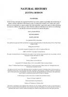
kazinols Q (21), and R (22) (flavans), and broussonols A-D (23-26) (flavonols), were isolated as moderate to weak cytotoxic principles against several human cancer cell lines with ED50 values ranging from 2.3 to 17.4 ^ig/mL [25,27]. Two flavans, kazinols A (19) and E (20), were reported as antioxidative principles using the l,l-diphenyl-2-picrylhydrazyl (DPPH) radical scavenging assay (IC50 41.4 and 33.4 |iM, respectively) [26]. These compounds (19 and 20) also exhibited inhibitory activity against tyrosinase, which is a key enzyme in melanin biosynthesis and plays a role in the conversion of tyrosine to DOPA and DOFA to dopaquinone [26,33]. An antioxidative effect and the suppression of melanin biosynthesis are useful for cosmetic products in relation to hyperpigmentation [34]. Broussonetine C (1), a monocyclic polyhydroxy pyrrolidine alkaloid, showed a yellow spot on TLC when sprayed with ninhydrin reagent and heated (ninhydrin reaction), and its molecular formula was determined by a positive high-resolution mass spectrometry (C18H36NO5, [M + H]^, m/z 346.2579). The IR spectrum displayed a hydroxy band at 3370 cm'^ and a carbonyl band at 1706 c m \ The ^H- and '^^C-NMR signals were assigned using the ^H-^H correlated spectroscopy (^H-^H COSY), heteronuclear single quantum coherence (HSQC), and distortionless enhancement by polarization transfer (DEFT) pulse sequences. The position of the carbonyl carbon and the linkage of the pyrrolidine ring and the aliphatic side chain were determined using the heteronuclear multiple bond coherence (HMBC) NMR technique [HMBC correlations were observed for the carbonyl carbon signal (5c 210.8) with the proton signals at 6H 2.71 (H-11') and 6H 2.12 (H-120, and for the C-5 carbon signal (5c 62.9) of the pyrrolidine ring with the proton signals at 5H 4.44 (H-4) and 5H 2.04 (H-l'), respectively] [16]. The relative stereochemistry of the pyrrolidine ring of broussonetine C (1) was determined from its coupling constants (vicinal coupling, ^2,3 = •/3,4 = •/4,5 = 6.4 Hz) and nuclear Overhauser enhancement effects (H-2/H-4 and H-3/H-5). The absolute stereostructure was disclosed as (2i?,3/?,4i?,5/?) using the benzoate chirality method [35]. A diacetylacetoamide was prepared from broussonetine C (1) by treatment with acetic anhydride in pyridine at room temperature, and then a dibenzoate (la) was obtained by benzoylation of the diacetylacetoamide. The circular dichroism (CD) curve of l a displayed a negative Cotton
11
effect (A8237 -15.9) and a positive effect (AS223 +16.4), which indicated a negative chirality as shown in Fig. (2) [16]. C0CH3
BzO
OBz la
Ae(nin): +16.4(223) -15.9(237)
Fig. (2). Determination of the absolute stereostnicture of broussonetine C (1) by the benzoate chirality method.
Broussonetine L (8) showed similar physical and spectroscopic properties to those of broussonetine C (1) [16], except for proton signals of a p-glucose (anomeric proton, 8H4.78, 1H, doublet, J- 7.8 Hz) moiety in the H-NMR spectrum. Hydrolysis of broussonetine L (8) with 1 N HCl provided broussonetine F (4) [17] and D-glucose ([a]D +40.6°). Therefore, the structure of broussonetine L (8) was determined to be 13'O-p-D-glucopyranosylbroussonetine F due to the glycosylation shift of C13' (6c 69.3) of broussonetine L (8) (broussonetine F, 4, 6c-i3' 61.6) and HMBC long-range correlations observed between H-13' (6H 3.69 and 4.09) and an anomeric carbon (5c 104.4), and between an anomeric proton and C-13'[20]. The absolute stereochemistry of broussonetine L (8) was determined by the combination of the benzoate chirality method and the Mosher's method [35-37]. A carbamate (8a) was prepared from broussonetine F (4) by reaction with phenyl chloroformate in tetrahydrofuran-H20 (7:3), and a diacetate (8b) was prepared from 8a with acetic anhydride in pyridine. Finally, a dibenzoate (8c) was obtained by benzoylation of 8b. The CD curve of 8c showed a negative Cotton effect (AE237 -30.9) and a positive effect (Ae223 +15.9) to confirm a counter-clockwise chirality between two benzoyl groups. Fig. (3) [20].
12
OH
IN.
nd
CH2OGIC
OH
H
OH CH2OH
HOHjC*^'^.*^'^
Hcf
O—CO
OH
PhOCOCl NaHCOj
QH CH2OH
CH2OAC
HO
OH PhCOCl pyridine
O—CO
OAc CH2OAC
Bz(f
8c
OBz OBz
BzQ
A8(nm): +15.9(223) -30.9 (237)
Fig. (3). Determination of the absolute stereostructure of the pyrrolidine ring of broussonetine L (8) by the benzoate chirality method.
13
The absolute configuration of C-l' of 8 was then investigated by the Mosher's method. The di- (R)- and (S)-2-methoxy-2-phenyl-2(trifluoromethyl)-acetic acid (MTPA) esters (8dR and 8d5) and tri- (R)and (iS)-MTPA esters (SeR and SeS) prepared from 8a, were analyzed by ' H - ' H C O S Y N M R (500 MHz) and A6 values (SS-SR) were measured. These values established the R configuration of C-l' of 8 by comparison of the di-MTPA esters (8d/? and 8d5) and the tri-MTPA esters (SeR and 8fty),Fig.(4)[20]. O—CO
OR4 CH2OR1
R-,(f
OR, 8dif, 8d5: Ri = R2 = MTPA, R3 - R4 = H 8ei?, ScJ: R, = R2 = R4 = MTPA. R3 = H
A^(^S-^R)
3
4
5
r
+0.100
-0.039
-0.019
0.000
+0.020
-0.332
-0.161
-0.037
-0.060
+0.013
1"
2
8d
+0.030
1 8e
-0.154
r 0.000 1 +0.050
Fig. (4). Determination of the absolute configuration of C-l' of broussonetine L (8) by the Mosher's method.
Also, the absolute stereochemistry of the pyrrolizidine ring and Cr of broussonetine N (15) was established by the Mosher's method. The tri- (Ry and (5)-MTPA esters (15a/? and 15a5) and penta- (Ry and (5)MTPA esters (15bif and IShS) were prepared from 15 and A6 values (658R) were measured. Accordingly, the R configuration of C-l of the pyrrolizidine ring from 15a and the R configuration of C-l' from 15b were determined, respectively, Fig. (5) [22]. A biosynthetic study of the 18-carbon chain skeleton of broussonetines was reported [38]. To verify the biosynthetic route of these alkaloids, the plant was grown on an aseptic medium and the enriched ^^C of the isolated alkaloids was analyzed by NMR after feeding with [l-^^C]glucose. The labeling pattem of broussonetine J (27) obtained
14
MTPAO -Z. -0.006 "L
!?-^^^_,,, H r
C-0.029
MTPAOH2C
+0.013 +0.001
c V l
CHoOMTPA
H
+0.061
OH
MTPAO -0.075'^
''-^\,ri^ -
-^-^'^
CH2OMTPA OMTPA
15b
Fig. (5). Determination of the absolute configuration of broussonetine N (15) by the Mosher's method.
from the feeding experiment indicated that C-4 through C-18 were formed via palmitoyl CoA through the acetate-malonate pathway, whereas C-1 through C-3 were derived via serine from 3-phosphoglyceric acid. Therefore, the 18-carbon chain of broussonetine J (27) was assumed to be formed initially by condensation of serine and palmitoyl CoA [38], As shown in Fig. (6), the absolute stereochemistry of the pyrrolidine rings of the broussonetines is related to o-serine and that of broussonetine U (28) is related to L-serine. Out of a series of over 30 alkaloids obtained from B, kazinoki, some of them showed potent glycosidase inhibitory activity as shovm in Table 1. Interestingly, only broussonetines E and F (3 and 4), which have a hydroxyl group on C-T, demonstrated potent inhibitory activity against a-glucosidase [17]. However, broussonetines G and H (5 and 6), which also have a hydroxyl group on C-l', did not inhibit a-glucosidase [18]. These results suggested that the inhibition of a-glucosidase might be attributed to the hydroxyl groups on both C-T and C-13' and the keto groups of C-9' or C-10' [17,18]. However, additional studies seem to be required to verify this suggestion [24].
15
CHjOH -O.
COOH
OH
OH
OH
OH OH D-[l-^^C]glucose
O
II
^
.COOH
HO SCoA
NH2 D-serine
NH2 L-serine
CoAS
OH
HO HO
OH
Fig. (6). Biosynthesis of broussonetines J and U (27 and 28).
COOH H O - ^ '
16
BIOACTIVE PAPYRIFERA
COMPOUNDS
FROM
BROUSSONETIA
The major types of bioactive constituents reported from Broussonetia papyrifera are the prenylated flavonoids, which include compoxmds of the diphenylpropane, chalcone, flavan, flavanone, flavone, flavonol, and aurone classes (Table 2), Fig. (7). An early study on B, papyrifera resulted in the isolation of two diphenylpropanes, broussonins A (29) and B (30), and a coumarin, marmesin (52), with antifungal activity [39]. Also, a diprenylated diphenylpropane derivative, kazinol F (31) [40], was reported as an antioxidant and tyrosinase inhibitory constituent [34]. Table 2. Bioactive Compounds from Broussonetia papyrifera Activity
Compound type/name
Reference(s)
FLAVONOroS Diphenylpropanes Broussonin A (29)
Antifungal activity* Inhibition of aromatase**
[39]
Broussonin B (30)
Antifungal activity*
[39]
Kazinol F (31)
Antioxidant activity (scavenging free radicals)^ Inhibition of tyrosinase**
[34]
[34]
1
Antioxidant activity (inhibition of lipid peroxidation)^ Inhibition of cyclooxygenase* Inhibition of nitric oxide production'^ Inhibition of respiratory burst in neutrophils* Platelet aggregation inhibitory activity**
[42]
1
Inhibition of aromatase**
[41]
Isogemichalcone C (34)
Inhibition of aromatase*'
[41]
1 2,4,2',4'-Tetrahydroxy-3'1 prenylchalcone (35)
Inhibition of aromatase**
[41]
1
[41]
1
Chalcones
Broussochalcone A (32)
1 3'-[y-Hydroxymethyl-(£)-ymethylallyl]-2,4,2',4'. tetrahydroxychalcone 1 r - 0 coumarate (33)
[43] [42] [44] [431
Flavans Broussoflavan A (36)
Antioxidant activity (inhibition of lipid peroxidation)*^ 1 Platelet aggregation inhibitory activity**
1 1
[43] [45]
1
17
Table 2. Bioactive Compounds from Broussonetia papyrifera Compound type/name KazinolA(19) KazinolB(37)
(continued)
Activity
RefereDce(s)
Antioxidant activity' Inhibition of tyrosinase' Platelet aggregation inhibitory activity** Inhibition of cyclooxygenase* Platelet aggregation inhibitory activity**
1
[26] [26] [43]
[43] [43]
1 1
Flavanones (25)-Abyssinone II (38)
Inhibition of aromatase**
[41]
(2.S)-2',4'-Dihydroxy-2"-(lhydroxy-1-methylethyl)dihydrofuro[2,3-Alflavanone (39)
Inhibition of aromatase*'
[41]
(2iS)-Euchrenone a? (40)
Inhibition of aromatase*'
[41]
(2.S)-Naringenin (41)
Inhibition of aromatase**
[41]
Inhibition of aromatase**
[41]
Inhibition of aromatase**
[41]
Platelet aggregation inhibitory activity*"
[43]
Antioxidant activity (inhibition of lipid peroxidation)' Antiproliferative activity* Inhibition of aromatase** Inhibition of cyclooxygenase* Platelet aggregation inhibitory activity*" Antioxidant activity (inhibition of lipid peroxidation)*^ Antiproliferative activity*
[45] [45] [41] [43] [43]
Inhibition of aromatase*'
[41]
(25)-5,7,2',4'1 Tetrahydroxyflavanone (42) Flavone 1 5,7,2',4'-Tetrahydroxy-31 geranylflavone (43) Flavonols Broussoflavonol £ (44)
Broussoflavonol F (45)
Broussoflavonol G (46) Isolicoflavonol (47)
[45]
'
Aurone Broussoaurone A (48)
[45]
Antioxidant activity (inhibition of lipid peroxidation)' Inhibition of cyclooxygenase* Platelet aggregation inhibitory activity*"
[45] [43]
L. _ [43I_.
MISCELLANEOUS AlbanoIA(49)
Inhibition of aromatase**
[41]
Betulinic acid (50)
Selective cytotoxic activity against melanoma cell lines'"
[46]
1
18
Table 2. Bioactive Compounds from Broussonetia papyrifera (continued) Compound type/name
Activity
Reference(s)
Demethylmoracin I (51)
Inhibition of aromatase**
[41]
Marmesin(52)
Antifungal activity*
[39]
MoracinN(53)
Inhibition of aromatase**
[41]
Ursolic acid (54)
Inhibition of HIV-1 protease dimerization''
[47]
"Antifungal activity (presented as a range) expressed as the minimum concentration (mM) required for complete inhibition of fungal growth including Fusarium roseum, F. lateritium, F. solani, Diaporthe nomuraU Stigmina mori, Sclerotinia sclerotiorum, Bipolaris leersiae, and Rosellinia necatrix\ 29: 0.2-0.9, 30: 0.05-0.9, 52: 0.9-4.0. ''Aromatase inhibitory activity determined as IC50 value (^iM); 29: 30.0, 33: 0.5, 34: 7.1, 35: 4.6, 38: 0.4, 39: 0.1,40: 3.4,41: 17.0,42: 2.2,43: 24.0,45: 9.7,47: 0.1,49: 7.5,51: 31.1,53: 31.1. ^Antioxidant activity expressed as IC50 value (pM); 31: 6.7 (jig/mL); 32:0.63,36: 2.1,45:2.7,46:1.0,48: 1.2. ''The tyrosinase inhibitory activity of 31 was IC50 0.39 |ig/mL. *Cyclooxygenase inhibitory effect determined as IC50 value (ng/mL); 32: 19.4,37: 155.3,45: 17.5,48: 22.7. ^Inhibitory effect (IC50) of 32 on nitric oxide production was 11.3 ^iM. ^Compound 32 inhibited O2 consumption in formylmethionyl-leucyl-phenylalanine- and phoibol 12-myristate 13-acetate-stimulated rat neutrophils with IC50 values of 70.3 and 63.9 ^M, respectively. •^Antiplatelet activity induced by arachidonic acid was expressed by IC50 value (^M); 19: 11.4, 32: 6.8, 36: 86.7,37: 32.6,44: 39.9,45:16.9,48: 15.4. 'Activity found as a constituent of Broussonetia kazinofd. ^Antiproliferation activity shown by the inhibition of ['H]thymidine incorporation into DNA in the proliferation of rat vascular smooth muscle cells. The effect was expressed as % of control; 45: 0-7.8,46: 0-0.4. ''Activity found as a constituent of a plant other than a Broussonetia species.
Broussochalcone A (32) [48], a prenylated chalcone, is one of the most completely studied constituents of B. papyrifera biologically. Broussochalcone A (32) inhibited platelet aggregation induced by arachidonic acid with an IC50 value of 6.8 |aM as well as induction by adrenaline in human platelet-rich plasma. The antiplatelet effect of 32 was partially due to an inhibitory effect on cyclooxygenase activity and by reducing thromboxane fomiation [43]. Also, broussochalcone A (32) inhibited O2 consiraiption in fomiylmethionyl-leucyl-phenylalanine- and phorbol 12-myristate 13-acetate-stimulated rat neutrophils with IC50 values of 70.3 and 63.9 jiM, respectively. This inhibitory effect of 32 on respiratory burst in neutrophils was not mediated by the reduction of phospholipase C activity, but was mediated by the suppression of protein kinase C activity through interference with the catalytic region and by the
19
RiO
29Ri = CH3,R2 = H 30Rj = H,R2 = CH3
31
33R = H 34R = OCH3
OH
36
20
Fig. (7). Continued
OH
OH
37
OH
OH
41R = H 42R = OH
21
Fig. (7). Continued
HO,
48
49
22
Fig. (7). Continued
COOH
50
HO-
51
o ^ ^ ^ -o- - o 52
CCX)H
54
Fig. (7). Structures of bioactive constituents of Broussonetia papyrifera.
attenuation of O2*" generation from the NADPH oxidase complex, which might inhibit the generation of toxic oxygen radicals and terminate the tissue damage [43]. Furthermore, broussochalcone A (32) showed antioxidant activity in iron-induced lipid peroxidation in a rat brain
23
homogenate model with an IC50 value of 0.63 |iM as well as in the DPPH system, and exhibited an inhibitory effect on nitric oxide (NO) production with an IC50 value of 11.3 jaM. This potent inhibitory effect on NO production was mediated by suppression of nuclear factor (NF)-KB activation, phosphorylation and degradation of iKBa (an inhibitory protein of NF-KB), and inducible NO synthesis expression, which have been associated with autoimmune or inflammatory diseases [42]. In an effort to investigate antioxidant constituents with antiproliferative effects in rat vascular smooth muscle cells (VSMC), broussoflavan A (36) [49], broussoflavonols F (45) [50] and G (46) [51], and broussoaurone A (48) [49] were found to inhibit the Fe^^-induced thiobarbituric acid-reactive substance formation in rat brain homogenate. Furthermore, broussoflavonols F (45) and G (46) inhibited fetal calf serum-, 5-hydroxytryptamine-, or ADP-induced [^H]thymidine incorporation into rat VSMC [45]. Antioxidant activities and inliibitory effects on proliferation of rat VSMC with potent antiplatelet activities of 45 and 46 may be useful for vascular diseases and atherosclerosis [43,45]. The concept of cancer chemoprevention is becoming wellestablished and refers to the pharmacological intervention to arrest or reverse the process of carcinogenesis, and thus prevent cancer [52,53]. It has become evident that various phytochemical components of the diet are able to prevent cancer formation in full-term carcinogenesis inhibition studies in animal models [54]. As part of a U.S. National Cancer Institutefunded program project conducted at the University of Illinois at Chicago [55-57], an ethyl acetate extract of the whole plants ofB, papyrifera was found to significantly inhibit aromatase activity in an in vitro assay [58,59] (74% inhibition at 80 |ig/mL) [41]. This was only one of a handful of extracts found to significantly inhibit aromatase activity with the bioassay protocol used, out of over 1,000 extracts screened [60]. This target was chosen for investigation, because aromatase catalyzes the final, rate-limiting step in estrogen biosynthesis [61], and is regarded as a target relevant to the treatment or prevention of breast and prostate cancers [62]. Several synthetic aromatase-inhibitory drugs have been developed, including aminoglutethimide, substrate androstenedione derivatives, imidazoles, and triazoles [63-65]. From the active extract of B, papyrifera were isolated several aromatase inhibitors with IC50 values in the range 0.1-31.1 ^M, inclusive of broussonin A (29) [66], 3'-[Y-hydroxymethyl-(£)-y-methylallyl]-
24
2,4,2',4'-tetrahydroxychalcone 11 '-O-coumarate (33) [41], isogemichalcone C (34) [41], 2,4,2',4'-tetrahydroxy-3'-prenylchalcone (35) [67], (25)-abyssinone II (38) [68], (25)-2',4'.dihycIroxy-2"-(lhydroxy-1 -methylethyl)-dihydrofuro[2,3-A]flavanone (39) [41], (25)euchrenone a7 (40) [69], (25)-naringenin (41) [70], (25)-5,7,2',4'tetrahydroxyflavanone (42) [71], 5,7,2',4'-tetrahydroxy-3-geranylflavone (43) [41], broussoflavonol F (45) [50], isolicoflavonol (47) [72], albanol A (49) [73], demethylmoracin I (51) [41], moracin N (53) [74]. Of these aromatase inhibitors, five of the compounds were new (33, 34, 39, 43, 51), and details of structure elucidation of 33, 34, and 43 are presented as examples in the following two paragraphs. The isolates 3'-[y-hydroxymethyl-(£^-Y-methylallyl]-2,4,2',4'. tetrahydroxychalcone 11'-O-coumarate (33) and 3'-[Y-hydroxymethyl(£)-y-methylallyl]-2,4,2',4'-tetrahydroxychalcone 11 '-O-ferulate (isogemichalcone C, 34) were obtained as orange powders and were shown by positive HRFABMS to possess molecular formulas of C29H26O8 (m/z [M + Na]^ 525.1884) and C30H28O9 (m/z [M + N a ] \ 555.1577), respectively. The ^H- and ^^C-NMR spectra of 33 and 34 exhibited characteristic chalcone signals, and signals for a coumarate group for 33 at 6H 7.54 (2H, 7 = 8.6 Hz, H-2" and H-6"), 6H 6.87 (2H, J = 8.5 Hz, H3" and H.5''), 5H 7.59 (IH, 7 = 16.0 Hz, H-T'), and 5H 6.35 (IH, J = 16.0 Hz, H-8") and signals for a ferulate group for 34 at 6H 7.34 (IH, 7 = 1.6 Hz, H-2"), 6H 6.85 (IH, / = 8.1 Hz, H.5"), 6H 7.12 (IH, 7 = 1.7 and 8.2 Hz, H-6"), 5H 7.57 (IH, J = 16.0 Hz, H.7"), 5H 6.40 (IH, J= 15.9 Hz, H8"), and 6H 3.91 (3H, singlet, OCH3). Based on these observations, the structures of 33 and 34 were concluded to be prenylated chalcones with a coumarate and a ferulate unit attached, respectively, which were confirmed by 2D-NMR techniques. Fig. (8). In case of isogemichalcone C (34), it was concluded to be a regioisomer of gemichalcone C by comparing its spectra with those of the latter compound [75]. This was confirmed using a NOESY NMR experiment. Thus, the NOE correlations between H-7' and H-10', and H-8' and H-IT clearly indicated E stereochemistry of the prenyl group. Moreover, the chemical shift differences at positions C-10' and C-1T of the E and Z isomers supported the stereochemistry proposed. Fig. (8) [41,75,76]. 5,7,2',4'-Tetrahydroxy-3-geranylflavone (43) exhibited a molecular ion [M]"^ at m/z 422.1719 by HREIMS, consistent with an
25
Carbon
6c 33
34
Gemichalcone C [75]
10'
14.2
14.2
64.2
ir
70.2
70.2
22.8
'
Fig. (8). Selected HMBC (->) and NOE (
COOEt Bu.Sn^
^COOEt
Fig (3). Treatment of 2,2,6-trimethylcyclohexanone with lithium diisopropylamide (LDA) followed by phenyltriflimide (7V-phenylbis (trifluoromethanesulphonimide) gave the corresponding triflate [24]. The
73
best coupling reaction could be achieved with Farina's 'soft' palladium (Pd2(dba)3) with AsPhs as ligand and DMPU, Fig (4) [25]. OTf
a)LDA
^COOEt
b) Bu3Sn"
phenyltriflimide
Pd2(dba)3, NMP, DMPU, AsPh3
^^COOU
COOEt c) K:OH, EtOH, H2O
Fig. (4). Dominguez, Iglesias, and De Lera (1998, 2001). As an extension of this procedure, they synthesized the side chain of 9Z-retinoate stereoselectively and attached it to the hydrophobic ring by a high yielding thallium accelerated Suzuki cross-coupling reaction, Fig. (5) [26]. The tetraenylstannate used for the coupling reaction was obtained by Mn02 oxidation of the known stannyldienol [27], to the corresponding aldehyde (86%), followed by condensation with the phosphonate (52%) and reaction of the tetraenylstannate with a solution of iodine. The product was immediately added to the organoborane, in the presence of Pd(PPh3)4 then TIOH was added, to provide the 9Zretinoate in 84% yield. The organoborane was freshly prepared from the cyclohexanone, via its hydrazone, which was transformed into the iodide. COOEt
a) BuLi,
1 BuSn
BuSnH, CuCN OH4
BuSn
^
^
b) Mn02 K2C03^
[ ""OH
-^:^ ^
BuSn''
OEt ^j^Q
c) BuLi, DMPU
•
COOEt
COOEt
I J) /BuLi, B(0Me)3
e) H2N>fH2 I2, Et3N, DBN
"
Pd(PPh3)4
*"
Fig. (5). Pazos, and De Lera (1999).
B(0H)2 J ^ ^ ^
kjl\
COOEt
74
In this exhaustive work De Lera et al described the syntheses of the retinoid skeleton via the Stille coupUng for the formation of side-chain single bonds [28]. C(7)-C(8) strategies: A stereoselective synthesis of all E retinal, via a condensation of a Cio chloroacetal with p-cyclogeranylsulfone was described by Julia et al. [29]. The chloroacetal was reacted with the silylenol ether, using TiCl4/Ti(OMe)4, to give in 63% yield, the chloromethoxyacetal derivative as a mixture of ElZ isomers (80/20). The aldehyde was converted in 97% yield into the corresponding acetal with HC(0Me)3 and camphorsulfonic acid in methanol, Fig. (6).
OMe OMe
OMe
OMe
a) Ti(0Me)4 TiCl4
OMe
b) HC(0Me)3 CHO
OMe CI
01
CI
Fig. (6). This building block was condensed with the anion of (icyclogeranylsulfone. During flash-chromatography the intermediate was hydrolyzed to the sulfone-aldehyde, as a mixture of three isomers in 95% yield. Retinal was obtained from this sulfone by treatment with MeONa, for 10 days, in the dark (90%), Fig. (7).
OMe I
OMe OMe
SO2
I
OMe I
CHO
Fig. (7). Chemla, Julia, and Ugen (1993).
75
Chabardes developed a process for the preparation of vitamin A and its intermediates, from cyclogeranylsulfone and Cio aldehyde-acetals [30], For example, chlorocitral reacted with ethylene glycol, HC(0Me)3 and pyridinium tosylate to provide the chloroacetal (40%), as a mixture of two isomers. Reaction of this allylchloride with A^-methylmorpholine oxide (NMO) and Nal furnished the aldehyde, as a mixture of four isomers. These compounds underwent condensation with pcyclogeranylsulfone. Further chlorination of the sulfone-alkoxide salts, led to a mixture of sulfone-chloride acetals and their products of hydrolysis in 45-50% yield. Double elimination of the chloride and the sulfone, followed by hydrolysis with pyridinium tosylate (PPTS) gave retinal, as a mixture of all E and 13Z isomers (78/22). The overall yield from the chloroacetal was 18%. In another 'one-pot' example, retinal was obtained in 52% yield from the aldehyde, and was then isomerised and reduced to retinol (all E: 95.5, 13Z: 4, 9Z: 0.5) Fig. (8).
s
a) NMO CI
I
Nal, DMF
I
O-^ ^) L A
^2
/PrMgCl c) SOCI2, pyridine
d) MeOK
j^^^^'-'^^::^.-'^^
e) PPTS
Fig. (8). Chabardes (1994). Honda et al described a highly Z stereoselective [2,3]-sigmatropic rearrangement that provided trisubstituted E,Z synthons, starting from A^tiglyl-p-methallyldimethylammonium salts [31]. The application of this key triene synthon to the stereoselective synthesis of 13Z-retinol was reported from a trieneester. Thus, prenylbenzyl ether was converted via ene-type chlorination followed by amination into internal allylamine. This was reacted with ethyl 3-bromotyglate in acetonitrile to give the
76
quaternary salt. Treatment of the latter with EtOK in ethanol resulted in the formation of an ylide. This latter underwent [2,3]-sigmatropic rearrangement to furnish the diene that possessed a newly formed Z and tiglyl-origin E stereochemistry, Fig. (9). a) Br
COOEt
OSi/BuMeo
OSi/BuMeo
h) EtOK
NMeo
"^ EtOOC 0SirBuMe2 COOEt
Fig. (9). Treatment of this synthon with peracetic acid resulted in the formation of a A^-oxide intermediate. A Cope elimination gave the triene, Fig. (10).
c) AcOOH
EtOOC
EtOOC 0Si/BuMe2
0Si/BuMe2 ^0°C
EtOOC 0Si/BuMe2
Fig. (10). ?BuMe2Si was then replaced by rBuPh2Si and the transformation of the ester group to formyl group was carried out by treatment with aluminium hydride (AIH3), followed by manganese dioxide oxidation. This triene aldehyde was reacted with the anion of p-cyclogeranylsulfone and quenched with AC2O. Desilylation to the acetoxysulfone (80%), and
77
reductive cleavage with sodium amalgam gave the desired 13Z-retinol (63%), Fig. (11). e)Bu4NF,y)TBDPSCl
»• OHC
EtOOC
^
g) AlCl3/LiAlH4, h) Mn02 OSi/BuMeo
OSi/BuPh,
OSi/BuPho
Fig. (11). Honda, Yoshii, and Inoue (1996). A one-pot procedure was developed by Otera et al from pcyclogeranylsulfone [32]. Its lithium salt reacted with 3,7-dimethyl-8oxo-2,6-octadienyl acetate to the sulfone-alcohol. The hydroxyl group was protected to the MOM ether with MeOCH2Cl. Double elimination could be achieved with potassium MeOK to provide vitamin A in 50% yield. Fig. (12).
.o 9 o.
OHC
^ ^ so. OAc
OAc OH
a) Nal, BuLi
b)MOM-ci
y^^^Yi^^^^ ^
\ X \
c) MeOK
OMOM
Fig. (12). Orita, Yamashita, Toh, and Otera (1997).
^^^^^^^^^^^V^^OAc
78
A similar synthesis was patented by Odera [33]. Two patents by Takahashi et al reported the synthesis of vitamin A via a Cio dihalogeno derivative [34,35]. In one procedure the halogenodiene was prepared by bromination of 3,7-dimethyl-2,5,7octatrien-1-yl acetate. Addition of the latter and /BuOK in DMF to the Cio sulfone provided the retinol sulfone (34%). Again, double elimination (MeOK), gave vitamin A acetate, Fig. (13).
a) Br2
Brv
OAc b) /BuOK, DMF
PY
SOo
I
Br
c) MeOK
-^::s^^^^
OAc
Fig. (13). Takahashi, Furutani, and Seko (2000). They also developed a second process via other dihalo-compounds [36]. Treatment of the 1,2-bromo-hydroxy chain with TiCU in DME, gave mainly the l-bromo-4-chloro unit. Condensation with the Cio sulfone in DMF, in the presence of /BuOK gave the retinylsulfoneacetate. Elimination of the tolylsulfmate with KOH in DMF produced vitamin A acetate in 87% yield. Fig. (14).
Fig. (14). Takahashi, and Seko (2001).
79
In a similar route, Takahashi et al made use of non-halogenated sulfones [37]. Similar processes were related. TiCU was added to a solution of the diol to give a crude mixture of isomers in which the 5-chlorosulfone was the main compound in 95% yield. The mixture was treated with MeOK to produce crude retinol. Acetylation with acetic anhydride (AC2O) in pyridine, in the presence of DMAP, provided the retinyl acetate in 70% from the diol [38,39], Fig. (15).
OH
c) AC2O, DMAP
Fig. (15). Takahashi, Furutani, and Seko (2000). C(io)-C(ii) strategies: Mestres et al [40] published a regioselective addition of a lithium trienediolate (generated from hexa-2,4-dienoic acid or dihydropyran-2one) to p-ionone. Dehydration of the hydroxyacid, afforded a mixture of 9EIZ, 13£'/Zretinoic acids which, isomerised in the presence of I2, led to all E retinoic acid in 35% and 30% yield, starting from dienic acid and pyranone, respectively, Fig. (16).
COOH
COOH
Fig. (16). Aurell, Parra, Tortajada, Gil, and Mestres (1990); Aurell, Came, Clar, Gil, Mestres, Parra, and Tortajada (1993); Aurell, Ceita, Mestres, Parra, and Tortajada (1995).
80
A concise preparation of retinoids via new enaminodiesters synthons was described by Valla et al [41]. For example, all £-retinoic acid was synthesized within one day by a 'one-pot' process. The enaminodiester synthon was prepared from methyl isopropylidenemalonate and dimethylformamide dimethylacetal (DMF-DMA) and then condensed with the lithium enolate of p-ionone. A Grignard reaction with the obtained ketodiester led to the retro carbomethoxyretinoate. Saponification and concomitant decarboxylation, provided mainly all E retinoic acid {all E/UZ: 90/10, 72% from (J-ionone), Fig. (17).
^v
COOMe
a)LDA
l^^^J^
b) MeMgBr
COOMe
COOMe COOMe COOMe
COOH
c)K0H,Me0H,H20
^ COOMe
^HCllM
Fig. (17). Valla, Cartier, Labia, and Potier (2001). A short synthesis of retinal was described by Taylor et al. [42] based on the addition of a Cn vinylalane to a methylpyrylium salt. The 13Zretinal (48%) was isomerised to all E retinal by a previous procedure [43]. p-Ionone was first converted into the alkyne and then into the vinylalane, using the Negishi methodology [44]. Addition of an excess of this alane to 4-methylpyrilium tetrafluoroborate [45] gave 13Z-retinal, being isomerized to the all E isomer (I2 in benzene/ether). Fig. (18). AlMejBuLi
a) MejAl, ZrClz
^ (TI-C5H5)2, BuLi
c)l2 CHO
Fig. (18). Hemming, De Meideros, and Taylor, (1994); Taylor, Hemming, and De Meideros (1995).
CHO
81
Through two successive Stille reactions, Parrain et al [44] realized a stereo selective synthesis of all E, 13Zand 9-A2or-retinoic acids. First, the coupling of £'-l,2-bis(tributylstannyl)ethene and Z- or E-tributylstannyl3-iodoalk-2-enoates was performed, followed by iododestannylation. The second step involved another vinyltin which was synthesized by stannylation of the Negishi dienyne, derived from p-ionone [47]. To obtain the substituted vinylstannate, the dienyne was treated with lithium butyltributylstannylcyanocuprate (Lipshutz reagent) [48] to yield the intermediate vinylcuprate, which was trapped with an excess of Mel in the presence of hexamethylphosphoramide (HMPA). The reaction occurred to the advantage of the terminal vinylstannate (up to 92%). The coupling partner was obtained from tetrolic acid, which was converted into E vinyliodide by stannylcupration of the generated stannate [49]. The Z vinyliodide was more classically obtained by hydroiodination [50]. Stille coupling of the P-iodovinylic acids (protected as the corresponding tributyltin esters) with £'-l,2-bis(tributylstannyl)ethene, catalyzed by dichlorobis(acetonitrile)palladium provided dienyltins with retention of the configuration of the two double bonds in fair yields. Iododestannylation yielded quantitatively the dienic acids, Fig. (19). a) Bu3SnBuCuLi, yr-——
^^
j T-
C00HBu3Sn^
^COOH
^
d) BuSnOMe, PdClzCMeCN); e)l2./)KF,HC\
V c)HI
j
I
SnBu3 g)PdCl2(MeCN)2,DMF
COOH
I
I
^ O ^ : ^ ^ ^ ^
^ \^-!v
COOH
Fig. (19). Thibonnet, Abarbri, Duchene, and Parrain (1999). The first palladium-catalyzed cross-coupling reaction used in the synthesis of retinoids was described by Negishi and Owczarczyk from a Ci4 alkenylzinc [51]. The synthesis was carried out via a Pd(PPh3)4
82
catalyzed coupling of the C14 alkenylzinc (obtained from the iodide) with the Ce iodide (derived from 3-methyl-2£'-penten-4-yn-l-ol), followed by further deprotection with BU4NF. Vitamin A was obtained in 38% yield based on p-ionone, with complete control of stereo- and regiochemistry, Fig. (20).
Q
a) LDA, Cl-P(0)(0Et)2, LDA b) MesAl, Cl2ZrCp2,12,
c) DIBAL-H, I2 ClSiPh2/Bu, EtsN, DMAP
d) /BuLi, ZnBr2 P(i(PPh3)4 e) BU4NF
Fig. (20). Negishi, and Owczarczyk (1991). A highly stereoselective synthesis of retinol vz^ a CM + C6 route was depicted by De Lera et al [52]. A Suzuki reaction of a C14 alkenyliodide with a C6 alkenylboronic acid afforded retinol in 83% yield, with retention of the geometries of the coupling partners. The alkenyliodide was obtained by a zirconium-mediated methylalumination and a subsequent Al/I exchange by slow addition of ICN. Coupling with the C6 boronic acid (12 hrs to reach completion), afforded retinol in 83% yield [53], Fig. (21).
Fig. (21). Torrado, Iglesias, Lopez, and De Lera (1995).
83
C(ii)-C(i2) strategies: Stereoselective syntheses of all E, 9Z-retinoic-acids and llZ-retinal were developed from p-ionone-tricarbonyliron complex [12]. Treatment of the complex (prepared from p-ionone and dodecacarbonyliron, (Fe3(CO)i2)), with the lithium salt of acetonitrile, Wada et al obtained the nitrile, in 88% yield, Fig. (22). CHO
O
a) LDA, MeCN ^ Fe(C0)3 Q (EtO)2P''''~V^COOEt
^ c) BuLi
COOEt
COOH
,)j^30H MeOH, HjG
Fig. (22). Contrarily, the reaction of the lithium enolate of ethyl acetate with subsequent dehydration gave predominantly the ethyl 9Zionylideneacetate in 89% yield, Fig. (23).
O
a) LDA, MeCOOEt ^
Fe(C0)3
/'-p^
CHO ^)BuUTHF
(C0)3 g) NaOH, ^
^ COOEt
MeOH, H2O COOH
COOEt Fig. (23). Wada, Hiraishi, Takamura, Date, Aoe, and Ito (1997); Wada, (2000).
84
These compounds were converted to the corresponding all E and 9Zretinoic acids via P-ionylideneacetaldehydes. Thus, the reaction with the Uthium sah of (EtO)2P(0)CH2C(Me)=CHCOOEt in THF made possible the C20 ester-complex. The complex was removed by CUCI2 in EtOH (98%) and saponification of the ethyl retinoate, the retinoic acids could be obtained {all E: 89%, 13Z: 8% and 9Z: 59%, 9Z,13Z: 12%, respectively). The Peterson reaction of the chlorovinyl-complex with ethyl trimethylsilylacetate provided the HZ isomer preferentially (77%), and the 1 IJE" isomer as a secondary product (15%). The ester was transformed into the Cig ketone (Ph3SnCH2l, BuLi, Et20, 79%). Reaction with (/PrO)2P(0)CH2CN afforded the llZ-retinonitrile in 73% yield. The complex was removed by CuCb (72%) and DIBAL-H reduction led quantitatively to llZ-retinal, Fig. (24). EtOOC
Fig. (24). Wada, Hiraishi, Takamura, Date, Aoe, and Ito (1997); Wada (2000). Wada et al. [13] have previously reported similar syntheses of all E, 9Z-retinoic acids and 1 IZ-retinal. A short access to retinal was reported by Duhamel et al. [54,55] via the enolate of prenal, prepared from the corresponding silyl enol ether or enol acetate. The diene reacted with p-ionylideneacetaldehyde to give the dihydropyranol as the single reaction product. The dihydropyranol was
85
easily converted into retinal (43% yield) by dehydration, ring opening and further dehydration in the presence of a catalytic amount of pyridinium chloride or boric acid, Fig. (25).
OAc
a) MeLi
J^„." t ^
OSiMe,
pyridinium chloride, DMF
Fig. (25). Duhamel, Guillemont, and Poirier (1991); Cahard, Duhamel, Lecomte, and Poirier (1998). These authors also described a three-step synthesis of 13Z-retinoic acid [56]. The obtained hydroxydihydropyrane (66%) was oxidized either by Jones's reagent (CrOs, water, H2SO4, 90%) or Corey's reagent (pyridinium chlorochromate (PCC), 65%). Finally, the dihydropyranone was transformed into retinoic acid (as a mixture of9E, 13Z, and 9Z,13Z), by /BuOK, according to a known procedure [57], Fig. (26).
or PCC
GOGH
Fig. (26). Cahard, Mammeri, Poirier, and Duhamel (2000); Cahard, Duhamel, Lecomte, and Poirier (1998). This French group patented a process for the preparation of vitamin A from vinyl-P-ionol, by BF3-Et20 catalyzed condensation with a C5 sulphide (50% yield) [58]. The phenylthioretinal was reduced with NaBH4 to give the corresponding alcohol (99.5%), which was acetylated (AC2O, -100%).
86
The resulting sulphide-acetate was oxidized with w-chloroperbenzoic acid (MCPBA) and the sulfoxide was eliminated by heating in CCI4 to supply vitamin A acetate in 76% yield. Fig. (27). SPh OH
OMe
^^O 6)NaBH4
a) BF3-Et20
c) Bi^N, AC2O
•Qll
^Y^vA.^^
SPh
d) MCPBA
^Y^^^A-^^^^
ecu ^
kA.
reflux
SOPh
OAc
Fig. (27). Ancel, Bienayme, Duhamel, and Duhamel (1992). Another work of Duhamel and Ancel [59] related this synthesis of retinal via p-ionylideneacetaldehyde. Condensation of methallylmagnesium chloride with diethyl phenyl orthoformate (Et02CH0Ph) led after bromination of the ene-acetal, deshydrohalogenation (NaOH 50%), ethanol elimination with hexamethyldisilazane (HMDS) and ISiMes, to the bromo-dienol ether. This latter was submitted to bromine lithium exchange and the lithio enol ether was then condensed with pionylideneacetaldehyde to give retinal. Fig. (28). GEt MgCl (Et0)2CH0Ph
GEt Br,
GEt
GEt
GEt NaGH
GEt Br
/BuLi
HMDS Ov-^CHG
Fig. (28). Duhamel, and Ancel (1992). In a similar approach, Duhamel et al [60] studied the catalyzed condensation (BF3-Et20 or ZnCb) of vinyl-P-ionol with a chloroenolether. The intermediary aldehyde {all EI9Z\ 65/35) had been
87
dehydrohalogenated (l,8-diazabicyclo[5.4.0]undec-7-ene (DBU), 86% or LiCl, 75%), to a mixture of retinals. This mixture had been isomerized to all E retinal, according to literature procedures, [61] Fig. (29).
a) BF3-Et20 or ZnCl2^
CHO orLiCl, DMF
Fig. (29). Duhamel, Duhamel, and Ancel (1994). In connection with a work related to the syntheses of C5 building blocks, Quintard et al [62] described a synthesis of retinal from Pcyclocitral. This aldehyde was condensed with the vinyl lithium salt of the C5 acetal. The lithiated compound was obtained via the vinyltin derivative which was first converted into the vinyl iodide before doing the halogen-metal exchange. Fig. (30 and 31). OEt J^^^^
OEt
.;Bu3SnMgMe,CuCN
i ^ J . ^ ^ ^ ^
c)BuUort^.U
u^J.^^^ OEt
b)\2
Fig. (30). In an iterative fashion, the hydroxyacetal (intermediately formed by condensation of vinyllithium salt with p-cyclocitral) was dehydrated with aqueous HBr. This allowed the simultaneous hydrolysis into pionylideneacetaldehyde, as a mixture of7E,9E (80%) and 7£,9Z (20%). The reaction had been repeated with the same C5 unit and finally, retinal could be obtained as a mixture of isomers, containing 68% of all E isomer (47%) yield from P-cyclocitral), Fig. (31).
>CcCHO
Fig. (31). Beaudet, Launay, Parrain, and Quintard, (1995); Launay, Beaudet, and Quintard (1997). Bienayme and Yezeguelian [63] described a new synthesis of retinal via a Heck vinylation of a C15 tertiary allylic alcohol with a C5 iodoacetal. Thus, the bromo acetal was prepared by a known procedure [64], by a bromination-dehydrobromination reaction sequence (E and Z isomers: 40/60). The iodo acetal could be easily obtained (as a mixture of E and Z isomers, 40/60), by a nickel catalyzed iodine-bromine exchange. This synthon reacted smoothly with the C15 tertiary allylic alcohol in the presence of a catalytic amount of palladium acetate and a stoechiometric amount of either a silver or a thallium salt. The C20 hydroxy-acetal was obtained in 38% yield, as a mixture of E and Z isomers (48/52). Finally retinal was obtained by treatment with dilute HBr in refluxing acetone, as a mixture of £" and Z isomers (C(9)=C(io) and C(i3)=C(i4)), Fig. (32).
I
GEt ""^^ 1 ^ 3
I
OEt
I ' c)lK,NiBrJn
^
OEt ^^^
I
OH
^Pd(OAc),
Fig. (32). Bienayme, and Yezeguelian (1994) In another study, Bienayme [65] obtained retinal in three steps from pionone, involving a Pd-catalyzed rearrangement of a mixed carbonate, derived from ethynyl-retro-ionol.
89
Thus, the P-ionone was smoothly deconjugated and ethynylated to give ethynyl-retro-ionol as a mixture of ElZ stereoisomers. Formation of the carbonate and its Pd-catalyzed rearrangement produced straightforward a mixture of aldehydes and a allene compound. After silica-gel chromatography, the allenic-aldehyde was conjugated with a catalytic amount of HBr in acetone. Retinal was obtained as a mixture of E and Z isomers (75/25), which could be converted into the all E isomer by simple equilibration. Fig. (33).
uC^^ /^^^N.x^^
T^
...^^
c) Pd(0Ac)2
^.
fl)MeONa,NMP
?$^ ^ " ^
^ QC
r^y^^^^'''''^^
—"
^^ ^E~MgCl
o 6)Pd(dba)3,P(napht)3
Y^
CHO
^HBr
CHO
Pd(dba)3, P(napht)3
Fig. (33). Bienayme (1994, 1995). A similar route was patented by Ancel and Meilland [66]. The ethynyl-retro-ionol was acetylated (Ac20-DMAP-Et3N) and this propargylic acetate was reacted with methyl butadiene acetate in the presence of BF3-Et20. The allenic-retinal, obtained in 61% yield was isomerised in retinal by HBr in acetone (yield: 50%), Fig. (34).
CHO
Fig. (34). Ancel, and Meilland (2000).
90
Salman et al [67] described a process for the preparation of 13Zretinoic acid (isotretinoin) in a single step from piony lideneacetaldehy de. Thus, isotretinoin was obtained by treating methyl-3,3dimethylacrylate with LDA, followed by addition of pionylideneacetaldehyde and further hydrolysis with 10% sulphuric acid. The pH had to be adjusted to 2.8 ±0.5, Fig. (35).
'
b)
MeO a) LDA
c)H2S04,pH=2.8
\ X \
COOH
Fig. (35). Salman, Kaul, Babu, and Kumar (2001). Recently Valla et al showed that new 'P-methylenealdehydes' synthons could be substituted to 7jE',9£'-ionylideneacetaldehydes (derived from a and P-ionones) in a Stobbe reaction [68,69]. Regioselective isomerization of these P-methylenaldehydes in Et2NH produce the compound {EIZ\ 97/3). These synthons were synthesized by formylation of ionones and concomitant acetalysation of the sodium salts of the hydroxymethylenic compounds. Wittig reaction and acidic hydrolysis of the p-methyleneacetals produced the pmethy lenealdehydes. Hence, Stobbe-like condensation with dimethyl-isopropylidene malonate and saponification of malonic acid, half-esters afforded the corresponding 14-carboxyretinoic acids, as a mixture of all E and 9Z isomers (80/20). The all E diacid was easily removed by crystallization from MeCN or ether, Fig. (36). A stereospecific decarboxylation in 2,6dimethylpyridine led to isotretinoin.
91
O >Q
a) MeONa, HCOOMe
OMe "OMe
c) ?h^?CH2
b) H2SO4, MeOH COOMe
OMe OMe
d) HCOOH
COOMe y)NaOH COOH
'
COOH
g) ether
COOH
h) 2,6.dimethyl pyridine
COOH
Fig. (36). Valla, Andriamialisoa, Prat, Giraud, Laurent, and Potier (1999); Giraud, Potier, Andriamialisoa, and Valla (1999). A related stereoselective synthesis of all E retinoic acid was also performed by Valla et al [70] from the 14-carboxyretinoic acid, derived from p-ionone, using pyridine (2 eq.) at room temperature for 20 hrs. The crude retinoic acid mixture {all E/13Z: 97/3) was crystallized in MeCN or AcOEt to provide pure all E retinoic acid. Fig. (37). COOH
COOH
a) pyridine ^
COOH
Fig. (37). Valla, Andriamialisoa, Prat, Laurent, Giraud, and Potier (2000). A new preparation of the Cig ketone, an important synthon for the synthesis of vitamin A had also been published by Valla et al [71]. Hence P-ionone and acetonitrile were condensed in the presence of KOH, to afford the nitrile (80%, ElZ isomers: 80/20). A Reformatsky reaction of ethyl bromoacetate with the nitrile provided the ethyl Pionylideneacetoacetate in 70% yield. Subsequent reduction with NaBH4, followed by esterification (MeS02Cl) and desulfonation of the unstable
92
ester, led to the acid {ElZ isomers, 80/20) in 80% yield. Reaction of the latter with MeLi afforded the Cig ketone in 70% yield, as a mixture of 9£/Z isomers (80/20), Fig. (38).
^V-'^^O
CN
ci) MeCN
b) Zn, BrCHsCOOEt
KOH
V^^^^^^/J^^^^
c) NaBH4
COOH
d) MeS02Cl-Me3N
Fig. (38). Andriamialisoa, Valla, Zenache, Giraud, and Potier (1993). In addition, these researchers described a series of 9- and 13methylene analogues. The synthesis of 9 and 13-methylene isomers of retinal has also been reported [72]. Hence, the above described Pmethylenealdehyde was condensed with the carbanion of diethyl 2oxopropylphosphonate, to give the methylene ketone in 51% yield. Condensation of the ketone with A^-ethylidenecyclohexylamine afforded the 9-methylene isomer of retinal, as a 13£/13Z mixture (80/20), Fig. (39).
CHO
a) (EtO)2POCH2COMe
^)MeCH=NC6Hii c) (C00H)2
Fig. (39). Laurent, Prat, Valla, Andriamialisoa, Giraud, Labia, and Potier (2000).
93
The synthesis of the 13-methylene isomer was performed from Pionylideneacetaldehyde {ElZ: 80/20). Condensation with acetone provided the conjugated ketone which, after formylation (MeONa/HCOOEt) and ketalisation (H2SO4/CH3OH), produced the pketoacetal {9EIZ\ 80/20). A Wittig reaction with methyltriphenyl phosphorane (/BuOK/PhaP^CHs, Br") followed by hydrolysis of the pmethyleneketal, produced the 13-methylene isomer of retinal, as a 9E and 9Z mixture (80/20), Fig. (40). O P>.
i.
|c)DIBAL-H
r^^^'^Y^''^^:-^^
y)Mn02
.COOEt
CHO
v^^^^^CHO O
^
(EtO)2P'xA^CN x ^ e) DIBAL-H CN
Fig. (41). Valla, Prat, Laurent, Andriamialisoa, Giraud, Labia, and Potier (2001). These French chemists described a synthesis of ethyl 9-methylene13£ and 13Z-retinoates via the Julia strategy [74]. The required new C15 sulfone was prepared by O-silylation of p-ionone, followed by catalytic condensation (ZnBr2) of the enol with PhSCH2Cl. A Peterson olefmation of the ketosulphide led to the methylenic sulphide. Oxidation (using bis(trimethylsilyl) peroxide [75]), gave the Ci5 9-methylenesulphone, without any detectable oxidation of the double bonds. Thus, condensation with ethyl 4-bromo-3-methyl-2-butenoate (2£/2Z: 50/50) provided the sulphone-ester, as a mixture of isomers (13£'/13Z: 50/50). Elimination to the ethyl 9-methylene-retinoate (2£/2Z: 50/50) was done by treating the crude mixture with EtONa in cyclohexane. Fig. (42).
95
OSiMe Q
b) PhSCHsCl, ZnBr2
a) LDA, Me3SiCl
O l^'^^^Y'^^
^) Me3SiCH2MgCl peroxyde
X^^^^^^A^ I
H
Br-
COOEt
SOoPh
r^^^Y^^^/^^^
COOEt
e) BuLi
Fig. (42). Valla, Laurent, Prat, Andriamialisoa, Cartier, Giraud, Labia, and Potier (2001). These researchers also described further syntheses of modified retinoids such as: 9-demethyl-14-carboxyretinoic acid [76], 9-methylene13-demethyl analogues of natural retinoids [77], aromatic 9-methylene and 13-demethyl-retinol, retinal, and ethyl 13-demethyl-9-methylene retinoate [78], Fig. (43). COOH COOH
R = CH20H;CH0; COOEt
Fig (43). Giraud, Andriamialisoa, Valla, Zennache, and Potier (1994); Valla, Prat, Laurent, Andriamialisoa, Cartier, Labia, and Potier (2001).
96
The Wittig reaction of lithium a-(dimethylamino)-alkoxydes and a Ci5 alkyltriphenylphosphonium salt was used by Wang et al to elaborate the ethylenic linkage of retinol [79]. This in situ method offers the unique advantage in its application to labile aldehydes, which otherwise would become isomerised or self-condensed, Fig. (44).
P"Ph3,Br- ^> /Bir^^'^0 /BuLi, /BuOK
Fig. (44). Wang, Wei, and Schlosser, (1999). Three analogous processes involved the reaction of the C15 phosphonium salt with the 5-hydroxy-4-methyl-2(5/i/)-furanone, in the presence of a base, as described below. To generate the phosphorane, Magnone [80,81], Wang et al [82] and John and Paust [83] used respectively sodium methoxide, triethylamine/MgCb in A^,A^-dimethylacetamide and LiOH in A^,A^dimethylformamide. For the isomerization step, the two first authors emploied rose Bengal as photosensitizer and the latter Erythrosine B, to give isotretinoin. Fig. (45). a)
BrMg"^
b) PhsP, HCl, EtOH OH
*^
c) NaOMe or Et3N, MgClj, AcNMe2 or LiOH, DMF
e) KOH, rose Bengal or Erythrosine B
Fig. (45). Magnone (1996,1999); Wang, Bhatia, Hossain, and Towne (1999); John, and Paust (1994).
97
White et al. developed a stereospecific synthesis of Z-olefins, including isotretinoin [84]. Thus, isotretinoin was obtained by a Reformatsky reaction of p-cyclocitral with the C5 bromoester, followed by DIBAL-H lactone reduction, lactol ring opening, selective olefin bond formation with ethyl 4-diethoxyphosphoryl-3-methyl-2-butenoate and further saponification, Fig. (46). OH
0
\
/
II
t^
\ /
\ >
>C^"« .)EtO^^^^
Uk
'A
1
zii
>
^
b) DIBAL-H
^
" L0
°
d) KOH, EtOH, H2O
JC"
I
•rS r^
r^^
COOH
Fig. (46). White, Hwang, and Winn (1996). Tanaka et al reported a synthesis of vitamin A derivatives from C15 phosphonates [85]. Vitamin A acetate was prepared in 92% yield by reaction of the C15 phosphonate with 2-methyl-4-acetoxy-2-butenal, Fig. (47).
a) /BuONa, DMF, PhMe
Fig. (47). Tanaka, Hanakoa, and Takanohashi (1994). Babler and Schlidt [86] described a route to a versatile C15 phosphonate, used for a stereoselective synthesis of all E retinoic acid and p-carotene. Base-catalyzed isomerization of the vinyl-phosphonate afforded the corresponding allyl-phosphonate as the sole product. Horner-Emmons olefination with ethyl 3-methyl-4-oxo-2-butenoate concluded the facile synthesis of all E ethyl retinoate. The C15 phosphonate was synthesized starting from the epoxide of p-ionone. Subsequent isomerization with MgBr2, afforded the C14 aldehyde in 93%
98
from p-ionone. A modified Homer-Emmons olefmation with tetraethyl methylenediphosphonate led to the vinyl phosphonate in 93% yield. Isomerization to the allylic phosphonate was perfomied with /BuOK. The synthesis of ethyl retinoate was carried out via Homer-Emmons olefination with ethyl 3-methyl-4-oxo-2£-butenoate (61%), Fig. (48).
^v
>Q fl)Me2S=CH2
c)CH2(P(OEt)2)2
Z>)MgBr2
/ ^ ^ ^ ^ C H O
r^V^V-^^^- 80 _g/ml) against human pathogenic fungi such as Candida albicans and Aspergillus fumigatus, and in this respect does not share the activity of certain other tetramic acid metabolites such as the aurantosides that are active against C albicans. Interestingly, P. oryzae is the most sensitive pathogen to cryptocin. This fungus, which causes rice blast and is responsible for significant crop losses, is one of the five targeted diseases in the development of fungicides [70]. Cryptocin is also active against R. solani, a representative of the basidiomycetes that cause cankers, heart and stem rots, root rots, and blights of woody and viney plants. A metabolite (CJ-17,572) from a strain of the fungus Pezicula sp appears to be identical to cryptocin, although the possible identity of the two was not mooted [71]. The lack of reported details, NMR parameters for
124
cryptocin and m.p. for the Pezicula metabolite, makes comparison difficult. Of some interest is the observation that attempted acetylation of the Pezicula metabolite yielded a derivative (37) in which the secondary alcohol had been eliminated and the enol oxygen at C4 acetylated. The metabolite (CJ-17,572) inhibited the growth of multi-drug resistant strains of Staphyllococcus aureus (MIC 10 |ig/ml) and Enterococcus faecalis (MIC 20 |ig/ml) and exhibited cytotoxicity against HeLa cells (ICg^ 7.1 Jig/ml).
NH2
39
40
Yet another analogue (CJ-21,058) (38) of equisetin was isolated from an unidentified soil fungus found at Nagasaki, Japan [72]. It showed marginally greater activity than CJ-17,572 against S. aureus (MIC 5 |ig/ml) and E. faecalis (MIC 5 ^g/ml). Interestingly, CJ-21,058 was discovered using an assay for SecA inhibiting activity. Sec A is a dimer of 102 kDa subunits found in the cytoplasm and bound to the inner membrane and is the peripheral domain of a core containing an integral domain comprising SecY, SecE and SecG proteins. SecA couples the energy from ATP binding and hydrolysis to protein translocation through repeated cycles of insertion and deinsertion of SecA. Compounds that inhibit association of the enzyme complex or of ATPase activity of SecA could provide a new class of antibiotics. CJ-21,058 showed an IC50 of 15 |ig/ml. Other examples in which the decalin system has been modified have been described. The epoxide (39) (PF1052) has been reported as a metabolite from an isolate of a Phoma sp. It showed good activity against Staphylococcus aureus (MIC 3.13 |ig/ml). Streptococcus parvulus (0.78 |Lig/ml) and Clostridium perfringens (0.39 |Lig/ml) [73]. A Microtetraspora sp isolate recovered at Andhra Pradesh in India, produced a metabolite BU-4514N assigned structure (40) from NMR data
125
[74]. It has been claimed to be active against Gram-positive bacteria and to be effective as a nerve growth factor (NGF) mimic. NGF is a protein known to be essential for the development and maintenance of certain sympathetic and sensory neurons in the peripheral nervous system. NGF appears to have functions in the cholinergic neurons in the basal forebrain. BU-4514N is useful for treating neurodegenerative disorders such as Alzheimer_s disease by mimicking the effect of NGF. Cultures of PCI2 rat pheochromocytoma cells respond to NGF by differentiating into sympathetic neuron-like cells. The cells stop dividing, produce nuritelike structures and produce increased levels of neurotransmitters and neurotransmitter receptors [75].
N—r^^ CO^Hs
o»»'
42
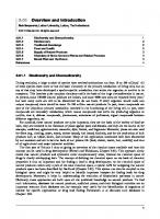
Vermisporin (41) is produced by the fungus Ophiobolus vermisporis [76]. Its structure was determined by chemical degradation to the derivative (42) which was studied by X-ray crystallography and provided the absolute configuration [77]. Vermisporin exhibits antimicrobial activity towards Bacteroides spp (0.25-2 |ig/ml), Clostridium perfringens (0.25-2 fig/ml) and methicillin-resistant Staphylococcus aureus (0.12-0.5 |Lig/ml). A metabolite of Ophiobolus rubellus produces the tetramic acid (43) that has been claimed to be an inhibitor of proline hydroxylase (IC5019|LiM) [78]. Three related tetramic acids have been reported from Chaetomium globosum. Two (44, 45) differ in the stereochemistry of the amino acid component, and the third is the methyl ester of 44 [79]. It is claimed that these compounds are chemokine receptor antagonists and can be used to treat HIV-1 infections.
126
44Ri = C02H;R2=OH 4 5 R i = OH;
R2 = C02F
An isolate of Streptomyces lydicus gave lydicamycin (46), a metabolite that showed activity against gram-positive bacteria, Bacillus subtilis (MIC kaempferol > luteolin. As for biflavones, the best radical scavengo* is amentoflavone, followed by bilobetin, ginkgetin, isoginkgetin, and sdadopitysin [137]. Recently, free radical scavenging activities of terpene-free EGb and quercetin w^e revealed by means of an in vitro electro-spin resonance assay [138]. Additionally, the in vivo experiments showed that terpene-free EGb inhibits cutaneous blood flux, whidi reflects the skin inflammatory level [138]. In regard to ginkgo terpenes, it has been revealed by means of electron paramagnetic resonance and U\7VIS spectroscopy that ginkgolides B, C, J and M, as well as bilobalide but not ginkgolide A, scavenge superoxide and hydroperoxyl radicals in dimethyl sulfoxide as an aprotic solvent [139]. Akiba et d. showed that EGb prevents the platelet aggregation induced by a combination of 100 f4M terr-butyl hydroperoxide and Fe^*. However, ginkgolides A, B and C, which are known to be PAF-antagonists, have no influence on this aggregation. Therefore, it was suggested that free radicals, but not FAF, might be involved in platelet aggregatk)n induced by oxidative stress [140]. Serotonin (5-HT) produces a rapid elevation of superoxide that stimulates the mitogenesis of bovine pulmonary artery smooth muscle ceUs (SMCs). EGb scavenges superoxide elevated by 5-HT, hence preventing 5-HT-induced mitogenesis on both SMCs and Chinese hamster lung fibroblasts. These results indicate that EGb inhibits the cellular transduction signaling process that leads to mitogenesis, as a result of its antioxidant activity [141]. In addition to radical scavenging properties, it has been reported that EGb reacts with nitric oxide (NO) in in vitro systems [136], and inhibits NO production induced by lipopolysaccharide plus intoferon-Y in maaophage cell Une RAW 264.7 [142]. Fre-treatment with oral administration of EGb reduced nitric oxide overproduction after transient brain ischemia in the MongoHan gerbil [143]. Further experiments showed that EGb inhibits NO production by attenuating the level of iNOS mRNA in a human endothelial cell line (ECV304) [144], also inhibits the activation of protein kinase C (PKC) induced by sodium nitroprusside (SNP), NO generator, and that its flavonoid constituents have protective properties against toxicity induced by SNP on cells of the hippocampus [145]. Recently, it was shown that ginkgolide A, ginkgolide B and bilobalide inhibit NO production in macrophages derived from a human monocytic cell line through attenuation of iNOS mRNA expression. However, these components have no effect on the eNOS-mediated NO production in endothelial ceUs [146].
181
Influences on the Neurotransmitters Numerous studies have demonstrated age-related changes in levels of neurotransmitters and their recq>tors in certain areas of the brain. There is a decrease in the levels of acetylcholine and in the numbers of muscarinic receptors and 6-adrenocqptors in the c^ebral cortex and hippocampus of the brain in patients suffering from Alzheimer's disease and in the brains of aging rodents, diaracterized behaviorally by a sevore impairment ia cognitive functions [147, 148, 149]. Numbers of 5-HT recq)tors and levels of dopamine and noradrcnalin and 5-HT have also shown age-related diminution [150, 151, 152], and are known to be involved in the regulation of mood [153]. Furthermore, it has been demonstrated that the activity of monoanune oxidase (NfAO), which r^ulates the brain concentrations of 5-HX norq)inephrine and other biogenic amines, inaeases with advancing age [154]. Hius, the inhibition of NfAO has been shown to produce antidepressant or anxiolytic responses in animal models and in man [155]. Brain Levels of Biogenic Monoamines Nforier-Teissier et d. [156] determined that administration of EGb alters the levels of catecholamines, indolamines and their metabolites in some brain areas of young rats and mice. Marked changes in the EGb-treated brain were found for norepinephrine, 5-HT, and its metabotite, 5-hydroxyindole-3-acetic add, whereas it was less effective for dopamine and its m^abolite 3,4-dihydroxy-phenylacetic add. EGb-induced changes depend on the route of administration (p. o. or L p.), dose and duration of treatment (acute or dironic). In old rats (26 months old), oral administration of EGb (10 mg/kg and 30 mg/kg, for 7 days) produces elevations of 5-HT in the frontal cortex, hippocampus, striatum and hypothalamus, and of dopamine levels in the hippocampus and hypothalamus compared with controls. On the other hand, EGb decreases the 5-HT level in the pons, and those of norepinephrine in the hippocampus and hypothalamus [157]. In this connection, Racagni et al, [158] showed that the O-methylated amine metaboUte of norepinephnne, normetanq)hrine, was markedly elevated (+500%) in the cerdjral cortex by du:onic oral administration of EGb (100 mg/kg, for 14 days), suggesting an increase of norq)inephrine turnover. In additbn, treatment with EGb (50 or 100 mg/kg/day, for 20 days) diminished the inareased plasma levels of epiDq)hrine, norepinephrine, and corticosterone induced by acute auditory stress in young and old rats [113]. GABA is the major inhibitory neurotransmitter in the CNS and acts to counter glutamateinduced exdtatk)n. Bilobalide (30 mg/kg/day, p.o., for 4 days) elevates GABA levels in the hippocampus and cerebral cortex in mice. These effects of bilobalide are due to a potentiation in glutamic add decarboxylase activity and an enhancement in the protein amount of 67 kDa glutamate decarboxylase. Furthermore, isoniazid and 4-O-methylpyridoxine, pot^t convulsants, induce reductk)ns in brain GABA levels, whereas bilobalide counteracts these effects. These results indicate that potentiation of GABAergic transmission induced by bilobalide might explain its anticonvulsant activity against isoniazid and 4-Omethylpyridoxine [159,160]. Monoamine Oxidase Activity White et al, [161] explored in rat brain mitodiondrial extracts the effect of EGb on MAO activity in vitro. MAOA and MAOB activities wore assayed using [^H]5-HT and [^*C]B-
182
phenetfaylamme as substrates, respectively. EGb inhibited both NfAOAand MAOB activities of rats and mice in vitro to similar extents. These results have suggested that the inhibition of MAO may be a mechanism underlying antidq)ressant or anxiolytk: responses of this extract obtained in animal models and man. Similar observations using a fluorimetric method were shown for EGb, but not for ginkgolide A and ginkgolide B [162]. Sloley et d. showed that kaempferol is a primal in vivo, but not ex vivo, rat brain MAQ-inhibitor in EGb [163]. Besides, the effects of long-term treatment with EGb (500 mg^g/day, for 7 months) on c^ebral MAO activity were investigated in mice subjected to a chronic mild stress. EGb induced reductions in basal MAO activity in 18-month-old mice. Hie age-related inaease in brain MAO activity was lower in die untreated mice subjected to stress and EGb potentiated this effect [164]. Recently, the effects of EGb on aggression woe investigated using MAO-A knockout nuce. EGb reduced their aggressive behavior in resident-intruder confrontations to levels seen in wild types, and decreased their [^H]ketanserin binding to 5-HT2A reoq)tors in the frontal cortex [165]. On the other hand. Fowler et d, recently measured MAO-A and MAO-B activities in the human brain using positron emission tomography and ["C]dorgyline and ["C]Ir dopamine > 5-HT [173]. Similar results were obtained by Ramassamy er d, [174]. These workers showed that EGb deaeased the specific uptakes of [^H]dopamine, [^H]5-HT and [^H]choline by synaptosomes prepared from tiie striatum of mice in a concentration-dependent manner. Tlie IQ^ values were 637 figfiol for [^H]dopamine uptake, 803 /ig/ml for [^H]5-HT uptake, >2000 //gAnl for [^H]choline uptake. However, they conduded that the inhibition of amine uptake caused by EGb appears to be non-specific, since EGb also prevents the specific binding of the dopamine uptake inhibitor [^H]GBR12783 to membranes prq)ared from striatum. EGb in vitro modifies the [^H]5-HT uptake by synaptosomes prepared firom nuce cerebral cortex in a biphasic manner. As mentbned above, the uptake of [^H]5-HT is inhibited by a high concentration of EGb [174]. On the other hand, low concentrations of EGb (4-16 //g/ml) A similar inaease was also obtained when significantly inaease [^H]5-HT uptake. synaptosomes were prepared from the cortk:es of mice treated orally with EGb, either acutely (100 mg/kg, 14 hours and 2 hours before death) or semi-dironically (2 x 100 mg/kg/day, for 4 days). Furthermore, such an inaement in the [^H]5-HT uptake is attributed to the flavonoid constituents of EGb [175], and may be associated with the mechanism of its antidq)ressant activity.
5'HT Receptors Adeaeased (22%) number of 5-HTi^ recq)tor binding sites labeled by [^H]8-hydroxy-2(di-/i-
184
propylainino)tetralin (pHJS-OH-DPAT), a S-HTi^ receptor agonist, in cerebral cortex membranes of Wistar rats was observed in aged (24 months old) rats as compared with young (4 months old) animals. Chronic treatment with EGb (5 mg/kg/day, for 21 days) did not alter the B ^ value in young rats, whereas it significantly inaeased it in aged rats (33%) [176]. On the other hand, Bolanos-Jim^iez et d, showed that chronic treatment with EGb (50 mg/kg/day, 14 days) produced a relatively small diminution in pHJS-OH-DPAT binding to hippocampal 5-HTi;^ receptors in 18-month-old rats [177]. There is at the moment no dear explanation for this disaepancy. An inhibitory effect of S-OH-DPAT on forskolin-stimulated adenylyl cyclase activity is observed in hippocampal membranes of the guinea pig and rat, and has been used as an index of the functional activities of S-HTj^ receptors [178]. Q)ld stress induces a reduction of the inhibitory effect of S-OH-DPAT in the hippocampus isolated from 18-month-old rats, although it has no influence on either the affinity or number of [^H]8-0H-DPAr binding sites. The administration of EGb (50 mg/kg p.o. for 14 days) prevents the cold stress-induced reduction in the inhibitory effect of 8-(Xl-DPAr on forskolin-stimulated adenylyl cyclase activity in old rats. These results indicate that EGb prevents the stress-induced desensitization of hippocampal 5-HTu^ receptors; thus, its effects might explain anti-stress and antidepressant properties of EGb [177].
NMDA Receptor Taylor [173] showed that EGb acts in vitro as an inhibitor of radioligand binding to the competitive and non-competitive sites of ^-methyl-i>-aspartate (NMDA) receptors. In addition, the most potent inhibition (K^ = 0.5 mg/ml) is observed for non-competitive NMDAsites labeled by ['H]MK-801. MPTP-Induced Dopaminergic Neurotoxicity It is known that l-methyl-4-phenyl-l,2,3,6-tetrahydropyridine (MPTP) selectively causes degeneration of the nigrostriatal dopaminergic neuronal pathway in several animal species, which is considered an animal model of Parkinson's disease [179]. As shown in Figure (2), MPTP administered systematically crosses the blood brain barrier, and is oxidized by MAOB into MPP*. This metaboUte is concentrated into dopaminergic neurons and consequently destroys such neurons by generation of ifree radicals [180, 181]. In mice implanted subcutaneously with osmotic minipumps releasing MPTP for 7 days (105 /^g/h/mouse) (approximately 100 mg/kg/day), a decrease in [^H]dopamine uptake by a synaptosomal fraction prepared from striatum was observed. This neurotoxic effect was prevented by the chronic injection of EGb (approximately 100 mg/kg/day in drinking tap water) for 17 days. Such a protective activity of EGb against MPTP neurotoxicity is unlikely to depend on inhibition of the MPTP uptake by dopamine neurons, since the concentration at which EGb prevents [^H]dopamine uptake is too high to reach the brain under in vivo experimental conditions [174, 182]. Therefore, it appears likely that a possible explanation lies in thefree-radicalscavenging property of EGb, which neutralizes free radicals generated from MPP* in the dopaminergic neurons. In this regard, another study showed that protective and curative treatments with EGb prevented the reduction of striatal dopamine levels induced by MPTP. MPTP (30 mg/kg/day.
185
BBB I i^Astrocyte
Nigrostriatal dopamine neuron
MPTP • ' - > MPTP injection
Fig. (2). Hypothesized mechanisms of neurotoxicity of MPTP. After injection of MPTP, its native fomi crosses the blood brain barrier (BBB) and is oxidized by monoamine oxidase B (MAO-B) into MPP^. This metabolite is transported and concentrated into nigrostriatal dopamine and exerts a neurotoxic effect
i.p. for 6 days) sigtdficantly reduced striatal dopamine levels in C57 mice. On the other hand, when C57 mice were pretreated with EGb (20, 50, 100 mg/kg/day, Lp.) for 7 days and then treated with the same extract 30 min before MPTP injection for 6 days, the neurotoxic effect of MPTP was antagonized in a dose-dependent manner. Moreover, in mice treated with EGb (50 mg/kg/day, Lp.) for 2 weeks after MPTP-lesion, the recovery of striatal dopamine levels was accelerated. MPTP is oxidized by MAO-B into MPP^ a positively charged species, whereas EGb, but not ginkgolides A and B, inhibits MAO-B activity. Therefore, another possible explanation for this protectk)n might lie in the inhibition of MAO activity caused by EGb to prevent the oxidization of MPTP into MPP* [162]. Effect of EGb on the Neuroendocrine System It is well known that neuro^docrine dianges with advancing age provide information about CNS functions [183]. The serum prolactin (PRL) level inaeases in old rats (26 months old) compared with young rats (3 months old). Administration of EGb (10 mg/kg/day, p.o., for 7 days) deaeases the blood PRL level, while greatly inaeasing adrenocorticotrophic hormone (ACTH) in old rats compared with age-matched controls. On the other hand, EGb, at a dose of 30 mg/kg, deaeases the serum level of growth hormone (GH) and ACTH in young rats compared with age-matdied controls [157]. Glucocorticoids are also essential for many aspects of normal brain development; however, hyperseaetion induces pathological states such as damage to the hippocampus [184]. Treatment with EGb (100 mg/kg/day, for 8 days) causes a 50% reduction of the plasma corticosterone level This reduction is probably due to deaeases in the number (B,^), mRNA
186
expression and protein levels of adrenal mitochondrial peripheral-type benzodiazepine receptors (PBR), whidi are a key element in the r^ulation of cholesterol transport Similar results have been observed in the chronic administration of ginkgolides A and B (2 mg/kg/day, i.p. for 8 days)[185]. Further study demonstrated that EGb and ginkgolide B also decreased PBR expression and cell proliferatk)n in the highly aggressive human breast cancer cell line MDA-231, which is ridi in PBR [186]. With regard to steroidogenesis, in addition to PBR, an ACTH-dependent process is also responsible for its regulatfon. Using cultured adrenocortical cells, Amri et al, [187] have shown that ex vivo treatment with EGb (100 mg/kg/day, p.o., for 8 days) and ginkgolide B (2 mg/kg/day, i p . , for 8 days) reduces ACTHstimulated cortkx)Sterone production by 50% and 80%, respectively. Moreover, Mardlhac et al. [188] showed that administration of EGb (50 or 100 mg/kg p.o., for 14 days) reduces basal cortkx)sterone seaetion and the subsequent inaease in oortkx>tropin-releasing hormone (CRH) and arginine vasopressin {/^/?) gene expression. Ginkgolide B (2 mg/kg/day i.p., for 14 days) reduces basal corticosterone seaetion without alteration in the subsequent CRH and /WF inorease. However, the stimulation of CRH gene expression by insulin-induced hypoglycemia is attenuated by ginkgolide B. These results indkrate that EGb and ginkgolide B are also able to affect the hypothalamic-pituitary-adrenal axis at the hypothalamic level Corticosteroids play a pivotal role for the development of behavbral sensitization to an^hetamine through the type II glucocorticoid receptor [189], Trovero et d. [190] showed that EGb reduces D-amphetamine (0.5 mg/kg, i.p., for 12-24 days)-induced behavioral sensitization as estimated by increasing values of locomotor activity, although EGb itself has no locomotor effect Furthermore, chronic administration of D-amphetamine reduces the density of [^H]dexamethasone binding sites of type II glucocorticoid recq)tors in the dentate gyrus and the CAl hippocampal regions of D-amphetamine-treated animals, indkating down-regulation of type II glucocorticoid receptors. On the other hand, pretreatment with EGb (50 or 100 mg/kg/day, p.o., for 20-24 days) prevents this down-regulation of type II glucocorticoid receptors, suggesting that EGb restores the density of these receptors. Taken together, these studies indicate that EGb is able to modukte both stress-induced and age-related behavioral sensitizatk>n by regulating alterations of the neuroendocrine system. Effect of EGb on the Phospholipid Metabolism Effects of EGb on ischemia-induced principal changes in the cerebral lipid metabolism were reviewed by Robin et al. [191]. Figure (3) shows diagram of cerebral lipid metabolism following ischemia. Pretreatment witii EGb could normalize the increased mitodiondrial lipid peroxide content and cytosolic lactase dehydrogoiase activity and the deaeased mitochondrial phospholipid contents and superoxide dismutase activity in rat brain after occlusion of common carotid arteries [192]. Rogue et d. [193] have shown that EGb is a potent inhibitor of phospholipase C (PKQ in vitro (ICJQ: 82 //g/ml). Furthermore, in isdiemic rats pretreated with a single injection of EGb (100 mg/kg, i.p.), a deaease in PKC activity is observed as compared with untreated ischemic animals. Impairment of membrane-bound Na,K-ArPase, whidi is responsible for maintaining and restoring membrane potential, and an increased level of malondialdehyde (MDA), which is a known as an index of lipid peroxidation, are seen aft^ unilateral focal cerebral ischemia in the mouse. Pretreatment witii EGb (100 mg/kg/day, p.o. for 10 days) preserves the Na,K-ArPase activity during cerebral ischemia and prev«its the kiaeased MDA levels caused by cerebral
187
Hydroperoxide Prostaglandin Leukotriene
Fig. (3). Diagram of cerebral lipid metabolism following ischemia. During ischemia, AIT-synthesis is disturbed by deficiencies in oxygen and glucose supplies. The subsequent energy failure stimulates phospholipase C (PLC) and Aj/Aj (PIAj/Ai), and thereby leads to formation of diacylglycerol (DAG), which is converted to free fatty adds (FFA) by Upasc, and leads to accumulation FFA and lysophospholipids. Among FFA, especially, the peroxidation of arachidonic add (AA) initiates a cascade leading to lq)oxygenase and cydooxygenase metabolites (prostaglandins and leukotrienes) and hydroperoxide, which are augmented during reperfusion following ischemia. Aoetylation of lyso-platelet-activating factor (lyso-PAF) leads to PAF, a mediator of inflammation.
ischemia [194]. Similar inhibitory effects of EGb on MDA production induced by hydrogen peroxide have been shown in erythrocyte membranes [195, 196]. Electroconvulsive shock (ECS), as well as ischemia, induces inaeases in free fatty add (FFA) and diacylglycerol (DAG) in the rat brain, probably due to the breakdown of membrane phospholipids through the activation of phospholipases (PLC, PLAj/Aj). EGb treatment (100 mg/kg/day, p.o. for 14 days) selectively decreases endogenous FFA levels and increases endogenous DAG levels in the hippocampus. Therefore, ECS-induced accumulation of FFAis prevented in the hippocampus of EGb-treated rats during clonic seizures (30 sec to 2 min after
188
ECS). Furthermore, the inaeased DAG levels induced by ECS are delayed by EGb treatment and the subsequent decrease in DAG levels is accelerated by EGb treatment in both the hippocampus and cortex [197]. Hypoxic or ischemic conditions led to an immediate release of free dioline via the breakdown of choline-containing phospholipids in rat hippocampus slices. Klein et d. [198] showed that bilobalide inhibited the hypoxia-induced dioline release in a dose-dependent manner both in vitro (ECjo*. 0.38 /^M) and ex vivo (2-20 mgAcg, p.o.). Asimilar reduction of dioline release was confirmed after administration of EGb (200 mg/kg, p.o.). Bilobalide also inhibits the ^-methyl-D-aspartate-induced, PLAj-dependent release of dioline from hippocampal phosphol4)ids both in vitro (10-100;^M) and in vivo (20 mg/kg, Lp.) [199]. Rabin et d. [200] investigated the effect of EGb on the rate of FFAreincorporation into brain phospholipids during reperfiision following ischemia in the gerbil brain by means of quantitative autoradiography and biodiemical analysis. Isdiemia-reperfusk>n selectively reincorporated arachidonic add (AA) into brain phospholipids. Pretreatmait with EGb (50 or 150 mg/kg/day, for 14 days) accelerated AA reincorporation following isdiemia, suggesting that EGb ameliorates the neurotoxic reaction caused by prolonged ^posure of the brain to high concentrations of AAand its metabolites, and stabilizes the membrane bilayer. Taken together, these results showed that EGb can prevent isdiemia-induced Na,K-ArPase injury, and suppress hypoxia- and ECS-induced membrane phospholipid breakdown in the brain, and bilobalide might be associated with its protective action. In addition, EGb reduces AA-induced neuronal damage as a consequence of the increase in reincorporation of A A Therefore, these medianisms might provide a possible explanation for neuroprotective properties of EGb and bilobalide against oxidative damage. Anti-PAF Platelet-activating factor (PAF), a potent phospholipid inflammatory mediator, enhances glutamatergic exdtatory synaptic transmission in the h^>pocampus [201]. Braquet et d, [202, 203] showed that ginkgolides, mainly ginkgolide B, act as potent antagonists of PAF in various cell types. Pretreatment of ginkgolides (10 mg/kg/day, p.o., for 7 days) ameliorated behavioral impairments assessed by the MacGraw stroke index, and inq)roved the mitodiondrial respiration evaluated by the respiratory control ratio (RCR) following c^ebral ischemia obtained by bilat^al ligature of the common carotid arteries in Mongolian g^bils [204]. The order of these effects in cerebral isdiemia was ginkgolide B > ginkgolide A > ginkgolide C > ginkgolide J, and was correlated with that of their PAF antagonistic properties described by Braquet et d, [203]. Consistent with this finding, liu et d. showed that both preand post-hypoxic treatment with ginkgolide B (25 mg/kg/dose, two serial doses) decreased the inddence of cerebral infarction in hypoxic ischemic brain injury of immature rats [205]. Moreover, Akisu et d, [206] found that endogenous PAF concentrations in brain tissue markedly inaeased in the hypoxic-ischemic brain in immature rats. Pretreatment with EGb reduces endogenous PAF concentrations in cerebral hypoxic-ischemic brain injury of immature rats as compared with controls. These results indicate that the PAF-antagonistic activity of the ginkgolides contributes to the neuroprotective effect against brain injury associated with an episode of the post-isdiemic phase. This idea was supported further by the observation that in primary neuronal cultures isolated from onbryonic rat cerebral cortex, ginkgolide B demonstrates protective effects against glutamate neurotoxidty involving PAF [207].
189
It has been demonstrated that ginkgolide B prevents bng-tenn potentiation (LIP) induced by PAF in the h^}pocampus [208] and in the voitral part of the medial vestibular nudei [209]. These results suggest that PAF might act as a retrograde messenger in ITP, which activates the presynaptic mechanisms enhancing the glutamate release. Izqui^do et d, found that pre- or immediate post-training intrahippocampai or intraamygdala infusion of the PAF antagonist, ginkgolide B, produces amnesia for avoidance tasks. These results support the idea that PAF may play a role in memory formatfon [210]. Amine Uptake and Membrane Fluidity Protonged incubatbn of synaptosomes prqiared from the striatum in the presence of ascorbk: add (10^ M) decreases the ability of synaptosomes to take up [^H]dopamiae. Similar inhibition is also observed in the ability of cortical synaptosomes to take up [^H]5-HT. Furthermore, this decrease is potaitiated by addition of Fe^* tons. EGb (4-16 /igAni), in particular its flavonoid fraction, prevents the reduction in the ability of synaptosomes to take up either 5-HT or dopamine [211]. Moreover, EGb (10 //g/ml) prevents a decrease in binding of [^H]GBR12783, the dopamine uptake inhibitor, to dopamine recq)tors in the presence of the combination of ascorbic add/Fe^* ions. These results suggest that EGb-induced prevention of the impainnent of the ability of synaptosomes to take up [^H]amine by the ascorbic add/Fe^* ions is related its inhibitory properties in generation of free radicals. However, EGb does not modify the inaeased [^H]dopamine release that is triggered by high potassium concentrations in the presence of the combination of ascorbate/Fe^*, suggesting that the vesicular exocytotic dopamine release does not seem to depend upon peroxidation [212]. Furthermore, Ramassamy et d. [213] showed that the combination of ascorbic acid/Fe^* ions could deaease synaptosomal membrane fluidity measured by fluorescence polarization using 1,6-diphenyl 1,3,5-hexatriene in a concentration-dq)endent manner. Free radical generation by ascorbic add/Fe^* results in a decrease of membrane fluidity through the peroxidation of neuronal membrane Upids. These membrane altCTations were prevented by either EGb (2-16 ;-Methylglabridin (123) Hispaglabridin A (3'-prenylglabridin) Glabrol (25) 3-Hydroxyglabrol (26)* Glabrone (DMP;4',3']-2',7-dihydroxyisoflavone) Medicarpin (3-hydroxy-9-niethoxypterocarpan) Shinpterocarpin (DMP;3,4]-9-hydroxypterocarpan) Euchrenone as (DMP;4',3']-7-hydroxy-8-prenylflavanone) Glyinflanin K (2DMP;7,8, ;2',3']-isoflavan) Glyinflanin G (2DMP;4,5, ;4',3']-2',3-dihydroxychalcone) Kanzonol U (DMP;2',3']-4',6-dihydroxy-2-arylbenzofuran] Kanzonol V (DMP;2',3']-4',6-dihydroxy5-prenyl-2-arylbenzofuran) Kanzonol W (DMP;7,8]-2',4'-dihydroxy-3-arylcoumarin) Kanzonol X (3',8-diprenyl-2',4',7-trihydroxyisoflavan) Kanzonol Y (3,5'-diprenyl-a,2',4,4'-tetrahydroxy-dihydrochalcone) Kanzonol Z (DMP;7,8]-3,4'-dihydroxy-3'-prenylflavanone) 3-Hydroxyparatocarpin C**
+-H-¥ +++ ++ ++ +++ + ++ ++ + ++ +++ ++ ++ -
European [50,51 ] -
+++ ++++ ++ ++-H+++ +++ ++ ++++ +++ ++ ++ ++ ++ +++ +++ +++ +++ +++ +++
Yields from dried licorice roots: -H-i-H=more than 0.01%; +-H-=between 0.01 and 0.001%; ++=0.001-0.0001%; +=1-0.1 ppm. • The compound was obtained from the stolons. ° 2,2-dimethylpyrano[b=DMP. ** Tentative name used here (DMP;4,5]-3'-prenyl-2',3,4'-trihydroxychalcone).
The difference of the substituents at C-5 is expected that European and Chinese licorices exhibit different actions in therapeutically use. For example, 5,6-disubstituted isoflavans do not showed a potency of
207
anti-HIV activity in vitro, but two isoflavans with no substituent at both 5- and 6-positions obtained from Erythrina lysistemon (Leguminosae) have the activity as described later [52]. As described the above, moraceous plants and Glycyrrhiza species are rich sources of isoprenylated phenolic compounds. The phenolic nuclei having the isoprenoid-derived substituents, e.g., simple isoprene or a monoterpenoid, vary over a wide range from a simple phenol to complicated ones. Some of the moraceous plants studied by our group have been used as traditional herbal medicines in the native countries. It is interesting to clarify the relationship between the usage and biological activities of the isoprenylated phenolic compounds. So we studied some of the biological activities of these compounds. This article reviews the biological activities of the isoprenylated flavonoids isolated from the moraceous plants and isoprenoid-substituted phenols (flavonoids, xanthones, dihydrostilbenes, and dihydrophenanthrenes) from Glycyrrhiza species by our group and other several groups. 11. HYPOTENSIVE ACTIVITY OF ISOPRENYLATED FLAVONOIDS FROM THE ROOT BARK OF MORUS SPECIES The first report for the hypotensive effect of the mulberry tree was presented by Fukutome in 1938, who asserted that oral administration of the hot water extract of the mulberry tree showed a remarkable hypotensive effect in rabbits [53]. Ohishi reported the hypotensive effect of the ethanol extract of mulberry root bark [54]. Suzuki and Sakuma reported that the hypotensive activity seemed to be due to phenolic substances, and that the effect disappeared on acetylation [55]. Later, Katayanagi, et aL reported that the ether extract of the root bark gives to rabbit (6 mg/kg, i.v.) showed a marked hypotensive effect and that the active constituents seemed to be a mixture of unstable phenolic compounds [56]. Tanemura ascribed the activity of mulberry root bark to acetylcholine and its analogous presumably contained in the alcohol soluble fraction, and that the hypotensive constituents produced a yellowish-brown precipitate on treatment with Dragendorff reagent [57]. Yamatake, et al reported that n-butanol- and water-soluble fractions of mulberry root bark had similar effect except for those on the cardiovascular system. Both fractions showed cathartic, analgesic, diuretic, antitussive, anti-edema, sedative, anticonvulsant, and hypotensive actions in mice, rats, guinea pigs and dogs [7]. On the beginning of our study of mulberry tree, the hypotensive constituents had not been identified. In view of the reports, we assumed that the hypotensive compounds of the plant would be a mixture of unstable
208
phenolic compounds and therefore undertook a study of the phenolic constituents of the root bark of the cultivated mulberry tree. The root bark of the cultivated mulberry tree was extracted successively with n-hexane, benzene, and methanol. The methanol extract, 1-20 mg, showed a dose-dependent decrease in arterial blood pressure in pentobarbital-anesthetized rabbit, Fig. (4). The extract was fractionated successively by silica gel column chromatography (C.C.), polyamide C.C, silica gel preparative (p.) TLC, and p. HPLC leading to isolated of kuwanons G (1, 0.2% yield) [9] and H (2, 0.13% yield) [10]. The root bark of Moms alba n-Hexane Residue Benzene
Extract
Residue I Methanol Residue
Extract -T extract Ethyl acetate soluble portion I C.C, p. TLC, p. HPLC
C.C, p. TLC Morusin (3), kuwanons C (42), D, E (43), F, oxydihydromorusin (46), mulberroftiran A (47)
Kbwanons G (1), H (2), L (44), M (35), albanol B (97) mulberrofurans C (28), F (29), and G (30) Fig. (4). Isolation procedure of flavonoids from the root bark of Morus alba.
mmHg
PN n > < l i < M H H H H M M ( H i t i i kuwanon G 1 mg/kg i.v. mmHg
tsmmm |ioo 50
iilSliSirJ'^^
kuwanon H I mg/kg i.v.
'
10 s
Fig. (5). Effects of kuwanon G (1) and kuwanon H (2) on blood pressure. Electrocardiogram (ECG), phrenic nerve discharge (PN), and electroencephalogram (EEG) in a gallamine-immobilized rabbit.
209
Both compounds (1 and 2) almost equally caused decrease of arterial blood pressure in a dose dependent and reversible manner at the dose of between 0.1 and 3 mg/kg, i.v. in pentobarbital-anesthetized as well as in un-anesthetized, gallamine-immobilized rabbits. Fig. (5), [58]. These hypotensive actions of kuwanons G (1) and H (2) were not modified by atropine or eserine, suggesting the non-cholinergic nature origin. Furthermore, neither propranol nor diphenhydramine affected their actions on the arterial blood pressure. Although they produced no significant change in both electrocardiogram (ECG) and respiration when administered intravenously in rabbits. The hypotensive effects of kuwanon G (1) and H (2) did not accompany with heart rate change [58]. In pentobarbital-anesthetized pithed dogs, kuwanons G (1) and H (2) also significantly decrees of femoral arterial blood pressure. These effects suggested that mechanism of hypotensive effects of kuwanons G (1) and H (2) mediated through peripheral system. Mulberrofurans C (28) [59], F (29) [60], and G (30) [60], Fig. (6), were also isolated as hypotensive components from the mulberry tree. Mulberrofuran C (28) is considered to be formed by a Diels-Alder type of enzymatic reaction process of a chalcone derivative and dehydromoracin C (31) or its equivalent. Furthermore, mulberrofurans F (29) and G (30) seems to be Diels-Alder type adducts derived from chalcomoracin (32) and mulberrofuran C (28), respectively, by the intra-molecular ketalization reaction of the carbonyl group with the two adjoining hydroxyl groups, 3'(5')-OH and 2"-0H. Intravenous injection of mulberrofuran C (28, 1 mg/kg) produced a significant hypotension (37 mmHg fall) in rabbit (male, 3.3 kg) anesthetized with pentabarbital sodium (30 mg/kg). Single intravenous injection of mulberrofurans F (29) and G (30) (both 0.1 mg/kg) caused a marked depressor effect in rabbit by 26 mm Hg and 16 mm Hg, respectively. On the other hand, in Japan, "Sang-Bai-Pi" (the root bark of Chinese mulberry tree) imported from China has been used as an herbal medicine, hence a study of the components of this crude drug purchased in the Japanese market was undertaken. Its phenolic components are different from those of Japanese mulberry tree. For example, morusin (3) and kuwanon G (1) are the main phenolic components of Japanese mulberry tree, in the case of "Sang-Bai-Pi", these components are minor ones, while sanggenons A (4) [16], C (5) [17], and D (33) [61] are the main components [24]. Sanggenons C (5) and D (33) showed the hypotensive effects as follows: Sanggenon C (5) caused transient decrease in arterial blood pressure at the doses of 1 mg/kg in pentobarbital-anesthetized rabbit by 15 mm Hg, while at the doses of 5 mg/kg the compound (5) caused a transient decrease by 100 mm Hg, which continued for more
210
mulberrofuran C (28): R = H chalcomoracin (32): R = CH2CH=CMe2
mulberrofuran F (29): R = CH2CH=CMe2 mulberrofuran G (30): R = H
kuwanon E (43) sanggenon B (45)
Fig. (6).
Structures of flavonoids (28 - 44) from moraceous plants.
211
than one hour by 15 mm Hg [17,62]. Sanggenon D (33) caused a transient decrease at the dose of 1 mg/kg in pentobarbital and urethane anesthetized male Wister strain rat by 35 mm Hg, while the compound (33) caused a decrease by 80 mmHg at the doses of 1 mg^g in spontaneously hypertensive rat [61,63]. III. ANTI-TUMOR PROMOTING ACTIVITY OF MORUSIN (3) Cancer chemoprevention is the most important subjects in cancer research at present and is a new medical strategy for cancer prevention, which was established by recent understanding of molecular multistage carcinogenesis in humans. To find nontoxic cancer preventive agents, Fujiki and his coworker studied natural products derived from marine and plant sources [64,65]. In 1987, Yoshizawa, et aL reported that (-)epigallocatechin gallate (EGCG), which is a main constituent of green tea, inhibited tumor promotion by teleocidin in mouse skin [66]. In 1988, Fujita, et aL reported the inhibitory effect of EGCG on carcinogenesis with 7V-ethyl-A^-nitro-A^-nitrosoguanidine in mouse duodenum [67]. On the other hand, in the course of our examination the constituents of the Morus root bark, we found the following novel photo-oxidative cyclization. When a solution of morusin (3) in chloroform (CHCI3) was irradiated using high-pressure mercury lamp, morusin hydroperoxide (34), Fig. (6), was obtained in ca, 80% yield [68]. The reaction did not occur in the dark and was depend on the solvent; the reaction occurred in low polar or nonpolar solvent such as CHCI3 and benzene, but not in protic solvent. The reaction mechanism was suggested as follows [69]: morusin (3) in the ground state interacts with an oxygen molecular to form a contact charge transfer complex [3 O2] (CCTC). On irradiation, the CCTC gives an excited charge transfer state that presumably leads to reactive species such as free radicals as described in Fig. (7). Recently, the proof of presence of the CCTC was provided by laser desorption/ionization time-of-flight mass spectrometry of 3 [70]. The hydroperoxide (34) was also obtained with the oxidation of morusin (3) with singlet oxygen or radical initiator [71]. HO^^s^^OH hv 34 •OOH
Fig. (7). Reaction mechanism of photo-oxidative cyclization of morusin (3).
212
This photoreaction and the relative reaction of morusin (3) along with the anti-tumor promoting activity of EGCG encouraged us to examine the anti-tumor promoting activities of a series of isoprenylated flavonoids isolated from Morus species. First we examined the inhibition against three biochemical effects; the specific binding of ^H-12-O-tetradecanolylphorbol-13-acetate (TPA) to mouse particulate fraction, the activation of Ca^'^-activated phospholipid-dependent protein kinase (protein kinase C) with teleocidin, and induction of ornithine decarboxylase (ODC) with teleocidin in mouse skin [72]. Interestingly, of the eight isoprenylated flavonoids, morusin (3), kuwanons G (1) and M (35), mulberroforan G (30), and sanggenon D (33) gave similar results in these biochemical tests as described in Table 2. Table 2.
Effects oi Morus flavonoids on biological and biochemical activities Inhibiting of specific [^H]TPA binding (ED50 jimol/L)
57 99 100 85 34 62 48 60
Morusin (3) Kuwanon G (1) Kuwanon H (2) Kuwanon M (35) Mulberrofuran G (30) Sanggenon A (4) Sanggenon C (5) Sanggenon D (33)
Inhibition of activation of protein kinase C (ED50 fimol/L)
80 40 80 22 46 80 46 42
Inhibition of ODC induction
(%) 43 34 -35 25 10 -62 -17 17
100
^
o
o
Concentration (mol/L) of morusin (3) Fig. (8). Effects of morusin (3) on specific binding of [^H]TPA to a mouse skin particulate fi-action. Various concentrations of morusin (•) or TPA (o) were incubated with a particulate fi-action of mouse skin in the presence of 4 nmol/L [^H]TPA for 2 h at 4°C, and the assay mixture was filtered on glass filter membrane with acetone cooled in a dry ice-ethanol bath. Non-specific bindings were measured in the presence of 500-fold excess of unlabelled TPA.
213
Of these five compounds, morusin (3) is the least toxic and can be isolated as one of the main phenolic compounds from the root bark. The more detailed data for the above these biochemical tests of morusin (3) were as follows [73]. As shown in Fig. (8), morusin (3) caused dose-dependent inhibition of the specific binding pHJTPA to a mouse skin particulate fraction. The concentration of morusin (3) for 50% inhibition (ED50) was 57 |amol/L, whereas that of unlabelled TPA was 4 nmol/L. As morusin (3) was assumed to interact with the phorbol ester receptor, we examined whether it inhibited the activation of protein kinase C by teleocidin in vitro [73]. Fig. (9) shows that morusin (3) inhibited the phosphorylation of histone type III-S by protein kinase C dose-dependent and that 80 |imol/L morusin caused 50% inhibition. 100
o
VA
50
a
0.
VA
10-
\o-
10-*
Concentration (mol/L) of morusin (3) Fig. (9). Inhibition by morusin (3) of activation of protein kinase C by teleocidin in vitro. The assay mixture (0.25 mL) contained 20 jimol/L CaCh, 7.5 |ag of phosphatidylserine, 2.3 (^mol/L teleocidin, and various concentrations of morusin (3) with 0.05 units of partially purified enzyme. Enzyme activity was measured as the incorporation of ^^P from [7-^^P]ATP into histone type III-S during incubation for 3 min. at 30^.
Furthermore, we examined the inhibition of the induction of ODC induction by teleocidin in mouse skin. Application of 11.4 nmol morusin (3) caused 43% inhibition of the induction of ODC by 11.4 nmol teleocidin [73]. From the results of these three tests, morusin (3) might inhibit the tumor-promoting activity of teleocidin on mouse skin. As shown in Figs. (10) and (11), the percentage of tumor bearing mice in the group treated with 7,12-dimethylbenz[a]anthracene (DMBA) plus teleocidin reached 100% by week 15, o in Fig. (10). In contrast, the onset of tumor formation was delayed 5 weeks by treatment with morusin (3), • in Fig. (10), and the percentage of tumor-bearing mice in the group treated with DMBA plus teleocidin and morusin (3) was 60% at week 20. The average number of tumors per mouse in week 20 was also reduced from 5.3, o in Fig. (11), to 1.1, • in Fig. (11), by morusin (3) treatment.
214
On the other hand, morusin (3) itself did not show a tumor promoting activity on mouse skin, x in Figs. (10) and (11). From these results, morusin (3) is an anti-tumor promoter judging from its ability to inhibit the short-term effects induced by tumor promoters.
100
to
10
20
Weeks of promotion
10
20
Weeks of promotion
Figs. (10) and (11). Inhibition by morusin (3) of tumor promotion by teleocidin in a two-stage carcinogenesis experiment on mouse skin. Inhibition was achieved by a single application of 100 ^g of DMBA, and teleocidin (2.5 }ig) and morusin (1 mg) were applied twice a week throughout the experiments.
As mentioned the above, morusin (3), kuwanon G (1), kuwanon M (35), mulberroforan G (30), and sanggenon D (33) showed inhibitory effects in the three biochemical tests. The anti-tumor promoting activities of later four flavonoids with one or two isoprenoid groups have not been tested in a two-stage carcinogenesis experiments, due to limitations of their amounts available, but their inhibitory potencies to the three biochemical tests were almost similar to that of morusin (3). Furthermore, the twelve isoprenylated flavonoids from the moraceous plants and two flavonol glycosides (48 and 49) from Epimedium species (Berberidacaceae) [74] along with quercetin (50) were tested for inhibitory effects on carcinogenesis by a test for inhibition of specific binding of [^H]TPA to a mouse skin particulate fraction. While the other biochemical tests and the inhibition of tumor promotion of teleocidin in a two-stage carcinogenesis experiment have not been carried out, due to limitation in their amounts available, some of isoprenylated flavonoids from the moraceous plants showed the similar inhibitory potencies to those of morusin (3) and the related compounds, Figs. (6) and (12), as shown in Table 3. On the other hand, EGCG and green tea extract are acknowledged cancer-preventive agents in Japan [75,76]. Natural products with antitumor promotion activity isolated from foodstuff and medicinal plants have been summarized by Konoshima and his co-worker and Akihisa and
215
his co-worker [77,78]. Considering these results as well as the results of biochemical tests and anti-tumor promoting activity of the isoprenylated flavonoids from the moraceous plants in a two-stage carcinogenesis experiment with teleocidin, the isoprenylated poly-phenolic compound seems to be interesting compounds for finding cancer preventive agents and the more detailed experiments should be carried out. Table 3.
Effects of the isoprenylated flavonoids on inhibition of specific [^H]TPA binding (ID50, ^mol/L)
Kazinol C (36) Kazinol E (37) Kazinol F (38) Kazinol J (39) Kazinol M (40) Kazinol N (41) Kuwanon C (42) Kuwanon E (43)
Kuwanon L (44) Sanggenon B(45) Oxydihydromorusin (46) Mulberroftiran A (47) Ikarisoside A (48) Ikarisoside B (49) Quercetin (50)
80 70 98 90 100 >100 80 83
80 95 95 >100 >100 >100 >100
OMe oxydihydromorusin (46) mulberrofuran A (47)
ikarisoside A (48): R = Rha ikarisoside B (49): R = Glu(1 ^ 2)Rha
OCH3 OH
O
quercetin (50): Ri = OH,R2=R3 = H cirsilioi (51): Ri = H, R2 = 0Me, R3 = Me
antiarone L (57) artonin H (56)
Fig. (12). Structures of flavonoids (46 - 57) from moraceous plants, Epimedium species, and test reagents (50 and 51).
IV. INHIBITION OF ARTONIN E (7) AND RELATED COMPOUNDS ON 5-LIPOXYGENASE Previously, we reported the effects of Morus flavonoids on arachidonate metabolism in rat platelet homogenates, such as inhibition of 12-hydroxy5,8,10-heptadecatrienoic acid (HHT), thromboxane B2, and 12-hydroxy5,8,10,14-eicosatetraenoic acid (12-HETE) [79,80]. As described in the
216
introduction, Artocarpus plants (Moraceae) have been used as traditional medicine in Indonesia for swelling and malarial fever. This usage seems to be expecting for effect of anti inflammation. As leukotrienes are known to be chemical mediators of anaphylaxis and inflammation, a number of compounds have been studied and developed as selective inhibitors of 5-lipoxygenase, the enzyme initiating leukotriene biosynthesis from arachidonic acid. So the inhibitory effect of the Artocarpus flavonoids against arachidonate 5-lipoxygenase was examined [81]. Yamamoto, et aL screened various flavonoids, and found that cirsiliol (51), Fig. (12), potently inhibited 5-lipoxygenase and proposed two structural factors of the flavonoids for the specific inhibitory activity, one is catechol type of the B ring and the other is the presence of an alkyl-like side chain at the C-3 position [82,83]. We had interesting for the inhibitory effects of a series of Artocarpus flavones on the 5-lipoxygenase activity. Seven Artocarpus flavonoids and morusin (3) were tested for their inhibitory actions on arachidonate-5-lipoxygenase purified from porcine leukocyte [84]. As shown in Fig. (13), the IC50 values varied depending on the structural modification of the compound. The compounds having three hydroxyl groups at positions 2\ 4\ and 5' on the B ring (compounds 7, 8, 52 and 55) were more potent inhibitors. Thus, the vicinal diol partial structure was important for 5-lipoxygenase inhibition.
OH o heterophyllin (52)
OH
0
artonin A (54)
cycloheterophyllin (53)
Inhibitory effects (IC50 ± SD, N=3, ^imol/L) on arachidonate 5-lipoxygenase activity
OH
0
artonin B (55)
Morusin (3) Artonin E (7) Artobiloxanthone (8) j Cycloartobiloxanthone (9) Heterophyllin (52) Cycloheterophyllin (53) Artonin A (54) Artonin B (55)
2.9 ± 0.4 0.36 db 0.03 0.55 db 0.20 1.3 ±0.2 0.73 ±0.21 1.6±1.0 4.3 ± 0.5 1.0 ±0.1
1
'
Fig. (13). The inhibitory effect (IC50 ± SD) on arachidonate 5-lipoxygenase activity.
As shown in Fig. (14), 5-lipoxygenase was inhibited depending on the concentration of artonin E (7), which gave the lowest IC50 (0.36 |Limol/L) of all the eight compounds. On the other hand, morusin (3), which
217
lacked the 5'-hydroxyl group of artonin E (7), was a less potent 5lipoxygenase inhibitor (IC5o=2.9 |Limol/L). Artonin E (7) was significantly more potent than cirsiliol (51, Fig. (12), IC5o=1.3 |Limol/L), which was reported as a 5-lipoxygenase inhibitor. This finding was consistent with the report that the inhibitory activity of cirsiliol (51) with 5-lipoxygenase was enhanced by introducing a lipophilic alkyl group at the C-3 position of theflavoneskeleton. Inhibitory actions of artonin E (7) and morusin (3) on other mammalian arachidonate oxygenases were examined. Artonin E (7) inhibited two 12-lipoxygenase from porcine leukocytes and human platelets, 15-lipoxygenase from rabbit reticulocytes, and fatty acid cyclooxygenase from bovine vesicular glands (IC5o=2.3, 11, 5.2, and 2.5 |amol/L, respectively). However, IC50 values for these oxygenases were higher by one order of magnitude than that for 5-lipoxygenase. Morusin (3) also inhibited these enzymes (except for human platelet 12lipoxygenase) with IC50 values of micro molar order as follows: two 12lipoxygenase from porcine leukocytes and human platelets, 15lipoxygenase from rabbit reticulocytes, and fatty acid cyclooxygenase from bovine vesicular glands; IC5o=3.4, > 30, 3.3 and 1.6 |imol/l, respectively. These results indicated that artonin E (7) was a relatively specific inhibitor for 5-lipoxygenase. Thus, which the selectivity for 5-lipoxygenase was not observed with morusin (3). Significant differences of IC50 values of artonin E (7) and morusin (3) between porcine leukocyte 12-lipoxygenase and the human platelet 12-lipoxygenase should be noted since the leukocyte and platelet 12-lipoxygenase were distinct both catalytically and immunologically.
Concentration (|imol/L) Fig. (14). Dose-dependent inhibition of 5-lipoxygenase by artonin E (7, • ) , morusin (3, o), and cirsiliol (51, A).
V. INHIBITION OF ARTONIN E (7) AND RELATED COMPOUNDS ON MOUSE TNF-a RELEASE AND THEIR CYTOTOXIC ACTIVITIES
218
As described in Chapter III, morusin (3) has been found to be anti-tumor promoter in a two-stage carcinogenesis experiment with teleocidin. Considering the similarity of the structures between morusin (3) and artonin E (7), artonin E (7) was expected to be an anti-tumor promoter. Furthermore we found a novel photo-oxidative cyclization of artonin E (7) as follow: photo-reaction of artonin E (7) in CHCI3 containing 4% ethanol solution with high-pressure mercury lamp produced artobiloxanthone (8) and cycloartobiloxanthone (9), and the treatment of artonin E (7) with radical reagent (2,2-diphenyl-l-picrylhydrazyl: DPPH) resulted in the same products, Fig. (15), [84].
(±)-artobiloxanthone (8)
artonin E (7)
hv, 24 h. CHCI3 DPPH, 24 h, CHCI3 (in the dark)
Fig. (15).
8
9
34% 70%
3% 4%
OH 0 (±) -cycloartobitoxanthone (9)
Photoreaction of artonin E (7) and the reaction with radical reagent.
As described in Chapter III, we have reported the photo-oxidative cyclization on morusin (3). These results suggested that the photo-
OH 0 (±) -cycloartobiloxanthone (9)
OH 0 (±)-artobiloxanthone (8)
Fig. (16). Plausible mechanism for the formation of artobiloxanthone (8) and cycloartobiloxanthone (9) from artonin E (7).
219
oxidative cyciization of artonin E (7) may proceed through phenol oxidation via the semiquinone radicals described in Fig. (16). This chemical reactivity and the similarity of the structures between morusin (3) and artonin E (7) encourage us to examine the anti-tumor promoting activity of artonin E (7). Recently, Fujiki, et al. proposed a new tumor promotion mechanism applicable to human cancer development on the basis of experiment with okadaic acid. They described that tumor necrosis factor-a (TNF-a) induced by okadaic acid acts as a mediator of human carcinogenesis [65]. As briefly summarized in Fig. (17), okadaic acid inhibits the action of protein phosphatase type 1 and 2A, resulting in the accumulation of phosphorylated protein. Fujiki's group has shown that TNF-a acts as a timior promoter in BALB/3T3 cell transformation in vitro. The results of the studies on the okadaic acid class tumor promoters suggest that inflammatory stimuli or chemical tumor promoters induce TNF-a release from target tissues, and TNF-a gene expression in the initiated cells. This released TNF-a acts as a tumor promoter in the autocrine and paracrine system. According to the assumption that TNF-a is an endogenous tumor promoter associated with inflammatory potential, many historical puzzles of tumor promotion, such as its relationship to inflammation, can be solved. Based on this new tumor-promotion pathway, inhibition of TNF-a production leads to inhibition of tumor promotion. Furthermore, recent investigation has revealed that TNF-a is involved in various diseased, such as rheumatoid arthritis, Crohn's disease, multiple sclerosis, graft-versus-host disease, HIV, malaria, sepsis, and cachexia associated with cancer [85-90]. So, specific inhibitions of TNF-a production will almost certainly be effective not only in cancer prevention but also in the therapy and prevention of these other diseases.
—1 protein i j — ' phosphatase 1
okadac acid
^ phosphorylated proteins
t 1
{jene expression
- c-fos ojun
NF-KB
TNF-a - •
p
phosporylated proteins
~3
V
t ' _
ODC TNF-a
— •
_
Fig. (17). Mechanism of tumor promotion with okadaic acid.
Based on the above descriptions, we examined the inhibitory effect of the Artocarpus flavonoids on TNF-a release stimulated by okadaic acid using BALB/3T3 cells. This experiment was carried out in co-operation with Dr. Fujiki's group (Saitama Cancer Center Research Institute, Japan). All the compounds tested inhibit the TNF-a release stimulated by
220
okadaic acid at suitable lower concentration. This result suggests that several Artocarpus flavonoids act as anti-tumor promoter against to the okadaic acid type promotion. However, the detail mechanism is not clear at present, Fig. (17). The comparison of the inhibitory effects of the Artocarpus flavonoids against the TNF-a release (Table 4) and arachidonate 5-lipoxygenase, Fig. (13), was carried out. Artonin E (7) was the most potent inhibitor on both tests and the other compounds, artobiloxanthone (8) and heterophyllin (52), inhibited stronger than cycloartobiloxanthone (9), cycloheterophyllin (53), and morusin (3). The compounds showing stronger activity, all have three hydroxyl groups in the B ring. This characteristic feature might be important factor for both biological activities [91,92]. It is also noteworthy that the bioactivities of these flavonoids may reflect the use of Artocarpus species to the treatment for inflammation and malarial fever in Jamu medicines as is stated above. Table 4. Inhibitory effects (IC50, Mmol/L) of six flavonoids for the release of TNF-a from BALB/3T3 cells by treatment of okadaic acid Morusin (3) Artobiloxanthone (8) Heterophyllin (52)
1.76 0.94 0.48
Artonin E (7) Cycloartobiloxanthone (9) Cycloheterophyllin (53)
0.43 1.94 7.8
We also examined the cytotoxic activities of the Artocarpus flavonoids, artonins A (54), B (55), E (7), H (56), heterophyllin (52), and cycloheterophyllin (53), against cancer cells, mouse L-1210 and colon 38. All compounds tested showed the cytotoxic activities against both cancer cells (Table 5) [93]. Among them, cytotoxicity of heterophyllin (52), artonins B (55) and E (7) w^ere stronger than critical drug, l-(2-tetrahydrofuryl)-5-fluorouracil (TFFU). While we examined the cytotoxic activities of three dihydrochalcone derivatives isolated from Antiaris toxicaria (Moraceae), antiarones J (19), K (20), and L (57), against the two cancer cells [94]. All the compounds showed the weak cytotoxic activities against both cancer cells. Artonin E (7) also exhibited Table 5. Cytotoxic activities (IC50, |ig/mL) of Artocarpus and Antiaris flavonoids against L-12i0 and Colon 38 cells
Artonin A (54) Artonin B (55) Artonin E (7) Artonin H (56) Heterophyllin (52) ' Positive control.
L-1210
Colon 38
8.8 23 2.2 8.8 2.3
14.3
1.4 1.9 3.5 1.3
L-1210 Cycloheterophyllin (53) Antiarone J (19) Antiarone K (20) Antiarone L (57) TFFU*
Colon 38
4.7
4.6
77.0 81.3 80.4
70.4 46.3 >100
2.9
3.9
221
cytotoxic activities against human oral cells and MT4-cells as shown in Chapter VII (Table 7). VI.
BOMBESIN RECEPTOR ANTAGONISTS, KUWANONS G (1) AND H (2), ISOLATED FROM MORUS SPECIES
Bombesin and its mammalian counterparts, gastrin-releasing peptide (GRP) and neuromedin B (NMB), have been shown to have a wide range of physiological and pharmacological functions [95]. Ligand-binding and molecular cloning studies have revealed two pharmacologically distinct G-protein-coupled receptor subtypes for mammalian bombesinlike peptides; a GRP-preferring (GRP-R) and an NMB-preferring bombesin receptor (NMB-R) [96]. A series of observations indicates that the mammalian bombesin-like peptides may act autocrine growth factors in human small cell lung carcinoma (SCLC) and other cancers. First, many human SCLC cell lines have been shown to express bombesin-like peptides [97]. Second, peptide bombesin receptor antagonists or anti-bombesin antibodies inhibit SCLC cell growth in vitro and in vivo [98,99]. These data suggested that the bombesin receptor antagonists might be useful for the treatment of some kinds of SCLC and other cancers. Because most antagonists reported thus far are peptides except for CP-70,030 and CP-75,998 (first synthetic non-peptide antagonists) [100-102], so, Fujimoto's group (Shionogi Research Laboratories, Shionogi & Co. Ltd., Osaka, Japan) screened the four hundred plant extract samples to search for non-peptide bombesin receptor antagonists. The methanol extract of the underground part of cultivated mulberry tree, Morus bombycis, was found to potently inhibit [^^^I]GRP binding to Swiss 3T3 cells. Bioassay-directed fractionation led to the isolation of two known flavone derivatives, kuwanons G (1) and H (2), which were identified by direct comparison with the authentic samples [103]. The antagonistic profiles of kuwanons G (1) and H (2) were characterized from the following results [103]. Kuwanon H (2) inhibited specific binding of [^^^I]GRP to GRP-referring receptors in murine Swiss 3T3 fibroblasts with K{ value of 290±50 nmol/L, which is more potent than that of kuwanon G (1), K\ value=470±60 nmol/L. The Ki value of 2 was about one order of magnitude more potent than those of CP-70,030 and CP-75,998, but had no effect on endothelin-1 or neuropeptide Y binding. While kuwanon H (2) inhibited specific binding of [^^^I]bombesin to rat esophagus membranes, the Ki value was about one order of magnitude less potent, Ki value of 2=6,500±2,000, than that of [^^^I]GRP toSwiss 3T3 cells. While bombesin (10 ^ mol/L) increased intracellular Ca^"^ levels in Swiss 3T3 cells, kuwanon H (2, 500 nmol/L) attenuated the bombesin-
222
induced increase in cytosolic free Ca^"^ concentration ([Ca^"^]!) by 60%, but not bradykinin- or endothelin-1-induced increase in [Ca^"^]}, Fig. (18).
r\
r
808
215
t\
< \
f t V
BOM
t
t
S
BOM
t
BK
t
S
t
BK
A^ Y1 t t t t V
ET-1
S
ET-l
Fig. (18). Effect of kuwanon H (2) on agonist-induced increases in [Ca^^\ in Swiss 3T3 cells. Cells were stimulated by 10"* mol/L bombesin (BOM), 10"* mol/L endothelin-1 (ET) or 10"* mol/L bradykinin (BK). Kuwanon H (S, 500 nmol/L at the final concentration) or dimethyl sulfoxide (V) was added 1 min before stimulation.
In Swiss 3T3 cells, GRP stimulates ["^H]thymidine incorporation in a concentration-dependent manner. Kuwanon H (2) inhibited GRPinduced DNA synthesis in Swiss 3T3 cells. The IC50 value was around 100 nmol/L, close to its K, value for [^^^I]GRP binding to Swiss 3T3 cells, Fig. (19). Kuwanon H (2) demonstrated selectivity toward GRP, as concentration of 10"^ mol/L uninfluenced basal and 5% serum-induced [ HJthymidine incorporation. From above results, kuwanon H (2) appears to be a selective antagonist for GRP-R.
B o.
B o
35000
25000
15000
-log (2) mol/L Fig. (19). Dose-dependent effects of kuwanon H (2) on basal (o) and GRP (10" mol/L)-induced DNA syntheses in Swiss 3T3 cells (•). Values are the mean ± S.E. for four determinations.
As bombesin family peptides are thought to be autocrine growth factors for SCLC, the results described above suggested that kuwanon H
223
(2) might be useful against SCLC. Unfortunately, however, kuwanon H (2) had no effect on the growth of two human SCLC lines, Lu-134 and NCI-HI 28. At the time, kuwanon H (2) was the most potent of non-peptide bombesin receptor antagonists (NPBRA) that had been reported. Its affinity might be too low to determine whether the non-peptide antagonist is effective against human lung cancers. However, kuwanon H (2), and possibly kuwanon G (1) also, can serve as lead compounds for more rational drug design in the synthesis of more potent antagonists. Furthermore, these compounds may be useful tools on the study of GRP-R. Recently, it was reported that NPBRA, PD 176252, with high binding affinity which was developed via the application of a peptoid drug design strategy [104]. VIL
EFFECTS OF PHENOLS AGAINST BACILLUS SUBTILIS (M45) (REC-ASSAY), HUMAN ORAL CELLS, AND HIV-INFECTED MT-4 CELLS
Rec-assay was developed by Kada et al. for screenings chemical and enveloped mutagens. Recombination less mutant strain of Bacillus subtilis (M45) is more sensitive to the cell-killing action of chemical mutagens, e.g., mytomycin C, A^-nitroso-A/-methylurethane, etc., than the wild-type bacteria (HI7) [105]. This assay was also useful for prescreening of anticancer drugs, such as enediyne-family antibiotics [106]. For the constituents of plants, the assay was modified and used exclusively for the detection of anti-mutagen compounds [107]. Since the sensitivity of the rec-assay to chemicals having induction activity of DNA damage is higher than from other screening technique, such as Ames test, this method may be useful for pre-screening of anticancer agents in crude drugs. Furthermore, the antibacterial compounds against the wild-type strain (HI7) may be expected that these antibacterial compounds have another bioactive potency. We tried the application of the rec-assay (unmodified) for the detection of bioactive phenolic compounds obtained from Glycyrrhiza species [51], and spore rec-assay [108,109] was used for moraceous flavonoids as shown in Table 7. Sixty-nine Glycyrrhiza phenols out of a total 108 compounds showed inhibitory activity against the growth of both HI7 and M45 strains. Cytotoxic activities of these antibacterial compounds {Glycyrrhiza phenols and moraceous phenols) against human oral squamous cell carcinoma (HSC-2) and human T-lymphoblastoid cell line MT-4 cells were also shown in Table 7 [110-113] along with other biological activities reported until the middle of 2002. In the Table, relatively strong-cytotoxic compounds against HSC-2 (CC5o50 55 22 6 18 2 2 10 ND >100
-H-
±
12
10
Glabridin (23)
+++
±
13
4
Glabrol (25) Glycycoumarin Glycyrin Glycyrol (76) (neoglycyrol) Glyasperin A (77) Glyasperin B Glyasperin C (61) Glyasperin D (62) Glyasperin J Glyasperin K Glisoflavanone Glyinflanin A (glycyrdione A) Glyinflanin B Glyinflanin C (glycyrdione C) Hispaglabridin A (124) 3-Hydroxyglabrol (26) 3-Hydroxyparatocharpin C^ Isoderrone Isoglycyrol(117) Isoliquiritigenin (70)
4-+ -f+
±
18 32 14 100 13 11 14
-
-
AFE, FDC
ABM ABM, EBV, LAT ABE ABH, APA, EBV, ODD ALR, 5LG ABM ABH, ABM
ALR, ICO, 5LG, NKA, IC0,5LG,NKA ALR, IOC, 5LG, NKA AIA, IPP ABE, ABH, ABM, AFE, AMA, AOA, EAA ABE, ABH, ABM, AFE, AOA, EAA, EAB, ETA, ICO, IMI, PSO, SAE ABE ABC, ABE, AOA, CPH ABC, ABE, ABH ABC, 5LG
-
10 ND ND 46 ND
++ ++
-
31 19
27 8
+
-
14 31
>100 12
ABE, AOA, MOS ABE
-
38 ND 16 22
12 ND >100 14
ALA
+ -H-H-H-
++ + ++ ++
-H-
+ ++
-
++
-
± ±
++
ABM ABM, ABH
ABC, ALR ABE, ATP, EAA, MCC, MGA, TFV, UFI
229
Table 7 (continued) Kanzonol B (81) Kanzonol G Kanzonol H Kanzonol P Kanzonol R Kanzonol S Kanzonol U (glabrocoumarone A) Kanzonol V Kanzonol W Kanzonol X (tenuifolin B) Kanzonol Y Kumatakenin (73)
+ + + + ++
—
Licochalcone A (59) Licochalcone B (82) Licoflavonol Licoisoflavanone (66) Licoisoflavone A (phaseoluteone) Licoisoflavone B (67) Licoricidin (63) Licoricone (120) Licorisoflavan A Medicarpin 1-Methoxyphaseollidin (125) 1 -Methoxyficifolinol 3-(9-Methylgancaonin P 4'-(9-Methylglabridin (123) Naringenin Paratocarpin L (macarangaflavanone B) Pinocembrin 6-PrenyleriodictyoF (71) S-PrenyleriodictyoF (72) 6-Prenylnaringenin (90) Semilicoisoflavone B (68) Shinpterocarpin Sigmoidin A Sigmoidin B (99) Topazolin Wighteone (84) (erythrinin B)
±
-
ND ND 46 ND ND ND ND
ND ND >10 ND ND ND ND
-
ND ND ND
ND ND ND
—
ND 375
-H-
—
20
ND 51 TCD 15
-
-
4 22 72 55
16 13 40 21
43 8
7 15
45 14 45 28 11 ND ND ND 24
64 47 53 12 8 ND ND ND 14
105 ND 35 29 78 ND 20 43 19 20
>100 ND 60 32 22 ND 29 26 4 12
+
17 100 11 >100 11 ND ND
-hH-
±
100 >100 50 50 >50 >50 >100 12.5 12.5 12.5 12.5 12.5 25 25 >50 >50 12.5 12.5 6.25 6.25 >50 >50 >100 >100 0.05 0.025
ATCC 43526 >100 >100 50 25 >100 >100 50 50 >50 >50 >100 12.5 12.5 12.5 12.5 12.5 25 25 >50 >50 12.5 12.5 6.25 6.25 >50 >50 >100 >100 0.05 0.025
ZLM 1007
GP98
>100 >100 >100 >100 50 50 25 25 >100 >100 >100 >100 50 50 50 50 >50 >50 >50 >50 12.5 >100 12.5 12.5 12.5 25 12.5 12.5 12.5 12.5 12.5 12.5 25 25 12.5 12.5 >50 >50 >50 >50 12.5 6.25 6.25 6.25 6.25 6.25 3.13 6.25 >50 >50 >50 >50 >100 >100 >100 >100 0.20 0.05 0.025; 0.10
(cfii)'* (a) (b) (a) (b) (a) (b) (a) (b) (a) (b) (a) (b) (a) (b) (a) (b) (a) (b) (a) (b) (a) (b) (a) (b) (a) (b) (a) (b) (a) (b)
source
G. glabra G. glabra G. inflata G. inflata G. uralensis G. uralensis G. uralensis G. uralensis
* (a): 2x10^ colony forming units (=cfti), (b): 2x10^ cfii. ** Positive control; amoxicillin (=AMOX). ATCC 43504, ATCC 43526, and ZLM 1007 are CLAR-sensitive strains.
Next, we attempted to isolate further flavonoids exhibiting anti-^. pylori activity from the extract of G. uralensis. In 1967, Takagi and Ishii reported that one of the flavonoid-rich fractions of G. uralensis (FMIOO), which also included about 15% glycyrrhizic acid (110), is effective in prevention of digestive gastric ulcer by suppressing gastric secretion [258,259]. The fraction was developed as an anti-ulcer drug and ten similar medicines containing licorice extract have been also supplied as prescribed drugs for treatment of gastric ulcer, duodenal ulcer, and gastritis [255]. Our study of FMIOO showed that the medicine exhibited anti-/f. pylori activity but did not contain licoricidin (63), which is the main isoprenoid-substituted flavonoid in G. uralensis [259,260] and exhibited anti-//. pylori activity as described above. The other antibacterial agent 67 was not detected in FMIOO on TLC analysis. The above investigations indicated strongly that licorice extract contains some anti-//. pylori flavonoids.
242
H3CO.
3-0-methylglycyrol (118)
OH glycyrin (121)
6,8-diprenylorobol (124)
0
isdicofiavonol (122)
1-methoxyphaseollidin (125) HO^ ^..^ ^ 0 ^
OH O
0CH3 gancaonin I (126)
gancaond C (127)
_ . ^ ^
dihydroisoflavone A (128)
OCH3 4'-0-methylglabriclin (129)
hispagiabridin A (130)
shinflavanone(131)
Fig. (31). Structures of compounds 117-128 isolated from the active fractions of the methanol extract of G. uralensis and compounds 129 -131 from the dichloromethane extract of G. glabra (Russian licorice).
The isolation of flavonoids from the methanol extract of G. uralensis was carried out under non-basic conditions, because some flavonoids isomerize under basic conditions, e.g. racemization of flavanones and isoflavanones, ring-open reaction of flavanones etc. Bioactive fractions were separated by some chromatographic methods and each step was monitored with anti-//. pylori activity with the paper disk method. Eighteen compounds were isolated from these bioactive fractions and
243
their anti-H. pylori activities were shown in Table 10. The MICs of the growth of H. pylori of vestitol (119), licoricone (120), 1-methoxyphaseollidin (125), and gancaonol C (127), Fig. (31), were similar to that of licoricidin (63). The activities of the other flavonoids were weak and similar to those of glycyrrhetic acid (111) and liquiritigenin (101). All the compounds investigated here had weaker anti-//. pylori activity; however, these compounds may be chemopreventive agent agents the H. pylori infection. Furthermore, these compounds may be bacteriostatic agents for the bacteria in the stomach and prevent peptic ulcer or gastric cancer disease in H. pylori-mfQCtcd people. However, further pharmacological and clinical studies including the antibacterial effect in liquid medium are required for confirmation of this hypothesis. Imakiire et al. also reported antibacterial activities of compound 23, 4'-0-methylglabridin (129), hispaglabridin A (130), glabrol (25) and shinflavanone (131), Fig. (31), from the lipophilic extract of Russian licorice, G. glabra; Maruzen P-TH® that is a material of medicines and cosmetics [126,127]. Table 10. Anti-Helicobacter pylori activities (MIC, |ig/mL) of the flavonoids from Glycyrrhiza uralensis ATCC 43504 ATCC 43526 ZLM 1007 Glyasperin D (62) 3-0-Methylglycyrol (118) Vestitol (119) Licoricone (120) Glycyrin (121) Isolicoflavonol (122) Gancaonol B (123) 6,8-Diprenylorobol (124) l-MethoxyphaseoUidin (125) Gancaonin I (126) Gancaonol C (127) DihydrolicoisoflavoneA(128)^ CLAR** AMOX**
25 25 >16 >16 12.5 12.5 12.5 12.5 50 50 50 25 >32 32 >50 50 16 16 50 50 16 16 >25 >25 0.025 0.0125 0.05 0.025
25 25 >16 >16 12.5 12.5 12.5 12.5 50 50 25 25 32 16 >50 50 16 8 50 50 16 8 >25 >25 0.0125 < 0.0063 0.05 0.025
12.5 12.5 >16 >16 12.5 12.5 12.5 12.5 50 25 25 25 32 32 50 50 16 16 50 50 32 16 25 25 < 0.0063 < 0.0063 0.05 0.025
* (a): 2x10^ cfu, (b): 2x10^ cfu. ^ Tentative name used here. ** Positive control; clarithromycin (=CLAR) and amoxicillin (=AMOX).
ZLM 1200 25 25 >16 >16 12.5 12.5 25 12.5 50 50 50 25 32 16 >50 50 16 16 >50 50 32 16 >25 >25 0.0125 < 0.0063 0.025 0.125
GP98
(cfu)*
12.5 6.25 >16 >16 12.5 6.25 12.5 12.5 25 25 25 12.5 16 16 50 50 16 8 50 50 16 16 25 25 50 12.5 0.2 0.1
(a) (b) (a) (b) (a) (b) (a) (b) (a) (b) (a) (b) (a) (b) (a) (b) (a) (b) (a) (b) (a) (b) (a) (b) (a) (b) (a) (b)
244
Protonpump inhibitor-based triple therapy is now the most commonly accepted eradication regimen for peptic ulcer patients with H. pylori infection. However, CLAR resistance is an increasing problem as its use has become more common in recent years [234,261]. It is interesting that licorice flavonoids exhibited anti-//. pylori activity against not only CLAR and AMOX-sensitive strains but also CLAR and AMOX-resistant strain CP98: Although licorice has been used as a crude drug in Japan from more than 1200 years [262], these strains have not developed resistance to the licorice flavonoids. These compounds may be useful as lead compounds in the development of a new class of anti-//. pylori agents. X. EFFECTS OF ISOPRENYLATED FLAVONOIDS FROM MORUS SPECIES ON TESTOSTERONE 5a-REDUCTASE In Japan, the extracts of mulberry tree have been used for promotion of hair growth and prevention of baldness [263]. Testosterone 5a-reductase catalyses the reduction of testosterone to its active form, 5a-dihydrotestosterone (5a-DHT). 5a-DHT has been implicated in certain androgen-dependent conditions such as benign prostatic hyperplasia, acne, and male pattern boldness [264]. And 5a-reductase activity is high in situ. Inhibitions of 5a-reductase may be usefuU for the treatment of these diseases. Therefore we studied on 5a-reductase inhibitory activity of some isoprenylated flavonoids isolated from the root bark of Japanese mulberry tree [265]. Table 11 shows the 5a-reductase inhibitory activity of flavonoids isolated from the root bark of Morus species. Most of the flavonoids had inhibitory activities against 5a-redactase, and showed the activity in the range of 10^ - lO"'' mol/L. Kuwanon E (43) had the most potent activity of these compounds and its IC50 value is 6.9x10"^ mol/L, while kuwanon G (1) had no effect at 10"^ mol/L. Fig. (32) shows the effects of kuwanon E (43) on the Lineweaver-Burk plots of rat prostate 5a-reductase activity using testosterone as a substrate. The addition of 3x10"^ mol/L kuwanon E (43) produced a parallel shift indicating un-competitive inhibitor. And the apparent K\ value is 7.6x 10~^ mol/L. Enzyme kinetic studies of inhibitor are very important for considering as a therapeutic agent. It is interesting to note that isoprenoid-substituted flavonoids having non-steroidal structures are potent un-competitive inhibitors of 5a-reductase. So, it would be expected that the isoprenoid-substituted flavonoid derivertive would be an interesting lead compounds for testosterone 5a-reductase inhibitor.
245
Table 11. Effects of Morus flavonoids on testosterone 5a-reductase Inhibition (%)* 59.6 35.6 63.0 94.0
Morusin (3) Oxydihydromorusin (46) Kuwanon C (42) Kuwanon E (43) Kuwanon G (1) Kuwanon H (2) Kuwanon L (44) Mulberrofiiran A (47) Mulberrofuran G (30)
0 100 53.0 24.2 37.0
IC50 (mol/L)
8.2x10"^ 6.9x10"' 1.8x10"^ 4.4x10"^
' Final concentration at 100 |imol/L.
lA'estosterone (1/10^ mol/L) Fig. (32). Lineweaver-Burk plots of inhibition of prostatic 5a-reductase by kuwanon E (43). The assay was carried out at varied concentration of [4-*'*C]testosterone in the absence (o) or in the presence of 0.3 Hmol/L kuwanon E (•).
REFERENCES [1] Kitamura, S.; Murata, G., Genshoku Nihon Shokubutu Zukan, Mokuhon Hen (Colored Illustrations of Woody Plants in Japan), Hoikusha Publishing Co.: Osaka, 1980; Vol. 2, p. 231. [2] Nanba, T. Genshoku Wakanyaku Zukan (Colored Illustrations of Shino-Japanese Medicines), Hoikusha Publishing Co.: Osaka, 1980; Vol. 2, pp. 154 - 155. [3] Kimura, K.; Kimura, T. Genshoku Nihon Yakuyo Syokubutu Zukan (Colored Illustrations of Japanese Medicinal Plants), Hoikusha Publishing Co.: Osaka, 1981, pp. 19-20. [4] Takahashi, S. Kampo-yaku to Sono Hattenshi (History and Development of Shino-Japanese Medicines), Kougensha: Toyama (Japan), 1976. [5] Kubo, M.; Tani, T. Kampo lyaku-gaku (Sino-Japanese Medication), Hirokawa Shoten: Tokyo, 1985, pp. 1 - 2 . [6] Otsuka, K.; Yakazu, D.; Shimizu, T.; Kampo Shinryo Iten (Dictionary of Medical Examination and Treatment using with Shino-Japanese Medicines), Nanzandou Publishing Co.: Tokyo, 2001; p. 71 and p. 285.
246
[7] Yamatake, Y.; Shibata, M.; Nagai, M.; Jpn. J. Pharmacol, 1976,26,461 - 469. [8] Nomura, T. In Progress in the Chemistry of Organic Natural Products', Herz, H.; Grisebach, H.; Kibry, G.W.; Tamm, Ch., Eds.; Springer: Vienna, 1988; Vol. 53, pp. 8 7 - 2 0 1 and references cited therein. [9] Nomura, T.; Fukai, T.; Chem. Pharm. Bull,, 1980, 28, 2548 - 2552. [10] Nomura, H.; Fukai, T.; Narita, T.; Heterocycles, 1980,14, 1943 - 1951. [11] Nomura, T.; Hano, Y.; Nat. Prod. Rep., 1994,11,205 - 218. [12] Nomura, T.; Hano, Y.; Ueda, S. In Studies in Natural Products Chemistry, Atta-ur-Rahman, Ed.; Elsevier Science B. V.: Amsterdam, 1995; Vol. 17, pp. 451 -478. [13] Nomura, T.; Yakugaku Zasshi, 2001,121, 535 - 556. [14] Ferrari, F.; Delle Monache, F.; Suarez, A. I.; Compagnone, R.S.; Fitoterapia, 2000,77,213-215. [15] Nomura, T.; Fukai, T.; Yamada, S.; Katayanagi, M.; Chem. Pharm. Bull, 1978, 26, 1394-1402. [16] Nomura, T.; Fukai, T.; Hano, Y.; Sugaya, Y.; Hosoya, T.; Heterocycles, 1980,7^, 1785-1790. [17] Nomura, T.; Fukai, T.; Hano, Y.; Uzawa, J.; Heterocycles, 1981, 16, 2141 2148. [18] Hano, Y.; Kanzaki, R.; Fukai, T.; Nomura, T.; Heterocycles, 1997, 45, %61 - 874. [19] Fukai, T.; Pei, Y.-H.; Nomura, T.; Xu, C.-Q.; Wu, L.-J.; Chen, Y.-J.; Heterocycles, 1996, 43, 425 - 436. [20] Fukai, T.; Pei, Y.-H.; Nomura, T.; Xu, C.-Q.; Wu, L.-J.; Chen, Y.-J.; Phytochemistry, 1998, 47,273 - 280. [21] Shen, R.; Lin, M.; Phytochemistry, 2001, 57, 1231 - 1235. [22] Shi, Y.-Q.; Fukai, T.; Ochiai, M.; Nomura, T.; Heterocycles, 2001, 55, 13 - 20. [23] Shi, Y.-Q.; Fukai, T.; Nomura, T.; Heterocycles, 2001, 54, 639 - 646. [24] Nomura, T.; Hano, Y.; Fukai, T. In Recent Research Developments in Phytochemistry and Phytobiology; Research Signpost: Trivandrum, 1998; Vol. 2, pp. 191-218. [25] Nomura, T.; Hano, Y. In Basic Life Sciences', Gross, G.G.; Hemingway, R.W.; Yoshida, T., Eds.; Kluwer Academic / Plenum Publisher: New York, 1999; Vol. 56, pp. 279-297. [26] Nomura, T.; Pure Appl. Chem., 1999, 77, 1115 - 1118. [27] Venkataraman, K.; Phytochemistry, 1972, 77, 1571 - 1586. [28] Venkataraman, K. In Recent Dev. Chem. Nat. Carbon Comp.; Publishing House of The Hungarian Academy of Sciences: Budapest, 1976; Vol. 7, pp. 39 - 61. [29] Sultanbawa, M.U.S.; Surendrakumar, S.; Phytochemistry, 1989, 28, 599 - 605. [30] Nomura, T.; Hano, Y,; Aida, M.; Heterocycles, 1998, 47, 1179 - 1205. [31] Hano, Y.; Yamagami, Y.; Kobayashi, M.; Isohata, R.; Nomura, T.; Heterocycles, 1990,57,877-882. [32] Hano, Y.; Aida, M.; Nomura, T.; Ueda, S.; J. Chem. Soc, Chem. Commun., 1992, 1177-1178. [33] Messana, I.; Ferrari, F.; Mesquita de Araujo, M. do C ; Tetrahedron, 1988, 44, 6693-6698. [34] Messana, I.; Ferrari, F.; Delle Monache, F.; Yunes, R.A.; Calixto, J.B.; Bisognin, T.; Heterocycles, 1991, 32, 1287 - 1296.
247
[35] Hano, Y.; Yamanaka, J.; Nomura, T.; Momose, Y.; Heterocycles, 1995, 41, 1035 - 1043. [36] Ferrari, F.; Messana, I.; Mesquita de Araujo, M. do C ; Planta Med., 1989, 55, 70 -72. [37] Hano, Y.; Matsumoto, Y.; Shinohara, K.; Sun, J.-Y.; Nomura, T.; Heterocycles, 1990,57,1339-1344. [38] Sun, N.-J.; Chang, C.-J.; Cassady, J.M.; Phytochemistry, 1998, 27, 951 - 952. [39] Hano, Y.; Mitsui, P.; Nomura, T.; Heterocycles, 1990, 30, 1023 - 1030. [40] Hano, Y.; Mitsui, P.; Nomura, T.; Kawai, T.; Yoshida, Y.; J. Nat Prod,, 1991, 5^1049-1055. [41] Hano, Y.; Mitsui, P.; Nomura, T.; Heterocycles, 1990, 31, 1315 - 1324. [42] Aida, M.; Hano, Y.; Nomura, T.; Heterocycles, 1995, 41, 2761 - 2768. [43] Nomura, T.; Fukai, T. In Progress in the Chemistry of Organic Natural Products; Herz, W.; Kirby, G.W.; Moore, R.E.; Steglich, W.; Tamm, Ch. Eds.; Springer: Vienna, 1998; Vol. 73, pp. 1-140 and references cited therein. [44] Fukai, T.; Med Plant Res. (Nagasaki), 2001, 24, 38 - 54. [45] Nomura, T.; Fukai, T.; Akiyama, T.; Pure Appl Chem., 2002, 74, 1199 - 1206. [46] Fukai, T.; Tantai, L.; Nomura, T.; Heterocycles, 1994, 37, 1819 - 1826. [47] Fukai, T.; Nishizawa, J.; Yokoyama, M.; Tantai, L.; Nomura, T.; Heterocycles, 1994,5(9,1089-1098. [48] Fukai, T.; Tantai, L.; Nomura, T.; Phytochemistry, 1996, 43, 531 - 532. [49] Fukai, T.; Cai, B.-S.; Nomura, T.; Nat. Med., 2001, 55, 311. [50] Fukai, T.; Cai, B.-S.; Horikoshi, T.; Nomura, T.; Phytochemistry, 1996, ^ i , 1119 -1124. [51] Fukai, T.; Cai, B.-S.; Maruno, K.; Miyakawa, Y.; Konishi, M.; Nomura, T. Phytochemistry, 1998, 49,2005 - 2013. [52] McKee, T.C.; Bokesch, H.R.; McCormick, J.L.; Rashid, M.A.; Spielvogel, D.; Gustafson, K.R.; Alavanja, M.M.; Cardellina, J.H., II; Boyd, M.R.; J. Nat. Prod., 1997,60,431-438. [53] Fukutome, K.; J. Physiolg. Soc Jpn., 1938, 3, 172. [54] Ohishi, T.; Technical Bull. Sericultural Experimental Station, 1941, 59, 1 - 8 . [55] Suzuki, B.; Sakuma, T.; Technical Bull Sericultural Experiment Station, 1941, 59,9. [56] Katayanagi, M.; Wakana, H.; Kimura, T.; Abstract Papers of The 12th Annual Meeting of Pharmaceutical Society of Japan', Osaka, 1959, p. 289. [57] Tanemura, I.; Nippon Yakurigaku Zasshi (Folia Pharmacol. Jpn.), 1960, 56, 704 -711. [58] Nomura, T.; Fukai, T.; Momose, Y.; Takeda, R.; Abstract Papers of The 3rd Symposium on the Development and Application of Naturally occurring Drug Materials; Tokyo, 1980, pp. 13 - 15. [59] Nomura, T.; Fukai, T.; Matsumoto, J.; Fukushima, K.; Momose, Y.; Heterocycles, 1981,16, 759 - 765. [60] Fukai, T.; Hano, Y.; Hirakura, K.; Nomura, T.; Uzawa, J.; Fukushima, K.; Chem. Pharm. Bull., 1985, i i , 3195 - 3204. [61] Nomura, T.; Fukai, T.; Hano, Y.; Uzawa, J.; Heterocycles, 1982,17, 381 - 389. [62] Zenyaku Kogyo Co., Ltd., Jpn. Kokai Tokkyo Koho, 1982, JP57145894; Chem. Abstr., 98, 40575.
248
[63] Zenyaku Kogyo Co., Ltd., Jpn. Kokai Tokkyo Koho, 1983, JP58041894; Chem. Abstr,, 99, 93725. [64] Fujiki, H.; Suganuma, M.; J. Toxicol Toxin Rev., 1996, 75, 129 - 156. [65] Fujiki, H.; Suganuma, M.; Okabe, S.; Sueoka, E.; Suga, K.; Imai, K.; Nakachi, K.; Cancer Detection Prevention, 2000,2^, 91 - 99. [66] Yoshizawa, S.; Horiuchi, T.; Fujiki, H.; Yoshida, T.; Okuda, T.; Sugimura, T.; Phytother. Res., 1987,1,44-47. [67] Fujita, Y.; Yamane, T.; Tanaka, M.; Kuwata, K.; Okuzumi, J.; Takahashi, T.; Fujiki, H.; Okuda, T.; Jpn. J. Cancer Res., 1989, 80, 503 - 505. [68] Nomura, T.; Fukai, T.; Yamada, S.; Katayanagi, M.; Chem. Pharm. Bull, 1978, 26, 1431-1436. [69] Nomura, T.; Fukai, T.; Heterocycles, 1978, 9, 635 - 646. [70] Fukai, T.; Nomura, T.; Rapid Commun. Mass Spectrom., 1998,12, 1945 - 1951. [71] Nomura, T.; Fukai, T.; Matsumoto, J.; J. Heterocyclic Chem., 1980, 77, 641 646. [72] Nomura, T.; Fukai, T.; Hano, Y.; Yoshizawa, S.; Suganuma, M.; Fujiki, H. In Progress in Clinical and Biological Research', Cody, V.; Middleton, E., Jr.; Harbome, J.B.; Beretz, A., Eds.; Alan R. Liss, Inc.: New York, 1988; Vol. 280, pp. 2 6 7 - 2 8 1 . [73] Yoshizawa, S.; Suganuma, M.; Fujiki, H.; Fukai, T.; Nomura, T.; Sugimura, T.; Phytother. Res., 1989, i, 193 - 195. [74] Fukai, T.; Nomura, T.; Phytochemistry, 1988, 27,259 - 266. [75] Okabe, S.; Ochiai, Y.; Aida, M.; Park, K.; Kim, S.-J.; Nomura, T.; Suganuma, M.; Fujiki, H.; Jpn. J Cancer Res., 1999, 90,133 - 739. [76] Sueoka, N.; Suganuma, M.; Sueoka, E.; Okabe, S.; Matsuyama, S.; Imai, K.; Nakachi, K.; Fujiki, H.; Ann. N. Y. Acad Sci., 2001, 928, 21A - 280. [77] Konoshima, T.; Takasaki, M. In Studies in Natural Products Chemistry; Atta-ur-Rahman, Ed.; Elsevier Science B.V.: Amsterdam, 2000; Vol. 24, pp. 215 -267. [78] Akihisa, T.; Yasukawa, K. In Studies in Natural Products Chemistry, Atta-ur-Rahman, Ed.; Elsevier Science B.V.: Amsterdam, 2001; Vol. 25, pp. 3 42. [79] Kimura, Y.; Okuda, H.; Nomura, T.; Fukai, T.; Arichi, S.; Chem. Pharm. Bull., 1986, 34, 1223 - 1227. [80] Kimura, Y.; Okuda, H.; Nomura, T.; Fukai, T.; Arichi, S.; J Nat. Prod., 1986, 49, 639-644. [81] Reddy, G.R.; Ueda, N.; Hada, T.; Sackeyfio, A.C.; Yamamoto, S.; Hano, Y.; Aida, M.; Nomura, T.; Biochem. Pharm., 1991, 41, 115-119. [82] Yamamoto, T.; Furukawa, M.; Yamamoto, S.; Horie, T.; Wakanabe-Kohno, S.; Biochem. Biophys. Res. Commun., 1983, 776, 612-618. [83] Yamamoto, S.; Ueda, N.; Hada, T.; Horie, Y.; In Flavonoids in Biology and Medicine: Current Issues in Flavonoids Research', Das, N.P., Ed.; National University of Singapore: Singapore, 1990, Vol. 3, pp. 435-446. [84] Aida, M.; Yamagami, Y.; Hano, Y.; Nomura, T.; Heterocycles, 1996, 43, 2361 2566. [85] Gorman, J.D.; Sack, K.E.; Davis, J.C, Jr.; New Engl. J Med., 2002, 346, 1349 1356. [86] KeatingG.M.; Perry, CM.; BioDrugs, 2002. 16,\\\148.
249
[87] Ghezzi, P.; Mennini, T.; Neuroimmunomodulation, 2001, P, 178-182. [88] Fowler, D.E.; Wang, P.; Int. J. Mol Med.; 2002, P, 443 - 449. [89] Siddiqi, N.J,; Alhomida, A.S.; Dutta, G.P.; Pandev, V.C; In Vivo, 2002, 16, 67 70. [90] Trotti, R.; Rondanelli, M.; Anesi, A.; Gabanti, E.; Brustia, R.; Minoli, L.; J. Hematother. Stem Cell Res.; 2002, / / , 369 - 375. [91] Aida, M.; Nomura, T.; Abstract Papers of The I15th Annual Meeting of Pharmaceutical Society of Japan; Sendai, 1995, p. 205. [92] Aida, M.; Hano, Y.; Fujiki, H.; Nomura, T.; Abstract Papers of 7PP5 International Chemical Congress of Pacific Basin Societies, Honolulu, 1995, Vol. 2, p. 873. [93] Hano, Y.; Aida, M.; Nomura, T.; Kozasa, M.; Fujimoto, M.; Abstract Papers of The Illth Annual Meeting of Pharmaceutical Society of Japan; Tokyo, 1991, Vol. 2, p. 229. [94] Uno, Y.; Mitsui, P.; Nomura, T.; Jpn. Kokai Tokkyo Koho, 1992, JP 04169548; Chem. Abstr.,ni,21>9U9. [95] Tache, Y.; Melchiorri, P.; Negri, L., Eds., Bombesin-Like Peptides in Health and Disease, In Ann. N.Y. Acad Sci., 1988, 547, 1-541. [96] Battey, J.; Wada, E.; Trends Neurosci., 1991,14, 524 - 528. [97] Moody,T.W.;Cuttitta,R.;Z//e5c/., 1993,52, 1161-1173. [98] Trepel, J.B.; Moyer, J.D.; Cuttitta, F.; Frucht, H.; Coy, D.H.; Natale, R.B.; Mulshine, J.L.; Jensen, R.T.; Sausville, E.A.; Biochem. Biophys. Res. Commun., 1988,/5ECg>EGC [185]. In the same study was also demonstrated that pyrogallol and gallic acid exert inhibitory activity and a mixture of these two compounds inhibited histamine release as strongly as EGCg. 7. Hepatoprotective activity Polyphenols are also endowed to have hepatoprotective effects. For example, quercetin reduces liver oxidative damage, ductural proliferation and fibrosis in biliary-ostructed rats, suggesting that it may
299
be a useful liver protective agent in patients with biliary obstruction [186]. 8. Antiviral and antimicrobial activity Polyphenols may act as antimicrobial and antiviral agents as demonstrated by several studies in vitro. Polyphenol-rich extracts from various plants, such as Betula pubescens^ Epilobium angustifolium, Perillafrutescens, Pinus sylvestris, Rubus chamaemorus, Rubus idaeus. Solarium tuberosum, propolis and pure compounds, were tested to evaluate their antimicrobial activity against different bacteria and yeasts species, such as Bacillus subtilis, Escherichia coli, Mycobacterium tuberculosis H37Rv, Pseudomonas aeruginosa. Salmonella spp, Staphylococcus aureus. Streptococcus piogenes, Aspergillus niger, Candida albicans, Saccharomyces cerevisiae and showed growth inhibitory and bactericidal effect at different concentrations [187-192]. Naturally occurring flavonoids with antiviral activity have been recognized since the 1940s [193]. Quercetin, morin, rutin, taxifolin, dihydrofisetin, leucocyanidin, pelargonidin chloride, apigenin, catechin, hesperidin, and naringin have been reported to possess antiviral activity against some of 11 types of viruses [193]. (-)-Epigallocatechin gallate and theaflavin digallate inhibited the infectivity of both influenza A virus and influenza B virus in Madin-Darby canine kidney cells in vitro [194]. 9. Oestrogenic activity Plant-derived oestrogens may exert both oestrogenic and antioestrogenic effects, depending on several factors, including their concentration, the concentrations of endogenous oestrogens, and individual characteristics, such as gender and menopausal status [195,196]. The anti-oestrogenic activity of phytoestrogens may be partially explained by their competition with endogenous 17p-estradiol for oestrogen receptors [197]. Many of the potential health benefits of phytoestrogens may be attributable to features that do not involve oestrogen receptors, such as their influence on enzymes, protein synthesis, cell proliferation, angiogenesis, calcium transport, Na"^/K"*" adenosine triphosphatase, growth factor action, vascular smooth muscle cells, lipid oxidation, and cell differentiation. Phytoestrogens may have
300
favorable effects on the risk of cardiovascular disease and are thought to be hypocholesterolemic, anticarcinogenic, antiproliferative, antiosteoporotic, and hormone altering [195,196,198,199]. Finally, flavonoids can bind to structural proteins and this feature could explain their ability to enhance the integrity of connective tissue. EPIDEMIOLOGIC EVIDENCE HEALTH BENEFITS
OF PLANT POLYPHENOL
1. Risk of CHD diseases Several epidemiological studies have reported inverse relation between intakes of flavonols and flavones and cardiovascular heart diseases (CHD). In a prospective study of 3454 men and women (age 55 years and older), a significant inverse association between the intake of catechinrich tea and radiographically quantified aortic atherosclerosis was found [200]. Similarly, inverse association between the consumption of red wine and CHD mortality (French paradox) have been suggested [201]. This beneficial effect of red wine may be due to the antioxidant ability of the wine phenolics to inhibit the oxidation of LDL to an atherogenic form [202], In the Zupthen Elderly Study [203] flavonol and flavone intake at baseline in 1985 of approximately 800 men (aged 65-85 years) was determined using the cross-check dietary history method. Men were divided into tertiles of flavonol and flavone intake. After five years of follow-up 43 men died from heart disease in this period. Flavonol and flavone intake, expressed as tertiles, was inversely associated with mortality from coronary heart disease and to a lesser extent with the incidence of first myocardial infarction. Furthermore, the association between long-term flavonol and flavone intake and risk of stroke in a cohort of 552 middle-aged Dutch men free fi"om history of stroke at baseline was also investigated within this study. Men were divided into quartiles of flavonol and flavone intake, and followed for 15 years. During this period 42 men had a first stroke event. Flavonol and flavone intake was strongly inversely associated with stroke risk. In both studies, the men in the highest category of flavonol and flavone intake (>30mg/day) had about one-third the risk of getting the disease compared
301
with men in the lowest category. The major sources of dietary quercetin and other flavonols were revealed as tea and onions (fruits and vegetables had minor importance). The same authors [204] confirmed these results in the Seven Country Study. The contribution of flavonols and flavones in explaining the variance in coronary heart disease mortaUty rates across 16 cohorts from seven countries was studied. Flavonol and flavone intake was inversely correlated with mortality from coronary heart disease. Thesefindingare in line with the results of a cohort study in Finnland [205], where a significant inverse gradient was observed between dietary intake of flavonoids and total and coronary mortality. A modest but not significant inverse correlation between the intake of flavonols and flavones and subsequent mortality rates was found in a prospective cohort study of US Health Professionals by Rimm et al [206]. The authors do not exclude thatflavonoidshave a protective effect in men with established coronary heart disease although strong evidence was missing. Also other studies failed to demonstrate a significant statistical association between the intake of polyphenols and CHD. In Great Britain for instance coronary and total mortality even rose with the intake of the majorflavonolsource, tea [207]. The most likely explanation for the latter observation is that in this study tea consumption merely acted as a marker for a lifestyle that favours the development of cardiovascular disease. Indeed, men with the highest intake of tea and flavonols tended to be manual workers, and they smoked more and ate more fat [208]. 2. Risk of cancer The epidemiological evidence for a beneficial support of polyphenols in cancer disease is contradictory and less clear than its role in CHD. The Zupthen Elderly Study found a weak inverse association between flavonoid intake from fruit and vegetables sources and cancer of the alimentary and respiratory tracts combined [209]. The same authors observed no independent association with mortality from other causes between flavonoid intake and cancer mortality in the Seven Country Study [204]. ICnekt et al [210] studied the relation between the intake of flavonoids and subsequent cancer among 9959 finnish men and women during a follow-up in 1967-1991, An inverse association was observed between the intake offlavonoidsand incidence of all sites of cancer combined. Of
302
the major flavonoid sources, the consumption of apples showed an inverse association with lung cancer incidence. The cancer protective effects of black and green tea consiraiption, important sources of flavonol in specific countries, have been investigated mainly in case-control studies. Kohlmeier et al [211] evaluated the epidemiologic literature about tea and cancer prevention, concluding that cohort studies do not suggest a protective role for tea drinking in the total risk of cancer. Site-specific studies give a more complex picture. For example, a protective effect of green tea on the development of colon cancer is suggested. On the other hand, evidence for black tea is less clear, with some indication of a risk of colon or rectal cancer associated with regular use of black tea. In another cohort study of a Japanese population, researcher surveyed more than 8000 individuals over 40 years of age on their living habits, including daily consumption of green tea. Results found a negative association between green tea consumption and cancer incidence, especially among females drinking more than 10 cups per day [212]. 3, Vasoprotective effects (Hypertension) Experimental studies have shown that the administration of green teaenriched water to laboratory animals is associated with a reduction in blood pressure [213]. Different epidemiologic studies have suggested that drinking either green or black tea may lower cholesterol concentration and blood pressure [214,215]. In a epidemiological study of Japanese women, a history of stroke was less common among those who drank more green tea. There was no statistically significant reduction in blood pressure alone among those women who drank more tea [206]. 4. Oestrogenic effects Phytoestrogens represent a family of plant compounds that have been shown to have both oestrogenic and anti-oestrogenic properties. Accumulating evidence from molecular and cellular biology experiments, animal studies and, to a limited extent, human clinical trials suggests that phytoestrogens may potentially confer health benefits related to
303
cardiovascular diseases, cancer, osteoporosis, and menopausal symptoms. These potential health benefits are consistent with the epidemiological evidence that the risk of heart disease, various cancers, osteoporotic fractures, and menopausal symptoms is lower among populations that consume plant-based diets, particularly among cultures with diets that are traditionally high in soy products. One study over 9 months noted a significant reduction in total cholesterol in premenopausal women when they consumed soy products with 45 mg conjugated isoflavones/day in comparison to levels during a control period when they were fed isoflavone-free soy products. The treatment group difference was significant despite the small sample size and the selection of healthy, normocholesterolemic women who had limited room for detectable improvements [216]. The pattem of soy intake and its association with blood lipid concentrations in the Hong Kong Chinese population was studied in a total of 500 men and 510 women with an age range of 24-74 years by Ho et al [217]. In men, soy intake and total plasma cholesterol were negatively correlated (r = 20.09, P = 0.04), as were soy intake and LDL cholesterol fr = 20.11, P = 0.02). The respective values in women tors. Crocin did not affect the inhibiticm of non-NMDA response by 100 mM ethanol, but significantly blocked the inhibition of NMDA response by 10-50 mM etiianol. We perfcnmed whole-cell patch receding with primary cultured rat hippocampal neurons, and confirmed tiiat crocin blocked etfianol inhibition of inward currents evoked by the application of NMDA. We also demonstrated that crocin suppresses the effect of tumor necrosis factor (TNF)-a on neuronally differentiated PC-12 cells. The modulating effects of crocin on the expression of Bcl-2 family proteins led to a marked reduction of a TNF-a~induced release of cytochrome c from the mitochondoria. Crocin also blocked the cyotochrome c-induced activation of caspase-3. We found that crocin inhibited the effect of daunorubicin as well. The present paper focuses on the pharmacological actions of crocin on the central nervous system and reviews briefly the findings of such studies on the prevention of neuronal progranmaed cell death (apoptosis).
INTRODUCTION Saffron {Crocus sativus L. ; Iridaceae)findsuse in medicine as well as a flavoring and coloring agent. It has three main chemical compounds. The bright red coloring carotenoids; a bitter taste, picrocrocin; and a spicy
314
aroma, safranal. The carotenoid pigments consist of oooelin (JKP4>^Jiioo^^ estei; crooetin13°C). A formulation without camphor showed the same positive results as well [78]. Apilife VAR was not toxic for honey bees and the residues (only thymol) found in honey and wax were innocuous to humans. Another registered product, Thymovar®, consists of a sponge cloth that functions as a vehicle for the drug thymol (15 g). The effects of this preparation were similar to that of Apilife VAR [80]. Thymol was also employed as "Frakno thymol frame". It consists of thymol crystals placed into an evaporation box built into a brood frame and hung next to the brood nest, to be replaced about 2-3 times a year. However, its efficacy was judged insufficient for long-term treatment [81]. Finally, the latest thymol-based acaricide is Apiguard®; a gel formulation designed for
392
a more controlled release. It was tested both against Varroajacobsoni and Acarapis woodi [82]. Varroa mortality in treated groups was about 4 times higher than natural mortality, whereas in treated Acarapis infested groups it was 6-8 times higher. Mattila et al. found this preparation not toxic for adult bees or for 4-5 days old larvae, but it was quite toxic for younger larvae [83]. Honey production during treatment was significantly reduced in colonies treated with Apiguard, though the yield of the entire season was not significantly different from controls [84]. A patent application has been presented for Varroa control by means of acyclic and cyclic terpenes, mainly linalool, linalyl acetate, eugenol and anethole; total control of the mite was observed when honey bees were fed 50% sugar syrup containing 1% linalool [85]. Besides thymol, other terpenes have been tested for their toxicity against Varroajacobsoni. Imdorf et al. determined in vitro the effective miticidal air concentrations, but with minimal effects on the bees as follows: 5-15 |ig/litre air for thymol, 50-150 jig/litre for camphor and 2060 |iig/litre for menthol; 1,8-cineole was too toxic for honey bees [86]. Another interesting paper considered the efficacy of different isomers of menthol on Acarapis woodi [87]. The natural crystals obtained from the plant, synthetic crystals and the L-form gave more than 96% mite mortality, while the D-form crystals only a 37% mortality. Colin et al. proposed a different method for characterizing the biological activity of essential oils on Varroa mites [12]. Starting from the supposition that all the field or laboratory experiments were based only on counting the dead mites after therapeutic administration, they affirmed that to describe a more real pattem of the biological activity of these compounds, lethality tests had to be complemented by behavioural tests. Authors used four essential oils: Thymus vulgaris (containing 30% of thymol). Salvia officinalis (with thujanols as major components), Chenopodium spp. (with ascaridole as main constituent), and Anona spp. (with geraniol and linalool among the principal components). They first determined the lethal dose, in order to choose the proper doses for the following behavioural tests. These consisted in repellency test with choice and without choice. They concluded that the essential oils of Thymus, Salvia and Chenopodium not only exhibited an acute toxicity in direct contact tests, but they prevented also the treated bee pupae from being parasitized: the repellent effect was so strong that a majority of
393
mites do not move close to the treated pupae and probably died from starvation [12]. A recent survey about essential oils and their pure constituents used to control Varroajacobsoni, contained three interesting tables that reported the toxicity of essential oils for V. jacobsoni and Apis mellifera after 24, 48 and 72 hours in a topical application and in an evaporation test, and the effects of essential oils on behavior and reproduction of V. jacobsoni and on the bee brood [63]. The most interesting oils were those of cinnamon and clove, with 100% mite mortality after 24 h and no significant toxicity on honey bees. Furthermore, clove essential oil produced small brood mortality, and it was an inhibitor of mite reproduction. Other effective oils were anise, fennel, lavender, rosemary and wintergreen, which killed 100% mites after 48-72 hours. On the contrary, the oils obtained from garlic, onion, oregano and thyme, were found to be very toxic for honey bees. Among pure constituents, camphor, linalool, linalyl acetate and pinene resulted small brood mortality and inhibited mite reproduction. The variable responses observed are probably the main drawback for the practical use of essential oil as miticides. It must be pointed out that the same plant species often produces essential oils with variable composition because of environmental and/or genetic factors; many species have varieties, the so called chemotypes; for instance at least seven chemotypes are known for Thymus vulgaris [88,89]. Also the extraction process influences the composition of the essential oils. For these reasons, it is advisable that authors report the composition of the essential oils used in the biological investigations. Unfortunately, only one paper reported this important information [64]. Summarizing, the use of natural products as miticides in apiculture, with the exception of some substances, is not widespread. In extensive laboratory tests many compounds showed significant acaricidal properties. However, very few of them have proven to be effective when applied in field trials. Considerable variations in local environmental and colony conditions can affect efficacy. In case of mixtures, such as essential oils, the difficulty in obtaining standardized compounds also affects treatment predictability. Nevertheless, identifying new acaricide compounds with low toxicity to honey bees is fundamental for providing candidate compounds for field trials. Furthermore, the development of
394
effective delivery systems could greatly contribute to the effectiveness of some promising molecules infieldconditions. VETERINARY AND HUMAN MEDICINE a) Ticks In many areas of the world, particularly the tropics, arthropod-borne diseases are among the major limiting factors to the efficient production of livestock and poultry. These diseases result in weakening, lameness, blindness, wasting, congenital defects, abortions, sterility, and death of the infested animals. Some exotic arthropod-borne diseases of livestock are zoonotic and affect humans as well as animals. Some of the most devastating of all animal diseases caused by arthropod-borne blood protozoa, include babesiosis of cattle, sheep, goats, horses, and swine; theileriosis, the East Coast fever syndrome, and Mediterranean fever; the trypanosomiases causing illness in cattle, sheep and goats, camels, pigs, dogs, and many wild game species; as well as several arthropod-borne protozoa that cause diseases of birds. The most prominent groups of arthropods that transmit etiological agents pathogenic to livestock are those that are blood-feeding (hematophagous), such as ticks. They are the most versatile vectors, for they parasitize all vertebrate groups, except fishes. The tick-borne diseases they transmit are among the most significant animal health deterrents to efficient livestock production. Ticks are obligate ectoparasites of vertebrates. The family Ixodidae comprises approximately 80% of all tick species, with the most economically important ixodid ticks attacking livestock in tropical regions belonging to the genera Amblyomma, Boophilus, Rhipicephalus and Hyalomma, Amblyomma sp. (Acari: Ixodidae) is a three-host hard tick, commonly found in cattle, sheep and goats in Asia and Africa. The African species are of the greatest economic significance because they transmit the rickettsial pathogens Cowdria ruminantium, which causes Heart-Water (Nairobi sheep disease) and Coxiella burnetii in cattle and sheep. Furthermore, it is associated with streptothricosis, the actinomycete infection of the skin of cattle, caused by Dermatophilus congolensis [90,91]. It also causes theileriosis in cattle [92]. Infected manraials bite the sites of infection or rub their bodies against hard objects damaging their skin. As a result, they do not fetch a good prices in livestock
395
markets and their skin can be worthless for use in the manufacture of leather goods. Furthermore, because of the blood sucking ticks, the animals become weak and anemic, resulting in reduced milk and meat production; severe infestations usually leads to premature death. Many synthetic acaricides have been used to control this tick, including organochlorine derivatives, organo-phosphorous compounds and carbamates. However, besides resistance problems, these compounds are expensive, especially for third world countries, and sometimes they cause toxicity problems in animals and farmers [93,94]. Ndumu et al. evaluated the effectiveness of Azadirachta indica seed oil against the larvae of this parasite [95]. They administered the oil as hydroalcoholic solutions ranging 4.2-100% and computed the mortality within 60 hours. Authors observed that the mortality of larvae was concentration and time dependent; 100% mortality was observed with 100% pure neem oil after 48 h. The LD50 of different concentrations were 33.3% (56 h) and 66.7% (48 h). Author also observed little or no adverse effects on treated animals. Furthermore, they stated that the open wound caused by tick bites and therefore exposed to potential fungal and bacterial attacks, could be protected by the microbicidal properties of the neem oil. Previously, the effectiveness of neem oil was also observed by Williams and Mansingh against another tick species of the same genus. A, cajennense, another cattle tick [96]. Malonza et al. observed that the leaves of a Capparidaceous plant, Gynandropsis gynandra, exhibited repellent properties against all stages of Amblyomma variegatum [97]. In field conditions the ticks were not found up to 2-5 m from the plant in areas where this species was predominant, while, in the laboratory, ticks which were continuously exposed to its leaves, died. The effectiveness of the plant was most pronounced on juvenile stages and least pronounced on adults. Another plant, snakeweed {Gutierrezia sarothrae and G. microcephala, Asteraceae) was found useful against A. americanum [98]. Domestic rabbits that had received diets containing 5% or 10% of this plant leaves showed 39% and 33% reduction of tick attachment, respectively. Dichloromethane extracts of the leaves, topically administered on sheep skin, significantly reduced tick attachment with respect to control. A third species, Margaritaria discoidea (Euphorbiaceae), showed acaricidal properties against A, variegatum [99]. Its water extract was effective on
396
nymphs, but not on adults. The hexane extract at 6.25% was found more effective, causing 100% mortality of adults. Its application at a 50% concentration on the ears of rabbits prevented the attachment of adult ticks for at least 4 days, while its direct application on engorging ticks induced mortality of 70% adults on rabbits and 50% adults on cattle in the field. Ivermectin is a macrolide antibiotic produced from a fungus first isolated from a soil sample in Japan, Streptomyces avermitilis. In the mid80s, ivermectin was introduced as probably the most broad-spectrum anti-parasite medication ever. It is effective against most common intestinal worms (except tapeworms), most mites, and some lice. It is also effective against larval heartworms (the "microfilariae" that circulate in the blood), but not against adult heartworms (that live in the heart and pulmonary arteries). Avermectins, to which ivermectin belongs, are agonist for the neurotransmitter y-aminobutyric acid (GABA). GABA is a major inhibitory neurotransmitter. In mammals, GABA-containing neurons and receptors are found in the Central Nervous System; while in arthropods and nematodes GABA is found primarily in the Peripheral Nervous System (neuromuscular junction). This difference in the localization of the GABA receptors, could be the reason for the large margin of safety of avermectins-containing products in mammals. The binding of avermectins to a neuronal membrane increases the release of GABA. GABA binds to the GABA receptor-chloride channel complex of postsynaptic neuronal membranes, causing an influx of chloride ions. This influx hyperpolarizes the neuronal membranes, making them less excitatory and decreasing nerve transmission. The hyperpolarization of neuronal membranes mediate a flaccid paralysis in arthropods and nematodes [100-101]. Ivermectin has been used in different way of administration for the control of Amblyomma. Soil et al. experimented an intraruminal bolus against natural infestations of five different African tick species on cattle, among which Amhlyomma hebraeum (the other species were Boophilus decoloratus, Hyalomma spp., Rhipicephalus appendiculatus ^ndi Rhipicephalus evertsi evertsi) [102]. Unfortunately, the observed reduction in the number of engorged female ticks was not statistically significant. An experiment aimed at controlling A. americanum in free-living white-tailed deer, Pound et al. prepared whole kernel com treated with 10 mg of ivermectin per 0.45 kg corn to use as
397
bait [103]. Monitoring of the free tick populations through two years of study, showed 83.4% fewer adults, 92.4% fewer nymphs, and 100.0% fewer larval masses in the treatment versus control. In 1997, Miller et al. compared the treatment of pasture cattle with ivermectin (administered orally at 200 |ig/kg or by injection at 40 |ig/kg) against A. americanum [104]. They observed no significant difference in the number of unengorged, small, and medium sized female ticks to the untreated control compared with those treated orally. However, the number of large female ticks was reduced. Significantly, fewer small, medium, and large female ticks were found on the injection treated cattle, compared with the untreated controls. There were also significantly more unengorged females on the animals treated by injection than on those treated orally or left untreated. Wilson et al. evaluated the effects of ivermectin on the volume of blood ingested by A, americanum [105]. Adult females were collected from Bos taurus hosts, treated and untreated with ivermectin, and they observed that the mites feeding on treated animals contained smaller quantities of blood. Recently, a bioabsorbable injectable microsphere formulation has been developed to provide long-lasting delivery of the drug [106]. Some researchers noticed that some chemicals attract adult of Amblyomma [107,108]. Among these substances many were of natural origin, such as nonanoic acid, methyl salicylate, benzyl alcohol, benzaldehyde, heptadecane and squalene. Other authors exploited this feature to prepare some drugs in which the acaricides were associated to the pheromone-like chemicals to control Amblyomma [109,110]. Another "natural" way to control Amblyomma is to use its hyperparasitic fungi, such as Beauveria bassiana and Metarhizium anisopliae [111,112]. In adult of ^. variegatum, M. anisopliae induced a mortality of 37%, while B, bassiana induced no mortality. However, both fiingi induced significant reduction in engorgement weights, egg masses and egg hatchability; B, bassiana completely inhibited egg hatchability. Authors concluded that these fimgal species were capable of inducing high mortalities, decreased fecundity and egg hatchability, and they could represent a great potential for tick control. Their ability to reduce fecundity and egg hatchability is of greater importance than adult mortality, in fact a reduction of egg hatchability by 99-100% may mean a very significant reduction in the next generation of ticks, and has a greater
398
impact on tick population than the direct mortaUty on engorging females, which may destroy only a few dozen ticks. Boophilus sp. are one-host ticks, which occur in all tropical and subtropical regions of the world, where they feed preferably on cattle. They are the main vectors of Babesia species, B, bovis and B. bigemina, causing babesiosis in cattle. Boophilus ticks, together with many other tick species, also transmit Anaplasma marginale, the rickettsia that causes anaplasmosis of cattle on all continents. Many natural remedies have been tested against this tick. Among them we can find essential oils and their purified constituents. Brazilian researchers evaluated the effectiveness of the essential oil and of its components a- and p-pinene, obtained from the grass Melinis minutiflora (Poaceae) [113]. All the chemicals showed lethal effect on Boophilus microplus larvae. Later, the same authors tested 1,8-cineole and «-hexanal, from the same oil, that showed individually 100% lethal effect on cattle-tick larvae within 10 min [114]. It must be pointed out that these two papers are among the few articles that report the composition of the tested essential oil. Two further essential oils, Cymbopogon citratus and C nardus (Poaceae), have been examined by Chungsamamyart and Jiwajinda [115]. They found that the oils extracted from fresh leaves, diluted in EtOH, exhibited a higher activity against adults and larvae of J5. microplus than the oils extracted from dried leaves. Fresh C citratus volatile oil exhibited 85-100% mortality against engorged female ticks at all the dilutions tested (up to 1:4), whereas fresh C. nardus oil exhibited 85-90% mortality up to 1:3 dilutions. On larvae, fresh C citratus volatile oil showed >90% mortality up to 1:16 dilution, while fresh C. nardus oil was endowed with a similar activity up to 1:12 dilution. The same research group also evaluated the activity of the peel oils and pure limonene of some cultivars and species of Citrus (Rutaceae) against the same ectoparasite [116]. The oils from C reticulata and C. maxima cv. Thong-dee showed a good acaricidal activity against engorged female ticks at the 1:10 dilution, activity being 2 times higher than that of limonene. C sinensis and C. maxima peel oils, diluted 1:10, exhibited high larvicidal activity, while the oils of C. hystrix, C. reticulata, C. suncris and C. maxima (immature fruits) showed moderate larvicidal activity, about 1.5 times stronger than that of limonene. The essential oils extracted from berries, bark, leaves and twigs of Pimenta dioica (Myrtaceae) were compared for their effectiveness against Boophilus microplus with that of
399
eugenol, isoeugenol and four commercial synthetic acaricides [300]. The berry essential oil was more effective at inhibiting oviposition (no egg laying at 3 mg/g body weight) and causing mortality of the ticks (100% at 3 mg/g) than all the other extracts, the synthetic products and methyl eugenol. Authors hypothesized that the activity of the berry essential oil could be attributed to eugenol (100% toxicity at 3 mg/g and no egg laying at 2 mg/g), which accounted for more than 65% of the whole oil. Korpraditkul et al. conducted an experiment using a vetiver {Vetiveria zizanioides, Poaceae) extract to control cattle tick [118]. Three ecotypes of vetiver grass were used, 'Si Sa Kef, 'Uthai Thani', and Thetchabun'. Two methods of essential oil extraction were employed, steam distillation and solvent extraction (using two solvents, ethanol and dichloromethane). When adjusting the oil's concentration at 10%, applied to treat dairy cow tick at larval and adult stages as well as egg-laying stage, the results indicated that the chemicals extracted from the roots of different ecotypes possessed different efficiency in controlling ticks. The extract obtained by steam distillation of the dried 'Uthai Thani' vetiver root killed ticks at both stages at the highest rates, with mortality rate of larvae and adults of 50.7 and 20.0%, respectively. In addition, the condition of the root also played a role in controlling ticks; extract from dry vetiver root was able to control larval stage ticks better than adult stage, while extract from fresh root was able to control adult stage of ticks better than larval stage. It was also found that the oil extracted from vetiver root showed no significant difference in controlling ticks when compared with citronella. The ethanol extract from dried roots of Thetchabun' ecotype showed very good results, giving 99.4% mortality of larvae and inhibiting adults from laying egg at 46.7%. The results indicated that the extract from vetiver roots was able to control the growth of ticks during larval and adult stage, and including egg-laying stage of ticks. The ethanol extract had the highest potential for controlling cow ticks. According to these authors, if the extraction method is improved, or its concentration is adjusted to the optimum level, the experiment may give better resuhs. Many non-volatile extracts have been studied for their effectiveness against Boophilus sp. Those obtained from legumes plants seem to be particularly promising. Cruz-Vazquez et al. evaluated the tropical species Stylosanthes humilis and S. hamata (Fabaceae) against the larvae of Boophilus microplus in plots experimentally infested [119]. They
400
observed that after four weeks the percentage larval survival was 5.1% for S, humilis, 7.5% for S. hamata, while in control plots it was 18.9%. Khudrathulla and Jagannath used the methanolic extract of another species of Stylosanthes, S. scabra, on different life stages of three tick species, B, microplus, Haemaphysalis intermedia and Rhipicephalus sanguineus [120]. In vitro trials, conducted by the tea bag method, showed a dose-dependent activity both on larval and nymphal mortality, with the exception of H. intermedia nymphs, which were not killed by the extract. Finally, authors declared that, in preliminary assays, the aqueous extract showed better acaricidal properties. Regassa reported the results of a questionnaire survey about the traditional tick control methods used in three provinces of Western Ethiopia [121]. The most frequently employed drugs were the latexes of Euphorbia obovalifolia (Euphorbiaceae) and Ficus brachypoda (Moraceae), the juice obtained from the leaves of Phytolacca dodecandra (Phytolaccaceae) and Vernonia amygdalina (Asteraceae), the fruit juice of Solanum incanum (Solanaceae), the seeds of Lepidium sativum (Brassicaceae) mixed with fresh cattle faeces, the juice of crushed leaves and bark of Calpurnea aurea (Papilionaceae), and the commercially available spice of Capsicum spp. (Solanaceae) mixed with butter. The same author tested in vitro the activity of these preparations of Capsicum spp., E. obovalifolia, S. incanum and F. brachypoda in vitro against Boophilus decoloratus, obtaining 30-100% killing effects. Following in vivo treatments with the same extracts ofE. obovalifolia and F. brachypoda on naturally infested indigenous cattle, reduced infestation up to 70%. In another survey on the activity on Boophilus microplus of several crude EtOH extracts of Jamaican plants, the authors evaluated the ability of the drugs to kill engorged ticks and inhibit the oviposition or embryogenesis [122]. They associated an acaricidal index, ranging from 0 to 100, on the basis of the effectiveness verified. The most active species were Quassia simarouba (100) (Simaroubaceae), Symphytum officinale (99) (Boraginaceae), Nicotiana tabacum (95) (Solanaceae), Hibiscus rosa-sinensis (93) (Malvaceae), Ricinus communis (82) (Euphorbiaceae), Salvia serotina (80) (Lamiaceae), Stachytarpheta jamaicensis (79) (Verbenaceae), Ocimum micranthum (76) (Lamiaceae), and Spigelia anthelmia (75) (Loganiaceae). Recently, Chungsamarnyart and Jansawan tested the extracts in water or in 10% EtOH of the mature Tamarindus indicus
401
(Fabaceae) fruits, from which the seeds had been removed, on engorged female of Boophilus microplus using the dipping method [123]. The pure organic acids contained in the fruits, oxalic, malic, succinic, citric and tartaric acids, were bioassayed at 0.5% and 1% concentrations. Among the extracts, the most effective were the crude 1:2 ones. Among the organic acids, 0.5% and 1% oxalic acid exhibited the highest acute acaricidal activity, while 1% tartaric acid showed the highest delayed activity. All the compounds caused patchy haemorrhagic swelling lesion on the parasite skin, as documented by photos. The EtOH extracts of five marine algae were assayed against Boophilus microplus [124]. The author evaluated the effects of topical applications on mortality, oviposition and embryogenesis in the ticks. The most toxic extracts were those of Laurencia obtusa (Rhodomelaceae) and Liagora elongata (Liagoraceae), whereas Liagora farinosa, Padina vickerisiae (Dictyotaceae) and Stypopodium lobatum (Dictyotaceae) resulted barely effective. On embryogenesis the results were different, with L obtusa, L farinosa and S. lobatum as the most effective extracts. Different preparations obtained from neem tree, Azadirachta indica, were tested on Boophilus sp. Williams investigated the adverse effect of EtOH extracts of neem (and of Artocarpus altilis, Moraceae) on egg laying and hatching in B, microplus [125]. Egg laying was inhibited by 50% at an extract concentration of 0.54 |ig/tick; the same dose also caused a 65% hatching failure; the extracts of ^. altilis were more effective (0.46 |Lig/tick and 80%, respectively). The activity of the extracts has been reported to be due to the inhibition of protein and lipid sequestration by ovaries and oocytes. Kalakumar et al. compared the activities in vitro and in vivo of neem oil with those of Annona squamosa (Annonaceae) seed oil and pyrethrins against B. microplus [126]. Neem oil was the least effective substance: it was only 60-75% effective in infested cattle and buffaloes and it was unable to inhibit oviposition, while the other two extracts were 100% effective both against infestation and oviposition. Neem oil was also tested in association with Eucalyptus (Myrtaceae) and Pongamia (Fabaceae) oils on Boophilus microplus infested cattle and goats [127]. The most effective mixture was neem/eucalyptus oils. Authors evidenced a significant reduction of total proteins and total lipids in the treated ticks. The herbal formulation AV/EPP/14, containing extracts of A cor us calamus (Araceae), Azadirachta indica (Meliaceae), Pongamia pinnata
402
(Fabaceae), Cedrus deodara (Pinaceae) and Eucalyptus globulus (Myrtaceae), and Pestoban, an Indian herbal solution containing extracts of Cedrus deodara, Azadirachta indica and Embelia ribes (Myrsinaceae), were tested by various researchers. AV/EPP/14 was 100% effective against larvae and nymphs of Boophilus microplus within 24-48 h from application; furthermore, it reduced 95% egg laying and hatchability [128,129]. Pestoban was found effective in a single application in light infestations in Buffaloes and cattle, while a second application was required for heavy infestations [130]. Similar results were obtained by Srivastava and Sinha, which observed that the larval and nymphal stages of ticks were more susceptible than the adults [131]. On calves, this herbal preparation did not induce inflammatory or other tissue changes, contrary to synthetic products which caused severe tissue reactions [132,133]. Many avermectin derivatives (see above) have been tested on Boophilus spp. Maske et al. observed tick elimination within 72 hours and prevention of reinfestation for 30-32 days with ivermectin [134]. Better results were obtained when ivermectin was administered as an intraruminal sustained-release bolus: treated animals showed significant less re-infestations of Boophilus annulatus for 90 days [102]. These results were recently confirmed by Miller et al. [135]. In Brazil a subcutaneous injection of 1% ivermectin (200 |lg/kg) showed a very good efficacy for about one month against B. microplus [136]. Also doramectin was highly effective in removing tick populations and in controlling reinfestations under conditions of continuous field challenge [137-140]. Many purified natural compounds were assayed for acaricidal activity against Boophilus sp. From the EtOH extract of the aerial parts oiBontia daphnoides L. (Myoporaceae), Williams et al. isolated the sesquiterpene furan epingaione. Fig. (2), that showed growth regulatory activities on gravid adult female of Boophilus microplus [141]. It inhibited 50%) egg hatching at 0.4 mg/g tick body weight. The activity was due to the inhibition of the sequestration of protein into eggs; histological examination of ovarian sections from the treated ticks revealed significant degeneration of funicle cells and reduction in yolk content. The dichloromethane extract of Hyptis verticillata (Lamiaceae) yielded the sesquiterpene cadina-4,10(15)-dien-3-one. Fig. (2) [142]. Besides its insecticidal activity against Cylas formicarius, this chemical inhibited the
403
metabolism of lipids during embryogenesis of 5. microplus eggs in a doserelated manner. Thus, 69.78% lipid was metabolized in the control eggs, compared to 59.61% and 35.93% in the eggs produced by ticks treated with 0.9 and 1.8 mg/g of the cadinene derivative, respectively. Authors speculated that the inhibition of oviposition could be due to the effect of this substance on neuromuscular binding sites, since egg laying in Acarina is a neuromuscular process.From the EtOH extract of the roots of the herbaceous tropical plant Petiveria alliacea L. (Phytolaccaceae), dibenzyltrisulfide. Fig. (2) was isolated [143]. This compound showed a LD50 value of 0.92 |Lig per adult tick in topical treatments; the same values for synthetic acaricides were 5-10 times higher. Dibenzyltrisulfide was also effective as inhibitor of oviposition, with a IOD50 of 0.221 |lg/tick (the doses of commercial acaricides were 1.3-29 times higher). The compound was capable in reducing the hatching success of eggs oviposited by the treated ticks.
Epingaione Cadina-4,10(15)-dien-3-one
Dibenzyltrisulfide Fig. (2). Structures of epingaione, cadina-4,10(15)-dien-3-one, and dibenzyltrisulfide
Williams et al. tested the acaricidal activity of five pure natural phenylpropanoid derivatives, the compounds that accounts for 80-85% of the essential oil of Pimenta dioica [144]. Eugenol was the most effective compound in killing the adult ticks and in reducing egg laying and hatching at 3.2 mg/kg body weight. For mortality, the effectiveness was 100% for eugenol, 50% for isoeugenol, 40% for safrole, 30% for methyleugenol, while benzo-l,3-dioxole was ineffective; famesynic acid, a
404
commercial insect growth regulator, was comparable to methyleugenol. Similar results were observed for oviposition and egg hatchability. Authors gave some consideration to the structure/activity relationships: with respect to eugenol, which was the most effective compound. Benzo1,3-dioxole, which lacks the propenyl substitution, was not active; substituting the hydroxyl by methoxyl reduced the activity, while changing the olefin center from 1-2' to 2 - 3 ' in isoeugenol reduced the killing efficacy by 50%. The methylene bridge in safrole was more effective than the methoxy groups in methyleugenol. The aqueous solutions of the monoterpenes piquerol A and B, purified from Piqueria trinervia (Asteraceae), showed an acaricidal potential on larvae of B. microplus, but neither compound prevented oviposition. Piquerol A, Fig. (3), was also toxic for gravid female ticks [145]. Spinosyns are natural metabolites produced under fermentation conditions by the actinomycete Saccharopolyspora spinosa. One such product, with the proposed common name of spinosad, a mixture of spinosyn A and spinosyn D, Fig. (3), has been developed by DowElanco and evaluated against B. microplus [146]. The results of this study demonstrated that a single whole-body spray treatment with spinosad, applied to cattle infested with all parasitic life stages of the tick, provided 85-90% control and almost complete protection against larval reinfestation for 1-2 weeks. Recently, many hyperparasites of the tick have been evaluated as control methods, in particular the entomogenous fungi Metarhizium anisopliae [147-149], Beauveria bassiana [112], Verticillium lecanii [150] and the bacteria Cedecea lapagei 117 and Bacillus thuringiensis var. kurstaki [151]. Other important tropical ixodid ticks species belong to the Rhipicephalus and Hyalomma genera. They can be vectors of many pathogens, such as the medically important Coxiella burnetii and Rickettsia conorii and the canine ones Anaplasma, Babesia and Ehrlichia spp. These two ticks have been also found on humans who venture into tick-infested caves and burrows [152,153]. In India, the two parasite species infesting goats, have been treated with an emulsion of tobacco leaf extract, mustard oil, DDT and copper sulphate, securing their removal within 2 days and preventing reinfestation for 33 days [154]. Comparison of the acaricide effectiveness of Annona squamosa (Annonaceae), Azadirachta indica (Meliaceae) oils
405
and pyrethrins against Rhipicephalus haemaphysaloides and Hyalomma anatolicum showed 100% efficacy for A, squamosa and pyrethrins, whereas neem oil was only 60-75% effective [126].
Piquerol A
R R=H spinosyn A R=CH3 spinosyn B Fig. (3). Structures of piquerol A and spinosad.
The Indian herbal preparation Pestoban (see above) have been evaluated by many authors, obtaining 50-100% control on adults or larval stages of the two ectoparasites [130-132,155]. Also, the herbal ectoparasiticide AV/EPP/14 (see above) showed similar results on different hosts [129,156-159]. An African ground mixture of natural products, containing dried tobacco leaves and 'Magadi Soda' (principally sodium bicarbonate), commonly sold in local markets in East, West and Central Africa, prevented the completion of all feeding phases of Rhipicephalus appendiculatus, suppressed the oviposition capacity of the engorged ticks and drastically reduced the hatchability of the eggs. Larvae and nymphs were killed within 24 h from the application of the substance, while many adults were killed within 2-3 days. This product could replace commercial acaricides among resource-poor farmers in Africa [160]. An African plant, Gynandropsis gynandra (L.) Brig.
406
(Capparidaceae) showed repellent and acaricidal activities against all life stages of Rhipicephalus appendiculatus, and was particularly effective on nymphs. Field investigations indicated that ticks were not found up to 25 m from the plant in areas where the plant was predominant; this species could be introduced, as pasture plant, for tick control among resourcepoor farmers in Africa [97]. The same research group evaluated the tickrepellent potential of the essential oil and its constituents obtained from the same plant [161]. The repellency of the essential oil hydrodistilled from the fresh aerial parts against R, appendiculatus was tested at four different test concentrations (lO-'^-lO"! |LI1) using a climbing bioassay. Apart from methyl isothiocyanate, all the identified constituents were assayed and their effectiveness compared with the commercial arthropod repellent DEBT (A^,A^-diethyl-toluamide). At the higher doses (0.1 and 0.01 ]\X) the percentage repellency of the oil was comparable to DEBT, while at lower doses it was slightly less effective. The most active components were m-cymene, nonanal, a-terpineol, a-cyclocitral, pcyclocitral, nerol, geraniol, carvacrol, a-ionone, (£')-geranylacetone, nerolidol and cedrene (isomer not specified), all of which had repellencies comparable to that of DEBT at the higher treatment levels; next in hierarchy benzaldehyde, phenylacetaldehyde, p-ocimene, linalool, phenylacetonitrile and methyl salicylate were found. The essential oil obtained from the leaves of another African plant, Ocimum suave (Lamiaceae), showed repellent and acaricidal properties against all the stages of 7?. appendiculatus [162]. The LC50 of the essential oil, dissolved in liquid paraffin, was 0.024% in vitro, while a 10% solution killed all the immature forms and more than 70% of adults on rabbits, and further protected them from reinfestation for at least five days. Another plant species, Margaritaria discoidea (Euphorbiaceae) showed acaricidal activity on R, appendiculatus [99]. Its water extract was effective both on nymphs and adults. The hexane extract from dry wood was found even more effective, causing 100% mortality of nymphs and adults at 6.25%. Its application at a 50% concentration on rabbit ears prevented the attachment of adults for at least 4 days, while its application directly on engorging ticks induced mortality of 70% adults on rabbits and 50% adults on cattle in the field, thus resulting as effective as the standard concentration (0.05%) of the synthetic acaricide chlorfenvinphos. Many papers report studies about the use of abamectins (see above)
407
against Rhipicephalus and Hyalomma spp. on various hosts, both as acaricides and as reinfestation preventives. All the papers reported good efficacy [102,134,155,163-165], with the exception of a case of Hyalomma dromedarii on dromedaries treated with ivermectin [166]. Finally, also for these ticks, the biological control with the entomogenous fungi Beauveria bassiana and Metarhizium anisopliae [111,112] or different strains of the bacterium Bacillus thuringiensis [167,168], showed different degrees, of effectiveness. Other important ticks, mainly distributed in North America, are Ixodes spp. (Acari: Ixodidae); certain species are vectors for Babesia spp. or Anaplasma spp. of cattle; /. dammini is the vector for Lyme disease (Borrelia burgdorferi). These bacteria are transmitted to humans by the bite of infected ticks; individuals who live, play or work in residential areas surrounded by tick-infested woods or overgrown brush are at risk of getting Lyme disease. Rash and flu-like symptoms are present in early, localized disease, while disseminated disease includes arthritis, carditis and neurologic disorders; in the U.S. about 15000 cases are reported annually. The control of the tick is performed mainly by means of synthetic acaricides, and very few papers are present in the literature about use of natural products or derivatives. The first one reports the effectiveness of Urariapicta (Fabaceae) against /. ricinus [169], a plant used in Nigerian folk medicine for control of ectoparasites. The aerial parts were extracted with MeOH, and the residue partitioned with EtOAc and water; the water-insoluble fraction was further partitioned into alkaline-soluble and alkaline-insoluble fractions. Furthermore, another aliquot of aerial parts was extracted with water. All the total and fractionated extracts were assayed on non-engorged /. ricinus ticks. All the extracts showed acaricidal activity. Water extract was the least effective (35% mortality at 5% concentration). The MeOH extract was very effective, showing 100% mortality at 1% concentration; its waterinsoluble fraction killed 98.86% of the ticks at 0.8%. The alkalineinsoluble and the alkaline-soluble constituents of the above fraction showed 80% and 97.76% acaricidal activity at 1% and 0.5% concentrations, respectively. The effective fractions were analyzed for the presence of different classes of compounds and authors suggested that it was attributable to more than one class; in fact the phytochemical screening indicated the presence of phenolic and flavonoid derivatives in
408
the alkaline-soluble fraction, while sterol and terpene derivatives were detected in the alkaline-insoluble fraction. Panella et al determined the acaricidal activity of the extracts of 13 plants on immature ticks (13-16 weeks old), using 10 different doses, ranging from 0.0001 to 2% [170]. Nine extracts resulted effective, in particular the essential oils obtained from Chamaecyparis nootkatensis, C. lawsoniana (Cupressaceae) (LC5o=0.151% and 0.487% w/v, resp.), Juniperus viriginiana and J. occidenatalis (Cupressaceae) (LC5o=0.328% and 0.633%, resp.), Calocedrus decurrens (Cupressaceae) (LC5o=0.343%), Thuja plicata (Cupressaceae) (LC5o=0.821%), Artemisia tridentata (Asteraceae) (LC5o=1.180%), Sequoia sempervirens (Taxodiaceae), (LC5o= 1.673%), Foeniculum vulgare (Apiaceae) (LC5o=0.744). Authors also assayed some pure terpenes, and four of them resulted effective, namely acedrene (LC5o=1.524%), cedryl acetate (LC5o=1.556%), thujopsene (LC5o=3.168%) and 4-terpineol (LC5o=3.860%). Another paper citing use of natural chemicals against Ixodes ticks, reports the isolation and the effectiveness against /. ricinus of two naphthoquinones from Calceolaria andina (Scrophulariaceae), Fig. (4) [171]. The nymphal stages were topically treated with 0.25 p.l of the compound in acetone, and the LD50 of the naphthoquinones resulted 120 and 50 ng, respectively. Highly significant, the acute toxicity of the two compounds on mammals was very low, 1366 and 1072 mg/kg respectively in oral tests, and >2000 mg/kg in dermal tests.
o BTG505R=H BTG504R=COCHb Fig. (4). Structures of naphthoquinones from Calceolaria andina.
Other "natural" methods to control Ixodes sp. contemplate the use of entomopathogenic nematodes belonging to the genera Steinernema and Heterorhabditis [172], or flocks of free range helmeted guineafowl (Numida meleagris) feedy on infested meadows [173].
409
b) Mange mites Mange is a common skin disease of animals caused by different species of mites. The most severe form of mange is caused by the mite Sarcoptes scabiei, which is also the cause of human scabies. Some forms of mange are known to all domesticated animals, no matter how well-taken care of or pampered they are. The main genera implicated are Sarcoptes, Notoedres (Acari: Sarcoptidae), Psoroptes, Chorioptes, Otodectes (Acari: Psoroptidae), Demodex (Acari: Demodicidae), and Knemidocoptes (Acari: Knemidocoptidae). In the management of livestock, both for production and breeding purposes, the mite genera mainly responsible for causing mange are Psoroptes, Chorioptes and Sarcoptes mites. Modem animal husbandry practices create favorable conditions for the multiplication of ectoparasites, such as mange mites. The subclinical form of the infestation, which reduces productivity considerably, is well recognized. Mange mites are mostly transmitted from animal to animal by contact. Since mange mites are able to survive outside the host, sheds, boxes and pasture fencing must be considered as sources of reinfestation. The increased incidence of mange and resistance to the usual forms of treatment, with increasingly frequent therapeutic failures, has prompted researchers to look for more efficient forms of acaricide therapy, which are more acceptable to human and animal patients. Although traditional topical treatments, including polysulfurous compounds, possess good antiparasitic action, they have numerous undesiderable characteristics including an unpleasant odour, require numerous applications, frequently provoke local irritation and occasionally treated human and animal patients develop signs of systemic toxicity. There are also other causes for the failure in treatment of scabies: reinfestation after the end of treatment, paucisymptomatic forms with rare mites (difficult to find), possible resistance of these mites to some antiparasitic compounds at atoxic concentrations, unmotivated human patients or with immune deficiencies that are strongly prone to parasitosis, and patients who are sensitive to the active ingredient or substances contained in commercial preparations [174]. There is no synthetic acaricide yet with which the eggs of the mange mites can be reliably destroyed. A second treatment is always necessary to kill the larvae, which have hatched from the eggs in the meantime. The mites are often concealed in the crusts and scabs.
410
where they are often not directly reached by the acaricide during spraying; for this reason a mange remedy with vapour effect should be favoured. Psoroptes mites prefer hairy parts of the body; by piercing the skin and sucking lymph fluid they cause pustules that spread rapidly, burst and form typical yellowish-sticky scabs and crusts. They are particularly active in the cold season and infest sheep, cattle (especially fattening bulls), and horses. The animals suffer from intense itching, become restless, bite and rub the affected parts and the hair or wool becomes detached. In young animals growth is arrested at first, followed by severe emaciation and death if the infestation is heavy. While in the past psoroptic mange in cattle was considered rare, it is today one of the main problems in bull fattening management. Sarcopies mites, which are also the cause of human scabies, prefer hairless parts of the body, which is why mange in the pig, for example, is always sarcoptic mange. The mites burrow tunnels in the skin, suck lymph and feed on young epidermal cells. At first acute symptoms, such as skin reddening and pustules, are observed on the affected parts of the skin, causing severe itching; even a few mites can cause severe clinical symptoms. In horses and sheep Sarcoptes mites cause the so-called "head mange", in susceptible horses this can spread to the neck and shoulder region. In cattle sarcoptic mange can become a problem particularly in dairy herds, the body areas most affected being the head, neck and udder. Sarcoptes mites can also cause mange in dogs and cats. Sarcoptes mites can survive for approx. 2 weeks off the host. Chorioptes mites preferentially attack the fetlocks and the base of the tail, where they chew at the skin surface, thus causing inflammations, scaly lesions and a powdery coating. The so-called foot and rump or tail mange can occur in the horse, sheep but especially in dairy cows. Demodectic mange is caused by small lancet-shaped follicular mites, belonging to Demodex genus, whose parasitism is not always accompanied by pathological lesions. These mites are also encountered in healthy animals and man, and need additional factors in order to produce clinical demodicosis. The disease has greatest significance in dogs. In other species, such as horse, cattle, pig, sheep, goat and rabbit, host-specific Demodex species can occur, but they rarely cause disease. The genera Psoroptes and Sarcoptes have been subjected to intense
411
Study, at least from the natural miticidal products point of view. Historically, in 180 BC, Cato the Censor advocated the anointing of sheep after shearing, with equal parts of olive oil dregs, water in which lupines had been steeped and the lees of good wine to control the disease. During the 19th Century, many internal cures appeared, including the feeding of sulphur, but all failed. Dipping was developed in the 19th Century, the method is still used today and it was the first to achieve any real success. William Cooper in 1843 produced the first commercial dip. Among natural products, many different materials were used, including hellebore and turpentine. Sulphur, nicotine and arsenic were the most commonly used and effective, however, they stained, damaged and devalued fleeces and caused sheep to lose weight [175]. In the literature, among the natural substances, there are more than 350 reports about the use of avermectins (see above) in mange control, too many for a complete survey (for the most recent ones, both in human and veterinary medicine, see i.e. [176-179]). Despite the large use of these microbial derivatives, no cases of resistant mites have been reported till now. Our research group has evaluated various natural substances for the control of sarcoptic mange. In 1994 we tested in vitro the essential oil of the lamiaceous plant Lavandula angustifolia Miller (composition reported in the paper) and of some of its main constituents (linalool, linalyl acetate and camphor), against the adult stage of Psoroptes cuniculi [180]. All the tested substances were placed in 6 cm airtight petri dishes and resulted toxic for the mites, that were immobilized within 15 min to 1 h. Linalyl acetate and camphor were active only at the highest doses (6 |il/dish), with 96.7% and 30.0% mortality, respectively. The essential oil showed 98.3% mortality at 0.50 |Lil (100% at 2 |il), while linalool was the most effective compound (96.7% at 0.25 ^il, 100% at 0.50 ^il). In further research on the essential oil of Z. angustifolia and its constituent linalool, we evaluated the activity of this essential oil on the same parasite using inhalation, rather than direct contact between mites and compounds [181]. The mites were placed into 6 cm petri dishes covered with filter paper, that enabled gas-exchange, and inserted then into 9 cm petri dishes containing the test substance. The essential oil caused a mortality significantly higher than the controls in the range 2.5-6.0 |il (59-100%, respectively), while linalool showed a similar action down to 0.7 jil. To verify if linalool was the only effective constituent, we prepared an
412
artificial mixture, in physiological saline, composed of 27% linalool, the percentage found in the essential oil, and tested in the same way. From the dose/mortality curves, linalool was the most active compound, while the mixture was the least active. We obtained the ED50 and ED90 values reported in Table (1). Table 1. Acaricidal activity of essential oil, linalool and artificial mixture from Lavandula angustifolia on Psoroptes cuniculL
EDsodil)
ED9o(lll)
Linalool
1.633
3.505
Essential oil
2.387
3.350
Mixture
5.418
10.558
Compounds
The static headspace analysis of the essential oil, showed that linalool was the most volatile constituent, and indicated that this substance could easily reach the mites in this kind of experiment. At higher dosages, the miticidal activity of the essential oil cannot be ascribed only to linalool, in fact the oil was more active than the artificial mixture. Moreover, the computed ED50 and ED90 did not overlap, giving a clear indication that these substances really had different activities, and demonstrating that linalool was not the only active compound in the oil. We also submitted to GC analyses the solution obtained by crushing the dead mites in Et20 after the bioassays with the essential oil. This examination revealed only linalool, so it can be stated that the miticidal activity by inhalation of the essential oil of L. angustifolia is mainly due to its linalool content. Linalool was one of the most active compounds, so we have investigated it effectiveness for the topical treatment of parasitic otitis caused by P. cuniculi in the rabbit and the goat [182]. The naturally infested animals were treated with 2.5 ml of three different linalool concentrations (10%, 5% and 3%) in a mixture composed of vaseline oil (2%) and physiological saline (98%). Both rabbits and goats, treated with the solution containing 5% linalool, recovered completely; no animal presented signs related to any toxicological effect of linalool, but in some rabbits, treated with higher concentrations, a transitory erythema of the ear skin was evidenced. The therapeutic efficacy of linalool was similar to the drugs commonly
413
employed for the therapy of ear mange (topical application of 2.5 ml of Acacerulen®, or systemic administration of 200 |ig/kg of Ivomec®), with the exception of a goat treated with Neguvon®, which remained positive even though the number of treatments (six) was much higher than that advised by the manufacturer (two). The same goat, treated later with 5% linalool, completely recovered. Previously, we have already observed the efficacy of linalool against rabbit psoroptic mange, but we also evaluated the presence of linalool residues in euthanized animals 24, 72 hours, 5,10 and 21 days after treatment. An analysis for residues in the skin, adipose tissue, skeletal muscles, liver, kidney, lung and in milk of pregnant females was also carried out. Significant amounts of linalool were found only in the skin and in the adipose tissue of animals sacrificied within 72 hours after the treatment. However, these residues were below the toxic dose of linalool, so it should not represent a hazard to human health if the meat of these rabbits was used as food [183]. In our opinion, a comparative study of the activity of each compound, even if it does not permit assessment of the potential synergy and antagonism among the components of an essential oil, could enable a determination of the necessary structures for their pharmacological action; this information should also allow the prediction of the biological activity of other structurally related chemicals, and the assessment of their possible modes of action. For this reason, we have also performed a study about the structure/activity relationships of some natural monoterpenes against P. cuniculi, both in direct contact and vapour diffusion assays [184]. In the former tests, all the hydrocarbons, either acyclic or cyclic (limonene, myrcene and y-terpinene), did not show any miticidal activity at all the doses tested (0.125-1% in physiological saline). Thus the double bond position and/or number seems to be unimportant for this kind of activity. In contrast, the terpene alcohols (linalool, geraniol, nerol, menthol, 4terpineol and a-terpineol) were able to kill nearly 100% of mites at the doses tested. Therefore, it appears that oxygenated functional groups potentiate the acaricidal properties among these compounds. Neither the acyclic nor the cyclic nature of the compound appeared to influence miticidal activity. Similarly, neither the site of linkage (to the ring or to a side-chain), nor the nature of the hydroxyl group (primary, secondary or tertiary), influenced the activity. The cis/trans isomerism (nerol and geraniol) also appeared unimportant. Thymol and eugenol killed nearly
414
100% of the mites at all dosages used, indicating that a phenolic function can enhance the miticidal properties of terpenes. The low susceptibility of parasites to linalyl acetate, particularly at the lowest doses, could be related to the esterification of the oxygenated function. Estragole, structurally close to eugenol, but with a methylated phenolic group exhibited, at 1% concentration, an activity comparable with the same dose of eugenol, but at 0.25% this miticidal action decreased (63%) and fell to zero at 0.125%. These results indicate that the best miticidal activity, in direct contact tests, can be related to compounds with free alcoholic or phenolic groups. In vapour diffusion tests, at 6 |Lil, the results were comparable to the direct contact tests. Thus, while hydrocarbons were ineffective, alcohols and phenols maintained almost 100% toxicity. Lowering the dose to 3 |Lil, all the alcohols preserved nearly the same acaricidal activity, except nerol (83.3%) and 4-terpineol (41.7%). At 1 |Ltl geraniol, menthol and thymol maintained about 100% effectiveness, whereas the activity of linalool, eugenol, a-terpineol and nerol was diminished. Linalyl acetate and estragole, like hydrocarbons, were partially or completely ineffective at all doses tested. We have evaluated also the activity, in vitro and in vivo, against eggs, larvae, nymphs and adults of P. cuniculi of the essential oil (10, 5, 2 and 1%) and two water extracts (20% and 7.5%) oiArtemisia verlotorum (Asteraceae) [185]. The in vitro studies indicated that the essential oil was highly effective against this mite; it killed 100% of larvae, nymphs and adults at all concentrations and inhibited 100% egg hatching at concentrations of 10, 5 and 2% and 95% mortality was registerd at 1%. Both the aqueous extracts killed 100% of larvae, nymphs and adults, and the 20% concentration strongly inhibited (94%) egg hatching. The in vivo efficacy was evaluated for the oil diluted at 5% and the water extract at 20%. The compounds were applied directly to the infected ears (2.5 ml). The treatment with the essential oil resulted in a clinical and parasitological recovery in all the treated rabbits: neither clinical symptoms nor mites in ear cerumen were found in these rabbits seven days after the treatment. This was not the case for the aqueous extract, which completely cured only one of the treated rabbits; in the other ones clinical lesions disappeared, but eggs and mites were still present in the ears. Many other herbs or pure compounds have been tested against manges. Among them the most studied are probably the extracts of Azadirachta indica. In
415
literature two contrasting studies are present: Dakshinkar et al. [186] reported a positive response against eggs, nymphs and adults of P. cuniculi, while O^Brian et al. [187] noticed a failure in the complete elimination of P. ovis on sheep. Other papers referred to positive effects, ranging from moderate to very promising, against Sarcoptes scabiei both on human and animal patients [188-190]. The same researchers evaluated also the activity of the tea tree {Melaleuca alternifolia, Myrtaceae) oil, which was found to give far better results than neem. Another promising extract against P, cuniculi seems to be the one obtained from garlic. Allium sativum (Liliaceae). Two different papers reported its effectiveness, both against adults or nymphs and eggs [186,191]. Dakshinkar et al. [186] also reported the efficacy of Annona squamosa extract. An emulsion obtained from tobacco leaves (Nicotiana tabacum, Solanaceae) and other synthetic ingredients gave protection for 29 days, besides Sarcoptes and Psoroptes, also against Demodex mites [154]. When used alone, tobacco was less effective than synthetic drugs [192]. During our research, we have investigated the effect in vivo of Thymus vulgaris essential oil (composition reported) in a group of budgerigars (Melopsittacus undulatus) with a natural infestation caused by Knemidocoptes pilae [193]. The animals were topically treated with 10% and 5% essential oil (diluted in DMSO) on beak, vent, legs, wings and aroimd the eyes. The results were compared with ivermectin and DMSOtreated controls. The clinical and parasitological recovery was observed only in the birds treated with 10% and 5% thyme essential oil and ivermectin. The two concentrations showed the same effectiveness, but at 10% the essential oil caused death in two animals, while in birds treated with 5% solution no adverse reaction was observed. No other investigation with natural compounds (apart from avermectins) against Knemidocoptes is present in literature. In India, a comparative study between the essential oil of Cedrus deodar a (Pinaceae) and benzyl benzoate have been performed [194] against sarcoptic mange in sheep. The drugs were topically applied (doses not given) and the essential oil, of unstated composition, was the most effective, producing a complete recovery of the treated animals after the fifth application, while treatment with benzyl benzoate gave only a partial recovery; the essential oil gave also better results in haematological responses. The general conditions of the animals improved after the first
416
application, and the itching and rubbing disappeared after the third one. Benzyl benzoate is one of the most studied pure natural derivatives in mange control, both in man and animals, but it can be unpleasant to use because of its unpleasant smell, can cause itching, buming and stinging [195-197]. Furthermore, in literature many conflicting reports may be found about its real effectiveness. Positive results were described on humans infested by Sarcopies scabiei [190,198], against Psoroptes and Sarcopies mites in rabbits [199] and against Demodex folUculorum in man [197]. In contrast, failures or incomplete recovery were obtained against sarcoptic mites on pigs [200], Psoropies in rabbits [201] and Demodex in dogs [202]. In Rwanda, scabies is the most important problem in parasitic dermatology; in order to find new anti-scabies agents, Heyndrickx et al. [203] tested a series of 15 plants used in the Rwandese traditional medicine to treat this disease. The plants were extracted in a percolator with EtOH and assayed at 30 mg/ml. The 100% active extracts were assayed at lO-l, 10"^, 10"^ and 10'"^ mg/ml. The hexane, CHCI3, water and EtOH extracts of the active plants were also bioassayed. Out of 15 plants tested, four showed a 100% Psoropies mortality: Heieromorpha irifoliaia leaves (Apiaceae), Neorauienenia miiis roots (Fabaceae), Penias longiflora roots (Rubiaceae), and Psorospermum febrifugum roots (Guttiferae). The EtOH extracts of A^. mitis and P. longiflora showed the greatest activity, killing the mites up to 10"^ and 10"^ mg/ml, respectively. Also the hexane and CHCI3 extracts of these two species showed good acaricidal activity: both the extracts were effective up to 10'^ mg/ml in the case of A^. miiis, and up to 10"^ mg/ml in the case of P. longiflora. Several commercial herbal preparations have been tested, mainly in India, against mange mites. Among these we can found Himax (M/s Indian Herbs, Saharanpur), containing Cedrus deodara, Polyalihia longifolia and P. excessa. This preparation always showed good acaricidal action against psoroptic, sarcoptic and demodectic mites on dogs, goats, sheep, rabbits [191,204-206]. Charmil, another herbal preparation (Dabur Ayurvet Ltd., India), containing extracts of Cedrus deodara and Pongamia pinnaia, showed even better results against Sarcopies, Psoropies and Noioedris mites on buffaloes, cattle, pigs, dromedaries, dogs and rabbits [207-212]. Other very effective commercial herbal preparations are Ectozee and Pestoban (containing extracts of Cedrus deodara, Azadirachia indica, Emhelia ribes) and AV/EPP/14 {Acorus calamus, Azadirachia indica,
417
Pongamia pinnata, Cedrus deodara. Eucalyptus globulus). They have been tested against various mange mites associated with different hosts (i.e. see [213-217]). In comparison, for the genera Otodectes and Chorioptes, with the exception of avermectins, no natural compound has been evaluated. c) House Dust Mites The term "house dust mites" is applied to a large number of mites found in association with dust in dwellings. Unlike some other kinds of mites, house dust mites are not parasites of living plants, animals, or humans. House dust mites primarily live on dead skin cells regularly shed by humans and their animal pets. Skin cells and squames, commonly called dandruff, are often concentrated in parlour and sitting room, mattresses, frequently used furniture and associated carpeted areas, and may harbour large numbers of these microscopic mites. For most people, house dust mites are not harmful. The medical significance of house dust mites arises because their microscopic moulted skins and faeces being a major constituent of house dust, induces allergic reactions in some individuals. For those individuals, inhaling the house dust allergen triggers rhinitis or bronchial asthma. Expert panel reports and position statements from the European Union, the US National Heart, Lung and Blood Institute (NHLBI), and the American Academy of Allergy, Asthma and Immunology (AAAAI) have recommended dust mite allergen avoidance as an integral part of asthma management [218-221]. House dust mites belong to different genera and species, the main ones are Dermatophagoides pteronyssinus, D. farinae and Euroglyphus maynei (Acari: Pyroglyphidae). However, there is a great variation in the acarid fauna among the different regions of the world. The diversity of mite fauna in a given (micro)habitat is not only due to the direct influence of environmental temperature and humidity upon the mite development and survival, but ecological and evolutionary factors may also play a role in mite diversity. The term 'house dust mites' is used originally to refer to those mites belonging to Pyroglyphidae. At present, the term 'dust mites' is more widely used, and this is in reference to all pyroglyphid and nonpyroglyphidites that are implicated in dust-borne respiratory allergy. Dermatophagoides pteronyssinus (literally "skin-eating mites") is considered as the true house dust mite and has a cosmopolitan
418
distribution. Together with D. farinae (=flour, also infests stored food), it accounts for 80-90% of the total mite population generally found in houses. No pesticides are currently labeled for house dust mites. However, some commercial products are available for treatment of house dust mites and their allergens. The active ingredients are benzyl benzoate and tannic acid. Benzoic acid esters, such as benzyl benzoate, are very effective acaricides in both laboratory and field evaluations. Health risks appear to be slight as benzoates are rapidly metabolized in the body to hippuric acid, which is excreted in the urine. Most of the studies evaluated the effectiveness of these compounds on house dust mites, and all the authors agree about their effectiveness and safety, both in laboratory tests and homes, i.e.: [222-226]. Other studies concerned the activity of some plant essential oils. Among these, Chang et al. investigated the antimite activity of the essential oil and their constituents obtained from the heartwood of Taiwania cryptomerioides (Cupressaceae) against Dermatophagoides pteronyssinus and D. farinae [227]. The tests were performed in resin plates, using Et20 solutions containing various concentrations of the essential oil or pure components; the mites were introduced after air drying. The results showed that, at either high (12.6 [xg/cm^) or low concentration (6.3 jiig/cm^), the activity in decreasing order was a-cadinol (100%), T-muurolol (100% and 80% on D. pteronyssinus, 83.3% and 56.7% on D, farinae), ferruginol (80% and 56.7%, 68.1% and 36.7%, respectively), and T-cadinol (70.0% and 4.7%, 20.4% and 14.1%, respectively). The mortalities due to the treatments with 12.6 [ig/cm^ of the whole essential oil were 67% and 36.7% on D, pteronyssinus and D. farinae, respectively. Authors suggested some structure-activity relationships, in particular an equatorial OH at C-9 (a-cadinol) seems to be an important factor for antimite activity. In contrast, the type of ring junction (C-5/C-10) was less important: whether in cis configuration (Tcadinol) or trans (T-muurolol) with axial C-9 OH, Fig. (5), their antimite activities were lower than a-cadinol. Yatagai et al. studied the essential oils of the leaves of six Melaleuca species (Myrtaceae), a well-known insect repellent plant [228]. The oil obtained from M hracteata exhibited the strongest activity against D. pteronyssinus, killing all mites after 24 hours at the two doses tested (0.13 and 1.28 jig/cm^). M argentea, M. dealbata and M saligna showed mild
419
activities, with mortalities greater than 50% at the lower dose after three days. Although the essential oils of M symphyocarpa and M acacoides at the higher dose killed all mites after three days, the mortalities at 1/10 of that dose were only 13% and 0%, respectively. Other Japanese researchers tested the activity of the essential oils obtained from the leaves of Lauraceae trees {Cinnamomum camphora, C. japonicum, Persia thunbergii, Actinodaphne lancifolia, Neolitsea sericea and Under a umbellata) [229]. A^. sericea oil showed the greatest activity against both D. pteronyssinus and D. farinae. The major active constituents were acadinal, caryophyllene oxide and the rare furane sesquiterpene isosericenine Fig. (5). D. farinae resulted more susceptible than D. pteronyssinus.
a-cadinol
T-cadinol
COOMe T-muurolol
isosericenine
Fig. (5). Sesquiterpenes from Taiwania cryptomerioides and Neolitsea sericea
House dust mites were of interest also for our research group. In particular, we have evaluated the activity of the essential oils of four plants, Lavandula angustifolia, L stoechas, Mentha x piperita (Lamiaceae) and Eucalyptus globulus (Myrtaceae), against a mite of stored food, Tyrophagus longior (Acari: Acaridae) [230,231]. We have analyzed by GC-MS all the essential oils and applied two different methods to test the activity of these compounds: one by direct contact and the other by vapour diffusion. In the direct contact assays five different quantities of
420
each undiluted substance (6, 2, 1, 0.5 |Lil) were spread on the internal surface of petri dishes. The activity by inhalation was tested using two petri dishes of different sizes: the smaller one, containing the mites, was covered with a filter-paper disk and enclosed in a bigger dish containing 6 or 2 |il of each undiluted substance. At the highest doses, the essential oils of the two lavender species and of peppermint killed 100% of the mites, both by direct contact and inhalation. Eucalyptus oil was the least active. We have also tested the activity of the main constituents of the above essential oils, specifically linalool (27.3% in L angustifolid), linalyl acetate (32.1% in L. angustifolia), camphor (10.0% in L stoechas), fenchone {A12% in L. stoechas), 1,8-cineole (76.0% in E, globulus), menthone and menthol (31.8% and 21.7%, resp., in M. piperita). Among these compounds, menthol showed the highest activity, killing 100% of the mites at the lowest dose (0.25 |il) by direct contact and at 6 |il by inhalation. Linalool, fenchone and menthone showed good acaricidal activity (100% mite deaths at 2.0, 1.0, 0.5 |LI1 in direct contact and 6.0 |Lil each by inhalation). 1,8-cineole was the least effective compound, killing 81.7% mites at 6.0 |Lil and 50% at 6.0 |LI1 in direct contact and inhalation tests, respectively. At present, we are evaluating the acaricidal activity of the essential oils and main pure components of the branches of four Pinus species against another pest of stored food, Tyrophagus putrescentiae (Acari: Acaridae) [301]. The species used in this study were P, pine a, P. halepensiSy P. pinaster and P. nigra, their essential oils being characterized by GC-MS. The acaricidal tests were performed against mites isolated from samples of seasoned Parma ham, avoiding direct contact of the substances with the mites, but evaluating only their volatile fractions. Each compound was tested at 8 and 6 |LI1. All the essential oils, with the exception of P. nigra, had a good acaricidal activity. P. pinea oil was the most effective one, killing 100% mites at 8 |LI1 and 20% at 6 |LI1. P. halepensis and P. pinaster oils killed 60% and 53% mites at 8 |Lil, respectively, while at 6 |il they were ineffective. P. nigra oil was completely ineffective, a-pinene, p-pinene, myrcene, limonene, 1,8cineole, P-caryophyllene and germacrene D, the main constituents of these oils, were assayed in the same way. 1,8-cineole was the most active compound, killing 100% mites at 8 and 6 |il, followed by limonene which killed 100% mites at 8 |il and 32% at 6 |xl. All the other compounds were completely ineffective. Thirteen terpenes were tested by Sanchez-Ramos
421
and Castanera against Tyrophagus putrescentiae [232]. The authors used myrtanol, pulegone, pinene, valencene, 1,8-cineole, linalool, linalyl acetate, fenchone, menthone, a-terpinene and y-terpinene, and unspecified isomers of caryophyllene and terpineol (valencene and caryophyllene were incorrectly considered monoterpenes). The miticidal assays were performed in cylindrical plastic cages by inhalation. Seven monoterpenes showed a high acaricidal activity, in particular pulegone, 1,8-cineole, linalool, fenchone, menthone, a-terpinene and y-terpinene. Their LC50 (vapour concentration, |il/l) were 3.7, 14.9, 7.0, 9.0, 4.7, 32.3 and 33.2, respectively. Other authors have concerned that the differences in our results [230,231] may either be due to the smaller size of T. putrescentiae in relation to T. longior and/or different specificity of the components on these two species. Interestingly, larvae and males of T. putrescentiae had a significantly higher mortality, about 2-fold, compared to females when exposed to the same dose of monoterpenes. All the compounds were ineffective on eggs. Essential oils can be also used as laundry additives for killing house dust mites [224,233]. In fact, both bedding and clothing may contain high populations of these mites and their allergens. Authors noted that washing in warm water removes most accumulated allergens but has little effect on mites, and effective long-term control requires killing of the mites. Higher water temperatures cannot always be used, so miticidal additives are required. Low concentrations of five essential oils have been evaluated: citronella, eucalyptus, spearmint, tea tree and wintergreen oils. They were mixed 4:1 with Tween 20 as dispersant and tested at 0.8%. All the oils killed more than 80% of mites after 30 mins, and with the exception of citronella, they killed more than 60% of mites after 10 mins. For shorter exposure times, tea tree oil was the most effective, killing 79% mites in 10 mins. Recently, French researchers extracted the bark of Uvaria pauciovulata (Annonaceae) with EtOH and with CH2CI2 and assayed the extracts on D. pteronyssinus [226]. The alcohol extract was weakly effective, killing less than 50% mites at 1.67 g/m^, while the CH2CI2 extract, at the same dose, killed 100% mites. Bioassay-guided fractionation of the non-polar extract led to the isolation of two active compounds, benzyl benzoate and a bis-tetrahydrofuran acetogenin, squamocin, Fig. (6). The EC50 were 0.33 and 0.06 g/m^ for benzyl benzoate after 1 and 24 hours, respectively, while for squamocin they
422
were 2.7 and 0.6 g/m^, respectively. From the bark of Neolitsea sericea (Lauraceae) two acaricidal lanostane triterpenes, 24Z-ethylidenelanost-8en-3-one and 24-methylenelanost-8-en-3-one, Fig. (6), were isolated and characterized [234].
Squamocin
R=CH-CI^ 24Z-ethylidenelanost-8-en-3-one R=CH2 24-methylenelanost-8-en-3-one Fig. (6). Structures of squamocin from Uvaria pauci-ovulata and lanostanes from Neolitsea sericea
When mites were exposed to 16 [ig/cm^ of 12 and 13 for 72 hours, the percentages of immobilized mites were 17.8 and 31.4%, respectively, and 26.9 and 31.4% when exposed to 32 iLig/cm^. Authors affirmed that these data suggested that triterpenes having a 24-methylene group in the side chain were more powerful miticides than compounds having the 24Zethylidene moiety. Since Nathanson demonstrated that caffeine and other methylxanthines interfere with insect feeding and reproduction [235], American researchers have investigated its acaricidal effect on D. pteronyssinus cultivated in vitro and its main allergen levels [236]. There was a significant inhibition in the growth of treated cultures and in the production of allergens. However additional studies are necessary to determine the safety of caffeine on humans and animals. Other effective substances for control of house dust mites seem to be fungicides because fungal digestion of skin scales is a prerequisite for mite utilization [237]. Finally, an alternative to organophosphorus pesticide treatment in stored grain has been found in the use of inert substances,
423
susch as diatomaceous earths [238,239]. These are dusts composed of Si02 from fossihzed diatoms. They act by absorbing waxy elements of parasite cuticle, causing desiccation. Diatomaceous earths have a very low^ mammalian toxicity and leave no chemical residues. These authors demonstrated the effectiveness of three different diatomaceous earths and an amorphous precipitated silica against two storage mites, Acarus siro and Lepidoglyphus destructor. Field treatments did not exceed 5 g/kg, and nearly 99% of any dust applied to the grain was removed during normal milling processes for flour production. AGRICULTURE Plant-feeding mites play important roles as agricultural pests of timber, fruits, vegetables, forage crops, and ornamentals. In many instances, lack of information about the correct identity of mites, as well as inadequate knowledge regarding their biology and ecology, have hampered our ability to effectively combat these mite pests. Their small size and cryptic appearance make mites difficult to detect, and thus infestations are often overlooked. Once established in a new area, certain biological characteristics allow rapid escalation to pest status. These include high egg production, various modes of reproduction (parthenogenesis, paedogenesis, and sexual), short life cycles, a myriad of dispersal techniques, and adaptability to diverse ecological conditions. These traits, combined with an exponential increase in world trade, have set the stage for potentially devastating situations that may threaten the sustainability of the world's agroecosy stems. Miticidal compounds, as in veterinary and human medicine, cannot be toxic for the plant host and no harmful residues must be found in foods: Furthermore, in agriculture an additional feature is requested: they must be devoid of undesirable effects on useful non-target organisms, like pollinators and predator arthropods [240-242]. There are several different species of mites that can cause damage to a wide variety of plants. The main species are Tetranychus sp., Oligonychus sp. (Acari: Tetranychidae), Phyllocoptruta oleivora, Tegolophus australis (Acari: Eriophyidae). Among these, the twospotted spider mite, Tetranychus urticae, a polyphagous pest, is probably one of the most dangerous for crops and ornamentals, particularly in glasshouse. Its high reproductive capacity enables it to cause serious damage in a short period of time. Furthermore, this parasite
424
has developed resistance to many synthetic acaricides [see i.e.: 243-246], apart from the fact that many of these substances are toxic to useful nontarget arthropods [see i.e.: 247-249]. Numerous papers about its control by mean of natural substances are present in literature. Serbian investigators prepared the 1:1 EtOH fluid extracts of the aerial parts of Taraxacum officinale (Asteraceae), flowers of Sambucus nigra (Caprifoliaceae) and leaves of Juglans regia (Juglandaceae) [250]. They assayed these extracts and their 50, 10 and 2% dilutions against Tetranychus urticae isolated from Lamium purpureum plants. Taraxacum was the most active extract, killing 100% of the mites at 50% and about 90% at 10% dilution; at 2% it killed 57% of the mites. Sambucus showed a very similar effectiveness, with 96.4% mites killed at 50% and 91% at 10%, but at 2% only 31.7% of the mites were killed. Juglans extract was less active, killing 100% of the mites only at 100% concentration, and only 73% at 10%. Hiremath et al. compared the activity of MeOH extracts obtained from 21 different African plant species against adults of T. urticae using the leaf-dipping method [251]. The most active ones were the extracts from the whole plant of Celosia trigyna (Amaranthaceae) and Combretum micranthum (Combretaceae), leaves of Combretum glutinosum, and leaves and fruits of Prosopis chilensis (Fabaceae). The insecticidal properties of Meliaceae plants have been known for a quite long time, so Ismail evaluated the relative toxicity of Melia azedarach extracts and some synthetic acaricides against newly hatched larvae of T. urticae and third-instar larvae of an useful arthropod, its predator Stethorus gilvifrons [252]. The methanol extract of the plant was the most effective among the tested extracts, followed by acetone and petroleum ether extracts, respectively. The toxicity of plant materials was far less against the predator compared with two-spotted spider mite, whereas a synthetic acaricide was equally toxic to the pest and its predator. The study of the joint action revealed a strong synergism in the mixture of bromopropylate with the methanolic extract of M azedarach; interestingly, this mixture showed no effect on the predator. Melia azedarach extracts also greatly affected the fecundity of the parasite, especially when mixed with synthetic acaricides. The author suggested that M azedarach extracts could be used in integrated pest management programs for mite control. The crude alkaloids, the EtOH extract and the oil of the bulb of the omamental plant Pancratium maritimum, a member
425
of the Amaryllidaceae family with strongly scented, white, narcissus-like flowers, was active against T. urticae. Their LC50 values were 0.2, 0.36 and 1.5%, respectively [253]. The lipophilic fraction of seeds, leaves and roots of Glossostemon bruguieri (Sterculiaceae), specifically the unsaponifiable part, was tested against T, urticae [254]. The leaves were the most toxic plant part to both adult and egg stages of T. urticae, with LC50 values against eggs of 1.7 mg/ml. At 1.25 mg/ml oviposition was totally inhibited. Artemisia absinthium (Asteraceae) is a well-known insecticide, and its water extract is used worldwide against aphids; however, the same extract showed very weak acaricidal activity [255]. The same was true for the water extract of Pinus sylvestris. When mixed 1:1, a clear synergistic interaction between absinth and pine shoot extracts was detected with the leaf dipping method against T. urticae, with 77.5% and 92.3% mortality on immature and adult stages, respectively. Synergism was also evidenced by mixing synthetic acaricides with jojoba {Simmondsia chinensis, Buxaceae) seed oil [256]. Thus, it was possible to reduce the dose of acaricides used for control of the two-spotted spider mite Tetranychus arabicus. Some correlations have been postulated about the natural concentration of foliar essential oils in six strawberry (Fragaria sp., Rosaceae) cultivars and their different degree of susceptibility towards T. urticae [257]. On the basis of leaf damage, the cultivars were classified as highly susceptible, intermediate to susceptible, intermediate to resistant and resistant. It was observed that the first two classes had a lower linalool, a-terpineol and p-cyclocitral content. Resistant plants could be used to obtain effective acaricidal compounds, as demonstrated by Amer and Rasmy [258]: when larvae of T. urticae were reared on excised leaves of Conyza dioscoridis (Asteraceae) or Carina indica (Cannaceae), they did not develop to the protonymphal stage, whereas when reared on Trigonella foenum-graecum (Fabaceae) or Brassica rapa (Brassicaceae) developed to the adult stage in a significantly longer period and the resulting females laid fewer eggs compared with controls. Crude extracts of C. dioscoridis and T. foenum-graecum leaves showed a remarkable toxic effect on adults and eggs, while extracts of C. indica and B, rapa showed less intense ovicidal action. Many essential oils and their pure constituents have been tested against Tetranychus mites. Egyptian authors tested Thymus vulgaris oil
426
and pure thymol against T. urticae and both compounds were found effective [259]. Thymol was more potent than thyme oil as a deterrent factor for reducing egg laying by the mite. Mortality percentage reached 100% with both materials used, however, at lower concentrations, the effect was more pronounced with thymol than thyme oil. A very interesting paper deals about the insecticidal and acaricidal activities of many monoterpenes and their possible phytotoxicity on host plants [260]. Twenty-nine compounds, belonging to different chemical classes, were assayed against T. urticae by mean of the leaf-dip method. In particular, the alcohols carveol, carvomenthenol, citronellol, geraniol, 10hydroxygeraniol, isopulegol, linalool, /-menthol, perillyl alcohol, aterpineol and verbenol, the phenols carvacrol, eugenol and thymol, the ketones t/-carvone, /-carvone, /-fenchone, menthone, pulegone, thujone and verbenone, the aldehydes citral and citronellal, the acid citronellic acid, the ether 1,8-cineole and the hydrocarbons limonene, a-terpinene and y-terpinene were used. All the compounds were tested, in water with Triton X-100 as wetting agent, at 10000 and 1000 ppm, and the activity was assessed 24, 48 and 72 hours after treatment. The toxicity differed depending on the concentrations and the exposure times. The monoterpenes tested, except for 1,8-cineole, 10-hydroxygeraniol, aterpineol, verbenol and verbenone, caused 100% mortality at the highest concentration after 24 hours. Carvacrol was the most effective at the lowest concentrations, followed by citronellol. Geraniol produced 100% mortality, whereas its analog 10-hydroxy geraniol showed 0% mortality. Longer exposure time increased acaricidal effects. The most effective monoterpenoids (carvacrol, carvomenthenol, carvone, citronellol, eugenol, geraniol, perillyl alcohol, 4-terpineol, thymol) were evaluated in more detailed tests. Of these, carvomenthenol and 4-terpineol showed greater acaricidal activity (LC5o= 59 and 96 ppm, respectively) than others. Furthermore, authors examined the phytotoxicity of some compounds to both com roots and leaves 3 and 10 days after treatments, /-carvone was the most phytotoxic compound, while pulegone was the safest. A table with detailed data is reported in the paper. Two species known for a long time as potential pesticides, particularly as insecticides and insect repellents, have also been investigated as acaricides against T. urticae [261]. The essential oils of Artemisia absinthium and Tanacetum vulgare (Asteraceae) were obtained from whole cultivated plants harvested in fiiU
427
bloom by three different methods of extraction: a microwave assisted process (MAP), distillation in water (DW) and direct steam distillation (DSD), and their relative toxicity assayed by direct contact. All the oils were tested at 1, 2, 4 and 8% as emulsions prepared in water containing 9% of denatured EtOH and 0.32% of Alkamul EL-620 as emulsifier, and mite mortality was assessed after 48 hours. All three oils of ^. absinthium were lethal to T. urticae, however there were differences in the degree of toxicity, depending on the extraction methods, as reported in a table in the paper. For example, at 4%, oil extracted by the MAP and the DW methods caused 52.7 and 51.1% mite mortality, whereas oil obtained by DSD resulted in 83.2% mortality. Consequently, the LC50 of the oil extracted by DSD was lower (0.043 mg/cm^) than those obtained by MAP (0.134 mg/cm2) and by DW (0.130 mg/cm2). The extracts of T. vulgare obtained by DW and DSD showed greater acaricidal activity than the extract prepared by MAP. At 4% concentration they showed 60.4, 75.6 and 16.7% mortality, respectively. Chemical analysis of the T. vulgare extracts indicated that p-thujone is by far the major compound of the oil (>87.6%), and probably contributes significantly to the acaricidal activity of the oil. The paper demonstrated the importance of a further factor affecting the variability in the effectiveness of extracts obtained from the same species, that is the technique used for the extraction. Once again, I would like to underline the significance of knowing the composition of the essential oils used in the tests; unfortunately, in the case of the three absinth oils, authors were not able to identify its major constituent; moreover, another sesquiterpene, present in the DSD oil and absent in the other two, that could be responsible of the greater toxicity of the former oil, was also unidentified. The toxicity of vapours of the essential oils obtained from four plant species, seeds of Cuminum cyminum and Pimpinella anisum (Apiaceae), leaves of Origanum syriacum var. bevanii (Lamiaceae) and fruits of Eucalyptus camaldulensis (Myrtaceae) against another species of the same genus, the carmine spider mite, T. cinnabarinus, was investigated in Turkey [262]. This mite is a major greenhouse pest in this country and throughout the world. It attacks a large range of 100 cultivated crops and weeds. It is a serious pest on beans, eggplant, pepper, tomatoes, cucurbits, and many other vegetables. It is also a pest of papaya, passion fruit, and many other fruits. The carmine spider mite also attacks many flowers and ornamental
428
plants such as carnation, chrysanthemum, cymbidium, gladiolus, marigold, pikake, and rose. The oils were assayed at 0.25, 0.50, 1.00 and 2.00 |il/l of air. The phytotoxicity of the vapours of these essential oils was estimated by exposing tomato, bean and cucumber seedlings to the highest dose for 96 hours. All essential oils, except that of eucalyptus, caused 100% mortality of the carmine spider mite at or below the maximum dose after 2-3 days of exposure. When the essential oils were compared on the basis of their LT50 and LT99 values, the order of toxicity was oregano>cumin>anise>eucalyptus. Phytotoxicity to seedlings was manifested as discoloration and eventual drying of the first two leaves. Cumin and anise oils were toxic to all plants tested, oregano was toxic only to tomato, whereas eucalyptus to none. Some terpenes, commonly found in essential oils, are pheromones of some arthropods species. Pheromones are volatile chemicals used for communication within individuals of the same species and, occasionally, between different species (usually the latter are known as allomones) [263]. Two examples are represented by famesol and nerolidol. Fig. (7), two highly attractive compounds to Tetranychus mites. These compounds have been added to common synthetic acaricides to improve their effectiveness against moving stages of mites [264,265].
nerolidol
CH.OH famesol Fig. (7). Nerolidol and famesol, two sesquiterpene pheromones
Also many non-volatile compounds have been found effective against Tetranychus mites. Among these chemicals, particularly interesting as a new class of acaricides, are naphthoquinones, in particular two derivatives isolated from Calceolaria andina (Scrophulariaceae), protected by patent application, and designated as BTG 505 and BTG 504, Fig, (4) [171,266,267]. These chemicals were found to exhibit high activity, even against strains of T, urticae that are most resistant to a wide range of
429
commercial acaricides. Furtheraiore, the levels of activity were low on many beneficial species, both insects and acari {Phytoseiulus persimilis and Typhlodromus piri). Naphthoquinones have long been known to inhibit mitochondrial respiration, however the primary site of action may vary, depending upon the nature of substituents. Complex III was found to be the primary site of action for the two 2-hydroxy-l,4naphthoquinones isolated from C. andina. Complex III (ubiquinol:cytochrome c oxidoreductase), is found in mitochondria, photosynthetic bacteria and other prokaryotes; the general function of the complex is electron transfer between two mobile redox carriers, ubiquinol (QH2) and cytochrome c. Electron transfer is coupled with the translocation of protons across the membrane thus generating an electrochemical proton potential that can drive ATP synthesis by ATP synthase. Since the primary loss mechanism of applied naphthoquinones from leaf surfaces was proved to be volatilization (though some degradation also occurred leading to a half life of about 20 hours), authors have shown that different types of formulations (not reported, patent pending) can increase the efficacy against parasites and reduce phytotoxicity. Concerning the commercial potential of these products, the authors observed that naphthoquinones can occur in concentrations up to 5% w/w in dried aerial parts of C andina, but this herb would be a poor source for commercial production of the natural products. However, this genus is amenable to hybridization, and from preliminary studies it seems that it could be possible to produce more vigorous and highyielding cultivars. Furthermore, these compounds can be synthesized in 2-3 steps. Other synthetic (and natural, see below) acaricides, i.e. tebufenpyrad, pyridaben, fenazaquin, have been shown to be active by inhibition of another electron transport system of the mitochondrial respiratory chain, Complex I (NADH:ubiquinone oxidoreductase). Because of their high activity against various mite species, these METI (mitochondrial electron transport inhibitors) acaricides are in widespread use, but some strains of T. urticae from different parts of the world have been reported to exhibit resistance to these substances. Very recently, a strain of T. urticae from hops in UK was confirmed to have crossresistance to all the METI acaricides, despite having only been exposed to a single compound. Naphthoquinones inhibit Complex III in the mitochondrial respiratory chain, a system distinct from the Complex I,
430
but given the unpredictability of cross-resistance patterns and the fact that metabolic resistance can confer resistance between chemical groups, it was important to evaluate whether METI resistance also protected mites against the naphthoquinones [268]. Experiments with METI acaricide resistant strains and the standard reference susceptible strain, ascertained that the activity of naphthoquinones against T. urticae remained uncompromised.
•Mil
Fig. (8). Acetogenins from Annona glabra
From the seeds of another species, Abrus precatorius (Fabaceae), other classes of compounds have shown promising effects against T. urticae. From the non-saponified fraction of a crude petroleum ether extract, coumarin, p-amyrin and a mixture of sterols were isolated and tested against females and eggs in laboratory conditions. P-amyrin was the most effective compound against both stages. Spraying females with sub-lethal doses of p-amyrin caused a significant reduction in fecundity and the viability of resulting eggs [269]. From the seeds of Annona glabra (Annonaceae), three acetogenins, squamocin. Fig. (6), desacetyluvaricin and asimicin, Fig. (8), have been extracted and their toxicity against insects and mites evaluated; they have shown good insecticidal activity, but no acaricidal effect against T. urticae [270] while, recently, squamocin was found effective against the house dust mite Dermatophagoides pteronyssinus [226]. The genus Pimpinella (Apiaceae) produces rare phenylpropanoids with an unusual substitution pattern at the phenyl ring: the (l^*)propenyl-2-hydroxy-5-methoxybenzene skeleton of these compounds
431
has been named pseudoisoeugenol [271], Fig (9). Also derivatives of (1£)propenyl-4-hydroxybenzene have been isolated in some Pimpinella species. The activity in the contact assay of 100 ppm of eight Pimpinella phenylpropanoids against the red spider mite, T. telarius, has been evaluated [272]. Epoxy-anoltiglate was the most effective compound, killing 100% of the mites, while four other substances, epoxypseudoisoeugenolisobutyrate, epoxy-pseudoisoeugenoltiglate, pseudoisoeugenolisobutyrate and isoeugenolisobutyrate showed lesser effectiveness, killing 80-90% of the mites.
X) HoCO'
H3CO' Epoxy-anoltiglate
epoxy-pseudoisoeugenolisobutyrate
isoeugenolisobutyrate
^ O H3CO"
"^^
epoxy-pseudoisoeugenoltiglate
H3CO" pseudoisoeugenolisobutyrate
Fig. (9). Phenylpropanoids from Pimpinella species
The other phenylpropanoids tested, anoltiglate, isoeugenolisobutyrate and isoeugenol were completely ineffective. Other uncommon natural derivatives are 2-acylcyclohexane-l,3-diones or p-triketones, typical of hops {Humulus lupulus, Cannabinaceae). The p-acids from hop belong to this class of chemicals and are by-products of hop processing for brewing. The fraction containing these products has been examined in a choice bioassay for its effect on the feeding behavior
432
of T. urticae [273]. The results showed that both the highest concentrations of culupulone, Fig. (10), the chiefs-acid component of the fraction, and the whole p-acid fraction repelled T. urticae and also affected its survival. The greatest difference between the pure compound and the crude fraction treatments was seen in the oviposition of the mites: significantly fewer eggs were found in the whole p-acid fraction. This suggested that culupulone was not the only active component in the pacid fraction. Some secondary metabolites, epitaondiol diacetate, stypetriol triacetate, epitaondiol monoacetate and epitaondiol. Fig. (10), isolated from the brown alga Stypopodium flabelliforme (Dictyotaceae) have been tested on adults of T. urticae [274]. Only epitaondiol showed little acaricidal activity at 500 ppm (14% mortality), while the other compounds resulted inactive at 1000 ppm.
epitaondiol culupulone
ajoene stypetriol Fig. (10). Structure of hop and algal metabolites
Ajoene, Fig. (10), an unsaturated sulfoxide disulfide, is the principal chemical responsible for garlic's anticoagulant properties. It has been also investigated for its acaricidal activity on T. urticae [275]. Complete mortality (100%) by ajoene was observed at 0.075% after 14 h of treatment, a dose comparable with other synthetic acaricides used in the experiment. At lower concentrations (0.05%), it affected female fecundity
433
and only 31.5% of the juvenile stages. These results suggested that ajoene, besides having direct acaricidal effect, could also control resurgence of the pest. Glandular trichomes of wild tomato, Lycopersicon hirsutum f. glabratum (Solanaceae), yielded two methyl ketones: 2tridecanone and 2-undecanone [276]. They are known to cause mortality in several herbivorous insect species; it seems that this kind of chemicals could be considered defensive substances against pollen-feeding animals, as confirmed also by their abundance in wind-pollinated plants [277]. Dutch researchers investigated the effects of these compounds on two strains of T. urticae, collected from tomato and cucumber crops in greenhouses [276]. The two ketones were tested separately, in combination in the ratio found in L hirsutum f glabratum and in several other ratios to detect any synergistic interaction between them. Synergistic effects were not detected. They measured both the direct contact and residual toxicity, as well as the viability of the eggs produced by ketone-treated females. Both compounds showed LC50 values comparable to the formulated acaricide amitraz; 2-tridecanone was slightly more toxic than 2-undecanone, but only against the tomato strain. In the bioassays for the residual effects, no significant mortality occurred, however the mites avoided feeding on the treated surface and the eggs were laid almost exclusively on the untreated area. Furthermore, there was no significant egg viability for most of the treatments. A new plant species. Quassia sp. aff. bidwillii (Simaroubaceae) was discovered in Australia and the MeOH extract of its aerial parts was tested against T, urticae [278]. Because of its effectiveness, subsequent fractionation by RP-chromatography gave the pure active quassinoid derivative chapparinone, which showed a LC5o=47 ppm. Neem extracts, pure constituents (i.e. azadirachtin) and formulated products showed positive results against Tetranichus mites [279-283]. Less polar extracts were considerably more toxic than polar ones or coldpressed neem oil or commercial neem oil, and reduced the fecundity of the mites on treated plants and the survival of nymphs hatched from treated eggs; application of pentane extract or neem oil in sublethal concentrations, caused growth disrupting effects on the nymphal stages and ovicidal effects. Quantification of the insecticidal substance azadirachtin in the extracts revealed that this compound was not the most active principle against the mites [284].
434
Other promising control agents are microbial metabolites. Abamectin was found much less toxic to the useful predatory mite Phytoseiulus persimilis than to the parasite mite T, urticae [285]. A strain of the soil bacterium Streptomyces platensis yielded three new substances having a marked acaricidal activity against T. urticae, AB3217 A, B and C, Fig. (11) [286,287], while another strain of Streptomyces, NKl 1687, produced gualamycin. Fig. (11), that was able to kill 100% of dicofol-sensitive and resistant mites (adults and larvae) at 250 |ig/ml [288].
AB3217A:R=H O
^
^^
11
I
AB3217 B:R= —c—(CH2)4—C-(CH3)2
(f
AB3217C:R= —C—(CH2)2—CH-(CH3)2 ^
H N ^ ^
gualamycin
Fig. (11). Acaricidal agents of bacterial origin
Finally, as altematives to chemical compounds or as part of integrated pest management programs, predatory arthropods or fungal pathogens have been used. Among predators, the most used are phytoseiid mites such as Phytoseiulus persimilis and Neoseiulus fallacis [289-293], while the fungus Neozygites adjarica was tested as pathogen agent [294,295]. The acaricidal properties of some of the previous compounds were
435
attributed to the interference with mitochondrial electron transport due to inhibition of Complex III. Some structurally diverse miticides are able to inhibit Complex I (NADHiubiquinone oxidoreductase), a system distinct from Complex III. Complex I is the first electron transport complex of the mitochondrial respiratory chain. It oxidizes NADH and transfers the electrons via a flavin mononucleotide cofactor and several iron-sulfur clusters to ubiquinone (Q). So, Complex I contributes to the protonmotive force that drives ATP synthesis. Besides many synthetic products, some secondary products from microbial and plant sources exhibit biological activity against agricultural pests because of their action on Complex I. Rotenone and piericidin A, Fig. (12), were known for a long time as high-affinity inhibitors of proton-translocating NADHiQ oxidoreductase [296]. Rotenone is the most widely used inhibitor of Complex I because of its high inhibitory potency and commercial availability. It is the most potent member of the rotenoids, a family of isoflavonoids extracted from Fabaceae plants. All known natural rotenoids have been isolated in the thermodynamically stable cis-B/C ring fusion; synthetic analogues in which B and C-rings planes are almost coplanar were about 100-fold less active than natural rotenone, indicating that the bent form is essential for the activity [297]. This conclusion is supported by the observation that rotenol, in which the whole conformation is not fixed due to opening of the C-ring, is about 200-fold less active than rotenone. Another important feature for the activity is the configuration of the isopropenyl group linked to the E-ring. Cube resin, the roots extract of Lonchocarpus utilis and L. urucu (Fabaceae), is an important acaricide. The four principal active constituents are rotenone, rotenolone, deguelin and tephrosin. Fang and Casida identified further 25 minor rotenoids having variations in the B, D and E-rings, thereby providing a new and unique set of compounds to elucidate structureactivity relationships for the activity on Complex I [298]. The rotenone series and the deguelin series, with modifications in the B, C and D-rings, followed similar overall substituent effects on activity. In particular, the parent compounds rotenone and deguelin were more potent than any of their derivatives. Hydroxylation or methoxylation in the A-D ring system considerably reduced the potency. The trans isomers were 7-100 fold less active than the corresponding cis isomers. Authors affirmed that, considering the potency and the amounts, the four
436
major rotenoids accounted for more than 95% and probably almost all of the biological activity of the cube resin as inhibitor of Complex I. Many kinds of Streptomyces strains produce piericidin homologues. Piericidin A is a very potent inhibitor of Complex I. The natural side chain of piericidin A is not essential for the activity since piericidin B, C and D analogues, in which the region from C-5 to C-13 differs, exhibit activity as high as or only slightly less than piericidin A. On the basis of studies with synthetic analogues, it was concluded that a branched methyl group at C-3 and unsaturation between C-2 and C-3 are important for potent activity [297].
OCHo
OCH OCHo
OCHo
DeguelinR=H Tephrosin R=OH
Rotenone R=H Rotenolone R=OH
HoC
H3CO'
Piericidin A
HO*
Capsaicin
Fig. (12). Complex I inhibitors
Capsaicin, Fig. (12), the pungent principle of Capsicum species
437
(Solanaceae), acts as competitive inhibitor for ubiquinone in Complex I. Methyl capsaicin is more potent than capsaicin, indicating that the phenolic OH is not essential for the activity [297]. Other natural inhibitors of Complex I are annonaceous acetogenins. These compounds belong to a wide group of natural products isolated from several species of the Annonaceae family, which include more than 250 molecules with diverse chemical structures. Among the various classes, it seems that monotetrahydrofuranic derivatives are less potent than other acetogenins [296,299]. CONCLUSIONS The control of parasitic diseases is mainly based on the use of effective drugs, both in agriculture or human and veterinary medicine; for this reason the lack of effective drugs often prevents the control of some parasitic diseases, making them more serious and important. At present, however, the use of commercial drugs involves many problems that strongly limit their use: foremost the drug-resistance problem shown by the most important parasites, the environmental damage and the toxicity of many synthetic drugs. In addition, drug residues in plant and animal food products are important reasons of considerable economic losses for farmers. The European Community's recent law (EC n. 1804/99) regarding biological animal farming, limits the use of synthetic drugs, while the use of homeopathic remedies and phytotherapies is allowed. All these problems are stimulating the search for new and alternative control methods, including the search of effective compounds characterized by smaller environmental impacts in terms of residues and toxicity. Since plant-derived compounds are generally more easily degradable and could show a reduced environmental damage with respect to synthetic drugs, at present the evaluation of the antiparasite activity of plant extracts is being increasingly investigated, as demonstrated by the recent studies that have evaluated and confirmed the effectiveness of many plant compounds on bacteria, fungi, protozoa, helmints and arthropods. Much of present day antiparasite chemotherapy is derived from practices and advances made in the 19-20th Centuries, during which the antiparasite activity of some plants has been scientifically confirmed. Nowadays higher plants are still important sources of new active principles, among which the antimalarial compound artemisinin is one of the most recently introduced.
438
Even so, the pharmacological control of some parasitic diseases is still very difficult; among them we can find arthropod-related diseases. Perhaps human and veterinary medicine are the most suitable fields for a real application of natural drugs, in fact the treatment of these pathologies is mostly topical, and particular drug-formulations are not required. Furthermore, generally only a few treatments are necessary to kill all the parasites. In agriculture, in spite of the studies performed to date, these substances are perhaps still far from their effective use: their main usefiil feature, that is their biodegradability, is also their weakness. Often, many products are not able to persist in the environment for a period of time sufficient for pests control. Further studies are necessary to prepare better formulations that allow us to solve this problem. Other important future research topics should concentrate on the evaluation of the toxicity of these compounds, an unknovm feature for many natural compounds. REFERENCES [I]
Massee, A.M.; Proc.lOth Int. Congr. Entom., Vol. 3,1958, pp. 155-194.
[2]
Zajac, A.M.; Sangster, N.C.; Geary, T.G.; Parasitol. Today, 2000, 76, 504-506.
[3]
Stern, V.M.; Smith, R.F.; Van den Bosch, R.; Hagen, K.S.; Hilgardia, 1959, 29, 81-101.
[4]
Kenmore, P.E.; Heong, K.L.; Putter, C.A.; In Integrated Pest Management in Asia; Lee B.S., Loke W.H., Heong K.L. Eds.; Malayasian Plant Prot. Soc, Kuala Lumpur, 1985, pp. 47-66.
[5]
Perrin, R.M.; Crop Prot., 1997,16, 449-456.
[6]
Taylor, M.A.; Vet. J., 2001,161, 253-268.
[7]
Dujin, T.; Jovanovic, V.; Suvakov, D.; Milkovic, Z.; Vet. Glas., 1991, 45, 851-855.
[8]
Lodesani, M.; Colombo, M.; Spreafico, M.; Apidologie, 1995, 26, 67-72.
[9]
Trouiller, J.; Apidologie, 1998, 28, 537-546.
[10] Elzen, P.J.; Baxter, J.R.; Spivak, M.; Wilson, W.T.; Am. Bee J., 1999, 139, 362. [II]
Milani, N.; Proc. Int. Conf. MOMEDITO, 2000, Prague, October, 1-15.
[12] Colin, M.E.; Ciavarella, P.; Otero-Colina, G.; Belzunces, L.P.; In
New
perspectives on Varroa; Matheson A. Ed.; Int. Bee Res. Assoc: Cardiff, 1994; pp. 109-114. [13]
Ritter, W.; Ruttner, F.; Allg. Dtsch. Imkentg., 1980,14, 151-155.
[14] Wachendorfer, G.; Fijalkowski, J.; Kaiser, E.; Seinsche, D.; Siebentritt, J.;
439
Apidologie, 1985, 76, 291-305. [15] Lupo, A.; Gerling, D.; Apidologie, 1990, 27, 261-267. [16] Buhlmann, G.; Schweiz. Bienen Zeit, 1991, 77^, 505-511. [17] Frilli, F.; Milani, N.; Barbattini, R.; Greatti, M.; Cliiesa, P.; lob, M.\ D'Agaro, M.; Prota, R.; Proc. Conv. "Stato attuale e sviluppo della ricerca in apicoltura'\ 25-26 Ottobre 1991, Sassari, publ. 1992, 59-77. [18] Greatti, M.; lob, M.; Barbattini, R.; D'Agaro, M.; Apic. Moderno, 1992, 83, 49-58. [19] Greatti, M.; Barbattini, R.; D'Agaro, M.; Obiett. Docum. Vet, 1993, 14, 3743. [20] Imdorf, A.; Charriere, J.D.; Maquelin, C ; Kilchenmann, V.; Bachofen, B.; Schweiz. Bienen Zeit, 1995, 778, 450-459. [21] Mutinelli, P.; Cremasco, S.; Irsara, A.; Baggio, A.; Nanetti, A.; Massi, S.; Apic. Mod., 1996, 87, 99-104. [22] Marceau, J.; Abeille Quebec, 1997,18, 11-14. [23] Marceau, J.; Abeille Quebec, 1997, 75, 10-11. [24] Skinner, J.A.; Parkman, J.P.; Studer, M.D.; Am. Bee J., 1997,137, 227-228. [25] van Veen, J.; Pallas, R.A.C.; Murillo, A.C.; Arce, H.G.A.; Bee World, 1998, 79, 5-10. [26] Calderone, N.W.; Nasr, M-E.; / Econ. Entomol, 1999, 92, 526-533. [27] Calderon, R.A.; Ortiz, R.A; Arce, H.G.; van Veen, J.W.; Quan, J.; /. Apic. Res.,2mQ,39,
177-179.
[28] Kochansky, J.; Shimanuki, H.; J. Agric. Food Chem., 1999, 47, 3850-3853. [29] Eguaras, M.; Del Hoyo, M.; Palacio, M.A.; Ruffinengo, S.; Bedascarrasbure, E.L.; . J. Vet. Med. Ser. B, 2001, 48, 11-14. [30] Westcott, L.C.; Winston, M.L.; Can. Entomol., 1999,131, 363-371. [31]
Sharma, S.D.; Kashyap, N.P.; Raj, D.; Kumar, A.; Ann. Biol. Ludhiana, 1994, 10,1\-1A.
[32] Laub, E.; Metzler, B.; Putz, A.; Roth, M.; Lebensmitt. Gerich. Chemie, 1987, 41, 107-109. [33] Defilippi, A.; Piancone, G.; Prandtatter, A.; Tibaldi, G.P.; Industr. Alim., 1995, 34, 495-497. [34] Hansen, H.; Guldborg, M.; Tidsskrift Planteavl, 1988, 92, 7-10. [35] Bogdanov, S.; Kilchenmann, V.; Pluri, P.; Buhler, U.; Lavanchy, P.; Am. Bee J., 1999,139, 61-63. [36] Capolongo, P.; Baggio, A.; Piro, R.; Schivo, A.; Mutinelli, P.; Sabatini, A.G.; Colombo, R.; Marcazzan, G.L.; Massi, S.; Nanetti, A.; Ape nostra amica.
440
1996,75, 4-11. [37]
Mutinelli, F.; Baggio, A.; Capolongo, F.; Piro, R.; Prandin, L.; Biasion, L.; Apidologie, 1997, 25, 461-462.
[38]
Floris, I.; Satta, A.; ISlutinelli, F.; Prandin, L.; Redia, 1998, 87, 143-150.
[39]
Higes, M.; Meana, A.; Suarez, M.; Llorente, J.; Apidologie, 1999, 30, 289-292.
[40]
Gregorc, A.; Planinc, I.; Apidologie, 2001, 32, 333-340.
[41]
Kraus, B.; Biene, 1991, 727, 427-430.
[42]
Kraus, B.; Biene, 1992, 728, 186-192.
[43]
Kraus, B.; Apidologie, 1992, 23, 385-387.
[44]
Kraus, B.; Biene, 1992, 728, 5-11.
[45]
Kraus, B.; Berg, S.; Exper. Appl AcaroL, 1994, 78, 459-468.
[46]
Br0dsgaard, C.J.; Hansen, H.; Hansen, C.W.; Apiacta, 1997,3, 81-88.
[47]
Rademacher, E.; Apidologie, 1992, 23, 381-382.
[48]
Ahmad, R.; Pak. J. Zool, 1991, 23, 363-364.
[49]
Gatoria, G.S.; Brar, H.S.; Jhajj, H.S.; 7. Insect, Sci„ 1995, 8, 157-159.
[50]
Sharma, S.D.; Kashyap, N.P.; Raj, D.; Sharma, O.P.; Int. J. Trop.
Agric,
1994, 72, 96-100. [51]
Garza, Q.C.; Dustmann, J.H.; Deutsch. Imker J., 1991, 2, 455-456.
[52]
Liu, T.P.; Am, Bee J„ 1995,135, 562-566.
[53]
Melathopoulos, A.P.; Winston, M.L.; Whittington, R.; Smith, T.; Lindberg, C ; Mukai, A.; Moore, M.; J. Econ. EntomoL, 2000, 93, 199-209.
[54]
Whittington, R.; Winston, M.L.; Melathopoulos, A.P.; Higo, H.A.; Am. Bee J., 2000,140, 567-572.
[55]
Melathopoulos, A.P.; Winston, M.L.; Whittington, R.; Higo, H.; le Doux, M.; J. Econ. EntomoL, 2000, 93, 559-567.
[56]
Majeed, Q.; Shashpa, 2000, 7, 49-52.
[57]
Peng, C.Y.S.; Trinh, S.; Lopez, J.E.; Mussen, B.C.; Hung, A.; Chuang, R.; J. Apicult. Res.,2Qm,39,
159-168.
[58]
Cobey, S.; Am. Bee J., 1994,134, 257-258.
[59]
Goodwin, M.; Van Eaton, C ; Control of Varroa. A Guide for New Zealand Beekeepers. Ministry of Agriculture and Forestry, 2001.
[60]
Br0dsgaard, C.J.; Kristiansen, P.; Hansen, H.; Proc. IX Int. Congr. Acarology, 17-22 July 1994; pp 1-5.
[61]
Lejeune, B.; Vennat, B.; Regerat, F.; Gardelle, D.; Foucher, D.; Pourrat, A.; Parf. Cosm. Aromes, 1984, 56, 65-68.
[62]
Angelloz-Nicoud, E.; Bee World, 1930,10, 12-14.
[63]
Imdorf, A.; Bogdanov, S.; Ochoa, R.I.; Calderone, N.W.; Apidologie, 1999, 30,
441
209-228. [64]
Colin, M.E.; J. Appl EntomoL, 1990,110, 19-25.
[65] Vecchi, M.A.; Giordani, G.; J. Invert. Path., 1968, 10, 390-416. [66]
Duff, S.R.; Furgala, B.; Am. Bee J., 1991,131, 315-317.
[67]
Duff, S.R.; Furgala, B.; Am. Bee J., 1993,133, 127-130.
[68]
Duff, S.R.; Furgala, B.; Am. Bee J., 1992,132, A16-A11.
[69]
Kevan, P.G.; Nasr, M.; Kevan, S.D.; Hivelights, 1999,12, 1-4.
[70]
Kevan, P.G.; Nasr, M.; Kevan, S.D.; Can. Entomol, 1999,131, 279-281.
[71] Lodesani, M.; Bergomi, S.; Pellacani, A.; Carpana, E.; Rabitti, T.; Apicoltura, 1990,6, 105-130. [72] Chiesa, F.; Apidologie, 1991, 22, 135-145. [73] Calderone, N.W.; Spivak, M.; /. Econ. Entomol, 1995, 88, 1211-1215. [74] Calderone, N.W.; Wilson, W.T.; Spivak M.; J. Econ. Entomol, 1997, 90, 1080-1086. [75]
Sammataro, D.; Degrandi-Hoffmann, G.; Needham, G.; Wardell, G.; Am. Bee /., 1998,138, 681-685.
[76] Rickli, M.; Imdorf, A.; Kilchenmann, V.; Apidologie, 1991, 22, 417-421. [77]
Schulz; S.; Apidologie, 1993, 5, 497-499.
[78] Imdorf, A.; Kilchenmann, V.; Maquelin, C ; Bogdanov, S.; Apidologie, 1994, 25, 49-60. [79] Imdorf, A.; Bogdanov, S.; Kilchenmann, V.; Maquelin, C ; Bee World, 1995,
76, n-S3. [80]
Bollhalder, F.; Bee Biz, 1999, 9, 10-11.
[81] Bogdanov, S; Kilchenmann, V.; Imdorf, A.; Fluri, P.; Am. Bee J., 1998,138, 610-611. [82] Mattila, H.R.; Otis, G.W.; Am. Bee J., 1999,139, 947-952. [83]
Mattila, H.R.; Otis, G.W.; Daley, J.; Schulz, T.; Am. Bee J., 2000,140, 6870.
[84] Mattila, H.R.; Otis, G.W.; Am. Bee J., 2000,140, 969-973. [85]
PrigU, M.; Suhayda, J.; Bekessy, G.; Int. Pat. Appl., 1991, WO 91 07 875.
[86]
Imdorf, A.; Kilchenmann, V.; Bogdanov, S.; Bachofen, B.; Beretta, C ; Apidologie, 1995, 26, 27-31.
[87]
Wilson, W.T.; Pettis, J.S.; Collins, A.M.; Am. Bee J., 1989,129, 826.
[88]
Brasseur T., Pharm. Belg., 1983, 38, 261-272.
[89]
Stahl-Biskup, E.; /. Essent. Oil Res., 1991, 3, 61-82.
[90]
Sewell, M.M.H.; Brocklesby, D.W.; Handbook of Animal Diseases in the Tropics, 4th ed., BailUere Tindall ELBS, London , 1992.
442
[91]
Soulsby, E.J.L.; Elminths, Arthropods and Protozoa of Domenstic Animals, 6th ed., The English Book Soc. and Balliere Ltd., London, 1968.
[92] Radostis, O.M.; Blood, D.C; Gay, C.C.; Veterinary Medicine, a Textbook of the Diseases of Cattle, Sheep, Pigs, Goats and Horses. 8th ed. Bailliere Tindall, London, 1995. [93] Kagaruki, L.K.; Trop. Pest Manag,, 1991, 37, 33-36. [94] Regassa, A.; De Castro, J.J.; Tropic. Anim. Health Prod., 1993, 25, 69-74. [95] Ndumu, P.A.; George, J.B.D.; Choudhury, M.K.; Phytother. Res., 1999, 13, 532-534. [96] Williams, L.A.D.; IMansingh, A.; Integr. Pest Manag. Rev., 1996,1, 133-145. [97] Malonza, M.M.; Dipeolu, O.O.; Amoo, A.O.; Hassan, S.M.; Vet. 1992,42,
Parasitol,
123-136.
[98] Miller, R.J.; Byford, R.L.; Smith, G.S.; Craig, M.E.; Vanleeuwen, D.; J. Agric. EntomoL, 1995,12, 137-143. [99]
Kaaya, G.P.; Mwangi, E.N.; Malonza, M.M.; Int. J. Acarol, 1995, 21, 123129.
[100] Lasota, J.A.; Dybas, R.A.; Amu. Rev. EntomoL, 1991, 36, 91-117. [101] Bloomquist, J.R.; Comp. Biochem. Physiol. C, 1993, 706, 301-314. [102] Soil, M.D.; Benz, G.W.; Carmichael, LH.; Gross, S.J.; Vet. Parasitol, 1990, 37, 285-296. [103] Pound, J.M.; Miller, J.A.; George, J.E.; Oehler, D.D.; Harmel, D.E.; / . Med. EntomoL, 1996, 33, 385-394. [104] Miller, J.A.; Garris, G.I.; Oehler, D.D.; J. Agric. EntomoL, 1997,14, 199-204. [105] Wilson, K.J.; Hair, J.A.; Sauer, J.R.; Weeks, D.L.; J. Med. EntomoL, 1991, 28, 465-468. [106] Miller, J.A.; Oehler, D.D.; Pound, J.M.; J. Econ. EntomoL, 1998,97, 655659. [107] Yunker, C.E.; Peter, T.; Norval, R.A.I.; Sonenshine, D.E.; Burridge, M.J.; Butler, J.F.; Exper. AppL AcaroL, 1992,13, 295-301. [108] Yoder, J.A.; Stevens, B.W.; Crouch, K.C.; J. Med. EntomoL, 1999, i 6 , 526529. [109] Norval, R.A.I.; Sonenshine, D.E.; Allan, S.A.; Burridge, M.J.; Exper. AppL AcaroL,1996,20,
31-46,
[110] Allan, S.A.; Barre, N.; Sonenshine, D.E.; Burridge, M.J.; Med. Vet EntomoL, 1998, 72, 141-150. [Ill] Kaaya, G.P.; Mwangi, E.N.; Ouna, E.A.; J. Invert. Pathol, 1996, 67, 15-20. [112] Kaaya, G.P.; Hassan, S.; Exper. AppL Acarol, 2000, 24, 913-926.
443
[113] Prates, H.T.; Oliveira, A.B.; Leite, R.C.; Craveiro, A.A.; Pesq. Agropec. Brasil, 1993, 28, 621-625. [114] Prates, H.T.; Leite, R.C.; Craveiro, A.A.; Oliveira, A.B.; J. Brazil. Chem. Soc, 1998, 9, 193-197. [115] Chungsamarnyart, N.; Jiwajinda, S.; Kasetsart 7., 1992, 26 SuppL, 46-51. [116] Chungsamarnyart, N.; Jansawan, W.; Kasetsart /., 1996, 30, 112-117. [117] Brum, J.G.W.; Teixeira, M.O.; Arq. Brasil, Med. Vet. Zootecn., 1992, 44, 543544. [118] Korpraditkul, R.; Ratanalcreetakul, C ; Jiuraijinda, S.; Swasdiphanich, S.; Tiraporn, R.; Proc. ICV-1, Chiang Rai, Thailand, 4-8 February 1996, 140. [119] Cruz Vazquez, C ; Fernandez Ruvalcaba, M.; Solano Vergara J.; Garcia Vazquez, Z.; Parasitol al Dia, 1999, 23, 15-18. [120] Khudrathulla, M.; Jagannath, M.S.; Indian J. Anim. Scl, 2000, 70, 1057-1058. [121] Regassa, A.; /. South Afr. Vet Assoc, 2000, 71, 240-243. [122] Mansingh, A.; Williams, L.A.D.; Insect Sci. Appl, 1998,18, 149-155. [123] Chungsamarnyart, N.; Jansawan, W.; Kasetsart J., 2001, 35, 34-39. [124] Williams, L.A.D.; Florida EntomoL, 1991, 74, 404-408. [125] Williams, L.A.D.; Invert. Repr. Devel, 1993, 23, 159-164. [126] Kalakumar, B.; Kumar, H.S.A.; Kumar, B.A.; Reddy, K.S.; J. Vet. Parasitol, 2000, 14, 171-172. [127] Sivaramakrishnan, S.; Kumar, N.S.; Jeyabalan, D.; Babu, R.; Raja, N.S.; Murugan, K.; Indian J. Environ. Toxicol, 1996, 6, 85-86. [128] Banerjee, P.S.; J. Vet. Parasitol, 1997, II, 215-217. [129] Kumar, R.; Chauhan, P.P.S.; Agrawal, R.D.; Shankar, D.; J. Vet. Parasitol, 2000, 14, 67-69. [130] Nooruddin, M.; Mahanta, P.N.; Nauriyal, D.C.; Pashudhan, 1990, 5, 4. [131] Srivastava, P.S.; Sinha, S.R.P.; Pashudhan, 1990, 5, 4. [132] Kumar, A.; Joshi, B.P.; Indian J. Vet. Med, 1993,13, 38. [133] Vatsya, S.; Singh, N.P.; Indian J. Vet. Pathol, 1997, 21, 30-31. [134] Maske, D.K.; Sardey, M.R.; Bhilegaonkar, N.G.; Indian Vet. J., 1992, 69, 5758. [135] Miller, J.A.; Davey, R.B.; Oehler, D.D.; Pound, J.M.; George, J.E.; Vet. Entomol, 2001, 94, 1622-1627. [136] Marques, A.O.; Arantes, G.J.; Silva, C.R.; Rev. Brasil Parasitol Vet., 1996, 4, 117-119. [137] Gonzales, J.C.; Muniz, R.A.; Farias, A.; Goncalves, L.C.B.; Rew, R.S.; Vet. Parasitol, 1993, 49, 107-119.
444
[138] Leite, R.C.; Muniz, R.A.; Oliveira, P.R.; Goncalves, L.C.B.; Rew, R.S.; Rev, Brasil Parasitol Vet, 1995, 4, 53-56. [139] Lombardero, OJ.; Moriena, R.A.; Racioppi, 0.; Errecalde, J.O.; Vet. Argent, 1995, 72, 318-324. [140] Muniz, R.A.; Hernandez, F.; Lombardero, O.; Leite, R.C.; Moreno, J.; Errecalde, J.; Goncalves, L.C.B.; Am, J. Vet Res., 1995, 56, 460-463. [141] Williams, L.A.D.; Gardner, M.T.; Singh, P.D.A.; The, T.L.; Fletcher, C.K.; Caled Williams, L.; Kraus, W.; Invert Reprod. Develop., 1997, 31, 231-236. [142] Porter, R.B.R.; Reese, P.B.; Williams, L.A.D.; Williams, D.J.; Phytochemistry, 1995, 40, 735-738. [143] Johnson, L.; Williams, L.A.D,; Roberts, E.V.; Pestic. ScL, 1997, 50, 228-232. [144] Williams, L.A.D.; Simpson, G.; Jackson, Y.; Trop. ScL, 1997, 37, 85-87. [145] de la Parra, G.M.; Chavez Pena, D.; Jimenez Estrada, M.; Ramos Mundo, C ; Pestic. Sci, 1991, 33, 73-80. [146] Davey, R.B.; George, J.E.; Snyder, D.E.; Vet Parasitol., 2001, 99, 41-52. [147] do Correia, C.B.; Fiorin, A.C.; Monteiro, A.C.; Verissimo, C.J.; /.
Invert
Pathol., 1998, 71, 189-191. [148] Bittencourt, V.R.E.P.; de Souza, E.J.; Peralva, S.L.F.; Reis, R.C.S.; Rev. Brasil, Med, Vet, 1999, 21, 78-82. [149] Guedes Frazzon, A.P.; da Silva Vaz Jr., I.; Masuda, A.; Schrank, A.; Henning Vainstein, M.; Vet Parasitol., 2000, 94, 117-125. [150] Camacho, E.R.; Navaro, G.; Rodriguez, R.M.; Murillo, E.Y.; Rev. Colomb, Entomol,,199S,24,
67-69.
[151] Abdel Megeed, K.N.; Abdel Rahman, E.H.; Hassanain, M.A.; Vet Med, J. Giza, 1997, 45, 389-395. [152] Estrada Pena, A.; Jongejan, F.; Experim. Appl. Acarol., 1999, 23, 685-715. [153] Waller, J.; Ball, C.; Kremer, M.; Bull. Soc. Franc. Parasitol, 1988, 6, 133-136. [154] Pandita, N.N.; Ram, S.; Small Ruminant Res., 1990, 3, 403-412. [155] Kumar, A.; Sharma, S.D.; Joshi, B.P.; Indian Vet J., 1992, 69, 62-64. [156] Panda, D.N.; Misra, S.C.; Indian Vet J., 1997, 74, 562-564. [157] Panda, D.N.; Misra S.C; J. Vet Parasitol, 1997,11, 155-159. [158] Bhilegaonkar, N.G.; Maske, D.K.; J. Vet Parasitol, 1998,12, 46-47. [159] Bagherwal, R.K.; Indian Vet J., 1999, 76, 196-198. [160] Dipeolu, O.O.; Ndungu, J.N.; Vet Parasitol, 1991, 38, 327-338. [161] Lwande, W.; Ndakala, A.J.; Hassanali, A.; Moreka, L.; Nyandant, E.; Ndungu, M.; Amiaani, H.; Gitu, P.M.; Malonza, M.M.; Punyua, D.K.; 1999,50,
401-405.
Phytochemistry,
445
[162] Mwangi, E.N.; Hassanali, A.; Essuman, S.; Myandat, E.; Moreka, L.; Kimondo, M.; Exper. Appl AcaroL, 1995,19, 11-18. [163] Mallo, O.; Vet. Argentina, 1990, 7, 631-633. [164] Jernigan, A.D.; IVlcTier, T.L.; Chieffo, C ; Thomas, C.A.; Krautmann, IVl.J.; Hair, J.A.; Young, D.R.; Wang, C ; Rowan, T.G.; Jacobs, D.E.; Vet, ParasitoL, 2000, 91 Spec. Issue, 359-375. [165] Morsy, T.A.; Haridy, P.M.; J. Egyp. Soc. ParasitoL, 2000, 30, 117-124. [166] van Straten, M.; Jongejan, F.; Exper. Appl. Acarol., 1993,17, 605-616. [167] Singh, S.; Kumar, R.; Chhabra, M.B.; J. Vet. ParasitoL, 1992, 6, 1-5. [168] Hassanain, M.A,; El Garhy, M.F.; Ghaffar, F.A.; El Sharaby, A.; Megeed, K.N.A.; ParasitoL Res., 1997, 83, 209-213. [169] Igboechi, A.C.; Osazuwa, E.G.; Igwe, U.E.; J. Ethnopharm., 1989, 26, 293298. [170] Panella, N.A.; Karchesy, J.; Maupin, G.O.; Malan, J.C.S.; Piesman, J.; J. Med. EntomoL, 1997, 34, 340-345. [171] Khambay, B.P.S.; Batty, D.; Cahill, M.; Denholm, I.; Mead-Briggs, M.; Vinall, S.; Niemeyer, H.M.; Simmonds, M.S.J.; /. Agric. Food Chem., 1999, 47, 110775. [172] Hill, D.E.;y. ParasitoL, 199%, 84, 1124-1127. [173] Duffy, D.C.; Downer, R.; Brinkley, C.; Wilson BulL, 1992,104, 342-345. [174] Ena, P; Spano, G.; Leigheb, G.; Giorn. ItaL DermatoL VenereoL, 1999, 134, 115-122. [175] O'Brien, D.J.; Vet. ParasitoL, 1999,83, 177-185. [176] Bridi, A.A.; Carvalho, L.A.; Cramer, L.G.; Barrick, R.A.; Vet. ParasitoL, 2001, 97, 277-283. [177] Kolbjornsen, O,; Gjerde, B.; Hanche Olsen, S.; Norsk Vet,2001,
113, 285-
290. [178] Madan, V.; Jaskiran, K.; Gupta, U.; Gupta, D.K.; J. DermatoL, 2001, 28, 481484. [179] Ochs, H.; Lonneux, J.F.; Losson, B.J.; Deplazes, P.; Vet. ParasitoL, 2001, 96, 233-242. [180] Perrucci, S.; Cioni, P.L.; Flamini, G.; Morelli, I.; Macchioni, G.; Parassitologia, 1994, 36, 269-271. [181] Perrucci, S.; Macchioni, G.; Cioni, P.L.; Flamini, G.; MorelH, I.; Taccini, F.; Phytother. Res., 1996,10, 5-8. [182] Perrucci, S.; Cioni, P.L.; Cascella, A.; Macchioni, F.; Med. Vet. Entom., 1997,11, 300-302.
446
[183] Perrucci, S.; Flamini, G.; Macchioni, F.; Parassitologia, 1996, 38, 239. [184] Perrucci, S.; Macchioni, G.; Cioni, P.L.; Flamini, G.; Morelli, I.; J. Nat. Prod., 1995,58,
1261-1264.
[185] Perrucci, S.; Flamini, G.; Cioni, P.L.; Morelli, I.; Macchioni, F.; Macchioni, G.; Vet. Record, 2001,148, 814-815. [186] Dakshinkar, N.P.; Sharma, S.R.; Kothekar, M.D.; Sapre, V.A.; Gore, A.K.; Indian Vet. Med. J., 1992,16, 288-291. [187] O'Brien, D.J.; Smyth, E.; McAuliffe, A.; Pike, K.; Indian Vet. Med J., 2000, 16, 288-291. [188] Charles, V.; Charles, S.X.; Trop. Geogr. Med, 1992, 44, 178-181. [189] Knust, F.J.; Kleeberg, H.; Micheletti, V.; Proc. 4th Workshop "Practice oriented results on use and production ofneem ingredients and pheromones" Bordighera, Italy, November 28th-December 1st 1994, 101-102. [190] Walton, S.F.; Myerscough, M.R.; Currie, B.J.; Trans. Royal Soc. Trop. Med. Hyg., 2000, 94, 92-96. [191] Joshi, S.S.; Dakshinkar, N.P.; Sapre, V.A.; Sarode, D.B.; Indian Vet. J., 2000, 77, 706-708. [192] Neog, R.; Borkakoty, M.R.; Lahkar, B.C.; Sharma, P.C; J. Vet.
Parasitol,
1990, 4, 35-39. [193] Perrucci, S.; Rossi, G.; Flamini, G.; Ceccherelli, R.; Mani, P.; Selez. Vet., 1997, 8-9, 837-842. [194] Sharma, D.K.; Saxena, V.K.; Sanil, N.K.; Singh, N.; Small Rum. Res., 1997, 26, 81-85. [195] Blanc, D.; Deprez, P.; Lancet Brit. Ed., 1990, 335, 1291-1292. [196] Nathan, A.; Pharm. J., 1997, 259, 331-332. [197] Forton, F.; Seys, B.; Marchal, J.L.; Song, M.; Brit. J. Dermatol,
1998,138,
461-466. [198] Blenkinsopp, J.; Pharm. J., 1989, 243, 274-275. [199] Sekar, M.; Ulaganathan, V.; Cheiron, 1989,18, 259-260. [200] Syaamasundar, N.; Chauduri, P.C; Reddy, K.K.; Indian J. Vet. Med., 1994,14, 38. [201] Rao, V.N.; Mandial, R.K.; Sanjeet, K.; Indian J. Vet. Med, 1992,12, 90-91. [202] Prasad, V.R.; Singari, N.A.; Choudhuri, P.C; Cheiron, 1999, 28, 52-54. [203] Heyndrickx, G.; Brioen, P.; van Puyvelde, L.; /. Ethnopharm., 1992, 35, 259262. [204] Tripathy, S.B.; Indian J. Indig. Med, 1990, 7, 55-60. [205] Parija, B.G.; Misra, S.C; Sahoo, P.K.; Panda, G.M.; Indian Vet. J., 1994, 71,
447
819-821. [206] Hirudkar, U.S.; Deshpande, P.D.; Narladkar, B.W.; Vadlamudi, V.P.; Indian Vet. J., 1997, 74, 506-508. [207] Singh, J.; Gill, J.S.; J. Vet. Parasitol, 1993, 7, 124-126. [208] Chhabra, M.B.; Jakhar, G.S.; Indian J. Vet. Med., 1994,14, 92-93. [209] Sangwan, A.K.; Sangwan, N.; Chaudhri, S.S.; Gupta, R.P.; Indian Vet. J., 1994, 77, 925-927. [210] Pathak, K.M.L.; Kapoor, M.; Shukla, R.C.; Indian Vet. J., 1995, 72, 494-496. [211] Das, S.S.; Vet. Parasitol, 1996, 63, 303-306. [212] Maske, D.K.; Bhilegaonkar, N.G.; Teeware, M.M.; Gore, A.K.; J. Vet. Parasitol,
1991,11,11-19.
[213] Das, S.S.; J. Vet. Parasitol, 1993, 7, 67-69. [214] Das, S.S.; / Parasitol Appl Anim. Biol, 1997, 6, 39-41. [215] Hazarika, R.A.; Deka, D.K.; Phukan, S.C; Saikia, P.K.; J. Vet. Parasitol, 1995,9, 143-145. [216] Maske, D.K.; Bhilegaonkar, N.G.; Indian J. Indig. Med., 1996, 75, 67-69. [217] Pathak, K.M.L.; Shukla, R.C.; /. Vet. Parasitol, 1998, 72, 50-51. [218] Bahir, A.; Goldberg, A.; Mekori, Y.A.; Confino-Cohen, R.; Morag, H.; Rosen, Y.; Monakir, D.; Rigler, S.; Cohen, A.H.; Horev, Z.; Noviski, N.; Mandelberg, A.; Ann. Allergy Asthma Immunol, 1997, 78, 506-512. [219] van der Heide, S.; Kauffman, H.F.; Dubois, A.E.J.; de Monchy, J.G.R.; Allergy, 1997, 52, 921-927. [220] Tovey, E.; Marks, G.; J. Allergy Clin. Immunol, 1999,103, 179-191. [221] Arlian, L.G.; Platts-Mills, T.A.E.; J. Allergy Clin. Immunol, 2001, 107, Suppl., s406-s413. [222] Bischoff, E.; Fischer, A.; Liebenberg, B.; Clin. Ther., 1990, 72, 216-220. [223] Hayden, M.L.; Rose, G.; Diduch, K.B.; Domson, P.; Chapman, M.D.; Heymann, P.W.; Platts-Mills, T.A.E.; J. Allergy Clin. Immun., 1992, 89, 536545. [224] McDonald, L.G.; Tovey, E.; J. Allergy Clin. Immun., 1993, 92, 771-772. [225] Chang, J.H.; Becker, A.; Ferguson, A.; Manfreda, J.; Simons, E.; Chan, H.; Noertjojo, K.; Chan-Yeung, M.; Ann. Allergy, Asthma Immun., 1996, 77, 187190. [226] Raynaud, S.; Fourneau, C ; Laurens, A.; Hocquemiller, R.; Loiseau, P.; Bories, C ; Planta Med., 2000, 66, 173-175. [227] Chang, S.T.; Chen, P.F.; Wang, S.Y.; Wu, H.H.; J. Med. Entomol, 2001, 38, 455-457.
448
[228] Yatagai, M.; Ohira, T.; Nakashima, K.; Biochem. Syst. EcoL, 1998, 26, 713722. [229] Furuno, T.; Terada, Y.; Yano, S.; Uehara, T.; Jodai, S.; Mokuzai
Gakkaishi,
1994, 40, 78-87. [230] Perrucci, S.; Cioni, P.L.; Flamini, G.; Morelli, I.; Macchioni, G.; Proc. Int. Meeting "Coltivazione e miglioramento di piante ojficinali'\ Trento, Italy, 2-3 giugno 1994, 579-584. [231] Perrucci, S.; J. Food Prot., 1995, JS, 560-563. [232] Sanchez-Ramos, I.; Castanera, P.; J. Stored Prod. Res., 2001, 57, 93-101. [233] McDonald, L.G.; Tovey, E.; J. Allergy Clin. Immun., 1997,100, 464-466. [234] Sharma, M.C.; Ohira, T.; Yatagai, M.; Phytochemistry, 1994, 37, 201-203. [235] Nathanson, J.A.; Science, 1984, 226, 184-186. [236] Russell, D.W.; Fernandez-Caldas, E.; Swanson, M.C.; Seleznick, M.J.; Trudeau, W.L.; Lockey, R.F.; J. Allergy Clin. Immun., 1991, 87, 107-110. [237] Walshaw, M.J.; Respir. Med., 1990, 84, 257-258. [238] Cook, D.A.; Armitage, D.M.; Experim. Appl. Acarol., 1999, 23, 51-63. [239] Cook, D.A.; Armitage, D.M.; Collins, D.A.; Postharv. News Inform., 1999, 10, 39N-43N. [240] Krishnamoorthy, A.; Rajagopal, D.; Pest Manag. Hort. Ecosys., 1995, 7, 7179. [241] Berrada, S.; Nguyen, T.X.; Fournier, D.; Z Angew. Entom., 1996, 120, 181185. [242] Childers, C.C; Villanueva, R.; Aguilar, H.; Chewning, R.; Michaud, J.P.; Exper. Appl. Acarol, 2001, 25, 461-474. [243] Flexner, J.L.; Westigard, P.H.; Hilton, R.; Croft, B.A.; J. Econ. EntomoL, 1995,88,
1517-1524.
[244] Herron, G.A.; Learmonth, S.E.; Rophail, J.; Barchia, I.; Exper. Appl. 1997,21,
Acarol,
163-169.
[245] Herron, G.A.; Rophail, J.; Exper. Appl Acarol, 1998, 22, 633-641. [246] Ho, C.C; Exper. Appl Acarol, 2000, 24, 453-462. [247] Fischer-Colbrie, P.; El-Borolossy, M.; Pflanzenschutzberichte, 1988,49, 125131. [248] Stolz,M.; Pflanzenschutzber., 1990, 51, 127-138. [249] Stolz, M.; Bull OILB SROP, 1994,17, 49-54. [250] Sekulic, D.; Jovanovic, Z.; Kostic, M.; Sekulovic, D.; Pharmazie, 1995, 50, 835. [251] Hiremath, I.G.; Ahn, Y.J.; Kim, S.I.; Choi, B.R.; Cho, J.R.; Korean J. Appl
449
EntomoL, 1995, 34, 200-205. [252] Ismail, S.M.M.; Ann. Agric. ScL Moshtohor, 1997, 35, 605-618. [254] El-Gengaihi, S.E.; Ibrahim, N.A.; Amer, S.A.A.; Acarologia, 1999, 40, 199204. [255] Kuusik, A.; Hiiesaar, K.; Metspalu, L.; Mitt, S.; Proc. Int. Conf. "Development of environmentally friendly plant protection in the Baltic Region", Tartu, Estonia, September 28-29, 2000, 90-93. [256] Abbassy, M.A.; El-Gougary, O.A.; El-Hamady, S.; Sholo M.A.; J. Egypt. Soc. Parasitol, 1998, 28, 197-205. [256] El-Duweini, F.K.; Sedrak, R.A.; Dugger, P.; Richter, D.; Proc.Beltwide Cotton Conf, San Diego, California, 5-9 January 1998, Vol. 2, 974-976. [257] Belanger, A.; Khanizadeh, S.; Proc. Internat. Meeting
"Coltivazione e
miglioramento dipiante qfficinali" Trento, Italy, 2-3 giugno 1994, 591-593. [258] Amer, S.A.A.; Rasmy, A.H.; Acta Phytopathol
EntomoL Hung., 1994, 29,
349-352. [259] El-Gengaihi, S.E.; Amer, S.A.A.; Mohamed, S.M.; Anz.
Schadlingskunde
Pflanz, Umwelt., 1996, 69 157-159. [260] Lee, S.; Tsao, R.; Peterson, C.; Coats, J.R.; J. Econ. Entom., 1997, 90, 883892. [261] Chiasson, H.; Belanger, A.; Bostanian, N.; Vincent, C ; Poliquin, A.; / . Econ. Entom., 2001, 94, 167-171. [262] Tunc, I.; Sahinkaya, S.; Entom. Exper. Appl, 1998, 86, 183-187. [263] Harborne, J.B.; In Introduction to Ecological Biochemistry. 3rd ed., Academic Press, London, 1989, pp. 240-276. [264] Papaioannu-Soulioti, P.; Bull SROP, 1991,14, 140-145. [265] Mitrofanov, V.I.; Popov, S.Y.; Kul'man, V.N.; Izv. Timir. Sel'skok.
Akad,
1994, / , 146-152. [266] Khambay, B.P.S.; Batty, D.; Beddie, D.G.; Denholm, I.; Cahill, M.R.; Pestic. ScL, 1997, 50, 291-296. [267] Khambay, B.P.S.; Jewess, P.; Crop Prot., 2000,19, 597-601. [268] Devine, G.J.; Khambay, B.P.S.; Pest Manag. ScL, 2001,57, 749-750. [269] Dimetry, N.Z.; El-Gengaihi, S.; Reda, A.S.; Amer, S.A.A.; Acarologia, 1990, 31, 361-366. [270] Ohsawa, K.; Atsuzawa, S.; Mitsui, T.; Yamamoto, I.; /. Pestic. ScL, 1991,16, 93-96. [271] Martin, R.; Reichling, J.; Becker, H.; Planta Med, 1985, 3, 198-202. [272] Reichling, J.; Merkel, B.; Hofmeister, P.; / Nat. Prod, 1991, 54, 1416-1418.
450
[273] Jones, G.; Campbell, C.A.M.; Pye, BJ.; Maniar, S.P.; Mudd, A.; Pestic. ScL, 1996,47,
165-169.
[274] Rovirosa, J.; Sepulveda, M.; Quezada, E.; San-Martin, A.;
Phytochemistry,
1992,57, 2679-2681. [275] Singh, R.N.; Prithiviraj, B.; Singh, U.P.; Singh, P.K.; Wagner, K.G.; Z. PflanzenL Pflanzen., 1996,103, 195-199. [276] Chatzivasileiadis, E.A.; Sabelis, M.W.; Exper, Appl. Acarol, 1997, 21, 473484. [277] Dobson, H.E.M.; Bergstrom, G.; PI Syst. EvoL, 2000, 222, 63-87. [278] Latif, Z.; Craven, L.; Hartley, T.G.; Kemp, B.R.; Potter, J.; Rice, M.J.; Waigh, R.D.; Waterman, P.G.; Biochem. Syst. Ecol, 2000, 28, 183-184. [279] Dimetry, N.Z.; Amer, S.A.A.; Reda, A.S.; 7. Appl Entom., 1993,116, 308312. [280] Sundaram, K.M.S.; Sloane, L.; 7. Environ. Sci. Health B, 1995, 30, 801-814. [281] Sundaram, K.M.S.; Campbell, R.; Sloane, L.; Studens, J.; Crop Prot., 1995, 14, 415-421. [282] Mansour, F.A.; Ascher, K.R.S.; Abo-Moch, F.; Phytoparasitica, 1997, 25, 333336. [283] Singh, R.N.; Singh, J.; Indian J. Entom., 1999, 61, 188-191. [284] Sanguanpong, U.; Schmutterer, H.; Z Pflanzenk. Pflanzen., 1992, 99, 637-646. [285] Zhang, Z.Q.; Sanderson, J.P.; J. Econ. Entom., 1990, 83, 1783-1790. [286] Kanbe, K.; Mimura, Y.; Tamamura, T.; Yatagai, S.; Sato, Y.; Takahashi, A.; Sato, K.; Naganawa, H.; Nakamura, H.; Takeuchi, T.; litaka, Y.; J. Antibiot., 1992, 45, 458-464. [287] Kanbe, K.; Takahashi, A.; Tamamura, T.; Sato, K.; Naganawa, H.; Takeuchi, T.; J. Antibiot., 1992, 45, 568-571. [288] Tsuchiya, K.; Kobayashi, S.; Harada, T.; Kurokawa, T.; Nakagawa, T.; Shimada, N.; Kobayashi, K.; /. Antibiot., 1995, 48, 626-629. [289] Benuzzi, M.; NicoH, G.; Inform. Agr., 1991, 41, 41-46. [290] Campbell, C.A.M.; Lilley, R.; Biocontrol Sci. Techn., 1999, 9, 453-465. [291] Lilley, R.; Campbell, C.A.M.; Biocontrol Sci. Techn., 1999, 9, 467-473. [292] Morris, M.A.; Berry, R.E.; Croft, B.A.; J. Econ. Entom., 1999, 92, 1072-1078. [293] Nicetic, O.; Watson, D.M.; Beattie, G.A.C.; Meats, A.; Zheng, J.; Exper. Appl. Acarol., 2001, 25, 37-53. [294] Dick, G.L.; Buschman, L.L.; J. Kansas Entom. Soc, 1995, 68, 425-436. [295] Dick, G.L.; Buschman, L.L.; Ramoska, W.A; Mycologia, 1992, 84, 729-738. [296] Lummen, P.; Biochim. Biophis. Acta, 1998,1364, 287-296.
4S1
[297] Miyoshi, H.; Biochim. Biophis. Acta, 1998,1364, 236-244. [298] Fang, N.; Casida, J.E.; J. Agric. Food Chem., 1999, 47, 2130-2136. [299] Tormo, J.R.; Gallardo, T.; Aragon, R.; Cortes, D.; Estornell, E.; Chem. Biol. Interact,1999,122, 171-183. [300] Brown, H.A.; Minott, D.A.; Ingram, C.W.; Williams, L.A.D.; Insect Sci. AppL, 1998, 78, 9-16. [301] Macchioni, F.; Cioni, P.L.; Flamini, G.; Morelli, L; Perrucci, S.; Franceschi, A.; Macchioni, G.; Ceccarini, L.; J. Agric, Food Chem., 2002, 50, 4586-4588.
This Page Intentionally Left Blank
Atta-ur-Rahman (Ed.) Studies in Natural Products Chemistry, Vol. 28 © 2003 Elsevier Science B.V. All rights reserved.
453
PODOLACTONES: A GROUP OF BIOLOGICALLY ACTIVE NORDITERPENOIDS ALEJANDRO F. BARRERO, JOSE F. QUILEZ DEL MORAL and M. MARHERRADOR Department of Organic Chemistry, Institute ofBiotechnology, University of Granada, Avda. Fuentenueva, 18071, Granada, Spain ABSTRACT: More than seventy podolactones have been isolated mainly from Podocarpus species. Some of these structures have also been found in filamentous fungi. The different biosynthetic origin of these molecules considering their vegetal or fungic source is discussed in this review. These molecules present a wide range of biological activities: anti-tumor activity, anti-inflammatory activity, fungicide activity, insecticidal activity and plant growth regulatory activity. These biological properties are analyzed in detail, and in the light of their results, structure/activity relationships are discussed. Finally, chemical reactivity, including interconversion reactions, and synthetic approaches to these compounds are summarized.
INTRODUCTION Podolactones are considered to be a group of natural products whose basic skeleton contains a y-lactone between carbons 19-6 and a 6-lactone between carbons 12-14, which are their characteristic functions Fig. (1) [1]. The numbering of the podolactone skeleton has been assigned on the basis of the totarane skeleton from which most of podolactones have been proposed to be derived.
Fig.{l)
The podolactones with a nor- or bisnorditerpenoid structure are mainly found in different species of the Podocarpus plant type [2-3]. Apart from
454
the Podocarpus species, five podolactones have also been isolated from a New Zealand mistletoe, Ileostylus micranthus, which was parasiting P. totara, from which it has been suggested that podolactones were assimilated [4]. A small number of tetranorditerpenoid dilactones isolated from the filamentous fungi Oidiodendron truncatum [5-6], O. griseum [7], Aspergillus wentii [8] and a non-identified species of the Acrostalagmus genus [9] have also been considered as fomiing a part of this group. These natural substances are of great interest due to both their unusual structures and the potent wide-ranging spectrum of biological activities that they possess: antitumor activity [10] in vitro and/or in vivo, antiinflammatory activity [7], fungicidal activity [11], herbivorous manmialian antifeedant activity [12], insecticide activity against house-fly larvae and other insects [13] and, lastly, a potent plant growth regulatory activity, both as inhibitors and as stimulants [14-15]. BIOSYNTHETIC ORIGIN All natural podolactones have been isolated from two types of natural sources, plants related to the genus Podocarpus, from which the majority of podolactones have been described, and the filamentous fungi. This different natural source also possesses a different biosynthetic origin. The first noticeable evidence supporting this difference is that, while podolactones isolated from plants are nor- orfcwnorditerpenoids(i.e. nagilactone C and nagilactone E), the dilactones isolated from fungi have lost four carbons from a diterpene precursor (i.e. oidilactone C).
Nagilactone C
Nagilactone E
Oidiolactone 0
Appearance of podolactones in the extracts of Podocarpus species along with various derivatives with a totarane skeleton (totarol, 12-
455
hydroxytotarol, 19-hydroxytotarol, totaral and 4p-carboxy-19-nortotarol) [16] led the Hayashi's team to postulate the following biosynthetic scheme for these substances (Scheme 1)* The pathway starts with 12hydroxytotarol, which suffers a meta-pyrochatecase-type fission to give a hydroxy-acid, intemiediate which decarbonylates leading to the corresponding a-pyrone, transformation already described in other catechols [17],
Scheme 1. Proposed biogenetic pathway for podoactones isolated from plants.
On the other hand, the C-16 terpenoid dilactones isolated from fiingi have been postulated to have a biosynthetic origin different from that reported for podolactones from plants. Two possible biosynthetic ways were envisaged, one supposing an oxidative loss of four carbons from a diterpene precursor, and secondly, the addition of a C-1 unit to a sesquiterpenoid [18]. The results obtained by administering isotopically labelled acetic and mevalonic acids to an Acrostalamus fungi, together with the isolation from this same fungus of acrostalidic acid, acrostalic acid and isoacrostalidic acid let us to conclude an attribution of a diterpenoid origin for these lactones with the following biogenetic pathway proposal[19] (Scheme 2).
456
.CO2H
-^
CO2H
*
ciS' and frans-communic acid
COgH
CO2H
aerostatic acid
CO2H
'OMe
0
^
"" CO2H III
acrostaiidic acid
^^ 'CO2H isoacrostalidic acid
Scheme 2. Proposed biogenetic pathway for podoactones isolated from fungi.
The route could start from the mixture of cis- and rrans-coimnunic acids (or also from other diterpenes, such as isocupresic acid). The isolation of hydroxyacid I as a natural product from Acrostalagmus reinforces the possiblity of the existence of diene II as a key precursor, not only of isoacrostalidic acid and acrostaiidic acid, but also of dilactone III. The straightforward chemical conversion of I into III in a recent publication by Barrero et al. [20] supports this hypothesis.
457
STRUCTURES OF NATURAL PODOLACTONES Natural podolactones can be classified into three major stractural types depending on the nature of the conjugated lactone system in the B/C ring moiety [21], Fig. (2). Type A: a-pirone [8(14),9(ll)-dienolide], Type B: 7a,8a-epoxy-9(ll)-enolide, Type C: 7,9(1 l)-dienolide.
Type A
TypeB
TypeC
Fig. (2).
Below we can see all the podolactones described up to date, distributed according to the aforementioned classification with an indication of the species from which they were identifed and their bibliographic reference. First, the podolactones isolated from Podocarpus species are presented and, later, the podolactones found in fungi will be listed.
458
Podolactones from plants Type A
nagilactone A 1 P. nagi, P. macrophyllus P. philippinensis, P. polystachyus [M, 22-22]
0
nagilactone B 2
P.nagi[\7\ 0
nagilactone C 3 P. nagi, P. nivalis, P. halii, P. macrophyllus, P. purdeanus, lleostylus micranthus [4,17,25-27] O
inumakilactone E 6 P. macrophyllus, P. polystachyus [24, 26] O
C02Me
hallactone A
7 P. ham [30]
1 -deoxy-2a-hydroxynagiiactone A 8 P./lag/[31]
15-methoxycarbonylnagilactone D
9 P. nag/ [31]
459
10 P. nagi [32]
O 1-deoxy-2p,3pepoxynagilactone A 13 P. nagi [33]
15-hydroxynagitactone D
3p-hydroxynagiiactone A 11 P. nagi [32]
urbalactone 14 P. urbanii [34]
12 P. nag/ [32]
O
R=p-D-glc nagilactoside A 15 P. nagi [35]
O
3-deoxynagilactone C 16 lleostylus micranthus [4]
3-ephnagilactone C 17
P. na^/ [36]
1-deoxynagiiactone A 18 P. nag/[16]
460
2,3-dehydro1-deoxynagilactone A
2,3-dehydronagilactone A
20 P. na^/[16]
19
P. nag/[16]
R=p-D-glc nagilactoside B 21 P. nagi [37]
R*,
epi-sellowin C 22 P. nagi [37]
R=p-D-glc-(1-^6)p-D-glc-nagilactoside E 25 P. nagi [38]
R=p-D-glc-(1 -•Sj-p-D-glcnagilactoside C 23 P. nagi [38]
R=p-D-glc-(1-*-3)p-D-glc-(1-^ 6)-p-D-glcnagilactoside F 26 P. nagi [39]
R=p-D-glc-(1^^6)p-D-glc-nagilactoside D 24 P. nagi [38]
R=:p-D-glc-{1-#-6)p-D-glc-(1-^ 3)-p-D-glcnagilactoside G
27 P. nag/ [39]
461
TypeB
OH
0 inumakilactone A 28 P. macrophyllus, P. philippinensis [40-41,22] O
O
podolactone A 29 P. neriifolius [42-43]
O podolactone B 30 P. neriifolius [42]
SOMe
0
inumakilactone B 31 P. polystachyus, P. macrophyllus P. neriifolius [23,44-45]
^
O
podolactone C 32 P. neriifolius P. milanjianus [45-48] O
podolactone D 33 P. neriifolius [45-47] 0
'%^
O
sellowin B 35 P. sellowii [29,47]
nagilactone E 36 P. nap/ [49]
462
SOaMe
::o I
0Glu(0H)4
inumakiiactone A giucoside 37 P. macrophylus P, philippinensis [40-41,22]
O
nagilactone Q 40 P. sellowii P. milanjianus [50]
2p,3p-epoxypodolide 43 P. nagi [52]
o;
hallactone B 38 P. hallii, P. sellowii P. polystachyus [24,30,47]
podolide
39 P. gracilor [9]
O
16-hydroxypodollde (salignone H) 41 P. saligna [51]
milanjilactone A 44 P. milanjianus [53]
O
2,3-dihydro16-hydroxypodolide 42 P. nagi [52]
salignone I 45 P. saligna [54]
463
K^
O 3-deoxy-2a-hydroxynagiiactone E 46
O
salignone M 47 P. saligna [55]
P. nagi lleostylus micranthus [4,36]
16-hydroxynagiiactone E 48
P. nagi [56]
TypeC
Gluc-0'
O
ponaiactone A
49
P. na/fa// [57]
-O d ' ip-hydroxynagilactone F 52 P. nagi
O ponaiactone A glucoside 50 P. r?a/fa// [57]
nubilactone A 53 P. nubigena [58]
podolactone E 51 lleostylus micranthus P. neriifolius [4,45]
O
3p-hydroxynagilactone F
54 P. naflf/ [33]
464
nagilactone F 55 P. nagi, P. milanjianus, P. sellowii P. macrophilus [49-50,25]
2,3-dehydro-16-hldroxinagilactone F
miianjilactone B 56
57 P. nagi [56]
P. milanjianus [53]
Ha,
nagilactone I 58 P. nagi [66]
2a-hydroxynagilactone F
59 P. nagi lleostylus micranthus [4,36]
465
Podolactones from fungi
-O PR 1388 60 Oidiodendron truncatum Oidiodendron griseum [6,7]
0^ Oidiolactone C (Oidiodendroiide C) 61 Oidiodendron truncatum Oidiodendron griseum [7,11] O
oidiolactone D (oidiodendroiide A) 62 Oidiodendron truncatum Oidiodendron griseum [7,11]
'''OMe
O
LL-21271a 0 LL-Z1271Y 64 63 Acrostalagmus Acrostalagmus Oidiodendron griseum [7,9,59] Oidiodendron griseum [7,9,59]
wentilactone A 65 Aspergillus wentii [8]
0
O wentilactone B 66 Aspergillus wentii [8,20]
oidiodendroiide B 67 Oidiodendron truncatum [11]
Oidiodendron griseum [7]
466
Miscellaneous
OH '''OH
HO'
inumakilactone C 69 P. macrophyllus [44]
inumakilactone D 70 P. macrophyllus [60]
saljgnone A 71 P.sa//flfna[51,61]
OH
"'^^x^OH H / —0
C02Me
0^
V ^ ^ x * \ ^
COaMe
saiignone J 73 P. sa//flfna [61]
saiignone B 72 P. saligna [61]
saiignone K 74 P. saligna [55]
C02Me
saiignone L 75 P. sa//p/7a [55]
nagilactone J 76 P. /lagf/ [62]
O dlhydrodeoxynubilactone A 77 P. saligna [63]
467
BIOLOGICAL ACTIVITY OF PODOLACTONES Anti-tumoral Activity. a) Yoshida Sarcoma. The in vitro bioactivitivity of 29 podolactones, 15 of them natural products, against cultured Yoshida Sarcoma cells [64-65] was investigated by Hayashi's group during the period between 1975 and 1979.
H2O3PO'
468
Table 1 summarizes the obtained results. Podolactones were grouped on the basis of the previously reported structural subgroups in which these natural compounds had been classified. Table 1. Citotoxidty of natural and synthetic podolactones against Yoshida Sarcoma Type A Lactones Nagilactone A (1) Nagilaaone B (2) Nagilactone C (3) Nagilactone D (4) 1 -deoxy-2a-hydroxynagilactone A(8) 3p-hydroxynagilactone A (11) 15-methoxycarbonyl nagilactone D (9) 10 78 79 80 81
TypeB Lactones ICso (x 10-^ M) 3l0 17.2 22.5 3.32 16.4 487.0 21.5 305.0 1460.0 1000.0 138.0 119.0
Nagilactone E (36) NagUactone G (40) 82 23-ciihydro-16hydroxypodolide (42) and 16-hydroxy podolide (41) Inumakilactone A( 28) Inumakilactone B (31) 83 84 85 86 87 88
TypeC ICso (xlO^M) 336 1.48 3.72 14.8 10.4 4.11 18.3 87.0 607.0 4,19 20.6 110.0
Lactones Nagilactone F (55) 89 90 91
ICso (XIQ-^ M)
o
12.2 16.1 18.9
469
Synthetic derivatives 92 and 93, which can not be not included in any of the three A-C subgroups, turned out to be inactive against this cell line.
On the basis of the reported data, the following structure-activity relationships were proposed: i) The dilactones with few or no polar substituents showed strong activity, a factor that could be related to the permeability of these substances through the cell membrane. Thus, the most active compounds (ICso -- 0.015 jig/mL), nagilactone F (55) and 7,8-epoxy-nagilactone F (91), do not have any hydroxyl group. The monohydroxylated dilactones nagilactone D (4), inumakilactone B (29) and nagilactone E (34) are slightly less active, but still maintain a substantial activity (IC5o=0.11-0.14 M-g/mL), while the dihydroxylated lactones nagilactone A (1), nagilactone C (3) and inumakilactone A (26), and the trihydroxylated 10 and 3p-hydroxynagilactone A (11) are, respectively, 10 and 100 times less active than the non-hydroxylated ones. ii) The dienic system conjugated with the y-lactone is one of the most important functional groups for anti-tumor activity. ///) The y-lactone group in positions 4p and 6P is important for the activity, but not essential. iv) The acetylation of hydroxyl groups reduces the activity by 10-50 times, probably due to steric effects. v) The 7,8-epoxy group of the type-B dilactone is crucial, since the hydrogenolysis product of the epoxide ring is completely inactive. vi) Activity decreases when the configuration of the substituent is changed on Ci? from a to p. vii) Oxidation of the hydroxyl groups to ketones or the introduction of a p epoxide in the A ring do not affect activity, but the introduction of an a epoxide in position 2,3 reduces activity up to ten times.
470
b) P-388 Mouse Linfome Podolide was the first dilactone reported to have antitumor activity in vivo against P-388 leukemia in mice and citotoxycity in vitro towards cells derived from P-388 murine leukemia [9]. More detailed reports on the antileukemic activity of nagilactone C (3) and nagilactone E (36) was given by Hayashi in 1975 [64]. Both lactones were effective with a dose of 20 mg/kg/day (T/C 125%). Podolactone C also showed antineoplasic activity against P 388 cells in vivo at 20 mg/kg/day (151% T/C) [48]. Finally, Bloor and Molly reported the cytotoxic activity of five podolactones isolated from a New Zealand mistletoe. The more active lactones were 3-deoxy-2a-hydroxynagilactone E (46), 2ahydroxynagilactone F (59) and podolactone E (51) showing IC50 (jxg/mL) values of 0.06, 0.06 and ) on distinct cell typesfrominnate immunity (monocytes/macrophages, NK cells, PMNs, dendritic cells) and adaptive immunity (lymphocytes). • ) other cells or directly the tumor, These cells produce cytokines or NO ( C 3 ) which target ( resulting in toxicity ( • « ^ ) . Lipid A can also inhibit angiogenesis, bloodflowin the tumor and the secretion of immunosuppressive TGF-p by the tumor cells.
533
Treatments in animal models LPS and lipids A treatments
LPS treatments In animals, the first in vivo experiments were performed with bacterial extracts in a guinea pig sarcoma model by Gratia and Linz [88], and with LPS in mouse primary subcutaneous tumors by Shear and Turner [4]. The antitumoral effect of LPS on the growth of subcutaneous or intramuscular tumors has been extensively investigated [61,147-153]. On ascitic tumors, treatment with LPS was shown to be efficient in some cases [153-155] while failing in others [61,156]. We tested the effect of LPS in a model of peritoneal carcinomatosis (solid tumor) induced by PROb colon cancer cells in syngeneic BDIX rats. We showed that i.p. injections of LPS from E. coli can cure 20 % of the rats [157]. Comparing the effect of LPS from different strains in this model, we found that the efficacy depends on the bacterial strain and on the structure of the lipid A used. Whatever the lipid A used, we have shown a correlation with in vitro macrophage secretion of IL-ip but not with NO, TNF-a or IL-6 [83]. Lipids A treatments Here, we will emphasize on treatments with lipid A, considering only curative treatments beginning after tumor cell injection. After a review of the literature, we will detail the effects and mechanisms of DT-5461, ONO-4007 and OM-174, the three lipids A which have been mostly documented. Parr et al. [61] showed that lipid A has the same antitumoral effect as whole endotoxin preparations on murine L5178Y lymphoma. The effects of LPS and synthetic lipid A treatments were compared by Shimizu et al. [158-161] on Meth A fibrosarcoma in BALB/c mouse. The antitumoral activity of different lipids A has also been investigated. Ribi et al. [162] used an extract from S, typhimurium containing lipid A, which when injected directly into hepatocarcinoma line 10 tumors in guinea pigs shows an antitumoral effect. This activity is attributed to a monophosphoryl diglucosamine derivative of lipid A [163]. Synthetic lipid A analogs also proved to be active in this system [164], as well as
534
when injected i.p. in Meth A fibrosarcoma-bearing BALB/c mice presensitized with Propionibacterium acnes. The i.v. injection of the monoglucosamine GLA-27 slows the growth rate of RL-1 lymphoma and Meth-A sarcoma in BALB/c mice [165]. Shimizu et al. [160] compared several lipid A analogs with regards to their antitumoral activity using Meth A tumors in BALB/c mice. Antitumoral activity is not correlated with mitogenicity of C3H/He mice splenocytes and NO production in Swiss mice macrophages, but is correlated with macrophage TNF-a production. A synthetic lipid A was shovm to inhibit the growth of tumors induced in nude mice by the injection of ML\ paca-2 or Panc-1 human pancreatic tumor cells, likely through TNF-a secretion by macrophages [134]. Association of lipids A with other immunomodulators Treatments with lipids A were tested in association with diverse immunomodulators. Intravenous injections of GLA-60 in association with IFN-Y were found to reduce B16 melanoma lung metastases in C57BL/6 mice [114]. DT-5461 injected i.v. in association with indomethacin increases the survival of BALB/c mice bearing peritoneal, liver and lung C26 colon carcinoma [166] through the inhibition of angiogenesis. MDP (muramyl dipeptide), a Mycobacteria derivative, potentiates the antitumoral effect on Meth A fibrosarcoma in BALB/c mice, of several lipids A (A-171, A-172, 56, A-606, A-607, A-608) injected i.v., but the associations were less efficient than LPS alone [160,167]. MDP also increases the efficacy of DT 5461 in the same model [158]. The increase was correlated with an in vitro mitogenic effect of DT 5461 on spleen cells, as well as the production of NO and TNF-a by macrophages. In 1996, the same team tested several lipid A analogues, finding that the association with MDP showed no better efficacy than LPS on Meth A sarcoma in BALB/c mice. In these mice, cyclophosphamide injected 7 days prior an ONO-4007 treatment in order to inhibit the immunosuppressive response, enhanced the efficacy of the lipid A [108]. Treatment with the lipid A DT 5461 The synthetic diglucosamine compound DT-5461 has been reported to reduce the weight of various tumors including murine Meth A
535
fibrosarcoma, MH134 hepatoma, MM46 mammary carcinoma, Lewis Lung carcinoma, and C38 colon carcinoma, but not that of C26 colon carcinoma [168] or L5178Y lymphoma [169]. The tumor size was reduced by a necrotic process. DT-5461 i.v. injections induced TNF-a secretion in subcutaneous Meth A tumors in BALB/c mice [112] as well as in B16-BL6 tumors in C57BL/6 mice, decreased angiogenesis, and reduced the number of spontaneous metastases [94]. The effect of DT5461 was accompanied by a reduced blood flow in the tumor which could be reversed by antisera directed against TNF-a, IFN-oc/|3, and IFNY [105,112]. No effect on the life span of animals bearing ascitic tumors was observed. The antitumoral effect of i.v. injections of DT-5461 in a rabbit hepatic carcinoma [113] was associated with blood flow reduction in the tumor area. In vitro, it was shown that in the murine macrophage cell line J774, DT-5461 enhanced cytokine production using LPS receptors [30], and that in the murine macrophage cell line RAW 264, signal transduction involved phosphorylation of MAP kinases [170]. Treatment with the lipid A ONO-4007 The monoglucosamine compound ONO-4007, injected i.v. slowed the growth of subcutaneous MM46 murine mammary carcinoma in C3H/He mice, increasing TNF-a secretion by intratumoral macrophages [80]. It induced TNF-a production by spleen cells from BALB/c mice bearing intradermic murine Meth-A fibrosarcoma, as well as their proliferation in vitro [135]. Intravenous injections increased the survival of WKAH rats bearing KDH-8 hepatocarcinoma subcutaneous tumors, but had no effect on rats bearing KMT-17 fibrosarcoma, or SST-2 mammary adenocarcinoma [171]. In WKAH rats, ONO-4007 acted by inducing the production of TNF-a [99], as was confirmed in C3H/He mice bearing MM46 mammary carcinoma or MH134 hepatoma [144]. The efficacy of this lipid A may be limited to TNF-a-sensitive tumors in WKAH rats [172]. While 3 i.v. injections every 7 days inhibited TNF-a production in liver and blood, no tolerization was found in tumors [99]. A similar effect was seen in hamsters bearing pancreatic carcinoma [173]. The prolongation of survival of WKAH rats bearing c-WRT-7 myelomonocytic leukemia by i.v. injections of ONO-4007 can be explained by a differentiating effect [174] that could not be reproduced
536
by a cytokine (BL-la, IL-6 or TNF-a) treatment. Recently, Mizushima et al. [175] showed that subcutaneous 13762NF mammary tumors, but not peritoneal or lung tumors, were cured by i.v. injections of ONO-4007 in F-344 rats. The efficacy of ONO-4007 was enhanced in mice bearing Meth A fibrosarcoma when cyclophosphamide was injected 7 days prior treatment in order to inhibit an immunosuppressive response [106]. Subcutaneous injections of ONO-4007 increased vascular permeability in the skin of normal mice by increasing the production of TNF-a and ILip [176]. In the same conditions, i.v. injections induced NO production in the lungs [177] but these effects were not studied in the context of tumor growth. Treatment with the lipid A OM-174 We investigated the antitumoral activity of OM-174 in a model of peritoneal carcinomatosis induced by PROb colon cancer cells in syngeneic BDIX rats. These cells are chemoresistant [178], NK-resistant [179] and TNF-a-resistant [81]. Without treatment, all rats die of their tumors. Treatment of peritoneal carcinomatosis (solid tumors) always started after the formation of macroscopic nodules up to 3 mm. The cumulative volume of these numerous nodules corresponds to the volume of a large tumor. An equivalent stage of tumors in himians cannot be resected and have always a fatal evolution. OM-174, which is a triacylated diglucosamine, has a partial structure ofE, coli lipid A [180]. Repeated i.v. injections of OM-174, every 2 days, cured 90 - 100 % of the rats. Such a success has never been obtained with a treatment of this kind. Tumor disappeared via the apoptotic pathway without an inflammatory reaction. OM-174 is not toxic to tumor cells in vitro^ therefore it does not induce tumor cell apoptosis directly. The establishment of a specific immune response was evidenced with a Winn-type assay, e.g. the protection of naive rats against a tumor by the injection of spleen cell from rats cured of the same tumor. Treatment efficacy depended on the number and frequency of injections which indeed induced the tolerance of macrophages to OM-174 decreasing their TNF-a production [81]. After a peak following the 2 first injections, TNF-a in tumors returned to basal levels. Moreover, PROb cells are resistant to TNF-a in vitro, in consequence in our model, TNF-a is probably not involved in tumor regression. On the contrary, the efficacy
537
of both DT-5461 in CDFl mice [169], and ONO-4007 in BALB/c mice [99], depended on TNF-a. In our model, during lipid A-induced txmior regression, NOS II mRNA and protein levels were induced in tumor cells with the concomitant production of NO [104]. Neither OM-174 nor TNFa induced NO production by tumor cells in vitro, whereas NOSII expression is induced by IFN-y and IL-ip in these cells [122]. Therefore, the in vivo NOS II induction may be indirectly due to the presence of IFN-y and IL-ip in the tumors of treated animals [96]. Accordingly, we determined that the treatment with OM-174 causes IFN-y and IL-ip accumulation in tumors, at the mRNA and proteins levels (unpublished results). The NO thus produced is autotoxic for tumor cells provoking their apoptosis. Moreover, treatment with OM-174 inhibited the synthesis of TGF-pi by PROb tumor cells [107], therefore abrogating its immunosupressive role [181]. Furthermore the inhibition of TGF-pl enhanced the synthesis of NOSII [107], thus increasing the autotoxic effect of NO on tumor cells. Therapeutic vaccination
Lipids A are also used in therapeutic cancer vaccination to cure tumors. In this case, lipids A are used as adjuvants, e.g. administered simultaneously with tumor extracts or tumor antigens, to increase the immunogenicity of the vaccine or to inhibit the tumor-induced tolerance. Therapeutic vaccines were tested in BALB/c mice bearing TA3-Ha mammary carcinoma. The treatment consisted of 4 subcutaneous injections, at 3-6 days intervals, of Detox [a commercial preparation of cell wall skeletons from Mycobacterium phlei and non-toxic monophosphoryl lipid A from Salmonella minnesota (S. minnesota) in squalane oil and Tween 80 from Ribi Immunochemical research, Montana, USA] mixed with Thomsen-Friedenreich (TF) antigen coupled with KLH (Keyhole Limpet Hemocyanin) performed 5 days after the tumor cell injection. This vaccination achieved the survival of 25 % of the mice. Pretreatment of mice with cyclophosphamide in order to inhibit any suppressive response, increased survival to 50 % when the treatment began 5 days after tumor cell injection, and to 90 % when the treatment began 2 days after tumor cell injection. Both antibody as well as delayed-
538
type hypersensivity (DTH) responses were obtained. Moreover, lymph node cells were protective in a Winn-type assay [182], In C3H/HeN mice bearing MH134 hepatoma, monophosphoryl lipids A from P. gingivalis or S, minnesota Re 595 increased the survival of mice when administered in combination with timior cell lysates and Freund*s incomplete adjuvant [31]. Conciusion In conclusion, various lipid A have been tested as treatments for tumorbearing animals, using different routes (intratumoral, intraperitoneal, intravenous, intradermic). While the intradermic route permits the use of greater doses without toxicity [76,77], most of the studies were performed using several i.v. injections of lipid A. Optimum doses range from 1 to 5 mg/kg for the 3 lipids A DT 5461, ONO 4007 and OM-174 in rats and mice. In our model of colon carcinoma in rats, we showed that the i.v. treatment is more efficient than an intraperitoneal one [81]. In general, most studies showed that the treatments increase survival, or slow the growth of established tumors in mice [80,93,94,99,105,112,135,144,160,168], rats [12,81,96,107,171174,181], and rabbits [113]. To our knowledge, only two laboratories reported the total cure of established tumors. Mizushima et al. [175] showed that ONO-4007 cures subcutaneous tumors but not intraperitoneal or lung ones. Onier et al. [81] reported that OM-174 cures 90-100 % of rats bearing peritoneal carcinomatosis consisting of a large number of nodules between 1 and 3 mm, while all untreated rats die of their cancer. Lipids A are generally considered to act through TNF-a secretion [112]. For instance ONO-4007 was shown to be efficient only on TNF-asensitive tumors [171,172], therefore, only well vascularized timiors can be affected. However, in our model, we showed that TNF-a is probably not involved since this cytokine peakes after the first two injections and then retums to basal levels before tumor regression. The efficacy of OM174 relies on an indirect induction of an autotoxic production of NO by the tumor cells [104]. Perhaps a more important aspect is the immunogenicity of tumor cells. After apoptosis or necrosis of tumor
539
cells, apoptotic bodies or debris can be phagocytosed by macrophages and dendritic cells. These cells will then present the immunogenic peptides to helper T lymphocytes which will induce a specific immune response. CLINICAL STUDIES The aim of phase I trials is to determine the toxicity of potential drugs. The following phase II trials are designed to study the pharmacological properties and the potential effectiveness of a drug. The aim of a phase III trial is to study the efficacy and safety of a particular protocol. Treatments with LPS Several phase I trials have been performed with LPS from Salmonella abortus equi administered i.v. in patients who suffered from disseminated cancer. White blood cell number decreased after each injection and retumed to basal level by 24 hours. There were no changes in coagulation parameters, and no disseminated intravascular coagulation was observed. After the first injection of LPS, increases in TNF-a concentration and IL-6 activity in serum were detected. However LPS tolerance which is accompanied by a decrease in TNF-a and IL-6 production depended on the intervals between repeated injections, but it was not determined whether it was a benefit or a draw-back. Injections of IFN-y prevented this decrease in TNF-a and IL-6, and ibuprofen attenuated LPS toxicity [183,186]. In a phase II trial, patients with colorectal cancer or non-small cell lung cancer, LPS showed a low grade toxicity and induced a reduction of TNF-a concentration in serum. One complete remission, stable for 36 months, was achieved [187]. In another phase I trial, i.d. injections of LPS from Pantoea agglomerans were given to patients who suffered from disseminated cancer and received cyclophosphamide, and ibuprofen, to attenuate fever. Increases in the serum concentrations of TNF-a, IL-6 and G-CSF were observed, without tolerance [188].
540
The low response of cancer patients to LPS treatments may be due to low maximal tolerated dose (MTD), 4 ng/kg. In order to avoid this problem, several trials were performed with lipids A. Treatments with lipids A A phase I trial was conducted with monophosphoryl lipid A (MPLA) prepared from S, typhimurium or S, minnesota injected i.v. in patients with disseminated cancer. Fever, chills andfetiguewae Ihe most OMnmoti side eflFects and the dose of 250 \x^jr^ i.e. (250x1.7): 65=6 jugl^ was estimated acceptable [189]. Another trial used i.v. injections of the synthetic lipid A SDZ MRL 953 in patients with disseminated cancer who received ibuprofen. The most common toxicity was fever, and the MTD was not reached. The lipid A had no significant effect on the serum concentrations of TNF-a, EL-ip, IL-8, G-CSF and IL-6. White cell number increased within 12 hours after the first injection, mainly due to PMNs, and then retumed to normal after 48 hours [37]. In a recent phase I trial the synthetic lipid A analog ONO-4007 was given by i.v. injections to patients with cancer unresponsive to the standard therapy. The limited systemic toxicity disappeared within 24 hours. The MTD was defined as 125 mg/patient [e.g. (125:65=2 mg/kg]. The lipid A increased serum concentrations of TNF-a and IL-6, without affecting the concentrations of GM-CSF, IFN-y and neopterin. There was a significant drop in lymphocyte counts after injections, but no effect on clotting parameters [190]. The results of phase I trials of LPS and lipids A in cancer patients show that the tested lipids A are approximately 30,000 to 500,000 fold less toxic than the LPS (table 1). The MTD in humans ranging from 6 |Lig/kg to 2 mg/kg, is lower than or close to the optimal doses of lipids A observed in rodents. Humans are more sensitive to lipid A than rodents, therefore it is possible that similarly to the toxic dose, the effective dose for humans is lower than for rodents. To this day the very few existing phase n trials cannot answer this question. These trials are summed up in Table 1.
541
Table 1. List of tfie clinical tiiab performed with LPS or lipids A injected to cancer patients.
Phase Vosika et al. Cancer Immunol Immunother 1984 Engelhardt et al JBiolRespModif 1990 1 Engelhardt et al. Cancer Res 1991 1 Mackensen et al. Blood 1991 1 Mackensen et al. Eur Cytokine Netw 1992 Ottoetal. Eur J Cancer 1996 1 Goto et al. Cancer Immunol Immunother 1996 1 Kiani et al. Blood 1997 1 DeBonoetal, Clin Cancer Res
1
2000
Product
Tumor
Route
MTD
Tested 1 Parameters CC
I
Lipid A Diverse (Salmonella)
i.v.
250 jig/m^ 6^g/kg
I
LPS (Salmonella)
Diverse
i.v.
4ng/kg
CC, CK
Diverse LPS (Salmonella)
i.v.
8ng/mg
CC, CK
LPS (Salmonella)
Diverse
i.v.
CK
LPS Diverse (Salmonella)
i.v.
CC,CK
CC, CK
I
II
LPS (Salmonella)
Diverse
i.v.
I
LPS (Pantoea)
Diverse
s.c.
I
Diverse Lipid A SDZMRL 953 1 Diverse Lipid A ONO-4007
I.v.
>1800ng/kg
CK
> 39.6 ng/kg
CC, CK
125 mg/patient 2mg/kg
1
CK
'
CC = cell count, CK = cytokines, i.v. = intravenously, s.c. = subcutaneously, MTD = maximum tolerated dose
Therapeutic vaccines Several trials of therapeutic vaccination used the adjuvant property of lipid A to enhance the vaccination efficacy against human tumors, which are often considered as weakly immunogenic, or even tolerogenic. The first trials of therapeutic vaccination against cancer using lipid A as adjuvant were performed on melanoma patients with 0.25 ml Detox. The composition of commercial vaccines used as therapeutic vaccines in humans is given in Table 2. Some trials used a pretreatment vsdth 300
542
mg/m of cyclophosphamide to inhibit an eventual suppressive response to the vaccine. Table 2 . Composition of commercial vaccines used as therapeutic vaccines in cancer patients.
DETOX (Ribi Immunochem Research, Inc., Hamilton, Montana, USA) 0.25 ml: 250 mg cell wall skeleton from Mycobacterium phlei, 25 mg monophosphoiyl lipid A from Salmonella minnesota R595 prepared as an oil-in-water emulsion with 2% squalane and 0.4% Tween 80 in 2X noraaal saline. Melacine (Ribi Immunochem Research, Inc., Hamilton, Montana, USA) Homogenates of melanoma tumor cell lines mixed with Detox. THERATOPE sTn-KLH (Biomira Inc., Edmonton,AB, Canada) 100 mg sTn-KLH emulsified in 0.25 ml Detox. (sTn = sialyl-Tn = DAcNeu2-6aGalNAc-0-Ser/Thr). OncoVax-P (Jenner Biotherapies, Inc, San Ramon, CA, USA) 1.2 ml: 100 mg/ml recombinant PSA + liposomes of dimyristoyl phosphatidylcholine, dimyristoyl phosphatidylglycerol, cholesterol + 200 mg /ml monophosphoiyl lipid A from Salmonella minnesota R595.
Melacine
The first phase I trial, performed by Mitchell et al. [191] and several further phase I and II trials by the same group, studying different parameters [192,193] are summed up by Mitchell et al. [194]. Homogenates of 2 melanoma cell lines, mixed with Detox were injected s.c. to melanoma patients. Toxicity was minimal and local. There was no correlation between the antibody response against melanoma determinants and the clinical response. The remissions were correlated with the presence of cytotoxic T lymphocyte (CTL) precursors. Most of the CD8+ and CD4+ isolated clones lysed MHC-matched melanoma target cells. Histological studies of regressing lesions showed the presence of CD3+ lymphocytes, mostly CD4H-, perivascularly and at the periphery of the tumor as well as the presence of macrophages throughout the lesions. Cyclophosphamide did not improve the number of responding patients, however lESf-a given to non-responding patients led to a clinical response. The vaccine, referred to as Melacine (Ribi Immunochemical Research, Inc.), contains homogenates of melanoma cells mixed with 0.25 ml Detox. In a multicenter phase II trial, with patients with
543
disseminated melanoma, the toxicity was moderate, mostly local. PreCTL number increased, and the expansion of CD8+ T cells was correlated with increased survival Clinical responses (remissions and disease stabilizations) were obtained [195]. The efficacy of Detox was contrasted with that of other adjuvants. Helling et al. [196], treated melanoma patients with cyclophosphamide, comparing Detox, BCG and the saponin QS-21 in a vaccine containing the ganglioside GM2, conjugated with KLH. Detox toxicity was only local. The antibody response was not increased by Detox, in contrast to the other adjuvants. Schultz et al. [197] used vaccines made of materials shed from melanoma cell lines, mixed with Detox or alum. Local side effects occurred in the Detox group. In this trial, an antibody response was present and more frequent in the Detox group than in the alum group while there was no difference in DTH. However, the disease-free survival was lower in the Detox groups than the alum group. Eton et al. [198] mixed irradiated melanoma cells with Detox. Toxicity was only local. Peripheral blood mononuclear cell cytotoxicity against autologous melanoma cells was correlated with survival, but no NK cell cytotoxicity occured. Two major responses were obtained, not correlated with DTH response. Jheratope
Detox was also used in immunotherapy directed against other types of cancers. Vaccines generally used sialylated (s)Tn, which are mucin epitopes expressed on epithelial tumors, conjugated with KLH. This vaccine was commercialized as Theratope (Biomira Inc., Edmonton, Canada). In a phase I study O'Boyle et al. [199] injected the vaccine to colorectal cancer patients. Toxicity was only local, and an antibody response was observed. Several trials were performed on breast cancer patients treated with cyclophosphamide. Little local toxicity was found, and an antibody response was evidenced. Partial clinical responses and disease stabilizations were obtained. [200-202]. Adluri et al. [203] compared vaccines using Detox or QS-21 in an adjuvant therapy for colorectal cancer patients. Toxicity was mostly local. An antibody response was
544
induced against synthetic epitopes but not against natural antigens. No DTH response was detected. A phase II trial was performed on metastatic breast cancer patients with or without cyclophosphamide [204]. The results showed a local toxicity. Cyclophosphamide was not efficient. An antibody response was evidenced. No complete remission was obtained. On the other hand, a randomized trial with breast, ovarian and colorectal cancer patients, showed an increase in the antibody response by cyclophosphamide. This antibody response was correlated with increased survival, when antibody levels to mucins were low before immunotherapy. The beneficial role of this response might be due to a blockage of immunosuppressive mucins [205-207]. In another study involving patients with metastatic breast, colorectal and ovarian cancer, increased anti-sTn titers were correlated with better survival. Even if before treatment, elevated titers of antibodies against the mucin MUCl, were correlated with a poor response to immunotherapy, CTL precursors to the MUCl were detected in carcinoma patients [208]. A vaccine using 10 |Lig/ml MUCl-KLH mixed with Detox was injected s.c. in breast cancer patients treated with cyclophosphamide. A weak antibody response, and an ex vivo CTL response against HLA-matched adenocarcinoma cell lines were seen. No correlation with the clinical outcome was available [209]. Theratope was given to patients with breast or ovarian cancer who received peripheral blood stem cell rescue after chemotherapy. Toxicity was mostly local. In vitro, NK activity which was low before immunization returned to normal values, toxicity against cells bearing sTn antigen appeared, and lymphocytes responded to sTn, by proliferation and IFN-y production. Antibodies against sTn were detected in 16 patients, while the anti-MUC-1 antibody titer decreased [210]. The remissions were longer in treated patients and there was a tendency to a decreased risk of relapse [211]. Another vaccine, formulated by mixing irradiated cells from colon carcinoma cell lines with Detox was injected to patients with colorectal metastatic adenocarcinoma, with or without DL-la [212]. The vaccine induced local toxicity, and fatigue which was increased in the group treated with IL-la. DTH occurred in both groups. No clinical response was available.
545
Since point mutations of the ras proto-oncogene are often found in cancer, a vaccine was made with mutated ras peptides mixed with Detox. In a phase I study, CD4+ proliferation and CD8+ cytotoxicity specific to the mutated peptide, were observed. The side effects were minimal and one patient showed a stabilisation of the disease [213]. OncoVax
Trials of therapeutic vaccination against prostate cancer used OncoVax-P (Jenner Biotherapies, Inc, San Ramon, California). OncoVax-P consists of 200 ng monophosphoryl lipid A (similar to that used in Detox) added to 1 ml liposomes and 100 ^g PSA (prostate-specific antigen). Patients received injections by different routes (intramuscular, intravenous or subcutaneous) according to the trial, with or without GM-CSF, IL-2 or BCG and cyclophosphamide pretreatment. No serious side effects were seen. DTH and antibody responses were achieved. Vaccination increased the PSA-reactive T cell frequency as determined by IFN-y secretion, but no toxicity against PSA-expressing target cells was detected. The most effective strategy could not be determined, and no conclusion about the clinical efficacy of the treatment was possible [214,215]. Conclusion
The phase I and phase II trials of therapeutic vaccines (Table 3.) show a weak toxicity of lipids A, furthermore they show a stimulation of the acquired antitumoral immune response. CTL responses correlate better than antibody responses to clinical outcomes, in agreement with current concepts of antitumor immunity. Phase HI trials are now necessary to determine the effective protocol.
546
Table 3.
List of clmical trials performed with lipids A as adjuvant of therapeutic vaccines administered to cancer patients.
Phase 1 Adjuvant 1 Mitchell et al. Cancer Res, 1988 Mitchell etal., AfmNYAcadSci,\99^
1
Schultzetal. Vaccine, 1995 | Elhot et al. Semin Surg Oncol, 1993 1 Eton et al. Clin Cancer Res, 1998 Longenecker et al. Ann NY Acad Sci, 1993 Adluri et al. Cancer Immunol Immunother, 1995 1 Miles et al. Brit J Cancer, 1996 Reddish et al. Cancer Immunol Immunother, 1996 MacLean et al. J Immunother, 1996 MacLean et al. J Immunother, 1996 1 MacLean et al. J Immunother, 1997 Sandmaier et al. J Immunother,\999 Holmberg et al. Bone Marrow transplant, 2000 1 Reddish et al. Int J Cancer, 1998 Kleif et al. J Immunother, 1999 Woodlock et al. J Immunother, 1999 1 Harris et al. 1 Semin Oncol, \999
Antigen
Tumor
I
Detox
Cell material 1
Melanoma
1
Detox
Cell material
Melanoma
II I
Detox
Cell material 1
Detox 1 Cell material
Melanoma Melanoma
II
Detox
Cell material]
Melanoma
I
Detox
Cell material
Melanoma
CTL,DHT
Breast
AR
AR,CTL AR,DTH
Detox
sTn-KLH j
I
Detox
sTn-KLH
Colorectal
AR,DTH
II
Detox
sTn-KLH
Breast
AR
Detox
sTn-KLH
AR
Detox
sTn-KLH
Theratope
sTn-KLH
Breast, ovarian, colorectal Breast, ovarian, colorectal Breast
Theratope J sTn-KLH
Breast, ovarian
AR
sTn-KLH
Breast, ovarian
I
Theratope
AR AR
1 sTn-KLH
Theratope
MUCl-KLH 1
Detox I
Detox
I
Detox
m\
QncoVax-P
1 Breast, ovarian Breast
Ras
Colorectal, pancreas, lung Cell material 1 Colorectal PSA
1
Prostate
1
AR = antibody response, CTL = cytotoxic T lymphocytes, DTH = delayed type hypersensitivity, PR = proliferative response
Tested i parameters AR,DTH, CTL AR,CTL
AR,CTL, PR CTL AR, CTL CTL, PR DTH AR,DTH
547
CONCLUSION LPS immunotherapy was the first immunotherapy for cancers assayed in patients in spite of its toxicity. The standardisation of animal models of cancer, the discovery of the LPS composition and of lipid A activity, the discovery of lipid A structure leading to its chemical synthesis, and the synthesis of lipid A derivatives far less toxic than the natural lipids A, restarted research in this field. At the same time, advances in immunology allowed a better understanding of the mechanisms of action of LPS and lipids A in whole organisms. Most of the articles report a significant enhancement of the life span of treated animals. However recent results show that it is now possible to definitely cure animals bearing large tumors while the untreated counterparts die of their cancer. The most effective structure so far consists of diglucosamine acylated by 3 long chain fatty acids, and the substitution of the diglucosamine backbone is now under investigation. The best treatments consist of repeated i.v. injections, where frequency is an important parameter, and the optimal dose is not necessarily the maximal one. With the same lipid A, the best treatment schedule changes from one animal model to the other. Comparative studies have not been performed to elucidate if species and tumor origin and/or the immunogenicity of tumor cells and their immunosuppressive effect are important parameters at the origin of these differences. Three lipids A have been more intensively studied in animal models, all of them having indirect effects, mediated in vivo by the immune system. For two of them, DT-5461 and ONO-4007, TNF-a is an important mediator acting at the vascular level that provokes tumor necrosis. For the third one, OM-174, the treatment induces the accumulation of IFN-y and DL-lp in tumors, which activate NOS II transcription in tumor cells that produce autotoxic NO, which then provokes the apoptosis of tumor cells. At the same time this treatment inhibits the production of TGF-pl by tumor cells which reduces the TGFpi induced immunosuppression and enhances NO production. Acquired immune response, probably completes the tumor regression started by the apoptosis process and, most probably induces specific memory. Important questions have to be answered to facilitate the definition of protocols for humans. For example, is the lipid A tolerance of
548
macrophages an important parameter for treatment effectiveness ? The answer will influence the choice of doses and frequency of injections, in order to determine whether to increase progressively the dose of lipid A injected to each patient or not. Several lipids A have been tested in cancer patients: MPLA, SDZ MRL 953, and ONO-4007 were injected i.v. in phase I trials. The maximal tolerated dose found is lower than or close to the optimal dose defined in animals. Humans are more sensitive to lipid A than rodents so it is possible that similarly to the toxic dose, the effective dose is lower in humans than in animals. Because it requires small amounts of lipid A generally injected s.c, the adjuvant effect of lipid A has been largely investigated in cancer patients, but only with MPLA. Phase I and phase II trials show weak toxicity of different vaccines with MPLA and the development of an immune response. Phase III are now necessary to find an effective protocol. Therefore it is now to soon to know or to predict whether the lipids A will become an antitimioral medicine. A great deal of data are now available, which justify and impose the necessity of phase III trials. New efforts have to be made quickly because of the thousands of patients who will die in the near future. ABBREVIATIONS APC = antigen-presenting cells; BPI = bactericidal permeabilityincreasing protein; CSF = colony stimulating factor; CTL: cytotoxic T lymphocytes; DG = diacyl-glycerol; DTH = delayed-type hypersensivity; ECSIT = Evolutionarily-Conserved Signaling Intermediate in Toll pathway; FADD = Fas associated death domain; G-CSF = granulocyte colony stimulating factor; GPI = glycosylphosphatidylinositol; HDL = high density lipoprotein; ICAM = intercellular adhesion molecule; i.d.: intradermal; IKK == IkB-inducing kinase; IL-lp = interleukine-lp; IFN-y = interferon-y; IP3 = inositol triphosphate; IL-IR = IL-1 receptor; IRAK = IL-lR-associated kinase; i.p. = intraperitoneal; i.v. = intravenous; KLH = Keyhole Limpet Hemocyanin; LAK = Lymphokine-activated killer; LBP = LPS binding protein; LDL = low density lipoprotein; LPS ==
549
lipopolysaccharide; MCP = monocyte chemoattractant protein; MDP = muramyl dipeptide; MIP = macrophage inflammatory protein; MHC = major histocompatibility complex; MPLA = monophosphorylated lipid A; MTD = maximum tolerated dose; NIK = K-inducing kinase; NFKB = nuclear factor KB; NK = natural killer; NO == nitric oxide; NOS == nitric oxide synthase; PAF = platelet-activating factor; PGE2 = prostaglandin E2; PKC = protein kinase C; PMN = polymorphonuclear neutrophils; PSA = prostate-specific antigen; RSLA = Rhodobacter sphaeroides lipid A; s.c. = subcutaneous; SCID == severe combined immuno deficiency; SLPI = secretory leukocyte protease inhibitor; TF = ThomsenFriedenreich; TLR = Toll Like Receptor; TNF-a = Tumor necrosis factor-a.
ACKNOWLEDGEMENTS The authors thank Conseil Regional de Bourgogne, association pour la Recherche contre le Cancer (ARC), Ligues contre le Cancer de Bourgogne et de Haute-Mame and Fondation pour la Recherche Medicale for theirfinancialsupport.
BIBLIOGRAPHY [1] [2] [3] [4] [5]
[6]
PfeifFer, R.; Z Hygiene, 1892, 11, 393-412. Boivin, A. and Mesrobeanu, L; C R. Acad Sc, 1935, 201, 168-172. Boivin, A Schweiz Z Pathol Bacteriol, 1946, 9, 505-541. Shear, M. J.; Turner, F.C.; J. Natl Cancer Inst., 1943, 4, 81-97. Galanos, C; Lehmann, V.; Luderitz, O.; Rietschel, E.T.; Westphal, O.; Brade, H. Brade, L.; Freudenberg, M.A.; Hansen-Hagge, T.; Liideritz, T.; McKenzie, G. Schade, U.; Strittmatter, W.; Tanamoto, K.-L; Zahringer, U.; Imoto, M. Yoshimura, H.; Yamamoto, M.; Shimamoto, T.; Kusumoto, S.; Shiba, T.; Eur, 1 Biochem., 1984, 140, 221-227. Takayama, K.; Qureshi, N.; Mascagni, P.; J. Biol Chem., 1983, 258, 1280112803.
550
[7] [8]
[9] [10]
[11] [12] [13] [14] [15] [16] [17] [18] [19] [20] [21] [22] [23] [24] [25] [26] [27] [28] [29]
Imoto, M.; Shiba, T.; Naoki, H.; Iwashita, T.; Rietschel, E.T,; Wollenweber, H.W.; Galanos, C ; Luderitz, O.; Tetrahedron Lett, 1983, 24,4017-4020. Westphal, O.; Luderitz, O.; Galanos, C ; Mayer, H., Rietschel, EX.; In Adv. Immunopharmacol, Chedid, Hadden, Spreafico, Dukor, Willoughby, Ed., Pergamon Press: Oxford, 1985; pp. 13-34. Imoto, M.; Yoshimura, H.; Sakaguchi, N.; Kusumoto, S.; Shiba, T.; Tetrahedron Lett,, 19SS, 26, 1545-1548. Deidier, A., Deux dissertations medicinales et chirurgicales, I 'une sur la maladie venerienne, Vautre sur la nature et la curation des tumeurs. 1725, D'Houzy, CM., Imprimeur: Paris. Coley, W.B.;y. Am, Med Assoc,, 1898, 31, 589-595. Jeannin, J.F.; Onier, N.; Lagadec, P.; von Jeney, N.; Stiitz, P.; Liehl, E.; Gastroenterology, 1991, 101, 726-733. Appelmelk, B.J.; An, Y.Q.; Thijs, B.G.; MacLaren, D.M.; de GraaflF, J.; InfecUmmun., 1994, 62, 3564-3567. Brackett, D.J.; Lemer, M.R.; Lacquement, M.A.; He, R.; Pereira, H.A.; Infect, Immun., 1997, 65, 2803-2811. Belanger, M.; Begin, C ; Jacques, M.; Infect, Immun., 1995, 63, 656-662. Tobias, P.S.; Soldau, K.; Ulevitch, R.J.; J. Biol. Chem., 1989, 264, 10867-10871. Taylor, A.H; Heavner, G.; Nedelman, M.; Sherris, D.; Brunt, E.; J, Biol, Chem., 1995,270,17934-17938. Reddy, T.S.; Kishore, V.; Immunopharmacol. ImmunotoxicoL, 1996, 18, 145159. Hampton, R.Y.; Golenbock, D.T.; Penman, M.; Krieger, M.; Raetz, C.R.; Nature, 1991, 352, 342-344. Ingalls, R.R.; Monks, B.G.; Savedra, R.; Christ, W.J.; Delude, R.L.; Medvedev, A.E.; Espevik, T.; Golenbock, D.T.,J. Immunol., 1998, 161, 5413-5420. Delude, R.L.; Savedra, R.; Zhao, H.; Thieringer, R.; Yamamoto, S.; Fenton, M.J., Golenbock, D.T. ; Proc, Natl. Acad, Sci, USA, 1995, 92, 9288-9292. Ingalls, R.R.; Monks, B.G.; Golenbock, D.T.; J. Biol, Chem., 1999, 274, 1399313998. Arditi, M.; Zhou, J.; Torres, M.; Durden, D.L.; Stins, M.; Kim, K.S.; J. Immunol, 1995, 155, 3994-4003. Loppnow, H.; Stelter, F.; Schonbeck, U.; Ernst, M.; Schutt, C ; Flad, H.D.; Infect Immun., 1995, 63, 1020-1026. Takeuchi, O.; Hoshino, K.; Kawai, T.; Sanjo, H.; Takada, H.. Ogawa, T.; Takeda, K.; Akira, S., Immunity, 1999, 11, 443-451. Hirschfeld, M.; Ma, Y.; Weis, J.H., Vogel, S.N.; Weis, J.J.; J, Immunol., 2000, 165, 618-622. Simard, J.M.; Tewari, K.; Kaul, A ; Nowicki, B.; Chin, L.S.; Singh, S.K.; PerezPolo, J.R; J, Neurosci. Res., 1996, 45, 216-225. Ogawa, T.; Shimauchi, H.; Uchida, H.; Mori, Y.; Immunobiol, 1996, 196, 399414. Chow, C.W.; Grinstein, S.; Rotstein, O.D.; NewHoriz., 1995, 3, 342-351.
551
[30] [31] [32] [33] [34] [35] [36] [37] [38] [39] [40] [41] [42] [43] [44] [45] [46] [47] [48] [49] [50] [51]
Akahane, K.; Someya, K.; Tsutomi, Y.; Akimoto, T.; Tohgo, A.; Cell Immunol, 1994, 155,42-52 Ogawa, T.; Nagazawa, M.; Masui, K.; Vaccine, 1996, 14, 70-76. Golenbock, D.T.; Hampton, R.Y.; Qureshi, N.; Takayama, K.; Raetz, C.R.; J. Biol Chem., 1991, 266, 19490-19498. Lien, E.; Means, T.K.; Heine, H.; Yoshimura, A.; Kusumoto, S.; Fukase, K.; Fenton, M.J.; Oikawa, M.; Qureshi, N.; Monks, B.; Finberg, R.W.; Ingalls, R.; Golenbock, D.T.; J. Clin Invest,, 2000, 105, 497-504. Henricson, B.E.. Manthey, C.L.; Perera, P.Y.; Hamilton, T.A.; Vogel, S.N.; Infect. Immun., 1993, 61,2325-2333. Madonna, G.S.; Peterson, J.E.; Ribi, E.E.; Vogel, S.N.; Infect. Immun., 1986, 52, 6-11. Henricson, B.E.; Perera, P.Y.; Qureshi, N.; Takayama, K.; Vogel, S.N.; Infect. Immun., 1992, 60, 4285-4290. Kiani, A.; Tschiersch, A.; Gaboriau, E.; Otto, F.; Seiz, A.; Knopf, H.-P.; Stutz, P.; Farber, L.; Haus, U.; Galanos, C; Mertelsmann, R.; Engelhardt, R,; Blood, 1997,90,1673-1683. Astiz, M.E.; Rackow, E.C.; Kim, YB.; Weil, M.H.; Circ. Shock, 1991, 33, 9297. Astiz, M.E.; Rackow, E.C.; Still, J.G.; Howell, S.T.; Cato, A.; Von Eschen, K.B.; Ulrich, J.T.; Rudbach, J.A.; McMahon, G.; Vargas, R.; Stem, W.; Crit. Care Aferf.,1995,23,9-17. Matsuura, M.; Kiso, M.; Hasegawa, A.; Nakano, M.; Eur. J. Biochem., 1994, 221,335-341. Carpati, CM.; Astiz, M.E.; Rackow, E.G.; Kim, J.W.; Kim, Y.B.; Weil, M.H; J. Lab. Clin. Med, 1992, 119, 346-353. Henricson, B.E.; Benjamin, W.R.; Vogel, S.N.; Infect Immun., 1990, 58, 24292437. Lam, C; Schutze, E.; Quakyi, E.; Liehl, E; Stutz, P.; Can. J. Infect. Dis., 1992, 3, 94-100. Yao, Z.; Foster, P.A.; Gross, GJ.; Circ. Shock, 1994,43, 107-114. Salkowski, C.A.; Detore, G.; Franks, A.; Falk, M.C.; Vogel, S.N.; Infect. Immun., 1998, 66, 3569-3578. Zuckerman, S.H.; Qureshi, N.; Infect. Immun., 1992, 60, 2581-2587. Ishida, H.; Irie, K.; Suganuma, T.; Fujii, E.; Yoshioka, T.; Muraki, T.; Ogawa, R. Inflamm. Res., 2002, 51, 38-43. Beutler, B; Krochin, N.; Milsark, I.W,; Luedke, C; Cerami, A.; Science, 1986, 232, 977-980. Amano, Y.; Lee, S.W.; AUison, A.C.; Mol Pharmacol, 1993,43, 176-182. Ziegler-Heitbrock, H.W.; Wedel, A.; Schraut, W.; Strobel, M.; Wendelgass, P.; Stemsdorf, T; Bauerle, PA.; Haas, J.G; RiethmuUer, G; J. Biol Chem., 1994, 269, 17001-17004. Knopf, H.P.; Otto, F.; Engelhardt, R.; Freudenberg, M.A.; Galanos, C; Herrmann, F.; Schumann, R.R.; J. Immunol, 1994, 153,287-299.
552
[52] [53] [54] [55] [56] [57] [58] [59] [60] [61] [62] [63]
[64] [65] [66] [67]
[68] [69] [70] [71] [72] [73] [74] [75] [76] [77]
Goldring, C.E.; Reveneau, S.; Pinard, D.; Jeannin, J.-F.; Eur. J. Immunol, 1998, 28, 2960-2970. Kitchens R.L.; Ulevitch, RJ.; Munford, R.S.; J, Exp. Med., 1992, 176, 485-494. Fahmi, H; Chaby, R.; Cell. Immunol, 1993, 150, 219-229. LaRue, K.E.; McCall, C.E.; J.Exp. Med., 1994, 180, 2269-2275. Jin, F.Y.; Nathan, C ; Radzioch, D.; Ding, A. Cell 1997, 88, 417-426. Reisser, D.; Pance, A.; Jeannm, J.-F.; Bioessays, 2002, 24, 284-289. Christophi, C; Winkworth, A; Muralihdaran, V; Evans, P.; Surg. Oncol, 1998, 7, 83-90. Pober, J.S. In Tumour necrosis factor and related cytoxins. Bock, G., Marsh, J. (eds) Chichester. Wiley, 1987; pp. 170-185. Sarraf, C.E.; Int. J. Oncology, 1994, 5, 1333-1339. Parr, L; Wheeler, E.; Alexander, R; Br. J. Cancer, 1973, 27, 370-389. Yamaguchi, N.; Sakai, T.; Yoshida, S.; Katayama, Y.; Kawai, K.; Gastroenterol Jpn., 1983, 18, 436-439. Schumann, R.R.; Kirschning, C.J.; Unbehaun, A ; Aberle, HP.; Knope, H.P.; Lamping, N.; Ulevitch, R.J.; Herrmann, F.; Mol Cell Biol, 1996, 16, 34903503. Gaynor, E.; Blood, 1973, 41, 797-808. Choi, K.B.; Wong, F.; Harlan, J.M.; Chaudhary, P.M.; Hood, L.; Karsan, A.; J. Biol Chem., 1998, 273, 20185-20188. Renzi, P.M.; Lee, C,-H.; Shock, 1995, 3, 329-336. Beck-Schimmer, B; Schimmer, R.C.; Warner, R.L.; Schmal, H.; Nordblom, G.; Flory, CM.; Lesch, M.E.; Friedl, HP.; Schrier, D.J.; Ward, PA.; Am. J. Respir. Cell Mol Biol, 1997, 17, 344-352. Taichman, D.B.; Cybulsky, M.L; Djaffar, L; Longenecker, B.M.; Teixido, J.; Rice, GE.; ArufFo, A ; Bevilacqua, M.P.; Cell Regul, 1991, 2, 347-355. Jibu, T.; Koike, S.; Ando, K.; Matsumoto, T.; Kimoto, M.; Kanegasaki, S.; Clin. Exp. Metastasis, 1993, 11, 306-312. Huang, K.T.; Kuo, L.; Liao, J.C; Biochem. Biophys. Res. Commun., 1998, 245, 33-37. Rocha, M.; Kruger, A ; Van Rooijen, N.; Schirrmacher, V.; Umansky, V.; Int. J. Cancer, 1995, 63, 405-411. Cybulsky, M.L; McComb, D.J.; Dinarello, C.A.; Movat, H.Z.; In Leukocyte emigration and its sequelae; Movat H.Z., Ed. Karger, 1987, pp. 38-50. Pickaver, A G ; Ratcliflfe, N.A.; Williams, AE.; Smith, H.; Nature New Biol, 1972,235,186-187. Fady, C ; Reisser, D.; Martin, F.; Immunobiology, 1990, 181, 1-12. Movat, H.Z.; Burrowes, C.E. In Handbook of endotoxins. Proctor, R.A. ed.; Elsevier: Amsterdam 1985; Vol. 3, pp. 260-302. Inagawa, H.; Nishizawa, T.; Takagi, K.; Goto, S.; Soma, G.-I.; Mizuno, D.; Anticancer Res., 1997, 17, 1961-1964. Inagawa, H.; Nishizawa, T.; Noguchi, K.; Minamimura, M.; Takagi, K.; Goto, S.; Soma, G.-I; Mizuno, D.; Anticancer Res., 1997, 17, 2153-2158.
553
[78] [79] [80] [81] [82] [83] [84] [85] [86] [87] [88] [89] [90] [91] [92] [93] [94] [95] [96] [97] [98] [99] [100] [101] [102] [103]
Alexander, P.; Evans, R.; Nat New Biol, 1971; 232: 76-78. Mannel, D.N.; Rosenstreich, D.L.; Mergenhagen, S.E.; Infect Immun., 1979, 24, 573-576. Yang, D.; Satoh, M.; Ueda, H.; Tsukagoshi, S.; Yamazaki, M.; Cancer Immunol Immunother, 1994, 38, 287-293. Onier N.; Hilpert, S.; Amould, L.; Saint-Giorgio, V.; Davies, J.G.; Bauer, J.; Jeannin, J.F.; Clin. Exp. Metastas., 1999, 17, 299-306. Lagadec, P., Jeannin, J.F., Pelletier, H., Reisser, D., Olsson, N.O.; Anticancer Res. 1989, 9, 421-425. Blondiau, C ; Lagadec, P.; Lejeune, P.; Onier, N.; Cavaillon, J.M.; Jeannin, J.F.; Immunobiology, 1994, 190, 243-254. Herberman, R.B.; Ortaldo, J.R.; Science, 1981, 214, 24-30. Ogawa, T.; Uchida, H.; Amino, K.; Microbiol, 1994, 140, 1209-1216. Nakatsuka, M.; Kumazawa, Y.; Homma, J.Y.; Kiso, M.; Hasegawa, A.; Int J. Immunopharmacol, 1991, 13, 1119. Takahashi, M.; Ogasawara, K.; Takeda, K.; Hashimoto, W.; Sakihara, H.; Kumagai, K.; Anzai, R ; Satoh, M.; Seki, S.; J. Immunol, 1996, 156, 2436-2442. Gratia, A.; Linz, R.; C R. Soc. Biol, 1931, 108, 427-428. Schwartzman, G., Michailovski, N.; Proc. Soc. Exp. Biol Med, 1932, 29, 737. Andervont, H.B.; Am. J. Cancer, 1936, 27, 77-83. Seligman, A.M.; Shear, M.J.; Leiter, J.; Sweet, B.; J. Natl Cancer Inst., 1948, 9, 13-18. Carswell, E.A.; Old, L.J.; Kassel, R.L.; Green, S.; Fiore, N.; Williamson, B.; Proc. Natl Acad Set USA, 1975, 72, 3666-3670. Van de Wiel, P. A.; van der Pijl, A.; Bloksma, N.; Cancer Immunol Immunother., 1991,33,115-102. Sato, K.; Yoo, Y.C.; Mochizuki, M.; Saiki, I.; Takahashi, T.A.; Azuma, I.; Jpn. J. Cancer Res., 1995, 86, 374-382. Berendt, M.J.; North, R.J.; Kirstein, D.P.; J. Exp. Med., 1978, 148, 1550-1559. Onier N.; Lejeune, P.; Martin, M.; Hanmiann, A.; Bauer, J.; Hirt, P.; Lagadec, P.; Jeannin J.F.; Eur. J. Cancer, 1993, 29A, 2003-2009. Larrick, J.W.; Wright, S.C; FASEBJ., 1990, 4, 3215-3223. Dighe, A.S.; Richards, E.; Old, L.J.; Schreiber, R.D.; Immunity, 1994; 1, 447456. Matsumoto, N.; Oida, H.; Aze, Y.; Akimoto, A.; Fujita, T.; Anticancer Res., 1998, 18,4283-4289. Salkowski, C.A.; Detore, G.R.; Vogel, S.N.; Infect Immun., 1997, 65, 32393578. Puren, A.J.; Fantuzzi, G.; Dinarello, C.A.; Proc. Natl Acad. Set USA, 1999, 96, 2256-2261. Tennenberg, S.D.; Weller, J.J.; J. Surg. Res., 1996, 63, 73-76. Brunda, M.J.; Wright, R.B.; Luistro, L.; Harbison, M.L.; Anderson, T.D.; Mclntyre, K.W.;y. Immunother. Emphasis Tumor Immunol, 1994, 15, 233-241.
554
[104] Onier, N.; Hilpert, S.; Reveneau, S.; Amould, L.; Saint-Giorgio, V.; Exbrayat, J.M.; Jeannin, J.F.; Int, J, Cancer, 1999, 81, 755-760, [105] Kumazawa, E.; Akimoto, T.; Kita, Y.; Jimbo, T.; Joto, N.; Tohgo, A.; J. Immunother. Emphasis Tumor Immunol, 1995, 17, 141-150. [106] Cavaillon, J.M. Interleukin-l. In Les cytokines, Cavaillon, J.M., Ed. Masson: Paris. 1996; pp. 93-117. [107] Lagadec, P.; Raynal, S.; Lieubeau, B.; Onier, N.; Amould, L.; Saint-Giorgio, V.; Lawrence, D.A.; Jeannin, J.F.; Am. J, Pathol, 1999, 154, 1867-1876. [108] Inagawa, H.; Nishizawa, T.; Honda, T.; Nakamoto, T.; Takagi, K.; Soma, G.I.; Anticancer Res., 1998, 18, 3957-3964. [109] Sousa, C.R.; Hieny, S.; Sharton-Kersten, T.; Jankovic, D.; Charest, H.; Germain, R.N.; Sher, A.; J, Exp, Med., 1997, 186, 1819-1829. [110] Gallagher, G.; Stimson, W.H.; Findlay, J.; al-Azzawi, F.; Cancer, Immunol Immunother., 1990, 31, 49-52. [ I l l ] MacPherson, G.G.; North, R.J.; Cancer Immunol Immunother., 1986, 21, 209216. [112] Akimoto, T.; Kumazawa, E.; Jimbo, T.; Joto, N.; Tohgo, A.; Anticancer Res., 1995, 15, 105-107. [113] Jimbo, T.; Akimoto, T.; Tohgo, A.; Anticancer Res., 1996,16, 359-364. [114] Saiki, I.; Sato, K.; Yoo, Y.C.; Murata, J.; Yoneda, J.; Kiso, M.; Hasegawa, A.; Azuma, L; Int, J, Cancer, 1992, 51, 641-645. [115] Sato, N.; Takishima, H.; Okisaka, S.; Sawasaki, Y.; Goto, T.; In Tumor necrosis factor/cachectin and related cytokines. Bonavida, B., GiflFord, G.E., Kirchner, H., Old, L.J., Eds.; Karger: Basel, 1988; pp.93-101. [116] Vukanovic, J.; Isaacs, IT.; Cancer Res., 1995, 55, 14991504. [117] Jenkins, D.C.; Charles, I.G.; Thomsen, L.L.; Moss, D.W.; Holmes, L.S.; Baylis, S.A.; Rhodes, P.; Westmore, K.; Emson, P.C.; Moncada, S.; Proc, Natl Acad ScL USA, 1995, 92, 4392-4396. [118] Okamura, H.; Tsutsi, H.; Komatsu, T.; Yutsudo, M.; Hakura, A.; Tanimoto, T.; Torigoe, K.; Okura, T.; Nukada, Y.; Hattori, K.; Akita, K.; Namba, M.; Tanabe, F.; Konishi, K.; Fukuda, S.; Kurimoto, M.; Nature, 1995, 378, 88-91. [119] Cao, R.; Famebo, J.; Kurimoto, M.; Cao, Y.; FASEB J,, 1999, 13, 2195-2202. [120] Hibbs, J.B.; Taintor, R.R.; Vavrin, Z.; Rachlin, E.M.; Biochem. Biophys. Res. Commun,, 1988, 157, 87-94. [121] Alleva, D.G.; Burger, C.J.; Elgert, K.D.; J. Immunol, 1994,153, 1674-1686. [122] Lejeune, P.; Lagadec, P.; Onier, N.; Pinard, D.; Oshima, H.; Jeannin, J.F.; J, Immunol, 1994, 152, 5077-5083. [123] Dong, Z.; Staroselsky, A.H.; Qi, X.; Xie, K.; Fidler, I.J.; Cancer Res., 1994, 54, 789-793. [124] Tsung, K.; Meko, J.B.; Peplinsky, G.R.; Tsung, Y.L.; Norton, J.A.; J, Immunol, 1997, 158, 3359-3365. [125] Kundu, N.; Dorsey, R.; Jackson, M.J.; Guiterrez, P.; Wilson, K.; Fu, S.; Ramanujam, K.; Thomas, E.; Fulton, A.M.; Int, J, Cancer, 1998, 76, 713-719.
555
[126] Ambs, S.; Merriam, W.G.; Ogunfusika, M.O.; Bennett, W.P.; Ishibe, N.; Hussain, S.P.; Tzeng, E.E.; Geller, DA.; Billiar, T.R.; Harris, C.C.; Nat Med, 1998, 4, 1371-1376. [127] Amigorena, S.; Medecine/Sciences, 1999; 15: 931-938. [128] De Smedt, T.; Pajak, B.; Muraille, E.; Lespagnard, L.; Heinen, E.; De Baetselier, P.; Urbain, J.; Leo, O.; Moser, M.; J. Exp, Med., 1996, 184, 1413-1424. [129] Matzinger, ?.;Anrm. Rev, Immunol., 1994, 12, 991-1045. [130] Vogel, S.N.; Hilfiker, M.L.; Caulfield, M.J.;y. Immunol., 1983, 130, 1774-1779. [131] Bismuth, G.; Duphot, M.; Theze, J.; J. Immunol., 1985, 134: 1415-1421. [132] Mattem, T.; Flad, H.-D.; Brade, L.; Rietschel, E.T.; Ulmer, A.J.; J, Immunol., 1998, 160,3412-3418. [133] Tough, D.F.; Sun, S.; Sprent, J.; J, Exp. Med., 1997, 185, 2089-2094. [134] Satake, K.; Yokomatsu, H.; Hiura, A.; Pancreas, 1996, 12, 260-266. [135] Matsumoto, N.; Aze, Y.; Akimoto, A.; Fujita, T.; Immunopharmacology, 1997, 36, 69-78. [136] Sinkovics, J.G.; Aheam, M.J.; Shirato, E.; Shullenberger, C.C; J. Reticuloendothel Soc., 1970, 8, 474-492. [137] Dye, E.S.; North, R.J.; J. Exp, Med., 1981, 154, 1033-1042. [138] Berendt, M.J.; Newborg, M.F.; North, R.J.; Infect, Immun., 1980, 28, 645-647. [139] Burger, C.J., Elgert, K.D.; Immunol. Commun., 1983, 12, 285-290. [140] Walker, T.M.; Burger, C.J.; Elgert, K.D.; Cell, Immunol, 1994, 154, 342-357. [141] Alleva, D.G.; Burger, C.J.; Elgert, K.D.; J, Leukoc, Biol, 1993, 53, 550-558. [142] Gardner, T.E.; Naama, H.; Daly, J.M.; J. Surg, Res., 1995, 59, 305-310. [143] Kumar, A.; Singh, S. M.; Sodhi, A.; Int. J. Immunopharmacol, 1998, 20, 99110. [144] Ueda, H.; Yamazaki, M.; J. Immunother., 1997, 20, 65-69. [145] Grossie, V.B.; Mailman, D.; J, Cancer Res, Clin, Oncol, 1997, 123, 189-194. [146] Rofe, A.M.; Bourgeois, C.S.; Coyle, P.; Immunol Cell Biol, 1992, 70, 1-7. [147] Strausser, H.R.; Bober, LA.; Cancer Res., 1972, 32, 2156-2159. [148] Berendt, M.J.; North, RJ.; Kirstein, D.P.; J, Exp. Med., 1978, 148, 1560-1569. [149] Abe, S.; Yoshioka, O.; Masuko, Y.; Tsubouchi, J., Kohno, M.; Nakajima, H.; Yamazaki, M.; Mizono, D.; Gann, 1982, 73, 91-96. [150] Bloksma, N.; Kuper, C.F.; Hofhuis, F.M.; Benaissa-Trouw, B.; Willers, J.M. ; Cancer Immunol Immunother., 1983, 16, 35-39. [151] Ribi, E.; Cantrell, J.L.; Takayama, K.; Amano, K. In Beneficial effects of endotoxin. Nowotny, A., Ed.; Plenum Press: New York; 1983; pp. 529-554. [152] Freudenberg, N.; Joh, K.; Westphal, O.; Mittermayer, C; Freudenberg, M.A.; Galanos, C; Virchows Arch, A Pathol Anat, Histopathol, 1984, 403, 377-389. [153] Miyamoto, K.; Koshiura, R.; Hasegawa, T.; Kato, N,; Jpn. J, Pharmacol, 1984, 36,51-57. [154] Dye, E.S.; North, R.J.; J. Immunol, 1980, 125, 1650-1657. [155] Nowotny, A.; Butler, R.C.; Adv, Exp. Med Biol, 1979, 121, 455-469. [156] Shinoda, H.; Yamazaki, M.; Mizuno, D.; Gann, 1977, 68, 567-571.
556
[157] Lagadec, P.; Jeannin, J.F.; Reisser, D.; Pelletier, H.; Olsson, O.; Invasion Metastasis, 1987, 7, 83-95. [158] Shimizu, T.; Iwamoto, Y.; Yanagihara, Y.; Ikeda, K.; Achiwa, K.; Int. J. ImmunopharmacoL, 1992, 14, 1415-1420. [159] Shimizu,!.; Sugiyama, K.; Iwamoto, Y.; Yanagihara, Y.; Asahara, T.; Ikeda, K.; Achiwa, K.;Int, J. Immunopharmac, 1994, 16, 659-665, [160] Shimizu, T.; lida, K.; Iwamoto, Y.; Yanagihara, L; Ryoyama, K.; Asahara, T.; Ikeda, K., Achiwa, K.; Int, J. Immunopharmacoi, 1995, 17, 425-431. [161] Shimizu, T.; Iwamoto, Y.; Yanagihara, Y.; Ryoyama, K.; Suhara, Y.; Ikeda, K.; Achiwa, K.; ImmunobioL, 1996, 196, 321-331. [162] Ribi, E.; Amano, K.; Cantrell, J.; Schwartzman, S.; Parker, R.; Takayama, K.; Cancer Immunol Immunother, 1982, 12, 91-96. [163] Qureshi, N.; Takayama, K.; Ribi, E.; J. Biol. Chem., 1982, 257, 11808-11815. [164] Ukei, S.; lida, J.; Shiba, T.; Kusumoto, S.; Azuma, L; Foccme, 1986,4, 21-24. [165] Nakatsuka, M.; Kumazawa, Y.; Ikeda, S.; Yamamoto, A ; Nishimura, C ; Homma, J.Y.; Kiso, M.; Hasegawa, A.; J. Clin. Lab. Immunol., 1988, 26, 43-47. [166] Jimbo, T.; Akimoto, T.; Tohgo, A ; Cancer Immunol Immunother., 1995, 40, 10-16. [167] Shimizu, T.; Ohtsuka, Y.; Yanagihara, Y.; Itoh, H.; Nakamoto, S.I.; Achiwa, K.; Int. J. Immunopharmacoi, 1991, 13, 605-611. [168] Kumazawa, E.; Tohgo, A.; Soga, T; Kusama, T.; Osada, Y.; Cancer Immunol Immunother., 1992, 35, 307-314. [169] Sato, K.; Yoo, Y.C.; Matsuzawa, K.; Watanabe, R.; Saiki, I.; Tono-Oka, S.; Azuma, I.; Int. J. Cancer, 1996, 66, 98-103. [170] Joto, N.; Akimoto, T.; Someya, K.; Tohgo, A.; Cell Immunol, 1995, 160, 1-7. [171] Kuramitsu, Y.; Nishibe, M.; Ohiro, Y.; Matsuhita, K.; Yuan, L.; Obara, M.; Kobayashi, M.; Hosokawa, M, Anti-Cancer Drugs, 1997, 8, 500-508. [172] Matsushita, K.; Kobayashi, M.; Totsuka, Y.; Hosokawa, M.; Anticancer Drugs, 1998, 9, 273-282. [173] Morita, S.; Yamamoto, M.; Kamigaki, T.; Saitoh, Y.; Kobe J. Med Sci., 1996, 42,219-231. [174] Kobayashi, M.; Nagayasu, H.; Hamada, J.I.; Takeichi, N.; Hosokawa, M.; Exp. Hematol, 1994, 22, 454-459. [175] Mizushima, Y.; Sassa, K.; Fujishita, T.; Oosaki, R.; Kobayashi, M.; J. Immunother, 1999, 22, 401-406. [176] Ishida, H.; Fujii, E.; Irie, K.; Yoshioka, T.; Muraki, T.; Ogawa, R.; Brit. J. Pharmacol, 2000, 130, 1235-1240. [177] Hattori, Y.; Szabo, C ; Gross, S.S.; Thiemermann, C ; Vane, J.R.; Eur. J. Pharmacol, 1995, 291, 83-90. [178] Chaufifert, B.; Martin, F.; Caignard, A; Jeannin, J.-F.; Leclerc, A ; Cancer Chemother. Pharmacol, 1984, 13, 14-18. [179] Pelletier, H.; Olsson, N.O.; Shimizu, T.; Lagadec, P.; Fady, C ; Reisser, D.; Jeannin, J.F., Immunobiology, 1987, 175, 202-213.
557
[180] Brandenburg, K.; Lindner, B.; Schromm, A.; Koch, M.H.; Bauer, J.; Merkli, A.; Zbaeren, C; Davies, J.G.; Seydel, U.; Eur. J. Biochem., 2000, 267, 3370-3377. [181] Lagadec, P.; Reveneau, S.; Lejeune, P.; Pinard, D.; Borman, T.; Bauer, J.; Jeannin, J.F.; J. Pharmacol Exp, Ther., 1996, 278, 926-933. [182] Fung, P.Y.; Madej, M.; Koganty, R.R.; Longenecker, B.M.; Cancer Res., 1990, 50,4308-4314. [183] Engelhardt, R.; Mackensen, A.; Galanos, C; Andreesen, R.; J. Biol. Response Aforf., 1990,9,480-491. [184] Engelhardt, R; Mackensen, A.; Galanos, C; Cancer Res., 1991, 51, 2524-2530. [185] Mackensen, A.; Galanos, C; Engelhardt, R.; Blood, 1991, 78, 3254-3258. [186] Mackensen, A.; Galanos, C; Wehr, U.; Engelhardt, R.; Eur. Cytokine Netw., 1992, 3, 571-579. [187] Otto, P.; Schmid, P.; Mackensen, A; Wehr, U.; Seiz, A.; Braun, M.; Galanos, C; Mertelsmann, R.; Engelhardt, R.; Eur. J. Cancer, 1996, 32, 1712-1718. [188] Goto, S.; Sakai, S.; Kera, J.; Suma, Y.; Soma, G.-I.; Takeuchi, S.; Cancer Immunol Immunother., 1996, 42, 255-261. [189] Vosika, G.J.; Barr, C; Gilbertson, D.; Cancer Immunol Immunother., 1984, 18, 107-112. [190] De Bono, J.S.; Dalgleish, A.G.; Carmichael, J.; Diffley, J.; Lofts, F.J.; FyfFe, D.; EUard, S.; Gordon, R.J.; Brindley, C.J.; Evans, T.R.; Clin. Cancer Res., 2000, 6, 397-405. [191] Mitchell, M.S.; Kan-MitcheU, J.; Kempf, R.A.; Hard, W.; Shau, H.Y.; Lind, S.; Cancer Res., 1988, 48, 5883-5893. [192] Mitchell, M.S.; Hard, W.; Kempf, RA.; Hu, E.; Kan-Mitchell, J.; Boswell, W.D.; Dean, G., Stevenson, L.; J. Clin. Oncol, 1990, 8, 856-869. [193] Mitchell, M.S.; Int. Rev. Immunol, 1991, 7, 331-347. [194] Mitchell, M.S.; Harel, W.; Kan-MitcheU, J.; LeMay, L.G.; Goedegebuure, P.; Huang, X.-Q.; Hofinan, F.; Groshen, S.; Ann. N. Y. Acad. Sci., 1993, 690,153166. [195] Elliott, G.T.; McLeod, R.A.; Perez, J.; Von Eschen, KB.; Semin. Surg. Oncol, 1993, 9, 264-272. [196] Helling, F.; Zhang, S.; Shang, A.; Adluri, S.; Calves, M.; Koganty, R.; Longenecker, B.M.; Yao, T.-J.; Oettgen, H.F.; Livingston, P.O.; Cancer Res., 1995, 55, 2783-2788. [197] Schultz, N.; Oratz, R.; Chen, D.; Zdeniuch-Jacquotte, A.; Abeles, G.; Bystryn, J.C; Vaccine, 1995, 13, 503-508. [198] Eton, O.; Kharkevitch, D.D.; Gianan, M.A.; Ross, M.I.; Itoh, K.; Pride, M.W.; Donawho, C; Buzaid, A.C.; Mansfield, P.P.; Lee, J.E.; Legha, S.S.; Plager, C; Papadopoulos, N.E.; Bedikian, AY.; Benjamin, R.S.; Balch, CM.; Clin. Cancer Res., 1998, 4, 619-627. [199] O'Boyle, K.P.; Zamore, R.; Adluri, S.; Cohen, A.; Kemeny, N.; Welt, S.; Lloyd, K.O.; Oettgen, H.F.; Old, LJ.; Livingston, P.O.; Cancer Res., 1992, 52, 56635667.
558
[200] MacLean, G.D.; Reddish, M.; Koganty, R.R.; Wong, T.; Gandhi, S.; Smolenski, M.; Samuel, J.; Nabholtz, J.M.; Longenecker, B.M.; Cancer Immunol Immunother., 1993, 36, 215-222. [201] Longenecker, B.M.; Reddish, M.; Koganty, R.; MacLean, G.D.; Arm. N, Y, Acad. 5c/., 1993, 12,276-291. [202] Longenecker, B.M.; Reddish, M.; Koganty, R.; MacLean, G.D.; Adv. Exp. Med. Biol., 1994, 353, 105-124. [203] Adluri, S.; Helling, F.; Ogata, S.; Zhang, S.; Itzkowitz, S.H.; Lloyd, K.O.; Livingston, P.O.; Cancer Immunol. Immunother., 1995,41, 185-199. [204] Miles, D.W.; Towlson, K.E.; Graham, R.; Reddish, M.; Longenecker, B.M.; Taylor-Papadimitriou, J.; Rubens, R.D.; Brit. J. Cancer, 1996, 74, 1292-1296. [205] MacLean, G.D.; Reddish, M.A.; Koganty, R.R.; Longenecker, B.M.; J. Immunother. Emphasis Tumor Immunol, 1996, 19, 59-68. [206] MacLean, G.D.; Miles, D.W,; Rubens, R.D.; Reddish, M.A.; Longenecker, B.M.; J. Immunother. Emphasis Tumor Immunol., 1996, 19, 309-315. [207] MacLean, G.D.; Reddish, M.A.; Longenecker, B.M.; J. Immunother., 1997, 20, 70-78. [208] Reddish, M.A.; MacLean, G.D.; Poppema, S.; Berg, A; Longenecker, B.M.; Cancer Immunol Immunother., 1996, 42, 303-309. [209] Reddish, M.A.; MacLean, G.D.; Koganty, R.R.; Kan-Mitchell, J.; Jones, V.; Mitchell, M.S.; Longenecker, B.M.; Int. J. Cancer, 1998, 76, 817-823. [210] Sandmaier, B.M.; Oparin, D.V.; Holmberg, L.A.; Reddish, M.A.; MacLean, G.D.; Longenecker, B.M.; J. Immunother., 1999, 22, 54-66. [211] Holmberg, L A ; Oparin, D.V.; Gooley, T.; Lilleby, K.; Bensinger, W.; Reddish, M.A.; MacLean, G.D.; Longenecker, B.M.; Sandmaier, B.M.; Bone Marrow Transplant., 2000, 25, 1233-1241. [212] Woodlock, T.J.; Sahasrabudhe, D.M.; Marquis, DM.; Greene, D.; Pandya, K.J.; McCune, C.S.; J. Immunother., 1999, 22, 251-259. [213] Khleif, S.N.; Abrams, ST.; Hamilton, J.M.; Bergmann-Leitner, E.; Chen, A; Bastian, A.; Bernstein, S.; Chung, Y.; Allegra, C.J.; Schlom, J.; J. Immunother., 1999,22, 155-165. [214] Harris, D.T.; Matyas, G.R.; Gomella, L.G.; Talor, E.; Winship, M.D.; Spitler, L.E.; Mastrangelo, M.J.; Semin. Oncol, 1999, 26, 439-447. [215] Meidenbauer, N.; Harris, D.T.; Spitler, L.E.; Whiteside, T.L.; Prostate, 2000, 43, 88-100.
Atta-ur-Rahman (Ed.) Studies in Natural Products Chemistryy Vol. 28 © 2003 Elsevier Science B.V. All rights reserved.
559
Prevention of Cancer Chemotherapy Drug-Induced Adverse Reaction, Antitumor and Antimetastatic Activities by Natural Products YOSHIYUKI KIMURA Second Department of Medical Biochemistry, School of Medicine, Ehime University, Shigenobu-cho, Onsen-gun, Ehime 791-0295, Japan. ABSTRACT: Although it has recently been thought that a number of medicinal plants and foodstuffs have antitumor and antimetastatic activities, the basis for this hearsay is unclear. Therefore, to clarify whether natural products have antitumor and antimtastatic actions, I have been using biochemical and pharmacological approaches to study the natural products isolated from various medicinal plants and foodstuffs. In the review, we will introduce the biological and pharmacological actions of various components isolated from some medicinal plants and foodstuffs on tumor growth and metastasis in tumor-bearing mice. Chitosan and fish oils prevented the adverse reactions such as gastrointestinal toxicity and myelotoxicity caused by cancer chemotherapy drugs without interfering the antitumor activity of chemotherapy drugs. Stilbenes derivatives isolated from Cassia or Polygonum species inhibited the tumor growth and metastasis to the lung in highly metastaic tumor-bearing mice. Furthermore, I found that stilbenes inhibited the angiogenesis in in vivo and in vitro models.
INTRODUCTION Cancer is the largest single cause of death in both men and women, claiming over 6 million lives each year v^orldwide. Cancer chemotherapy drugs such as 5-fluorouracil (5-FU) derivatives, cisplatin (CDDP), mitomycin, doxorubicin, taxisol, etc. have been used extensively for the treatment of certain types of cancer. However, with these treatments, severe gastrointestinal toxicity with diarrhea and mucosis, and hematologic toxicity with leukopenia and immunosuppression, appear to be dose-limiting factors. Furthermore, the removal of malignant tumor by surgical operation, radiation therapy and/or adjuvant therapy with cancer chemotherapy drugs may be curative. However, the removal of certain cancers, for example, breast carcinoma, colon carcinoma and osteogenic sarcoma, may be followed by the rapid growth of distant metastases to lung, liver etc. Therefore, efforts are underway to develop new modulators that inhibit the adverse reactions without loss of antitumor activity and new drugs having antitumor and antimetastatic activities without adverse
560
reactions.
In this review, I describe the following articles.
1) Prevention by Natural Products of Adverse Reactions Induced by Cancer Chemotherapy Drug without Loss of Antitumor Activity. a) Prevention by Chitosan of Myelotoxicity, Gastrointestinal Toxicity and Immunocompentent Organic Toxicity Induced by S-Fluorouracil (5-FU) [1] or Doxorubicin [2] without Loss of Antitumor Activity in Tumor-Bearing Mice, Chitin and chitosan are polymers with molecular weight of about 1000 kDa, and contain more than 5000 acetylglucosamine and glucosamine units, respectively. Chitin is widely distributed in natural products such as the protective cuticles of crustaceans and insects, as well as being found in the cell walls of some fungi and microorganisms, and is usually prepared from the shells of crabs and shrimps. Chitin is converted to chitosan by deacetylation with 45% NaOH at lOO'^C for 2 h. Though chitosan is reported to augment the natural killer activity, the antitumor activity of chitosan is not clear yet. First, I examined the antitumor effects of chitosan, but it had no effect. Gastrointestinal toxicity and myelotoxicity are caused by the 5-FU after the phosphorylation in the digestive tract and bone marrow tissue. To clarify whether chitosan enhances the antitumor activity of 5-FU or doxorubicin and prevents the adverse reactions induced by 5-FU or doxorubicin, I examined the antitumor activity and adverse reactions, such as myelotoxicity, immunocompetent organ toxicity, and gastrointestinal toxicity of combined treatment with chitosan and 5-FU or doxorubicin in sarcoma 180-bearing mice. 5-FU (12.5 mg/kg twice daily) plus chitosan (150, 375 and 750 mg/kg twice daily) inhibited the tumor growth as well as 5-FU alone. Chitosan (150 and 750 mg/kg twice daily) blocked the reduction of blood leukocyte number caused by 5-FU administration, and it prevented the injury of the small intestinal mucosa membrane and delayed the onset of diarrhea induced by 5-FU "Fig. (1), Fig. (2) and Fig. (3)". Furthermore, chitosan (750 mg/kg twice daily) prevented the reduction of spleen weight induced by 5-FU in sarcoma 180-bearing mice "Fig. (4)", and the reduction of lymphocyte, CDS^ and NKl.l.^ T cell numbers induced by 5-FU "Fig. (5)". Intraperitoneal doxorubicin (5 mg/kg on days 1 and 8 after inoculation of tumor cells) significantly inhibited tumor volume and tumor weight, compared with sarcoma 180-bearing mice. Similarly, doxorubicin plus chitosan (200 and 800 mg/kg twice daily) also inhibited the tumor growth, compared with tumor-bearing mice "Fig. (6)". On the other hand, a remarkable reduction in body weight of mice after 8 days was observed in the mice receiving intraperitoneal doxorubicin compared with tumor-bearing mice. Oral administration of chitosan (400 and 800 mg/kg
561
I I
I sarcoma 180-bearing mice
H
^-^
B
I sarcoma ISO-bearing mice SFU
f^J
Chitosan
E 2 I Chitosan
i E E o E 51
5
22)-Xyi; A" 3i Cladoloside B [24] R = [Glc-(l->4)]-[3-0-Me-Glc-2)-4-OS03Na-Xyi; A^
Fig. (4). Structure of 3p,12a-dihydro3Qiiolost-9(l l)-ene aglyoone based glycosides
Some glycosides contain two hydroxyl groups at positions 12a and 17a of the holostanol skeketon, Fig. (5):
HO O
5a Echinoside B [28] R = Qui-(1^2)-4-OS03Na.Xyl; R' = H 5b Echinoside A [28] R = 3-0-Me-Glc3>Glo4>Qiii-(l->2>4-OS03Na-Xyl; R* = H 5c 22-Acetoxy-echinoside A [29] R = 3-0-Me-Glc3)-Glc2)^M3S03Na-Xyl; R* = OAc 5d Holothurin A, [30] R = 3-2>4-0S03Na-Xyl; R* = C« 5e 24-Dehydroechinoside B [31] R = Qui-(l->2)^K)SOjNa-XyI; R* = H; A^ 5f 24-Dehydroechiiioside A [31] R = 3-0-MeGlc4)-Qui-2)-4-0S03Na.Xyl; R^ = H; A^ 5g 22-Hydroxy-24-dehydroechiiioside A [29] R = 3-0-Me-Glc-(l->3)-Glc-(l->4)-Qui-2)-4-0S03NarX>i; R' = OH;A^
Fig. (5), Stiucture of 3p,12a, 17a-trihydrox54iolost-9(l l)-«ie aglycone based glycosides
Glycosides 5c, 5d and 5g together with glycosides 6, Fig. (6) and 7, Fig. (7) are characterized by additional acetoxy or hydroxy groups in the side chain.
592
OH
R
O'
6 24(5)-hydroxy-25-dehydroechinoside A [29] R = 3-0-Me-Glc-(l->3)-Glc-(l->4)-Qui-(l-^2)-4-OS03Na^Xyl Fig. (6). Structure of a sul&ted tetraglycoside isolatedfixMnthe sea oxccashei Actinopygaflammea
HO O^
^O
OAc
7a Holothurinosidc B [32] R = [3-0-Me-Glc-(l->3)-CHc-(l->4)-Qui-2)]-[CHc-(l->4)]-Xyl; R ' = OH; A^ 7b Pervicoside A (Neothyosidc A) [27] R = 3-C>-Me-Glc-(l->3)-Glc-(l-^)-Qui-(l->2)-4-OS03Na-Xyl; R^ = H 7c Neothyoside B [33] R = Qui-(l->2)-4-OS03Na-Xyl; R ' = H Fig. (7). Structure of 25-acetoxi-3p,12a-dihydroxjiiolost-9(l l)-€ne aglycone based glycosides
Holothurins A (8a) and B (8b) isolated from the sea cucumber Holothuria leucospilota [34] as well as Desholothurin A (8d), and Holothurinosides A (8c), C (8e) and D (8f), Fig. (8)fromHolothuria forskali [32] are the only examples of glycosides containing the side chaininafiiranform. Compounds 3a, 3g-31 and 7c are the only A^'^ ^-glycosides isolated from sea cucumbers belonging to the order Dendrochirotida. In general, 3|3-hydroxyholost-9(ll)-ene based aglycones were characterized in holothurins isolated from animals of the order Aspidochirota.
593
8a Holothurm B [34] R = Qui-(1^2)-4-OS03Na-Xyl; R ' = OH 8b Holothurm A [34] R = 3-0-Me-Glc-(l->3)-Glc-(l-^)-Qui-(l-^2)-4-OS03Na-X)4; R^ = OH 8c Holothurinoside A [32] R = [Glc-(1^4)]-[3-0-Me-Glc-(l-^3)-Glc-(l->4)-Qiii-(l->2)]-Xyi; R^ = OH 8d Desholothurm A [32] R = 3-0-Me-CHc-(l->3)-Glc-(l->4)-Qui-(l->2)-Xyl; R ' = OH 8e Holothurinoside C [32] R = 3-0-Me-Glc-(l->3)-Glc-(l-^)-Qui-(l->2)-Xyi; R ' = H 8f Holothurinoside D [32] R = Qui-(1^2)-Xyi; R ' = H Fig. (8). Structures of glycosides isolatedfromtbe sea cucumbers Holothuria leucospilota and Holothuria forskalii
3P-HydroxyhoIost-7-ene aglycones Frondoside B (9a), Cucumariosides A2-4 (9b) and A7-3 (9c), Fig. (9) as well as several triterpene glycosides isolated from the sea cucumbers Stichopus chloronotus (lOa-lOh) and Thelenota ananas (lOi, lOj), Fig. (10) contain the simple 3P-hydroxyholost-7-ene as the aglycone. An additional acetoxyl group in the side chain is present in compounds 10alOj.
9a Frondoside B [35] R = [3-0-Me-Glc-(l-^3)-6-OS03Na-Glc-(l->4)]-pCyl-2)-4-OS03Na.Xyl; A'; A^ 9b Cucumarioside K2-A [36] R = [3-0-Me-Glo3)-Glc-(l->4)]-pCyl-(l->2)]-Qui-2)-40S03Na-Xyl;A';A^ Fig. (9). Structures of glycosides isolatedfromthe sea cucumbers Cuctanariafrondosa and Cucumaria japonica
594
Glycosides lOa-lOj were isolated from Stichopus chloronotus and Thelenota ananas, two sea cucumbers belonging to the order Aspidochirota [37].
lOa Stichloroside C, (Stichoposide C) [37] R = [3-0-Me-Glo3)-Glc-(l->4)]-[3-0-Me-ac3)-Xyi-(l-»4)Qum-(l-^2)]-Xyi 10b Stichloroside B, (Stichoposide D) [37] R = [3-0-Me-Glc-(l->3)-Glc-(l-^)]-[3-0-Me-Glc-2)]-X>4; A^' lOg Stichloroside B2 [37] R = [3-0-Me-Glc-(l->3)-Glc-(l->4)]-[3-0-Me-Glo3)-Xyi-(l-M)-Glc-(l-^2)]-Xyi; A^^ lOh Stichloroside A2 [37] R = [3-0-Me-Glc-{l->3)-Glc-3)-CHc-2)]-Xyl; A^ lOi Thelenotoside A [37] R = 3-0-Me-Glc-(l->3)-Xyi-(l-^)-Qui-(1^2)-Xyl lOj Thelenotoside B [37] R = 3-3)-X>i-(l->4)-Glc-(l->2)-Xyl Fig. (10). Structures o f glycosides isolated fixjm the sea cucunibers Stichopus chloronotus and Thelenota ananas
3p-Hydroxyholost-7-ene aglycones with a carbonyl group at C-16 have been isolated exclusively from the sea cucumber Cucumaria japonica, Fig. (11).
R—a
l l a Cucumarioside A2-3 [36] R = [3-0-Me-Glc-(l-^3)-Glc-2)-Xyi
595 l i b Cucumarioside Ar-2 [36] R = [6-OS03Na-3-0-Me-Glc-(l->3)-6-OS03Na-Glc-(1^4)]-[X>d-(l->2)]-Qui-(l->2)4-OS03N2hXyl l i e Cucumarioside Ao-3 [38] R = [3-0-Me-Glc-(l->3)-Xyi-2)]-Qui-(l-^2)-4-OS03Na.X)d; A" U d Cucumarioside A,-2 [38] R = [6-OAc^c-(l->3)-Glc-(l->4)]-pCyi-(l->2)]-Qui-(l->2H-OS03Na-Xyl; A^' l i e Cucumarioside A2-2 [36] R = [3-0-Me-CHc-(l->3)-Glc-(l-^)]-pCyi-(l->2)]-Qui-(1^2)-XyI; A" l l f Cucumarioside A7-I [36] R = [6-OS03Na-3-0-Me-Glc-(l-^3)-6-OS03Na-Glc-(l->4)]-[X>d-(l->2)]-Qui-(l->2)4-OS03Na-Xyl;A^' l l g Cucumarioside A3 [39] R = [3-(9-Me-ac4)]-[X54-(l->2)]-Qui-(l->2)-4-OS03NaXyi;A" l l h Cucumarioside A6-2 [39] R = [6-OS03Na-3-0-Me-Gac-(l->3)-Glc-(l->4)]-pCyi-(l->2)]-Qui-(l->2)-4-OS03NaXyl;A" Hi Cucumarioside A4-2 [36] R = [GIc-(l->3)-ac-2)-4-OS03Na-Xyl 12b Frondoside Ai [41] R = 3-0-Me-Glc-(l->3)-X>i-(l->4)-Qui-(l->2)-4-OS03Na-Xyl 12c Liouvilloside B [42] R = 6-OS03Na-3-0-Me-Glc-(l->3)-6-OSOjNa-Glc-(l->4)-Qui-(l->2)-4-OS03Na-Xyi 12d Cucumarioside Ao-2 [38] R = [3-0-Me-Glc-(l-^3)-Xyi-(l-^)]-pCyl-(l-^2)]-Qui-(l-^2)-4-OS03Na-Xyl; A^^ 12e Neothyonidioside C [43] R = 6-OS03Na-3-6>-Me-Glc-(1^3)-X5d-(l->4)-Qui-(l->2)-4-OS03Na-Xyi; A^ 12f Cucumarioside G, [44] R = 3-0-Me-Xyi-(l->3)-GJc-(l->4)-Qui-(l->2)-4-OS03N»-Xy!; A^ 12g Liouvilloside A [42] R = 6-OS03Na-3-0-Me-Glc-3)-6-OS03Na-Glc-(1^4)-Qui2)-4-OS03Na-Xyi; A^ 12h Cucumarioside C^ [45] R = [3-0-Me-Xyl-(l->3)-Glc4)]-[Xyi-(l->2)]-Qui-(l->2)-Xyi; HE, A^'* 12i Cucumarioside H [46] R = 3-0-Me-Xyl-3)-2)-4-OS03Na-X>4; 22£'; A^ 12k Cucumarioside Cj [45] R = [3-0-Me-X)d-(l->3)-Glc-2)]-Qui-(l->2)-Xyl; 22Z; A^ 121 Cucumarioside G3 [47] R = 3-0-Me-ac-(l-^3)-Cac4)-Qui-(l-^2)-4-OS03Na.Xyl; 22Z; A^ Fig. (12). structure of 16p-acetoj^-3p4iydroxjiiolost-7-eoe aglycone based glycosides
Some of the glycosides containing a 16p-acetoxy group also present an allylic hydroxyl group at C-25, Fig. (13).
596
OH
13a Cucumarioside G4 [47] R = 3-(9-Me-X)4-(l->3)-Glc-{l->4)-Qui-(1^2)^U)S03Na.Xyl 13b Eximisoside A [48] R = 3-0-Me-Glc-(l-^3)-Xyl-(l->4)-Glc-(l->2)-X5d 13c Caldgeroside E [49] R = [6-OS03Na.3-0-Me-Glc3)-a(Kl->4)]-[Glc-(l-^2)]-Qui-{l->2)-4-OS03Na-Xyl Fig. (13). Structure of 16p-acetoxy-3p,25-dihydrox>iiolosta-7,22-diene aglyccaie based glycosides
Four glycosides isolated from the sea cucumber Cucumaria lefevrei [50] are the only examples of holothurins with a 16a-acetoxy group in their aglycones, Fig. (14). Lefevreiosides A2 (14b), B (14c) and C (14d) show the same monosulfated tetrasaccharide chain and differ in the degree of unsaturation or the position of the double bond in their side chains. Lefevreioside Ai (14a) is the desulfated analog of glycoside 14b.
14a Lefevreioside A, [50] R = 3-0-Me-Glc-(l->3)-Glc-(l->4)-Qui-(l-^2)-Xyl 14b Lefevreioside A2 [50] R = 3-0-Me-Glc-(l-^3)-Cac-(l->4)-Qui-2)-4-OS03Na-X>d 14c Lefevreioside B [50] R = 3-0-Me-Glc-(l-^3)-Glc-(l->4)-Qiii-(l->2)-4-OS03Na-Xyi; A^ 14d Lefevreioside C [50] R = 3-0-Me-Glc-(l->3)-G!c-(l->4)-Qui-(l->2)-4-OS03Na-Xyl; A^^ Fig. (14). Structures o f glycosides isolatedfixatnthe sea cucumber Cucumaria
lefevrei
Several triterpene glycosides isolated from the sea cucumbers Cucumaria echinata and Pentamera calcigera contain a carbonyl group at C-23 in the side chain, Fig. (15). This structural feature is absent in 3phydroxyholost-9(ll)-ene aglycones.
597
15a Cucumechinoside C [51] R = 3-(9-Me-Glc-(l->3)-6-OS03Na-ac-(l->4)-Qui-(l->2)-4-OS03Na-Xyl; R^ = H 15b Cucumedimoside F [51] R = 6-OS03Na-3-0-Me-Glc-(l->3)-6-OS03Na^c-(l->4)-Qui-(1^2)-4-OS03Na-Xyl; R' = H 15c Caldgeroside Cj [52] R = [3-0-Me-XyKl->3)-Glc-(l->4)]-[CHc-(l->2)]-Qui-(l->2)-4-OSOjNa-Xyl; R^ = H 15d Caldgeroside D2 [49] R = [3-C>-Me-XyH1^3)-6-OS03Na.Glc-(l->4)]4Glc2)]-Qm-3)-6-OS03Na-Glc-(1^4)-Qui-(l->2)-4-OS03Na-Xyi; R* = O 15f Cucumedimoside B [51] R = 3-O-Me-Glc-(l->3)-2-OSO3N2hXyi-(l->4)-Qui-(l->2)-4-OS03Na-Xyl; R' = O 15g Cucumechinoside D [51] R = 6-OS03Na-3-0-Me-d; R^ = O 15i Cucumarioside Ao-1 [38] R= [3-O-Me-Glc-(l->3)-X>i-(l-^)]-[Xyi-2)]-Qui-(l->2)-4-OS03Na-Xyi; R^ = P-OAc Fig. (15). Structures of glycosides isolated fix>tn the sea cucumbers Cucumaria echinata and Pentamera calcigera
Recently, we have isolated an antifungal holothurin from the sea cucumber Psolus patagonicus [53]. Patagonicoside A (16), Fig. (16) is the &st example of a 3p-hydroxyholost-7-ene aglycone substituted with 12a- and 17a-hydroxy groups. H%o-Me-Glc-(l->3)-6-OS03NarGlc-4)-Qui-(l->2)-4-OS03N»-Xyl Fig. (16). Structure of patagonicoside A, an antifungal oligoglycoside isolated fixxn the sea cucumber Psolus patagonicus
Non-holostane aglycones
598
Recently, some examples of holothurins having uncommon nonholostane aglycones have appeared in the literature. These glycosides have been isolated from seven species of sea cucimibers belonging to the order Dendrochirota. All are sulfated compounds, the majority monosulfated at the glucose or xylose units. Five glycosides contain aglycones with an 18(16)-lactone and a A^unsaturation, Fig. (17) and (18). OAc
17 Psolusoside B [54] R = [6-OSO,Na-Gl(Kl->4)]-[Glc-(l->4)-Glo(l-^2)]-X>i Fig. (17). Structure of Psolusoside B, isolatedfixmithe sea cucumber Psoltds fabricii
^N^^ •iiiH
18a Cucumarioside G^ [55] R = 3-0-Me-Xyl-(l-^3)-Glc(l->4)-Qui-(l-^2)-4-OS03Na.Xyl 18b Caldgeroside B [52] R= [3-0-Me-X>i-(l->3)-Glc(l->4)]-[Qw-2)]-Qiii-(l-^2)-4-OS03Na-Xyi 18c Caldgeroside C, [52]R = [3-0-Me-X>d3)-CHc(l->4)]-[Glc-(l-^2)]-Qui-(l->2)-4-OS03Na.X>1 18d Caldgeroside D, [49] R = [3-0-Me-Xyl-(l->3)-6-OS03Na-Glc(l-^)]-[CHcji-(l->2)-4-OS03Na-X^ Fig. (18). structures of noo4iolostane glycosides isolated fixxn the sea cucumbers Et^ntacta fraudatrix and Pentamera cahigera
Avilov et al. [56,57] reported three holothurins that are devoid of a lactonefiinctionand have a shortened side chain. Kurilosides A (19a) and
599
C (19b) contain a 9(ll)-double bond aglycone moiety and 16a-acetoxy group, Fig. (19).
OAc
R-
19« Kuriloside A [56] R = [3-O.Me-Glc-(1^3)-6-OS03N»4)-Qiii-(l->2)]-Xyl 19b Kuriloside C [56] R = [3-(9-Me-Glc-(l->3)-6-OS03Na^c-(l->4)]-[Qui-2)]-Xyl Fig. (19). Structures of ^yootsides isolatedfixxnthe sea cucumber Duasmodactyla kurilensis
Koreoside A (20) isolated from Cucumaria koraiensis is one of the two examples of non-holostane glycosides with three sulfete groups in the oligosaccharide chain, Fig. (20). COCH, IIH
20 Koreoside A [57] R = [6-OS03Na-3-C>-Me-3)-6-OS03Na-Glc-(l->4)]-PCyl-(l->2)]-Qui-(l->2)-4OSO^a-Xyi Fig. (20). Glycoside isolatedfixxntbe sea cucumber Cucumaria koraiensis
Ds-Penaustrosides A (21a) and B (21b), as well as Frondoside C (21c), also lack the lactone function and have an additional hydroxyl group at C20, Fig. (21).
600
21a Ds-Penaustroside A [19] R = [3-0-Me-Xyi4)]-[Qui-(l-^2)]-Qui-(1^2)-4-OS03Na-Xyl; R^ = H 21b Ds-Penaustroside B [19] R = [3-0-Me-Xyi-2)]-Qui-(l->2)-4-OS03Na-X>4; R^ = H; 21c Frondoside C [58] R = [3-0-Me-Xyi4)]-[Qiii-(l->2)]-Qui-2)-4-OS03Na-Xyl; R' = OAc; A^ Fig. (21). Structures of tiOQ4K>lostane glyoosides isolated from the sea cucumbers Pentacta australis and Cucumariafrondosa
Most of sea cucumber triterpene glycosides are tetra- or pentaglycosides. The few disaccharides that have been isolated show a Qui-(1^2)-4-OS03Na-Xyl chain attached to C-3 of the triterpenoid aglycone [28, 31, 33, 34, 37]. Bivittoside A (4a) and Holothurinoside D (8f) show no sulfate group while Stichoposide B (lOe) is the only example of a disaccharide with a glucose unit attached to C-2 of the xylose unit. Some hexasaccharides have been isolated from sea cucumbers of the order Aspidochirota: Stichopus japonica [21], Stichopus chloronotus [37], Parastichopus californius [20] and Bohadschia bivittata [18]. They are non-sulfated glycosides with a linear 3-0-Me-Glc-(l->3)Glc-(l->4)-Xyl chain and a branching of a linear trisaccharide at C-2 of the xylose unit. The only example with a glucose unit instead of the terminal 3-0-Me-glucose is Holotoxin Bi (3d). Most tetrasaccharides show a linear chain with the most common 3-0In some Me-Glc-(l->3)-Glc-(l->4)-Qui-(l->2)-Xyl structure. tetrasaccharides the glucose imit is replaced by a xylose [22, 24, 37, 38, 40, 43, 51] while Cucumariosides Gi (12f) and G4 (13a) show a terminal 3-0-Me-xylose unit. Thelenotoside B (lOj) and Eximioside A (13b) show a different tetrasaccharide chain: 3-0-Me-Glc-(l-^3)-Xyl-(l->4)-Glc(1~>2)-Xyl with no quinovose unit. Non-holostane triterpenoids, such as Psolusoside B (17), Kuriloside C (19b) and Bivittoside B (4b) are the only examples of tetrasaccharides with a non-linear chain. Most tetrasaccharides are sulfated at C-4 of the xylose unit. Additional sulfete
601
groups at C-6 of the 3-0-Me-glucose unit and at C-6 of the glucose unit have been found in trisulfeted tetraglycosides. Pentaglycosides isolated from sea cucumbers show a variety of carbohydrate chains, Fig. (22). Most glycosides contain chains I-IV. Chain IV is typical for glycosides isolated from the sea cucumber Pentamera calcigera: Calcigerosides Ci (18c), C2 (15c), Di (18d), D2 (15d) and E (13c). Cucumarioside Ai-2 (lid) is the only example of a triterpene glycoside containing an acetate group at C-6 of the terminal glucose unit (chain XII). Pentasaccharide chains with glucose as the terminal sugar are uncommon and were found in a few glycosides, such as Cucumarioside A4-2 (Hi) (chain VII), Cladoloside B (3i) (chain X) and Holothurinoside A (8c) (chain XT). [3-^-M&2)]s^^^^0^.Av^'10 |Lig/mL for kuanoniamine B (134), 5 [Xg/mL for kuanoniamine D (136), to 1 |Lig/mL for kuanoniamine A (133) [145]. Kuanoniamine A (133) has also been synthesised [146,147].
133
640
NHR 134 R = COCH2CHMe2 135 R = COEt 136 R = Ac
Polycarpamines A-E (137-141) are unusual sulfur-containing antifungal agents from Polycarpa auzata from the Philippines. The structures were elucidated by interpretation of spectral data [148]. Polycarpine (142), a cytotoxic, dimeric, disulfide alkaloid, the corresponding dihydrochloride (143) and two sulfur-containing related monomers (144-145) were isolated from Polycarpa clavata from Western Australia [149]. Polycarpine (142) was also isolated with two monomers (144,146) from P. aurata from Chuuk [150] and later it was synthesised in three steps from p-methoxyphenacyl bromide [151]. NMeo
NMe2
Y\S
MeO.
MCO^'YTC^ MeSS
R OMe
NMe7
MeO.
HO,
MeSS
MeOS
R
137 R = H
138 R = O
140 R = COMe
139 R = S
OMe
141
NH2 OMe
01U'
MeO
Me^^^Y
NH2
c
Me
VN
jr^'^" ..o^^
142
144 R = OMe
143 = .2HC1
145 R = OH
146
The in vivo antitumour activity of extracts of the tunicate Ecteinascidia turbinata was noted in the late 1960s [152] but the active metabolites were only isolated and identified much later by two research groups. These complex alkaloids were termed the ecteinascidins and are
641
abbreviated as Et with a number representing the value of the highest mass ion observed in the positive ion FAB mass spectrum. The Harbor Branch group [153] identified two compounds that were identical to ecteinascidins 729 (147) and 743 (148), identified at Illinois where compounds 745 (149), 759A (150), 759B (151) and 770 (152) were also reported [154]. The stereochemical representations at the 11,13 bridgehead differ between the two groups. Ecteinascidins 759A (150) and 759B (151) were tentatively assigned as A^-oxides of ecteinascidin 743 (148) [154]. X-ray crystal structures of the N12-formyl derivative of ecteinascidin 729 and of the natural N12-oxide (153) of ecteinascidin 743 (apparently different from compounds 150 and 151) were determined [155]. An enantioselective total synthesis of ecteinascidin 743 (148), which entered phase I clinical trials as an anticancer agent, has been reported [156] and synthesis of 148 from the fermentation product cyanosafracin B can provide sufficient quantity for clinical trials [157]. OMe HO^ Js^^Me
OMe H0,^Js^Me
MeO. J ^
147 Ri = H, R2 = OH
-NH
153
148 Rj = Me, R2 = OH 149Ri=Me, R2 = H 150 Ri = Me, R2 = OH, TV-oxide 151 Ri = Me, R2 = OH, N-oxide 152Ri=Me,R2 = CN
Ecteinascidins 597 (154), 583 (155), 594 (158) and 596 (158) are putative biosynthetic precursors of ecteinascidins and were isolated from £". turbinata from the Caribbean [158]. A recent review on the chemistry and pharmacology of the ecteinascidins has been published [159].
642
OMe
OMe
OMe
MeO
Four simple sulfates (158-161) were identified as antimicrobial constituents of Halocynthia roretzi from Japan [160]. Sodium (or potassium) 2,6-dimethylheptyl sulfate (161) was also found in Polycitor adriaticus from Croatia [161]. The absolute configuration of 2,6dimethylheptyl sulfate (161), which has also been found in other Mediterranean ascidians, has been determined using Mosher's method [162].
The Mediterranean ascidian Halocynthia papillosa contained two cytotoxic sulfates, 6-methylheptyl sulfate (162) and (F)-oct-5-enyl sulfate (163) [163]. ^OSOjNa
^OSOjNa 162
163
Ascidia mentula from the Mediterranean was the source of two antiproliferative alkyl sulfates, sodium salts of 3,7,11,15tetramethylhexadecane-l,19-diyl disulfate (164) and heneicosane-1,21diyl disulfate (165) respectively [164]. Microcosmus vulgaris, also
643
collected in the Mediterranean, was the source of the sodium (or potassium) salt of (3Z)-4,8-dimethylnon-3-en-l-yl sulfate (166) [165]. ^OSOgNa OSO.Na 164 NaO^; 165
166
The Mediterranean tunicate Sidnyum turbinatum contained four alkyl sulfates, 1-heptadecanyl sulfate (167), 1-octadecanyl sulfate (168), sodium (25)-2,6,10,14-tetramethylpentadeca-l,18-diyl sulfate (169) and 1-hexyl sulfate (170). The structures were determined by spectroscopic and chemical methods. All exhibited antiproliferative activity in vitro against the murine fibrosarcoma cell line, WEHI 164 [166]. NaO.SO
NaOiSO 168
167 ^0S03Na NaOaSO
169
Na03S0 170
The structure of polyclinal (171), an aromatic sulfate from a Califomian specimen of Polyclinum planum, was determined by X-ray crystallography [167]. OH ^CHO ^OSOsNa OH 171
644
Uoamines A (172) and B (173) are piperidine alkaloids, isolated from Aplidium uouo from Maui, Hawaii. They differ only in the geometry of the 3-thiomethylacrylate ester group [168]. OH
A^A^ O
SMe
H 172
173
Tasmanian collections of Clavelina cylindrica yielded the alkaloids cylindricines F (174) and G (175), the first thiocyanates isolated from an ascidian [169]. Cylindricines H-J (176-178) were later isolated from the same species [170].
,0Ac
,0Ac SCN
NCS"
RH2C
174R = (CH2)4Me
176 R = SCN
175 R = (CH2)2Me
177 R = NCS
A Micronesian ascidian, Nephteis fasicularis, was the source of fasicularin (179), a tricyclic, thiocyanate-containing alkaloid that was active in a DNA damaging assay [171]. The structure was confirmed by total synthesis [172]. C^Hv NCS.
The virenamides A-C (180-182), thiazole-containing cytotoxic linear peptides, were isolated from the colonial ascidian Diplosoma virens collected on the Great Barrier Reef, Australia. Their structures were
645
deduced from NMR spectral data and confirmed using Marfey's procedure [173]. Virenamides D (183) and E (184) were also obtained from D. virens from the Great Barrier Reef [174] and virenamide B (181) has been synthesised [175].
^ . \
,,
180
181 R = CHMej 182 R = CHjPh
183
184
An enediyne antitumour antibiotic, namenamicin (185) was isolated from Polysyncraton lithostrotum from Fiji [176].
NHC02Me
HO-^'
wo
o^
Y
OH
HN.,
185
646
Cnidaria (Coelenterates) The Cnidaria comprise about 8,000 living species and include jellyfish, corals, soft corals or gorgonians, sea anemones and hydrozoans. They are the lowest members of the animal kingdom with cells organised into specialised organs [177]. Cnidarians have a single internal cavity, which acts as a stomach and a single opening above, which is encircled by tentacles and through which food enters and waste escapes [178]. Some Cnidaria are solitary and consist of a single polyp such as sea anemones and others are colonial such as corals but all Cnidaria are radially symmetrical [178]. Many have nematocysts or stinging cells but these organisms are less likely to contain secondary metabolites for use in chemical defence, as they are not really required. Terpenoids are very commonly isolated from this phylum but very few sulfur-containing compounds have been found in Cnidarians. The marine hydroid Tridentata marginata contained the aromatic alkaloids tridentatols A-C (186-188). Tridentatol A (186) inhibited feeding by the planehead filefish. The structure of tridentatol C (188) was elucidated by a single crystal X-ray diffraction study [179]. ^QXJ
SMe
^QXJ
NySMe
kjk ^
SMe 186
1 >-SMe
187
188
^
A zoanthid from the Indian Coast, Zoanthus sp., contained the sulfated sphingolipid hariamide (189) [180]. o C9H19
Two new ultraviolet (UV) absorbing compounds, palythrinethreonine-sulfate (190) and palythrine-serine-sulfate (191) were isolated from the reef-building coral Stylophora pistillata [181].
647
jj
R
OMe
OH yC HO ^OSOjH 190R=Me 191 R = H
The sea anemone Anthopleura elegantissima was the source of the sulfonic acid-containing compound mycosporine-taurine (192) [182], o HOH2CJ X
^
SOH
H 192
Molluscs The phylum MoUusca comprises approximately 100,000 species, making it one of the largest animal phyla [177]. The name mollusc means "softbodied". Molluscs are non-segmented, have a head with tentacles and move by crawling on a foot. For bivalves such as mussels and oysters, the foot is a digging tool and for cephalopods such as squid and octopuses, it is formed into tentacles. The outer body covering is termed the mantle and usually secretes a shell to protect the body [183]. The shell-less molluscs such as the carnivorous nudibranchs (sea slugs) and herbivorous ophistobranchs (sea hares), are well known sources of bioactive secondary metabolites but in many cases the mollusc itself does not produce the compound but sequesters it from its diet. Similarly, filter-feeding bivalves have been the sources of large toxic compounds but the actual producers of these compounds are thought to be microorganisms. Adenichrome is an Fe (Ill)-containing pigment from bronchial heart of Octopus vulgaris. It consists of a mixture of closely related peptides derived from glycine and the isomeric amino acids adenochromines A, B and C (193-195) [184,185].
648
.CO2H
193 Ri= ^-S^
NH
. R,= f - S
R4N^N
194Ri = H.
R2= ^-S^ =(
^pNH2
h
R3 = H
R4 = H or Me .CO2H R3= ^ NH2 R4N^N
R4 = H or Me 195 R
NH9
R4N^N
R4 = H or Me CO2H
ro^H /-
256
255
254
The structure of 5-isothiocyanatopupukeanane (257), a sesquiterpene isothiocyanate from an Axinyssa species from Guam, was determined by X-ray analysis [260]. Two isomeric sesquiterpene thiocyanates, 2thiocyanatoneopupukeanane (258) and 4-thiocyanatoneopupukeanane (259) were isolated from an unidentified sponge from Pohnpei and from Phycopsis terpnis from Okinawa [261]. A sample of Axinyssa (= Trachyopsis) aplysinoides from Palau yielded a rare thiocyanate, 2thiocyanatopupukeanane (260), while two specimens from Pohnpei yielded 13-isothiocyanatocubebane (261), 1-isothiocyanatoaromadendrane (262) and 2-thiocyanatoneopupukeanane (258) [262]. This last compound had previously been assigned different stereochemistry at C2 [261]. (-)-4Thiocyanatoneopupukeanane has been synthesised in an enantiospecific manner (259) [263]. Both enantiomers of 2-thiocyanatoneopupukeanane (258) have been synthesised from (/?)-carvone [264].
SCN\
NCS
,
. H '
258 Ri = SCN, R2 = H ^^'
259Ri=H,R2 = SCN
^60
261
262
A sesquiterpene isothiocyanate, halipanicine (263) has been isolated from an Okinawan specimen of Halichondria panicea [265]. The relative stereochemistry of halipanicine (263) was established by synthesis [266] and later, a total synthesis was achieved [267].
SCN
CO w 263
662
Three new antiparasitic sesquiterpene isothiocyanates, 4isothiocyanato-9-amorphene (264), 10-isothiocyanato-4,6-amorphadiene (265) and 10-isothiocyanato-5-amorphen-4-ol (266) were isolated from a Fijian specimen of Axinyssa fenestratus. The compounds were identified by spectral data interpretation [268]. Two isomeric isothiocyanates (267268) were isolated from Acanthella klethra from the Great Barrier Reef and their structures were determined by X-ray crystallography and spectral data examination [269]. . .Ncs SCN»
A sesquiterpene thiocyanate, cavemothiocyanate (269) was isolated from Acanthella cf. cavernosa and the structure was elucidated on the basis of spectral data. The nudibranch Phyllidia ocellata also contained cavemothiocyanate [270]. Acanthene B (270) is a sesquiterpene isothiocyanate isolated from a British Columbian Acanthella sp. [271]. The sesquiterpene thiol, T-cadinthiol (271) was isolated from Cymbastela hooperi from Kelso Reef on the Great Barrier Reef [272]. A sesquiterpene isothiocyanate that displayed modest in vitro antimalarial activity, (-)-9-isothiocyanatopupukeanane (272) was isolated from an Axinyssa sp. from the Great Barrier Reef [273]. Great Barrier Reef samples of A. cavernosa contained lO-isothiocyanatocadin-4-ene (273) [274]. H\^SH
269
^^^
270
271
^^^^f^
272
HTNCS
273
Two isothiocyanates, epipolasins A and B and the corresponding (3phenylethylamine adducts, epipolasinthioureas A (274) and B (275) were isolated from the sponge, Epipolasis kushimotoensis. The structures of the epipolasins were determined by chemical degradation to known compounds [275]. The structure of epipolasin A is identical to that
663
previously assigned to a metabolite of the nudibranch Cadlina luteomarginata (240) [249] and the physical data is also similar except for the sign and magnitude of the optical rotation. The structure of epipolasin B is identical to that previously assigned to axisothiocyanate 2 (232) [239]. Synthesis of (-)-(10/?)-10-isothiocyanoaromadendrane indicated that it was the enantiomer of epipolasin B, previously isolated from E, kushimotoensis [276]. .-'^"S"'^
w)
CQ ^K!^
^
240 R = NCS
232 R = NCS
274 R = NHC(S)NHCH2CH2Ph
275 R = NHC(S)NHCH2CH2Ph
The structure of a diterpenoid isothiocyanate (276) extracted from a Halichondria sponge, was determined from chemical and spectral data [277].
NCS 276
Kalihinols G (277) and H (278) were trace components of a species of Acanthella from Guam and kalihinol X (279) was isolated from a Fijian species of Acanthella, All inhibited growth of Bacillus subtilis. Staphylococcus aureus and Candida albicans [278]. 10-Epi-isokalihinol H (280) and 15-isothiocyanato-l-epi-kalihinene (281) were isolated from Acanthella cavernosa from the Seychelles [279]. A Japanese specimen of A. cavernosa contained a sesquiterpene isothiocyanate (282) and lOPformamido-5P-isothiocyanatokalihinol A (283). Structures were assigned by spectral data interpretation [280]. Phakellia pulcherrima from the Philippines contained the minor diterpene isothiocyanates kalihinol L (284), 10-isothiocyanatokalihinol G (285), 10-epi-kalihinol H (286) and 10-isothiocyanatokalihinol C (287) [281]. 10-Epi-kalihinol I (288) and 5,10-bisisothiocyanatokalihinol G (289) were isolated from an Acanthella sp. from Okinawa [282].
664
HOJ
HOj
1 HJ
1 HJ THJ[>
C N ^
A^^
NCS
H CI
279
277 Ri = NC, R2 = NCS
280
281
278 Ri = NCS, R2 = NC
NCS
H\.NHCHO
^K
- ^
282
283
SCN ^yJC P
SCN V C
CN V Q
CI
NCS
NCS
NCS
NCS
HO-
289
A Japanese sponge of the Adociidae family contained 10isothiocyanatobiflora-4,15-diene (290), which was identified by spectral analysis [283]. A. cavernosa from Fiji yielded a diterpene isothiocyanate (291) [284]. NHCHO
NCS
NCS 291
665
Cymbastela hooperi from the Great Barrier Reef contained four diterpene isothiocyanates (292-295) amongst other diterpenes [285]. An amphilectene isonitrile (296) was isolated from a Caribbean Cribochalina sp. [286]. NCS
NCS
A series of eighteen long chain, aUphatic a,a)-bis-isothiocyanates (297-314) and three a-isothiocyano-c?-formyl analogues (315-317) was isolated from a Fijian species of Pseudaxinyssa [287]. The major constituents (297), (305) and (315) all have the same length of aliphatic chain (CI8). Unlike terpenoid isothiocyanates, this series was not accompanied by the corresponding isocyanides or formamides. SCNr ^ ( C H 2 ) n / ^ ^ I NCS
SCN-(CH2)n
297n=14 301n = l l
3 0 5 n = 1 6 310n = 13
315 n = 15
298n = 8
302n = 12
306n = 9
316 n = 9
299n = 9
303n=13
3 0 7 n = 1 0 312n = 15
300n=10
304n=14
308n=ll
313n=17
309n=12
314n=18
NCS
311n=14
SCN
(CH2)n-CH0
317n=16
Dysidea herbacea contains linear polychlorinated peptides with a thiazole residue. The metabolites can be divided into the dysidenin, the isodysidenin and the dysideathiazole series of compounds [22]. Dysidenin (318) was isolated from D. herbacea from Cooktown, Australia without stereochemical assignments [288]. The structure of isodysidenin (319), isolated from a sample of D, herbacea from Papua New Guinea, was determined by X-ray diffraction analysis [289]. It was proposed that the two compounds differ in stereochemistry at C5 [290]. The absolute configurations were later revised [291].
666
Me
^H JL
Me
Q H
318 R = Me 325 R = H
*^
-^H J ^
Q H '^
319 R = Me, X = CI, Y = CI 322 R = H, X = CI, Y = CI 323R = H,X = C1,Y = H 324R = H,X = H,Y = C1
Two thioacetates, thiofurodysin acetate (320) and thiofurodysinin acetate (209) were isolated from a Dysidea species from Sydney, Australia. They were converted by treatment with Raney nickel to a mixture containing furodysin and furodysinin respectively [214]. These were the first thiol acetates isolated from natural sources. The absolute configurations of (-)-(6/?,ll/?)-thiofurodysinin acetate (209), (-)-(6/?,ll/?)furodysinin disulfide (208) and (+)-(6/?,ll/?)-methoxythiofurodysinin acetate lactone (321), isolated from a Fijian specimen of D. herbacea were determined by chemical interconversion [292]. H
Acs
HA 320
^o
^""YT^^) ^""^^
OMe O
^ 209 R = SAc
321
208 R = -S-S- (dimer)
A collection of D. herbacea from near Bowen, Australia yielded 13demethylisodysidenin (322), 11 -monodechloro-13-demethylisodysidenin (323) and 9-monodechloro-l3-demethylisodysidenin (324), all derivatives of isodysidenin, and a dysidenin derivative, 13-demethyldysidenin (325). 13-Demethylisodysidenin (322) and 13-demethyldysidenin (325) were epimeric at C5 by direct comparison of the dechlorinated derivatives of each [293]. Syntheses of both (+)-13-demethyldysidenin (325) and (-)-13demethylisodysidenin (322) have been described [294]. The results of this synthetic study imply that absolute configurations at C2 and C7 in all of the natural materials are 5, opposite to those assigned by X-ray crystallography to isodysidenin (319). In an Australian specimen of D, herbacea, 13-demethylisodysidenin (322) was found to be localised in cells of the cyanobacterium Oscillatoria spongeliae, while two sesquiterpenes were associated with the sponge cells [295].
667
Thiofurodysinin (326), a furanosesquiterpene from Dysidea avara from Australia, was the first report of a sesquiterpene mercaptan from a sponge [296]. HS^V^r^^x 326
A Palauan species of Dysidea contained 15-acetylthioxyfurodysinin lactone (327), that binds to human leukotriene B4 (LTB4) receptor. The structure was determined by spectral data analysis and confirmed by synthesis involving photo-oxidation of 15-acetylthioxyfurodysinin (328), which co-occurs with it in the sponge [297,298]. An Australian species of Euryspongia also contained 15-acetylthioxyfurodysin (329) and 15acetylthioxyfurodysinin (328) [299]. A sample of Z). herbacea from the Great Barrier Reef contained (-)-neodysidenin (330) and the absolute configuration was determined by capillary electrophoresis of Marfey's derivatives [300]. H OH AcS''"V**'"'^f'^^^^>^Q
Acs
H/ 327
H
329
OH
W
NH
330
The dysideathiazoles A-E (331-335) are a series of polychlorinated amino acid derivatives from Pacific Island collections of D. herbacea. The structures were determined by X-ray analyses and the absolute configurations were determined by X-ray crystallography of a brominated derivative [301]. Herbamide A (336), a chlorinated amide was isolated from a Papua New Guinean sample of D. herbacea as a minor component [302]. D. herbacea from the southern Great Barrier Reef contained a thiazole (337) amongst other known metabolites [303]. A Dysidea sp.
668
from Okinawa contained the benzothiazole S1319 (338), as a Padrenoreceptor agonist [304]. XCI2C.
CCI2Y
331 R = H, X = CI, Y = CI
336
332 R = Me, X = CI, Y = CI 333R=:Me,X = Cl,Y = H 334 R = H, X = CI, Y = H 335 R = Me, X = H, Y = H Me ^ C ^ N ^ , CHCI2
NFP 337
NHMe
y
338
Dysidea avara from the Solomon Islands contained the melemeleones A (339) and B (340), which were identified by spectroscopic analyses [305]. They consist of a sesquiterpene linked to a quinone with an attached taurine residue [22].
SOaH
340
Dysidea sp. from Bararin Island in the Philippines, has yielded the dysideaprolines A-F (341-346), proline-derived analogues of dysidenin (318). The barbaleucamides A (347) and B (348), which are structural analogues of the cyanobacterial metabolite barbamide, were also isolated. The structures were elucidated by NMR spectroscopic analysis. It is most probable that all of these compounds are derived from a symbiotic cyanobacterium found in close association with the Dysidea sp. [306].
669
OMe
^^A, X2HC' " ^ ^N R2
Cl^C
R .^N,^-^CCl3
O
341 Ri = H, R2 = Me, X = CI, R3 = CHCI2
347 R = H
342 Ri = Me, R2 = Me, X = CI, R3 = CHCI2
348 R = Me
343 Ri = H, R2 = H, X = CI, R3 = CHCI2 344 Ri = H, R2 = Me, X = H, R3 = CHCI2 345 Ri = H, R2 = Me, X = CI, R3 = CH3
346 Ri = H, R2 = Me, X = CI, R3 = CH2CI
The burrowing sponge Siphonodictyon coralliphagum and other species of the same genus, contain a series of sesquiterpene hydroquinones. Siphonodictyal D (349), and siphonodictyols G (350) and H (351) occur as sodium sulfates and the structure of siphonodictyal D (349) was determined by X-ray crystallography [307]. A deep water collection of S. coralliphagum contained bis(sulfato)cyclosiphonodictyol A (352) which inhibits binding of LTB4 to human neutrophils [308]. Na03S0,^^^CH0
349
OH
350
Na03S0^^
Na03S0-f3-0S03Na "CH2OH
351
Na03S0
670
Agelas nakamurai from Japan produced the sesquiterpene sulfone, agelasidine A (353), which possessed antispasmodic activity. The structure was deduced from spectral data [309]. A simple synthesis of agelasidine A (353) utilised a hetero-Claisen rearrangement [310]. A biomimetic synthesis of 353 was also reported [311] and another synthesis of agelasidine A (353) was carried out in three steps from famesol [312].
353
Two diterpene derivatives of hypotaurocyamine, agelasidines B (354) and C (355) were also isolated from Agelas nakamurai The structures were determined by interpretation of spectral data. Both are antimicrobial, inhibit smooth muscle contraction and enzyme activity of Na"*'/K"^transporting adenosine triphosphate (ATP)ase [313]. Agelasidine C (355) has been synthesised [314]. (-)-Agelasidine C (356) and (-)-agelasidine D (357) were isolated from the Caribbean sea sponge Agelas clathrodes. The structures were confirmed by interpretation of the spectral data and by comparison of this data with those of the known antipode (+)-agelasidine C (355) [315]. NH
o \ 354
355
O'^O
356
H .N>^NH2 NH
NH
357
Suvanine (358), an acetyl cholinesterase inhibitor was first isolated from species of Ircinia [316] and then later from a Coscinoderma species from Fiji and Palau when the structure was revised [317].
671
Me,
+ NHo
Me
NHo
A Califomian sponge of the Halichondriidae family contained a sulfated sesterterpene hydroquinone and five sulfated sesterterpenes. The structures of the halisulfates 1-5 (359-363) were determined by interpretation of spectral data and a structure was proposed for halisulfate 6 (364). The halisulfates are antimicrobial and antiinflanmiatory [318]. The absolute configuration of halisulfate 3 (361), which was also isolated from Ircinia sp. from the Philippines, has been determined by application of the chiral amide method and by chemical degradation techniques [319]. Halisulfate 7 (365) is a sesterterpene sulfate from a Coscinoderma sp. from Yap, Micronesia [320].
359
360 NaOgSO-
CH20S03Na-0 361
362
672
NaOaSO'
Bioassay directed isolation of serine protease inhibitors from Coscinoderma mathewsi yielded the 1-methylherbipoline salts (366-367) of known sesterterpenes halisulfate-1 (359) and suvanine (358) [321]. Coscinosulfate 1 (368), a sesquiterpene sulfate, was isolated from a New Caledonian collection of C mathewsi. It displayed significant activity as an inhibitor of the protein phosphatase Cdc25 [322]. A total synthesis starting from (+)-sclareolide was described [323].
OSOa
^O
Me
367
OS03Na
Sulfircin (369), an antifungal sesterterpene sulfate was isolated as the N,A^-dimethylguanidinium salt from a deepwater Ircinia species and its structure was determined by X-ray analysis [324]. Two sesterterpene sulfates, hipposulfates A (370) and B (371), were isolated from Hippospongia cf. metachromia from Okinawa and their structures were elucidated by interpretation of spectroscopic data. Both compounds possess an enolsulfate functionality [325].
673
oso
NaO^SO^
370R = H 371 R = OH
Akaterpin (372) is an inhibitor of phosphatidylinositol-specific phospholipase C from a Callyspongia sp. [326]. The relative stereochemistry of the ring junction in the upper decalin moiety of akaterpin was shown to be cis by synthesis of model compounds [327]. NaOsSO-f
VoSOgNa
Four unstable sulfate esters (373-376) of known furanosesterterpenes were obtained from Ircinia variabilis and from /. oros from the northern Adriatic Sea [328]. The 22-(9-sulfates of palinurin (377) and fasiculatin (378) were isolated from /. variabilis and from /. fasiculata respectively. Both were toxic to brine shrimp [329]. Ircinianin sulfate (379) was isolated from /. (Psammocinia) wistarii from the Great Barrier Reef as a very unstable metabolite [330]. PSO3K
674
379
Adociasulfates 1-6 (380-385) were isolated from a Haliclona (aka Adocia) sp. from Palau and were all inhibitors of kinesin motor proteins [331]. Adociasulfate 2 (381) had earlier been shown to inhibit the activity of the motor protein kinesin by interference with its binding to microtubules [332]. An Adocia sp. from the Great Barrier Reef contained adociasulfates 1 (380), 7 (386) and 8 (387), which inhibit vacuolar YCATPase [333]. Adociasulfates 5 (384) and 9 (388) were obtained from Adocia aculeata from the Great Barrier Reef [334]. The structure of adociasulfate 1 (380) was confirmed by an enantioselective total synthesis [335]. Adociasulfate 10 (389) from Haliclona sp. from Palau also inhibits the kinesin motor proteins [336].
NaOaSO-ZT^OSOsNa
HO^: 380Ri = SO3Na,R2 = SO3Na
381R = S03Na
384 Ri = SOsNa, R2 = H
385 R = H
386 Ri = H, R2 = SOsNa
382
675
H0-/^-0S03Na
OSOaNa
387
383
NaOaSO.
COoH
OSOaNa
389
388
Shaagrockols B (390) and C (391) from the Red Sea sponge Toxiclona toxius, are antifungal hexaprenylhydroquinone disulfates and were identified by spectral data interpretation [337]. Toxicols A (392), C (393) and toxiusol (394) are hexaprenoid hydroquinones that were also isolated from Toxiclona toxius. The structures were determined by spectral data examination [338].
390
OSOaNa
GSOsNa
0S03Na
OSO^Na
676
OSOsNa
OSOgNa
392 R = SOgNa
394
393 R = H
A hexaprenyl-hydroquinone sulfate (395) was identified as an H^/K"^ATPase inhibitor from a Japanese species of Dysidea [339]. Sarcotragus spinulosus from deep water contained the Na"^/K"^-ATPase inhibitors sarcochromenol sulfates A-C (396-398) and sarcohydroquinone sulfates A-C (399-401) [340]. The structures were determined by spectral data analysis of the natural products and of derivatives.
.rd"*^"
NaOaSO'
396 n = 5 397 n = 6 398 n = 7
OSOaNa 399 R = H, n = 6 400R = SO3Na,n = 7 401 R = H, n = 8
A heptaprenylhydroquinone derivative (402) was isolated from an Indian sample of Ircinia fasciculata [341]. Ircinia spinulosa from the Adriatic contained three sulfated 2-prenylhydroquinones (403-405) that are toxic to brine shrimp [342]. An Ircinia sp. from New Caledonia contained a sulfated 2-prenylhydroquinone (406) and a sulfated furanoterpene (407) [343]. An Australian Sarcotragus sp. contained octaprenylhydroquinone sulfate (408) and nonaprenylhydroquinone sulfate (409) as inhibitors of al,3-fucosyltransferase VII [344].
677
OSO^Na
402
ry^^^yiry^
0S03Na OSO^Na
407
The yellow pigment halenaquinol sulfate (410) has been isolated from the Okinawan sponge Xestospongia sapra and is a pentacyclic hydroquinone [345]. The absolute stereochemistry was determined to be 65 by comparing the CD spectrum of a derivative with a theoretically calculated spectrum [346]. Theoretical calculation of CD spectra of halenaquinol sulfate (410) isolated from X exigua and X sapra determined that the absolute stereostructure was 12b5' [347]. The pentacyclic compound, xestoquinol sulfate (411) has been isolated from an Okinawan collection of X. sapra and its structure was elucidated on the basis of spectroscopic data and a chemical conversion [348]. NaOjSO. OSOaNa
Xestoquinolide B (412) was obtained from Xestospongia cf. carbonaria and the protein kinase activity of it and related compounds reported. The structure of this merosesquiterpenoid was elucidated by spectral data interpretation [349]. A Xestospongia species from the
678
Philippines contained the topoisomerase n inhibitors, secoadociaquinones A (413) and B (414) [350]. HN"^
0 ' "SOjNa NH
HNO'ITV ^ , ^ C O N H
426
680
Halicylindramides A-C (427-429) are antifungal and cytotoxic depsipeptides from Halichondria cylindrata from Japan [358]. Two additional peptides, halicylindramides D (430) and E (431) of which the former is antifungal and cytotoxic, were also isolated from a Japanese collection of//, cylindrata [359]. H V.
.
O
/
***, „ ' O
^ ^ ° / O rf^ H
Ph
CONH2 427 Ri = H, R2 = Me 428 Ri = Me, R2 = H 429 Ri = Me, R2 = Me j ^
O^^NHCHO
r S
?ONH2
Ph O H2N
N "
H2NOC
u ^NH O
-^
430
Br-f
V-,
O. NHCHO A-
-(L-proline-L-thioproline) (498) was isolated from Tedania ignis but a bacterial origin for the metabolite was suggested [423]. Cyd(7-(L-proline-L-methionine) (499) was isolated from Pseudomonas aeruginosa associated with the Antarctic sponge, Isodictya setifera. The structure was elucidated by spectroscopic methods and confirmed through synthesis [424]. o
o
498
499
o
693
There have been three reports of the same dimeric disulfide. It was first isolated from an unidentified sponge from Guam and the structure elucidated by analysis of spectral data. The (E,E) stereochemistry of the disulfide (500) was defined by comparing the ^^C NMR spectroscopic data with those of the (E,Z)Asomer (501) that was obtained as an unstable minor product [425]. Compound 500 was isolated from a species of Psammaplysilla and was called psammaplin A [426]. It was also isolated from Thorectopsamma xana, collected from the same location in Guam, together with a minor dimeric metabolite bisaprasin (502). Both compounds inhibited growth of Staphylococcus aureus and Bacillus subtilis [427]. Psammaplin A (bisprasin) (500) was later isolated from a Dysidea species of sponge and shown to act on Ca^^-induced Ca^"^ release channels of skeletal muscle [428].
500n= !(£,£) 501 n = 1 (E,Z) 502n = 2
Four minor metabolites, psammaplins B-D (503-505) and presammaplin A (506) were isolated from Psammaplysilla purpurea, in addition to psammaplin A (500). Psammaplin B (503) is a thiocyanate bromotyrosine derivative, while psammaplin C (502) is a sulfanamide. Psanmiaplin D (505) displayed antimicrobial activity and mild tyrosine kinase inhibition [429]. The psammaplins Ai (507) and A2 (508) and aplysinellins A (509) and B (510) were isolated from Aplysinella rhax from both Pohnpei and Palau. These compounds inhibit famesyl protein transferase and leucine aminopeptidase [430]. Another sample of A. rhax from the Great Barrier Reef, Australia contained psammaplin A 11'sulfate (511) and bisaprasin 11'-sulfate (512), both of which inhibited [ H]-l,3-dipropyl-8-cyclopentylxanthine binding to rat brain adenosine Ai receptors [431].
694
O
H
T
H
0
HO' 506
503 R = SCN 504 R = SO2NH2 505 R=
—S-S^^^^^
o N H
A
OMe
507 Ri = H, R2 = SO3', n = 1 508 Ri = SO3-, R2 = 503', n = 2 511 Ri = H, R2 = S03Na, n = 0
^^H
OH Br Br^
HO2C
695
34-Sulfatobastadin 13 (513) is an inhibitor of endothelin A receptor from lanthella sp. from the Great Barrier Reef [432]. Three new bastadin analogues including 15,34-0-disulfatobastadin 7 (514) and lO-Osulfatobastadin 3 (515) were isolated from lanthella basta from Exmouth Gulf, Western Australia. They showed moderate differential activity as sarcoplasmic reticulum-Ca^'*"-channel agonists of the skeletal muscle receptor-protein complex, RylR FKBP12 [433]. NOH
ON,5j^^Na03SO ^>Ss^Br
NOH NOH
0--V^Br 0S03Na
NaOjSO^ H(
OlP
v^
Br' NOH 514
NOH 515
lantherans A (516) and B (517) are dimeric tetrabrominated benzofuran derivatives that were isolated from an Australian lanthella species. The structures were determined by spectroscopic and chemical methods. lantheran A (516) includes a (Z,Z)-1,3-butadiene moiety, whereas iantheran B (517) is the geometric isomer possessing a (Z,£:)-1,3butadiene moiety. Both compounds were Na"^/K'^-ATPase inhibitors [434,435]. lanthesines C (518) and D (519) showed potent NaVK^-
696
ATPase activity and are additional dibromotyrosine derivatives from an Australian lanthella sp. [436].
516
517 R = S03Na
MeO
.SOgNa H
N^xv^O
OMe 518
Br,
i)H
MeOBr HN^^^^^Oyk
Hj^^S03Na
B,A:A^C02H 519
A two Sponge association of a thin crust of Haliclona sp. overlaying an unidentified choristid (probably not Jaspis) sponge contained two enol sulfates, presumed to be from the choristid sponge [437]. These enol sulfates were also found as the sodium salts jaspisin (520) [438] and isojaspisin (521) [439] and (£)- and (Z)-narains (522-523) [440] from
697
Japanese specimens of Jaspis sp. The jaspisins (520-521) inhibited hatching of sea urchin embryos and the narains (522-523) induced metamorphosis in ascidian larvae. Three 3,4-dihydroxystyrene sulfate dimers (524-526) were also isolated from the same Jaspis species [441]. H0^^^5yx%^OS03-R
H0.,^ - !NH2 ^ - - NT
ROsSO' 6SO3R 533
A Sterol disulfate (534) and a sterol trisulfate (535), both closely related to halistanol, have been found in a species of Halichondria [447] and in Trachyopsis halichondrioides [448] respectively.
699
.-kAJ
H4N"'03SO'
OSOsNa 534
535
A Japanese species of Epipolasis contained five sterol sulfates named halistanol sulfates A-E (536-540), which differ from the original halistanol sulfate (532) from Halichondria moorei [449]. Structures were elucidated by spectroscopic and chemical techniques. Halistanol sulfates F-H (541543) are three additional sterol sulfates from Pseudaxinyssa digitata that inhibit HIV in vitro [450].
>s.-4Ct/
Na03S04 3380
H • 0S03Na
b=
536 Ri = a, R2 = H 537 Ri = b, R2 = H
c=
538 Ri = c,R2 = H 539 Ri = d, R2 = H
d=
540 Ri = e, R2 = O H
The sterol sulfate, halistanol disulfate B (544) was isolated from a South African Pachastrella sp. The structure and stereochemistry of compound 544 were established mainly by interpretation of spectral data. Halistanol disulfate B (544) was active in the endothelin converting enzyme (ECE) assay at a micromolar concentration [451]. Three sterol trisulfates (545-547) have been isolated from the sponges Trachyopsis halichondrioides and Cymbastela coralliophila [452].
700
R Na03Sa
I
I J H
NaOjSO' OSOgNa 544
545 R = ^^.^.^-^^ 546 R = ^^Y^ 'VV,
547 R =
f
The structure of sokotrasterol sulfate (548), isolated from sponges of the family Halichondriidae was determined by X-ray analysis [453-455]. The steroid, 26-norsokotrasterol sulfate (549), was isolated from the marine sponge Trachyopsis halichondrioides and was identified by NMR spectroscopic analysis [456].
HOaSQ HO3SO' OSOjNa 548
Ibisterol sulfate (550) is a sulfated sterol from a deepwater Topsentia sp. that was cytoprotective against HIV-1 in the NCI primary screen [457].
0S03Na 550
701
A Topsentia sp. from Okinawa contained five antimicrobial 14-methyl sterol sulfates, topsentiasterol sulfates A-E (551-555) [458]. Ophirapstanol trisulfate (556) from deepwater Topsentia ophiraphidites showed inhibition in the guanosine diphosphate/G-protein RAS exchange assay [459].
Na03S04
I^T'TI''^
NaOjSO'' HO ^ OSOjNa 551 R =
^ "r"^^ '^ o V-OH 552 R = ^^t^^r=\ -OH HO^O
553 R =
o-^^o 554 R =
il \ O
555 R =
OSOaNa 556
702
An unusual 6a-sterol sulfate (557) was isolated from Dysideafragilis, from the Venetian lagoon and displayed cytotoxicity against two different tumour cell lines in vitro [460]. Tamosterone sulfates (558-559) are a C14 epimeric pair of polyhydroxylated sterols isolated from a new species of Oceanapia [461]. The Japanese marine sponge Epipolasis sp. contained the steroid polasterol B sulfate (560) along with the known compound halistanol sulfate (532). The structure of compound 560 was determined on the basis of spectroscopic evidence and a chemical conversion [462].
NaOjSQ
HO Y Y ^OH GSOsNa
OH OH 558 R = a-H 559 R = P-H
557
NaOaSO^i 4 y . ^ L i x ^ Na03S0' OSOjNa 560
An Acanthodendrilla sp. from Japan contained ten steroidal sulfates, acanthosterol sulfates A-J (561-570). Acanthosterol sulfates I (569) and J (570) showed antifungal activity against Saccharomyces cervisiae and its mutants [463]. Clathsterol (571), was isolated from the Red Sea sponge Clathria sp. The structure was established mainly by interpretation of spectral data and a chemical transformation. Clathsterol (571) was active against HIV-1 reverse transcriptase (RT) at a concentration of 10 |LiM [464]. Toxadocia zumi contains three sterol sulfates (572-574) that are antimicrobial, cytotoxic, ichthyotoxic and larvicidal [465].
703
RjO. R20^ 0S03Na 561
R,0,
^ T1 l Tii OSOaNa
OSOsNa 562Ri=H,R2 = Ac
564 Ri = H, R2 = Ac
563Ri = H,R2 = H
566 Ri = H, R2 = H
568Ri=Ac,R2 = H
569 Ri = Ac,R2 = H
NaOjSOi HI H NaO^SO' " ^ ^^
v^
OH' OAc
«^jdb
I JL J NaOaSO^^-^^^
0S03Na 565 Ri = H, R2 = Ac
571
572 R =
567 Rj = H, R2 = H
573 R =
570Ri=:Ac,R2 = H 574R= .
The sterol sulfates haplosamates A (575) and B (576) are inhibitors of HFV-l integrase from two Philippines Haplosclerid sponges and were reported to be the first naturally occurring sulfamates [466] but the structures were revised after re-examination of spectral data [467].
-O •bP02H0Me
NaOsSO' Y Y ^OR OH OH 575 R = H 576R = P03H2
704
A sterol sulfate, 3P,4P-dihydroxypregn-5-en-20-one 3-sulfate (577), was isolated from Stylopus australis from New Zealand and was the first known sterol sulfate with a 5-pregnene skeleton [468].
HO3SO'
The Pacific deepwater sponge Poecillastra laminaris contained annasterol sulfate (578), which had glucanase inhibitory activity [469].
'^OAc
NaOaSa 578
Polymastiamide A (579), an antimicrobial steroid with an unusual side chain modification involving an amide bond to a non-protein amino acid, was isolated from the Norwegian marine sponge Polymastia boletiformis. The structure of polymastiamide A (579) was elucidated by analysis of spectroscopic data and chemical interconversions [470]. Polymastiamides B-F (580-584), additional amino acid conjugates of steroids, were later isolated from the same sponge [471]. O
'CO2H NaOsSO'^ >
MeO 579
NaOjSa 580Ri = H,R2 = OMe 581Ri=Me,R2 = H
CO2H
705
O
NaOsSO^^^V""^"--^
CO2H
582 Ri = Me, R2 = OMe 583Ri = H,R2 = OMe 584Ri=Me,R2 = H
Echinoclasterol sulfate phenethylammonium salt (585), an antifungal and cytotoxic steroid, was isolated from the South Australian sponge Echinoclathria subhispida [472].
^x:!;55^^\^NH3
0380^
585
Three antiviral sterol disulfate orthoesters, orthoesterol disulfates A-C (586-588) were isolated from Petrosia weinbergi and their structures were determined by spectral data elucidation [473].
NaOaSa
I
I J H
NaOjSO'
586R= / .
587
R=c5^0C
588R:
c^^
706
Weinbersterol disulfates A (589) and B (590) are also antiviral metabolites from P. weinbergi [474].
589Ri = H,R2 = OH 590Ri = OH,R2 = H
Haliclostanone sulfate (591) is an unusual polyhydroxylated sterol sulfate from Haliclona sp. from Malaysia [475].
HO' Y ' y ^OH O l f OH 591
Crellastatin A (592) is the first of a series of cytotoxic bis-steroidal sulfates isolated from a Crella sp. from Vanuatu [476].
,0S03Na
592 Ri = OH, R2 = OH 593 Ri = H, R2 = OH 594 Rj = OH, R2 = H 595 Ri = H, R2 = H
707
Crellastatins B-M (593-604) are twelve additional cytotoxic, dimeric 4,4'-dimethylsterols from the same Crella sp. [477,478].
,0S03Na
596
.OSOaNa
597 R = OH 598 R = H
.OSOjNa
OSOjNa
600
708
^OSOjNa
^OH
r^^. 602
,0S03Na
OSO^Na
^OH OSOjNa
Benzylthiocrellidone (605) was isolated from Crella spinulata and the structure was confirmed by synthesis [479]. It is the first reported example of a natural product containing a dimedone unit [22].
709
Ph O
S^
OH
00 605
Pateamine (606), a potent cytotoxin containing a dilactone functionality, was isolated from a New Zealand species of Mycale and identified by analysis of spectral data [480]. Total synthesis of pateamine A (606) involved a P-lactam based macrocyclisation [481,482], while another total synthesis of pateamine employed a concise and convergent route [483].
A sulfated galactolipid, M-6 (607) was isolated from Phyllospongia foliascens. M-6 (607) consisted of an inseparable mixture of compounds with variations occurring in the carboxylic acid portion of the molecule. Compound 607 has resistant activity against complement fixation in serological reactions [484]. CH20S03"Na'"
OH 607
hOR ^O"^
R = (a:b=l:2) a =C0(CH2)6CH=CHC7Hi5 b = C0Ci5H3i
710
The cytotoxic polyether acanthifolicin (608), which is structurally very similar to okadaic acid, was isolated from Pandaros acanthifolium. It is unique in having an episulfide group on a long-chain polyether (C38) backbone. The structure and absolute configuration were determined by X-ray analysis [485]. Acanthifolicin (608) is thought to be a product of a microbial or microalgal symbiont of the sponge. Desulfurisation of acanthifolicin with a Zn/Cu couple yielded okadaic acid [486]. HQH
Mycothiazole (609) is a novel thiazole-containing lipid with anthelmintic properties isolated from Spongia mycofijiensis. The structure was established by analysis of spectral data [487]. A total synthesis of (-)mycothiazole (609) utilised a convergent strategy. The optical rotation of the product was the same sign as that of the natural material but significantly larger [488].
NHC02Me 609
The theonezolides are 37-membered macrocycles, consisting of fatty acid chains with attached functionalities such as a sulfate ester and a thiazole [22]. Theonezolide A (610) is a cytotoxic metabolite of Theonella sp. from Okinawa. The structure was reported without stereochemical details [489], The structures of theonezolides B (611) and C (612) from a Japanese Theonella sp. were determined by spectroscopic methods but without stereochemistry, except at one centre [490].
711
Toxadocials A-C (613-615) and toxadocic acid (616) are sulfated long chain alcohols isolated from Toxadocia cylindrica that inhibit thrombin. Their structures were determined by chemical and spectral means [491,492]. NaOgSO
GSOsNa
NaOsSO 613 R = CHO 616 R = CO2H
0S03Na
712
NaOsSO
OSOgNa
CHO
NaOaSO
OSO^Na
614 OHC NaOaSO NaOsSO
0S03Na
OSOaNa 615
Callyspongins A-B (617-618) are sulfated compounds from a Japanese sample of Callyspongia truncata. They inhibit fertilisation of starfish (Asterias amurensis) gametes [493].
"OSO^Na 617 R = SGjNa 618 R = H
Mycale sp. from Japan contained thiomycalolides A (619) and B (620) as minor metabolites. They are highly cytotoxic glutathione adducts of the known metabolites mycalolides A and B [494]. OHC
^o HO2C
NH
HO2C o'^-'^s^ o
I O
VQ OMe
619R = 0 620 R = H, 0C0CH(0Me)CH20Me
713
Penares sp. from Japan contained penarolide sulfates Ai (621) and A2 (622), which were a-glucosidase inhibitors [495]. O
•3"7 o c.H
C4H9
An Oceanapia sp. collected off the northern Rottnest Shelf, Australia, has yielded three novel dithiocyanates, thiocyanatins A-C (623-625). The structures were determined by spectroscopic analysis and confirmed by total synthesis. The thiocyanatins contain an unprecedented dithiocyanate functionality and an unusual 1,16-difunctionalised n-hexadecane carbon skeleton. They possess nematocidal activity [496]. NCS^
^SCN
SCN 624
623 "'SCN 625
Three sulfated ceramides, calyceramides A-C (626-628) were isolated as inhibitors of neuraminidase from the marine sponge Discodermia calyx. Their structures were determined by spectroscopic and chemical methods [497].
NaOjSO.
NaOjSi
714
NaOaSO.
Irciniasulfonic acid (629) was obtained from Ircinia sp. from Japanese waters. Spectroscopic and chemical analyses revealed it to consist of three different kinds of acids; common fatty acids, a novel unsaturated branched CIO fatty acid and an isethionic acid. Irciniasulfonic acid (629) reverses multidrug resistance in human carcinoma cells caused by overexpression of membrane glycoprotein [498].
R=
629
Bastaxanthins B, C, D, E, and F (630-634) are novel carotenoid sulfates from the marine sponge lanthella basta from the Great Barrier Reef, Australia [499]. The stereostructure of bastaxanthin C (631) was determined on the basis of infrared (IR), ^H and ^^C NMR, and CD spectra, and by chemical transformations [500]. Bastaxanthins were also isolated from /. flabelliformis from the Great Barrier Reef including bastaxanthin C (631) (major), B (630), D (632), and F (634) and bastaxanthin G (635) [501]. Bastaxanthin G (635) was not fully characterised but was the most polar of the carotenoids isolated and was tentatively described as a disulfate [501],
715
630 R = CH2OH
Na03S0'
631 R = CHO 633 R = CO2H
632 R = CH2OH
NaOgSa
634 R = CO2H
A sulfone (636) is a minor constituent of the Mediterranean sponge Anchinoe tenacior [502]. Sulfolane (637), a familiar industrial chemical, was isolated from a mixture of the sponge Batzella sp. and a Lissoclinum tunicate from Victoria, Australia [503]. It is possibly an absorbed compound rather than a natural product [12].
(!) 637
636
5-Thio-D-mannose (638), the first example of a naturally occurring 5thio-sugar has been isolated from Clathria pyramida [504] and was later synthesised in 12 steps from D-mannose [505]. HOpH
HOAOH
638
716
(2-Hydroxyethyl)dimethylsulfoxonium chloride (1), the causative agent of Dogger Bank Itch which has previously been isolated from the marine bryozoan Alcyonidium gelatinosum [26], has now been isolated as a cytotoxic component of the marine sponge Theonella aff. mirabilis [506].
Echinoderms The phylum Echinodermata comprises about 7000 living species [177]. Echinoderm means "spiny-skinned" and these organisms are characterised by the tube feet, which they use to move about. These have suction discs on the ends, which operate by an internal bulb pumping water in and out of the foot, causing expansion and contraction. The phylum is sub-divided into five classes; the asteroids (sea stars), the holothurians (sea cucumbers), the crinoids (sea lilies), the echinoids (sea urchins) and the ophiuroids (brittle stars) [178]. As stated in the introduction to this review, sulfated sterols and saponins, which comprise the majority of echinoderm metabolites containing sulfur, are not included here. The histidine derivatives, l-methyl-5-thiolhistidine (639) and its disulfide (640) were isolated from unfertilised eggs of the sea urchin Paracentrotus lividus [507] and from those of other echinoderms [508]. Their structures were revised after an unambiguous synthesis [509]. LOvothiol A disulfide (640) was also shown to be the egg release pheromone of the marine polychaete worm Platynereis dumerilii [510]. Me CO2H NNf--^NH2 /—< S-S NH2 H2N Y\ N^N-Me H02C^^-^N
HS
639
640 ^^
The ovothiols, a family of mercaptohistidine compounds, have been isolated from marine invertebrate eggs. Ovothiol B (641) from the scallop
717
Chlamys hastata, ovothiol C from the sea urchin Strongylocentrotus purpuratus (642) and ovothiol A from the starfish Evasterias troschelli were isolated from eggs of ovarian tissue [511,512]. The structure of ovothiol A is identical to that of l-methyl-5-thiolhistidine (639). CO2H
641 R = H 642 R = Me
The ovaries of the Japanese sea urchin Hemicentrotus pulcherrimus contained a bitter tasting amino acid, pulcherrimine (643) [513]. A sulfonoglycolipid isolated from the shell of the sea urchin Anthocidarias crassispina was a 96:4 mixture of r-0-palmitoyl-3'-0-(6-sulfo-a-Dquinovopyranosyl)glycerol (644) and the myristoyl counterpart (645) [514]. o
OH
^^ 643
OH^ 644R = Ci5H3i 645R = Ci3H27
Many starfish cause an escape response in usually sessile marine invertebrates [7]. The starfish Dermasterias imbricata causes the sea anemone Stomphia coccinea to release its basal disc from the substratum and swim away on contact. Bioassay-directed fractionation of the starfish extract led to the isolation of the compound found to elicit this response, the benzyltetrahydroisoquinoline alkaloid imbricatine (646). The structure of compound 646 was elucidated by spectral data interpretation. The amino acid residue in imbricatine is related to the thiol containing amino acids ovothiols A-C. Imbricatine (646) is active in both L1210 and P388
718
assays [515,516]. The structural elucidation and partial synthesis of imbricatine (646) were later reported fully [517].
Forbesin (647), a sulfated glycolipid and a disodium salt, eicosane1,16-disulfate (648) were isolated from the sea star Asterias forbesi, Forbesin was also isolated from A. vulgaris [518]. NaOsSO
647
HOMO
NaOaSO OSOgNa
648
The sea star Henricia laeviuscula contained the anthraquinone sodium isorhodoptilometrin-2'-sulfate (649) [519].
OSOjNa
719
The naphthopyrone, comantherin sulfate (650) was isolated from the crinoid Comantheria perplexa [520] and three naphthopyrones (651-653) were isolated from Comanthus parcivirrus timorensis. Both species were collected off Australia [521].
NaOsSO' 651 Ri = OH, R2 = H
650
652 Ri = OH, R2 = OMe 653Ri = OMe,R2 = OMe
The crinoid Comatula pectinata contained three anthraquinones (654656) [522] while two further anthraquinones (657-658) were isolated from the crinoid Ptilometra sp. l,8-Dihydroxy-3-propyl-9,10-anthraquinone-60-sodium sulfate (657) and l,8-dihydroxy-3-(r-hydroxypropyl)-9,10anthraquinone-6-O-sodium sulfate (658) have not previously been isolated from a natural source. l,8-Dihydroxy-3-(l-hydroxypropyl)-9,10anthraquinone-6-O-sodium sulfate (658) was cytotoxic [523]. OH O NaOgSO.
.OMe NaOsSO' OR2 0
ORi
0
654 Ri = H, R2 = H
657R = H
655 Rj = Me, R2 = H
658R = OH
656 Ri = Me, R2 = Me
The deepwater stalked crinoid Gymnocrinus richeri contained the gymnochromes C (659) and D (660) and isogymnochrome D (661). These compounds have a helical chirality and chiral atoms in the sidechains give rise to isomers [524].
720
HO
0
OH
fWS
Brv
HO" HO.
HO -%H
Wy^ OH 0 OH 659
riT
HOs ^B,6S03H
rl
\J
Br^
OH
0
if' ^ II ""^
HO"JLX
TI T11 TL
Br''
Brv
HO ,Br
O
OH
9SO3H
II
1
11^
^
JS ^
PSO3H 'Br
OH 0 OH 660
PSO3H OH O OH 661
The holothurian Cucumaria frondosa contains 2,6-dimethylnonane-lsodium sulfate (662) and 2,4,6-trimethyl-nonane-l-sodium sulfate (663). The structures were proposed without stereochemical detail [525].
LAjJx.'
OSOsNa
662 R = H 663 R = Me
The Japanese sea cucumber Cucumaria echinata contained a ganglioside CG-1 (664) with neuritogenic activity toward the rat pheochromocytoma PC-12 cell line [526]. Similar activity was reported for the ganglioside HPG-8 (665) isolated from the sea cucumber Holothuria pervicax from Japan [527]. 0SO3H HO^^^A^OH HN^3H ..OH HO' ^ *OHHO^ OH 664
721
OH H
loX>^2C0^0.
H03SO'
HO' ^ 'OHHO^ OH 665
A carotenoid sulfate, ophioxanthin (666), was isolated from the ophiuroid Ophioderma longicaudum from the Mediterranean Sea and shown to be 5,6,5',6-tetrahydro-P,P-carotene-3,4,3',4-tetraol 4,4'-disulfate [528]. The carotenoid, dehydroophioxanthin (667) was isolated from the ophiuroid Ophiocomina nigra off Spain and the structure was determined by spectral data analysis [529].
(3Z)-4,8-Dimethylnon-3-en-l-yl sodium sulfate (166), which is also found in the ascidian Microcosmus vulgaris [166], is a sulfated alkene that was isolated from the ophiuroid Ophiocoma echinata from Colombia [530].
722
ABBREVIATIONS ALL
= acute lymphoblastic leukaemia
Anti-fflV
= anti-human immunodeficiency virus
ATP
= adenosine triphosphate
CD
= circular dichroism
DNA
= deoxyribonucleic acid
DSP
= diarrhetic shellfish poisoning
EC50
= effective concentration needed to reduce cell growth by 50%
ECE
= endothelin converting enzyme
ED50
= effective dose needed to reduce cell growth by 50%
FAB
= fast atom bombardment
fflV-1
= human immunodeficiency virus type 1
IC50
= inhibitory concentration needed to reduce cell growth by 50%
IR
= infrared
mg
= milligram
mL
= millilitre
mM
= millimolar
[iM
= micromolar
nM
= nanomolar
NCI
= National Cancer Institute
NMR
= nuclear magnetic resonance
NOEDS
= nuclear Overhauser enhancement difference spectroscopy
723
NSP
= neurotoxic shellfish poisoning
RT
= reverse transcriptase
SPLA2
= secreted phospholipase A2
REFERENCES [I]
Faulkner, D.J., Chem, Br., 1995, J7, 680-684.
[2]
Kornprobst, J.-M.; Sallenave, C ; Bamathan, G.; Comp. Biochem. Physiol, 1998,7795,1-51.
[3]
Faulkner, D.J.; Nat. Prod. Rep., 1984, 7, 251-280.
[4]
Faulkner, D.J.; Nat. Prod. Rep., 1984, 7, 551-598.
[5]
Faulkner, D.J.; Nat. Prod. Rep., 1986,3, 1-33.
[6]
Faulkner, D.J.; Nat. Prod. Rep., 1987,4, 539-576.
[7]
Faulkner, D.J.; Nat. Prod. Rep., 1998, 5, 613-663.
[8]
Faulkner, D. J.; Nat. Prod. Rep., 1990, 7, 269-309.
[9]
Faulkner, D.J.; Nat. Prod. Rep., 1991, 8,97-147.
[10]
Faulkner, D.J.; Nat. Prod. Rep., 1992, 9, 323-364.
[II]
Faulkner, D.J.; Nat. Prod. Rep., 1993,10, 497-539.
[12]
Faulkner, D.J.; Nat. Prod. Rep., 1994, 77, 355-394.
[13]
Faulkner, D.J.; Nat. Prod. Rep., 1995, 72, 223-269.
[14]
Faulkner, D.J.; Nat. Prod. Rep., 1996,13, 75-125.
[15]
Faulkner, D.J.; Nat. Prod. Rep., 1997,14, 259-302.
[16]
Faulkner, D.J.; Nat. Prod. Rep., 1998,15, 113-158.
[17]
Faulkner, D.J.; Nat. Prod. Rep., 1999,16, 155-198.
[18]
Faulkner, D.J.; Nat. Prod. Rep., 2000,17, 7-55.
[19]
Faulkner, D.J.; Nat. Prod. Rep., 2001, 78, 1-49.
[20]
Faulkner, D.J.; Nat. Prod. Rep., 2002, 79, 1-48.
[21 ]
Christophersen, C.; Anthoni, U.; Sulfur Reports, 1986,4, 365-442.
[22]
Jimenez, C. In Studies in Natural Products Chemistry, Atta-ur-Rahman, Ed.; Elsevier Science B.V: Amsterdam, 2001; Vol. 25, pp. 811-917.
[23]
Christophersen, C ; Acta Chem. Scand., 1985,39B, 517-529.
724
[24]
Ryland, J.S. Bryozoans, Hutchinson and Co. Ltd: London, 1970.
[25]
Gordon, D.P. In New Zealand Coastal Invertebrates, de Cook, S. Ed.; Canterbury University Press, Christchurch, 2002. In press.
[26]
Christophersen, C ; Carle, J.S.; J. Am. Chem. Soc, 1980,102, 5107-5108.
[27]
Prinsep, M.R.; Blunt, J.W.; Munro, M.H.G.; /. Nat. Prod., 1991, 54, 10681076.
[28]
Blackman, A.J.; Ralph, C.E.; Skelton, B.W.; White, A.H.; Aust. J. Chem., 1993, ^6,213-220.
[29]
Choi, Y.-H.; Park, A.; Schmitz, F.J.; J. Nat. Prod., 1993, 56, 1431-1433.
[30]
Rizvi, S.K.; Hossain, M.B.; van der Helm, D.; Acta Crystallogr. Sect. C, 1993, 49,151-154.
[31]
Park, A.; Schmitz, F.J.; Tetrahedron Lett,, 1993,34, 3983-3984.
[32]
Shindo, T.; Sato, A.; Kasanuki, N.; Hasegav^a, K.; Sato, S.; Iwata, T.; Hato, T.; Experientia, 1993,49, \11-\1%.
[33]
Sato, S.; Shindo, T.; Hasegawa, K.; Sato, S.; Chem. Abstr., 1991,116, 67164p.
[34]
Kelly, T.R.; Fu, Y.; Sieglen, J.T. Jr.; De Silva, H.; Org. Lett., 2000, 2,2351-2352.
[35]
Dakaira subovoidea is a synonym for Watersipora subovoidea. See Gordon, D.P.; New Zealand Oceanographic Institute Memoir 97\ New Zealand Oceanographic Institute, Wellington, 1989, pg 16.
[36]
Morris, B.D.; Ph.D. thesis. Studies of Natural Products from New Zealand Marine Bryozoans, University of Waikato, 1998.
[37]
Ojika, M.; Yoshino, G.; Sakagami, Y.; Tetrahedron Lett., 1997,38, 4235-4238.
[38]
Morris, B.D.; Prinsep, M.R.; J. Org. Chem., 1998, 63, 9545-9547.
[39]
Davidson, B.S.; Chem. Rev., 1993, 93, 1771-1791.
[40]
Hamamoto, Y.; Bndo, M.; Nakagawa, M.; Nakanishi, T.; Mizukawa, K,; J. Chem. Soc. Chem. Commun., 1983, 323-324.
[41]
Hamada, Y.; Kato, S.; Shioiri, T.; Tetrahedron Lett., 1985, 26, 3223-3226.
[42]
Ishida, T.; Inoue, M.; Hamada, Y.; Kato, S.; Shioiri, T.; J. Chem. Soc. Chem. Commun., 1987, 310-371.
725
[43]
Ishida, T.; Tanaka, M.; Nabae, M., Inoue, M.; Kato, S.; Hamada, Y.; Shioiri, T.; J. Org. Chem., 1988, 53, 107-112.
[44]
Ireland, C ; Scheuer, PJ.; /. Am. Chem. Soc, 1980,102, 5688-5691.
[45]
Wasylyk, J.M.; Biskupiak, J.E.; Costello, C.E.; Ireland, CM.; J. Org. Chem., 1983,48,4445-4449.
[46]
Hamamoto, Y.; Endo, M.; Nakagawa, M.; Nakanishi, T.; Mizukawa, K.; J. Chem. Soc. Chem. Commun., 1983, 323-324.
[47]
Schmidt, U.; Weller, D.; Tetrahedron Lett., 1986, 27, 3495-3496.
[48]
Kato, S.; Hamada, Y.; Shioiri, T.; Tetrahedron Lett., 1986, 27, 2653-2656.
[49]
Sugiura, T.; Hamada, Y.; Shioiri, T.; Tetrahedron Lett., 1987, 28, 2251-2254.
[50]
Ishida, T.; Ohishi, H.; Inoue, M.; Kamigauchi, M.; Sugiura, M.; Takao, N.; Kato, S.; Hamada, Y.; Shioiri, T.; J. Org. Chem., 1989, 54, 5337-5343.
[51]
Williams, D.E.; Moore, R.E.; J. Nat. Prod., 1989, 52, 732-739.
[52]
Ireland, CM.; Durso, A.R.; Newman, R.A.; Hacker, M.P.; / Org. Chem., 1982, 47,1807-1811.
[53]
Schmidt, U.; Utz, R.; Gleich, P.; Tetrahedron Lett., 1985, 26, 4367-4370.
[54]
Hamada, Y.; Shibata, M.; Shioiri, T.; Tetrahedron Lett., 1985,26,5155-5158.
[55]
Hamada, Y.; Shibata, M.; Shioiri, T.; Tetrahedron Lett., 1985, 26, 6501-6504.
[56]
Schmidt, U.; Griesser, H.; Tetrahedron Lett., 1986, 27, 163-166.
[57]
Hamada, Y.; Shibata, M.; Shioiri, T.; Tetrahedron Lett., 1985, 26, 5159-5162.
[58]
Sesin, D.F.; Gaskell, S.J.', Ireland, CM.; Bull. Soc. Chim. Belg., 1986, 95, 853867.
[59]
In, Y.; Doi, M.; Inoue, M.; Ishida, T.; Hamada, Y.; Shioiri, T.; Chem. Pharm. Bull., 1993,41, 16S6-1690.
[60]
In, Y.; Doi, M.; Inoue, M.; Ishida, T.; Hamada, Y.; Shioiri, T.; Acta Crystallogr., Sect. C, 1994, 50, 432-434.
[61]
Ishida, T.; In, Y.; Shinozaki, P.; Doi, M.; Yamamoto, D.; Hamada, Y.; Shioiri, T.; Kamigauchi, M.; Sugiura, M.; /. Org. Chem., 1995, 60, 3944-3952.
[62]
Patil, A.D.; Freyer, A.J.; Killmer, L.; Chambers-Myers, C ; Johnson, R.K.; Nat. Prod. Utt., 1997, 9,
[63]
ISl-lSl.
Zabriskie, T.; Mayne, C ; Ireland, C ; J. Am. Chem. Soc, 1988,110, 7919-7920.
726
[64]
Corley, D.; Moore, R.; /. Am. Chem. Soc, 1988,110, 7920-7922.
[65]
Degnan, B.M.; Hawkins, CJ.; Lavin, M.F.; McCaffrey, EJ.; Parry, D.L.; van den Brenk, A.L.; Walters, D.J.; J. Med. Chem., 1989,52, 1349-1354.
[66]
Schmitz, F.J.; Ksebati, M.B.; Chang, J.S.; Wang, J.L.; Hossain, M.B.; van der Helm, D.; J. Org. Chem., 1989, 54, 3463-3472.
[67]
Williams, A.B.; Jacobs, R.S.; Cancer Utt., 1993, 71,97-102.
[68]
Boden, C ; Pattenden, G.; Tetrahedron Lett., 1994,35, 8271-8274.
[69]
Boden, C.D.J.; Pattenden, G.; Tetrahedron Utt., 1995, 36, 6153-6156.
[70]
Boden, C.D.J.; Pattenden, G.; J. Chem. Soc. Perkin Trans. 1, 2000, 875-882.
[71]
Hawkins, C.J.; Lavin, M.F.; Marshall, K.A.; van den Brenk, A.L.; Walters, D.J.; J. Med Chem., 1990, 33, 1634-1638.
[72]
Wipf, P.; Fritch, P.C; J. Am. Chem. Soc, 1996,118, 12358-12367.
[73]
Morris, L.A.; Kettenes van den Bosch, J.J.; Versluis, K.; Thompson, G.S.; Jaspars, M.; Tetrahedron, 2000, 56, 8345-8353.
[74]
Fu, X.; Su, J.; Xeng, L.; Sci. China, Ser. B: Chem., 2000,43, 643-648.
[75]
McDonald, L.A.; Ireland, CM.; / Nat. Prod., 1992, 55, 376-379.
[76]
Rashid, M.A.; Guslafson, K.R.; Cardellina, J.H. II.; Boyd, M.R.; /. Nat. Prod., 1995, 58, 594-597.
[77]
Fu, X.; Do, T.; Schmitz, F.J.; Andrusevich, V.; Engel, M.H.; /. Nat. Prod., 1998,67,1547-1551.
[78]
Degnan, B.M.; Hawkins, C.J.; Lavin, M.F.; McCaffrey, E.J.; Parry, D.L.; Wallers, D.J.; J. Med Chem., 1989,32, 1354-1359.
[79]
Foster, M.P.; Concepcion, G.P.; Caraan, G.B.; Ireland, CM.; J. Org. Chem., 1992,57,6671-6675.
[80]
Aguilar, E.; Meyers, A.I.; Tetrahedron Utt., 1994,35, lAll-lA^Q.
[81]
Downing, S.V.; Aguilar, E.; Meyers, A.I.; / Org. Chem., 1999,64, 826-831.
[82]
Carroll, A.R.; Coll, J.C; Bourne, D.J.; Macleod, J.K.; Zabriskie, T.M.; Ireland, CM.; Bowden, B.F.; Aust. J. Chem., 1996,49,659-667.
[83]
Zabriskie, T.M.; Foster, M.P.; Stout, T.J.; Clardy, J.; Ireland, CM.; J. Am. Chem. Soc, 1990,112, 8080-8084.
[84]
Wipf, P.; Uto, Y.; Tetrahedron Utt., 1999,40, 5165-5169.
727
[85]
Wipf, P.; Uto, Y.; J. Org, Chem., 2000, 65, 1037-1049.
[86]
McKeever, B.; Pattenden, G.; Tetrahedron Lett., 2001, 42, 2573-2577.
[87]
McDonald, L.A.; Foster, M.P.; Phillips, D.R.; Ireland, CM.; Lee, A.Y.; Clardy, J.; / Org. Chem., 1992, 57, 4616-4624.
[88]
Carroll, A.R.; Bowden, B.F.; Coll, J.C; Hockless, D.C.R.; Skelton, B.W.; White, A.H.; Aust. J. Chem., 1994, 47, 61-69.
[89]
McKeever, B.; Pattenden, G.; Tetrahedron Lett., 1999, 40, 9317-9320.
[90]
Toske, S.G.; Fenical, W.; Tetrahedron Lett., 1995, 36, 8355-8358.
[91]
Boden, C.D.J.; Norley, M.C.; Pattenden, G.; Tetrahedron Lett., 1996, 57, 91119114.
[92]
Norlay, M.C.; Pattenden, G.; Tetrahedron Lett., 1998, 39, 3087-3090.
[93]
Boden, C.D.J.; Norley, M.C.; Pattenden, G.; J. Chem. Soc. Perkin Trans. 1, 2000, 883-888.
[94]
Rudi, A.; Aknin, M.; Gaydou, E.M.; Kashman, Y.; Tetrahedron, 1998, 54, 13203-13210.
[95]
Pika, J.; Faulkner, D.J.; Nat. Prod Lett., 1995, 7, 291-296.
[96]
Riccio, R.; Kinnel, R.B.; Bifulco, G.; Scheuer, P.J.; Tetrahedron Lett., 1996, 37, 1979-1982.
[97]
Reddy, M.V.R.; Faulkner, D.J.; Venkateswarlu, Y.; Rao, M.R.; Tetrahedron, 1997,53, 3457-3466.
[98]
Davis, R.A.; Carroll, A.R.; Pierens, G.K.; Quinn, R.J.; J. Nat. Prod., 1999, 62, 419-424.
[99]
Reddy, M.V.R.; Rao, M.R.; Rhodes, D.; Hansen, M.S.T.; Rubins, K.; Bushman, F.D.; Venkateswarlu, Y.; Faulkner, D.J.; /. Med Chem., 1999, 42, 1901-1907.
[100]
Vasquez, M.J.; Quifioa, E.; Riguera, R.; Ocampo, A.; Iglesias, T.; Debitus, C ; Tetrahedron Lett., 1995, 36, 8853-8856.
[101]
Exposito, M.A.; Lopez, B.; Fernandez, R.; Vazquez, M.; Debitus, C ; Iglesias, T.; Jimenez, C ; Quinoa, E.; Riguera, R.; Tetrahedron, 1998, 54, 7539-7550.
[102]
Mitchell, S.S.; Rhodes, D.; Bushman, F.D.; Faulkner, D.J.; Org. Lett., 2000, 2, 1605-1607.
728
[103]
Heitz, S.; Durgeat, M.; Guyot, M.; Brassy, C ; Bachet, B.; Tetrahedron Lett., 1980,27,1457-1458.
[104]
Hogan, I.T.; Sainsbury, M.; Tetrahedron, 1984,40, 681-682.
[105]
Rinehart, K.L.; Kobayashi, J.; Harbour, G.C.; Hughes, R.G.; Mizsak, S.A.; Scahill, T.A.; / Am. Chem. Soc, 1984,106, 1524-1526.
[106]
Blunt, J.W.; Lake, R.J.; Munro, M.H.G.; Toyokuni, T.G.; Tetrahedron Lett., 1987,28,1825-1826.
[107]
Lake, R.J; Brennan, M.M.; Blunt, J.W.; Munro, M.H.G.; Pannell, L.K.; Tetrahedron Lett., 1988, 29, 2255-2256.
[108]
Lake, R.J.; McCombs, J.D.; Blunt, J.W.; Munro, M.H.G.; Robinson, W.T.; Tetrahedron Lett., 1988,29, 4971-4972.
[109]
Lake, R.J.; Blunt, J.W.; Munro, M.H.G.; Aust. J. Chem., 1989,42, 1201-1206.
[110]
Ibanez-Calero, S.; Rinehart, K. L.; Rev. Boliv. Quim., 1998, 75, 39-51.
[Ill]
Liu, J.-J.; Nakagawa, M.; Harada, N.; Tsuruoka, A.; Hasegawa, A.; Ma, J.; Hino, T.; Heterocycles, 1990, 31,229-232.
[112]
Nakagawa, M.; Liu, J.; Hino, T.; J. Am. Chem. Soc, 1989, 777, 2721-2722.
[113]
Liu, J.-J.; Hino, T.; Tsuruoka, A.; Harada, N.; Nakagawa, M.; J. Chem. Soc. Perkin Trans. 1, 2000, 3487-3494.
[114]
Kobayashi, J.; Cheng, J.-F.; Ohta, T.; Nozoe, S.; Ohizumi, Y.; Sasaki, T.; J. Org. Chem., 1990,55, 3666-3670.
[115]
Murata, O.; Shigemori, H.; Ishibashi, M.; Sugama, K.; Hayashi, K.; Kobayashi, J.; Tetrahedron Lett., 1991, 32, 3539-3542.
[116]
Rashid, M.A.; Gustafson, K.R.; Boyd, M.R.; J. Nat. Prod., 2001, 64, 14541456.
[117]
Roll, D.M.; Ireland, CM.; Tetrahedron Lett., 1985,26,4303-4306.
[118]
Moriarty, R.M.; Roll, D.M.; Ku, Y.-Y.; Nelson, C ; Ireland, CM.; Tetrahedron Utt., 1987,28,749-752.
[119]
Schumacher, R.W.; Davidson, B.S.; Tetrahedron, 1995,57, 10125-10130.
[120]
Schumacher, R.W.; Davidson, B.S.; Tetrahedron, 1999, 55, 935-942.
[121]
Kikuchi, Y.; Ishibashi, M.; Sasaki, T.; Kobayashi, J.; Tetrahedron Lett., 1991, 32, 797-798.
729
[122]
Arabshahi, L.; Schmitz, FJ.; Tetrahedron Lett., 1988, 29, 1099-1102.
[123]
Copp, B.R.; Blunt, J.W.; Munro, M.H.G.; Pannell, L.K.; Tetrahedron Lett., 1989,30, 3703-3706.
[124]
Pearce, A.N.; Babcock, R.C.; Battershill, C.N.; Lambert, G.; Copp, B.R.; / Org. Chem., 2001, 66, 8257-8259.
[125]
Litaudon, IVl.; Guyot, M.; Tetrahedron Lett., 1991, 32, 911-914.
[126]
Litaudon, M.; Trigalo, F.; Martin, M.T.; Frappier, F.; Guyot, M.; Tetrahedron, 1994, 50, 5323-5334.
[127]
Searle, P.A.; Molinski, T.F.; / Org. Chem., 1994,59, 6600-6605.
[128]
Charyulu, G.A.; McKee, T.C.; Ireland, CM.; Tetrahedron Lett., 1989, 30, 4201-4202.
[129]
Szczepankiewicz, B.G.; Heathcock, C.H.; / Org. Chem., 1994,59, 3512-3513.
[130]
Patil, A.D.; Freyer, A.J.; Killmer, L.; Zuber, G.; Carte, B.; Jurewicz, A.J.; Johnson, R.K.; Nat. Prod Lett., 1997,10, 225-229.
[131]
Davidson, B.S.; Molinski, T.F.; Barrows, L.R.; Ireland, CM.; J. Am. Chem. Soc.,1991, J 1,4709-4710.
[132]
Behar, V.; Danishefsky, S.J.; /. Am. Chem. Soc, 1993, 775, 7017-7018.
[133]
Ford, P.W.; Davidson, B.S.; /. Org. Chem., 1993, 58, 4522-4523.
[134]
Ford, P.W.; Narbut, M.R.; Belli, J.; Davidson, B.S.; /. Org. Chem., 1994, 59, 5955-5960.
[135]
Toste, F.D.; Still, I.W.J.; / Am. Chem. Soc, 1995, 777, 7261-7262.
[136]
Compagnone, R.S.; Faulkner, D.J.; Carte, B.K.; Chan, G.; Freyer, A.; Hemling, M.E.; Hofmann, G.A.; Mattern, M.R.; Tetrahedron, 1994, 50, 12785-12792.
[137]
Makar'eva, T.N.; Stonik, V.A.; Dmitrenok, A.S.; Grebnev, B.B.; Iskov, V.V.; Rebachyk, N.M.; J. Nat. Prod., 1995,58, 254-258.
[138]
Molinski, T.F.; Ireland, CM.; /. Org. Chem., 1989, 54, 4256-4259.
[139]
Cooray, N.M.; Scheuer, P.J.; Parkanyi, L.; Clardy, J.; J. Org. Chem., 1988, 53, 4619-4620.
[140]
Carroll, A.R.; Cooray, N.M.; Poiner, A.; Scheuer, P.J.; /. Org. Chem., 1989, 54, 4231-4232.
[141]
Rudi, A.; Kashman, Y.; J. Org. Chem., 1989, 54, 5331-5337.
730
[142]
McDonald, L.A.; Eldredge, G.S.; Barrows, L.R.; Ireland, CM.; /. Med. Chem., 1994,57,3819-3827.
[143]
Koren-Goldshlager, G.; Aknin, M.; Gaydou, E.M.; Kashman, Y.; /
Org.
C/i^m., 1998,65,4601-4603. [144]
Koren-Goldshlager, G.; Aknin, M.; Kashman, Y.; / Nat. Prod., 2000, 63, 830831.
[145]
Carroll, A.R.; Scheuer, P.J.; J. Org. Chem., 1990, 55, 4426-4431.
[146]
Kitahara, Y.; Nakahara, S.; Yonezawa, T.; Nagatsu, M.; Kubo, A.; Heterocycles, 1993, 36, 943-946.
[147]
Lindsay, B.S.; Pearce, A.N.; Copp, B.R.; Synth. Comm., 1997, 27, 2587-2592.
[148]
Lindquist, N.; Fenical, W.; Tetrahedron Lett, 1990, 31, 2389-2392.
[149]
Kang, H.; Fenical, W.; Tetrahedron Lett., 1996, 37, 2369-2372.
[150]
Abas, S.A.; Hossain, M.B.; van der Helm, D.; Schmitz, F.J.; Laney, M.; Cabuslay, R.; Schatzman, R.C.; 7. Org. Chem., 1996,61, 2709-2712.
[151]
Radchenko, O.S.; Novikov, V.L.; Willis, R.H.; Murphy, P.T.; Elyakov, G.B.; Tetrahedron Lett., 1997, 38, 3581-3584.
[152]
Sigel, M.M.; Welham, L.L.; Lichter, W.; Dudeck, L.E.; Gargus, J.L.; Lucas, L.H.; in Food-Drugs from the Sea, Proceedings, 1969, Youngken, W.H., Ed.; Marine Technology Society, Washington, DC, 1970, pp. 281-295.
[153]
Wright, A.E.; Forleo, D.A.; Gunawardana, G.P.; Gunasekera, S.P.; Koehn, F.E.; McConnell, O.J.; / Org. Chem., 1990, 55,4508-4512.
[154]
Rinehart, K.L.; Holt, T.G.; Fregeau, N.L.; Stroh, J.G.; Keifer, P.A.; Sun, F.; Li, L.H.; Martin, D.G.; / Org. Chem., 1990,55, 4512-4515.
[155]
Guan, Y.; Sakai, R.; Rinehart, K.L.; Wang, A.H.J.; J. Biomol. Struct. Dyn., 1993,70,793-818.
[156]
Corey, E.J.; Gin, D.Y.; Kania, R.S.; / Am. Chem. Soc, 1996,118, 9202-9203.
[157]
Cuevas, C ; Perez, M.; Martin, M.J.; Chicharro, J.L.; Fernandez-Rivas, C ; Flores, M.; Francesch, A.; Gallego, P.; Zarzuelo, M.; de la Calle, F.; Garcia, J.; Polanco, C ; Rodriguez, I.; Manzanares, I.; Org. Lett., 2000, 2, 2545-2548.
[158]
Sakai, R.; Jares-Erijman, E.A.; Manzanares, I.; Silva Elipe, M.V.; Rinehart, K.L.; J. Am. Chem. Soc, 1996,118, 9017-9023.
731
[159]
Manzanares, I.; Cuevas, C ; Garcia-Nieto, R.; Marco, E.; Gago, F.; Curr. Med. Chem.: Anti-Cancer Agents, 2001, 7, 257-276.
[160]
Tsukamoto, S.; Kato, H.; Hirota, H.; Fusetani, N.; J. Nat. Prod., 1994, 57, 1606-1609.
[161]
Crispino, A.; de Giuiio, A.; de Rosa, S.; de Stefano, S.; Milone, A.; Zavodnik, N.; J. Nat. Prod., 1994, 57, 1575-1577.
[162]
de Rosa, S.; Milone, A.; Crispino, A.; Jaklin, A.; de Giuiio, A.; / Nat. Prod., 1997, 60, 462-463.
[163]
Aiello, A.; Carbonelli, S.; Esposito, G.; Fattorusso, E.; luvone, T.; Menna, M.; J. Nat. Prod., 2000, 63, 1590-1592.
[164]
Aiello, A.; Fattorusso, E.; Menna, M.; Carnuccio, R.; Dacquisto, F.; Tetrahedron, 1997, 53, 5877-5882 .
[165]
Aiello, A.; Fattorusso, E.; Menna, M.; Carnuccio, R.; luvone, T.; Tetrahedron, 1997,55,11489-11492.
[166]
Aiello, A.; Carbonelli, S.; Fattorusso, E.; luvone, T.; Menna, M.; J. Nat. Prod., 2001,6^,219-221.
[167]
Lindquist, N.; Fenical, W.; Parkanyi, L.; Clardy, J.; Experientia, 1991, 47, 503504.
[168]
McCoy, M.C.; Faulkner D.J.; / Nat. Prod., 2001,64, 1087-1089.
[169]
Li, C ; Blackman, A.J.; Aust. J. Chem., 1994,47, 1355-1361.
[170]
Li, C ; Blackman, A.J.; Aust. J. Chem., 1995,48, 955-965.
[171]
Patil, A.D.; Freyer, A.J.; Reichwein, R.; Carte, B.; Killmer, L.B.; Fucette, L.; Johnson, R.K.; Faulkner, D.J.; Tetrahedron Lett.', 1997, 38, 363-364.
[172]
Abe, H.; Aoyagi, S.; Kibayashi, C ; J. Am. Chem. Soc, 2000,122, 4583-4592.
[173]
Carroll, A.R.; Feng, Y.; Bowden, B.F.; Coll, J.C; J. Org. Chem., 1996, 61, 4059-4061.
[174]
Feng, Y.; Bowden, B.F.; Aust. J. Chem., 1997,50, 337-339.
[175]
Moody, C.J.; Hunt, J.C.A.; J. Org. Chem., 1999,64, 8715-8717,
[176]
McDonald, L.A.; Capson, T.L.; Krishnamurthy, G.; Ding, W.-D.; Ellestad, G.A.; Bernan, V.S.; Maiese, W.M.; Lassota, P.; Discafani, C ; Kramer, R.A.; Ireland, CM.; / Am. Chem. Soc, 1996,118, 10898-10899.
732
[177]
Nielsen, C ; Animal Evolution: Interrelationships of the Living Phyla, Oxford University Press, Oxford, 1995.
[178]
Grace, R.; in The New Zealand Diver's Guide, Doak, W. Ed., Reed Publishing New Zealand Ltd, Auckland, 1993.
[179]
Lindquist, N.; Lobkovsky, E.; Clardy, J.; Tetrahedron Lett., 1996, 37, 91319134.
[180]
Babu, U.V.; Bhandari, S.P.S.; Garg, H.S.; J. Nat. Prod., 1997,60, 1307-1309.
[181]
Won J.J.W.; Chalker, B.E.; Rideout, J.A.; Tetrahedron Lett., 1997, 38, 25252526.
[182]
Stochaj, W.R.; Dunlap, W.C; Shick, J.M.; Mar. Biol. (Berlin), 1994, 118, 149156.
[183]
Westerkov, K.; Probert, K.; The Seas Around New Zealand, A.H. and A.W. Reed Ltd, Wellington, New Zealand, 1981.
[184]
Ito, S.; Nardi, G.; Prota, G.; / Chem. Soc. Chem. Commun., 1976, 1042-1043.
[185]
Ito, S.; Nardi, G.; Palumbo, A.; Prota, G.; J. Chem. Soc. Perkin Trans. 1, 1979, 2617-2623.
[186]
Kigoshi, H.; Imamura, Y.; Yoshikawa, K.; Yamada, K.; Tetrahedron Lett., 1990,37,4911-14.
[187]
Pettit, G.R.; Kamano, Y.; Brown, P.; Gust, D.; Inoue, M.; Herald, C.L.; J. Am. Chem. Soc., 1982,104, 905-907.
[188]
Hamada, Y.; Kohda, K.; Shioiri, T.; Tetrahedron Lett., 1984,25, 5303-5306.
[189]
Pettit, G.R.; Holzapfel, C.W.; J. Org. Chem., 1986,57,4586-4590.
[190]
Kelly, R.C.; Gebhard, I.; Wicnienski, N.; J. Org. Chem., 1986,57,4590-4594.
[191]
Pettit, G.R.; Holzapfel, C.W.; J. Org. Chem., 1986,57,4580-4585.
[192]
Pettit, G.R.; Kamano, Y.; Holzapfel, C.W.; van Zyl, W.J.; Tuinman, A.A.; Herald, C.L.; Baczynskyj, L.; Schmidt, J.M.; J. Am. Chem. Soc, 1987, 709, 7581-7582.
[193]
Holzapfel, C.W.; Van Zyl, W. J.; Roos, M.; Tetrahedron, 1990,46, 649-660.
[194]
Bredenkamp, M. W.; Holzapfel, C.W.; Van Zyl, W.J.; Liebigs Ann. Chem., 1990,871-875.
733
[195]
Pettit, G.R.; Kamano, Y.; Herald, C.L.; Tuinman, A.A.; Boettner, F.E.; Kizu, H.; Schmidt, J.M.; Baczynskyj, L.; Tomer, K.B.; Bontems, R.J.; J. Am. Chem. Soc, 1987, 709, 6883-6885.
[196]
Pettit, G.R.; Singh, S.B.; Hogan, F.; Lloyd-Williams, P.; Herald, D.L.; Burkett, D.D.; Clewlow, PJ.; J. Am. Chem. Soc, 1989, 777, 5463-5465.
[197]
Hamada, Y.; Hayashi, K.; Shioiri, T.; Tetrahedron Lett.. 1991, 32, 931-934.
[198]
Tomioka, K.; Kanai, M.; Koga, K.; Tetrahedron Lett., 1991, J2, 2395-2398.
[199]
Shioiri, T,; Hayashi, K.; Hamada, Y.; Tetrahedron, 1993, 49, 1913-24.
[200]
Sone, H.; Kondo, T.; Kiryu, M.; Ishiwata, H.; Ojika, M.; Yamada, K.; J. Org. a ^ m . , 1995, 60, 4774-4781.
[201]
Ojika, M.; Nemoto, T.; Nakamura, M.; Yamada, K.; Tetrahedron Lett., 1995, 36, 5057-5058.
[202]
Nakamura, M.; Shibata, T.; Nakane, K.; Nemoto, T.; Ojika, M.; Yamada, K.; Tetrahedron Lett., 1995,36, 5059-5062.
[203]
Sone, H.; Kigoshi, H.; Yamada, K.; Tetrahedron, 1997, 53, 8149-8154.
[204]
Kigoshi, H.; Yamada, S.; Tetrahedron, 1999,55,12301-12308.
[205]
Pettit, G.R.; Xu, J.-P.; Williams, IVI.D.; Hogan, F.; Schmidt, J.IVl.; Bioorg. Med. Chem. Lett., 1997, 7, 827-832.
[206]
Cimino, G.; Crispino, A.; Spinella, A.; Sodano, G.; Tetrahedron Lett., 1988, 29, 3613-3616.
[207]
De Medeiros, E.F.; Herbert, J.M.; Taylor, R.J.K.; /. Chem. Soc, Perkin Trans. 1,1991, 2725-30.
[208]
Mebs, D.; / Chem. EcoL, 1985, 77, 713-716.
[209]
Okuda, R.K.; Scheuer, P.J.; Experientia, 1985,4, 1355-1356.
[210]
Kashman, Y.; Groweiss, A.; Shmueli, U.; Tetrahedron Lett., 1980, 27, 36293632.
[211]
Groweiss, A.; Shmueli, U.; Kashman, Y.; / Org. Chem., 1983,48, 3512-3516.
[212]
Kakou, Y.; Crews, P.; Bakus, G.J.; J. Nat. Prod., 1987,50, 482-484.
[213]
Cimino, G.; Crispino, A.; de Stefano, S.; Gavagnin, M.; Sodano, G.; Experientia, 1986, 42, 130M302.
[214]
Ksebati, IVI.B.; Schmitz, F.J.; / Nat. Prod., 1988,57, 857-861.
734
[215]
Kazlauskas, R.; Murphy, P.T.; Wells, RJ.; Daly, J.J.; Schonholzer, P.; Tetrahedron Lett., 1978,4951-4954.
[216]
Kassuhlke, K.E.; Potts, B.C.M.; Faulkner, DJ.; J. Org, Chem., 1991, 56, 37473750.
[217]
Wesson, K.J.; Hamann, M.T.; J. Nat, Prod., 1996, 59, 629-631.
[218]
Edmonds, J.S.; Francesconi, K.A.; Healy, P.S.; White, A.H.; /. Chem. Soc. Perkin Trans, 1,1982, 2989-2983.
[219]
Murata, M.; Kumagai, M.; Lee, J.S.; Yasumoto, T.; Tetrahedron Lett., 1987, 28, 5869-5872.
[220]
Satake, M.; Terasawa, K.; Kadowaki, Y.; Yasumoto, T.; Tetrahedron Lett., 1996,37, 5955-5958.
[221]
Takahashi, H.; Kusumi, T.; Kan, Y.; Satake, M.; Yasumoto, T.; Tetrahedron Lett., 1996, 37, 7087-7090.
[222]
Morohashi, A.; Satake, M.; Oshima, Y.; Yasumoto, Y.; BioscL, Biotechnol. Biochem., 2000, 64, 1761-1763.
[223]
Satake, M.; Tubaro, A.; Lee, J.-S.; Yasumoto, T.; Nat. Toxins, 1997, 5, 107110.
[224]
Ciminiello, P.; Fattorusso, E.; Forino, M.; Magno, S.; Poletti, R.; Viviani, R.; Tetrahedron Utt., 1998, 39, 8897-8900.
[225]
Daiguji, M.; Satake, M.; Ramstad, H.; Aune, T.; Naoki, H.; Yasumoto, T.; Nat. Toxins, 1998, 6, 235-239.
[226]
Ciminiello, P.; Fattorusso, E.; Forino, M.; Poletti, R.; Viviani, R.; Chem. Res. Toxicol„2000,13,770-774,
[227]
Ciminiello, P.; Fattorusso, E.; Forino, M.; Poletti, R.; Viviani, R.; Eur. J. Org. Chem., 2000, 291-295
[228]
Ciminiello, P.; Fattorusso, E.; Forino, M.; Poletti, R.; Chem. Res. Toxicol., 2001,14,596-599.
[229]
Ciminiello, P.; Fattorusso, E.; Forino, M.; J. Org. Chem., 2001,66, 578-582.
[230]
Ishida, H.; Nozawa, A.; Totoribe, K.; Muramatsu, N.; Nukaya, H.; Tsuji, K.; Yamaguchi, K.; Yasumoto, T.; Kaspar, H.; Tetrahedron Lett., 1995, 36, 725728.
735
[231]
Morohashi, A.; Satake, M.; Murata, K.; Naoki, H.; Kaspar, H.F.; Yasumoto, T.; Tetrahedron Lett, 1995,36, 8995-8998.
[232]
Murata, K.; Satake, M.; Naoki, H.; Kaspar, H.F.; Yasumoto, T.; Tetrahedron, 1998, 54, 735-742.
[233]
Morohashi, A.; Satake, M.; Naoki, H.; Kaspar, H.F.; Oshima, Y.; Yasumoto, T.; Nat, Toxins, 1999, 7, 45-48.
[234]
Chou, T.; Kuramoto, M.; Otani, Y.; Shikano, M.; Yazawa, K.; Uemura, D.; Tetrahedron Lett., 1996, 37, 3871-3874.
[235]
Jones, R.M.; Bulaj, G.; Curr. Pharm. Des., 2000, 6, 1249-1285.
[236]
Olivera, B.M.; Cruz, L.J.; Toxicon., 2000, 39, 7-14.
[237]
Hagadone, M.R.; Scheuer, P.J.; Holm, A.; J. Am. Chem. Soc, 1984,106, 24472448.
[238]
Cafieri, P., Fattorusso, E.; Magno, S.; Santacroce, C ; Sica, D.; Tetrahedron, 1973, 29, 4259-4262.
[239]
Fattorusso, E.; Magno, S.; Mayol, L.; Santacroce, C ; Sica, D.; Tetrahedron, 1975,57,269-270.
[240]
Di Blasio, B.; Fattorusso, E.; Magno, S.; Mayol, L.; Pedone, C ; Santacroce, C ; Sica, D.; Tetrahedron, 1976, 32, 473-478.
[241]
lengo. A.; Mayol, L.; Santacroce, C ; Experientia, 1977, 33, 11-12.
[242]
Adinolfi, M.; De Napoli, L.; Di Blasio, B.; lengo. A.; Pedone, C ; Santacroce, C ; Tetrahedron Lett., 1977, 2815-2816.
[243]
Chenera, B.; Chuang, C.-P.; Hart, D.J.; Lai, C.-S.; /. Org. Chem., 1992, 57, 2018-2029.
[244]
Da Silva, C.C.', Almagro, V.; Marsaioli, A.J.; Tetrahedron Lett.; 1993, 34, 6717-6720.
[245]
Ciminiello, P.; Fattorusso, E.; Magno, S.; Mayol, L.; /. Org. Chem., 1984, 49, 3949-3951.
[246]
Ciminiello, P.; Fattorusso, E.; Magno, S.; Mayol, L.; /. Nat. Prod., 1985, 48, 64-68.
[247]
Ciminiello, P.; Fattorusso, E.; Magno, S.; Mayol, L.; Experientia, 1986, 42, 625-627.
736
[248]
Burreson, B.J.; Christophersen, C; Scheuer, P.J.; J. Am. Chem. Soc, 1975, 97, 201-202.
[249]
Thompson, J.E.; Walker, R.P.; Wratten, S.J.; Faulkner, D.J.; Tetrahedron, 1982,55,1865-1873.
[250]
Wratten, S.J.; Faulkner, D.J.; Van Engen, D.; Clardy, J.; Tetrahedron Lett., 1978,16, 1391-1394.
[251]
Nakamura, H.; Kobayashi, J.; Ohizumi, Y.; Hirata, Y.; Tetrahedron Lett., 1985, 25,5401-5404.
[252]
Ciminiello, P.; Fattorusso, E.; Magno, S.; Mayol, L.; Can. J. Chem., 1987, 65, 518-522.
[253]
Ciminiello, P.; Magno, S.; Mayol, L.; Piccialii, V.; J. Nat. Prod., 1987,50, 217220.
[254]
Mayol, L.; Piccialii, V.; Sica, D.; Tetrahedron, 1987,43,5381-5388.
[255]
Sullivan, B.W.; Faulkner, D.J.; Okamoto, K.T.; Chen, M.H.M.; Clardy, J.; /. Org. Chem., 1986,51,5134-5136.
[256]
Tada, H.; Tozyo, T.; Shiro, M.; J. Org. Chem., 1988,53, 3366-3368.
[257]
Capon, R.J.; MacLeod, J.K.; Aust. J. Chem., 1988,41,979-983.
[258]
He, H.Y.; Faulkner, D.J.; Shumsky, J.S.; Hong, K.; Clardy, J.; J. Org. Chem., 1989,54,2511-2514.
[259]
Iwasawa, N.; Funahasi, M.; Narasaka, K.; Chem. Lett., 1994, 1697-1700.
[260]
Marcus, A.H.; Molinski, T.F.; Fahy, E.; Faulkner, D.J.; Xu, C ; Clardy, J.; / Org. Chem., 1989,54, 5184-5186.
[261]
Pham, A.T.; Ichiba, T.; Yoshida, W.Y.; Scheuer, P.J.; Uchida, T.; Tanaka, J.; Higa, T.; Tetrahedron Lett., 1991,32, 4843-4846.
[262]
He, H.Y.; Salva, J.; Catalos, R.F.; Faulkner, D.J.; / Org. Chem., 1992, 57, 3191-3194.
[263]
Srikrishna, A.; Gharpure, S.J.; Tetrahedron Lett., 1999,40, 1035-1038.
[264]
Skririshna, A.; Gharpure, S.J.; J. Chem. Soc. Perkin Trans. 1, 2000, 3191-3193.
[265]
Nakamura, H.; Deng, S.; Takamatsu, M.; Kobayashi, J.; Ohizumi, Y.; Hirata, Y.;Agric. Biol. Chem., 1991,55, 581-583.
[266]
Nakamura, H.; Ye, B.; Murai, A.; Tetrahedron Lett., 1992,33, 8113-8116.
737
[267]
Ye, B.; Nakamura, H.; Murai, A.; Tetrahedron, 1996, 52, 6361-6372.
[268]
Alvi, K.A.; Tenenbaum, L.; Crews, P.; /. Nat. Prod., 1991, 54, 71-78.
[269]
Konig, G.M.; Wright, A.D.; Sticher, O.; Fronczek, F.R.; J. Nat. Prod., 1992, 55, 633-638.
[270]
Fusetani, N.; Wolstenholme, H.J.; Shinoda, K.; Asai, N.; Matsunaga, S.; Onuki, H.; Hirota, H.; Tetrahedron Lett., 1992, 33, 6823-6826.
[271]
Burgoyne, D.L.; Dumdei, E.J.; Andersen, R.J.; Tetrahedron, 1993, 49, 45034510.
[272]
Konig, G.IVl.; Wright, A.D.; / Org. Chem., 1997, 62, 3837-3840.
[273]
Simpson, J.S.; Garson, M.J.; Hooper, J.N.A.; Cline, E.I.; Angerhofer, C.K.; Aust. J. Chem., 1997, 50, 1123-1127.
[274]
Clark, R.J.; Stapleton, B.L.; Garson, IVI.J.; Tetrahedron, 2000,56, 3071-3076.
[275]
Tada, H.; Yasuda, F.; Chem. Pharm. Bull, 1985, 33, 1941-1945.
[276]
Da Silva, C.C.; Almagro, V.; Zukerman-Schpector, J.; Castellano, E.E.; Marsaioli, A.J.; J. Org. Chem., 1994, 59, 2880-2881.
[277]
Burreson, B.J.; Scheuer, P.J.; J. Chem. Soc. Chem. Commun., 1974, 1035-1036.
[278]
Chang, C.W.J.; Patra, A.; Baker, J.A.; Scheuer, P.J.; /. Am. Chem. Soc, 1987, 79,6119-6123.
[279]
Trimurtulu, G.; Faulkner, D.J.; /. Nat. Prod., 1994,57, 501-506.
[280]
Hirota, H.; Tomono, Y.; Fusetani, N.; Tetrahedron, 1996,52, 2359-2368.
[281]
Wolf, D.; Schmitz, F.J.; /. Nat. Prod., 1998, 61, 1524-1527.
[282]
Miyaoka, H.; Shimomura, M.; Kimura, H.; Yamada, Y.; Kim, H.-S.; Wataya, Y.; Tetrahedron, 1998, 54, 13467-13474.
[283]
Sharma, H.A.; Tanaka, J.; Higa, T.; Lithgow, A.; Bernardinelh, G.; Jefford, C.W.; Tetrahedron Lett., 1992,33, 1593-1596.
[284]
Rodriguez, J.; Nieto, R.lVl.; Hunter, L.M.; Diaz, IVL.C; Crews, P.; Lobkovsky, E.; Clardy, J.; Tetrahedron, 1994,50, 11079-11090.
[285]
Konig, G.IVI.; Wright, A.D.; Angerhofer, C.K.; J. Org. Chem., 1996, 61, 32593267.
[286]
Ciavatta, JVl.L.; Fontana, A.; Puliti, R.; Scognamiglio, G.; Cimino, G.; Tetrahedron, 1999, 55, 12629-12636.
738
[287]
Karuso, P.; Scheuer, PJ.; Tetrahedron Lett., 1987,28,4633-4636.
[288]
Kazlauskas, R.; Lidgard, R.O.; Wells, RJ.; Tetrahedron Lett., 1977, 31833186.
[289]
Charles, C ; Braekman, J.C; Daloze, D.; Tursch, B.; Karlsson, R.; Tetrahedron Lett., 1918,1519-1520.
[290]
Charles, C ; Braekman, J.C; Daloze, D.; Tursch, B.; Tetrahedron, 1980, 36, 2133-2135.
[291]
Biskupiak, J.E.; Ireland, C.IM.; Tetrahedron Lett., 1984, 25, 2935-2936.
[292]
Horton, P.; Inman, W.D; Crews, P.; J. Nat. Prod., 1990, 53, 143-151.
[293]
Erickson, K.L.; Wells R.J.; Aust. J. Chem., 1982,35, 31-38.
[294]
De Laszlo, S.E.; Williard, P.O.; J. Am. Chem. Soc, 1985,107, 199-203.
[295]
Unson, M.D.; Faulkner, D.J.; Experientia, 1993,49, 349-353.
[296]
Capon, R.J.; MacLeod, J.K.; J. Nat. Prod., 1987, 50, 1136-1137.
[297]
Carte, B.; Mong, S.; Poehland, B.; Sarau, H.; Westly, J.W.; Faulkner, D.J.; Tetrahedron Lett., 1989, 30, 2725-2726.
[298]
Mong, S.; Votta, B.; Sarau, H.M.; Foley, J.J.; Schmidt, D.; Carte, B.K.; Poehland, B.; Westley, J.; Prostaglandins, 1990,39, 89-97.
[299]
van Altena, LA.; Miller, D.A.; Aust. J. Chem., 1989,42, 2181-2190.
[300]
MacMillan, J.B.; Trousdale, E.K.; Molinski, T.F.; Org. Lett., 2000, 2, 27212723.
[301]
Unson, M.D.; Rose, C.B.; Faulkner, D.J.; Brinen, L.S.; Steiner, J.R.; Clardy, J.; J. Org. Chem., 1993,58, 6336-6343.
[302]
Clark, W.D.; Crews, P.; Tetrahedron Lett., 1995,36, 1185-1188.
[303]
Dumdei, E.J.; Simpson, LS.; Garson, M.J.; Byriel, K.A.; Kennard, C.H.L.; Aust. J. Chem., 1997,50, 139-144.
[304]
Suzuki, H.; Shindo, K.; Ueno, A.; Miura, T.; Takei, M.; Sakakibara, M.; Fukamachi, H.; Tanaka, L; Higa, T.; Bioorg. Med. Chem. Lett., 1999, 9, 13611364.
[305]
Alvi, K.A.; Diaz, M.C.; Crews, P.; Slate, D.L.; Lee, R.H.; Moretti, R.; J. Org. Chem., 1992, 57, 6604-6607.
739
[306]
Harrigan, G.G.; Goetz, G.H.; Luesch, H.; Yang, S.; Likos, J.; J. Nat. Prod., 2001,64,1133-1138.
[307]
Sullivan, B.W.; Faulkner, DJ.; Matsumoto, G.K.; Cun-Heng, H.; Clardy, J.; / Org. Chem., 1986, 57, 4568-4573.
[308]
Killday, K.B.; Wright, A.E.; Jackson, R.H.; Sills, M.A.; J. Nat. Prod., 1995, 58, 958-960.
[309]
Nakamura, H.; Wu, H.; Kobayashi, J.; Ohizumi, Y.; Hirata, T.; Higashijima, T.; IVIiyazawa, T.; Tetrahedron Lett., 1983, 24, 4105-4108.
[310]
Ichikawa, Y.; Tetrahedron Lett., 1988, 29, 4957-4958.
[311]
Ichikawa, Y.; Kashiwagi, T.; Urano, N.; /
Chem. Soc. Chem. Commun., 1989,
987-988. [312]
Ichikawa, Y.; Kashiwagi, T.; Urano, N.; / Chem. Soc. Perkin Trans. I, 1992, 1497-1500.
[313]
Nakamura, H.; Wu H.; Kobayashi, J.; Kobayashi, M.; Ohizumi, Y.; Hirata, Y.; J. Org. Chem., 1985, 50, 2494-2497.
[314]
Asao, K.; lio, H.; Tokoroyama, T.; Chem. Lett., 1989, 1813-1814.
[315]
Morales, J.J.; Rodriguez, A.D.; J. Nat. Prod., 1992,55, 389-394.
[316]
Manes, L.V.; Naylor, S., Crews, P.; Bakus, G.J.; J. Org. Chem., 1985, 50, 284286.
[317]
Manes, L.V.; Crews, P.; Kernan, M.R.; Faulkner, D.J.; Fronczek, F.R.; Candour, R.D.; J. Org. Chem., 1988, 53, 570-573.
[318]
Kernan, M.R.; Faulkner, D.J.; J. Org. Chem., 1988,53, 4574-4578.
[319]
Muller, E.L.; Faulkner, D.J.; Tetrahedron, 1997,53, 5373-5378.
[320]
Fu, X.; Ferreira, M.L.G.; Schmitz, F.J.; Kelly, M.; /. Nat. Prod., 1999, 62, 1190-1191.
[321]
Kimura, J.; Ishizuka, E.; Nakao, Y.; Yoshida, W.Y.; Scheuer, P.J.; KellyBorges, M.; / Nat. Prod., 1998,61, 248-250.
[322]
Loukaci, A.; Le Saout, I.; Samadi, M.; Leclerc, S.; Damiens, E.; Meijer, L.; Debitus, C ; Guyot, M.; Bioorg. Med Chem., 2001, 9, 3049-3054.
[323]
Poigny, S.; Nouri, S.; Chiaroni, A.; Guyot, M.; Samadi, M.; /. Org. Chem., 2001, 66, 7263-7269.
740
[324]
Wright, A.E.; McCarthy, P.J.; Schulte, O.K.; / Org. Chem., 1989, 54, 34723474.
[325]
Musman, M.; Ohtani, I.L; Nagaoka, D.; Tanaka, J.; Higa, T.; J. Nat. Prod., 2001, 64, 350-352.
[326]
Fukami, A.; Ikeda, Y.; Kondo, S.; Naganawa, H.; Takeuchi, T.; Furuya, S.; Hirabayashi, K.; Shimoike, K.; Hosaka, S.; Watanabe, Y.; Umezawa, K.; Tetrahedron Lett., 1997, 38, 1201-1202.
[327]
Kawai, N.; Takao, K.; Kobayashi, S.; Tetrahedron Lett., 1999,40,4193-4196.
[328]
de Rosa, S.; Milone, A.; de Giulio, A.; Crispino, A.; lodice, C ; Nat. Prod. Lett., 1996,5,245-251.
[329]
de Rosa, S.; de Giulio, A.; Crispino, A.; lodice, C ; Tommonaro, G.; Nat. Prod. Lett., 1997,10,1-12.
[330]
Coll, J.C; Kearns, P.S,; Rideout, J.A.; Hooper, J.; /. Nat. Prod., 1997, 60, 1178-1179.
[331]
Blackburn, C.L.; Hopmann, C ; Sakowicz, R.; Berdelis, M.S.; Goldstein, L.S.B.; Faulkner, D.J.; J. Org. Chem., 1999, 64, 5565-5570.
[332]
Sakowicz, R.; Berdelis, M.S.; Ray, K.; Blackburn, C.L.; Hopmann, C ; Faulkner, D.J.; Goldstein, L.S.B.; Science, 1998, 280, 292-295.
[333]
Kalaitzis, J.A.; de Leone, P.; Harris, L.; Butler, M.S.; Ngo, A.; Hooper, J.N.A.; Quinn, R.J.; J. Org. Chem., 1999,64, 5571-5574.
[334]
Kalaitzis, J.A.; Quinn, R.J.; J. Nat. Prod., 1999, 62, 1682-1684.
[335]
Bogenstatter, M.; Limberg, A.; Overman, L.E.; Tomasi, A.L.; J. Am. Chem. Soc, 1999, 727, 12206-12207.
[336]
Blackburn, C.L.; Faulkner, D.J.; Tetrahedron, 2000,56, 8429-8432.
[337]
Isaacs, S.; Kashman, Y.; Tetrahedron Lett., 1992,33,2227-2230.
[338]
Isaacs, S.; Hizi, A.; Kashman, Y.; Tetrahedron, 1993,49,4275-4282.
[339]
Fusetani, N.; Sugano, M.; Matsunaga, S.; Hashimoto, K.; Shikama, H.; Ohta, A.; Nagano, H.; Experientia, 1987,43, 1233-1234.
[340]
Stonik, V.A.; Makar'eva, T.N.; Dmitrenok, A.S.; J. Nat. Prod., 1992, 55, 12561260.
[341]
Venkateswarlu, Y.; Reddy, M.V.R.; / Nat. Prod., 1994, 57, 1286-1289.
741
[342]
de Rosa, S.; Crispino, A.; de Giulio, A.; lodice, C ; Milone, A.; / Nat. Prod., 1995,58,1450-1454.
[343]
Bifulco, G.; Bruno, I.; Minale, L.; Riccio, R.; Debitus, C ; Bourdy, G.; Vassas, A.; Lavayre, J.; J. Nat. Prod., 1995, 5S, 1444-1449.
[344]
Wakimoto, T.; Maruyama, A.; Matsunaga, S.; Fusetani, N.; Shinoda, K.; Murphy, P.T.; Bioorg. Med. Chem. Lett., 1999, 9, 727-730.
[345]
Kobayashi, M.; Shimizu, N.; Kyogoku, Y.; Kitagawa, I.; Chem. Pharm. Bull, 1985, i i , 1305-1308.
[346]
Kobayashi, M.; Shimizu, N.; Kitagawa, L; Kyogoku, Y.; Harada, N.; Uda, H.; Tetrahedron Lett., 1985, 26, 3833-3836.
[347]
Harada, N.; Uda, H.; Kobayashi, M.; Shimira, N.; Kitagawa, I.; J. Am. Chem. 5oc., 1989, 777, 5668-5674.
[348]
Kobayashi, J.; Hirase, T.; Shigemori, H.; Ishibashi, M.; Bae, M.A.; Tsuji, T.; Sasaki, T.; J. Nat. Prod., 1992,55, 994-998.
[349]
Alvi, K.A.; Rodriguez, J.; Diaz, M.C.; Moretti, R.; Wilhelm, R.S.; Lee, R.H.; Slate, D.L.; Crews, P.; J. Org. Chem., 1993, 58, 4871-4880.
[350]
Concepcion, G.P.; Foderaro, T.A.; Eldredge, G.S.; Lobkovsky, E.; Clardy, J.; Barrows, L.R.; Ireland, CM.; / Med. Chem., 1995,38,4503-4507.
[351]
Schmitz, FJ.; Bloor, S.J.; J. Org. Chem., 1988,53, 3922-3925.
[352]
Harada, N.; Sugioka, T.; Soutome, T.; Hiyoshi, N.; Uda, H.; Kuriki, T.; Tetrahedron: Asymmetry, 1995, 6, 375-376.
[353]
Matsunaga, S.; Fusetani, N.; Konosu, S.; Tetrahedron Lett., 1985, 26, 855-856.
[354]
Matsunaga, S.; Fusetani, N.; Konosu, S.; /. Nat. Prod., 1985,48, 236-241.
[355]
Ryu, G.; Matsunaga, S.; Fusetani, N.; Tetrahedron Lett., 1994,35, 8251-8254.
[356]
Ryu, G.; Matsunaga, S.; Fusetani, N.; Tetrahedron, 1994, 50, 13409-13416.
[357]
Gulavita, N.K.; Gunasekera, S.P.; Pomponi, S.A.; Robinson, E.V.; J. Org. Chem.,1992, 57, 1161A112.
[358]
Li, H.; Matsunaga, S.; Fusetani, N.; J. Med. Chem., 1995,38, 338-343.
[359]
Li, H.; Matsunaga, S.; Fusetani, N.; J. Nat. Prod., 1996,59, 163-166.
[360]
Rashid, M.A.; Gustafson, K.R.; Gartner, L.K.; Shigematsu, N.; Pannell, L.K.; Boyd, M.R.; J. Nat. Prod., 2001, 64, 117-121.
742
[361]
Itagaki, F.; Shigemori, H.; Ishibashi, M.; Nakamura, T.; Sasaki, T.; Kobayashi, J.; J. Org. Chem,. 1992, 57, 5540-5542.
[362]
Kobayashi, J.; Itagaki, F.; Shigemori, H.; Takao, T.; Shimonishi, Y.; Tetrahedron, 1995, 57, 2525-2532.
[363]
Uenoto, H.; Yahiro, Y.; Shigemori, H.; Tsuda, M.; Takao, T.; Shimonishi, Y.; Kobayashi, J.; Tetrahedron, 1998,54, 6719-6724.
[364]
Tsuda, M.; Ishiyama, H.; Masuko, K.; Takao, T.; Shimonishi, Y.; Kobayashi, J.; Tetrahedron, 1999,55, 12543-12548.
[365]
Tsuda, M.; Shimbo, K.; Kubota, T.; Mikami, Y.; Kobayashi, J.; Tetrahedron, 1999,55,10305-10314.
[366]
Sowinski, J.A.; Toogood, P.L.; Chem. Commun., 1999, 981-982.
[367]
Chill, L.; Kashman, Y.; Schleyer, M.; Tetrahedron, 1997,53, 16147-16152.
[368]
Bonnington, L.S.; Tanaka, J.; Higa, T.; Kimura, J.; Yoshimura, Y.; Nakao, Y.; Yoshida, W.S.; Scheuer, P.J.; 7. Org. Chem., 1997,62, 7765-7767.
[369]
Pettit, G.R.; Xu, J.; Cichacz, Z.A.; Williams, M.D.; Dorsaz, A.-C; Brune, D.C.; Boyd, M.R.; Cemy, R.L.; Chapuis, J.-C; Cerny, R.L.; Bioorg. Med. Chem.
Utt.,\994,4,209\'2096. [370]
Pettit, G.R.; Toki, B.E.; Xu, J.-P.; Brune, D.C.; J. Nat. Prod., 2000, 63, 22-28.
[371]
Kobayashi, J.; Nakamura, T.; Tsuda, M.; Tetrahedron, 1996,52, 6355-6360.
[372]
Tan, L.T.; Williamson, R.T.; Gerwick, W.H.; Watts, K.S.; McGough, K.; Jacobs, R.; J. Org. Chem., 2000, 65, 419-425.
[373]
Mau, C.M.S.; Nakao, Y.; Yoshida, W.Y.; Scheuer, P.J.; Kelly-Borges, M.; /. Org. Chem., 1996, 61, 6302-6304.
[374]
Rashid, IVI.A.; Gustafson, K.R.; Boswell, J.L.; Boyd, M.R.; /. Nat. Prod., 2000, 63, 956-959.
[375]
Kobayashi, J.; Cheng, J.; Ishibashi, M.; Nakamura, H.; Ohizumi, Y.; Hirata, Y.; Sasaki, T.; Lu, H.; Clardy, J.; Tetrahedron Lett., 1987,28,4939-4942.
[376]
Cheng, J.; Ohizumi, Y.; Walchli, M.R.; Nakamura, H.; Hirata, Y.; Sasaki, T.; Kobayashi, J.; J. Org. Chem., 1988, 53, 4621-4624.
[377]
Perry, N.B.; Blunt, J.W.; Munro, M.H.G.; Tetrahedron, 1988,44, 1727-1734.
743
[378]
Perry, N.B.; Blunt, J.W.; Munro, M.H.G.; Higa, T.; Sakai, R.; J. Org. Chem., 1988,55,4127-4128.
[379]
Kobayashi, J.; Cheng, J.-F.; Yamamura, S.; Ishibashi, M.; Tetrahedron Lett., 1991,32, 1227-1228.
[380]
Dijoux, M.-G.; Gamble, W.R.; Hallock, Y.F.; Cardellina, J.H. II; van Soest, R.; Boyd, M.R.; /. Nat. Prod., 1999,62, 636-637.
[381]
Ford, J.; Capon, R.J.; J. Nat. Prod., 2000,63, 1527-1528.
[382]
Sakemi, S.; Sun, H.H.; Jefford, C.W.; Bernardinelli, G.; Tetrahedron Lett., 1989, JO, 2517-2520.
[383]
Sun, H.H.; Sakemi, S.; Burres, N.; McCarthy, P.; / Org. Chem., 1990, 55, 4964-4966.
[384]
Alvarez, M.; Bros, M.A.; Gras, G.; Ajana, W.; Joule, J.A.; Eur. J. Org. Chem., 1999,1173-1183.
[385]
Radisky, D.C.; Radisky, E.S.; Barrows, L.R.; Copp, B.R.; Kramer, R.A.; Ireland, CM.; J. Am. Chem. Soc, 1993, 7/5,1632-1638.
[386]
Kita, Y.; Egi, M.; Tohma, H.; Chem. Commun., 1999, 143-144.
[387]
Kita, Y.; Egi, M.; Takada, T.; Tohma, H.; Synthesis, 1999, 885-897.
[388]
Mancini, I.; Guella, G.; Debitus, C; Duhet, D.; Pietra, F.; Helv. Chim. Acta, 1994, 77, 1886-1894.
[389]
Gunawardana, G.P.; Kohmoto, S.; Gunasekera, S.P.; McConnell, O.J.; Koehn, F.E.; / Am. Chem. Soc., 1988,110,4856-4858.
[390]
Burres, N.S.; Sazesh, S.; Gunawardana, G.P.; Clement, J.J.; Cancer Res., 1989, 49, 5267-5274.
[391]
Gunawardana, G.P.; Kohmoto, S.; Burres, N.S.; Tetrahedron Lett., 1989, 30, 4359-4362.
[392]
Gunawardana, G.P.; Koehn, F.E.; Lee, A.Y.; Clardy, J.; He, H.Y.; Faulkner; D.J.; J. Org. Chem., 1992,57, 1523-1526.
[393]
Bishop, M.J.; Ciufolini, M.A.; / Am. Chem. Soc, 1992,114,10081-10082.
[394]
Salomon, C.E.; Faulkner, D.J.; Tetrahedron Lett., 1996, 37, 9147-9148.
744
[395]
Eder, C ; Schupp, P.; Proksch, P.; Wray, V.; Steybe, K.; Muller, C.E.; Frobenius, W.; Herderich, M.; van Soest, R.W.M.; J. Nat. Prod., 1998, 67, 301305.
[396]
Debitus, C ; Cesario, M.; Guilhem, J.; Pascard, C ; Pais, M.; Tetrahedron Lett., 1989,30,1535-1538.
[397]
de Silva, E.D.; Racok, J.S.; Andersen, R.J.; Allen, T.M.; Brinen, L.S.; Clardy, J.; Tetrahedron Lett., 1991,32, 2707-2710.
[398]
Kourany-Lefoll, E.; Pais, M.; Sevenet, T.; Guittet, E.; Montagnac, A.; Fontaine, v.; Guernard, D.; Debitus, C ; J. Org. Chem., 1992, 57, 3832-3835.
[399]
Kourany-Lefoll, E.; Laprevote, O.; Sevenet, T.; Montagnac, A.; Pais, M.; Debitus, C ; Tetrahedron, 1994, 50, 3415-3426.
[400]
Kashman, Y.; Groweiss, A.; Lidor, R.; Blasberger, D.; Carmely, S.; Tetrahedron, 1985, 41, 1905-1914.
[401]
Spector, I.; Shochet, N.R.; Kashman, Y.; Groweiss, A. Science, 1983, 219, 493495.
[402]
Zibuck, R.; Liverton, N.J.; Smith, A.B. Ill; J. Am. Chem. Soc, 1986,108, 24512453.
[403]
Blasberger, D.; Green, D.; Carmely, S.; Spector, I.; Kashman, Y.; Tetrahedron Lett., 1987, 28,459-462.
[404]
White, J.D.; Kawasaki, M.; J. Am. Chem. Soc, 1990,112,4991-4993.
[405]
Smith, A.B. Ill; Noda, I.; Remiszewski, S.W.; Liverton, N.J.; Zibuck, R.; J. Org. Chem., 1990, 55, 3977-3979.
[406]
Smith, A.B. Ill; Leahy, J.W.; Noda, I.; Remiszewski, S.W.; Liverton, N.J.; Zibuck, R.; J. Am. Chem. Soc, 1992,114, 2995-3007.
[407]
White, J.D.; Kawasaki, U.', J. Org. Chem., 1992,57, 5292-5300.
[408]
Blasberger, D.; Carmely, S.; Cojocaru, M.; Spector, I.; Shochet, N.R.; Kashman, Y.; LiebigsAnn. Chem., 1989, 1171-1188.
[409]
Jefford, C.W.; Bernardinelli, G.; Tanaka, J.; Higa, T.; Tetrahedron Lett., 1996, 37, 159-162.
[410]
Tanaka, J.; Higa, T.; Bernardinelli, G.; Jefford, C.W.; Chem. Lett., 1996, 255256.
745
[411]
Jimenez, C ; Crews, P.; Tetrahedron Lett., 1994,35, 1375-1378.
[412]
Kobayashi, J.; Inaba, K.; Tsuda, M.; Tetrahedron, 1997, 53, 16679-16682.
[413]
He, H.; Faulkner, D.J.; Lee, A.Y.; Clardy, J.; / Org. Chem., 1992, 57, 21762178.
[414]
Ishiyama, H.; Ishibashi, M.; Ogawa, A.; Yoshida, S.; Kobayashi, J.; / Org. C/zem., 1997, 62, 3831-3836.
[415]
Fattorusso, E.; Taglialatela-Scafati, O.; Tetrahedron Lett., 2000,41, 9917-9922.
[416]
Diop, M.; Samb, A.; Costantino, V.; Fattorusso, E.; Mangoni, A.; J. Nat. Prod., 1996,59,271-272.
[417]
Killday, K.B.; Yarwood, D.; Sills, M.A.; Murphy, P.T.; Hooper, J.N.A.; Wright, A.E.; J. Nat. Prod., 2001, 64, 525-526.
[418]
Sato, A.; Morishita, T.; Shiraki, T.; Yoshioka, S.; Horikoshi, H.; Kuwano, H.; Hanzawa, H.; Hata, T.; J. Org. Chem., 1993, 58, 7632-7634.
[419]
Bourguet-Kondracki, M.L.; Martin, M.T.; Guyot, M.; Tetrahedron Lett., 1996, 31, 3457-3460.
[420]
Cafieri, F.; Fattorusso, E.; Taglialatela-Scafati, O.; /. Nat. Prod., 1998, 61, 1171-1173.
[421]
Kitamura, A.; Tanaka, J.; Ohtani, I.; Higa, T.; Tetrahedron, 1999, 55, 24872492.
[422]
De Marino, S.; lorizzi, M.; Zollo. F.; Debitus, C ; Menou, J.-L.; Ospina, L.F.; Alcaraz, M.J.; Paya, M.; /. Nat. Prod., 2000, 63, 322-326.
[423]
Dillman, R.L.; Cardellina, J.H.; / Nat. Prod., 1991,54, 1159-1161.
[424]
Jayatilake, G.S.; Thornton, M.P.; Leonard, A.C.; Grimwade, J.E.; Baker, B.J.; J. Nat. Prod., 1996, 59, 293-296.
[425]
Arabshahi, L.; Schmitz, F.J.; J. Org. Chem., 1987, 52, 3584-3586.
[426]
Quinoa, E.; Crews, P.; Tetrahedron Lett., 1987, 28, 3229-3232.
[427]
Rodriguez, A.D.; Akee, R.K.; Scheuer, P.J.; Tetrahedron Lett., 1987, 28, 49894992.
[428]
Suzuki, A.; Matsunaga, K.; Shin, H.; Tabudrav, J.; Shizuri, Y.; Ohizumi, Y.; J. Pharmacol. Exp. Ther., 2000, 292, 725-730.
[429]
Jimenez, C ; Crews, P.; Tetrahedron, 1991, 47, 2097-2102.
746
[430]
Shin, J.; Lee, H.-S.; Seo, Y.; Rho, J.-R.; Cho, K.W.; Paul, V.J,; Tetrahedron, 2000,56,9071-9077.
[431]
Pham, N.B.; Butler, M.S.; Quinn, R.J.; 7. Nat. Prod., 2000, 63, 393-395.
[432]
Gulavita, N.K.; Wright, A.E.; McCarthy, P.J.; Pomponi, S.A.; Kelly-Borges, M.; Chin, M.; Sills, M.A.; / Nat. Prod., 1993,56, 1613-1617.
[433]
Franklin, M.A.; Penn, S.G.; Lebrilla, C.B.; Lam, T.H.; Pessah, LN.; Molinski, T.F.; J. Nat. Prod., 1996, 59, 1121-1127.
[434]
Okamoto, Y.; Ojika, M.; Sakagami, Y.; Tetrahedron Lett., 1999,40, 507-510.
[435]
Okamoto, Y.; Ojika, M.; Suzuki, S.; Murakami, M.; Sakagami, Y.; Bioorg. Med Chem.; 2001, 9, 179-183.
[436]
Okamoto, Y.; Ojika, M.; Kato, S.; Sakagami, Y.; Tetrahedron, 2000, 56, 58135818.
[437]
Cerda-Garcfa-Rojas, CM.; Harper, M.K.; Faulkner, D.J.; J. Nat. Prod., 1994, 57, 1758-1761.
[438]
Ohta, S.; Kobayashi, H.; Ikegami, S.; Biosci. Biotech. Biochem., 1994, 58, 1752-1753.
[439]
Ohta, S.; Kobayashi, H.; Ikegami, S.; Tetrahedron Lett., 1994, 35,4579-4580.
[440]
Tsukamoto, S.; Kato, H.; Hirota, H.; Fusetani, N.; Tetrahedron Lett., 1994, 35, 5873-5874.
[441]
Tsukamoto, H.; Kato, H.; Hirota, H.; Fusetani, N.; Tetrahedron, 1994, 50, 13583-13592.
[442]
Gulavita, N.K.; Pomponi, S.A.; Wright, A.E.; J. Nat. Prod., 1995, 58, 954-957.
[443]
Ovenden, S.P.B.; Capon, R.J.; /. Nat. Prod., 1999, 62, 1246-1249,
[444]
Fusetani, N.; Matsunaga, S.; Konosu, S.; Tetrahedron Lett., 1981, 22, 19851988.
[445]
Slate, D.L.; Lee, R.H.; Rodriguez, J.; Crews, P.; Biochem. Biophys. Res. Commun., 1994,203, 260-264.
[446]
Tsukamoto, S.; Kato, H.; Hirota, H.; Fusetani, N.; Fish. Sci., 1997,63, 310-312.
[447]
Makar'eva, T.N.; Shubina, L.K.; Kalinovskii, A.I.; Stonik, V.A.; Khim. Prir. Soedm., 1985,272-273.
747
[448]
Makar'eva, T.N.; Shubina, L.K.; Stonik, V.A.; Khim. Prir, Soedin., 1987, 107, 111-115.
[449]
Kanazawa, S.; Fusetani, N.; Matsunaga, S.; Tetrahedron, 1992,48, 5467-5472.
[450]
Bifulco, G.; Bruno, I.; Minale, L.; Riccio, R.; J. Nat Prod., 1994, 57, 164-167.
[451]
Patil, A.D.; Freyer, A.J.; Breen, A.; Carte, B.; Johnson, R.K.; J. Nat. Prod., 1996.59, 606-608.
[452]
Makar'eva, T.N.; Stonik, V.A.; Dmitrenok, A.S.; Krosokhin, V.B.; Svetashev, V.I.; Vysotskii, M.V.; Steroids, 1995, 60, 316-320.
[453]
Makar'eva, T.N.; Kalinovskii, A.I.; Zhakina, T.I.; Stonik, V.A.; Khim. Prir. Soedin., 19S3, 114-115.
[454]
Makar'eva, T.N.; Shubina, L.K.; Kalinovskii, A.I.; Stonik, V.A.; Elyakov, G.B.; Steroids, 19S3,42, 261-2SI.
[455]
Ilyin, S.G.; Reshetnyak, M.V.; Schedrin, A.P.; Makar'eva, T.N.; Shubina, L.K.; Stonik, V.A.; Elyakov, G.B.; Sobolev, A.N.; J. Nat. Prod., 1992, 55, 232-236.
[456]
Makar'eva, T.N.; Dmitrenok, P.S.; Shubina, L.K.; Stonik, V.A.; Khim. Prir. Soedin., 19S8,37U375.
[457]
McKee, T.C.; Cardellina, J.H.I.; Tischle, M.; Snader, K.M.; Boyd, M.R.; Tetrahedron Lett., 1993, 34, 389-392.
[458]
Fusetani, N.; Takahashi, M.; Matsunaga, S.; Tetrahedron, 1994,50, 7765-7770.
[459]
Gunasekera, S.P.; Sennett, S.H.; Kelly-Borges, M.; Bryant, R.W.; J. Nat. Prod., 1994,57,1751-1754.
[460]
Aiello, A.; Fattorusso, E.; Menna, M.; Carnuccio, R.; luvone, T.; Steroids, 1995.60, 666-673.
[461]
Fu, X.; Ferreira, M.L.G.; Schmitz, F.J.; Kelly, M.; J. Org. Chem., 1999, 64, 6706-6709.
[462]
Umeyama, A.; Adachi, K.; Ito, S.; Arihara, S.; J. Nat. Prod., 2000, 63, 11751177.
[463]
Tsukamoto, S.; Matsunaga, S.; Fusetani, N.; / Nat. Prod., 1998, 61, 13741378.
[464]
Rudi, A.; Yosief, T.; Loya, S.; Hizi, A.; Schleyer, M.; Kashman, Y.; J. Nat. Prod., 2001, 64,1451-1453.
748
[465]
Nakatsu, T.; Walker, R.P.; Thompson, J.E.; Faulkner, DJ.; Experientia, 1983, 59,759-761.
[466]
Qureshi, A.; Faulkner, DJ.; Tetrahedron, 1999, 55, 8323-8330.
[467]
Fujita, M.; Nakao, Y.; Matsunaga, S.; Seiki, M.; Itoh, Y.; van Soest, R.W.M.; Heubes, M.; Faulkner, D.J.; Fusetani, N.; Tetrahedron, 2001, 57, 3885-3890.
[468]
Prinsep, M.R.; Blunt, J.W.; Munro, M.H.G.; /. Nat. Prod., 1989, 52, 657-659.
[469]
Makar'eva, T.; Stonik, V.A.; D'yachuk, O.G.; Dmitrenok, A.S.; Tetrahedron Lett., 1995, 36, 129-132.
[470]
Kong, F.; Andersen, R.J.; J. Org. Chem., 1993, 58, 6924-6927.
[471]
Kong, F.; Andersen, R.J.; J. Nat. Prod., 1996, 59, 379-385.
[472]
Li, H.Y.; Matsunaga, S.; Fusetani, N.; Fujiki, H.; Murphy, P.T.; Willis, R.H.; Baker, J.T.; Tetrahedron Lett., 1993, 34, 5733-5736.
[473]
Koehn, F.E.; Gunasekera, M.; Cross, S.S.; J. Org. Chem., 1991, 56, 1322-1325.
[474]
Sun, H.H.; Cross, S.S.; Gunasekra, M.; Koehn, F.E.; Tetrahedron, 1991, 47, 1185-1190.
[475]
Sperry, S.; Crews, P.; / Nat. Prod., 1997, 60, 29-32.
[476]
D'Auria, M.V.; Giannini, C ; Zampella, A.; Minale, L.; Debitus, C ; Roussakis, C ; J. Org. Chem., 1998, 63, 7382-7388.
[477]
Zampella, A.; Giannini, C ; Debitus, C ; Roussakis, C ; D'Auria, M.V.; Eur. J. Org. Chem., 1999, 949-953.
[478]
Giannini, C ; Zampella, A.; Debitus, C ; Menou, J.-L.; Roussakis, C ; D'Auria, M.V.; Tetrahedron, 1999, 55, 13749-13756.
[479]
Lam,
H.W.;
Cooke,
P.A.;
Pattenden,
G.;
Bandaranayake,
W.M.;
Wickramasinghe, W.A.; J. Chem. Soc. Perkin Trans. 1,1999, 847-848. [480]
Northcote, P.T.; Blunt, J.W.; Munro, M.H.G.; Tetrahedron Lett., 1991, 32, 6411-6414.
[481]
Rzasa, R.M.; Shea, H.A.; Romo, D.; J. Am. Chem. Soc, 1998,120, 591-592.
[482]
Romo, D.; Rzasa, R.M.; Shea, H.A.; Park, K.; Langenhan, J.M.; Sun, L.; Akhiezer, A.; Liu, J.O.; /. Am. Chem. Soc, 1998,120, 12237-12254.
[483]
Remuinan, M.J.; Pattenden, G.; Tetrahedron Lett., 2000,41, 7367-7371.
749
[484]
Kikuchi, H.; Tsukitani, Y.; Manda, T.; Fujii, T.; Nakanishi, H.; Kobayashi, M.; Kitagawa, I.; Chem. Pharm. Bull, 1982, 30, 3544-3547.
[485]
Schmitz, FJ.; Prasad, R.S.; Gopichand, Y., Hossain, M.B.; van der Helm, D.; Schmidt, P.; / Am. Chem. Soc, 1981,103, 2467-2469.
[486]
Tachibana, K.; Scheuer, P.; Tsukitani, Y.; Kikuchi, H.; Van Engen, D.; Clardy, J.; Gopichand, Y.; Schmitz, F.J.; /. Am. Chem. Soc, 1981,103, 2469-2471.
[487]
Crews, P.; Kakou, Y.; Quinoa, E.; J. Am. Chem. Soc, 1988,110,4365-4368.
[488]
Sugiyama, H.; Yokokawa, F.; Shiori, T.; Org. Lett., 2000, 2, 2149-2152.
[489]
Kobayashi, J.; Kondo, K.; Ishibashi, M.; Walchli, M.R.; Nakamura, T.; J. Am. Chem. Soc, 1993,115, 6661-6665.
[490]
Kondo, K.; Ishibashi, M.; Kobayashi, J.; Tetrahedron, 1994, 50, 8355-8362.
[491]
Nakao, Y.; Matsunaga, S.; Fusetani, N.; Tetrahedron Lett., 1993, 34, 15111514.
[492]
Nakao, Y.; Matsunaga, S.; Fusetani, N.; Tetrahedron, 1993, 49, 11183-11188.
[493]
Uno, M.; Ohta, S.; Ohta, E.; Ikegami, S.; /. Nat. Prod., 1996, 59, 1146-1148.
[494]
Matsunaga, S.; Nogata, Y.; Fusetani, N.; J. Nat. Prod., 1998, 61, 663-666.
[495]
Nakao, Y.; Maki, T.; Matsunaga, S.; van Soest, R.W.M.; Fusetani, N.; Tetrahedron, 2000, 56, 8977-8987.
[496]
Capon, R.J.; Skene, C ; Liu, E.H.-T.; Lacey, E.; Gill, J.H.; Heiland, K.; Friedel, T.; J. Org. Chem., 2001, 66, 7765-7769.
[497]
Nakao, Y.; Takada, K.; Matsunaga, S.; Fusetani, N.; Tetrahedron, 2001, 57, 3013-3017.
[498]
Kawakami, A.; Miyamoto, T.; Higuchi, R.; Uchiumi, T.; Kuwano, M.; van Soest, R.W.M.; Tetrahedron Lett., 2001,42, 3335-3337.
[499]
Ramdahl, T.; Kazlauskas, R.; Bergquist, P.; Liaaen-Jensen, S.; Biochem. Syst. £c^/., 1981, 9, 211-13.
[500]
Hertzberg, S.; Ramdahl, T.; Johansen, J.E.; Liaaen-Jensen, S.; Acta Chem. Scand. Ser. B, 1983,37, 267-280.
[501]
Hertzberg, S.; Bergquist, P.; Liaaen-Jensen, S.; Biochem. Syst. EcoL, 1989,17, 51-53.
[502]
Casapullo, A.; Minale, L.; Zollo, F.; Tetrahderon Lett., 1994,35, 2421-2422.
750
[503]
Barrow, R.A.; Capon, R.J.; / Nat. Prod., 1992, 55, 1330-1331.
[504]
Capon, RJ.; MacLeod, J.K.; J. Chem. Soc. Chem. Commun., 1987, 1200-1201.
[505]
Yuasa, H.; Izukawa, Y.; Hashimoto, H.; J. Carbohydr. Chem., 1989, S, 753763.
[506]
Warabi, K.; Nakao, Y.; Matsunaga, S.; Fukuyama, T.; Kan, T.; Yokoshima, S.; Fusetani, N.; Comp. Biochem. Phys., B, 2001,128B, 27-30.
[507]
Palumbo, A.; d'Ischia, M.; Misuraca, G.; Prota, G.; Tetrahedron Lett., 1982, 23, 3207-3208.
[508]
Palumbo, A.; Misuraca, G.; d'Ischia, M.; Donaudy, F.; Prota, G.; Comp. Biochem. Physiol, 1984, 78, 81-83.
[509]
Holler, T.P.; Ruan, F.; Spaltenstein, A.; Hopkins, P.B.; J. Org. Chem., 1989,54, 4570-4575.
[510]
Rohl, I.; Schneider, B.; Schmidt, B.; Zeeck, E.; Z Naturforsch., Sect. C: J. Biosci., 1999,54,1145-1147.
[511]
Turner, E.; Klevit, R.; Hopkins, P.B.; Shapiro, B.M.; J. Biol. Chem., 1986, 261, 13056-13063.
[512]
Turner, E.; Klevit, R.; Hager, L.J.; Shapiro, B.M.; Biochemistry, 1987, 26, 4028-4036.
[513]
Kitagawa, I.; Hamamoto, Y.; Kobayashi, M.; Chem. Pharm. Bull., 1979, 27, 1934-1937.
[514]
Murata, Y.; Sata, N.U.; J. Agric. Food Chem., 2000,48,5557-5560.
[515]
Pathirana, C ; Andersen, R.J.; J. Am. Chem. Soc, 1986,108, 8288-8289.
[516]
Elliott, J.K.; Ross, D.M.; Pathirana, C ; Miao, S.; Andersen, R.J.; Singer, P.; Kokke, W.C.M.C; Ayer, W.A.; Biol. Bull, 1989,776, 73-78.
[517]
Burgoyne, D.L.; Miao, S.; Pathirana, C ; Andersen, R.J.; Ayer, W.A.; Singer, P.P.; Kokke, W.C.M.C; Ross, D.M.; Can. J. Chem., 1991, 69, 20-27.
[518]
Findlay, J.A.; He, Z.-Q.; Calhoun, L.A.; / Nat. Prod., 1990, 53, 1015-1018.
[519]
Utkina, N.K.; Maksimov, O.B.; Khim. Prir. Soedin., 1979, 148-151.
[520]
Kent, R.A.; Smith, I.R.; Sutherland, M.D.; Aust. J. Chem., 1970, 23, 23252335.
[521]
Smith, I.R.; Sutherland, M.D.; Aust. J. Chem., 1971, 24,1487-1499.
751
[522]
Rideout, J.A.; Sutherland, M.D.; Aust J. Chem., 1981, 34, 2385-2392.
[523]
Lee, N.K.; Kim, Y.H.; Bull. Korean Chem. Soc, 1995, 76, 1011-103.
[524]
De Riccardis, F.; lorizzi, M.; Minale, L.; Riccio, R.; Richer de Forges, B.; Debitus, C; J. Org. Chem., 1991, 56, 6781-6787.
[525]
Findlay, J.A.; Yayli, N.; Calhoun, L.A.; J. Nat. Prod., 1991, 54, 302-304.
[526]
Yamada, K.; Hara, E.; Nagaregawa, Y.; Miyamoto, T.; Higuchi, R.; Isobe, R.; Honda, S.; Eur. J. Org. Chem., 1998, 371-378.
[527]
Yamada, K.; Harada, Y.; Nagaregawa, Y.; Miyamoto, T.; Isobe, R.; Higuchi, R.; Eur. J. Org. Chem., 1998, 2519-2525.
[528]
D'Auria, M.V.; Riccio, R.; Minale, L.; Tetrahedron Lett., 1985,26, 1871-1872.
[529]
DAuria, M.V.; Minale, L.; Riccio, R.; Uriarte, E.; J. Nat. Prod., 1991, 54, 606608.
[530]
Roccagliata, A.J.; Maier, M.S.; Seldes, A.M.; Zea, S.; Duque, C; /. Nat. Prod., 1997, 60, 285-286.
This Page Intentionally Left Blank
753
SUBJECT INDEX (Vol. 28) Abamectin 434,406 eigmnst Hyalomma spp, 407 against Phytoseiulus persimilis 434 against Rhipicephalus spp. 407 against Tetranichus urticae 434 Absolute stereostructure 11 of broussonetine C 11 ofbroussonetineL 12 Aburatubolactam A 139 f^om Streptomycessp. 139 Aburatubolactam C 139 fvom Streptomycessp. 139 (2S)-Abyssinone II 17 as aromatase inhibitor 17 Acacia honey 386 aroma of 386 Acanthella 663 kalihinol G of 663 kalihinol H of 663 Acanthella cavernosa 663 10-^/?/-isokalihinolHfrom 663 15-isothiocyanato-1 -epi-kalihinene from 663 Acanthella klethra 662 isothiocyanates from 662 A canthella pulcherhma 660 isothiocyanates from 660 sesquiterpenes from 660 Acanthifolicin 710 as cytotoxic agent 710 episulfide group of 710 from Pandaros acanthifolium 110 similarity to okadaic acid 710 Acanthodendrilla sp. 702 acanthosterol sulfate A of 702 acanthosterol sulfate B of 702 acanthosterol sulfate C of 702 acanthosterol sulfate D of 702 acanthosterol sulfate F of 702 acanthosterol sulfate G of 702 acanthosterol sulfate H of 702 acanthosterol sulfate I of 702 acanthosterol sulfate J of 702 against Saccharomyces cerevisiae
702 antifimgal activity of 702 A carapis woodi 387,390 in honey bees' tracheal tubes 387 infestations in Minnesota 390 Acaricidal activities 403,406,412,422 against Dermatophagoides pteronyssinus 422 against Psoroptes cuniculi 412 against Rhipicephalus appendiculatus 406 for killing adult ticks 403 of caffeine 422 of Cuminum cyminum 427 of essential oils 412,427 of Eucalyptus camaldulensis All of eugenol 403 of extract prepared by microwave assisted process (MAP) 427 of Lavandula angustifolia 412 of linalool 412 of Margaritaria discoidea 406 of Origanum syriacum var. bevanii All of phenylpropanoid derivatives 403 of Pimenta dioica 403 of Pimpinella anisum All of Tanacetum vulgare 's extracts 427 of P-thujone 427 Acaricidal properties 400,406 of benzaldehyde 406 of carvacrol 406 of cedrene 406 of a-cyclocitral 406 of P-cyclocitral 406 of Euphorbia obovalifolia 's latex 400 of Ficus brachypoda's htex 400 of geraniol 406 of (£)-geranylacetone 406 of a-ionone 406 of linalool 406 of w-cymene 406 of methyl salicylate 406 ofnerol 406
754
of nerolidol 406 of nonanal 406 of P-ocimene 406 of phenylacetaldehyde 406 of phenylacetonitrile 406 of a-terpineol 406 Acaricide 381,429,435 cross-resistance of 429 deguelinas 435 effectiveness of 404 fenazaquinas 429 from Annona squamosa 404 from Azadirachta indica 404 literature about 381 Lonchocarpus urucu as 435 of natural origin 381 pyridabenas 429 rotenoloneas 435 rotenoneas 435 tebufenpyrad as 429 tephrosinas 435 A cams siro 382 as mite species 382 Acrostalamus fungi 455 acrostalidic acid from 455 acrostalic acid from 455 isoacrostalidic acid from 455 3-Acyl tetramic acid 111,112,114 from Alternaha alternata 114 from Alternaha longipes 114 from Alternaha tenuis 114 biosynthetic pathways of 111 from Pyricularia oryzae 114 tautomeric forms of 112 Adenichrome 647 Fe(III)-containing pigment as 647 from Octopus vulgaris 647 Adociasp. 674 adociaquinone A from 678 adociasulfate from 674 from Great Barrier Reef 674 structure of 674 Adociasulfates 1-6 674 as kinesin motor proteins inhibitors 674 from Haliclona (aka Adocia) sp. 674
Adociidae family 664 10-isothiocyanatobiflora-4,15diene of 664 spectral analysis of 664 P-Adrenoceptors 183 with [^H]dihydroalprenolol 183 AflastatinA 127,128 as aflatoxin inhibitor 128 from Streptomyces griseochromogenes 127 structure of 128 AflastatinB 127 as aflatoxin inhibitor 128 from Streptomyces griseochromogenes 127 Anatoxins 128 from Aspergillus flavus 128 from Aspergillus nomius 128 from Aspergillus parasiticus 128 from Aspergillus tamarii 128 African tick species 396 Amblyomma hebraeum as 396 Boophilus decoloratus as 396 Hyalomma sp. as 396 Rhipicephalus appendiculatus as 396 Rhipicephalus evertsi evertsi as 396 Agelas dispar 692 from Bahamas 692 pyridinebetaine B of 692 Agelas nakamurai 670 agelasidine A from 670 agelasidine B from 670 antispasmodic activity of 670 Na^/K^-transporting adenosine triphosphate (ATP)ase inhibitor from 670 spectral data of 670 structure of 670 synthesis of 670 Agricultural pests 423 of forage crops 423 of fruits 423 of ornamentals 423 of timber 423 of vegetables 423
755
Ajoene 432 acaricidal activity of 432 against Tetranyehus urticae 432 anticoagulant properties of 432 Akaterpin 673 as phosphatidylinositolphospholipase C inhibitor 673 from Callyspongia sp. 673 stereochemistry of 673 AlbanolA 17 as aromatase inhibitor 17 Albanol B 234 from Morus uralensis 234 Alcyonidium gelatinosum sp. 619 (2-hydroxyethyl) dimethylsulfoxonium ion from 619 Dogger Bank itch by 619 Aldose reductase inhibitor 691 from Dictyodendhlla sp. 691 a-Alkyl-p-hydroxyproline moiety 367 construction of 367 Allelopathic activity 483 of natural podolactones 484 of synthetic podolactones 484 Allium sativum (LilidicediQ) 415 Alphitolic acid 40 from Licania heteromorpha var. heteromorpha 40 Althiomycin 143 from Cystobacter fuscus 144 frovci Myxococcus xanthus 144 from Streptomyces althioticus 143 from Streptomyces matensis 143 Amblyomma 394,397 by Beauveria bassiana 397 by hyperparasitic fimgi 397 by Metarhizium anisopliae 397 control of 397 in goats 394 in sheeps 394 Amblyomma variegatum 395 repellent properties of 395 [^H]Amine uptake 183 of Ginkgo biloba L. 183 Ancorinoside A 120 fvom Ancorinasp. 120
Ancorinoside A Mg salt 120 ficom Ancorinasp. 120 Ancorinoside B 120 ficom Ancorinasp. 120 Ancorinoside C 120 from Ancorina sp. 120 Ancorinoside D 120 from Ancorinasp. 120 Annona glabra seeds 430 acetogenins from 430 against Dermatophagoides pteronyssinus 430 against Typhlodromus urticae 430 asimicinfrom 430 desacetyluvaricin from 430 squamocinfrom 430 Annona squamosa 415 extract of 415 Anthelmintics 331,332 broad spectrum activity of 332 diminished activity of 331 new class of 332 Anthocyanidins 275 cyanidinas 275 delphinidin as 275 malvidinas 275 pelargonidin as 275 structure of as 275 Anthocyanins 275,276,277,292 cyanidin-3-glucoside as 275 delphinidin-3-glucoside as 275 for coronary heart disease 292 from black grapes 276 in blackberry 277 in blueberry 277 in cabbage, red 277 in cherry 277 inchokeberry 277 in cranberry 277 in currant (black) 277 in food plants 276 in grape (red) 277 in onion 277 in organe, blood O'uice) 277 in raspberry, red 277 in strawberry 277 in wines, porto 277 in wines, red 277
756
malvidin-3-glucoside as 275 pelargonidin-3-glucoside as 275 Anthopleura elegantissima 647 mycosporine-taurine from 647 Anti-apoptotic effects 175 Antiahs toxicaria 203 antiarone A from 203 antiarone B from 203 antiarone E from 204 antiarone J from 203 antiarone K from 203 ficusins A from 204 uses for arrow poison 203 Anti-atherosclerotic activity 257,293 of polyphenols 257,293 Anti-bacterial activity 140 of ikarugamycin 140 Anti-carcinogenic activity 257,293 of polyphenols 257,293 Anti-con vulsant activity 176 ofbilobalide 176 Anti-depressant effects 177 of Ginkgo biloba L. 177 Anti-feedant activity 480 against mammals 480 of l-deoxy-2P,3p-epoxynagilactoneA 480 of nagilactone A 480 of nagilactone C 480 Anti-ftingal activity 66,473,475 of2-hydroxynagilactoneF 475 of intrapetacin A 66 of intrapetacin B 66 of LL-Z1271a 473 of nagilactone C 475 of nagilactone E 475 of oidiodendrolideB 475 ofoidiolactoneD 475 Anti-fungal holothurin 597 from Psolus patagonicus 597 Anti'Helicobacter pylori activities 234,241,243 of 6,8-diprenylorobol 243 of dihydrolicoisoflavone A 243 of formononetin 241 of gancaonini 243 ofgancaonol B 243
of gancaonol C 243 ofglabrene 241 ofglabridin 241 ofglyasperinD 243 ofglycyrin 243 ofglycyrol 241 ofglycyrrheticacid 241 ofglycyrrhizicacid 241 of isoglycyrol 241 of isolicoflavonol 243 oflicochalcone A 241 oflicochalconeB 241 oflicoisoflavoneB 241 of licorice flavonoids 234 of licorice-saponin 241 oflicoricidin 241 oflicoricone 243 ofliquiritigenin 241 ofliquiritin 241 of l-methoxyphaseoUidin 243 of3-(9-methylglycyrol 243 ofvestitol 243 Anti-Human inmiunodeficiency virus (HIV) activity 225,226 of antiarone I 226 of broussoflavonol B 226 of broussoflavonol C 226 ofgancaoninR 226 ofglyasperin A 226 ofglycyrol 226 ofkazinolB 226 of kumatakenin 226 ofkuwanonH 225 oflicochalconeB 226 ofmoracinC 226 of morusin 225 of mulberry tree 225 ofnorartocarpetin 226 ofprenylflavones 225 of wighteone 226 Anti-HIV flavonoids 226 2-arylbenzofiiran as 226 from Glycyrrhiza species 226 from moraceous plants 226 Anti-inflammatory activity 200,257,293 of genus Morw^ 200 of polyphenols 257,293
757
Anti-metastatic activity 559 of natural products 559 Anti-microbial activity 62,224,257,293 ofalphitolicacid 62 of AMOX 224 ofbetulinicacid 62 of formononetin 224 ofgancaonini 224 ofgancaonolB 224 of glabrene 224 of glabridin 224 ofglyasperinD 224 of glycyrin 224 of glycyrol 224 of isoglycyrol 224 of isolicoflavonol 224 of Licania heteromorpha var. heteromorpha 62 of licochalcone A 224 of licochalconeB 224 of licoisoflavoneB 224 of licorice flavonoids 224 of licoricidine 224 of licoricone 224 of liquiritigenin 224 of liquiritin 224 of 3-0-methylglycyrol 224 of 3|3-0-d5-/?-coumaroyl maslinic acid 62 of 3P-0-cw-/?-coumaroyl alphitolic acid 62 of 3 ^'O'tranS'P'C0\xmdC[0y\ alphitolic acid 62 of 3 p-(9-^rv^Wea species 666 (I5*,45*,65*,7/?*)-4-Thiocyanato-9cadinene 660 from Trachyopsis aplysinoides 660 X-ray analysis of 660 5-Thio-D-mannose 715 as naturally occurring 5-thiosugar 715 from Clathria pyramida 115 Thiofiirodysinin 667 asfiiranosesquiterpene667 from Dysidea avara 667 Thymol 391 employed as Frakno thymol frame 391 Thymovar 391 employed as Frakno thymol frame 391 Thymus vulgaris 391,415 against Knemidocoptes pilae 415 essential oils from 391,415 Tick-borne diseases 394 in livestock 394
Ticks toxicity 403 byeugenol 403 byisoeugenol 403 by methyleugenol 403 bysafrole 403 Tirandamycin A 131 biological activity of 131 from Streptomyces tirandis 131 Topsentiasp. 701 in guanosine diphosphate/G-protein RAS exchange assay 701 sulfates of 701 topsentiasterol sulfate A from 701 topsentiasterol sulfate B from 701 topsentiasterol sulfate C from 701 topsentiasterol sulfate D from 701 topsentiasterol sulfate E from 701 Tormentic acid 40 from Licania licaniaeflora 40 from Licania pyrifolia 40 Total synthesis 502 of nagilactone F 502 ToxadocialA 711 as sulfated long chain alcohols 711 from Toxadocia cylindrica 711 thrombin inhibition by 711 ToxadocialB 711 as sulfated long chain alcohols 711 from Toxadocia cylindrica 711 thrombin inhibition by 711 ToxadocialC 711 from Toxadocia cylindrica 111 thrombin inhibition by 711 Toxic essential oils 393 from Apis mellifera 393 from Varroajacobsoni 393 Toxiclona toxius 675 toxicol A from 675 toxiusol from 675 Trachyopsis halichondrioides 700 26-norsokotrasterol sulfate from 700 Tracheal mites 389 effects ofvegetable oils on 389 Triandamycin B 131 from Streptomyces flaveolus 131 Trichostrongylus colubriformis 342
796
Tridacna maxima 652 arsenic-containing sugar sulfate from 652 Tridentata marginata 646 tridentatol A from 646 tridentatol B from 646 tridentatol C from 646 Trididemnum sp. 638 from Guam 638 shermilamine A from 638 P-Triketone 117 from Apiosordaria effusa 117 Triterpene glycoside 596,587,589 antifiingal activity of 589 cytotoxic activity of 589 cytostatic activity of 589 from Cucumaria echinata 596 from Pentamera calcigera 596 from sea cucumbers 587 hemolytic activity of 589 immunomodulatory activity of 589 Tropical ixodid ticks 404 Hyalomma genera as 404 Rhipicephalus genera as 404 Tryptophan derivative 369 construction of 369 Tumor 532 effectof lipid A on 532 in host response to LPS 531 Tumor growth in LLC-bearing mice 581 effects of 2,3,5,4'-tetrahydroxystilbene-2-O-D-glucoside on 581 effects of piceid on 581 Tumor necrosis factor-a (TNF-a) 219 as tumor promoter 219 by okadaic acid 219 Tunicates (Ascidians) 621 active metabolites of 622 ascidiacyclamide from 622 bistratamides from 622 from Phylum chordata 621 lissoclinamides from 622 patellamides from 622 Tyrindoxyl sulfate 652 as Tyrian purple dye 652 from Murex truncatus 652
Tyrophagus putrescentiae 421 against 1,8-cineole 421 against fenchone 421 against isomers of caryophyllene 421 against linalool 421 against linalyl 421 against menthone 421 against myrtanol 421 against Pinus halepensis 420 digdxnsX Pinus nigra 420 against Pinus pinaster 420 against Pinus pinea 420 against pinene 421 against pulegone 421 against a-terpinene 421 against y-terpinene 421 against terpineol 421 against valencene 421 UoamineA 644 from Aplidium uouo 644 UoamineB 644 of Aplidium uouo 644 3-thiomethylacrylate ester group of 644 Urbalactone 459 from Podocarpus urbanii 459 Ursolic acid 18,40 from Licania carii 40 from Licania licaniaeflora 40 from Licania pittieri 40 from Licania pyrifolia 40 inhibition of HI V-1 protease dimerization by 18 Uvaria pauciovulata ATI effects on Dermatophagoides pteronyssinus All squamocin from 422 structure of 422 Vancoresmycin 150 activity against gram-positive bacteria 150 from Amycolatopsin sp. 150
797
Varroajacobsoni 387 honey bees tolerant to 387 toxicity of 387 VarroaxmiQS 383,384,392 as honey bee parasites 384 biological activity of 392 Vasoprotective effects 302 of green tea 302 Vermisporin 125 antimicrobial activity of 125 from Ophiobolus vermisporis 125 Vernonia amygdalina 400 tick toxicity of 400 Veterinary medicine 383 for ectoparasites control 383 Vetiver grass 399 for controlling ticks 399 Phetchabunas 399 'Si Sa Kef as 399 'Uthai Thani' as 399 VirenamideA 644 as cytotoxic linear peptides 644 from Diplosoma virens 644 Virenamide B 644 as cytotoxic linear peptides 644 from Diplosoma virens 644 Virenamide C 644 as cytotoxic linear peptides 644 from Diplosoma virens 644,645 Vitamin A synthesis 71 by Baadische Anilin by 71 by Hoffrnann-LaRoche 71 by Rhone-Poulenc 71 SodaFabrik 71 Waiakeamide 684 as cyclic hexapeptide 684 from Ircinia dendroides 684 Watersipora subtorquata 620 5,7-dihydroxy-6-oxo-6//anthra[ 1,9-6c]thiophene-1 carboxylic acid from 620 Wentilactone A 465 from Aspergillus wentii 465 Wentilactone B 465 from Aspergillus wentii 465
Wheat flour 259 vanillic acid in 259 syringic acid in 259 Xanthobaccin A 140 asfiingitoxicmetabolite Xanthobaccin B 140 asftmgitoxicmetabolite Xanthobaccin C 140 asfiingitoxicmetabolite Xestoquinolide B 677 from Xestospongia cf. carbonaria 611 protein kinase activity of spectral data of 677
140 140 140
677
Yessotoxin 653 analogues of 653 from Patinopecten yessoensis 653 in diarrhetic shellfish poisoning (DSP) 653 Zoanthus sp. 646 sphingolipid hariamide from 646 zoanthid A from 646 Zyzzya cf marsailis 686 discorhabdin A from 686 makaluvamine F from 686 total synthesis of 686
This Page Intentionally Left Blank