Nucleic Acids in
Innate Immunity
Nucleic Acids in
Innate Immunity Edited by
Ken J. Ishii Shizuo Akir a
Boca Raton...
37 downloads
998 Views
5MB Size
Report
This content was uploaded by our users and we assume good faith they have the permission to share this book. If you own the copyright to this book and it is wrongfully on our website, we offer a simple DMCA procedure to remove your content from our site. Start by pressing the button below!
Report copyright / DMCA form
Nucleic Acids in
Innate Immunity
Nucleic Acids in
Innate Immunity Edited by
Ken J. Ishii Shizuo Akir a
Boca Raton London New York
CRC Press is an imprint of the Taylor & Francis Group, an informa business
CRC Press Taylor & Francis Group 6000 Broken Sound Parkway NW, Suite 300 Boca Raton, FL 33487-2742 © 2008 by Taylor & Francis Group, LLC CRC Press is an imprint of Taylor & Francis Group, an Informa business No claim to original U.S. Government works Printed in the United States of America on acid-free paper 10 9 8 7 6 5 4 3 2 1 International Standard Book Number-13: 978-1-4200-6825-2 (Hardcover) This book contains information obtained from authentic and highly regarded sources Reasonable efforts have been made to publish reliable data and information, but the author and publisher cannot assume responsibility for the validity of all materials or the consequences of their use. The Authors and Publishers have attempted to trace the copyright holders of all material reproduced in this publication and apologize to copyright holders if permission to publish in this form has not been obtained. If any copyright material has not been acknowledged please write and let us know so we may rectify in any future reprint Except as permitted under U.S. Copyright Law, no part of this book may be reprinted, reproduced, transmitted, or utilized in any form by any electronic, mechanical, or other means, now known or hereafter invented, including photocopying, microfilming, and recording, or in any information storage or retrieval system, without written permission from the publishers. For permission to photocopy or use material electronically from this work, please access www. copyright.com (http://www.copyright.com/) or contact the Copyright Clearance Center, Inc. (CCC) 222 Rosewood Drive, Danvers, MA 01923, 978-750-8400. CCC is a not-for-profit organization that provides licenses and registration for a variety of users. For organizations that have been granted a photocopy license by the CCC, a separate system of payment has been arranged. Trademark Notice: Product or corporate names may be trademarks or registered trademarks, and are used only for identification and explanation without intent to infringe. Library of Congress Cataloging-in-Publication Data Nucleic acids in innate immunity / [edited by] Ken J. Ishii and Shizuo Akira. p. ; cm. Includes bibliographical references and index. ISBN 978-1-4200-6825-2 (alk. paper) 1. Natural immunity. 2. Nucleic acids. I. Ishii, Ken J. II. Akira, S. III. Title. [DNLM: 1. Immunity, Natural--physiology. 2. Autoimmunity. 3. DNA--immunology. 4. RNA--immunology. 5. Receptors, Pattern Recognition--immunology. 6. Self Tolerance. QW 541 N964 2008] QR185.2.N82 2008 616.07’9--dc22 Visit the Taylor & Francis Web site at http://www.taylorandfrancis.com and the CRC Press Web site at http://www.crcpress.com
2007047405
Contents Preface......................................................................................................................vii Editors........................................................................................................................xi Contributors............................................................................................................ xiii Section I Roles of Nucleic Acids in Immunity Chapter 1 Structural Analysis of Toll-Like Receptors.......................................... 3 Jungwoo Choe and Ian A. Wilson Chapter 2 Antiviral Signaling through TLRs and RLHs.................................... 17 Taro Kawai Chapter 3 Recognition of Virus Invasion by Toll-Like Receptors and RIGI-Like Helicases................................................................................... 31 Hiroki Kato and Osamu Takeuchi Chapter 4 Characteristics of Dendritic Cell Responses to Nucleic Acids........... 43 Tsuneyasu Kaisho, Takahiro Sugiyama, and Katsuaki Hoshino Chapter 5 Dendritic Cells as Sensors for Foreign and Self Nucleic Acids.......... 59 Anne Krug and Wolfgang Reindl Section II Mechanisms and Therapeutic Applications of Immunomodulatory DNA Chapter 6 Natural DNA Recognition by Toll-Like Receptor 9 Does Not Rely upon CpG Motifs: Role of Endosomal Compartmentation........ 77 Tobias Haas, Frank Schmitz, Antje Heit, and Hermann Wagner Chapter 7 Discrimination of Self and Non-Self DNAs....................................... 85 Katryn J. Stacey, Francis Clark, Greg R. Young, Tara L. Roberts v
vi
Nucleic Acids in Innate Immunity
Chapter 8 Therapeutic Potential of Immunosuppressive Oligonucleotides Expressing TTAGGG Motifs............................................................ 101 Dennis M. Klinman, Debbie Currie, Chiaki Fujimoto, Igal Gery, and Hidekazu Shirota Chapter 9 Structure/Function of IFNα-Inducing CpG ODNs........................... 113 Daniela Verthelyi and Montserrat Puig Chapter 10 Clinical Development of Oligodeoxynucleotide TLR9 Agonists..... 129 Julie L. Himes and Arthur M. Krieg Chapter 11 Prospects for TLR9-Based Immunotherapy for Asthma and Allergy........................................................................................ 145 David Broide Chapter 12 Toll-Like Receptors in Development of Systemic Autoimmune Disease: In Vitro Artifact or In Vivo Paradigm?............................... 159 Ann Marshak-Rothstein and Mark J. Shlomchik Chapter 13 Impacts of Nucleoside Modification on RNA-Mediated Activation of Toll-Like Receptors..................................................... 171 Katalin Karikó and Drew Weissman Chapter 14 Activation of Innate Pattern Recognition Pathways by SingleStranded Ribonucleic Acids.............................................................. 189 Sandra S. Diebold Chapter 15 RNA Interference in Scope of Immune System................................207 Andrea Ablasser, Gunther Hartmann, and Veit Hornung Chapter 16 Recognition of RNA and Synthetic Compounds by TLR7 and TLR8.......................................................................................... 227 Svetlana Hamm and Stefan Bauer Chapter 17 Helicases at Frontline of RNA Virus Recognition............................ 241 Leonid Gitlin and Marco Colonna Index....................................................................................................................... 273
Preface All living organisms are continuously exposed to foreign entities including food, microorganisms, and unnecessary self-metabolites, thus creating a need to discriminate dangerous non-self from safe self entities, particularly life-threatening microorganisms that invade the body. The vertebrate immune system has evolved two arms of defense against invading pathogens: innate (natural) immunity and adaptive (acquired) immunity. A great deal of immunology research revealed that adaptive immunity has two sophisticated systems designated self- and non-self-discrimination by which T and B cells, both of which express highly diverse antigen receptors generated through DNA rearrangement, are thereby able to respond to a wide range of potential antigens. In contrast, innate immunity had been regarded as a relatively non-specific system whose two main roles were engulfing and destroying pathogens. The innate system also triggers pro-inflammatory responses and is involved in antigen presentations to prime adaptive immune responses. Recent studies have shown that the innate immune system has a greater degree of specificity than was previously thought. The system has a highly developed ability to discriminate between self and foreign entities, including microorganisms and unnecessary self molecules including proteins, and lipids, as well as nucleic acids that constitute the main topic of this book. Why and how the innate immune system discriminates self and non-self nucleic acids will be discussed in particular detail by the groups of Wagner and Stacey (DNA) and Kariko, Diebold, Hornung, Bauer, and Colonna (RNA). This discrimination relies, to a great extent, on pattern-recognition receptors (PRRs) including Toll-like receptors (TLRs), Nod-like receptors (NLRs) and the recently described RIG-I-like receptors (RLRs) that play a crucial role in early host defenses against invading pathogens, as described in the chapters contributed by the groups of Wilson, Kawai, Kato, Kaisho and Krug. These germ line-encoded PRRs are expressed constitutively on both immune and non-immune cells, and recognize conserved microbial components known as pathogen-associated molecular patterns (PAMPs).1 After recognition, each PRR activates specific signaling pathways, leading to robust but highly defined innate immune responses, followed by protective adaptive (antigen-specific) immune responses to pathogens. PAMPs have triggered considerable interest in nucleic acids in the field of immunology. While nucleic acids such as DNA and RNA are essential components of all living organisms, accumulating evidence over the last several decades suggests that nucleic acids function as essential ultimate units of life and also stimulate the immune system when they are released from pathogens.2,3 Their connection to pathogens attracted little attention in the past, but the link is in the limelight after the recent discovery of TLRs.4,5 Structure- and sequence-dependent immune recognitions of nucleic acids by TLRs were shown to play an important role in both innate and adaptive immune vii
viii
Nucleic Acids in Innate Immunity
responses to infectious organisms, including bacteria, viruses, and parasites.6,7 Novel therapeutics include nucleic acid-based agonists and antagonists via TLR-mediated immunomodulation, and including the use of CpG DNA as a potent TLR9 agonist are under development for multiple applications to prevent or treat infectious diseases, allergic disorders, and cancers (detailed by Krieg, Verthelyi, Klinman and Broide). On the other hand, the innate immune system that fights infection also seems to have an important role in clearing unnecessary or abnormal host molecules including nucleic acids. In fact, the system possesses specialized sets of genes including TLRs that facilitate clearance in cases of trauma, tumor, and autoimmune diseases,8,9 as summarized in the chapter by Rothstein. This role of the innate immune system is important. Initial dogma dictating that the system including TLRs discriminates infectious non-self from non-infectious self nucleic acids has been challenged by findings that host (self) nucleic acids are no longer inert in the immune system under certain conditions. Thus, the one or more elements within DNA and RNA (sequence, modification, structure) recognized by the innate immune system constitute an important issue that must be clarified to further explain this system. This book reviews recent advances in our understanding of the innate immune recognition of nucleic acids, and describes the resulting immune modulation through TLRdependent or -independent pathways. We would like to acknowledge some of the pioneering works that appeared long before the studies in this book were published. In 1963, two independent groups, including one led by Alick Isaacs who discovered interferon, reported that DNA and RNA derived from pathogens or host cells activated chicken and mouse fibroblasts to produce interferon (IFN).11 The other pioneering work in 1984 by Tokunaga and colleagues showed that a DNA fraction isolated from BCG activated both human and mouse non-B, non-T cells to produce type I IFNs.12 They also demonstrated that bacterial, and not mammalian, DNAs are immunostimulatory and can be reproduced by short, single-stranded (ss) oligodeoxyribonuleotides (ODNs) containing palindromic GC-rich sequences.13 Finally, we hope that our book will provide insight into the new areas of immunology, nucleic acid recognition, and regulation by innate immune systems. Ken J. Ishii and Shizuo Akira Research Institute for Microbial Diseases Osaka University
References
1. C. A. Janeway, Jr. and R. Medzhitov, Annu .Rev. Immunol. 20, 197 (2002). 2. A. Isaacs, R. A. Cox, and Z. Rotem, Lancet 2, 113 (1963). 3. T. Tokunaga, T. Yamamoto, and S. Yamamoto, Jpn. J. Infect. Dis. 52, 1 (1999). 4. R. Medzhitov and C. A. Janeway, Jr., Science 296, 298 (2002). 5. S. Akira and K. Takeda, Nat. Rev. Immunol. 4, 499 (2004). 6. H. Wagner, Trends Immunol. 25, 381-386 (2004). 7. K. J. Ishii and S. Akira, Trends Immunol. 27, 525 (2006). 8. P. Matzinger, Science 296, 301 (2002).
Preface
9. B. Beutler, Nature 430, 257 (2004). 10. Z. Rotem, R. A. Cox, and A. Isaacs, Nature 197, 564 (1963). 11. K. E. Jensen, A. L. Neal, R. E. Owens, and J. Warren, Nature 200, 433 (1963). 12. T. Tokunaga et al., J. Natl. Cancer Inst. 72, 955 (1984). 13. S. Yamamoto et al., J. Immunol. 148, 4072 (1992).
ix
Editors Dr. Ken J. Ishii is currently an associate professor at the Research Institute for Microbial Diseases of Osaka University in Japan. He is also affiliated with the Japan Science and Technology Agency where he serves as a group leader of the Akira Innate Immunity Project. Dr. Ishii graduated from the medical school of Yokohama City University, also in Japan. After years of clinical work, he joined the United States Food and Drug Administration where he trained as an immunologist and reviewer for vaccine clinical trials. Since then, his research has focused on how the immune system can recognize nucleic acids, including RNA and DNA, and the physiological relevance of RNA and DNA to infectious diseases, allergies, cancers, and autoimmune diseases. Dr. Ishii is also interested in developing nucleic acid-based immunotherapies including vaccines against infectious, allergic, neoplastic, and autoimmune diseases. He was singled out in September 2007 as a Rising Star by Thomson Scientific, Inc. (Institute for Scientific Information) after achieving the highest percentage increase in total citations in the immunology field. He was recently named an expert and member of the Independent Peer Review Panel of the National Institutes of Health in 2007. Dr. Shizuo Akira is currently a professor at the Research Institute for Microbial Diseases of Osaka University, and a group leader of the Akira Innate Immunity Project of the Japan Science and Technology Agency. He was recently appointed a director of the Frontier Immunology Research Center at Osaka University. Dr. Akira is a leading immunologist and has made many contributions establishing the importance of innate immunity. He has received many awards in Japan and abroad, including the Robert Koch Prize. His work on Toll-like receptors is highly regarded worldwide, and he has lectured at a number of international conferences including Keystone Symposia, Gordon Conferences, Nobel Forum, and meetings dealing with immunology and infectious diseases. He has authored over 600 papers, and is one of the most cited immunologists. He was recognized in 2006 and 2007 by the Institute for Scientific Information as the scientist who published the greatest number of “hot papers” during the preceding two years.
xi
Contributors Andrea Ablasser Division of Clinical Pharmacology Department of Internal Medicine University of Munich Munich, Germany
Sandra Diebold Peter Gorer Department of Immunobiology King’s College and Guy’s Hospital London, UK
Stefan Bauer Institute for Immunology Philipps University Marburg Marburg, Germany
Chiaki Fujimoto Laboratory of Immunology National Eye Institute National Institutes of Health Bethesda, Maryland, USA
David Broide Department of Medicine University of California San Diego, California, USA Jungwoo Choe Department of Life Sciences University of Seoul Seoul, Korea Francis Clark Institute for Molecular Bioscience and Cooperative Research Centre for Chronic Inflammatory Diseases University of Queensland Brisbane, Australia Marco Colonna Department of Pathology and Immunology School of Medicine Washington University St. Louis, Missouri, USA Debbie Currie Section of Retroviral Research Center for Biologics Evaluation and Research United States Food and Drug Administration Bethesda, Maryland, USA
Igal Gery Laboratory of Immunology National Eye Institute National Institutes of Health Bethesda, Maryland, USA Leonid Gitlin Department of Pathology and Immunology School of Medicine Washington University St. Louis, Missouri, USA Tobias Haas Institute for Medicine, Microbiology, Immunology, and Hygiene Technical University of Munich Munich, Germany Svetlana Hamm Institute for Medicine, Microbiology, Immunology, and Hygiene Technical University of Munich Munich, Germany Gunther Hartmann Division of Clinical Pharmacology University Hospital University of Bonn Bonn, Germany xiii
xiv
Antje Heit Institute for Medicine, Microbiology, Immunology, and Hygiene Technical University of Munich Munich, Germany Julie L. Himes Coley Pharmaceutical Group, Inc. Wellesley, Massachusetts, USA Veit Hornung Division of Clinical Pharmacology Department of Internal Medicine University of Munich Munich, Germany Katsuaki Hoshino Laboratory for Host Defense Riken Research Center for Allergy and Immunology Yokohama, Japan Tsuneyasu Kaisho Laboratory for Host Defense Riken Research Center for Allergy and Immunology Yokohama, Japan Katalin Karikó School of Medicine University of Pennsylvania Philadelphia, Pennsylvania, USA Hiroki Kato Department of Host Defense Research Institute for Microbial Diseases Osaka University Osaka, Japan Taro Kawai Department of Host Defense Research Institute for Microbial Diseases Osaka University Osaka, Japan
Nucleic Acids in Innate Immunity
Dennis M. Klinman Section of Retroviral Research Center for Biologics Evaluation and Research United States Food and Drug Administration Bethesda, Maryland, USA Arthur M. Krieg Coley Pharmaceutical Group, Inc. Wellesley, Massachusetts, USA Anne Krug Department of Medicine Technical University of Munich Munich, Germany Ann Marshak-Rothstein Department of Microbiology School of Medicine Boston University Boston, Massachusetts, USA Montserrat Puig Division of Therapeutic Proteins Center for Drug Evaluation United States Food and Drug Administration Bethesda, Maryland, USA Wolfgang Reindl Department of Medicine Technical University of Munich Munich, Germany Tara L. Roberts Institute for Molecular Bioscience and Cooperative Research Centre for Chronic Inflammatory Diseases University of Queensland Brisbane, Australia Frank Schmitz Institute for Medicine, Microbiology, Immunology, and Hygiene Technical University of Munich Munich, Germany
xv
Contributors
Hidekazu Shirota Section of Retroviral Research Center for Biologics Evaluation and Research United States Food and Drug Administration Bethesda, Maryland, USA Mark J. Shlomchik Department of Laboratory Medicine School of Medicine Yale University New Haven, Connecticut, USA Katryn J. Stacey Institute for Molecular Bioscience and Cooperative Research Centre for Chronic Inflammatory Diseases University of Queensland Brisbane, Australia Takahiro Sugiyama Laboratory for Host Defense Riken Research Center for Allergy and Immunology Yokohama, Japan Osamu Takeuchi Department of Host Defense Research Institute for Microbial Diseases Osaka University Osaka, Japan
Daniela Verthelyi Division of Therapeutic Proteins Center for Drug Evaluation United States Food and Drug Administration Bethesda, Maryland, USA Hermann Wagner Institute for Medicine, Microbiology, Immunology, and Hygiene Technical University of Munich Munich, Germany Drew Weissman School of Medicine University of Pennsylvania Philadelphia, Pennsylvania, USA Ian A. Wilson Department of Molecular Biology Skaggs Institute for Chemical Biology Scripps Research Institute La Jolla, California,USA Greg R. Young Institute for Molecular Bioscience and Cooperative Research Centre for Chronic Inflammatory Diseases University of Queensland Brisbane, Australia
I Roles of Nucleic Acids in Immunity
Analysis 1 Structural of Toll-Like Receptors Jungwoo Choe and Ian A. Wilson Abstract Toll-like receptors (TLRs) represent a primary line of defense against invading pathogens, including bacteria, viruses, fungi, and parasites.1,2 Recognition of conserved microbial components by these receptors triggers innate immune responses that result in inflammation, antiviral responses, and maturation of dendritic cells, and ultimately leads to the clearance of the infectious agents. TLRs contain extracellular domains with leucine-rich repeats for ligand binding and intracellular Toll/interleukin-1 receptor (TIR) domains for signaling. The ligand binding domain of a TLR recognizes various pathogen-associated molecules, including lipopeptide (ligand of TLR2), double-stranded RNA (TLR3), lipopolysaccharide (TLR4), flagellin (TLR5), single-stranded RNA (TLR7), and unmethylated CpG DNA (TLR9).3,4 The intracellular TIR domain interacts with TIR domains in adaptor molecules such as MyD88, TIRAP, TRIF, and TRAM to initiate pathogen-specific immune responses,5,6
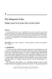
Introduction All living organisms face the challenge of defending themselves against microorganisms in the environment. Although the adaptive immune system has been subject to considerable study, the contribution of the innate immune system to defense against microbial pathogens has been less well appreciated. Innate immunity is often regarded as relatively non-specific. However, recent studies have shown that the innate immune system has a much greater specificity than previously thought, and can indeed respond to specific antigens. The first step in innate immunity is the recognition of microorganisms by receptors that recognize specific molecules that are present in the pathogen but not in self tissues. In mammals, a family of Toll-like receptors (TLRs) plays a central role in this discrimination, and ten human TLRs have been identified to date (Figure 1.1). These pathogen-associated molecules are likely to be essential for the survival of microbes and therefore cannot change rapidly under innate immunity selection pressure. A TLR contains a number of leucine-rich repeats (LRRs) in its ectodomain, and a Toll/interleukin-1 receptor (TIR) domain in the cytoplasmic region. The TLR ectodomain is responsible for ligand binding, and the TIR domain recruits cytoplasmic adapter proteins that also contain TIR domains such as MyD88, TIRAP, TRIF, and TRAM to carry signals into the cytoplasm. Most TLRs activate the MyD88-dependent pathway to induce a core response such as inflammation. However, individual TLRs 3
4
Nucleic Acids in Innate Immunity Triacyl Diacyl lipopeptide lipopeptide
TLR1/2
TLR6/2
LPS
Flagellin
dsRNA
ssRNA
ssRNA CpG DNA
TLR4
TLR5
TLR3
TLR7
TLR8
TLR9
Figure 1.1 Human Toll-like receptors and their representative ligands.
can also induce immune responses tailored to a specific microbial infection.7,8 In addition, TLRs control adaptive immune responses via multiple dendritic cell functions.9 Knowledge of the innate immune system, particularly related to TLRs as key receptor molecules and downstream adaptor molecules involved in the signaling pathway, has provided the research community with novel targets for drug design.10,11 Potential therapeutic agents include: (1) small molecule antagonists or neutralizing antibodies to TLR ectodomains to block ligand binding; (2) small molecule agonists such as imiquimod that can exert adjuvant effects for immunostimulation; and (3) small molecules that can interfere with the downstream signaling cascade, such as the TIR–TIR domain interaction between TLR and adaptor molecules.12 These small molecules can be used to interrupt specific TLR signaling pathways while leaving other beneficial TLR signaling intact because of the different adaptor usage and signaling specificity of TLRs.
Structure of Human Toll-Like Receptor 3 (TLR3) Overall Structure The human TLR3 ectodomain structure at 2.1 Å revealed a large horseshoe-shaped solenoid structure assembled from 23 LRRs forming a right-handed solenoid13 (Figure 1.2). The inner and outer diameters of the horseshoe are about 50 and 80 Å, respectively, thus representing the largest structure of an LRR-containing protein known to date. The inner concave surface forms a continuous β sheet composed of 25 parallel β strands. Twenty-three of the 25 parallel β strands come from the LRRs in the TLR3 ectodomain, and two come from the N- and C-terminal regions, respectively. The concave surface contains mostly β strands, while the outer convex surface contains more diverse secondary structures such as loops, α helices, 310 helices, and β strands. The outer surface of TLR3 also contains TLR3-specific insertions, with segments LRR12 and LRR20 containing insertions longer than 10 residues that may
5
Structural Analysis of Toll-Like Receptors
10
15
5
20
23
1 N-term
C-term
Figure 1.2 Structure of human TLR3. Leucine-rich repeats (LRRs) are numbered 1 through 23, and observed glycosylation sites are shown in detail.
be involved in TLR3 function. Superposition of the 23 LRRs revealed that the residues that form the inner concave surface are structurally similar, with RMSDs ranging from 0.3 to 0.8 Å. The outer surface adopts more diverse structures, with RMSDs ranging from 1.0 to 2.5 Å when equivalent Cα atoms are used for the superposition.
Leucine-Rich Repeats As do other TLRs, the LRRs of the TLR3 ectodomain contain the typical 24-residue LRR motif consisting of xLxxLxLxxNxLxxLxxxxFxxLx, where L represents hydrophobic residues (mostly leucines), F a conserved phenylalanine, and N a conserved asparagine. Fourteen of the 23 LRRs in TLR3 follow the 24-residue motif, and only LRR12 and LRR20 have insertions longer than five residues. The lengths and positions of insertions in LRR motifs vary from TLR to TLR and can be important in determining the ligand specificity of each TLR.14 Hence, the location and structure of LRR12 and LRR20 in TLR3 may be important for its function and will be discussed later. Within each LRR, seven conserved hydrophobic residues at LRR motif positions 2, 5, 7, 12, 15, 20, and 23 point in toward the solenoid and form a tight hydrophobic core that contributes to the structural stability of the elongated solenoid structure (Figure 1.3A). The side chains of the asparagine residues at LRR motif position 10 form extensive hydrogen bonding networks linking neighboring LRR motifs, and further contribute to the stability of the overall structure (Figure 1.3B). The inner concave surface is formed by a continuous standard parallel β sheet, except that at both ends of the β strand the backbone carbonyl group forms bifurcated hydrogen bonds with two main chain amide groups (Figure 1.3C).
6
Nucleic Acids in Innate Immunity A
B
15
20
23
12
N596
2
5
7
3.28 N572
3.09 2.94
2.81 3.30
2.92
N540
3.23 2.87
2.94 3.11
2.98 3.04
3.11 N516
2.88 2.75
2.87
C 3.11 2.88 2.89
2.92
3.08 2.77
3.34
3.49
2.75
2.85
2.70 3.13
2.94
2.91
2.95
3.07
3.06 2.82
2.91
2.89 2.86
2.96 2.75
Figure 1.3 Contribution of leucine-rich repeats to the structural stability of the TLR ectodomain (A) Seven conserved hydrophobic residues form a tight hydrophobic core within each leucine-rich repeat as shown for LRR7. (B) The conserved asparagines at position 10 form three hydrogen bonds with the previous LRR motif and a single hydrogen bond within their own motif. (C) A concave surface is formed by 25 continuous β strands connected by numerous hydrogen bonds including bifurcated hydrogen bonds at both ends of the β strands.
Glycosylation Pattern In the crystal structure, electron density is observed for carbohydrates at 8 of the 15 possible N-linked glycosylation sites. If all the 15 N-linked glycosylation sites were post-translationally modified, the TLR3 surface would be largely masked by carbohydrate. The inner concave surface also contains three predicted glycosylation sites in LRR1, LRR9, and LRR15, and glycosylation was observed in LRR9 and LRR15. The carbohydrates from these sites can fill the space inside the concave surface and may hinder the binding of large ligand molecules such as double-stranded RNA. Only one face is glycosylation-free [Figure 1.4(B) and (C)], suggesting its potential role in ligand binding or oligomerization. Investigation of predicted glycosylation sites in other human TLRs shows that all human TLRs contain at least two predicted N-linked glycosylation sites in the concave surface (residues 2 through 9 in the 24-residue motif). The exact in vivo compositions and structures of carbohydrates on different TLRs are not well characterized
7
Structural Analysis of Toll-Like Receptors
and may depend on the localization of each TLR. For example, TLRs that reside in the endosome, including TLR7, TLR8, and TLR9, may have different glycosylation patterns from the TLR4 targeted to the cell surface. Results indicate that some glycosylation sites may play critical roles in protein stability and targeting.15–17 Other results have shown that glycosylation may be important in ligand recognition.18
Surface Electrostatic Potential Analysis of the surface electrostatic potential of the TLR3 ectodomain using the GRASP program revealed that the inner concave surface is predominantly negatively charged [Figure 1.5(B)]. This finding was surprising because the inner concave surface was thought to be the binding site for double-stranded RNA, which is also highly negatively charged. The largest cluster of positively charged residues was found on the glycosylation-free face of TLR3 [Figure 1.5(C)]. A recent paper showed that the ligand binding of TLR3 is pH-dependent (optimum pH around 5), and that ligand binding is abolished at pH values above 7.19 The A
B
N
C
C
C
N
Figure 1.4 Predicted glycosylation of TLR3 ectodomain. (A) When oligomannose-type glycosylation is modeled for all the 15 predicted N-linked glycosylation sites, the carbohydrates cover a large portion of the TLR3 ectodomain including the concave surface. (B) View rotated 180° from (A) showing the glycosylation-free face. (C) View rotated 90° from (A) showing dramatic differences in the extent of glycosylation on the two side faces. A
B
C
K355
K371 K345 R394 K418 K335 K467 K493 K416 R331
R251 R325 R222 K531
R489
K210 K182 K137 K139
K145
K117 R635
N
C
N
C
C
N
Figure 1.5 Surface electrostatic potential of TLR3 ectodomain calculated with GRASP. (A) same orientation as Figure 1.4(A). (B) Inside concave surface shows predominantly negative electrostatic potential. (C) Glycosylation-free face contains the largest number of positively charged residues that may be involved in double-stranded RNA recognition. The locations of four histidine residues near the N-terminus are indicated by dots.
8
Nucleic Acids in Innate Immunity
authors suggested that double-stranded RNA may undergo pH-dependent conformational changes, thus affecting its binding to TLR3. Alternately, the pH-dependent change in histidine protonation may affect the ability of TLR3 to bind ligand. Interestingly, a patch of four histidine residues is located near the N-terminal region on the glycosylation-free face, as indicated by dots in Figure 1.5(C).
Conserved Surface Residues and Possible Dimerization In the crystal, two molecules related by two-fold crystallographic symmetry form a putative homodimer. The dimer interface is near the C-terminus on the glycosylation-free face. The dimer interaction is largely hydrophilic, including ionic interactions (Glu442–Lys467 and Lys547–Asp575), hydrogen bonds between side chains, and water-mediated hydrogen bonds. The buried surface area is relatively small at 640 Å2, and the dimer interface largely overlaps with the most highly conserved surface residues on TLR3. (Figure 1.6 A–E). The dimer interface also includes a TLR3-specific insertion from LRR20 and three residues (Lys547, Ala549, and Pro551) from the LRR20 motif. The dimerization of TLR3 in vivo has not yet been experimentally confirmed, but some evidence indicates multimerization of TLR3.19 However, other TLRs are known to form functional homodimers or heterodimers in vivo. For example, TLR2 forms heterodimers with TLR1 and TLR6.20 TLR4 is known to form a homodimer for signaling.21
TLR3-Specific Insertions The TLR-specific insertions are likely to be involved in the specific functions (ligand recognition and signaling) of individual TLRs. In the case of TLR3, two large insertions are found in LRR12 (10 residues) and LRR20 (11 residues). Although the insertion in LRR12 is disordered in the crystal structure, it was observed in a second human TLR3 structure.22 The insertion protrudes about 13 Å from the horseshoe and contributes to the proposed dimer formation. Although the LRR12 insertions within the proposed homodimer are spatially close, interactions between them are limited. The role of the LRR12 insertion is not clear and its deletion does not have an effect on the ligand binding ability of TLR3.19,23 It is interesting that TLR3 contains a structure that protrudes so far from the horseshoe. The patch of positively charged residues on the glycosylation-free face is only about 15 Å from the LRR12 insertion; thus LRR12 may interact with the grooves of dsRNA. It is also possible that this LRR12 insertion may act as a spacer to separate the dimer to make space for the dsRNA interaction. The other TLR3-specific insertion at LRR20 plays a significant role in the dimer interface, thus supporting the idea that two TLR3s form a biologically relevant dimer. The LRR20 insertion may play an important role in reinforcing ligand-induced dimerization and conformational changes. Other TLRs contain varying numbers of insertions at different positions and can give rise to the structural differences of TLRs.14 The positions of the insertions are usually on the sides or outside convex surfaces. Interestingly, two groups of TLRs (1, 6, and 10 in one group, and 7, 8, and 9 in the other) have similar patterns of insertions different from other TLRs. The 1–6–10 and the 7–8–9 groups share high sequence
9
Structural Analysis of Toll-Like Receptors A
B
N
C
C
N
D
C
C
N
N
C
E
Figure 1.6 Conserved residues on TLR3 surface. (A) through (D) Completely conserved residues among five TLR3s (Homo sapiens, Pan troglodytes, Bos taurus, Rattus norvegicus, and Takifugu rubripes). (E) Residues involved in proposed dimerization. The dimer interface largely overlaps with the large patch of conserved residues in (B).
identities. Each group is known to recognize structurally- and chemically-related ligands (lipopeptides and nucleotides, respectively). TLRs 7, 8, and 9 also contain undefined regions with no sequence similarity to one of the known LRR motifs. These trends imply that the locations and structures of insertions in different TLRs are likely to be important in the functioning of TLRs by shaping ligand binding sites and determining the ligand binding specificity of each TLR.
Site-Directed Mutagenesis and Effects on Function To identify amino acid residues that are important in ligand recognition, over 50 residues in TLR3 have been mutated using site-directed mutagenesis.23 The most dramatic change in ligand binding ability was observed for H539E and N541A mutations (Figure 1.7). H539 coordinates one of the two sulfate molecules observed in the
10
Nucleic Acids in Innate Immunity A
C
Patch 1
LRR12
Patch 2
? LRR20
?
90°
B H565 R489 K493
N517
D536 H539
K589
D641
H643 H674
Patch 1
N541 R544
Figure 1.7 Residues mutated by site-directed mutagenesis. (A) and (B) Residues mutated on concave and lateral surfaces. Bell et al.23 showed that H539 and N541 are important for functioning. (C) LRR12 and LRR20 insertions and positively charged residues in patches 1 and 2.
crystal structure that may mimic the phosphate groups of dsRNA. However, other residues that coordinate the sulfate ion with H539, including R488, N515, Q538, and E570, had no effect on ligand binding when mutated. Residues that interact with the second sulfate, such as Y326, H359, and N361, also had no effect on RNA binding when mutated, thus indicating that the two sulfate binding sites located in the concave surface are not directly involved in ligand binding. Mutation of residues proximal to H539 revealed that the N541A mutation almost completely abrogates activity. N541, located on the glycosylation-free face, is also essential for RNA recognition (Figure 1.7). H539E and N541A mutants showed negligible change in their size-exclusion chromatography elution patterns in contrast to wild-type TLR3-ECD, whose elution pattern changes in the presence of its ligand, poly I:C. The authors suggested that these two residues, H539 and N541, are directly involved in dsRNA binding by interacting with the phosphate group and 2′ hydroxyl group of the RNA ribose, respectively. It is, however, noteworthy that these two residues are intimately involved in the proposed dimer interface, and the lack of RNA binding affinity may also reflect disruption of dimerization necessary for ligand recognition.
Structural Analysis of Toll-Like Receptors
11
Deletion mutants of TLR3-specific insertions (LRR12 and LRR20) showed that the loss of LRR12 had no effect on TLR3 stimulation, but the LRR20 deletion resulted in significant, but not total, loss of activity.23 The mutation of individual positively charged residues in the two basic patches on the glycosylation-free face also had no significant effect on ligand recognition. Only when a number of basic residues were mutated together (eight residues in patch 1 and seven in patch 2) was a significant loss of activity observed [Figure 1.7(C)]. Because such a large number of mutations were required for this loss of activity, whether these positively charged residues are directly involved in dsRNA binding, or whether loss of activity is due to alteration of the TLR3 structure or stability, is inconclusive. Mutation of each of the 15 asparagine residues in consensus N-linked glycosylation motif (Asn-X-Ser/Thr) to aspartate also had no significant effect on TLR3 expression or activity.
Proposed Double-Stranded RNA Binding Site Contrary to the original idea that the inner concave surface would act as the ligand binding site of TLRs,24 evidence from the TLR3 structure indicates that the inner concave surface is not suitable for double-stranded RNA binding. First, the inner concave surface contains three predicted glycosylation sites that fill most of the space. Although the exact nature and extent of the glycosylation are still unknown, it is likely that any degree of glycosylation may hinder the binding of dsRNA, considering the dimensions of the inner space (diameter ~50 Å) and dsRNA (diameter ~26 Å). It is possible that the carbohydrate groups in the inner surface may interact with the ligand, but examples of carbohydrates involved in ligand recognition are limited. The results of extensive site-directed mutagenesis experiments also indicated that residues located in the inner concave surface are not critical for dsRNA binding.19 The electrostatic potential of the inner concave surface is predominantly negatively charged. This negative potential would make interaction with highly negatively charged dsRNA unfavorable. Interestingly, two patches of positively charged residues and a TLR3-specific insertion in LRR12 are located in proximity to each other on the glycosylation-free side surface and may provide the binding site for double-stranded RNA, although mutagenesis experiments to confirm this theory were inconclusive.23 This putative RNA binding site is on the same face as the dimer interface seen in the crystal, and dimerization of TLR3 could then create a deep binding cleft between two TLR3 molecules for dsRNA binding (Figure 1.8). Based on site-directed mutagenesis results, Bell et al.23 proposed that the dsRNA ligand binding site is located on the glycosylation-free face near the C-terminus. Specifically, H539 can interact with the phosphate group in the minor groove of RNA and N541 can form a hydrogen bond with the 2′ OH group of the ribose in the minor groove. These interactions can provide a model in which two TLR3-ECDs related by a 180° rotation can bind both sides of one dsRNA molecule (Figure 1.9). TLRs including TLR3 should discriminate self from non-self molecules to specifically respond to invasions of pathogens. A subgroup of TLRs has become specialized to detect non-self nucleic acids derived primarily from viruses. TLR3 recognizes dsRNA; TLR7 and TLR8 bind single-stranded RNA and small immunomodulatory
12
Nucleic Acids in Innate Immunity A
B
Figure 1.8 Proposed double-stranded RNA binding site of TLR3. (A) Dimerization of TLR3 ectodomain brings the positively charged residues (dots) and TLR3-specific insertion at LRR12 close together to form a potential binding site for a dsRNA molecule. Cα positions of the disordered LRR12 region are indicated as dashed lines. (B) Top view of (A). A
B
90°
Figure 1.9 Potential double-stranded RNA binding site of TLR3 proposed by Bell et al.23 (A) H539 and N541 of TLR3-ECD interact with minor grooves of dsRNA to bring the two TLR3 molecules close together for intracellular signaling (B). TLRs with TIR domains (Protein Data Bank ID code 1FYV). Linker regions between TLR3-ECD–TM (7 residues) and TM–TIR (19 residues) domains are denoted by black dotted lines. The homologous position of the Lpsd mutation in the BB loop of the TLR4 TIR domain postulated to interact with MyD8825 is indicated. The separation of TLR3-ECD in the proposed oligomer easily accommodates the formation of a TIR dimer.
Structural Analysis of Toll-Like Receptors
13
compounds such as imidazoquinolines; and TLR9 interacts with unmethylated CpG DNA. It is unclear how these TLRs achieve their specificities. The key to this question may be found in the intracellular localization of these subtypes of TLRs. A recent paper indicates that subcellular localization is very important for specificity and, in particular, for discrimination of viral nucleic acids from self nucleic acids26 by TLRs. The intracellular localization of TLR9 is specified by the transmembrane domain. When a chimeric receptor composed of the TLR9 ectodomain and the TLR4 transmembrane and cytoplasmic domain is made, it can be redirected to the cell surface. These hybrids are then able to signal in response to exogenous self nucleic acids, whereas TLR9 (present in the endoplasmic reticulum, endosomes, and lysosomes) responds only to viral or bacterial CpG-containing DNA. Thus, the discrimination of viral DNA from self DNA is due to delivery by the virus of its nucleic acid to an intracellular compartment where self DNA is not normally found.26 Although the exact localization of TLR3 is still unclear, it is very likely that the intracellular compartmentalization of TLR3 plays a critical role in discriminating self and non-self dsRNA. It is interesting that CD14 is involved in enhancing dsRNA-mediated TLR3 activation by physical interaction and internalization of dsRNA.27
Possible Modes of Dimerization and Signal Transduction Although the exact binding sites for dsRNA recognition are different in the two proposed models,13,23 the proposed RNA binding sites still reside within the putative dimer interface. This suggests that binding of dsRNA may strengthen dimerization or induce conformational changes of the dimer that then bring the intracellular TIR domains closer together for signal transduction (Figure 1.10). This type of signal transduction mechanism is often used by cell surface receptors.28,29 The basic mechanisms of signaling by Toll and TLRs seem to involve ligandinduced dimerization and oligomerization or conformational changes.30,31 Evidence from the interaction of Spätzle with Drosophila Toll showed, both in vivo and in vitro, that the binding of Spätzle crosslinks two Drosophila Toll ectodomains. This crosslinking is necessary and sufficient for signal transduction by Toll.32 Recent evidence illustrates that chimeric TLR4 molecules in which the TLR4 ectodomain is replaced by Drosophila Toll are activated by Spätzle and have very similar biochemical characteristics to Toll. This finding also indicates that the TIR domains can initiate downstream signaling if the receptor extracellular domain is crosslinked in a symmetrical manner. Although receptor crosslinking is a crucial event in signal transduction, recent data suggest that the signaling process is also likely to involve a series of conformational changes.33 Studies using analytical ultracentrifugation showed that truncation of the N-terminal region of Toll ECD allows the formation of stable dimeric complexes in solution and causes constitutive activation of the signaling cascade.34 These results also indicate that the N-terminal domain in Toll provides structural hindrance, preventing the self-association of the receptors, and that binding of Spätzle can relieve this constraint by inducing conformational changes. After TLR activation by possible ligand-induced dimerization and/or conformational changes, TLRs recruit a specific set of adaptor proteins that give rise to
14
Nucleic Acids in Innate Immunity A
Ligand-induced dimerization RNA binding
Signaling RNA binding Pre-existing dimer B TLR3
dsRNA
+
Ligand recognition
Signaling complex
Receptor oligomerization
Figure 1.10 Hypothetical mechanism of ligand-induced signal transduction. (A) Doublestranded RNA binds to positively charged residues located on the glycosylation-free face and can induce either dimerization or conformational changes in pre-existing dimers to bring intracellular TIR domains closer together for signal transduction. (B) Residues near the C-terminal region of TLR3-ECD, such as H539 and N541, may interact with dsRNA and form a signaling complex by receptor oligomerization.
pathogen-specific immune responses.7 These adaptors also contain TIR domains. Ligand binding to TLRs must create a new arrangement of the TIR domains formed by the TLR dimer that provides the binding site for the recruitment of appropriate adaptor molecules. Understanding this TLR–adaptor specificity requires detailed structural information of the TIR–TIR domain complexes. Although certain regions of the TIR domain, for example, the BB-loop region, are known to be involved in the TIR–TIR domain interaction, it is likely that other areas are also involved in the interaction with different adaptor molecules. Recent data obtained from germ line mutagenesis experiments revealed that mutations in the TIR domain can block signaling with one adaptor molecule, but allow signaling via another.35 Determining how such adaptor specificity is achieved awaits further biochemical and structural information. Understanding adaptor specificity can then provide opportunities for designing specific inhibitors to disrupt specific signaling pathways without disturbing other TLR-initiated signaling pathways that may be beneficial or essential to the host.
Structural Analysis of Toll-Like Receptors
15
Acknowledgement IAW is supported by Grant AI-42266 of the National Institutes of Health..
References
1. Akira, S. 2003, Curr. Opin. Immunol., 15, 5. 2. Medzhitov, R. 2001, Nat. Rev. Immunol., 1, 135. 3. Barton, G. M. and Medzhitov, R. 2002, Curr. Top. Microbiol. Immunol., 270, 81. 4. Takeda, K. and Akira, S. 2005, Int. Immunol., 17, 1. 5. Takeda, K. and Akira, S. 2004, Semin. Immunol., 16, 3. 6. O’Neill, L. A., Fitzgerald, K. A., and Bowie, A. G. 2003, Trends Immunol., 24, 286. 7. Yamamoto, M., Takeda, K., and Akira, S. 2004, Mol. Immunol., 40, 861. 8. O’Neill, L. A. 2003, Biochem. Soc. Trans., 31, 643. 9. Iwasaki, A. and Medzhitov, R. 2004, Nat. Immunol., 5, 987. 10. Zuany-Amorim, C., Hastewell, J., and Walker, C. 2002, Nat. Rev. Drug Discov., 1, 797. 11. Ulevitch, R. J. 2004, Nat. Rev. Immunol., 4, 512. 12. Bartfai, T. et al. 2003, Proc. Natl. Acad. Sci. USA, 100, 7971. 13. Choe, J., Kelker, M. S., and Wilson, I. A. 2005, Science, 309, 581. 14. Bell, J. K. et al. 2003, Trends Immunol., 24, 528. 15. Sun, J. et al. 2006, J. Biol. Chem., 281, 11144. 16. Weber, A. N., Morse, M. A., and Gay, N. J. 2004, J. Biol. Chem., 279, 34589. 17. da Silva Correia, J. and Ulevitch, R. J. 2002, J. Biol. Chem., 277, 1845. 18. Kataoka, H. et al. 2006, Cell Microbiol., 8, 1199. 19. de Bouteiller, O. et al. 2005, J. Biol. Chem., 280, 38133. 20. Ozinsky, A. et al. 2000, Proc. Natl. Acad. Sci. USA, 97, 13766. 21. Lee, H. K., Dunzendorfer, S., and Tobias, P. S. 2004, J. Biol. Chem., 279, 10564. 22. Bell, J. K. et al. 2005, Proc. Natl. Acad. Sci. USA, 102, 10976. 23. Bell, J. K. et al. 2006, Proc. Natl. Acad. Sci. USA, 103, 8792. 24. Kobe, B. and Kajava, A. V. 2001, Curr. Opin. Struct. Biol., 11, 725. 25. Xu, Y. et al. 2000, Nature, 408, 111. 26. Barton, G. M., Kagan, J. C., and Medzhitov, R. 2006, Nat. Immunol., 7, 49. 27. Lee, H. K. Et al. 2006, Immunity, 24, 153. 28. Livnah, O. et al. 1999, Science, 283, 987. 29. de Vos, A. M., Ultsch, M., and Kossiakoff, A. A. 1992, Science, 255, 306. 30. Gay, N. J., Gangloff, M., and Weber, A. N. 2006, Nat. Rev. Immunol., 6, 693. 31. Gangloff, M., Weber, A. N., and Gay, N. J. 2005, J. Endotoxin Res., 11, 294. 32. Weber, A. N. et al. 2003, Nat. Immunol., 4, 794. 33. Weber, A. N. et al. 2005, J. Biol. Chem., 280, 22793. 34. Winans, K. A. and Hashimoto, C. 1995, Mol. Biol. Cell, 6, 587. 35. Jiang, Z. et al. 2006, Proc. Natl. Acad. Sci. USA, 103, 10961.
Signaling 2 Antiviral Through TLRs and RLHs Taro Kawai Abstract Innate immune cells, such as dendritic cells and macrophages, detect invading microorganisms including viruses through a limited number of receptors. Two pathways for the detection of viral nucleic acids have been identified. One is mediated by members of the Toll-like receptor (TLR) family that recognize viral double-stranded RNA, single-stranded RNA, and DNA. The other pathway is utilized by RIG-I-like RNA helicases (RLHs) that detect viral RNA in the cytoplasm. These receptors use specific intracellular adaptor proteins to activate the key IRF and NF-κB transcription factors that promote synthesis of various cytokines required to eliminate infected viruses. This review summarizes recent insights into the antiviral signaling pathways activated by TLRs and RLHs.
Introduction Virus infection is sensed by the innate immune system. Within a host, innate immune cells such as dendritic cells (DCs) and macrophages express a variety of pattern recognition receptors (PRRs) that recognize specific pathogen-associated molecular patterns (PAMPs) within microbial structures such as viral nucleic acid.1,2 Following recognition, PRRs initiate signaling pathways that induce the production of a variety of cytokines including inflammatory cytokines such as IL (interleukin)-6 and TNFα, type I interferons (IFNs; both α and β), chemokines, and IL-12, along with increased surface expression of co-stimulatory molecules to support the proliferation of T cells and their differentiation into Th cells. Type I IFN in particular enhances DC maturation, natural killer (NK) cell cytotoxicity, and differentiation of virus-specific cytotoxic T lymphocytes. Type I IFN also upregulates transcription of many IFN-inducible genes that influence protein synthesis, growth arrest, and apoptosis to establish an antiviral state.3 PRRs that recognize viral PAMPs are classified into several families. The Toll-like receptor (TLR) family consists of more than ten members that respond to PAMPs derived from many pathogens. Within the TLR family, TLR3, TLR7, TLR8, and TLR9 represent a subfamily that recognizes viral nucleic acids. RLHs such as RIG-I and Mda5 participate in the detection of RNAs of infected viruses. Although TLRs and RLHs recognize viral nucleic acids to induce antiviral responses, they differ in cellular localization, ligand specificity, and downstream signal transduction pathways.1–4 17
18
Nucleic Acids in Innate Immunity
TLRs 3, 7, 8, and 9 Double-stranded (ds) RNA is synthesized during the course of replication of many viruses and serves as a potent activator of innate immune cells that induce type I IFN. A synthetic analog of viral dsRNA, polyinosinic acid:cytidylic acid (poly I:C), has been used extensively to mimic viral infection. Induction of type I IFN and pro-inflammatory cytokines in response to poly I:C or genomic RNA purified from a dsRNA virus such as reovirus was abrogated in macrophages derived from TLR3deficient mice.5 TLR3-deficient mice are consistently resistant to I:C-induced shock, indicating that TLR3 recognizes poly I:C and possibly senses viral dsRNA. TLR3 is also implicated in recognizing dsRNA derived from ssRNA viruses such as the respiratory syncytial, encephalomyocarditis, and West Nile viruses.6,7 TLR3 is expressed on a CD4 – CD8+ subset of cDC that has high phagocytic activity. The apoptotic bodies of virus-infected or dsRNA-loaded cells are taken up by CD8+ DC, and TLR3 recognizes the dsRNA within these cells. This process triggers crosspresentation, a pathway important for the development of the CD8 cytotoxic T cell response against viruses that do not infect DC. Thus, the TLR3-dependent pathway is important for cross-presentation of CD8+ DC.6 TLR7 was initially identified as a receptor that recognizes imidazoquinoline derivatives with antiviral activity, such as imiquimod and resiquimod (R-848), and guanine analogues such as loxoribine.8 Subsequently, guanosine- or uridine-rich ssRNA derived from the human immunodeficiency virus (HIV) and the influenza virus was identified as a natural ligand for TLR7.9,10 In addition, TLR7 recognizes synthetic poly U RNA and certain small interfering RNAs.9,11 TLR8 is phylogenetically similar to TLR7. Human TLR8 preferentially mediates the recognition of HIV-derived ssRNA and R-848, although mice deficient for TLR8 respond normally to these molecules, suggesting that mouse TLR8 may not be functional.9,10,12 TLR9 recognizes unmethylated 2′ deoxyribo(cytidine-phosphate-guanosine) (CpG) DNA motifs that are frequently present in pathogens such as viruses and bacteria but are rare in vertebrates.13 TLR7 and TLR9 are highly expressed on plasmacytoid DCs (pDCs; also known as IFN-producing cells)—a subset of DCs that have plasmacytoid morphology and primarily secrete vast amounts of type I IFN in response to viral infection.14–16 In TLR7-deficient mice, IFNα production by pDCs is impaired after infection with influenza virus or vesicular stomatitis virus.9,17 Moreover, IFNα production by pDCs in response to DNA viruses such as mouse cytomegalovirus, HSV-1, and HSV-2 is dependent on TLR9.18–20 Inactivated HSV-2 or genomic DNA purified from this virus triggers IFNα secretion in pDCs in a TLR9-dependent manner.20 pDCs rely on TLR7 and TLR9 to detect viral infection, but viral detection by pDCs does not seem to require viral replication within cells. Unlike other TLRs, TLR 3, 7, 8, and 9 are not expressed at the plasma membrane and are exclusively localized to intracellular compartments like endosomes, suggesting that these intracellular TLRs recognize nucleic acids following the internalization and lysing of viruses.1 Intracellular localization is also important to prevent contact with self DNA, because TLR9 can respond to self DNA if it is relocalized to plasma membranes.21
Antiviral Signaling Through TLRs and RLHs
19
RIG-I and Mda5 Because TLRs are localized to endosomes, they are unable to sense viruses that have entered the cytosol and initiated replication to produce dsRNA. Numerous studies implicated TLR-independent mechanisms in the detection of viral infection. For example, induction of IFNβ following transfection of poly I:C or infection with RNA viruses is normally observed in the absence of TLR3 or TRIF, suggesting that host cells have a mechanism to sense actively replicating viruses in the cytoplasm.22–25 RIG-I, a member of the RNA helicase family, was identified as a molecule that senses dsRNA and induces type I IFN responses.25 RIG-I contains a DExD/H box RNA helicase and two caspase recruiting domain (CARD)-like domains. The helicase domain interacts with dsRNA, whereas the CARD-like domains are required for activating downstream signaling pathways. Furthermore, Mda5 and LGP2 were subsequently identified as members of the RLH’s.26,27 Mda5 contains two CARDlike domains and a helicase domain. LGP2 lacks the CARD-like domains and is thought to negatively regulate RIG-I and Mda5. Studies of RIG-I- and Mda5-deficient mice revealed that RIG-I is essential for the recognition of a series of ssRNA viruses including flaviviruses, paramyxoviruses, orthomyxoviruses, and rhabdoviruses. Mda5 is required for the recognition of a different set of RNA viruses that includes picornaviruses.28–30 Furthermore, Mda5 and RIG-I detect poly I:C and long dsRNA, respectively, indicating that these RNA helicases detect different RNA viruses.29 RIG-I-mediated detection of RNA has recently been shown to depend on the 5’ triphosphate end of RNA generated by viral polymerases.31,32 The production of type I IFN is still observed in pDCs derived from RIG-Ior Mda5-deficient mice, although cDCs, macrophages, and fibroblast cells derived from these mice showed impaired type I IFN induction after infection with their respective RNA viruses.28,29 It is notable that the TLR system is required for pDC induction of the antiviral response. Collectively, these observations indicate that the TLR system plays a pivotal role in the detection of viruses by pDCs.
TLR7 and TLR9 Signaling TLRs contain extracellular LRRs that mediate ligand recognition, a transmembrane domain, and a cytosolic TIR domain required for downstream signaling pathways.33 Upon recognition of nucleic acids, TLR7 and TLR9 recruit a TIR-containing MyD88 adaptor molecule that is universally utilized by all TLRs with the exception of TLR3 (Figure 2.1). The association of TLRs and MyD88 results in the recruitment of members of the IRAK family, including IRAK1, IRAK2, IRAK4, and IRAK-M. In particular, IRAK4 and IRAK1 are sequentially phosphorylated and involved in activation of the MyD88-dependent signaling pathway, while IRAK-M negatively regulates the MyD-88-dependent pathway. The function of IRAK2 remains unknown. Once phosphorylated, IRAK4 and IRAK1 dissociate from MyD88 and interact with TRAF6, an E3 ligase that forms a complex with Ubc13 and Uev1A and promotes the synthesis of lysine 63-linked polyubiquitin chains.
20
Nucleic Acids in Innate Immunity
DNA
ssRNA
DNA Endosome in cDC
Endosome in pDC TLR7
MyD88
IRAK4
TLR9
TLR7
TRAF6
IRF1
IRF5 IRF7
IκB NF-κB AP-1 AP-1
IRF7
IRF7 Nucleus
IFNα
NF-κB
NF-κB
TRAF6
TRAF3 IKKα IRAK1
NEMO IKKα IKKβ
MAPK
TLR9
MyD88
IRAK4
Ubc13 TAK1
TRAF3 OPNi IKKα IRAK1 IRF7
ssRNA
IRF5
Inflammatory cytokines
IRF7 IRF7
IRF1
IFNβ
Figure 2.1 TLR7 and TLR9 signaling, which recruits MyD88, IRAK4, and TRAF6. TRAF6 then ubiquitin-dependently activates TAK1. The TAK1 complex activates the IKK complex consisting of IKKα, IKKβ, and NEMO/IKKγ to catalyze phosphorylation of IκB. IκBs are destroyed by the proteasome pathway, allowing NF-κB to translocate to the nucleus. TAK1 simultaneously activates the MAPK pathway, resulting in phosphorylation and activation of AP-1. NF-κB and AP-1 control inflammatory responses by inducing pro-inflammatory cytokines. MyD88 forms a signaling complex with IRAK1, IKKα, TRAF3, OPN-I, and IRF7 in pDC. In response to ligand stimulation, IRF7 is phosphorylated in a IRAK1- and IKKαdependent manner, forms a dimer, and translocates to the nucleus to regulate expression of type I IFN genes, especially IFNα. IRF5 and IRF1 also interact with MyD88 and participate in induction of inflammatory cytokines and type I IFN, respectively, in cDCs.
TAK1, a member of MAPKKK, is activated by TRAF6-dependent ubiquitination, and in combination with TAB1, TAB2 and TAB3, activates two downstream pathways involving the IKK complex and the MAPK family. The IKK complex composed of the catalytic subunits IKKα, IKKβ, and a regulatory subunit known as NEMO/IKKγ, induce the phosphorylation and subsequent degradation of the IκB proteins that allow the NF-κB transcription factor to translocate into the nucleus. The MAPK family (JNK, p38, ERK) phosphorylates and activates the NF-κB transcription factor AP-1, a dimer of basic region leucine zipper proteins of the Jun, Fos and ATF subfamilies. NF-κB and AP-1 play central roles in the induction of genes encoding inflammatory cytokines33 (Figure 2.1). Although in vitro analyses have implicated Ubc13 in the control of both NF-κB and MAPK activation, studies of Ubc13-deficient mice have demonstrated that it is dispensable for NF-κB activation.34 Macrophages deficient in Ubc13 show defective
Antiviral Signaling Through TLRs and RLHs
21
induction of inflammatory cytokines after treatment with TLR7 and TLR9 ligands. It is notable that they display impaired MAPK activation, but normal NF-κB activation. TAK1 activation and TRAF6 ubiquitination are normally observed in Ubc13deficient cells, suggesting that Ubc13 may activate the MAPK pathway through a TAK1/TRAF6-independent pathway, or a pathway located downstream of TAK1/ TRAF6. The finding that Ubc13 deficiency results in a loss of NEMO ubiquitination suggests a link between NEMO ubiquitination and MAPK activation.34 TLR7- and TLR9-mediated type I IFN induction by pDCs is dependent on MyD88. IRF7—structurally the most similar to IRF3—is present in the cytoplasm and translocates to the nucleus after phosphorylation by one or more virus-activated kinases. IRF7 potently activates the promoters of IFNα and IFNβ genes. The expression of the IRF7 gene is weak in unstimulated conditions, but rapidly upregulates in response to TLR ligands or viral infection in most cell types, suggesting a positive feedback regulation of type I IFN induction. In pDCs, however, IRF7 is constitutively expressed and it (but not IRF3) binds to MyD8835,36 (Figure 2.1). pDCs lacking IRF7 consistently fail to produce IFNα in response to CpG DNA, whereas IRF3 is dispensable in these pathways.37 IRF7 also forms a complex with IRAK1, IRAK4, IKKα, and TRAF6 in addition to MyD88.35,36,38,39 While mice deficient in MyD88, IRAK4, or TRAF6 exhibit defects in both IRF7 and NF-κB activation associated with impaired induction of type I IFN and inflammatory cytokines in response to CpG DNA, pDCs derived from IRAK1or IKKα-deficient mice specifically show loss of IRF7 activation and type I IFN induction. Moreover, IRAK1 and IKKα (but not IRAK4) are capable of phosphorylating IRF7.38,39 Together, IRAK1 and IKKα are most likely the kinases that catalyze the phosphorylation of IRF7 in pDCs. However, the functional relationship of IRAK1 and IKKα remains unclear. It is possible that they function as a heterodimer to potentiate IRF7 activation, or they may phosphorylate different residues of IRF7, both of which are required for the activation. Several additional components of the MyD88–IRF7 complex have recently been identified (Figure 2.1). TRAF3 binds MyD88 and IRAK1, and is critical for type I IFN induction in TLR7 and TLR9 signaling.40,41 TRAF3 is also necessary for the induction of the IL-10 anti-inflammatory cytokine, but not for the induction of pro-inflammatory cytokines, in response to ligands for TLR7 and TLR9.40 Osteopontin (OPN) is a secreted protein involved in diverse cellular functions such as bone resorption, vascularization, inflammation, and Th1 polarization. OPN expression is induced by a TLR9 ligand through T-bet, a master transcription factor for Th1 differentiation. pDCs derived from T-bet- and OPN-deficient mice show defects in the induction of type I IFN in response to a TLR9 ligand, whereas IL-6 induction and NF-κB activation are unaffected in these cells. An OPN precursor, designated OPN-i, is sequestered in the cytoplasm, suggesting that OPN-i functions as an intracellular signaling molecule. OPN-i interacts and colocalizes with MyD88, and nuclear translocation of IRF7 in response to a TLR9 ligand is impaired in pDCs derived from OPN-deficient mice.42 Thus, OPN-i is a component of the MyD88IRF7 complex in pDCs. IRF8 is also implicated in TLR9-mediated responses in pDCs. pDCs derived from IRF8-deficient mice show a loss of TLR9-mediated induction of type I IFN and
22
Nucleic Acids in Innate Immunity
inflammatory cytokines linked to impaired NF-κB DNA binding activity, suggesting the possibility that IRF8 facilitates NF-κB DNA-binding.43 TLR9 activates different signaling pathways between pDCs and cDCs (Figure 2.1). While cDCs derived from IRF1-deficient mice display impaired induction of IFNβ, inducible nitric oxide synthase, and IL-12 p35 in response to a TLR9 ligand, pDCs derived from IRF1-deficient mice show normal induction of IFNβ and IFNα.44 IRF1 also interacts with MyD88 and is released into nuclei in response to ligand stimulation. Cytokine induction in response to TLR ligands is enhanced by pretreatment of cells with IFNγ. Consistent with the findings that IFNγ stimulation induces IRF1 expression, IFNγ-mediated enhancement is impaired in IRF1-deficient mice. Thus, IFNγ-induced IRF1 is recruited to MyD88 and translocated into nuclei in response to TLR stimulation to induce a set of genes including IFNβ in cDCs. IRF5 is also involved in TLR signaling. IRF5-deficient cDCs and macrophages exhibit impaired inflammatory cytokine production in response to multiple TLR ligands, but exhibit normal secretion of type I IFN by pDCs.45 IRF5 binds MyD88 and TRAF6 and translocates to nuclei after phosphorylation. In the nucleus, IRF5 binds ISRE motifs found in the promoter regions of genes encoding inflammatory cytokines to cause their expression, presumably via collaborative activation with NF-κB. IRF5-mediated responses are negatively regulated by IRF4, which competes with IRF5 for interaction with MyD88.46 Studies of synthetic CpG ODN led to classification of into three groups, based on biological effects. D/A type ODN induces a secretion of type I IFN by pDCs but has a low ability to induce B cell activation and IL-12 production. In contrast, K/B type ODN stimulates B cell activation and IL-12 production, but poorly induces type I IFN. C type ODN has the ability to induce both type I IFN induction and B cell activation. A/D type CpG ODN colocalizes with TLR9, MyD88, and IRF7 in endosomes in pDCs and is rapidly transferred and degraded in lysosomes in cDCs. However, when A/D type CpG ODN relocalizes to the endosomes in cDCs using a cationic lipid, these cells can produce IFNα through activation of the MyD88-IRF7 pathway.47 B/K type CpG ODN also induces secretion of IFNα if it is manipulated to remain in the endosomes of cDCs for longer periods. These findings suggest that retention of the CpG DNA–TLR9 complex in endosomes may cause the induction of robust IFNα production. Thus, the spatiotemporal regulation of TLR9 signaling is also important in controlling IFNα production.
TLR3 Signaling Signaling through TLR3 activates IRF3 and NF-κB via a TIR domain-containing adapter molecule designated TRIF (also known as TICAM1), but not via MyD88.33 (Figure 2.2). TRIF is also shared by TLR4, which recognizes LPS.33 TRIF then recruits non-canonical IKKs, TBK1 (also known as NAK or T2K), and IKKi (also known as IKKε), rather than IRAK1 and IKKα, which phosphorylate IRF3.48,49 Phosphorylated IRF3 forms a dimer, translocates into the nucleus, and binds to target sequences to induce the secretion of IFNβ and IFNα4. These subtypes of IFN subsequently activate a transcriptional complex known as ISGF3 consisting of STAT1, STAT2, and
23
Antiviral Signaling Through TLRs and RLHs
Poly IC dsRNA Endosome TLR3 TRIF
RIP1
TRAF6 Ubc13
TRAF3
TBK1
TAK1
IKKi
IRF3
MAPK
NEMO IKKα IKKβ NF-κB
Nucleus
IRF3 IRF3
IFNβ
AP-1 AP-1
IκB
NF-κB NF-κB
Inflammatory cytokines
Figure 2.2 TLR3 signaling. TLR3 recruits the TRIF adapter that interacts with TRAF3, TBK1, and IKKi. TBK1 and IKKi mediate phosphorylation of IRF3. Phosphorylated IRF3 dimerizes and translocates to the nucleus where it binds DNA and induces expression of IFNβ. TRIF also interacts with TRAF6 and RIP1 to mediate NF-κB and AP-1 activation via TAK1.
IRF9 through the type I IFN receptor, stimulating the expression of IFN-inducible genes required for antiviral responses, including IRF7.3 IRF7 is likely phosphorylated by TBK1 and IKKi in a manner similar to IRF3, inducing type I IFN. TLR3-mediated inflammatory cytokine induction is controlled by TRIF-dependent NF-κB activation (Figure 2.2). The N-terminal and the C-terminal regions of TRIF have distinct functions with regard to the recruitment of downstream signaling molecules required for NF-κB activation. The N-terminal region recruits TRAF6 in addition to TBK1/IKKi.50 Dominant-negative TRAF6 prevents TRIF-induced NF-κB activation, and mutations in the TRAF6 binding motifs of TRIF abrogate NF-κB activation. The C-terminal region of TRIF mediates its interaction with RIP1, a member of the RIP family involved in TNFR-mediated NF-κB activation, via its RIP homotypic interaction motif.51 TLR3-mediated NF-B activation and the subsequent induction of target genes are impaired in the absence of RIP1. Furthermore, RIP1 polyubiquitination and complex formation with TRAF6 and TAK1 have been reported.52 Thus, TRIF recruitment of RIP1 and TRAF6 may facilitate TAK1 activation, resulting in activation of NF-κB and MAPK. Whether Ubc13 is involved in RIP1 polyubiquitination is still unclear. It has been suggested that TLR3-signaling is negatively regulated by several independent mechanisms. The TIR domain-containing protein SARM inhibits the TRIF-dependent pathway in human cell lines; however, the physiological function of
24
Nucleic Acids in Innate Immunity
SARM in mice remains unknown.53 While phosphorylation of the C-terminal serine and threonine clusters of IRF3 by TBK1/IKKi is essential for transcriptional activity, phosphorylation at Ser339 is linked to IRF3 destabilization. The Pin1 cytoplasmic peptidyl–prolyl–isomerase that catalyzes the cis–trans isomerization of peptide bonds located the N-terminus to proline residue to modulate substrate function binds IRF3 when phosphorylated at Ser339. This triggers ubiquitination and subsequent degradation of IRF3 by a proteasome-dependent pathway to terminate IFN responses.54 A protein kinase responsible for Ser339 phosphorylation has not been identified.
RLH Signaling Infection of viruses recognized by RIG-I or Mda5 results in the induction of type I IFN that is dependent on TBK1/IKKi but independent of TRIF and MyD88, indicating that RIG-I and Mda5 use an adaptor other than TRIF and MyD88.22 IPS-1 (also known as MAVS, Cardif, or VISA) was identified as a potent activator of the IFNβ promoter55–58 (Figure 2.3). IPS-1 contains a CARD-like domain that shows a homology to the CARD-like domains of RIG-I and Mda5. Overexpression of IPS-1 activates IFNα4, IFNα6, and NF-κB promoters in addition to the IFNβ promoter, and results in production of type I IFN sufficient for inhibition of viral replication. The RIG-I
dsRNA
Mda5
Poly IC IPS-1 Mitochondria FADD Casp8/10
TRAF3 TBK1
IRF3
IRF7
Nucleus
NEMO IKKα IKKβ
IKKi
IRF3
IRF7
IRF3
IRF7
IFNα
IFNβ
IκB
NF-κB
NF-κB
NF-κB
Inflammatory cytokines
Figure 2.3 RLH signaling. RIG-I and Mda5 interact with adapter IPS-1 via CARD-like domains. IPS-1 is localized to mitochondria and initiates intracellular signaling pathways that lead to activation of IRF3, IRF7, and NF-κB via activation of TBK1/IKKi and IKKα/IKKβ, respectively. A complex consisting of FADD, caspase-8, and caspase-10 has been implicated in IPS-1-dependent NF-κB activation, while TRAF3 provides a physical link between IPS-1 and TBK1/IKKi.
Antiviral Signaling Through TLRs and RLHs
25
activation of the IFNβ promoter after IPS-1 overexpression was ablated in TBK1and IKKi-null cells. The CARD-like domain of IPS-1 mediates interactions with RIG-I and Mda5. IPS-1 is most likely an adaptor interacting with RIG-I and Mda5. IPS-1-deficient mice have been recently developed.59,60 Unlike RIG-I-deficient mice that are embryonic lethal because of liver degeneration, IPS-1-deficient mice are viable and develop normally. Macrophages, cDCs, and embryonic fibroblasts derived from IPS-1-deficient mice failed to activate NF-κB and IRF3 with a concomitant loss of type I IFN and inflammatory cytokine induction after infection with RNA viruses recognized by RIG-I or Mda5. Cytokine induction in response to poly I:C or long dsRNA was also impaired in IPS-1-deficient mice. These mice are consistently susceptible to infection with RNA viruses, indicating the importance of IPS-1 in antiviral responses in vivo. However, IPS-1 is dispensable for type I IFN induction after infection with a DNA virus such as modified Vaccinia Ankara, as well as stimulation with TLR ligands. Collectively, IPS-1 is an essential adapter utilized by both RIG-I and Mda5 that mediate antiviral innate immune responses. In pDCs, however, TLR7 and TLR9 contribute more preferentially than RLHs to type I IFN induction after virus infection, indicating a cell type-specific role of TLR and RLH in the recognition of viruses and induction of antiviral immune responses. IPS-1-mediated activation of IRF3 and IRF7 is dependent on TBK1/IKKi, although IPS-1 does not directly interact with them. One report indicated that TRAF3 binds both IPS-1 and TBK1/IKKi, and TRAF3 deficiency results in impaired type I IFN induction after virus infection, indicating that TRAF3 is a link between IPS-1 and TBK1/IKKi61 (Figure 2.3). IPS-1 also interacts with FADD, a death domain containing adapter involved in death receptor signaling and RIP-1.55 Cells deficient for FADD display reduced induction of IFNβ and inflammatory cytokines in response to poly I:C stimulation.62,63 FADD forms a complex with caspase-10 and caspase-8. These caspases are cleaved in response to poly I:C stimulation. The cleaved fragments of the caspases (encoding a death effecter domain) can activate NF-κB but not the IFNβ promoter, suggesting that caspase-10 and caspase-8 selectively participate in NF-κB activation downstream of FADD (Figure 2.3). Cells derived from caspase-8-deficient mice accordingly show reduced NF-κB and inflammatory cytokine induction in response to poly I:C.63 How these cleaved fragments activate the IKK complex remains unclear. IPS-1 contains a transmembrane domain in the C-terminal tail that targets IPS-1 to the mitochondria56 (Figure 2.3). Notably, mitochondrial retention of IPS-1 is essential for IRF3 and NF-κB activation, suggesting that signaling from mitochondria plays an important role in antiviral immune responses. A report indicates that the NS3/4A serine protease in the hepatitis C virus (HCV) targets IPS-1 for cleavage.57,64–66 A putative cleavage site located upstream to the transmembrane domain and HCV infection result in cleavage of IPS-1, as demonstrated in an in vitro cell culture infection system. A cleaved form of IPS-1 lacking a transmembrane region has no ability to activate IRF3 and NF-κB. The NS3/4A combination is likely to change the cellular localization of IPS-1 from mitochondria to the cytoplasm by cleavage, thereby inhibiting type I IFN and the inflammatory response. NS3/4A also mediates the cleavage of TRIF to inhibit the TLR3-dependent antiviral response.67,68
26
Nucleic Acids in Innate Immunity
Perspectives Host cells express multiple PRRs for the detection of viruses. These PRRs are expressed in different cellular compartments and recognize different types of nucleic acids. TLR3 recognizes dsRNA that is released into endosomes following phagocytosis of apoptotic bodies from virally infected cells, or detects dsRNA viruses that are internalized by endocytosis. In contrast, Mda5 and RIG-I are expressed in the cytoplasms of a variety of cells and recognize the dsRNA produced during viral replication. TLR7 and TLR9 are expressed by pDCs and act as sensors for viral ssRNA and DNA that trigger the production of large amounts of IFNα. Although these PRRs induce type I IFN, intracellular signaling pathways are different. TLR3 relies on the TRIF adapter to recruit TBK1/IKKi and induces IRF3 phosphorylation. TLR7 and TLR9 use MyD88 as an adapter to induce type I IFN via IRAK1/IKKαdependent phosphorylation of IRF7. In RIG-I and Mda5 signaling, IPS-1 functions as a common adapter that mediates IRF3 and IRF7 activation via TBK1/IKKi. Cells appear to express additional intracellular PRRs, such as a family of NOD proteins that serve to detect various PAMPs including RNA, and an unidentified receptor or receptors that recognize right-handed dsDNA released by DNA viruses, bacteria, and damaged host cells.1,69,70 It will be important to understand how PPRs detect nucleic acid and induce antiviral innate immune responses. Such knowledge could improve therapeutic strategies for the treatment of infectious diseases and also autoimmune diseases associated with viral infection.
References
1. Akira, S., S. Uematsu, and O. Takeuchi. 2006. Pathogen recognition and innate immunity. Cell 124:783. 2. Kawai, T. and S. Akira. 2006. Innate immune recognition of viral infection. Nat Immunol. 7:131. 3. Honda, K., A. Takaoka, and T. Taniguchi. 2006. Type I interferon gene induction by the interferon regulatory factor family of transcription factors. Immunity 25:349. 4. Meylan, E. and J. Tschopp. 2006. Toll-like receptors and RNA helicases: two parallel ways to trigger antiviral responses. Mol. Cell 22:561. 5. Alexopoulou, L. et al. Flavell. 2001. Recognition of double-stranded RNA and activation of NF-κB by Toll-like receptor 3. Nature 413:732. 6. Schulz, O. et al. 2005. Toll-like receptor 3 promotes cross-priming to virus-infected cells. Nature 433:887. 7. Wang, T. et al. 2004. Toll-like receptor 3 mediates West Nile virus entry into the brain causing lethal encephalitis. Nat Med. 10:1366. 8. Hemmi, H. et al. 2002. Small anti-viral compounds activate immune cells via the TLR7 MyD88-dependent signaling pathway. Nat Immunol. 3:196. 9. Diebold, S. S. et al. 2004. Innate antiviral responses by means of TLR7-mediated recognition of single-stranded RNA. Science 303:1529. 10. Heil, F. Et al. 2004. Species-specific recognition of single-stranded RNA via Toll-like receptor 7 and 8. Science 303:1526. 11. Hornung, V. et al. 2005. Sequence-specific potent induction of IFN-α by short interfering RNA in plasmacytoid dendritic cells through TLR7. Nat Med. 11:263. 12. Jurk, M. et al. 2002. Human TLR7 or TLR8 independently confer responsiveness to the antiviral compound R-848. Nat Immunol. 3:499.
Antiviral Signaling Through TLRs and RLHs
27
13. Hemmi, H. et al. 2000. A Toll-like receptor recognizes bacterial DNA. Nature 408:740. 14. Liu, Y. J. 2005. IPC: professional type 1 interferon-producing cells and plasmacytoid dendritic cell precursors. Annu Rev Immunol. 23:275. 15. Colonna, M., G. Trinchieri, and Y. J. Liu. 2004. Plasmacytoid dendritic cells in immunity. Nat Immunol. 5:1219. 16. Kaisho, T. and S. Akira. 2006. Toll-like receptor function and signaling. J Allergy Clin Immunol. 117:979. 17. Lund, J. M. et al. 2004. Recognition of single-stranded RNA viruses by Toll-like receptor 7. Proc Natl Acad Sci USA 101:5598. 18. Krug, A. et al. 2004. TLR9-dependent recognition of MCMV by IPC and DC generates coordinated cytokine responses that activate antiviral NK cell function. Immunity 21:107. 19. Krug, A. et al. 2004. Herpes simplex virus type 1 activates murine natural interferonproducing cells through toll-like receptor 9. Blood 103:1433. 20. Lund, J. et al. 2003. Toll-like receptor 9-mediated recognition of Herpes simplex virus-2 by plasmacytoid dendritic cells. J Exp Med. 198:513. 21. Barton, G. M., J. C. Kagan, and R. Medzhitov. 2006. Intracellular localization of Tolllike receptor 9 prevents recognition of self DNA but facilitates access to viral DNA. Nat Immunol. 7:49. 22. Hemmi, H. et al. 2004. The roles of two IκB Kinase-related kinases in lipopolysaccharide and double stranded RNA signaling and viral infection. J Exp Med. 199:1641. 23. Yamamoto, M. et al. 2003. Role of adaptor TRIF in the MyD88-independent toll-like receptor signaling pathway. Science 301:640. 24. Hoebe, K. et al. 2003. Upregulation of costimulatory molecules induced by lipopolysaccharide and double-stranded RNA occurs by TRIF-dependent and TRIF-independent pathways. Nat Immunol. 4:1223. 25. Yoneyama, M. et al. 2004. The RNA helicase RIG-I has an essential function in double-stranded RNA-induced innate antiviral responses. Nat Immunol. 5:730. 26. Yoneyama, M. et al. 2005. Shared and unique fnctions of the DExD/H box helicases RIG-I, MDA5, and LGP2 in antiviral innate immunity. J. Immunol. 175:2851. 27. Rothenfusser, S. et al. 2005. The RNA helicase Lgp2 inhibits TLR-independent sensing of viral replication by retinoic acid-inducible gene-I. J Immunol. 175:5260. 28. Kato, H. et al. 2005. Cell type specific involvment of RIG-I in antiviral response. Immunity 23:19. 29. Kato, H. et al. 2006. Differential role of MDA5 and RIG-I in the recognition of RNA viruses. Nature 441:101. 30. Gitlin, L. et al. 2006. Essential role of mda-5 in type I IFN responses to polyriboinosi nic:polyribocytidylic acid and encephalomyocarditis picornavirus. Proc Natl Acad Sci USA 103:8459. 31. Hornung, V. et al. 2006. 5′ triphosphate RNA is the ligand for RIG-I. Science 314:994. 32. Pichlmair, A. et al. 2006. RIG-I-mediated antiviral responses to single-stranded RNA bearing 5′ phosphates. Science 314:997. 33. Akira, S. and K. Takeda. 2004. Toll-like receptor signalling. Nat Rev Immunol. 4:499. 34. Yamamoto, M. et al. 2006. Key function for the Ubc13 E2 ubiquitin-conjugating enzyme in immune receptor signaling. Nat Immunol. 7:962. 35. Kawai, T. et al. 2004. Interferon-α induction through Toll-like receptors involves a direct interaction of IRF7 with MyD88 and TRAF6. Nat Immunol. 5:1061. 36. Honda, K. et al. 2004. Role of a transductional-transcriptional processor complex involving MyD88 and IRF-7 in Toll-like receptor signaling. Proc Natl Acad Sci USA. 101:15416.
28
Nucleic Acids in Innate Immunity
37. Honda, K. et al. 2005. IRF-7 is the master regulator of type-I interferon-dependent immune responses. Nature 434:772. 38. Uematsu, S. et al. 2005. Interleukin-1 receptor-associated kinase-1 (IRAK-1) plays an essential role for TLR7- and TLR9-mediated interferon-α induction. J Exp Med. 201:915. 39. Hoshino, K. et al. 2006. IκB kinase-α is critical for interferon-α production induced by Toll-like receptors 7 and 9. Nature 440:949. 40. Hacker, H. et al. 2006. Specificity in Toll-like receptor signalling through distinct effector functions of TRAF3 and TRAF6. Nature 439:204. 41. Oganesyan, G. et al. 2006. Critical role of TRAF3 in the Toll-like receptor-dependent and -independent antiviral response. Nature 439:208. 42. Shinohara, M. L. et al. 2006. Osteopontin expression is essential for interferon-alpha production by plasmacytoid dendritic cells. Nat Immunol. 7:498. 43. Tsujimura, H. et al. 2004. Toll-like receptor 9 signaling activates NF-κB through IFN regulatory factor-8/IFN consensus sequence binding protein in dendritic cells. J Immunol 172:6820. 44. Negishi, H. et al. 2006. Evidence for licensing of IFN-γ-induced IFN regulatory factor 1 transcription factor by MyD88 in Toll-like receptor-dependent gene induction program. Proc Natl Acad Sci USA 103:15136. 45. Takaoka, A. et al. 2005. Integral role of IRF-5 in the gene induction programme activated by Toll-like receptors. Nature 434:243. 46. Negishi, H. et al. 2005. Negative regulation of Toll-like-receptor signaling by IRF-4. Proc Natl Acad Sci USA 102:15989. 47. Honda, K. et al. 2005. Spatiotemporal regulation of MyD88–IRF-7 signalling for robust type-I interferon induction. Nature 434:1035. 48. Sharma, S. et al. 2003. Triggering the interferon antiviral response through an IKKrelated pathway. Science 300:1148. 49. Fitzgerald, K. A. et al. 2003. IKKε and TBK1 are essential components of the IRF3 signaling pathway. Nat Immunol. 4:491. 50. Sato, S. et al. 2003. Toll/IL-1 receptor domain-containing adaptor inducing IFN-β (TRIF) associates with TNF receptor-associated factor 6 and TANK-binding kinase 1, and activates two distinct transcription factors, NF-κB and IFN-regulatory factor-3, in the Toll-like receptor signaling. J Immunol. 171:4304. 51. Meylan, E. et al. 2004. RIP1 is an essential mediator of Toll-like receptor 3-induced NF-κB activation. Nat Immunol 5:503. 52. Cusson-Hermance, N. et al. 2005. RIP1 mediates the TRIF-dependent toll-like receptor 3 and 4-induced NF-κB activation but does not contribute to IRF-3 activation. J Biol Chem. 280:36560. 53. Carty, M. et al. 2006. The human adaptor SARM negatively regulates adaptor protein TRIF-dependent Toll-like receptor signaling. Nat Immunol. 7:1074. 54. Saitoh, T. et al. 2006. Negative regulation of interferon-regulatory factor 3-dependent innate antiviral response by the prolyl isomerase Pin1. Nat Immunol. 7:598. 55. Kawai, T. et al. 2005. IPS-1; an adaptor triggering RIG-I- and Mda5-mediated type I interferon induction. Nat. Immunol. 6:981. 56. Seth, R. et al. 2005. Identification and characterization of MAVS, a mitochondrial antiviral signaling protein that activates NF-κB and IRF3. Cell 122:669. 57. Meylan, E. et al. 2005. Cardif is an adaptor protein in the RIG-I antiviral pathway and is targeted by hepatitis C virus. Nature 437:1167. 58. Xu, L. G. et al. 2005. VISA is an adapter protein required for virus-triggered IFN-β signaling. Mol. Cell 19:727. 59. Kumar, H. et al. 2006. Essential role of IPS-1 in innate immune responses against RNA viruses. J. Exp. Med. 203:1795.
Antiviral Signaling Through TLRs and RLHs
29
60. Sun, Q. et al. 2006. The specific and essential role of MAVS in antiviral innate immune responses. Immunity 24:633. 61. Saha, S. K. et al. 2006. Regulation of antiviral responses by a direct and specific interaction between TRAF3 and Cardif. EMBO J. 25:3257. 62. Balachandran, S., E. Thomas, and G. N. Barber. 2004. A FADD-dependent innate immune mechanism in mammalian cells. Nature 432:401. 63. Takahashi, K. et al. 2006. Roles of caspase-8 and caspase-10 in antiviral innate immune responses. J. Immunol. in press. 64. Li, X. et al. 2005. Hepatitis C virus protease NS3/4A cleaves mitochondrial antiviral signaling protein off the mitochondria to evade innate immunity. Proc Natl Acad Sci USA. 102:17717. 65. Lin, R. et al. 2006. Dissociation of a MAVS/IPS-1/VISA/Cardif-IKKepsilon molecular complex from the mitochondrial outer membrane by hepatitis C virus NS3-4A proteolytic cleavage. J Virol. 80:6072. 66. Loo, Y. M. et al. 2006. Viral and therapeutic control of IFN-β promoter stimulator 1 during hepatitis C virus infection. Proc Natl Acad Sci USA. 103:6001. 67. Ferreon, J. C. et al. 2005. Molecular determinants of TRIF proteolysis mediated by the hepatitis C virus NS3/4A protease. J Biol Chem. 280:20483. 68. Li, K. et al. 2005. Immune evasion by hepatitis C virus NS3/4A protease-mediated cleavage of the Toll-like receptor 3 adaptor protein TRIF. Proc Natl Acad Sci USA. 102:2992. 69. Ishii, K. J. et al. 2006. A Toll-like receptor-independent antiviral response induced by double-stranded B-form DNA. Nat Immunol. 7:40. 70. Stetson, D. B. and R. Medzhitov. 2006. Recognition of cytosolic DNA activates an IRF3-dependent innate immune response. Immunity 24:93.
of Virus 3 Recognition Invasion by Toll-Like Receptors and RIG-ILike Helicases Hiroki Kato and Osamu Takeuchi Abstract During viral infection, host cells sense the invasion by detecting viral components. Toll-like receptors (TLRs) comprise one of the systems that recognize viral nucleotides. TLR-mediated recognition of viruses leads to the production of type I interferons and pro-inflammatory cytokines. The TLR system plays an important role in plasmacytoid dendritic cells. In addition, the RIG-I and MDA5 RNA helicases located in the cytoplasm function as viral detectors by recognizing viral doublestranded (ds) RNA. Gene targeting has shown that RIG-I and MDA5 recognize different types of RNA viruses, as well as synthetic double-stranded RNAs. Moreover, 5′ triphosphate single-stranded (ss) RNA has also been shown to be a RIG-I ligand. The way these helicases sense distinct dsRNA structures, and the reason only RIG-I recognizes ssRNA structures, remain to be determined. Structural studies of the interactions of ligands with RIG-I and MDA5 are critical to the further understanding of antiviral immunity.
Introduction When mammalian cells are infected with viruses, they produce type I interferons (IFNs) that interfere with viral replication. Many researchers have searched for the key molecules that sense viral invasion and trigger type I IFNs. One group of these molecules are called Toll-like receptors (TLRs). A TLR has a receptor with a transmembrane domain that recognizes components of pathogens at either cell surfaces or at lysosome or endosome membranes.1–5 Activation of intracellular signaling pathways triggered by TLRs induces the nuclear translocation of transcription factors, such as nuclear factor (NF)-κB and IFN regulatory factors (IRFs), that lead to the expression of IFN-inducible genes and genes encoding pro-inflammatory cytokines.6 TLRs 3, 7, 8, and 9 are localized on the endosomal membrane. They recognize nucleotides derived from viruses and bacteria. TLR3 detects double-stranded (ds) RNA, while TLR7 and TLR9 recognize single-stranded (ss) RNA and unmethylated DNA with CpG motifs, respectively.7–9 31
32
Nucleic Acids in Innate Immunity
TLR7 and TLR9 play important roles in the recognition of viruses in plasmacytoid dendritic cells (pDCs) that produce large amounts of type I IFNs in response to viral infection.10 This system, however, may be ineffective for detecting pathogens that have invaded cytosols. Accumulating evidence suggests that microorganisms invading a cell are recognized by cytoplasmic pattern recognition receptors (PRRs) such as RLHs (RIG-like helicases) and NLRs (NOD-like receptors).11,12 RLHs are comprised of N-terminal caspase recruitment domains (CARDs) and C-terminal DExD/H box helicase domains. The RLH-mediated signaling triggered by RNA virus infection induces the expression of type I IFN genes by activating IRFs and NF-κB. This chapter will discuss the TLR and RLH system that recognizes viral nucleotides.
TLRs and Viral Recognition Viruses contain nucleotides as their genomes and these are surrounded by structural proteins. Although TLR2 and TLR4 are also involved in the recognition of viral envelope proteins, this recognition mainly produces pro-inflammatory cytokines in a variety of cells.13,14 By contrast, viral nucleotides are known to induce type I IFNs in immune cells. TLRs 3, 7, 8 and 9 recognize viral nucleotides.7–9,15 TLR3 recognizes dsRNA present as a viral genome or generated in virally-infected cells in the course of virus replication.7 Upon stimulation with a synthetic dsRNA analog, polyinosine–polycytidylic acid (poly I:C), TLR3 triggers a signaling cascade via a cytoplasmic adaptor molecule known as TRIF (TIR domain-containing adaptor inducing interferon-β). TRIF associates with the downstream signaling molecules: TNF receptor-associated factor 3 (TRAF3), TRAF6, and receptor-interacting protein-1 (RIP1).16–20 TRAF6 and RIP1 Cell type specific requirement of RIG-I family and the TLR system in viral recognition
Cytoplasm Fibroblasts conventional DC etc.
RLHs
Endosome
Type I IFNs
Plasmacytoid DC TLRs
Figure 3.1 Cell type-specific requirements of RLHs and the TLR system for viral recognition
Recognition of Virus Invasion by Toll-Like Receptors and RIG-I-Like Helicases
33
activate NF-κB. TRAF3 is responsible for inducing type I IFNs. TRAF3 activates two IκB kinase-related kinases: TANK-binding Kinase 1 (TBK1) and inducible IκB Kinase (IKK-i), both of which are involved in the activation of IRF3 and/or IRF7.21,22 The role of the TLR3-dependent pathway is still controversial. It was shown that TLR3 –/– mice were more resistant to infection with West Nile and influenza and other viruses, suggesting that these viruses take advantage of TLR3-mediated inflammatory responses for establishing infection.23 Conversely, TLR3 has been reported to promote crosspresentation of virus-infected cells through the engagement of virus-derived RNAs.24 TLR7 and TLR8 have been shown to recognize ssRNA derived from RNA viruses. In addition, mouse TLR7 and human TLR8 recognize synthetic antiviral imidazoquinoline components including R-848 and imiquimods.25 TLR7 and TLR8 are expressed within endosomal membranes. Although the role of mouse TLR8 is yet to be discovered, TLR7–/– mice are not responsive to challenge with R-848 or synthetic ssRNA. Furthermore, it was shown that pDCs from TLR7–/– mice failed to produce IFN-α in response to influenza virus infection. pDCs constitute a DC subpopulation characterized by the ability to secrete high levels of IFN-α in response to viral infection, indicating that TLR7-mediated recognition is critical for the recognition of viruses especially in pDCs. DNAs from viruses and bacteria are known to be detected by TLR9 at the endosomes. Synthetic oligonucleotides with CpG motifs (CpG-DNAs) are known to be ligands for TLR9. They are subclassified into A/D- or B/K-type CpG-DNAs based on their sequences. A/D-type CpG-DNAs are characterized by phosphorothioatemodified poly G stretches at the 5′ and 3′ ends, and a phosphodiester CpG motif in the central portion; they are highly potent in inducing type I IFN in pDCs.26.27 By contrast, B/K-type CpG-DNAs, conventional phosphorothioate-modified CpG-DNAs, are less potent in producing type I IFNs, although they can stimulate cells to produce pro-inflammatory cytokines. Recent studies suggested that A/D-type CpG-DNAs are retained in the endosomal vesicles for longer periods compared to B/K-type CpGDNAs, facilitating TLR9-mediated recognition. TLR9 has been shown to recognize DNA viruses, including herpes simplex virus type 1 (HSV-1), HSV-2, and murine cytomegalovirus (MCMV), whose genomes are abundant in CpG-DNA motifs.28–31 In contrast to TLR3, TLR7 and TLR9 signaling is mediated by MyD88, an adaptor molecule with a TIR domain.32 In various cell types, MyD88-dependent signaling leads to the activation of NF-κB and MAP kinases, and the ultimate expression of pro-inflammatory cytokine genes. By contrast, MyD88 can directly associate with IRF7 in pDCs, and activation of TLR7 or TLR9 signaling induces production of type I IFNs in this cell type.33–35 Mice lacking MyD88 are susceptible to infection from viruses such as MCMV.30,36 On the other hand, pDCs lacking both TBK1 and IKK-i can produce IFN-α in response to CpG-DNA stimulation or RNA virus infection, indicating that pDCs activate IRF7 independent of TBK1 and IKK-i, in contrast to other cell types.37. Although the involvement of TLRs in virus-induced cytokines and IFN production is well established, fibroblasts lacking both MyD88 and TRIF are still capable of expressing IFN-inducible genes in response to RNA virus infection. Therefore, the existence of virus detectors independent of TLRs had been predicted.
34
Nucleic Acids in Innate Immunity
Cytoplasmic Helicases for Viral Detection An RNA helicase retinoic-acid-inducible gene I (RIG-I) that induces type I IFNs in a TLR-independent manner was identified as a cytosolic sensor for viral invasion.38 Melanoma differentiation-associated gene 5 (MDA5) is homologous to RIG-I in possessing two N-terminal caspase recruitment domains (CARDs) followed by an RNA helicase domain. RIG-I and MDA5 recognize dsRNAs, unwind them with energy from ATPase activity, and undergo conformational changes for their signal transduction.39,40 Their CARDs are responsible for this signal transduction via their adaptor molecule IFN-β promoter stimulator 1 (IPS-1), also called Cardif, MAVS, or VISA, leading to the activation of NF-κB, IRF3, and IRF7.41–44 TBK1 and IKK-i are also responsible for the activation of a TLR3-dependent pathway, indicating that the signaling pathways triggered by TLR stimulation and RIG-I converge at the level of TBK1/IKK-i.21,22,45 The third member of the RLH group, LGP2, also contains a DExD box RNA helicase domain highly homologous to that of RIG-I, but lacks the CARD.46,47 Overexpression of LGP2 inhibited RIG-Iand MDA5-mediated IFN responses, suggesting that LGP2 negatively regulates antiviral responses. LGP2 binds to dsRNA and interferes with the recognition of viral RNA by RIG-I or MDA5. It is also suggested that LGP2 competes with IKK-i for a common interaction site on IPS-1,48 or shares a repressor domain with RIG-I.49 However, the functional role of LGP2 in vivo remains to be determined. Based on recent observations by gene targeting, we discuss how the RIG-I and MDA5 RNA helicases are involved in RNA viral recognition. Distinct RNA viral recognition by RIG-I and MDA5
In vitro transcribed dsRNA 5'-tri-phosphate ssRNA
VSV, JEV SeV etc.
Picornavirus (EMCV etc.) poly I:C
Cytoplasm RIG-I
MDA5 IPS-1
Mitochondoria
Type I IFNs production
Figure 3.2 RIG-I and MDA5 sense distinct RNA viral infection.
Recognition of Virus Invasion by Toll-Like Receptors and RIG-I-Like Helicases
35
Reverse Genetic Approaches for Assessing Functions of RIG-I and MDA5 in Vivo To investigate the functional role of RIG-I and MDA5, mice lacking these helicases were generated by gene targeting. RIG-I–/– mice on a 129Sv/C57BL/6 background generally showed embryonic lethality due to liver degeneration at e13.5.50 Although abrogation of TNF signaling failed to rescue liver apoptosis, adult RIG-I–/– mice were obtained by crossing with ICR mice.51 RIG-I–/– mouse embryonic fibroblasts (MEFs) did not produce IFN-β, activate IFN-inducible genes, or induce pro-inflammatory cytokines in response to infection with RNA viruses, including Newcastle disease virus (NDV), Sendai virus, and vesicular stomatitis virus (VSV).50,51 NDV-induced activation of NF-κB- and ISRE-containing genes was abrogated in RIG-I–/– cells. RIG-I overexpression in TBK1/IKK-i doubly deficient cells failed to activate the IFN-β promoter, indicating that RIG-I acts upstream of TBK1/IKK-i and governs both NF-κB and IRFs. cDCs derived from the few RIG-I–/– mice born alive also failed to induce type I IFNs and cytokines upon exposure to NDV. Interestingly, upon NDV stimulation, pDCs from RIG-I–/– mice produced amounts of IFN-α comparable to production of pDCs from wild-type mice, indicating that cDCs and pDCs mainly utilize different mechanisms for the induction of IFNs (Figure 3.1). By contrast, MyD88 –/– pDCs showed severely impaired IFN-α production in response to NDV infection, confirming that TLRs are essential for the recognition of viruses in pDCs. In contrast, MDA5–/– mice showed no obvious developmental defects.51,52 MDA5–/– MEFs produced IFN-β normally in response to infection with several RNA viruses, such as NDV, Sendai, and VSV, during which type I IFNs are produced in a RIG-Idependent manner.51 Production of type I IFNs in response to encephalomyocarditis virus (EMCV) was abrogated in MDA5–/– cells, but the response was not impaired in RIG-I–/– cells.51 EMCV belongs to the picornavirus family whose members have positive sense ssRNA genomes. Members of the picornavirus family, including Theiler’s virus and Mengo virus, were also recognized by MDA5, but not RIG-I, suggesting that MDA5 is responsible for the recognition of picornaviruses. These data indicate that RIG-I and MDA5 sense distinct RNA viral infections (Figure 3.2). During EMCV infection, cDCs derived from MDA5–/– mice failed to induce type I IFNs, while normal induction was seen in MDA5–/– pDCs, further supporting the cell type-specific involvement of the RIG-I family in RNA viral recognition (Figure 3.1). Also in EMCV infection, pDCs produced type I IFNs in a TLR-dependent manner, indicating that the TLR system is essential for viral detection in pDCs (Figure 3.2). Compared with their littermate controls, MDA5 –/– mice were highly susceptible to EMCV infection due to cardiomyopathy, while a deficiency in RIG-I did not affect the survival of mice infected with EMCV. These results are consistent with in vitro data showing that EMCV-induced IFNs are dependent on MDA5, but not on RIG-I. Furthermore, MyD88–/– mice were modestly susceptible to EMCV infection compared to wild-type mice, implying that pDC-mediated responses are not critical for eliminating infected EMCV.34,51 In contrast, RIG-I–/– mice were susceptible to infec-
36
Nucleic Acids in Innate Immunity
tion with VSV, while MDA5 –/– mice were not, strongly suggesting that RIG-I and MDA5 sense distinct viral infections. Some viruses, such as human hepatitis C (HCV), infect cells of human (but not mouse) origin. Analysis of a human hepatocyte cell line selected for permissiveness to HCV infection indicated that the cause of permissiveness to HCV infection was the mutational inactivation of RIG-I.53,54 Also, HCV genomic RNA can bind RIGI, but not MDA5. HCV is a member of the Flaviviridae that contain positive-sense ssRNA genomes. Therefore, this result suggests that RIG-I and MDA5 do not distinguish between positive and negative senses of the RNA virus genome. One recent suggestion is that RNA viruses and also a DNA virus can be recognized by RIG-I. The Epstein Barr (EB) virus that produces dsRNAs with stem loops in infected cells induced IFNs in RIG-I-expressing cells.55 However, the sensors for other DNA viruses such as herpes simplex are still unclear, although they are known to generate dsRNAs.56 It is well known that viruses evade host innate immune systems in various ways, and that the RIG-I/MDA5 pathway is one of the targets of viruses. Signaling molecules such as IPS1 and TBK1, were shown to be inactivated by proteins encoded in the viral genome. For example, IPS1 can be cleaved by HCV NS3/4A, disrupting its ability to induce type I IFNs.42,57 Both the signaling molecules and receptors are inhibited by RNA viruses. The influenza virus NS1 protein has been shown to associate with RIG-I and inhibit RIG-I-mediated influenza virus recognition.58–61 Furthermore, MDA5 is known to interact with V proteins from paramyxoviruses.39,62 V proteins associate with MDA5 and inhibit its functions. However, generation of RIG-I–/– and MDA5–/– mice revealed that paramyxoviruses are recognized by RIG-I, but not MDA5.51 Therefore, the functional role of the MDA5–V protein association remains unclear.
Synthetic Ligands for RIG-I and MDA5 Both RNA viruses and synthetic RNAs are differentially recognized by RIG-I and MDA5. For example, poly I:C induced the expression of type I IFNs in a RIG-Iindependent and MDA5-dependent manner. By contrast, in vitro transcribed dsRNAs generated with T7 RNA polymerase induced the expression of type I IFNs normally in MDA5–/– MEFs, but not in RIG-I–/– MEFs. ssRNAs generated by in vitro transcription with T7 RNA polymerase possess triphosphate groups at their 5′ ends. It was previously shown that these 5′ triphosphate ssRNAs induced IFN production, whereas those without 5′ phosphate failed to activate cells. An ssRNA with a 5′ triphosphate group has been shown to be a RIG-I ligand. RIG-I–/– MEFs failed to express type I IFNs in response to 5′ triphosphate ssRNA.58,63 The 5′ end of the influenza genome harbors a triphosphate group. Genomic RNA purified from influenza virus has been shown to be sensed by RIG-I. Treatment of 5′ triphosphate RNA and influenza genomic RNA with a phosphatase greatly weakened the ability of these RNAs to stimulate cells, implicating the importance of the 5′ phosphates in activating innate immune cells. Eukaryotic mRNA contains a 5′7-methylguanosine residue called the cap structure that suppresses nuclease-mediated mRNA degradation and facilitates
Recognition of Virus Invasion by Toll-Like Receptors and RIG-I-Like Helicases
37
translation. Since eukaryotic mRNA prevents recognition by host immune cells, it is possible that the 5′ cap structure also functions to prevent surveillance by the innate immune system. However, it is not clear whether the 5′ triphosphate RNA recognition recapitulates the function of RIG-I in the recognition of RNA viruses. It is well known that genomic RNAs of various positive-sense ssRNA viruses contain 5′ cap structures for initiating the expression of viral proteins. Japanese encephalitis virus (JEV), one such RNA virus whose genome is 5′ capped, is also detected by RIG-I, leading to production of type I IFNs and cytokines.51,64 One possibility is that JEV genomic RNA transcribed in the cells is immediately recognized by RIG-I before the capping of the 5′ genomic end is completed. On the other hand, RIG-I may recognize RNA structures other than 5′ triphosphate ssRNA. dsRNA generated in the course of JEV replication is the candidate for the ligand. Because RIG-I exerts helicase activity to unwind short dsRNAs without 3′ overhangs, it is still possible that dsRNA in virus infected cells is the major target for RIG-I-mediated recognition.65 The precise structure of EMCV that is responsible for MDA5-mediated recognition is also unclear. Although EMCV genomic RNA does not have a 5′ cap structure, the 5′ end of this RNA is covalently linked to VPg, a peptide encoded by the EMCV genome. The EMCV appears to mask its 5′-triphosphate structure and block the detection by RIG-I through VPg. It has been reported that EMCV-infected cells are abundantly stained with an anti-dsRNA antibody, whereas dsRNA was hardly detected in cells infected with the influenza virus that is recognized by RIG-I. These findings suggest that dsRNAs generated during EMCV replication may be the predominant targets for MDA5-mediated recognition.56 Indeed, poly I:C is recognized by MDA5, but not by RIG-I. It is still possible that the sequence of the dsRNA determines the differential recognition by RIG-I and MDA5. Virulent picornavirus strains contain polycytidine stretches at the 5′ terminals of their genomes.66 Although evidence is still insufficient, this poly C tract may be the target for MDA5-mediated recognition. Further studies are required to identify the precise structures recognized by RIG-I and MDA5.
Perspectives This chapter describes the two types of RNA virus-recognizing receptors, TLRs and RLHs, and the specificity of their recognition. Distinct TLRs recognize different viral components, and TLR-mediated recognition plays a central role in the production of cytokines and type I IFNs in pDCs that are known to produce type I IFNs to combat viral infections. The recent expansion in our knowledge of cytoplasmic viral detectors has clarified their essential roles in the production of type I IFNs in immune cells such as cDCs, and non-immune cells including fibroblasts. These observations were made in gene targeting studies showing that RIG-I and MDA5 are effective for viral recognition in fibroblasts and cDCs, but not in pDCs in which the TLR system is required for sensing viral invasion. Generation of IPS1–/– mice revealed that IPS1 is essential for the expression of type I IFNs and cytokines in response to viruses recognized by both RIG-I and
38
Nucleic Acids in Innate Immunity
MDA5.67 Nevertheless, IPS-1–/– mice and MDA5–/– mice are born in Mendelian ratios, grow healthily, and do not show gross developmental abnormalities.51,52,67 In contrast, RIG-I–/– mice showed embryonic lethality due to liver degeneration at e13.5, depending on background. This observation implies that RIG-I is activated by unknown ligands during the development of embryonic hepatocytes, and that this activation may not require IPS-1 to induce responses. Understanding of the mechanisms by which RIG-I contributes to embryonic development will be interesting to explore. RIG-I and MDA5 perform non-redundant functions in sensing different RNA viruses and distinct viral RNA products. The recognition of 5′ triphosphate ssRNA by RIG-I may explain the responses against infection with an influenza virus whose genome contains a 5′ triphosphate residue.58 Whether triphosphate recognition is extended to the mechanisms used by RIG-I to recognize other RNA viruses is still unclear. We still do not know the precise structures of MDA5 ligands in picornaviruses. Further detailed studies of the functions of these two RNA helicases will help us understand how viral invasion is sensed.
Acknowledgments We thank all our laboratory colleagues for their help and thank M. Hashimoto for secretarial assistance. This work was in part supported by grants from the Japan Ministry of Education, Culture, Sports, Science, and Technology, the Twenty-First Century Center of Excellence Program of Japan, and the National Institutes of Health in the United States (Grant AI070167).
References
1. Akira, S. and Takeda, K. 2004. Toll-like receptor signalling. Nat Rev Immunol 4: 499. 2. Akira, S., Uematsu, S., and Takeuchi, O. 2006. Pathogen recognition and innate immunity. Cell 124: 783. 3. Beutler, B. 2004. Inferences, questions and possibilities in Toll-like receptor signalling. Nature 430: 257. 4. Janeway, C. A., Jr. and Medzhitov, R. 2002. Innate immune recognition. Annu Rev Immunol 20: 197. 5. Hoffmann, J. A. 2003. The immune response of Drosophila. Nature 426: 33. 6. Taniguchi, T. et al. 2001. IRF family of transcription factors as regulators of host defense. Annu Rev Immunol 19: 623. 7. Alexopoulou, L. et al. 2001. Recognition of double-stranded RNA and activation of NF-κB by Toll-like receptor 3. Nature 413: 732. 8. Heil, F. et al. 2004. Species-specific recognition of single-stranded RNA via toll-like receptors 7 and 8. Science 303: 1526. 9. Diebold, S. S. et al. 2004. Innate antiviral responses by means of TLR7-mediated recognition of single-stranded RNA. Science 303: 1529. 10. Siegal, F. P. et al. 1999. The nature of the principal type 1 interferon-producing cells in human blood. Science 284: 1835. 11. Meylan, E., Tschopp, J., and Karin, M. 2006. Intracellular pattern recognition receptors in the host response. Nature 442: 39. 12. Stetson, D. B. and Medzhitov, R. 2006. Antiviral defense: interferons and beyond. J Exp Med 203: 1837.
Recognition of Virus Invasion by Toll-Like Receptors and RIG-I-Like Helicases
39
13. Kurt-Jones, E. A. et al. 2004. Herpes simplex virus 1 interaction with Toll-like receptor 2 contributes to lethal encephalitis. Proc Natl Acad Sci USA 101: 1315. 14. Kurt-Jones, E. A. et al. 2000. Pattern recognition receptors TLR4 and CD14 mediate response to respiratory syncytial virus. Nat Immunol 1: 398. 15. Hemmi, H. et al. 2000. A Toll-like receptor recognizes bacterial DNA. Nature 408: 740. 16. Yamamoto, M. et al. 2003. Role of adaptor TRIF in the MyD88-independent toll-like receptor signaling pathway. Science 301: 640. 17. Hoebe, K. et al. 2003. Identification of Lps2 as a key transducer of MyD88-independent TIR signalling. Nature 424: 743. 18. Meylan, E. et al. 2004. RIP1 is an essential mediator of Toll-like receptor 3-induced NF-κB activation. Nat Immunol 5: 503. 19. Hacker, H. et al. 2006. Specificity in Toll-like receptor signaling through distinct effector functions of TRAF3 and TRAF6. Nature 439: 204. 20. Oganesyan, G. et al. 2006. Critical role of TRAF3 in the Toll-like receptor-dependent and -independent antiviral response. Nature 439: 208. 21. Fitzgerald, K. A. et al. 2003. IKK-ε and TBK1 are essential components of the IRF3 signaling pathway. Nat Immunol 4: 491. 22. Sharma, S. et al. 2003. Triggering the interferon antiviral response through an IKKrelated pathway. Science 300: 1148. 23. Wang, T. et al. 2004. Toll-like receptor 3 mediates West Nile virus entry into the brain causing lethal encephalitis. Nat Med 10: 1366. 24. Schulz, O. et al. 2005. Toll-like receptor 3 promotes cross-priming to virus-infected cells. Nature 433: 887. 25. Hemmi, H. et al. 2002. Small anti-viral compounds activate immune cells via the TLR7 MyD88-dependent signaling pathway. Nat Immunol 3: 196. 26. Krug, A. et al. 2001. Identification of CpG oligonucleotide sequences with high induction of IFN-α/β in plasmacytoid dendritic cells. Eur J Immunol 31: 2154. 27. Verthelyi, D. et al. 2001. Human peripheral blood cells differentially recognize and respond to two distinct CPG motifs. J Immunol 166: 2372. 28. Lund, J. et al. 2003. Toll-like receptor 9-mediated recognition of herpes simplex virus-2 by plasmacytoid dendritic cells. J Exp Med 198: 513. 29. Hochrein, H. et al. 2004. Herpes simplex virus type-1 induces IFN-α production via Toll-like receptor 9-dependent and -independent pathways. Proc Natl Acad Sci USA 101: 11416. 30. Krug, A. et al. 2004. TLR9-dependent recognition of MCMV by IPC and DC generates coordinated cytokine responses that activate antiviral NK cell function. Immunity 21: 107. 31. Krug, A. et al. 2004. Herpes simplex virus type 1 activates murine natural interferonproducing cells through toll-like receptor 9. Blood 103: 1433. 32. Adachi, O. et al. 1998. Targeted disruption of the MyD88 gene results in loss of IL-1and IL-18-mediated function. Immunity 9: 143. 33. Kawai, T. et al. 2004. Interferon-α induction through Toll-like receptors involves a direct interaction of IRF7 with MyD88 and TRAF6. Nat Immunol 5: 1061. 34. Honda, K. et al. 2005. IRF7 is the master regulator of type-I interferon-dependent immune responses. Nature 434: 772. 35. Honda, K. et al. 2004. Role of a transductional-transcriptional processor complex involving MyD88 and IRF7 in Toll-like receptor signaling. Proc Natl Acad Sci USA 101: 15416. 36. Delale, T. et al. 2005. MyD88-dependent and -independent murine cytomegalovirus sensing for IFN-α release and initiation of immune responses in vivo. J Immunol 175: 6723.
40
Nucleic Acids in Innate Immunity
37. Matsui, K. et al. 2006. Cutting edge: Role of TANK-binding kinase 1 and inducible IκB kinase in IFN responses against viruses in innate immune cells. J Immunol 177: 5785. 38. Yoneyama, M. et al. 2004. The RNA helicase RIG-I has an essential function in double-stranded RNA-induced innate antiviral responses. Nat Immunol 5: 730. 39. Andrejeva, J. et al. 2004. The V proteins of paramyxoviruses bind the IFN-inducible RNA helicase, MDA5, and inhibit its activation of the IFN-β promoter. Proc Natl Acad Sci USA 101: 17264. 40. Kang, D. C. et al. 2002. MDA-5: An interferon-inducible putative RNA helicase with double-stranded RNA-dependent ATPase activity and melanoma growth-suppressive properties. Proc Natl Acad Sci USA 99: 637. 41. Kawai, T. et al. 2005. IPS-1, an adaptor triggering RIG-I- and Mda5-mediated type I interferon induction. Nat Immunol. 42. Meylan, E. et al. 2005. Cardif is an adaptor protein in the RIG-I antiviral pathway and is targeted by hepatitis C virus. Nature 437: 1167. 43. Seth, R. B. et al. 2005. Identification and characterization of MAVS, a mitochondrial antiviral signaling protein that activates NF-κB and IRF 3. Cell 122: 669. 44. Xu, L. G. et al. 2005. VISA is an adapter protein required for virus-triggered IFN-α signaling. Mol Cell 19: 727. 45. Hemmi, H. et al. 2004. Roles of two IκB kinase-related kinases in lipopolysaccharide and double stranded RNA signaling and viral infection. J Exp Med 199: 1641. 46. Rothenfusser, S. et al. 2005. The RNA helicase Lgp2 inhibits TLR-independent sensing of viral replication by retinoic acid-inducible gene-I. J Immunol 175: 5260. 47. Yoneyama, M. et al. 2005. Shared and unique functions of DExD/H box helicases RIG-I, MDA5, and LGP2 in antiviral innate immunity. J Immunol 175: 2851. 48. Komuro, A. and Horvath, C. M. 2006. RNA- and virus-independent inhibition of antiviral signaling by RNA helicase LGP2. J Virol 80: 12332. 49. Saito, T. et al. 2007. Regulation of innate antiviral defenses through a shared repressor domain in RIG-I and LGP2. Proc Natl Acad Sci USA 104: 582. 50. Kato, H. et al. 2005. Cell type-specific involvement of RIG-I in antiviral response. Immunity 23: 19. 51. Kato, H. et al. 2006. Differential roles of MDA5 and RIG-I helicases in the recognition of RNA viruses. Nature 441: 101. 52. Gitlin, L. et al. 2006. Essential role of MDA-5 in type I IFN responses to polyriboinos inic:polyribocytidylic acid and encephalomyocarditis picornavirus. Proc Natl Acad Sci USA 103: 8459. 53. Foy, E. et al. 2005. Control of antiviral defenses through hepatitis C virus disruption of retinoic acid-inducible gene-I signaling. Proc Natl Acad Sci USA 102: 2986. 54. Sumpter, R., Jr. et al. 2005. Regulating intracellular antiviral defense and permissiveness to hepatitis C virus RNA replication through a cellular RNA helicase, RIG-I. J Virol 79: 2689. 55. Samanta, M. et al. 2006. EB virus-encoded RNAs are recognized by RIG-I and activate signaling to induce type I IFN. EMBO J 25: 4207. 56. Weber, F. et al. 2006. Double-stranded RNA is produced by positive-strand RNA viruses and DNA viruses but not in detectable amounts by negative-strand RNA viruses. J Virol 80: 5059. 57. Lin, R. et al. 2006. Dissociation of a MAVS/IPS-1/VISA/Cardif-IKK-ε molecular complex from mitochondrial outer membrane by hepatitis C virus NS3-4A proteolytic cleavage. J Virol 80: 6072. 58. Pichlmair, A. et al. 2006. RIG-I-mediated antiviral responses to single-stranded RNA bearing 5′ phosphates. Science 314: 997. 59. Guo, Z. et al. 2006. NS1 Protein of influenza A virus inhibits the function of intracytoplasmic pathogen sensor RIG-I. Am J Respir Cell Mol Biol.
Recognition of Virus Invasion by Toll-Like Receptors and RIG-I-Like Helicases
41
60. Mibayashi, M. et al. 2007. Inhibition of retinoic acid-inducible gene I-mediated induction of β interferon by NS1 protein of influenza A virus. J Virol 81: 514. 61. Opitz, B. et al. 2006. IFN-β induction by influenza A virus is mediated by RIG-I which is regulated by the viral NS1 protein. Cell Microbiol. 62. Childs, K. et al. 2006. MDA-5, but not RIG-I, is a common target for paramyxovirus V proteins. Virology. 63. Hornung, V. et al. 2006. 5′ triphosphate RNA is the ligand for RIG-I. Science 314: 994. 64. Chang, T. H., Liao, C. L., and Lin, Y. L. 2006. Flavivirus induces interferon-β gene expression through a pathway involving RIG-I-dependent IRF3 and PI3K-dependent NF-κB activation. Microbes Infect 8: 157. 65. Marques, J. T. et al. 2006. A structural basis for discriminating between self and nonself double-stranded RNAs in mammalian cells. Nat Biotechnol 24: 559. 66. Martin, L. R. et al. 2000. Mengovirus and encephalomyocarditis virus poly(C) tract lengths can affect virus growth in murine cell culture. J Virol 74: 3074. 67. Kumar, H. et al. 2006. Essential role of IPS-1 in innate immune responses against RNA viruses. J Exp Med 203:1795.
of 4 Characteristics Dendritic Cell Responses to Nucleic Acids Tsuneyasu Kaisho, Takahiro Sugiyama, and Katsuaki Hoshino Abstract Dendritic cells (DCs) are professional antigen-presenting cells linking innate and adaptive immunity. DCs recognize and are activated by nucleic acid adjuvants through the Toll-like receptor (TLRs) or non-TLR systems. Compared with lipid or protein adjuvants, nucleic acid adjuvants are distinguished by the ability to induce type I interferon (IFN), especially IFN-α. DCs are heterogeneous and respond to nucleic acids in a subset-specific manner. Plasmacytoid DCs (PDCs) constitute a unique DC subset specialized for nucleic acid sensing through TLRs that can secrete vast amounts of type I IFNs in response to TLR signaling. We review DC responses to nucleic acid adjuvants, focusing on PDCs. Clarifying the molecular mechanisms by which TLR-stimulated PDCs produce type I IFN should contribute to the establishment of effective therapeutic strategies to regulate antiviral immunity, autoimmunity, and allergies.
Introduction Dendritic cells (DCs) sense pathogen infection through pattern recognition receptors (PRRs) consisting of internalizing or signaling PRRs. Internalizing PRRs such as lectins mainly function to incorporate invading microorganisms and are involved in Ag processing and presentation to Ag-specific T cells. Signaling PRRs can activate the signal transduction pathways that can induce production of pro-inflammatory cytokines or type I interferons (IFNs). Activated DCs can support T cell proliferation and direct helper T (Th) cell differentiation through a variety of costimulatory molecules or cytokines such as IL-12. Thus, with the help of PRRs, DCs function as innate immune cells and also contribute to the establishment of adaptive immunity.1 Toll-like receptor (TLR) family members are typical transmembrane signaling PRRs.2,3 Cytosolic signaling PRRs include nucleotide binding oligomerization domain (NOD) family members and RNA helicases.4.5 These PRRs can recognize a variety of molecular structures expressed mainly in microorganisms. These structures include lipids, proteins, and nucleic acids and can be referred to as microorganism-associated molecular patterns (MAMPs). They usually behave as non-self and 43
44
Nucleic Acids in Innate Immunity
possess common activities as immune adjuvants. However, nucleic acid adjuvants have several characteristics in comparison to other adjuvants. This chapter summarizes the ways DCs recognize and respond to nucleic acid adjuvants, focusing on a discrete DC subset, plasmacytoid DC (PDC).
Nucleic Acid Recognition by PRRs Human TLR family members are numbered 1 through 10 and 12. Mouse TLRs are numbered 1 through 9 and 11 through 13. TLR3, TLR7, TLR8, and TLR9 are involved in recognizing nucleic acids and are localized in the endosomes. Lipid- or protein-recognizing TLRs are in the plasma membranes (Figure 4.1). Bacterial and viral genomes contain DNAs with unmethylated CpG motifs (CpG DNAs); the motifs are rare in vertebrate genomes. Vertebrates possess DNA methyltransferase activity that can methylate cytosine residues. Because methylated cytosines can be read as thymidines during the replication phase, the enzyme activity decreases the frequency of unmethylated CpG motif in vertebrate DNA. CpG DNA can exert potent immune-stimulatory activity and is recognized by TLR9 (Figure 4.1).6 Synthetic oligonucleotides (ODNs) with such motifs also exhibit adjuvant activity. Screening a variety of CpG ODNs revealed functionally distinct types.7,8 D/Atype CpG ODNs showed stronger effects in inducing type I IFNs, but less ability to induce B cell activation and IL-12 production than classical or K/B-CpG ODNs.
O
N
N O N NH2
Br
NH2 HO
N
N
N
N
O
Endosome dsRNA
O
OH OH
NH2 N
Resiquimod (R848) N
Plasma membrane
Imiquimod
N
N
Broprimine
NH2
Loxoribine
CpG DNA CpG
O
N O H
TLR7
TLR3
ssRNA
TLR8
dsDNA
?
TLR9 MDA 5
5'-triphosphate RNA PPP-5' RIG-I
Figure 4.1 Nucleic acid recognition by signaling PRRs. TLRs and RNA helicases function in endosomes and cytosols, respectively. dsDNA is recognized in cytosols, although its receptor has not yet been defined.
Characteristics of Dendritic Cell Responses to Nucleic Acids
45
Around the CpG motif, D/A-type CpG ODNs should have central palindromic base sequences that should be linked by phosphodiester bonds. D/A-type CpG ODNs should also carry phosphorothiolated polyG stretches at the 3′ tails. Classical CpG ODNs do not necessarily require palindromic sequences or polyG tails, but their activity is stronger if bases are linked by phosphorothioated bonds. Vollmer et al.9 reported C-type CpG ODNs that can both activate B cells and induce type I IFNs. All these CpG ODN effects are dependent on TLR9.10 D/A type CpG ODNs can form multimers through their polyG tails and palindromic sequences. The formation is facilitated by a flexible structure of phosphodiester-linked bases.11 These multimers look like viral nanoparticles and are incorporated by DCs. Incorporated nanoparticles can be retained in the endosome longer than conventional CpG ODNs that are rapidly transported to lysosomes. This intracellular localization contributes to the type I IFN-inducing ability of D/A-type CpG ODNs.12,13 TLR7 ligands were first identified as antiviral chemical compounds [imidazoquinoline derivatives including imiquimod and resiquimod (R848)].14 See Figure 4.1. R848 is about 100 times as potent as imiquimod. Several guanosine analogs and certain anti-tumor compounds, including 7-allyl-8-oxoguanosine (loxoribin) and 2-amin-5-bromo-6-phenyl-4(3)-pyrimidone (bropirimine), were also found to be TLR7 ligands.15 These are synthetic compounds, but natural TLR7 ligands were later found to be single-stranded RNAs (ssRNAs) from certain viruses such as influenza and HIV-1.16,17 Among synthesized homopolymeric ssRNAs, only polyuridylic RNAs can function as TLR7 ligands. TLR8 closely resembles TLR7 in amino acid structure, and both genes are located about 50 kilobases apart on the X chromosome. R848 and other chemical compounds function as human TLR8 ligands15,18 (Figure 4.1), but no evidence to date indicates that murine TLR8 functions in immune cells. Upon viral infection, dsRNAs are produced in infected cells. Both dsRNAs and their synthetic mimic known as polyinosinic–polycytidylic acid [poly (I:C)] are recognized by TLR3 (Figure 4.1). Importantly, these dsRNAs are also recognized by cytosolic molecules such as retinoic-acid inducible gene-I (RIG-I) and melanoma differentiation associated gene 5 (MDA5).19 Gene targeting experiments clarified their differential roles in pathogen recognition. RIG-I is essential for responses to paramyxoviruses and the influenza and Japanese encephalitis viruses. MDA5 is critical for detecting picornaviruses and poly I:C.20 Extensive analysis clarified that RIG-I recognizes uncapped 5′ triphosphate RNA rather than dsRNA.21,22 Hostderived RNAs are modified at the 5′ portion and not detected by RIG-I. Another type of DNA-dependent IFN induction was also reported.23–25 dsDNA functions as a ligand but its sequence specificity is not so strict and depends on the conformation.23 The right-handed B-form (but not the left-handed Z-form) functions as a ligand. While the receptors have not been identified, the recognition is TLR-independent and mediated by cytosolic molecules (Figure 4.1). Thus, nucleic acids are recognized by TLRs and RNA helicases in endosomes and cytosols, respectively.
46
Nucleic Acids in Innate Immunity
DC Subsets and Nucleic Acid Ligands PDC as Type I IFN-Producing Subset Effects of nucleic acid adjuvants depend on DC subsets. PDC is one of a number of well characterized subsets.26,27 For decades, a discrete type of cell similar to the plasma cell was known to exist in the T cell areas of human lymphoid tissues. It was designated the T-associated plasma cell or plasmacytoid T cell. Their identity and function were unclear, but CD4+CD3 – CD11c– cells were found to be identical to plasmacytoid T cells and able to differentiate into mature DCs with IL-3 or IL3+CD40L.28 The cells acquired the ability to support Th2 cell differentiation after certain stimuli. Because of these characteristics, the cells were initially called type 2 DC precursors (pDC2s). Although most cell types can produce type I IFNs upon viral infection, a subset of leucocytes can produce vigorous amounts of type I IFNs with viral infection.29 The subset was found to be identical to pDC2.30 These PDCs are now called IFNproducing cells (IPCs) based on morphology and function. PDCs were also identified in mice.31–33 PDCs originate from bone marrow, circulate in the blood, and reside in T cell areas in lymph nodes and spleen. PDCs express several lymphocyte-specific genes, including pre-T cell receptor-α or λ5, and carry DJ rearrangement in Ig heavy chain genes. Thus, the PDC is ontogenetically related to a lymphoid cell lineage. PDCs express TLR7 and TLR9 exclusively, and therefore can be regarded as a nucleic acid-sensing DC. Whether PDCs utilize TLR-independent cytosolic recognition pathways is controversial,34–36 but PDCs mainly utilize TLRs to sense nucleic acids. TLR7 and 9 signaling can induce both IFN-α and IFN-β. Type I IFNs consist of more than ten subtypes of IFN-α and a single IFN-β. All type I IFNs act through the same receptor complex, but expression of IFN-α and IFN-β is differentially regulated. In mice, both PDCs and conventional DCs (cDCs) express TLR7 and TLR9. In response to TLR7 and TLR9 signaling, both PDCs and cDCs can secrete IFN-β and other pro-inflammatory cytokines, and upregulate surface expression of costimulatory molecules. However, only PDCs can produce IFN-α. Thus, PDCs constitute a unique DC subset that can produce IFN-α in response to TLR7 and TLR9 agonists. In humans, PDCs and myeloid DCs (MDCs) can be defined as DC subsets in the peripheral blood. Human TLR9 is expressed on PDC and B cells, but not on MDCs. Human PDCs can produce type I IFN, but show poor ability to produce inflammatory cytokines. Thus, the DC systems of humans and mice are different. The DC system is conserved in that PDCs express TLR7 and TLR9 exclusively among TLRs, and PDCs possess a characteristic ability to produce type I IFNs. It is also notable that PDCs exist as IPCs in monkey, pigs, and rats. How PDCs function except as IPCs remains unclear. PDCs show poor ability to induce T cell activation, likely due to low expression of major histocompatibility complex (MHC) class II and several co-stimulatory molecules including CD80 and CD86. Activated PDCs can still exhibit T cell stimulatory activity to an extent, and can activate CD8 T or NK cells through type I IFNs. Interestingly activated PDCs can induce interleukin (IL)-10 production from T cells and support regulatory T cell generation.37 A tolerogenic function of PDCs was demonstrated in a murine asthma
Characteristics of Dendritic Cell Responses to Nucleic Acids
47
model. PDC depletion induced cardinal manifestations of atopic asthma, including Th2 responses and adoptive transfer of PDCs before sensitization prevented the disease.38 PDCs are also involved in regulating graft rejection by presenting alloantigen.39 Further studies are required to clarify whether PDC is tolerogenic in general.
CD8+ DC as Crosspresenting Subset Murine cDCs can be further divided into three populations: CD4+CD8–, CD4-CD8+, and CD4 – CD8– cells, according to the expression pattern.40 A CD4 – CD8+ DC is characterized by its ability to ingest apoptotic cells and involvement in crosspresentation.41–44 Crosspresentation is an MHC-class I-restricted exogeneous includes crosspriming and crosstolerance, depending on whether they provoke or suppress immune responses. For CD4 T cell activation, DCs incorporate pathogen products, process them into peptides to be associated with MHC class II, and present these Ags to T cells. The class II-restricted responses are directed at exogenous Ags. For CD8 T cell activation, Ags should be presented with MHC class I, which targets endogenous Ags expressed in DCs. If DCs are infected by a virus, antiviral CD8 T responses can be efficiently generated. However, not all viruses can infect DCs, and some infected DCs are functionally compromised by virus-derived proteins that can inhibit type I IFN expression and signaling. Therefore, crosspriming, which depends on the ability of non-infected DCs to capture virally-infected cells, is important for efficient antiviral immunity, especially against viruses that do not directly infect DCs. TLR3 is expressed on CD4 – CD8+ DCs but not on PDCs.45 TLR3 is required for activation of CD4 – CD8+ DCs by dsRNA from virally infected cells.46 When immunized with virus-infected cells or dsRNA-containing cells, TLR3-dependent cytotoxic T cell responses are generated. Thus, TLR3 is critical for crosspresentation by CD4 – CD8+ DCs.
Liaison of Nucleic Acids and Autoimmunity through Type I IFN Autoimmune Potentials of Nucleic Acid Adjuvants Lipid or protein TLR ligands, such as LPS or flagellin, are apparently not expressed in vertebrates and therefore regarded definitely as non-self for a host. However, in the case of nucleic acids, structural distinctions between self and non-self are ambiguous. For example, CpG DNAs are observed in the host, albeit at a low frequency. Furthermore, host-derived mRNA and mRNA encoding green fluorescence protein can function as TLR7 ligands.17 Host-derived nucleic acids can be released, for example, upon apoptosis in the body and function to stimulate the host immune system. In this context, nucleic acids have the potential to cause autoimmunity. This potential danger is contained by several mechanisms. First, nucleic acids are prone to be degraded by nucleases and are unstable when released. Second, self nucleic acids and TLRs localize separately within cells. Self RNA is present in the cytoplasm, and self DNA is localized mainly in the nucleus. Thus, self nucleic acids
48
Nucleic Acids in Innate Immunity
normally fail to translocate into endosomal compartments where TLR7 and TLR9 are expressed. In contrast, virus-derived nucleic acids can be delivered into endosomes after viruses or virally-infected cells are endocytosed. The surfaces of plasma membranes are also unfavorable places for TLR9 expression because enforced expression on plasma membranes renders TLR9 responsive to self DNA.47 Thus, endosomal expression of TLR7 and TLR9 is advantageous for the host to avoid self recognition and provoke antiviral immune responses. Furthermore, a regulated TLR expression pattern among DC subsets also contributes to prevention of self recognition. TLR7 expression is lowest in a CD4 – CD8+ DC subset that has high phagocytic activity.45 Thus, self RNA derived from incorporated cells has little chance to act through TLR7. It is also notable that certain regions of the host DNA can inhibit TLR9 signaling. The frequency of the CpG motif in mammals is about 1/200 of the frequency in microorganisms. However, dose responses clarified that activity of mammalian DNA as CpG DNA was far less than anticipated.48 Several inhibitory sequences have been identified in the telomere,49 and TLR9 signaling can be inhibited by certain synthetic ODNs.50 Interestingly, host-derived RNA is abundant in modified nucleosides and includes 5-methylcytidine, N6-methyladenosine, and pseudouridine. This modification significantly reduces immune-stimulatory activity.51,52
Type I IFN and Autoimmunity Type I IFNs can induce expression of a variety of anti-viral genes that encode factors to inhibit viral replication or induce apoptosis of infected cells. Type I IFNs also upregulate expression of MHC genes and induce DC maturation. They can also induce crosspriming during viral infection.53 These effects are critical to anti-viral immunity.54. Clinical findings indicate potential pathogenic roles of type I IFN in autoimmune diseases including systemic lupus erythematosus (SLE).55 SLE-like manifestations were observed in cancer patients during IFN-α therapy.56,57 In addition, serum type I IFN levels are elevated in SLE patients and correlate with the severity of the disease.58 DNA microarray analysis indicated that SLE patient-derived peripheral mononuclear cells show enhanced expression of type I IFN and IFN-inducible genes. This IFN signature can be diminished after treatment with steroids. Thus, type I IFN likely contributes to pathogenesis of autoimmunity.
TLR7 and TLR9 as Triggers of Type I IFN In autoimmune diseases, auto antibodies (Abs) against DNAs and small nuclear ribonuclear proteins (RNPs) are generated. Such Abs form immune complexes (ICs) and prevent nucleic acids from degrading in serum (Figure 4.2). The ICs are rich in DNAs with unmethylated CpG motifs or uridine-rich RNAs that can function as TLR7 or TLR9 ligands. Thus, TLR7 and TLR9 signaling is a likely candidate as a type I IFN induction trigger in autoimmunity. Several findings suggest that PDCs are responsible for enhanced production of type I IFN. PDC numbers in blood are decreased, but activated PDCs infiltrate the skin lesions of SLE or psoriasis,59,60 likely due to mobilization of activated PDCs into damaged tissues. ICs can interact with fragment, crystallizable (Fc) receptors on PDCs, and these
49
Characteristics of Dendritic Cell Responses to Nucleic Acids
T cell
cDC
CpG DNA or ssRNA
PDC
Anti-DNA or Anti-RNA
FcγR
Activation & differentiation
RF
TLR7/9
Plasma cell
Costimulatory molecules
RF (anti Ig) BCR Type I IFN B cell
Activation & differentiation
Anti-DNA or RNA BCR
Figure 4.2 Autoimmune model of self nucleic acids. ICs consisting of Igs and nucleic acids are incorporated into the endosomes of DCs and stimulate DCs to produce type I IFN or inflammatory cytokines through TLR7 or TLR9. Type I IFN then activates DCs to maturate or further produce type I IFN. Activated DCs can also produce inflammatory cytokines and further support T cell activation and differentiation. ICs or nucleic acids can also stimulate activation and differentiation of B cells bearing RF-BCR or anti-DNA/RNA-BCR through BCR and TLR7/9. B cell activation is also augmented by type I IFN or IL-6 from DCs.
interactions facilitate ICs to activate PDCs through internalization or dual engagement with TLR961,62 (Figure 4.2). It is still possible that cDCs also contribute to pathogenesis because cDCs can produce IFN-β in response to TLR7 and TLR9 signaling. Thus, type I IFN production through this signaling process contributes to both antiviral immune responses and pathogenesis of autoimmune diseases such as SLE (Figure 4.2).
TLR7- and TLR9-Induced B Cell Activation and Autoimmunity TLR7/9 signaling can also activate both DCs and B cells. Rheumatoid factor (RF) is an immunoglobulin that reacts with certain types of IgG found in the sera of patients with autoimmune disorders. RF-positive B cells can recognize ICs through the interaction of their B cell receptors (BCRs) with Igs (Figure 4.2). Furthermore, B cells bearing anti-DNA or RNA BCR can recognize nucleic acids directly. These BCRs function in a similar manner to FcRs in PDCs by internalizing the nucleic acids into endosomes. TLR7 and TLR9 signaling then activates B cells to produce auto Abs, in cooperation with BCR.63 Type I IFN can also activate B cells and upregulate TLR7 expression, thereby augmenting B cell activation through TLR7.64
50
Nucleic Acids in Innate Immunity
TLR9 deficiency leads to decrease of anti-dsDNA and antichromatin Abs in lupus-prone mice.65 Curiously TLR9 deficiency does not ameliorate the disease; on the contrary, the deficiency exacerbates it.66 Auto Abs against RNA-containing Ags such as Smith (Sm) Ag are not decreased, and lymphocytes and PDCs are activated in TLR9-deficient lupus-prone mice. Importantly, these anti-RNA Ab titers are decreased and pathological findings are significantly improved in cases of TLR7 deficiency. This points to the interesting possibility that TLR7 and TLR9, although similar in various aspects, play opposite roles in the pathogenesis of autoimmunity. Other murine lupus models also indicate that TLR7 signaling contributes to pathogenesis of autoimmunity. TLR7 deficiency led to decreases of auto Ab production in knock-in mice expressing Ig heavy and light chains that recognize RNA, ssDNA, and nucleosomes.67 BXSB mice manifested higher incidences of autoimmune nephritis, with earlier onset in males.68 The Y chromosome from BXSB mice is responsible for autoimmune-enhancing effects and called Yaa (Y-linked autoimmune accelerator). Yaa cells contain duplication of genomic DNA regions including the TLR7 gene. TLR7 expression is enhanced in Yaa-carrying B cells.69,70 Yaa B cells are biased toward nuclear Ags and hyperactive to TLR7 signaling likely due to enhanced expression of TLR7. This should account for the ability of Yaa to promote autoimmunity. D/A-type CpG DNA has poor ability to activate B cells. However, B cells from lupus-prone mice can respond well to this type of CpG DNA.71 The responsive B cells show phenotypes of marginal zone B cells that have immediate access to bloodborne particulate Ags and are involved in early Ig production. Thus, although the underlying mechanism is unknown in this lupus model, B cell hyper-responsiveness to TLR9 signaling is also suggested to contribute to lupus pathogenesis.
IKK Family and TLR Signaling We recently clarified IκB kinase (IKKα as a critical molecule for type I IFN production from TLR-stimulated PDCs.72 Before discussing the signaling mechanisms of PDCs, we will describe the functions of IKK family members.
IKKβ The IKK family consists of four members: IKKα, IKKβ, TANK-binding kinase 1 (TBK1), and inducible IκB kinase (IKKι), also known as IKKε and shown as IKKε/ι in this section. All four members are serine threonine kinases.73,74 IKKα and IKKβ were originally identified as kinases that can phosphorylate IκB.75 IκB retains a transcription factor, NF-κB, as an inactive form in the cytoplasm. Upon various stimuli, IκB is phosphorylated and degraded. Then NF-κB is freed from IκB, translocates into the nucleus, and activates expression of target genes. Therefore, IκB phosphorylation by IKKs is a key step for NF-κB activation. All IKKs carry leucine zipper and helix-loop-helix domains, as well as a kinase domain, and IKKα and IKKβ show about 50% homology in amino acid structures. In vitro analysis did not reveal functional differences between them. Gene targeting experiments, however, clarified that IKKβ was more critically involved in NF-κB activation than IKKα.
51
Characteristics of Dendritic Cell Responses to Nucleic Acids TNF-R
LT-βR, BAFF-R,
IL-1R, TLR
CD40 Plasma membrane
Classical NF-κB activation
Noncanonical NF-κB activation
IKKα/β
IKKα/α
Limb formation and keratinocyte differentiation
MyD88
P
P NF-κB (p50/p65)
IκB P100/RelB
Nucleus P
p52/RelB
Chemokines etc. (BLC, SLC)
Inflammatory cytokines (IL-6, TNF, IL-12)
Figure 4.3 Distinct roles of IKKα and IKKβ in NF-κB activation. IKKα and IKKβ form heterodimers that are critically involved in the classical NF-κB pathway activated by TNF-R, IL-1R, and TLRs. The pathway induces expression of a variety of immune response genes including inflammatory cytokine genes. IKKα forms homodimers that mediate the non-canonical NF-κB pathway activated by certain TNF-R family members. The pathway leads to expression of certain chemokine genes. IKKα is also critical for limb formation and keratinocyte differentiation, although this pathway is not well characterized at present.
IKKβ-deficient mice are embryonic lethal due to liver apoptosis.74 This phenotype is similar to that of mutant mice lacking the p65 NF-κB component. IKKβdeficient cells showed severe impairment in NF-κB activation in response to TNF, IL-1, and TLR ligands. Impaired NF-κB activation leads to defective expression of various immune response genes, including pro-inflammatory cytokine genes. IKKβ forms heterodimers with IKKα and is critically involved in activation of a p50/p65 heterodimer. IKKα deficiency does not impair this pathway, implying that an IKKβ/ IKKβ homodimer can function in the absence of IKKα. Thus, IKKβ plays a major role in this NF-κB pathway, also known as the classical or canonical NF-κB pathway (Figure 4.3).
TBK1 AND IKKε/ι TBK1 and IKKε/ι are more similar to each other in amino acid structures than to IKKα or IKKβ. They fail to fully phosphorylate IκB, and IκB is not degraded by TBK1 or IKKε/ι. Furthermore, TBK1 and/or IKKε/ι deficiency do not lead to defective NF-κB activation. Thus, these kinases are not essential for NF-κB activation. They are, however, involved in phosphorylation and activation of IFN-regulatory factor (IRF-3).76,77 See Figure 4.4. IRF-3 is a transcription factor essential for IFN-β gene induction. IRF-3 is phosphorylated, homodimerized, and translocated into the nucleus by signaling through
52
Nucleic Acids in Innate Immunity Plasma membrane
TLR4 TRIF
MyD88
TLR3
TRIF
IPS-1 IKKα/β
Endosome
RIG-I/MDA5
IKKε/ι TBK1
P
NF-κB (p50/p65)
IκB
P Inflammatory cytokines (IL-6, TNF, IL-12)
IRF-3 P
IRF-7 P
IFN-β
Nucleus
IFN-α
Figure 4.4 Roles of IKKs in TLR3/4 and RIG-I/MDA5 signaling. IKKα and IKKβ heterodimers are activated by TLR3/4 and RIG-I/MDA5. TLR3/4 and RIG-I/MDA5 require TRIF and IPS-1 as adapters, respectively. TBK1 and IKKε/ι form heterodimers that are critically involved in IRF-3 activation and subsequent IFN-β production. RIG-I/MDA5 can also activate IRF-7 and lead to production of IFN-α; TLR3/4 cannot do so.
TLR4 or TLR3. These TLRs activate IRF-3 through an adaptor, the TIR domaincontaining adaptor-inducing IFN-β (TRIF).78 This IRF-3 activation and subsequent IFN-β induction were impaired in TBK1-deficient (but not in IKKε/ι-deficient) cells, indicating that TBK1 plays a dominant role in vivo.79,80 However, TBK1 can form heterodimers with IKKε/ι and analysis of mutant mice lacking both TBK1 and IKKε/ι demonstrated that IKKε/ι also functions cooperatively with TBK1. In addition to TLR3 and TLR4, cytosolic viral recognition molecules such as RIG-I and MDA5 can also activate IRF-3 and induce IFN-β through IFN-β promoter stimulator-1 (IPS-1, also known as MAVS, VISA, and CARDIF).81–84 This is also dependent on TBK1 and IKKε/ι. Thus, TBK1 and IKKε/ι are critical for IRF-3 activation and IFN-β induction in response to various PRRs (Figure 4.4).
IKKα IKKα-deficient mice do not exhibit liver cell apoptosis. NF-κB activation was moderately impaired or almost normal in IKKα-deficient cells. The mutant mice, however, showed defects in skeletal morphogenesis and keratinocyte differentiation.85–87 These phenotypes were not observed in IKKβ deficiency. IKKα deficiency also caused B cell maturation defect and lack of Peyer’s patch formation.88–90 B cell and Peyer’s patch phenotype can be ascribed to a defect in B cell activation factor belonging to the TNF family receptor (BAFF-R) and lymphotoxin (LT)-βR signaling, respectively. These TNF-R family members require IKKα as a homodimer to lead to activation of NF-κB components such as p52 or RelB. This pathway is also known as the alternative or non-canonical NF-κB pathway74 (Figure 4.3). Thus, IKKα plays
53
Characteristics of Dendritic Cell Responses to Nucleic Acids
a unique role in signaling pathways in a manner distinct from the other IKKs. However, the involvement of IKKα in innate immunity remained unclear until recently.
IKKα as Critical Molecule for PDC Function TLR7/TLR9 signaling can activate PDCs to produce type I IFNs including IFN-α and IFN-β and inflammatory cytokines. All these effects require a cytoplasmic adapter molecule known as MyD8810 and associated with intracytoplasmic domains of all TLRs except TLR3. All other MyD88-like adapters including TRIF are dispensable. As noted above, TLR3 and TLR4 can induce IFN-β, but this induction is dependent on TRIF—not on MyD88. TLR2 signaling that is totally dependent on MyD88 cannot induce any type I IFNs. Thus, TLR7/9 signaling is unique in that it can induce type I IFNs in a MyD88-dependent manner2 (Figure 4.5). TLR7/9-induced IFN-α production requires the IRF-7 transcription factor along with MyD88.91 Interestingly, IRF-7-deficient PDCs retained the ability to produce inflammatory cytokines, indicating that IRF-7 is selectively involved in IFN-αinducing pathways and functions independent of NF-κB. IRF-5 also functions independently of (but cooperatively with) the NF-κB activating pathway, thereby leading to production of inflammatory cytokines.92 MyD88 can directly interact with IRF-7,93,94 but certain kinases should be involved because IRF-7 must be phosphorylated for its nuclear translocation and activation. In response to TLR7/9 signaling, PDCs can produce type I IFNs independently Plasma membrane
TLR7/9
Endosome
MyD88
IRF-5
IKKα/β
NF-κB (p50/p65)
IKKα/α
P IκB
P Inflammatory cytokines (IL-6, TNF, IL-12)
IRF-7
P
P IFN-β
Nucleus
IFN-α
Figure 4.5 Roles of IKKs in TLR7/9-stimulated PDCs. TLR7/9 signaling can activate PDCs through MyD88. Inflammatory cytokines are induced by cooperative functions of IRF-5 and NF-κB. IKKα is critical for IRF-7 activation and subsequent induction of IFN-α and IFN-β. Type I IFN induction requires IRAK-1, osteopontin, and TRAF3 as well. TBK1 and IKKε/ι are dispensable in TLR7/9 signaling.
54
Nucleic Acids in Innate Immunity
0
IL-12p40 (ng/ml)
0
0
0.8
2
0.4
1
0
2 0 4 2
0 0 0 0.3 1 3
0.025 0.25 2.5 25
TNF-α (ng/ml)
0.5
2
0
4
4
1.0
PolyU RNA (µg/ml) TLR7
D/A CpG DNA (µM)
K/B CpG DNA (µM) TLR9
30
3 2 1
0.3 0.2 0.1 0
20 10
20 10
4
2
2 0 4 20 100
1 0
R848 (nM) TLR7
1
0
1.5
Control Ikkα-/-
0.1
4
cDC 3 2 1
0
15
PDC
0.01
8
0 0.03 0.1 0.3 1 3
30
0 0.01 0.03 0.1 0.3 1
IFN-α (ng/ml)
of TBK1 and IKKε/ι, indicating that TLR7/9 can induce type I IFNs in a distinct manner from TLR3/4. IKKα-deficient mice die soon after birth because of abnormal keratinocyte differentiation. In order to investigate the function of IKKα in DCs, bone marrow chimeric mice were generated by transferring fetal liver cells into irradiated mice.72 IKKα-deficient PDCs and cDCs were generated normally in terms of surface phenotype, indicating that DC development does not require IKKα. Analysis of purified PDCs and cDCs demonstrated that severe defects were observed in TLR7- or TLR9induced IFN-α production from PDCs72 (Figure 4.6). Production of inflammatory cytokines such as IL-12p40 and TNF-α was marginally or modestly impaired. Vesicular stomatitis virus (VSV) can induce IFN-α production from PDCs in a TLR7-dependent manner. Production was also severely impaired in IKKα-deficient PDCs. Notably, the Newcastle disease virus can induce type I IFN production from IKKα-deficient embryonic fibroblasts. Because this type I IFN induction is dependent on RIG-I,34 it can be assumed that the cytosolic viral recognition pathway does not require IKKα. Thus, IKKα plays a minor role in induction of inflammatory cytokines, but is critical for IFN-α production in TLR7/9-stimulated PDCs.72 IKK expression has been ubiquitously detected and is not specific to PDCs. This implies that IKKα is involved in type I IFN induction through certain PDC-specific molecules. IRF-7 is a critical molecule for type I IFN induction, as described above. Its protein level is constitutively high in PDCs.95 Its phosphorylation must be activated, and it is thus a candidate target for IKKα. Several findings support the targeting of IRF-7 is targeted by IKKα.72 First, overexpression experiments indicate a critical role of IKKα in IFN-α promoter activation. Enforced expression of IRF-7 can activate the IFN-α promoter, and this ability is enhanced by co-expression of MyD88. This synergistic effect of MyD88 and IRF-7 was inhibited by expression of kinase-deficient
D/A K/B CpG DNA CpG DNA (µM) (µM) TLR9
Figure 4.6 Cytokine production from TLR7/9-stimulated PDCs and cDCs. Bone marrow-derived DCs from control or IKKα-deficient mice was purified to PDCs or cDCs by FACS sorting and stimulated with various concentrations of TLR7/9 agonists. Amounts of cytokines were measured by ELISA.
Characteristics of Dendritic Cell Responses to Nucleic Acids
55
IKKα. Second, IKKα associates with IRF7 in DCs. Molecular complexes containing IKKα and IRF-7 increase along with elevated expression levels of IRF-7 after TLR7/9 stimulation. Third, IRF-7 is as efficient an in vitro substrate as IκB for IKKα. Furthermore, the binding of IRF-7 to the 5′ regulatory region for IFN-α expression is prominently decreased, while NF-κB activity is retained in TLR-stimulated IKKα-deficient PDCs. Thus, IKKα is critically involved in IRF-7 activation in PDCs. In the non-canonical NF-κB pathway, IKKα functions as a homodimer and requires another kinase, NF-κB-inducing kinase (NIK).74 In TLR7/9-stimulated PDCs, IRF-7 is coprecipitated with IKKα and IKKβ, indicating the possibility that IKKβ is also involved in IRF-7 phosphorylation. Importantly, PDCs from aly/aly mice in which NIK is mutated showed normal IFN-α production in response to TLR7/9 signaling, suggesting that IKKα is involved in IRF-7 activation in a distinct manner from non-canonical NF-κB activation that depends on NIK.72 Further studies are necessary to clarify the underlying molecular mechanism. Several other molecules are also reported to be involved in this PDC-specific pathway. IRAK-1 is also a critical serine threonine kinase for IRF-7 activation.96 Osteopontin, a phosphoprotein, was previously considered to be a cytokine to be secreted, but suggested to function as a MyD88-interacting intracellular molecule critical for type I IFN induction by TLR7/9.97 Another adapter designated TRAF3 is a major regulator for type I induction by a variety of innate stimuli including TLR7/9.98,99 These molecules may form a large molecular complex and contribute to the signaling pathway. Further analysis is required to clarify how these molecules cooperate.
Perspectives We have clarified a novel function of IKKα, a well-known kinase, in TLR7/9induced type I IFN induction from PDCs. The induction is involved in autoimmune pathogenesis, as well as antiviral immunity. Therefore, the signaling molecules including IKKα, should be potential targets for immune-regulatory manipulations for autoimmunity. Notably, nucleic acids can be more easily manipulated than lipids or protein adjuvants because they are relatively small and can be synthesized in vitro. CpG ODNs can exhibit strong Th1-skewing effects and are expected to improve Th2-mediated allergic responses.100 Successful treatments of human allergies were reported when CpG ODNs were conjugated with Ags.101,102 Determining the molecular and cellular mechanisms by which nucleic acid adjuvants activate immune cells should also be useful in optimizing therapeutic regimes for allergies.
Acknowledgments We thank our laboratory colleagues for useful discussions and suggestions. This work was supported by the Special Coordination Fund for Promoting Science and Technology of the Japan Ministry of Education, Culture, Sports, Science, and Technology (MEXT), the Grant-in-Aid Program for Scientific Research of MEXT and JSPS, the Japan Science and Technology (JST) Corporation, the Uehara Memorial Foundation, the Naito Foundation, and the Novartis Foundation for the Promotion of
56
Nucleic Acids in Innate Immunity
Science. The receptor for dsDNA (Figure 4.1) was found to be DNA-dependent activator of IFN-regulatory factors (DAI) (Takaoka, A. et al. 2007, Nature, 448, 502).
References
1. Banchereau, J. and Steinman, R. M. 1998, Nature, 392, 245. 2. Takeda, K., Kaisho, T., and Akira, S. 2003, Annu Rev Immunol, 21, 335. 3. Medzhitov, R. 2001, Nat Rev Immunol, 1, 135. 4. Inohara, N. and Nunez, G. 2003, Nat Rev Immunol, 3, 371. 5. Akira, S., Uematsu, S., and Takeuchi, O. 2006, Cell, 124, 783. 6. Hemmi, H. et al. 2000, Nature, 408, 740. 7. Krug, A. et al. 2001, Eur J Immunol, 31, 2154. 8. Klinman, D. M. 2004, Nat Rev Immunol, 4, 249. 9. Vollmer, J. et al. 2004, Eur J Immunol, 34, 251. 10. Hemmi, H. et al. 2003, J Immunol, 170, 3059. 11. Kerkmann, M. et al. 2005, J Biol Chem, 280, 8086. 12. Honda, K. et al. 2005, Nature, 434, 1035. 13. Guiducci, C. et al. 2006, J Exp Med, 203, 1999. 14. Hemmi, H. et al. 2002, Nat Immunol, 3, 196. 15. Lee, J. et al. 2003, Proc Natl Acad Sci USA. 16. Heil, F. et al. 2003, Eur J Immunol, 33, 2987. 17. Diebold, S. S. et al. 2004, Science, 303, 1529. 18. Gorden, K. B. et al. 2005, J Immunol, 174, 1259. 19. Yoneyama, M. et al. 2004, Nat Immunol, 5, 730. 20. Kato, H. et al. 2006, Nature, 441, 101. 21. Hornung, V. et al. 2006, Science, online. 22. Pichlmair, A. et al. 2006, Science, online. 23. Ishii, K. J. et al. 2006, Nat Immunol, 7, 40. 24. Stetson, D. B. and Medzhitov, R. 2006, Immunity, 24, 93. 25. Okabe, Y. et al. 2005, J Exp Med, 202, 1333. 26. Liu, Y. J. 2005, Annu Rev Immunol, 23, 275. 27. Colonna, M., Trinchieri, G., and Liu, Y. J. 2004, Nat Immunol, 5, 1219. 28. Grouard, G. et al. 1997, J Exp Med, 185, 1101. 29. Perussia, B., Fanning, V. and Trinchieri, G. 1985, Nat Immun Cell Growth Regul, 4, 120. 30. Siegal, F. P. et al. 1999, Science, 284, 1835. 31. Nakano, H., Yanagita, M., and Gunn, M. D. 2001, J Exp Med, 194, 1171. 32. Asselin-Paturel, C. et al. 2001, Nat Immunol, 2, 1144. 33. Bjorck, P. 2001, Blood, 98, 3520. 34. Kato, H. et al. 2005, Immunity, 23, 19. 35. Hochrein, H. et al. 2004, Proc Natl Acad Sci USA, 101, 11416. 36. Krug, A. et al. 2004, Blood, 103, 1433. 37. Moseman, E. A. et al. 2004, J Immunol, 173, 4433. 38. de Heer, H. J. et al. 2004, J Exp Med, 200, 89. 39. Ochando, J. C. et al. 2006, Nat Immunol, 7, 652. 40. Liu, Y. J. 2001, Cell, 106, 259. 41. Iyoda, T. et al. 2002, J Exp Med, 195, 1289. 42. Heath, W. R. et al. 2004, Immunol Rev, 199, 9. 43. Bevan, M. J. 1976, J Exp Med, 143, 1283. 44. den Haan, J. M. et al. 2000, J Exp Med, 192, 1685. 45. Edwards, A. D. et al. 2003, Eur J Immunol, 33, 827.
Characteristics of Dendritic Cell Responses to Nucleic Acids
46. Schulz, O. et al. 2005, Nature, 433, 887. 47. Barton, G. M., Kagan, J. C., and Medzhitov, R. 2006, Nat Immunol, 7, 49. 48. Sun, S. et al. 1997, J Immunol, 159, 3119. 49. Gursel, I. et al. 2003, J Immunol, 171, 1393. 50. Duramad, O. et al. 2005, J Immunol, 174, 5193. 51. Kariko, K. et al. 2005, Immunity, 23, 165. 52. Sioud, M. 2006, Trends Mol Med, 12, 167. 53. Le Bon, A. et al. 2003, Nat Immunol, 4, 1009. 54. Taniguchi, T. and Takaoka, A. 2002, Curr Opin Immunol, 14, 111. 55. Ronnblom, L., Eloranta, M. L., and Alm, G. V. 2006, Arthritis Rheum, 54, 408. 56. Gota, C. and Calabrese, L. 2003, Autoimmunity, 36, 511. 57. Stewart, T. A. 2003, Cytokine Growth Factor Rev, 14, 139. 58. Bennett, L. et al. 2003, J Exp Med, 197, 711. 59. Farkas, L. et al. 2001, Am J Pathol, 159, 237. 60. Nestle, F. O. et al. 2005, J Exp Med, 202, 135. 61. Bave, U. et al. 2003, J Immunol, 171, 3296. 62. Means, T. K. et al. 2005, J Clin Invest, 115, 407. 63. Leadbetter, E. A. et al. 2002, Nature, 416, 603. 64. Bekeredjian-Ding, I. B. et al. 2005, J Immunol, 174, 4043. 65. Christensen, S. R. et al. 2005, J Exp Med, 202, 321. 66. Christensen, S. R. et al. 2006, Immunity, 25, 417. 67. Berland, R. et al. 2006, Immunity, 25, 429. 68. Izui, S. et al. 1995, Immunol Rev, 144, 137. 69. Pisitkun, P. et al. 2006, Science, 312, 1669. 70. Subramanian, S. et al. 2006, Proc Natl Acad Sci USA, 103, 9970. 71. Brummel, R. and Lenert, P. 2005, J Immunol, 174, 2429. 72. Hoshino, K. et al. 2006, Nature, 440, 949. 73. Hayden, M. S. and Ghosh, S. 2004, Genes Dev, 18, 2195. 74. Bonizzi, G. and Karin, M. 2004, Trends Immunol, 25, 280. 75. Karin, M. 1999, J Biol Chem, 274, 27339. 76. Fitzgerald, K. A. et al. 2003, Nat Immunol, 4, 491. 77. Sharma, S. et al. 2003, Science, 300, 1148. 78. Yamamoto, M. et al. 2003, Science, 301, 640. 79. Hemmi, H. et al. 2004, J Exp Med, 199, 1641. 80. Perry, A. K. et al. 2004, J Exp Med, 199, 1651. 81. Kawai, T. et al. 2005, Nat Immunol. 82. Meylan, E. et al. 2005, Nature, 437, 1167. 83. Seth, R. B. et al. 2005, Cell, 122, 669. 84. Xu, L. G. et al. 2005, Mol Cell, 19, 727. 85. Takeda, K. et al. 1999, Science, 284, 313. 86. Hu, Y. et al. 1999, Science, 284, 316. 87. Li, Q. et al. 1999, Genes Dev, 13, 1322. 88. Kaisho, T. et al. 2001, J Exp Med, 193, 417. 89. Matsushima, A. et al. 2001, J Exp Med, 193, 631. 90. Senftleben, U. et al. 2001, Science, 293, 1495. 91. Honda, K. and Taniguchi, T. 2006, Nat Rev Immunol, 6, 644. 92. Takaoka, A. et al. 2005, Nature, 434, 243. 93. Kawai, T. et al. 2004, Nat Immunol, 5, 1061. 94. Honda, K. et al. 2004, Proc Natl Acad Sci USA, 101, 15416. 95. Izaguirre, A. et al. 2003, J Leukoc Biol, 74, 1125. 96. Uematsu, S. et al. 2005, J Exp Med, 201, 915. 97. Shinohara, M. L. et al. 2006, Nat Immunol, 7, 498.
57
58
Nucleic Acids in Innate Immunity
98. Oganesyan, G. et al. 2006, Nature, 439, 208. 99. Hacker, H. et al. 2006, Nature, 439, 204. 100. Hessel, E. M. et al. 2005, J Exp Med, 202, 1563. 101. Tulic, M. K. et al. 2004, J Allergy Clin Immunol, 113, 235. 102. Simons, F. E. et al. 2004, J Allergy Clin Immunol, 113, 1144.
Cells as 5 Dendritic Sensors for Foreign and Self Nucleic Acids Anne Krug and Wolfgang Reindl Introduction In the search for inducers of anti-viral interferons (IFNs), several groups in the early 1960s found that viral and other nucleic acids (RNA as well as DNA) can trigger the production of interferons that protect host cells from the cytopathic effects of viruses.1–3 Isaacs et al. noted that influenza virus particles containing nucleic acids were able to induce virus interference; virus particles lacking nucleic acids cannot do so.3 In a series of papers by Hilleman’s group published in 1967 in PNAS, it was shown that double-stranded (ds) RNA isolated from reovirus, virus-infected bacteria, and fungi, as well as dsRNA in the form of synthetic polynucleotides, were highly active IFN inducers.4–7 Field et al. demonstrated that poly I:C was one of the most potent polynucleotides to induce type I interferon responses in rabbits and mice.5 Several decades after these observations were made, the molecular pathways that mediate immune responses to dsRNA were recognized. Toll-like receptor (TLR) 3 was identified as a receptor for dsRNA,8 which we now know is mainly active intracellularly in specific dendritic cell (DC) subpopulations.9 Two cytosolic receptors, the caspase recruitment domain (CARD) containing RNA-helicases RIG-I (retinoic acid-inducible gene I) and Mda5 (melanoma differentiation antigen 5), that sense RNAs of replicating viruses in all cell types, have been found only recently.10–12 In addition to these cytoplasmatic dsRNA recognition mechanisms, TLR7 and TLR8, which are localized in endosomal compartments, have been identified as receptors for GU-rich ssRNA derived from viruses (such as influenza) and autoimmune complexes containing self RNA.13–16 The significance of these findings for anti-viral immune defense and autoimmunity will be discussed in detail below. Tokunaga et al. recognized in 1984 that bacterial DNA is responsible for the immuno-stimulatory and anti-tumor activities of the Mycobacterium bovis BCG vaccine17 currently used for immunotherapy of superficial bladder cancer.18 Their studies showed for the first time that bacterial DNA was more immune-stimulatory than mammalian DNA, and that CG-rich oligodeoxyribonucleotides (ODNs) could mimic the effects of bacterial DNA.19,20 Krieg et al. expanded this concept in 1995 by identifying unmethylated CpG dinucleotides with specific flanking sequences as stimulatory CpG motifs.21 59
60
Nucleic Acids in Innate Immunity
The pattern recognition receptor for bacterial DNA and oligonucleotides containing CpG motifs was found to be TLR922 which, like TLR7, is localized in endosomes. TLR9, however, is not the only receptor capable of mediating immune responses to DNA. As indicated by the study of Aleck Isaacs’ group in 1963, even mammalian DNA that lacks unmethylated CpG motifs can trigger immune responses.3 Subsequent studies demonstrated that dsDNA derived from dying mammalian cells, viruses, and bacteria, as well as synthetic dsDNA molecules, can activate dendritic cells or macrophages to upregulate costimulatory molecules and produce cytokines including type I IFN.23,24 This activation was about 100-fold more potent when the dsDNA was transfected into the cells using a cationic liposome formulation. This novel intracellular DNA recognition pathway appears to be critically involved in type I IFN responses to intracellular bacteria,25 and possibly DNA viruses in myeloid and non-immune cells. The receptors and signaling adaptors involved in recognition of intracellular non-CpG dsDNA have not been identified.
Dendritic Cells Sense Nucleic Acids within Endosomes via TLRs In contrast to the CARD-containing RNA helicases expressed ubiquitously in immune and non-immune cells throughout an entire organism, TLR7, TLR8, and TLR9 are active mainly in antigen-presenting cell types such as dendritic cells (DCs), monocytes, macrophages, and B cells. An exception to this rule was reported recently: regulatory T cells express TLR8 and respond to TLR8 ligands with reversal of their regulatory function.26 Occasional reports have also shown responsiveness of epithelial cells to TLR7 and TLR9 ligands. DC subpopulations differ remarkably in their TLR expression patterns. Human and murine plasmacytoid DCs express endosomally localized TLR7 and TLR9, but lack TLR2, TLR4, and TLR3 that are expressed in myeloid cells.27–29 Human monocytes and myeloid DCs express mainly TLR8, whereas murine myeloid DCs express TLR7.30,31 The role of TLR8 in the murine system remains to be elucidated. In mice, a specialized DC subpopulation known as CD8α+ that is essential for the uptake of apoptotic cells and induction of tolerance has been identified.32 Interestingly, this cell type lacks TLR7, possibly as a protective mechanism against recognition of self RNA from apoptotic material.33 However, the CD8α+ DCs preferentially express TLR3 that is activated by dsRNA contained in internalized apoptotic virus-infected cells. TLR3-mediated activation of CD8α+ DCs is required for crosspriming of cytotoxic T lymphocytes against viruses that do not themselves infect DCs. In contrast to the human system, all murine DC subpopulations express functional TLR9.33 Due to their high endocytic capacity, DCs are especially well equipped to recognize nucleic acids contained in bacteria or viruses via endosomal TLRs. TLR engagement by particulate antigens (e.g., bacteria or viruses) within endosomes and phagosomes leads to specific maturation processes in the endosomes and phagosomes that prevent complete lysosomal degradation of the antigen and allow efficient antigen processing and presentation on major histocompatibility complex (MHC)
61
Dendritic Cells as Sensors for Foreign and Self Nucleic Acids
class II. This mechanism of inducible phagosome maturation is a specific function of maturing DCs that contributes to their professional antigen-presenting capacity.34,35
Cell Type-Specific Nucleic Acid Recognition Pathways Trigger Innate Anti-Viral Immunity The hallmark of plasmacytoid DCs is their ability to produce large amounts of type I IFN in response to viruses or synthetic TLR7 and TLR9 ligands.36–38 Human plasmacytoid DCs produce 10- to 100-fold more IFN-α than other cell types such as myeloid DCs or macrophages. One possible explanation for this extraordinary IFN production capacity is the high level of constitutive expression of IRF7 in plasmacytoid DCs.39,40 Transcription of IFN-α genes is controlled by IRF7. However, IRF7 is not expressed constitutively in most cell types; its expression is induced by a first wave of IFN-β and IFN-α4 released upon initial contact with the virus and signal through the type I IFN receptor, thus allowing further induction of IFN-α genes via IRF7 (type I IFN positive feedback loop).41 Plasmacytoid DCs are unique in that IFN-α expression is induced by ligands of TLR7 and TLR9 via a supramolecular complex formed by MyD88, TRAF6, IRAK1/4, and IRF7 that directly activates IRF742,43 (Figure 5.1). IRAK-1, IKK-α, and osteopontin have been implicated to participate in the phosphorylation of IRF7, specifically in plasmacytoid DCs.44–46 Sustained production of type I IFN, however, depends on further IRF7 upregulation via the type I IFN feedback loop also in plasmacytoid DCs, confirming the role of IRF7 as the master regulator of the type I IFN response.47 Interestingly, the recruitment of signaling molecules necessary for direct type I IFN induction in plasmacytoid DCs by TLR9 ligands is dependent on retention of the ligand–receptor complexes within early endosomes at a pH value of 6.2 to 6.4.48,49 RNA virus ssRNA
CpG-DNA
TLR7
TLR9
Endosome
MyD88 IKKα Opn
IRAK1/4 TRAF6
IRF-7
RNA virus
DNA virus
dsRNA
dsDNA
DNA virus
5'-ppp
RIG-I Mda5
DNA-rec.?
IPS1 Mitochondrium IKKα IKKβ
IKKα IKKβ
NFκB
NFκB
IL-6 TNF... Nucleus
IFNα/β
Plasmacytoid DC
? IKKα TBK1 IKKε IRF-5 IKKβ
IRF-3 IRF-7
NFκB
IL-6 IFNβ IFNα IL-6 TNF TNF Nucleus Myeloid DC, Nonimmune cells
Figure 5.1 Cell-type specific nucleic acid recognition by the endosomal and cytosolic pathways.
62
Nucleic Acids in Innate Immunity
Delivery of TLR9 ligands to late endosomes with lower pH values leads to lower type I IFN induction and higher expression of inflammatory cytokines and co-stimulatory molecules. Endosomal retention occurs spontaneously in plasmacytoid DCs when TLR9 ligands form aggregates as, for example, CpG ODN 2216,38,50,51 or are delivered within cationic liposomes,49,52 immune complexes,53 and viral particles. Honda et al. proposed that conventional DCs can also respond to TLR9 ligands with significant type I IFN production when endosomal retention of the ligands is achieved with cationic liposome delivery.49 A similar mechanism has been proposed for type I IFN induction in plasmacytoid and conventional DCs by TLR7/8 ligands. We and others have found that in murine plasmacytoid DCs isolated from spleen or cultured bone marrow cells, the production of type I IFN and pro-inflammatory cytokines in response to many enveloped DNA and RNA viruses is largely dependent on MyD88 signaling. In vitro studies identified TLR9 as the predominant receptor for herpes simplex virus (HSV) types 1 and 2,54,55 murine cytomegalovirus (MCMV),56,57 adenovirus,58 and baculovirus59 in plasmacytoid DCs. IL-12 production by conventional DCs isolated from murine spleen in response to HSV-1 and MCMV was also largely TLR9-mediated. However, several studies including our own data have shown that myeloid DCs and macrophages can produce type I IFN in response to DNA viruses in the absence of TLR9.57,60–62 The significant contribution of both TLR9–MyD88-dependent and -independent type I IFN induction pathways in the innate immune response against viral infection in vivo has been thoroughly dissected in the MCMV infection model.56,57 We found significant reductions in IFN-α serum levels in TLR9- and MyD88-deficient mice 36 hours after infection when peak levels of IFN-α are observed in the sera of wild-type mice. Only 12 hours later, however, TLR9- and MyD88-deficient mice produced similar amounts of IFN-α as wild-type mice, suggesting that TLR9–MyD88-independent type I IFN production involves delayed kinetics. Similarly, depletion of plasmacytoid DCs affected only the very early peak of type I IFN expression. In contrast, production of IL-12 (and other inflammatory mediators) was severely impaired in TLR9 –/– and MyD88–/– mice as compared to wild-type mice at all time points, reflecting the requirement of TLR9 for IL-12 responses to MCMV in both plasmacytoid and conventional DCs. As a consequence, IFN-γ production by natural killer (NK) cells induced by IL-12 (as well as IL-18 and IL-15) was also reduced in TLR9 –/– and MyD88–/– mice. TLR9 –/– and MyD88–/– mice are therefore more susceptible to MCMV infection than wild-type mice in the early phases of infection. To date, the MCMV model is the only viral infection model that demonstrates a significant role for TLR9 in the innate antiviral immune response in vivo. However, the results obtained with this model clearly demonstrate the existence of an additional TLR9–MyD88-independent pattern recognition pathway that is critical for type I IFN production in response to herpes viruses in vivo. The putative intracellular dsDNA receptor postulated in several reports24,25 may also be involved in this response. Several studies convincingly demonstrated that TLR7 is responsible for recognition of influenza virus,63,64 Newcastle disease virus (NDV),65 vesicular stomatitis virus (VSV),66 coronaviruses,67 and probably the human immunodeficiency virus (HIV)13,68 in plasmacytoid DCs. However we have observed that MyD88 –/– mice are
Dendritic Cells as Sensors for Foreign and Self Nucleic Acids
63
similarly susceptible as wild-type mice to intranasal infections with influenza virus. The type I IFN response to intravenously injected influenza virus was only partially reduced in the absence of MyD88.64 The early IFN-α response to systemic infection with VSV is TLR7-dependent and mediated by plasmacytoid DCs only when high viral doses are used. At lower viral doses, TLR7-independent type I IFN production by other cell types is predominant.66,69 Interestingly, VSV can act directly on TLR7 in B cells to induce early antiviral IgM secretions.69 In many cell types except plasmacytoid DCs (e.g., myeloid DCs, macrophages, and fibroblasts), the CARD-containing RIG-I RNA helicase is responsible for triggering the expression of type I IFN and NF-κB target genes in response to influenza virus,70 VSV,10 NDV,12,65 Sendai virus (SV),10,12,71 and Japanese encephalitis virus (JEV)12 (Figure 5.1). During transcription and replication, these viruses generate 5′ triphosphate ssRNA in cytosols. Two recent papers demonstrate that uncapped 5′ triphosphate ssRNA is specifically recognized by RIG-I.72,73 In contrast, picornavirus family members such as encephalomyocarditis virus (EMCV) that generate cytosolic dsRNA lacking 5′ triphosphate ends are specifically recognized by the CARD-containing Mda5 RNA helicase,12,74 but not by RIG-I.72,73 These results demonstrate that protective innate immune responses to RNA viruses are generated by triggering endosomal TLRs and cytosolic RNA helicases in a cell type-specific manner. Depending on route and kinetics of infection, cellular tropism, viral entry and replication, and immune evasion mechanisms of individual virus strains, one or the other nucleic acid recognition pathways may dominate innate anti-viral immune responses in vivo. For example, RIG-I –/– mice are clearly more susceptible to infection with VSV and JEV than wild-type or MyD88–/– mice.12 This suggests that despite TLR7-dependent activation of plasmacytoid DCs by these viruses, triggering of the RIG-I pathway in other cell types is essential for immune defense against these infections. In the case of coronavirus infection [mouse hepatitis virus (MHV) or SARS coronavirus], murine and human plasmacytoid DCs (but not conventional DCs) can produce protective amounts of type I IFN and this response is TLR7-mediated. Depletion of plasmacytoid DCs during MHV infection led to abrogation of the type I IFN response within the first 48 hours post-infection, and to increased disease severity, suggesting a critical role of the initial TLR7-mediated immune defense in infection by rapidly replicating coronaviruses.67 Most of the viral infection models described above involve intravenous or intraperitoneal infection leading to systemic spread of the virus. In contrast, Smit and colleagues achieved intratracheal infection of mice with the respiratory syncytial virus (RSV) that replicates in the lungs and causes severe pulmonary inflammation with airway hypersensitivity.75 In the RSV model, plasmacytoid and conventional DCs were recruited to the lungs and depletion of plasmacytoid DCs led to abrogation of local type I IFN expression, an increase in RSV titers, and exacerbation of inflammatory responses in the lungs. These results support the notion that plasmacytoid DCs perform important immune-stimulatory and regulatory functions during organ-specific viral infection in vivo. Results obtained with knockout mice indicate that RIG-I, Mda5, and IPS-1 (the central signaling adaptor downstream of RNA helicases) are not operative in
64
Nucleic Acids in Innate Immunity
plasmacytoid DCs.65,76 However, transfection with in vitro-generated RNA or infection with replication-competent RSV triggers IFN-α production in isolated plasmacytoid DCs independently of endosomal TLRs.77,78 We therefore propose the existence of an additional mechanism for TLR-independent detection of intracellular viral RNA in plasmacytoid DCs that may be relevant for anti-viral defenses. Most likely, TLR-dependent and TLR-independent nucleic acid recognition mechanisms are partially redundant and cooperate in generating protective anti-viral immune responses, especially with respect to type I IFNs that are critical for survival during viral infections before an effective adaptive immune response is generated. This principle of redundant viral pattern recognition mechanisms is underlined by the finding that patients with IRAK4-deficiency who cannot respond to MyD88-dependent TLR ligands are resistant to natural infection by most common viruses.79 Peripheral blood mononuclear cells (PBMCs) from these patients can produce type I IFN in response to many DNA and RNA viruses.80 Most studies to date have concentrated on the role of nucleic acid recognition mechanisms during early phases of innate immune response to infection. Activation of DCs, B cells, and possibly subpopulations of T cells via pattern recognition receptors also shapes the adaptive immune response.26,35,81,82 The addition of TLR7/8 or TLR9 agonists to vaccine preparations during the priming phase dramatically augments Th1 and CD8 T cell responses after boosting with the same antigen in mice and non-human primates.83,84 Future studies in infection models will reveal the functions of the different nucleic acid recognition pathways for the adaptive immune response to infection.
Participation of Nucleic Acid Recognition Receptors in Pathogenesis of Systemic Autoimmune Diseases Humans with systemic lupus erythematosus (SLE) and mouse strains suffering from SLE-like disease often show excesses of apoptotic cells, in large part due to defects in the clearance of apoptotic materials.85 For example, increased percentages of apoptotic neutrophils are found in the blood of SLE patients,86,87 and apoptotic cells are found to accumulate in germinal centers in SLE patients and mice due to defects in removal by tingible body macrophages.88 Several genetic defects predisposing to SLE in humans and affecting the clearance of apoptotic cells (complement C1q, C2, C3, C4, and DNAse I)89 have been identified. The nucleic acid recognition receptors, especially TLR9 and TLR7, have been implicated in the activation of autoreactive B cells, plasmacytoid DCs, and conventional DCs by DNA- and RNA-containing autoantigens. It has been proposed that in specific situations (overloading with apoptotic and necrotic cells, defects in degradation, simultaneous stimulation by infection or exposure to ultraviolet light, defects in peripheral tolerance), tolerance can be broken and self nucleic acids can be recognized as autoadjuvants, directly by autoreactive B cells or by professional antigen-presenting cells, leading to the formation of antinuclear autoantibodies that are hallmarks of human SLE and murine SLE-like syndromes. The autoantibodies bind to the circulating nuclear material and form
Dendritic Cells as Sensors for Foreign and Self Nucleic Acids
65
immune complexes that can then be internalized via the BCR into B cells or via Fc receptors into the endosomal compartments of dendritic cells. Several groups including ours have found that these DNA- and RNA-containing immune complexes trigger the production of type I IFN and other inflammatory cytokines in plasmacytoid and conventional DCs90,91 via TLR992.93 and TLR7,15,16,51 respectively. In addition, direct activation of B cell proliferation and antibody secretion via TLR9 and TLR7 has been demonstrated in murine transgenic B cells.94,95 Type I interferons (IFN-α and IFN-β) are found at high levels in the sera of most patients with active SLE.96 In PBMCs of SLE patients, the typical IFN “signature” of gene expression that correlates with disease activity is found.97,98 Studies of the New Zealand black (NZB)/New Zealand white (NZW) mouse model of lupus,99,100 and the recently appreciated association between trisomy of the type I IFN cluster on human chromosome 9p and lupus-like autoimmunity,101 support a central role of IFN-α and -β in SLE pathogenesis. Type I IFN supports B cell differentiation and antibody production102.103 along with DC differentiation and maturation,104 and thus may contribute to autoimmunity in SLE. Plasmacytoid DCs identified as the major sources of IFN-α and IFN-β in human SLE90 are thought to be important players in this systemic autoimmune disorder.105 Several lines of evidence support an important in vivo role for TLR7 in SLE. Two recent papers have shown that the y chromosome autoimmune accelerator (yaa) mutation is due to a duplication and y chromosome translocation of a cluster of x-linked genes containing TLR7. Mice carrying the yaa mutation showed increased expression of TLR7 and augmented responses to TLR7 ligands. Addition of the yaa mutation to B6.Sle1 or B6/FcγRIIb –/– mice leads to disease exacerbation in these mild SLE-like disease models.106.107 Berland and colleagues generated knockin mice on the C57BL/6 background expressing autoreactive immunoglobulin that recognizes RNA, ssDNA, and nucleosomes. Backcrossing these mice with TLR7–/– mice showed that these knockin B cells were activated to produce autoantibodies in vivo in a TLR7-dependent manner, ultimately causing kidney pathology by immune complex deposition.108 A paper by Christensen et al. shows that in the absence of TLR7, MRL/Mplpr/lpr mice failed to produce antibodies against RNA autoantigens, but did produce anti-dsDNA antibodies.109 TLR7-deficient mice showed lower activation of plasmacytoid DCs and lymphocytes and less kidney disease. The roles played by different cell types expressing TLR7—B cells, plasmacytoid DCs, and conventional DCs—in the pathogenesis of the disease are still unclear. Results from mouse models focusing on the role of TLR9 in SLE are more controversial. Christensen et al. found impressive decreases in anti-dsDNA autoantibody production in TLR9-deficient MRL/Mplpr/lpr mice (measured by Hep2 cells and Crithidia luciliae kinetoplast staining)109,110 that were not confirmed in other studies measuring anti-DNA antibody titers by ELISA using the MRL/Mplpr/lpr, C57BL/6lpr/ lpr or Ali5 model of SLE.111–113 However, all models studied revealed significant increases in disease activity measured by glomerulonephritis severity and decreases in survival rate in TLR9-deficient mice. In some models, this exacerbation of disease in the absence of TLR9 was accompanied by an increase in the percentage of mice producing antibodies against RNA autoantigens and higher titers of these RNAreactive autoantibodies.
66
Nucleic Acids in Innate Immunity
The divergent results obtained with TLR7- and TLR9-deficient mice in murine lupus were unexpected, because both receptors share similar expression patterns and signaling pathways. The results can be interpreted in various ways. Triggering of TLR9 (but not TLR7) by nuclear autoantigens within immune complexes may serve a regulatory role in this disease, for example, by stimulating plasmacytoid DCs to promote regulatory T cell development, as demonstrated recently.114 Differences in the signaling pathways downstream of TLR9 and TLR7 that have not been elucidated yet may confer different functions on these receptors in the same cell types. One report indicates that DNA-reactive antibodies may promote the clearance of apoptotic material and exert protective effects.115 Another important difference between TLR7 and TLR9 is the fact that TLR7 is efficiently upregulated in B cells and DCs upon stimulation by type I IFNs,15,30,95 whereas TLR9 is not upregulated. This provides an enhancement mechanism for type I IFN induction by RNA autoantigens that is not available for TLR9 ligands. Therefore RNA-containing immune complexes that expand when DNA-containing immune complexes are reduced in the absence of TLR9 may be more pathogenic and lead to disease exacerbation in TLR9-deficient mice.109 It will be interesting in the future to investigate the specific contributions of the different cell types responding to autoantigens via TLR7 and TLR9 (mainly DC and B cells) in the course of lupus disease development. The roles TLR7 and TLR9 play in human SLE are also unclear. Several studies failed to find an association between polymorphisms in the TLR9 gene and occurrence of SLE.116,117 Similar studies of TLR7 have not yet been performed. Other nucleic acid recognition receptors such as RIG-I, Mda5, and the putative cytosolic dsDNA receptor may be involved in the generation of autoimmune responses, but their functions in this regard remain to be investigated.
Prevention of Immune Activation by Self Nucleic Acids in Healthy Organisms In the face of the constant emergence of a huge number of apoptotic cells in tissues with high cell turnover rates, the rapid clearance of apoptotic material and degradation of free nucleic acids by nucleases are critical to prevent immune stimulation by self nucleic acids leading to autoimmunity. The essential protective role of nucleases is revealed by the development of severe autoimmune disorders in humans and mice with genetic defects in these enzymes. DNAse I-deficient mice develop SLE-like diseases along with antinuclear antibodies and glomerulonephritis due to immune complex deposition in the kidneys.118 Interestingly, SLE patients have lower levels of DNAse I in their sera than healthy individuals,118 and mutations in the DNAse I gene were found in two Japanese patients with SLE.119 An association of DNAse I polymorphisms with anti-RNP and anti-DNA antibody production was found in Korean SLE patients.120 In addition to the presumed function of this extracellular nuclease in preventing autoimmunity leading to SLE, Nagata’s group found that DNAse II localized in the phagosomal compartments of macrophages may also play an important role in preventing immune responses to self DNA. DNAse II-deficient embryos die in utero
Dendritic Cells as Sensors for Foreign and Self Nucleic Acids
67
because of a strong activation of type I IFN and IFN-responsive genes that disturbs hematopoiesis and leads to lethal anemia.121 DNAse II is reponsible for the cleavage of DNA from apoptotic cells and nuclei expelled from erythroid progenitors that are engulfed by macrophages.121,122 In the absence of DNAse II, the macrophages in liver, thymus, spleen, and lymph nodes contain undigested DNA and strongly express IFN-β and IFN-stimulated genes.121–123 Interestingly, however, TLR9 was not involved in the generation of this type I IFN response.123 Genetic association studies did not reveal an increased risk for SLE in general, but noted a higher risk of renal disease in SLE patients with single nucleotide polymorphisms in their DNAse II genes.124 Mice lacking DNAse III (also called Trex1), a 3′-5′ DNA exonuclease localized in the nucleus, developed inflammatory myocarditis leading to heart failure and premature death.125 The exact function of Trex1 and the mechanism of its protective role against inflammation are still unclear. However, four mutations were found recently in patients with an enigmatic autoimmune disease called Aicardi–Goutieres syndrome (AGS), one of which was reported in the Trex1 gene.126 Three other mutations were found in genes encoding subunits of ribonuclease H2.127 AGS is an inherited encephalopathy that in many ways resembles encephalitis caused by congenital viral infection not detected in this syndrome. Elevated IFN-α and increased numbers of lymphocytes in the cerebrospinal fluid are characteristic of AGS.128,129 A critical role in the pathogenesis of this disorder has been attributed to IFN-α because transgenic mice overexpressing IFN-α in astrocytes showed similar pathology.130 Significant overlap between AGS and SLE was noted; both are associated with increased IFN-α levels.131 Some AGS patients develop vasculitic skin lesions resembling those of SLE patients. Intracranial calcifications similar to those seen in AGS are found in some SLE patients with cerebral disease manifestations.132 The nucleic acid recognition receptors leading to IFN-α induction in these syndromes and the cell types responsible for their production are still unknown. Plasmacytoid DCs may very well be involved because they are recruited to the brain during infection and inflammation.133,134 These reports demonstrate that nucleases are critical for avoiding autoimmunity, and underscore the role of nucleic acid recognition pathways leading to type I IFN expression in different autoimmune disorders. In addition to degradation of excess nucleic acids, other means of preventing immune activation by self nucleic acids have been suggested. For example, eukaryotic cells can distinguish intracellular self from viral RNA. The RNA transcribed in the nuclei of eukaryotic cells undergoes extensive post-transcriptional modification such as incorporation of 2′ O-methylated nucleotides, pseudouridines, and 2-thiouridines, as well as addition of 7-methyl-guanosine caps. These modifications can abrogate activation of TLR7/8 and triggering of RIG-I by RNA.15,72,73,135 Thus inadvertent triggering of the anti-viral immune response program by self RNA is avoided. How DNA derived from pathogens can be distinguished from mammalian DNA on a molecular basis has not been determined conclusively; both DNAs can trigger immune responses when transfected into mammalian cells.
68
Nucleic Acids in Innate Immunity
References
1. Isaacs, A., Cox, R. A., and Rotem, Z. 1963. Foreign nucleic acids as the stimulus to make interferon. Lancet 2: 113. 2. Jensen, K. E. et al. 1963. Interferon responses of chick embryo fibroblasts to nucleic acids and related compounds. Nature 200: 433. 3. Rotem, Z., Cox, R. A., and Isaacs, A. 1963. Inhibition of virus multiplication by foreign nucleic acid. Nature 197: 564. 4. Field, A. K. et al. 1967. Inducers of interferon and host resistance IV. Double-stranded replicative form RNA (MS2-Ff-RNA) from E. coli infected with MS2 coliphage. Proc Natl Acad Sci USA 58: 2102. 5. Field, A. K. et al. 1967. Inducers of interferon and host resistance II. Multistranded synthetic polynucleotide complexes. Proc Natl Acad Sci USA 58: 1004. 6. Lampson, G. P. et al. 1967. Inducers of interferon and host resistance I. Double-stranded RNA from extracts of Penicillium funiculosum. Proc Natl Acad Sci USA 58: 782. 7. Tytell, A. A. et al. 1967. Inducers of interferon and host resistance III. Double-stranded RNA from reovirus type 3 virions (reo 3-RNA). Proc Natl Acad Sci USA 58: 1719. 8. Alexopoulou, L. et al. 2001. Recognition of double-stranded RNA and activation of NF-κB by Toll- like receptor 3. Nature 413: 732. 9. Schulz, O. et al. 2005. Toll-like receptor 3 promotes crosspriming to virus-infected cells. Nature 433: 887. 10. Yoneyama, M. et al. 2004. The RNA helicase RIG-I has an essential function in double-stranded RNA-induced innate antiviral responses. Nat Immunol 5: 730. 11. Yoneyama, M. et al. 2005. Shared and unique functions of the DExD/H box helicases RIG-I, MDA5, and LGP2 in antiviral innate immunity. J Immunol 175: 2851. 12. Kato, H. et al. 2006. Differential roles of MDA5 and RIG-I helicases in the recognition of RNA viruses. Nature 441: 101. 13. Heil, F. et al. 2004. Species-specific recognition of single-stranded RNA via toll-like receptors 7 and 8. Science 303: 1526. 14. Diebold, S. S. et al. 2003. Viral infection switches non-plasmacytoid dendritic cells into high interferon producers. Nature 424: 324. 15. Savarese, E. et al. 2006. U1 small nuclear ribonucleoprotein immune complexes induce type I interferon in plasmacytoid dendritic cells through TLR7. Blood 107: 3229. 16. Vollmer, J. et al. 2005. Immune stimulation mediated by autoantigen binding sites within small nuclear RNAs involves Toll-like receptors 7 and 8. J Exp Med 202: 1575. 17. Tokunaga, T. et al. 1984. Antitumor activity of deoxyribonucleic acid fraction from Mycobacterium bovis BCG I. Isolation, physicochemical characterization, and antitumor activity. J Natl Cancer Inst 72: 955. 18. Totterman, T. H., Loskog, A., and Essand, M. 2005. The immunotherapy of prostate and bladder cancer. BJU Int 96: 728. 19. Yamamoto, S. et al. 1992. Unique palindromic sequences in synthetic oligonucleotides are required to induce IFN [correction of INF] and augment IFN-mediated [correction of INF] natural killer activity. J Immunol 148: 4072. 20. Yamamoto, S. et al. 1992. DNA from bacteria, but not from vertebrates, induces interferons, activates natural killer cells and inhibits tumor growth. Microbiol Immunol 36: 983. 21. Krieg, A. M. et al. 1995. CpG motifs in bacterial DNA trigger direct B cell activation. Nature 374: 546. 22. Hemmi, H. et al. 2000. A Toll-like receptor recognizes bacterial DNA. Nature 408: 740. 23. Ishii, K. J. et al. 2001. Genomic DNA released by dying cells induces the maturation of APCs. J Immunol 167: 2602.
Dendritic Cells as Sensors for Foreign and Self Nucleic Acids
69
24. Ishii, K. J. et al. 2006. A Toll-like receptor-independent antiviral response induced by double-stranded B-form DNA. Nat Immunol 7: 40. 25. Stetson, D. B. and Medzhitov, R. 2006. Recognition of cytosolic DNA activates an IRF3-dependent innate immune response. Immunity 24: 93. 26. Peng, G. et al. 2005. Toll-like receptor 8-mediated reversal of CD4+ regulatory T cell function. Science 309: 1380. 27. Krug, A. et al. 2001. Toll-like receptor expression reveals CpG DNA as a unique microbial stimulus for plasmacytoid dendritic cells which synergizes with CD40 ligand to induce high amounts of IL-12. Eur J Immunol 31: 3026. 28. Kadowaki, N. et al. 2001. Subsets of human dendritic cell precursors express different Toll-like receptors and respond to different microbial antigens. J Exp Med 194: 863. 29. Jarrossay, D. et al. 2001. Specialization and complementarity in microbial molecule recognition by human myeloid and plasmacytoid dendritic cells. Eur J Immunol 31: 3388. 30. Bekeredjian-Ding, I. et al. 2006. T cell-independent, TLR-induced IL-12p70 production in primary human monocytes. J Immunol 176: 7438. 31. Gorden, K. B. et al. 2005. Synthetic TLR agonists reveal functional differences between human TLR7 and TLR8. J Immunol 174: 1259. 32. Liu, K. et al. 2002. Immune tolerance after delivery of dying cells to dendritic cells in situ. J Exp Med 196: 1091. 33. Edwards, A. D. et al. 2003. Toll-like receptor expression in murine DC subsets: lack of TLR7 expression by CD8α+ DCs correlates with unresponsiveness to imidazoquinolines. Eur J Immunol 33: 827. 34. Blander, J. M. and Medzhitov, R. 2006. Toll-dependent selection of microbial antigens for presentation by dendritic cells. Nature 440: 808. 35. Blander, J. M. and Medzhitov, R. 2006. On regulation of phagosome maturation and antigen presentation. Nat Immunol 7: 1029. 36. Cella, M. et al. 1999. Plasmacytoid monocytes migrate to inflamed lymph nodes and produce large amounts of type I interferon. Nat Med 5: 919. 37. Siegal, F. P. et al. 1999. The nature of the principal type 1 interferon-producing cells in human blood. Science 284: 1835. 38. Krug, A. et al. 2001. Identification of CpG oligonucleotide sequences with high induction of IFN-alpha/beta in plasmacytoid dendritic cells. Eur J Immunol 31: 2154. 39. Izaguirre, A. et al. 2003. Comparative analysis of IRF and IFN-alpha expression in human plasmacytoid and monocyte-derived dendritic cells. J Leukoc Biol. 40. Kerkmann, M. et al. 2003. Activation with CpG-A and CpG-B oligonucleotides reveals two distinct regulatory pathways of type I IFN synthesis in human plasmacytoid dendritic cells. J Immunol 170: 4465. 41. Honda, K. and Taniguchi, T. 2006. IRFs: master regulators of signalling by Toll-like receptors and cytosolic pattern-recognition receptors. Nat Rev Immunol 6: 644. 42. Honda, K. et al. 2004. Role of a transductional-transcriptional processor complex involving MyD88 and IRF-7 in Toll-like receptor signaling. Proc Natl Acad Sci USA 101: 15416. 43. Kawai, T. et al. 2004. Interferon-α induction through Toll-like receptors involves a direct interaction of IRF7 with MyD88 and TRAF6. Nat Immunol 5: 1061. 44. Hoshino, K. et al. 2006. IκB kinase-α is critical for interferon-α production induced by Toll-like receptors 7 and 9. Nature 440: 949. 45. Shinohara, M. L. et al. 2006. Osteopontin expression is essential for interferon-α production by plasmacytoid dendritic cells. Nat Immunol 7: 498. 46. Uematsu, S. et al. 2005. Interleukin-1 receptor-associated kinase-1 plays an essential role for Toll-like receptor (TLR)7- and TLR9-mediated interferon-α induction. J Exp Med 201: 915.
70
Nucleic Acids in Innate Immunity
47. Honda, K. et al. 2005. IRF-7 is the master regulator of type I interferon-dependent immune responses. Nature 434: 772. 48. Guiducci, C. et al. 2006. Properties regulating the nature of the plasmacytoid dendritic cell response to Toll-like receptor 9 activation. J Exp Med 203: 1999. 49. Honda, K. et al. 2005. Spatiotemporal regulation of MyD88-IRF-7 signalling for robust type-I interferon induction. Nature 434: 1035. 50. Kerkmann, M. et al. 2005. Spontaneous formation of nucleic acid-based nanoparticles is responsible for high interferon-α induction by CpG-A in plasmacytoid dendritic cells. J Biol Chem 280: 8086. 51. Barrat, F. J. et al. 2005. Nucleic acids of mammalian origin can act as endogenous ligands for Toll-like receptors and may promote systemic lupus erythematosus. J Exp Med 202: 1131. 52. Yasuda, K. et al. 2005. Endosomal translocation of vertebrate DNA activates dendritic cells via TLR9-dependent and -independent pathways. J Immunol 174: 6129. 53. Means, T. K. et al. 2005. Human lupus autoantibody–DNA complexes activate DCs through cooperation of CD32 and TLR9. J Clin Invest 115: 407. 54. Krug, A. et al. 2004. Herpes simplex virus type 1 activates murine natural interferonproducing cells through toll-like receptor 9. Blood 103: 1433. 55. Lund, J. et al. 2003. Toll-like receptor 9-mediated recognition of herpes simplex virus-2 by plasmacytoid dendritic cells. J Exp Med 198: 513. 56. Krug, A. et al. 2004. TLR9-dependent recognition of MCMV by IPC and DC generates coordinated cytokine responses that activate antiviral NK cell function. Immunity 21: 107. 57. Delale, T. et al. 2005. MyD88-dependent and -independent murine cytomegalovirus sensing for IFN-α release and initiation of immune responses in vivo. J Immunol 175: 6723. 58. Basner-Tschakarjan, E. et al. 2006. Adenovirus efficiently transduces plasmacytoid dendritic cells resulting in TLR9-dependent maturation and IFN-α production. J Gene Med. 59. Abe, T. et al. 2005. Involvement of the Toll-like receptor 9 signaling pathway in the induction of innate immunity by baculovirus. J Virol 79: 2847. 60. Hochrein, H. et al. 2004. Herpes simplex virus type-1 induces IFN-α production via Toll-like receptor 9-dependent and -independent pathways. Proc Natl Acad Sci USA 101: 11416. 61. Malmgaard, L. et al. 2004. Viral activation of macrophages through TLR-dependent and -independent pathways. J Immunol 173: 6890. 62. Feyer, G., Drechsel, L., and Freudenberg, M. 2006. Adenovirus infection induces strong early production of type I IFN from splenic myeloid DCs in vivo resulting in LPS hypersensitivity. 20th Annual Meeting of EMDS (abstract). 63. Diebold, S. S. et al. 2004. Innate antiviral responses by means of TLR7-mediated recognition of single-stranded RNA. Science 303: 1529. 64. Barchet, W. et al. 2005. Dendritic cells respond to influenza virus through TLR7- and PKR-independent pathways. Eur J Immunol 35: 236. 65. Kato, H. et al. 2005. Cell type-specific involvement of RIG-I in antiviral response. Immunity 23: 19. 66. Lund, J. M. et al. 2004. Recognition of single-stranded RNA viruses by Toll-like receptor 7. Proc Natl Acad Sci USA 101: 5598. 67. Cervantes-Barragan, L. et al. 2006. Control of coronavirus infection through plasmacytoid dendritic cell-derived type I interferon. Blood. 68. Beignon, A. S. et al. 2005. Endocytosis of HIV-1 activates plasmacytoid dendritic cells via Toll-like receptor-viral RNA interactions. J Clin Invest 115: 3265.
Dendritic Cells as Sensors for Foreign and Self Nucleic Acids
71
69. Fink, K. et al. 2006. Early type I interferon-mediated signals on B cells specifically enhance antiviral humoral responses. Eur J Immunol 36: 2094. 70. Siren, J. et al. 2006. Retinoic acid inducible gene-I and Mda-5 are involved in influenza A virus-induced expression of antiviral cytokines. Microbes Infect 8: 2013. 71. Rothenfusser, S. et al. 2005. RNA helicase Lgp2 inhibits TLR-independent sensing of viral replication by retinoic acid-inducible gene-I. J Immunol 175: 5260. 72. Hornung, V. et al. 2006. 5′ triphosphate RNA is the ligand for RIG-I. Science. 73. Pichlmair, A. et al. 2006. RIG-I-mediated antiviral responses to single-stranded RNA bearing 5′ phosphates. Science. 74. Gitlin, L. et al. 2006. Essential role of Mda-5 in type I IFN responses to polyriboinosi nic:polyribocytidylic acid and encephalomyocarditis picornavirus. Proc Natl Acad Sci USA 103: 8459. 75. Smit, J. J., Rudd, B. D., and Lukacs, N. W. 2006. Plasmacytoid dendritic cells inhibit pulmonary immunopathology and promote clearance of respiratory syncytial virus. J Exp Med 203: 1153. 76. Sun, Q. et al. 2006. The specific and essential role of MAVS in antiviral innate immune responses. Immunity 24: 633. 77. Loseke, S. et al. 2006. In vitro-generated viral double-stranded RNA in contrast to polyinosinic:polycytidylic acid induces interferon-α in human plasmacytoid dendritic cells. Scand J Immunol 63: 264. 78. Hornung, V. et al. 2004. Replication-dependent potent IFN-α induction in human plasmacytoid dendritic cells by a single-stranded RNA virus. J Immunol 173: 5935. 79. Casanova, J. L. and Abel, L. 2004. The human model: a genetic dissection of immunity to infection in natural conditions. Nat Rev Immunol 4: 55. 80. Yang, K. et al. 2005. Human TLR-7-, -8-, and -9-mediated induction of IFN-α/β and -λ is IRAK-4 dependent and redundant for protective immunity to viruses. Immunity 23: 465. 81. Schnare, M. et al. 2001. Toll-like receptors control activation of adaptive immune responses. Nat Immunol 2: 947. 82. Pasare, C. and Medzhitov, R. 2005. Toll-like receptors: linking innate and adaptive immunity. Adv Exp Med Biol 560: 11. 83. Heit, A. et al. 2005. Protective CD8 T cell immunity triggered by CpG-protein conjugates competes with the efficacy of live vaccines. J Immunol 174: 4373. 84. Wille-Reece, U. et al. 2006. Toll-like receptor agonists influence the magnitude and quality of memory T cell responses after prime-boost immunization in nonhuman primates. J Exp Med 203: 1249. 85. Gaipl, U. S. et al. 2005. Impaired clearance of dying cells in systemic lupus erythematosus. Autoimmun Rev 4: 189. 86. Courtney, P. A. et al. 1999. Increased apoptotic peripheral blood neutrophils in systemic lupus erythematosus: relations with disease activity, antibodies to double stranded DNA, and neutropenia. Ann Rheum Dis 58: 309. 87. Ren, Y. et al. 2003. Increased apoptotic neutrophils and macrophages and impaired macrophage phagocytic clearance of apoptotic neutrophils in systemic lupus erythematosus. Arthritis Rheum 48: 2888. 88. Hanayama, R. et al. 2004. Autoimmune disease and impaired uptake of apoptotic cells in MFG-E8-deficient mice. Science 304: 1147. 89. Alarcon-Riquelme, M. E. 2005. The genetics of systemic lupus erythematosus. J Autoimmun 25 Suppl: 46. 90. Vallin, H. et al. 1999. Patients with systemic lupus erythematosus (SLE) have a circulating inducer of interferon-α (IFN-α) production acting on leucocytes resembling immature dendritic cells. Clin Exp Immunol 115: 196.
72
Nucleic Acids in Innate Immunity
91. Bave, U., Alm, G. V., and Ronnblom, L. 2000. The combination of apoptotic U937 cells and lupus IgG is a potent IFN-α inducer. J Immunol 165: 3519. 92. Means, T. K. and Luster, A. D. 2005. Toll-like receptor activation in the pathogenesis of systemic lupus erythematosus. Ann NY Acad Sci 1062: 242. 93. Boule, M. W. et al. 2004. Toll-like receptor 9-dependent and -independent dendritic cell activation by chromatin-immunoglobulin G complexes. J Exp Med 199: 1631. 94. Leadbetter, E. A. et al. 2002. Chromatin–IgG complexes activate B cells by dual engagement of IgM and Toll-like receptors. Nature 416: 603. 95. Lau, C. M. et al. 2005. RNA-associated autoantigens activate B cells by combined B cell antigen receptor/Toll-like receptor 7 engagement. J Exp Med 202: 1171. 96. Shi, S. N. et al. 1987. Serum interferon in systemic lupus erythematosus. Br J Dermatol 117: 155. 97. Bennett, L. et al. 2003. Interferon and granulopoiesis signatures in systemic lupus erythematosus blood. J Exp Med 197: 711. 98. Baechler, E. C. et al. 2003. Interferon-inducible gene expression signature in peripheral blood cells of patients with severe lupus. Proc Natl Acad Sci USA 100: 2610. 99. Mathian, A. et al. 2005. IFN-α induces early lethal lupus in preautoimmune (New Zealand Black x New Zealand White) F1 but not in BALB/c mice. J Immunol 174: 2499. 100. Santiago-Raber, M. L. et al. 2003. Type-I interferon receptor deficiency reduces lupuslike disease in NZB mice. J Exp Med 197: 777. 101. Zhuang, H. et al. 2006. Lupus-like disease and high interferon levels corresponding to trisomy of the type I interferon cluster on chromosome 9p. Arthritis Rheum 54: 1573. 102. Jego, G. et al. 2003. Plasmacytoid dendritic cells induce plasma cell differentiation through type I interferon and interleukin 6. Immunity 19: 225. 103. Le Bon, A. et al. 2001. Type I interferons potently enhance humoral immunity and can promote isotype switching by stimulating dendritic cells in vivo. Immunity 14: 461. 104. Blanco, P. et al. 2001. Induction of dendritic cell differentiation by IFN-α in systemic lupus erythematosus. Science 294: 1540. 105. Banchereau, J. and Pascual, V. 2006. Type I interferon in systemic lupus erythematosus and other autoimmune diseases. Immunity 25: 383. 106. Subramanian, S. et al. 2006. A TLR7 translocation accelerates systemic autoimmunity in murine lupus. Proc Natl Acad Sci USA 103: 9970. 107. Pisitkun, P. et al. 2006. Autoreactive B cell responses to RNA-related antigens due to TLR7 gene duplication. Science 312: 1669. 108. Berland, R. et al. 2006. Toll-like receptor 7-dependent loss of B cell tolerance in pathogenic autoantibody knockin mice. Immunity 25: 429. 109. Christensen, S. R. et al. 2006. Toll-like receptor 7 and TLR9 dictate autoantibody specificity and have opposing inflammatory and regulatory roles in murine model of lupus. Immunity 25: 417. 110. Christensen, S. R. et al. 2005. Toll-like receptor 9 controls anti-DNA autoantibody production in murine lupus. J Exp Med 202: 321. 111. Wu, X. and Peng, S. L. 2006. Toll-like receptor 9 signaling protects against murine lupus. Arthritis Rheum 54: 336. 112. Yu, P. et al. 2006. Toll-like receptor 9-independent aggravation of glomerulonephritis in a novel model of SLE. Int Immunol 18: 1211. 113. Lartigue, A. et al. 2006. Role of TLR9 in anti-nucleosome and anti-DNA antibody production in LPR mutation-induced murine lupus. J Immunol 177: 1349. 114. Moseman, E. A. et al. 2004. Human plasmacytoid dendritic cells activated by CpG oligodeoxynucleotides induce generation of CD4+CD25+ regulatory T cells. J Immunol 173: 4433.
Dendritic Cells as Sensors for Foreign and Self Nucleic Acids
73
115. Werwitzke, S. et al. 2005. Inhibition of lupus disease by anti-double-stranded DNA antibodies of the IgM isotype in the (NZB x NZW)F1 mouse. Arthritis Rheum 52: 3629. 116. De Jager, P. L. et al. 2006. Genetic variation in toll-like receptor 9 and susceptibility to systemic lupus erythematosus. Arthritis Rheum 54: 1279. 117. Hur, J. W. et al. 2005. Association study of Toll-like receptor 9 gene polymorphism in Korean patients with systemic lupus erythematosus. Tissue Antigens 65: 266. 118. Napirei, M. et al. 2000. Features of systemic lupus erythematosus in DNAse I-deficient mice. Nat Genet 25: 177. 119. Yasutomo, K. et al. 2001. Mutation of DNAse I in people with systemic lupus erythematosus. Nat Genet 28: 313. 120. Shin, H. D. et al. 2004. Common DNAse I polymorphism associated with autoantibody production among systemic lupus erythematosus patients. Hum Mol Genet 13: 2343. 121. Yoshida, H. et al. 2005. Lethal anemia caused by interferon-β produced in mouse embryos carrying undigested DNA. Nat Immunol 6: 49. 122. Kawane, K. et al. 2003. Impaired thymic development in mouse embryos deficient in apoptotic DNA degradation. Nat Immunol 4: 138. 123. Okabe, Y. et al. 2005. Toll-like receptor-independent gene induction program activated by mammalian DNA escaped from apoptotic DNA degradation. J Exp Med 202: 1333. 124. Shin, H. D. et al. 2005. DNAse II polymorphisms associated with risk of renal disorder among systemic lupus erythematosus patients. J Hum Genet 50: 107. 125. Morita, M. et al. 2004. Gene-targeted mice lacking the Trex1 (DNAse III) 3′-5′ DNA exonuclease develop inflammatory myocarditis. Mol Cell Biol 24: 6719. 126. Crow, Y. J. et al. 2006. Mutations in the gene encoding the 3′-5′ DNA exonuclease TREX1 cause Aicardi–Goutieres syndrome at the AGS1 locus. Nat Genet 38: 917. 127. Crow, Y. J. et al. 2006. Mutations in genes encoding ribonuclease H2 subunits cause Aicardi–Goutieres syndrome and mimic congenital viral brain infection. Nat Genet 38: 910. 128. Goutieres, F. 2005. Aicardi–Goutieres syndrome. Brain Dev 27: 201. 129. Lebon, P. et al. 1988. Intrathecal synthesis of interferon-α in infants with progressive familial encephalopathy. J Neurol Sci 84: 201. 130. Akwa, Y. et al. 1998. Transgenic expression of IFN-α in the central nervous system of mice protects against lethal neurotropic viral infection but induces inflammation and neurodegeneration. J Immunol 161: 5016. 131. De Laet, C. et al. 2005. Phenotypic overlap between infantile systemic lupus erythematosus and Aicardi–Goutieres syndrome. Neuropediatrics 36: 399. 132. Raymond, A. A. et al. 1996. Brain calcification in patients with cerebral lupus. Lupus 5: 123. 133. Pashenkov, M. et al. 2002. Recruitment of dendritic cells to the cerebrospinal fluid in bacterial neuroinfections. J Neuroimmunol 122: 106. 134. Curtin, J. F. et al. 2006. Fms-like tyrosine kinase 3 ligand recruits plasmacytoid dendritic cells to the brain. J Immunol 176: 3566. 135. Kariko, K. et al. 2005. Suppression of RNA recognition by Toll-like receptors: impact of nucleoside modification and the evolutionary origin of RNA. Immunity 23: 165.
II Mechanisms and Therapeutic Applications of Immunomodulatory DNA
DNA Recognition 6 Natural by Toll-Like Receptor 9 Does Not Rely upon CpG Motifs Role of Endosomal Compartmentation Tobias Haas, Frank Schmitz, Antje Heit, and Hermann Wagner Abstract We discuss why evolutionary pressure may have exiled nucleic acid sensing TLRs (TLR3, TLR7/8, and TLR9) to endosomal compartments of immune cells. This expression pattern contrasts with that of TLR1, 2, 4, 15, and 6 and TLR5 known to be anchored on the cell surface. We argue that cell-surface expressed TLRs primarily recognize unique and conserved pathogen-derived structures such as endotoxin that are not present in the host. Thus, a failure of self/non-self discrimination is less probable. Conversely, structural differences among pathogen-derived versus hostderived nucleotides are not as stringent as to exclude the potential for self reactivity. Specifically, we review experimental conditions under which self DNA and singlestranded (ss) DNA devoid of any CpG motifs activate TLR9. The type of endosomal of DNA in plasmacytoid dendritic cells (PDCs) trafficking (early versus late endosomes) rather than the DNA sequence itself determines the type of cytokines produced (type I interferons versus pro-inflammatory cytokines such as tumor necrosis factor (TNF)-α and interleukin (IL)-12). Since enforced endosomal translocation of host (self) DNA activates TLR9, it appears that under homeostatic conditions the poor endosomal translocation of self DNA represents a major control mechanism for self versus non-self (pathogen) DNA discrimination.
Introduction Recently, germ line-encoded, thus non-clonal Toll-like receptors (TLRs) have attracted much attention. Upon recognition of distinct microbial components known as pathogen-associated molecular patterns (PAMPs), TLRs activate immune cells 77
78
Nucleic Acids in Innate Immunity
including macrophages, dendritic cells, B cells, and certain subsets of T cells.1–3 TLRs are divided into subfamilies. For example, the subfamily consisting of TLR1, TLR2, and TLR6 recognizes lipopeptides, while TLR3, TLR7/8, and TLR9 recognize nucleic acids. Certain TLRs (TLR1, 2, 4, 5, and 6) are expressed on the cell surface while the TLRs that recognize nucleic acids (TLR3, 7, 8, 9) are confined to endosomes. It follows that cell surface membrane-expressed TLRs sample the extracellular milieu for their respective ligands, while ligands for endosomal TLRs, i.e., nucleic acids, require internalization (endosomal translocation) before signaling can be initiated. Why nucleic acid-specific TLRs were exiled to endosomes in the course of evolution is unclear. The current argument holds that TLRs recognize PAMPs, which represent conserved molecular patterns essential for pathogen survival but not present in the host. From this point of view, TLRs appear to be ideal candidates to discriminate self from non-self. However, the ligand specificity of TLRs is still poorly understood. For example, TLR4 senses lipopolysaccharide (LPS) and reportedly also structurally unrelated ligands such as the fusion protein of respiratory syncytial virus (RSV) and heat shock protein (HSP).1 Information from direct ligand–TLR interaction studies is scarce. This chapter discusses experimental data on TLR9, which has been shown to get activated by ssDNA rich in unmethylated CpG motifs.4,5 Since CpG motifs are suppressed in the vertebrate genome and, apart from active promoter regions, cytosines are mostly methylated,6 self versus non-self (pathogen) DNA discrimination by TLR9 has been thought to rely on such structural differences, although most of the relevant data was generated using phosphorothioate (PS)-modified DNA instead of natural phosphodiester (PD) DNA. Under conditions of “enforced” endosomal translocation, however, TLR9 recognizes both host DNA and ss natural (PD) DNA devoid of CpG motifs. Furthermore, the endosomal compartmentation (early versus late endosomes) rather than the DNA sequence itself controls the type of cytokines (type I interferon versus pro-inflammatory cytokines) produced by PDCs. These data imply that endosomally expressed TLR9 is in principle poor in discriminating self from foreign DNA. The endosomal localization, however, appears to prevent the access of extra cellular self-DNA to TLR9, thus protecting it from constant activation by natural PD self-DNA. On the other hand, this barrier can be breached easily, for example, by infectious DNA viruses. As a consequence, the endosomal localization of TLR9 helps to ensure that its activation is driven primarily by foreign (infectious) DNA.
Results and Discussion From a historical perspective, Yamamoto and coworkers7 and Pisetsky and coworkers8 made the landmark observations that (1) a palindromic DNA motif centered on a CpG dinucleotide activates mouse spleen cells and human peripheral blood mononuclear cells (PBMCs) to produce cytokines and (2) naked bacterial DNA is mitogenic to B cells. Since DNA and oligodeoxynucleotides (ODNs) containing natural PD linkages become rapidly degraded by ubiquitous DNase activity (its in vitro half life is about 20 min), most subsequent studies used PS-modified ODNs to mimic pathogenderived DNA.5 By iteratively changing the sequence of immune-stimulating ss (PS)
Natural DNA Recognition by Toll-Like Receptor 9
79
ODNs, Krieg and associates reported the key observation that a (PS) ODN motif in which an unmethylated CpG dinucleotide is flanked by two 5′ purines and two 3′ pyrimidines displays mitogenicity to B cells.9 This important observation suggested a link between the immune defense based on the recognition of CpG motifs (enriched in microbial DNA) and poor expression of CpG motifs in vertebrate DNA (CpG-motif suppression). Krieg et al. also noted that at low concentrations of mitogenic CpG ODN, B cell activation was strongly augmented by B cell receptor (BCR) crosslinking, somehow anticipating the situation in “rheumatoid” B cells. In line with Krieg’s data on B cells, our own studies revealed that immune-stimulating (PS) CpG ODN sequences specifically activated macrophages and dendritic cells (DCs) to express co-stimulatory molecules (CD40, CD80) and produce an array of pro-inflammatory cytokines.10 Our subsequent finding that (PS) CpG ODNs signal via MyD8811 led to the discovery that (PS) CpG ODNs are sequence specifically recognized by TLR9.12,13 Later it was shown that TLR9, as with other TLRs that recognize nucleic acids, is expressed within endosomes, but not at the cell surface.14,15 Why then do we question whether ssDNAs are sequences specifically recognized by endosomally expressed TLR9? The first caveat arose when we analyzed what we termed sequence- and speciesspecific TLR9 activation. By genetic complementation of TLR9 deficient cells with either human or mouse TLR9, we noted that PS ODN driven activation of human or murine TLR9 required different PS/ODN CpG sequences, respectively.13 However, when we later repeated these experiments with natural PD CpG ODNs, no such species-specific CpG ODN sequence requirements for TLR9 activation were observed.16 These results were surprising and raised the question whether the immunobiology of natural PD CpG ODN varies from that of the chemically modified PD CpG ODN. Indeed, many sequence-independent effects have been described for PS ODNs, ranging from avid non-specific protein binding to blockade of immune-stimulatory activities.5 Although PS CpG ODNs activate TLR9 in a sequence-specific manner,4,5 we experimentally readdressed the question whether TLR9 mediated recognition of natural PD ssDNA recapitulates these rules. We used surface plasmon resonance (Biacore™) to determine whether TLR9 physically and directly binds to CpG ODNs in this reductionist system. We were disappointed to note that PS ODN binding was non-specific and thus no informative data could be collected; similar observations were reported by other groups.15,16 However, PD CpG ODNs bound specifically to the extracellular domain (fused to human Fc) of TLR9 (high affine binding), while binding of methylated CpG ODNs was lower.17 Notably, similar results (specific, lower-affine binding to TLR9) were also observed with ODNs displaying no CpG motif.18 We had thus obtained apparently conflicting experimental data, as the Biacore binding data showed relative CpGmotif-independence of PD-ODN-TLR9 binding, while PS ODN mediated TLR9 activation seemed to be totally CpG-dependent. Due to the endosomal expression pattern of TLR9, the uptake and endosomal translocation of CpG ODNs may represent a functional bottleneck, in particular for PD ODNs known to be sensitive to DNase-1 mediated degradation. On the other
80
Nucleic Acids in Innate Immunity
hand, PS CpG ODNs efficiently translocated into endosomes via ill-defined receptormediated endocytosis mechanisms.19 We therefore argued that if we were able to enforce endosomal translocation (and in parallel protect PD ODNs against DNase activity), we might find out about the functional relevance of CpG-motif-independent PD ODN binding by TLR9 observed in the Biacore system. To this end we used the cationic lipid DOTAP™ which forms complexes with ODNs to protect ODNs from DNase degradation and enforces endosomal translocation. Our experiments provided two distinct pieces of information. First, PS CpGODNs complexed to DOTAP displayed enhanced cellular uptake paralleled by enhanced activation of myeloid or plasmacytoid DCs although the CpG motif requirement remained. In other words, PS ODNs lacking CpG motifs failed to activate TLR9 even under conditions of enforced endosomal translocation. Second, the opposite applied to for PD ODNs. Under conditions of DOTAP-mediated enforced endosomal translocation, both PD CpG ODNs as well as PD ODNs lacking CpG motifs activated DCs via TLR9.20 Overall, these new functional data corresponded well with the Biacore binding studies. Although the use of DOTAP had allowed us to define ssRNA (vulnerable to RNase) as a ligand for TLR721,22 and enabled others to characterize the spatio-temporal regulation of type I interferon production by pDCs as initiated in early endosomes,23, we viewed our results on the CpG-independence of TLR9 activation with caution because DOTAP may have confounding effects on DC-activation, as yet poorly defined. We therefore explored whether a relative (or absolute) CpG motifindependent TLR9 activation by natural PD DNA could be reproduced by 3′ extension of the respective ODNs with poly-guanosine (poly-G) tails. This reasoning was based on reports indicating that 3′ extensions of PD ODNs with poly-G tails protected against DNase activity,24 and enhanced cellular uptake.25 While confirming that 3′ poly-G extended PD ODNs translocated to endosomal compartments more efficiently, we could show that 3′ poly-G extended PD ODNs lacking CpG-motifs also activated both enhanced mDCs and pDCs via TLR9.26 Taken together, these data confirmed that under conditions of enforced endosomal translocation, ss natural DNA activates TLR9 in a CpG motif-independent manner. CpG ODNs characterized by PS-modified poly-G tails at the 5′ and 3′ ends, and a palindromic phosphodiester CpG motif in the central position (A/D-type ODNs), are potent inducers of type I interferon.27,28 In contrast, conventional PS-modified CpG ODNs (B/K- type ODNs) fail to stimulate pDCs to produce type I IFNs, although they stimulate production of pro-inflammatory cytokines.4 Interestingly, DOTAPmediated retention of B-type ODN in early endosomes allowed pDCs to produce type I IFNs in response to these ODNs.23 We therefore analyzed whether in pDCs the natural PD version of B-type CpG ODN 1668 extended with a 3′ poly-G tail, which, as we have shown, efficiently induced type I IFN, would also preferentially locate to early, as opposed to late endosomes, where regular PD B-type CpG ODN is found.23,29 These experiments revealed that natural phosphodiester ss PD ODNs with or without CpG motifs when 3′ extended with poly-G tails, extended with preferentially locate to early endosomes.26 Even with ODNs lacking CpG motifs, this differential compartmentation was associated with robust type I interferon but poor proinflammatory cytokine production by pDCs. In contrast, PS-modified ODNs lacking
Natural DNA Recognition by Toll-Like Receptor 9
81
poly-G tails translocated preferentially to late endosomes and lysosomes. The latter localization correlated with the production of pro-inflammatory cytokines but not type 1 interferon. It follows that the intracellular compartmentation (early versus late endosomes) of ODNs, but not the sequence control the type of cytokine in pDCs. From these data, we concluded that PS-modified ODNs activate TLR9 primarily in a CpG-dependent manner, whereas natural ss PD ODNs activate TLR9 in a CpGmotif-independent fashion upon enforced endosomal translocation. Bacterial genomic DNA stimulates immune cells via TLR9.1 Because TLR9 resides in endosomes, bacterial or viral DNA must be delivered to these intracellular compartments where acidic and reducing conditions lead to denaturation and degradation of dsDNA and subsequent TLR9 ligation. In mammalian genomes (P) CpG motifs are suppressed and cytosines are largely methylated. Since ss PD ODN can nevertheless activate TLR9 independent of its methylation status and CpG motif content, we must correct our view — based on the use of PS-modified ODNs — that methylated mammalian DNA lacking CpG motifs generally fails to activate TLR9. Obviously, the rules governing TLR9 activation by PS ODNs do not apply to PD ODNs. Compelling evidence for the ability of TLR9 to recognize mammalian DNA has recently been provided by creating a chimeric, cell surface expressed. By exchanging the intracellular/transmembrane portion of TLR9 with the intracellular/transmembrane portion of TLR4, receptor specificity for DNA was retained while the chimeric TLR localized to the cell membrane (as does TLR4).30 Under these conditions, both mammalian and vertebrate DNA activated immune cells via TLR9, surprisingly without requiring acidic pH. The latter aspect contrasts with our findings that bafilomycin and chloroquine both inhibit CpG DNA-driven signaling in endosomes,19 and that in vitro (Biacore) TLR9 interacted with CpG DNA more strongly at acidic pH.17 Since this chimeric TLR9 relies on the TLR4 signaling pathway,30 further experiments are required to clarify the role of endosomal acidification in TLR9-dependent DNA recognition. At face value, however, these data support the contention that the endosomal localization of TLR9 regulates the access of TLR9 ligands. Furthermore, to prevent mammalian self DNA recognition under homeostatic conditions, the host ensures degradation of extracellular DNA, for example, by DNase-1. Interestingly, mice deficient in DNase-1 develop features of systemic lupus erythematosus (SLE).31 SLE is a prototypical human autoimmune disease in which increased serum levels of type 1 IFN correlate with disease activity and severity. The sera of SLE patients contain immune complexes (ICs) of host DNA and anti-DNA Ig antibodies and/or host RNA and anti-RNA Ig molecules. In vitro, these anti-DNA ICs activate rheumatoid factor positive (RF+) B cells via sequential engagement of B cell receptors (BCRs) and endosomal TLR9. In this system, the RF+ BCRs first recognize the isotype of the anti-DNA autoantibody, and subsequently translocate the ICs into TLR9 expressing endosomes.32 This mechanism ensures enforced endosomal translocation of its self DNA cargo. Similarly, enforced endosomal translocation of anti-DNA ICs can be mediated by mouse Fcγ RIII or human Fcγ RIIa on DCs and activate the production of cytokines via TLR9.33,34
82
Nucleic Acids in Innate Immunity
Similar data have been reported for human anti-RNA ICs, which, upon endosomal translocation, activate TLR7.35–37 These findings support two conclusions: (1) Self-DNA (or -RNA) is recognized by TLR9 (or TLR7) upon enforced endosomal translocation; (2) since recognition of self-DNA or -RNA is potentially harmful to the host, acid recognition, evolutionary pressure may have exiled nucleic acid-specific TLRs to intracellular compartments in order to minimize access of these selfligands while preserving recognition of pathogen-derived nucleic acids (from DNA or RNA viruses). We argue that TLR9-mediated “natural” DNA recognition does not rely on CpG motifs. An important question to be addressed now is which structural components of ss PD DNA are recognized by TLR9. It is known that PS-modified CpG ODNs represent powerful immunopharmocological tools.4 Nevertheless, it is now clear that the rules governing recognition of natural PD DNA by TLR9 differ from those governing PS-modified DNA recognition. We believe that the findings related to PS DNA have clouded our understanding of PD DNA recognition.
References
1. Akira, S., Uematsu, S., and Takeuchi, O. 2006, Pathogen recognition and innate immunity. Cell 124, 783. 2. Beutler, B. et al. 2006, Genetic analysis of host resistance: Toll-like receptor signaling and immunity at large. Annu. Rev. Immunol., 24, 353. 3. Pasare, C. and Medzhitov, R. 2005, Toll-like receptors: linking innate and adaptive immunity. Adv. Exp. Med. Biol., 560, 11. 4. Krieg, A. M. 2002, CpG motifs in bacterial DNA and their immune effects. Annu. Rev. Immunol., 20, 709. 5. Wagner, H. 1999, Bacterial CpG DNA activates immune cells to signal infectious danger. Adv. Immunol., 73, 329. 6. Bird, A. P. 1980, DNA methylation and the frequency of CpG in animal DNA. Nucleic Acids Res., 8, 1499. 7. Yamamoto, S. et al. 1992, Unique palindromic sequences in synthetic oligonucleotides are required to induce IFN and augment IFN-mediated natural killer activity. J. Immunol., 148, 4072. 8. Pisetsky, D. S. 1996, Immune activation by bacterial DNA: a new genetic code. Immunity, 5, 303. 9. Krieg, A. M. et al. 1995, CpG motifs in bacterial DNA trigger direct B-cell activation. Nature, 374, 546. 10. Sparwasser, T. et al. 1998, Bacterial DNA and immunostimulatory CpG oligonucleotides trigger maturation and activation of murine dendritic cells. Eur. J. Immunol., 28, 2045. 11. Hacker, H. et al. 2000, Immune cell activation by bacterial CpG-DNA through myeloid differentiation marker 88 and tumor necrosis factor receptor-associated factor (TRAF)6. J. Exp. Med. 192, 595. 12. Hemmi, H. et al. 2000, A Toll-like receptor recognizes bacterial DNA. Nature, 408, 740. 13. Bauer, S. et al. 2001, Human TLR9 confers responsiveness to bacterial DNA via species-specific CpG motif recognition. Proc. Natl. Acad. Sci. USA, 98, 9237. 14. Ahmad-Nejad, P. et al. 2002, Bacterial CpG-DNA and lipopolysaccharides activate Toll-like receptors at distinct cellular compartments. Eur. J. Immunol., 32, 1958.
Natural DNA Recognition by Toll-Like Receptor 9
83
15. Latz, E. et al. 2004, TLR9 signals after translocating from the ER to CpG DNA in the lysosome. Nat. Immunol., 5, 190. 16. Roberts, T. L. et al. 2005, Cutting edge: species-specific TLR9-mediated recognition of CpG and non-CpG phosphorothioate-modified oligonucleotides. J. Immunol., 174, 605. 17. Rutz, M. et al. 2004, Toll-like receptor 9 binds single-stranded CpG-DNA in a sequenceand pH-dependent manner. Eur. J. Immunol., 34, 2541. 18. Yasuda, K. et al. 2006, CpG motif-independent activation of TLR9 upon endosomal translocation of “natural” phosphodiester DNA. Eur. J. Immunol., 36, 431. 19. Hacker, H. et al. 1998, CpG-DNA-specific activation of antigen-presenting cells requires stress kinase activity and is preceded by non-specific endocytosis and endosomal maturation. EMBO J., 17, 6230. 20. Yasuda, K. et al. 2005, Endosomal translocation of vertebrate DNA activates dendritic cells via TLR9-dependent and -independent pathways. J. Immunol., 174, 6129 21. Heil, F. et al. 2004, Species-specific recognition of single-stranded RNA via toll-like receptor 7 and 8. Science, 303, 1526. 22. Diebold. S. S. et al. 2004, Innate antiviral responses by means of TLR7-mediated recognition of single-stranded RNA. Science, 303, 1529. 23. Honda, K. et al. 2005, Spatiotemporal regulation of MyD88-IRF-7 signalling for robust type-I interferon induction. Nature, 434, 1035. 24. Bishop, J. S. et al. 1996, Intramolecular G-quartet motifs confer nuclease resistance to a potent anti-HIV oligonucleotide. J. Biol. Chem., 271, 5698. 25. Roberts, T. L. et al. 2005, Differences in macrophage activation by bacterial DNA and CpG-containing oligonucleotides. J.Immunol., 175, 3569. 26. Haas, T. et al. 26. The DNA sugar backbone 2′ deoxyribose determines Toll-like receptor 9 activation. Immunity, in press. 27. Krug, A. et al. 2001, Identification of CpG oligonucleotide sequences with high induction of IFN-alpha/beta in plasmacytoid dendritic cells. Eur. J. Immunol., 31, 2154. 28. Verthelyi, D. et al. 2001, Human peripheral blood cells differentially recognize and respond to two distinct CPG motifs. J. Immunol., 166, 2372. 29. Guiducci, C. et al. 2006, Properties regulating the nature of the plasmacytoid dendritic cell response to Toll-like receptor 9 activation. J. Exp. Med., 203, 1999. 30. Barton, G. M., Kagan, J. C. and Medzhitov, R. 2006, Intracellular localization of Tolllike receptor 9 prevents recognition of self DNA but facilitates access to viral DNA. Nat. Immunol., 7, 49. 31. Napirei, M. et al. 2000, Features of systemic lupus erythematosus in Dnase1-deficient mice. Nat. Genet., 25, 177. 32. Leadbetter, E. A. et al. 2002, Chromatin-IgG complexes activate B cells by dual engagement of IgM and Toll-like receptors. Nature, 416, 603. 33. Boulé, M. W. et al. 2004, Toll-like receptor 9-dependent and -independent dendritic cell activation by chromatin-immunoglobulin G complexes. J. Exp. Med., 199, 1631. 34. Means, T. K. et al. 2005, Human lupus autoantibody-DNA complexes activate DCs through cooperation of CD32 and TLR9. J. Clin. Invest., 115, 407. 35. Vollmer, J. et al. 2005, Immune stimulation mediated by autoantigen binding sites within small nuclear RNAs involves Toll-like receptors 7 and 8. J. Exp. Med., 202, 1575. 36. Lau, C. M. et al. 2005, RNA-associated autoantigens activate B cells by combined B cell antigen receptor/Toll-like receptor 7 engagement. J. Exp. Med., 202, 1171. 37. Savarese, E. et al. 2006, U1 small nuclear ribonucleoprotein immune complexes induce type I interferon in plasmacytoid dendritic cells through TLR7. Blood, 107, 3229.
of Self 7 Discrimination and Non-Self DNAs Katryn J. Stacey, Francis Clark, Greg R. Young, and Tara L. Roberts Abstract Cellular recognition of pathogen DNA is used as a means to signal infection. Tolllike receptor 9 (TLR9) recognizes DNA containing unmethylated CpG motifs within the endosomes of B cells, dendritic cells, and mouse macrophages. A number of factors appear to contribute to the generation of TLR9 responses to bacterial and viral DNAs, but not self DNA: (1) the CpG dinucleotide occurs at relatively low frequency in vertebrate genomes; (2) cytosine methylation of CpG motifs, prevalent in vertebrates, greatly reduces cellular responses; (3) saturable DNA uptake determines that only DNA with a certain threshold frequency of active sequences will accumulate to sufficient levels to activate cells; and (4) inhibitory DNA motifs may limit activation by self DNA. Apart from TLR9 responses, recent evidence indicates that cells can respond to double-stranded (ds) DNA within the cytoplasm as an indication of viral infection. In this case, the definition of the DNA as foreign or dangerous may rely on its abnormal location. Evidence suggests that discrimination of self from non-self DNA is defective in the autoimmune disease known as systemic lupus erythematosus (SLE), and this may involve both TLR9 and TLR-independent pathways.
Introduction From an overall structural viewpoint, the DNA molecules of all species are very similar. It therefore came as a scientific surprise that the immune system utilizes recognition of pathogen DNA as a means to signal infections. Responses to bacterial DNA were first recognized by Tokunaga et al. who showed natural killer (NK) cell activation, interferon induction, and anti-tumor activity.1,2 Messina et al. subsequently observed B cell proliferation in response to bacterial but not vertebrate DNA.3 The B cell response was demonstrated to require sequences containing unmethylated CG dinucleotides (CpG motifs),4 and subsequent work showed macrophages had similar sequence requirements for activation.5 Hemmi et al.6 showed that responses to CpG DNA required TLR9, and Biacore binding studies have shown direct binding of DNA to TLR9.7 Experiments with tagged TLR9 showed it to reside within the endoplasmic reticulum and move to localize with incoming DNA in the endosomal and lysosomal compartments.8,9 85
86
Nucleic Acids in Innate Immunity
Responses to CpG DNA appear to require maturation of the endosomal compartment, since agents such as chloroquine and bafilomycin A that prevent endosomal acidification are inhibitory.10,11 Discrimination of self from foreign DNA requires recognition of a distinctive structural or sequence motif common to numerous pathogen DNAs and rarely found in self DNAs. When CpG motifs were elucidated, this discrimination was proposed to be based on (1) the suppression of CpG motifs in the vertebrate genome, and (2) the fact that remaining CpG sequences are predominantly methylated on the cytosine residue, rendering them inactive.4 In addition, CCGG sequences that are frequently unmethylated in CpG islands in the promoters of active vertebrate genes presented little activity.12 The tendency of 5-methyl cytosine to spontaneously deaminate, yielding a thymine residue, is thought to be responsible for the low frequency of CpG dinucleotides in vertebrate genomes. The frequency of CpG dinucleotides in mouse DNA is 12.5% of the frequency found in Escherichia coli DNA.15 Approximately 70 to 80% of CpG dinucleotides in vertebrate DNA contain 5-methyl cytosine,13,14 a modification shown to prevent activity when introduced into bacterial DNA.4,5 Thus, unmethylated CpG dinucleotides are present in the mouse genome at 2.5 to 3.8% of the frequency at which they occur in E. coli DNA. Many of these remaining unmethylated CpG sequences may be relatively inactive CCGG sequences. In addition to CpG methylation and suppression controlling activity of self DNA, G-rich inhibitory DNA motifs may play a role.15,16 Finally, the intracellular location of TLR9 means that responses are controlled by the uptake of DNA or pathogens. Limited uptake of DNA is proposed to help control responses to free circulating self DNA.15,17,18 This chapter reviews the factors involved in self versus non-self discrimination of DNA via the TLR9 pathway, and suggest that it is not the only pathway involved in recognition of foreign DNA. dsDNA (e.g., viral DNA) within the cytoplasm is under consideration as a further route for cellular activation. In this case, the means of self and non-self discrimination may merely be the abnormal location of the DNA. A knowledge of the normal routes for distinguishing self and foreign DNA is relevant to understanding SLE, which appears to involve inappropriate responses to self DNA that are both TLR and non-TLR-mediated.19–21
Activities of Vertebrate and Escherichia coli DNAs Unmethylated CpG dinucleotides are present in vertebrate genomes, as described above, at around 3% of the frequency seen in E. coli DNA. For this reason, we assessed whether vertebrate DNA activates macrophages at high concentrations. The mouse macrophage cell line RAW264 was stably transfected with the nuclear factor-kappa-B (NF-κB)-dependent endothelial leukocyte adhesion molecule (ELAM) promoter driving green fluorescent protein (GFP),15 providing a convenient assay of CpG DNA responses. Figure 7.1 shows that while the activity of E. coli DNA was detectable at 0.03 µg/ ml, calf thymus DNA showed no activity up to 100 µg/ml. This does not discount the possibility that vertebrate DNA may stimulate other responses. We sometimes observed
87
Discrimination of Self and Non-Self DNAs
ELAM Promoter Activity
150
100
Calf DNA E.coli DNA
50
0 0.01
0.1
1
10
100
DNA Conc. (µg/ml)
Figure 7.1 High fidelity for distinction between vertebrate and bacterial DNA by mouse macrophages. DNA immunostimulatory activity measured using the ELAM mouse cell line, a clone of RAW264 containing an integrated ELAM (E-selectin) promoter driving a green fluorescent protein (GFP) reporter. E. coli and calf thymus DNAs were added to cells at indicated concentrations and incubated for 10 hr before measurement of GFP levels by flow cytometry. Results are typical of four experiments. (Source: Stacey, K. J. et al. 2003, J. Immunol 170: 2614. With permission.)
low level of nitric oxide production induced by vertebrate DNA in the presence of interferon-γ, although the effect was difficult to reproduce reliably. However, at least for this assay of NF-κB-dependent transcription, the fidelity of the distinction between self and foreign DNA is remarkable. Reasons for the lack of activity of the residual unmethylated CpG sequences in vertebrate DNA may include: (1) preferential methylation of active sequences, with unmethylated sequences being predominantly the poorly stimulatory CCGG sequences; (2) the presence of immunosuppressive sequences in vertebrate DNA; and (3) limited uptake of DNA imposing a threshold on the density of CpG motifs required for activation. The first possibility is difficult to assess because a genomewide assessment of the frequency of unmethylated stimulatory sequences would be a difficult undertaking. Evidence in support of roles for the second and third possibilities will be discussed below.
Role of Cytosine Methylation in Inhibiting TLR9 Responses The fact that CpG methylation of bacterial DNA inhibits its immunostimulatory activity4,5 led to the assumption that this is a major factor involved in the lack of activity of self DNA. However, evidence suggests methylation does not completely prevent the activities of oligodeoxynucleotides (ODNs).22,23 Figure 7.2 shows that methylation clearly decreased CpG ODN activity at low concentrations, but at higher concentrations, both methylated CpG ODNs and CCGG-containing ODNs considered to be of low activity24 induced NO production by mouse macrophages to a similar extent as fully active CpG ODNs. As noted earlier, NO production was sometimes observed
88
Nucleic Acids in Innate Immunity
Nitrite (µM)
50 CG MeCG
25
CCGG GC
0
0 1 2 3 4 5 6 7 8 9 10 (oligonucleotide) µM
Figure 7.2 Methylated CpG and CCGG sequences can display immunostimulatory activities at high concentrations. RAW264 mouse macrophage cells were primed with 20 U/ml interferon-γ for 2 hr followed by ODNs for 24 hr before nitrite was measured in culture supernatant as indication of NO production.70 Error bars that generally fall within the size of the symbol show the difference in activity between two separate syntheses of each ODN. Natural phosphodiester ODNs based on AO-171 were used and sequences varied only in CpG motifs (underlined). MeC = 5-methyl cytosine. CG = GCTCATGACGTTCCTGATGCTG MeCG = GCTCATGAmeCGTTCCTGATGCTG CCGG = GCTCATGCCGGTCCTGATGCTG GC = GCTCATGAGCTTCCTGATGCTG
in response to vertebrate DNA, so it is possible that this response is more readily induced by methylated DNA than some other measures of TLR9 activation. Methylation of CpG motifs decreased the affinity of an ODN for TLR9 binding but did not prevent binding.7 TLR9 had measurable binding to all ssDNA sequences tested, although dsDNA binding was barely detectable.7,25 This suggests that ODN with low affinity for TLR9 may not induce signaling at low concentrations, but at saturating levels of DNA, continuous weak interactions may induce TLR9-mediated signaling. Thus it is possible that methylation does not lead to complete prevention of activation by self DNA as has been commonly assumed. One issue that has not been examined is the effect of methylation in the context of dsDNA. Evidence suggests that TLR9 recognizes ssDNA but not dsDNA,7 but how endosomal DNA becomes single stranded is unknown. Helicases or strandspecific nucleases acting at endosomal pH may be involved. It is possible that methylation may be effective in silencing the activity of dsDNA by inhibiting endosomal DNA strand separation. This would explain results from enzymatic methylation of E. coli dsDNA that showed a complete loss of activity.4,5 The possibility that DNA strand separation is another step at which self versus non-self discrimination occurs is worthy of further investigation. A second finding questioning the role of methylation in silencing activity of self dsDNA was that hypomethylated genomic DNA obtained from ES cells deficient in maintenance methyltransferase Dnmt1 was found to be non-stimulatory.26 The hypomethylated DNA used in that study was anticipated to retain approximately 18% CpG methylation.13,15 Since it was possible that the remaining methylation specifically targeted the most active CpG motifs, we investigated the role of methylation by
89 16
125 100
ELAM Promoter Activity
ELAM Promoter Activity
Discrimination of Self and Non-Self DNAs
Calf PCR Human PCR Mouse PCR E.coli PCR Calf genomic
75 50 25 0
0
2
4
6
8
10
Mouse PCR E.coli PCR Calf genomic
14 12 10 8
0
0.25
0.5
0.75
DNA Conc. (µg/ml)
DNA Conc. (µg/ml)
(a)
(b)
1
Figure 7.3 PCR amplification of vertebrate DNA reveals a role for methylation in preventing DNA activity. A. Calf thymus, human, mouse and E. coli DNAs were partially digested with four base cutters and ligated to ds primer sequences. DNAs were then amplified by PCR to remove epigenetic modifications. The abilities of various concentrations of these DNAs and calf thymus genomic DNA to activate the ELAM promoter was assessed after 10-hr incubation with ELAM9 cells (see Figure 7.1). Results are typical of three experiments. B. Data from Figure 7.2A on an expanded scale to allow estimation of demethylated mouse DNA activity relative to E. coli. Error bars showing the values obtained with duplicate samples fall within the symbols. (Source: Stacey, K. J. et al. 2003, J. Immunol 170: 2614. With permission.)
generating randomly PCR-amplified genomic DNAs devoid of base modifications.15 Comparison of PCR-amplified DNA with normal genomic DNA in activation of NF-κB-dependent transcription in macrophages showed that unmethylated mouse and human DNA gained limited immunostimulatory activity (Figure 7.3). Thus base modification does play a role in preventing activation by self dsDNA.
Limitation of Responses to DNA with Low Frequency of CpG Motifs by Saturable DNA Uptake The intracellular location of TLR9 is obviously necessary for recognition of nucleic acids released from phagocytosed and degraded organisms. Exposure of TLR9 to DNA is limited to self or foreign DNA taken up by receptor-mediated endocytosis or DNA released from phagocytosed organisms or apoptotic cells. Endocytic uptake of DNA is clearly saturable in macrophages and dendritic cells.27,28 Figure 7.3A shows that the responses to both E. coli DNA and unmethylated vertebrate DNA reach a plateau despite the fact that the response to unmethylated vertebrate DNA is still far from the maximum possible. To determine whether DNA uptake is limiting for either of these responses, we compared the saturation of uptake of radiolabelled genomic DNA with increasing DNA concentrations to the saturation of immunostimulatory activity. Figure 7.4A shows that although the response to E. coli DNA reaches a plateau, it does so at a lower concentration than that where DNA uptake begins to saturate. Thus uptake does not seem to be limiting for DNA with a high frequency of stimulatory motifs.
90 300
5000
ELAM promoter activity DNA uptake
250
4000
200
3000
150
2000
100
1000
50 0
0
1
2
3
4
5
DNA Uptake (cpm)
Induced ELAM Promoter Activity (arbritary units)
Nucleic Acids in Innate Immunity
0
DNA (µg/ml)
6000
100
5000
80
4000
60
3000
40
DNA uptake ELAM promoter activity
20 0
0.0
2.5
5.0
7.5
2000 1000
DNA Uptake (cpm)
Induced ELAM Promoter Activity (arbritary units)
(a)
0 10.0
DNA (µg/ml) (b)
Figure 7.4 Saturable DNA uptake into macrophages limits response to DNA with a low frequency of stimulatory motifs. A. Responses to E. coli DNA are not limited by macrophage DNA uptake. To measure DNA uptake, DNA was labeled by nick translation with 33P-dCTP. RAW264 cells adherent in 96-well plates were incubated with radiolabelled DNA for 30 min, then washed four times with serum-free medium containing 0.2% BSA. Cell-associated radioactivity was assessed by scintillation counting. The experiment was performed twice and gave identical results. Due to differences in DNA concentrations, results of only one experiment are shown. Data points indicate the means and error bars the range obtained with duplicate samples. Immunostimulatory activity of E. coli DNA was assessed using the ELAM9 derivative of RAW264 as for Figure 7.1. The low basal expression from the ELAM promoter was subtracted, and results of three separate experiments normalized to the result for the highest concentration. Results shown are the means obtained in three experiments. Error bars show range of values. B. Responses to PCR-amplified CT DNA are limited by macrophage DNA uptake. Experiments showed no difference between uptake of calf thymus and E. coli DNA. Results shown here are for E. coli DNA as per Figure 7.4A. Activity of PCR-amplified CT DNA was assessed as per Figure 7.4A, and results shown are means of three independent experiments. Error bars show range of values obtained.
However, Figure 7.4B shows that the activity of unmethylated calf thymus DNA follows a similar curve to the amount of DNA taken up, with minimal increase in activity or DNA uptake above 5 µg/ml. Thus DNA uptake does seem to limit the responses to unmethylated calf thymus DNA. DNA with a low density of highly active CpG motifs, such as methylated vertebrate DNA, may never reach a sufficient concentration within the endosomes for a TLR9 response to occur. There then arises the concept of a threshold of CpG motif frequency below which responses will not
Discrimination of Self and Non-Self DNAs
91
occur. Since this depends on efficiency of DNA uptake, the threshold may vary from one cell type to another. Although saturable DNA uptake limits the accumulation of DNA in endosomal compartments, large amounts of self DNA are present in phagosomes following uptake of apoptotic cells. The control of responses to apoptotic DNA is an interesting and unresolved area. Conceivably, TLR9 activation may be inhibited by other signals resulting from recognition of apoptotic cells (e.g., engagement of phosphatidyl serine on apoptotic cell membranes). In addition, the signals leading to trafficking of TLR9 from the ER to incoming endosomes are uncharacterized. It is possible that TLR9 does not traffic to endosomes containing apoptotic cells. However, both these proposals would prevent TLR9 recognition of viral DNA within apoptotic cells, unless viral-induced apoptosis can be distinguished from other forms of apoptosis. In support of the idea that normal DNA uptake limits responses to self DNA, another group suggested that if vertebrate DNA uptake into endosomes is increased by chemical transfection, TLR9-dependent responses are seen.17 However, transfected DNA also activates a TLR9-independent pathway for recognition of cytoplasmic dsDNA (discussed below).29,30 Another group proposed that the intracellular localization of TLR9 plays a role in restricting responses to self DNA.18 A fusion protein of TLR9 ligand binding domain–TLR4 transmembrane and cytoplasmic domain localized to the cell surface responded to vertebrate DNA added outside the cell. This result could be interpreted several ways. First, the low pH of the endosome may be required to allow TLR9 to distinguish methylated from unmethylated CpG motifs. Another possibility is that the fidelity of distinction between self and foreign DNA requires appropriate glycosylation. The processing of glycosylated sites on TLR9 that has reached the surface would be different from native TLR9 retained in the ER that does not traffic through the Golgi apparatus.8,9 Finally, the response to normal methylated vertebrate DNA when TLR9 is expressed at the cell surface may represent a response to a very low density of stimulatory motifs—a response normally prevented by limited DNA uptake. Unfortunately, no comparison of the response of cell surface TLR9 to bacterial DNA and vertebrate DNA was provided,18 so the relative magnitude of response to self DNA cannot be assessed.
Relative Frequencies of Stimulatory Motifs in Genomic DNAs The level of activation achievable with unmethylated vertebrate DNA in no way approached levels seen with bacterial DNA (Figure 7.3A). This leads to consideration of the relative frequencies of activating motifs in bacterial and unmethylated vertebrate DNA. Table 7.1 shows that E. coli DNA has 8.4-fold higher frequency of the three best characterized activating CpG hexamers than mouse DNA. If these sequences alone determine the immunostimulatory potential of unmethylated DNA, we might expect that PCR-amplified E. coli DNA would be 8.4 times as stimulatory as PCR-amplified mouse DNA.
92
Nucleic Acids in Innate Immunity
Table 7.1 Frequencies of Stimulatory CpG Motifs in Vertebrate and Bacterial Genomes
Genome size %GC fAACGTT fGACGTT fGTCGTT Total f
E. coli K12
P. aeruginosa
H. influenzae Rd KW20
Human
4639670 50.8 3.675 3.207 2.543 9.425
6264399 66.6 0.506 1.751 2.418 4.675
1829487 38.1 4.624 1.311 2.001 7.936
2.858 × 10 40.8 0.5675 0.3319 0.2376 1.137
Mouse 9
2.550 × 10⁹ 41.7 0.5094 0.3305 0.2778 1.118
Note: Frequency (f) = occurrences in analyzed genome/length of genome analyzed × 104. Total f = total frequency of three analyzed CpG motifs
Figure 7.3B shows an expanded view of results of stimulation with PCR-amplified DNAs at low concentration before uptake or CpG responses begin to saturate. The unmethylated mouse DNA at a concentration of 0.5 µg/ml gave a response equivalent to that of amplified E. coli DNA at 0.022 µg/ml. Thus in the absence of epigenetic modification, E. coli DNA was 23 times more active than mouse DNA. This difference in activity is clearly not completely accounted for by the 8.4-fold difference in frequency of activating sequences, and other factors must limit vertebrate DNA activity.
Role for Inhibitory Motifs in Suppression of Responses to Self DNA? Evidence indicates that some G-rich DNA sequences can potently suppress responses to CpG motifs.15,16,21,31,32 While these G-rich sequences administered as ODNs are efficient inhibitors of CpG responses, evidence that such inhibitory sequences act within vertebrate DNA to limit immunostimulation is difficult to obtain. Inhibition by these motifs used as ODNs does not appear to be due to inhibition of DNA uptake,15,31 and direct competition for binding to TLR9 is a possibility. Inhibitory sequences contain a run of three or four G residues capable of interacting in a planar quartet array to generate four stranded DNA structures.33 Duramad et al.34 noted that such structures form with inhibitory ODNs, but all inhibitory activity resides in the single stranded form. Although most work studied phosphorothioate-modified inhibitory motifs, they do act potently as natural phosphodiesters.15 Thus it is conceivable that G-rich inhibitory sequences within vertebrate DNA may limit activities of self DNA. Added vertebrate DNA inhibits macrophage responses to CpG ODNs, but the effect is largely a competition for uptake.15 However, some residual inhibition remains unexplained by effects on uptake. This, along with the fact that unmethylated self DNA has a lower activity than accounted for by CpG suppression, suggests that inhibitory motifs may have some role within vertebrate DNA. Analysis of the frequencies of inhibitory motifs within bacterial and vertebrate genomes shows that these motifs are less frequent in E. coli and Haemophilus influenzae genomes than in mouse and human genomes (Table 7.2). On average, the two
f f/f exp
f f/f exp
f f/f exp
CCNDDNNGGGG
GGG
GGGG 0.00264 0.215 0.00114 0.862
0.00593 0.858
0.0000252 0.803
0.000104 0.172 0.0162 0.441
0.0000339 1.08
H. influenzae
0.0000829 0.137
P. aeruginosa E. coli
0.00188 0.451
0.01024 0.625
0.0000642 0.430
0.0000608 0.407
Human
0.00309 1.79
0.0132 1.55
0.0000938 2.05
0.0000790 1.73
Mouse
0.00348 1.84
0.0135 1.49
0.0000888 1.73
0.0000843 1.64
Note: Frequency (f) = occurrences in analyzed genome/length of genome analyzed. f exp = expected frequency based on content of each base in genome (see Table 7.1), assuming random distribution of bases. D = A, T, or G in the sequences. Inhibitory sequences based on analysis of base requirements for inhibition by phosphorothioate-modified ODNs.69 Inhibitory motifs conforming to this consensus are also active as phosphodiester ODNs.15
f f/f exp
CCNDDNGGGG
Table 7.2 Frequencies of Inhibitory Motifs in Vertebrate and Bacterial Genomes
Discrimination of Self and Non-Self DNAs 93
94
Nucleic Acids in Innate Immunity
inhibitory sequences analyzed are 1.4-fold higher in mouse and human DNA than E. coli DNA. However Pseudomonas aeruginosa, with a high GC percent shows no difference from vertebrate genomes in the frequency of GC-rich inhibitory motifs. Of interest, compared to the expected frequency based on the base content of each genome, inhibitory motifs are profoundly suppressed in P. aeruginosa and E. coli (i.e., f/f exp