1BFEB. 2002
Gas Chromatography and Mass Spectrometry A Practical Guide
Fulton G. Kitson II
Barbara S. Larsen II
Cha...
286 downloads
1076 Views
7MB Size
Report
This content was uploaded by our users and we assume good faith they have the permission to share this book. If you own the copyright to this book and it is wrongfully on our website, we offer a simple DMCA procedure to remove your content from our site. Start by pressing the button below!
Report copyright / DMCA form
1BFEB. 2002
Gas Chromatography and Mass Spectrometry A Practical Guide
Fulton G. Kitson II
Barbara S. Larsen II
Charles N. McEwen
ACADEMIC PRESS A Harcourt Science and Technology Company
San Diego San Francisco New York Boston London Sydney
Tokyo
This book is printed on acid-free paper.
8
Copyright © 1996 by ACADEMIC PRESS All Rights Reserved. No pm1 of this publication may be reproduced or transmitted in any fonn or by any means, electronic or mechanical, including photocopy, recording, or any infonnation storage and retrieval system, without pennission in writing trom the publisher. Requests for pennission to make copies of any part of the work should be mailed to: Pennissions Department, Harcourt, Inc., 6277 Sea Harbor Drive, Orlando, Florida 32887-6777
This book is the culmination of Fulton G. Kitson's highly successful 3D-year career in gas chromatography and mass spectrometry. We express our gratitude to Fulton for the many hours he spent after retirement collecting and preparing the documents that are incorporated into this book. We also thank Shirley Kitson for her gracious hospitality during our frequent visits to her home. Barbara S. Larsen Charles N. McEwen
To my wife, Shirley, for her encouragement and patience during the preparation of this book.
Academic Press Fulton G. Kitson
A Harcourt Science and Technology Company 525 B Street, Suite 1900, San Diego, California 92\ 0 1-4495, USA http://www.academicpress.com
Academic Press Harcourt Place, 32 Jamestown Road, London NWI 7BY, UK http://www.academicpress.com Library of Congress Cataloging-in-Publication Data Kitson, FuJton G. Gas chromatograrhy and mass spectrometry: a practical guide / by Fulton G. Kitson, Ba;bara S. Larsen, Charles N. McEwen. p. em. Includes bibliographi~al references and index. ISBN 0- 12-483385-3 (dlk. paper) I. Gas chromatography. 2. Mass spectrometry. I. Larsen, Barbara Seliger. II. McEwen, Charles N., date. Ill. Title. 1996 QD79.C45K57 95-47357 543'.0873---
.~
oct
This 100-ml volumetric flask contains an accurately known concentration that is close to 100 ppm = 100 ILg/ml = 100 ngl ILL Label the flask using the concentration determined from the actual weights.
3 2
O~---l-----If----+----l------l
o
2
3
4
5
W"/Wis Figure 3.1.
• Accurately pipet 1.00 ml to a second 100-ml volumetric flask. • Fill this flask to the mark with solvent. Shake the flask vigorously to ensure homogeneity.
Internal standard method for quantitation.
lected ion monitoring of the sample of interest and the internal standard is required. At this point, the solution containing the component to be measured (Ax) also contains any other compounds from the original matrix that are soluble in the solvent used in the analysis. For the analysis to be accurate. other components in the matrix cannot interfere by eluting at the same retention time as the components to be measured. For accurate MS analyses, the matrix component must not interfere with production of the ions being measured for either the internal standard or the component to be measured. In some cases, to eliminate interferences, it may be necessary to increase the resolution of the mass spectrometer by narrowing the mass window being monitored. Alternatively, MS/MS can be used to avoid chemical interference (see Chapter 1). The peak area of the unknown (Ax) relative to the peak area of the internal standard (A is ) is obtained. Conversion of the measured ratio to a concentration is achieved by comparing it to area ratios of the solutions of known analyte concentration, to which the same quantity of internal standard has been added. A graph of the ratio of the peak area of the component to be measured (An) to the peak area of the internal standard (A is ) versus the ratio of the weight of the component to be measured (Wn ) to the weight of the internal standard (Wis ) for the known solutions results in a graph from which the concentration of the component(s) in the unknown matrix (Ax) can be determined (Figure 3.1). Making Standard Solutions
The following procedure may be useful in making standard solutions: • Accurately weigh ca. 10 mg of the standard using an analytic balance. • Quantitatively transfer this amount to a clean, dry 100-ml volumetric flask. Fill the flask to the mark with solvent.
This flask contains ca. 1 ppm the actual concentration.
=
1 ILl/ml
=
1 ngl ILL Label the flask with
Part I References Useful GC/MS Books
Blau, K., and Halket, J., Eds., Handbook of Derivatives for Chromatography. New York: Wiley, 1993. Gudzinowicz, B. J., Gudzinowicz, M. J., and Martin, H. F. Fundamentals ofIntegrated GC-MS (Vols. 1-3). New York: Marcel Dekker, 1976. Karasek, F. W., and Clement, R. E. Basic Gas Chromatography-Mass Spectrometry-Principles and Techniques. New York: Elsevier, 1991. Linskens, H. F., and Jackson, J. F., Eds. Gas Chromatography/Mass Spectrometry (Vol. 3). New York: Springer, 1986. Message, G. M. Practical Aspects of Gas Chromatography/Mass Spectrometry. New York: Wiley & Sons, 1984. The following books are now out of print, but if available through your library or a private collection, are a valuable resource.
Beynon, J. H., Saunders, R. A., and Williams, A. E. The Mass Spectra of Organic Compounds. New York: Elsevier, 1968. Budzikiewicz, H., Djerassi, c., and Williams, D. H. Interpretation of Mass Spectra of Organic Compounds. San Francisco: Holden-Day, 1964. Domsky, I. I., and Perry, 1. A. Recent Advances in Gas Chromatography. New York: Marcel Dekker, 1971. Hamming, M. c., and Foster, N. G. Interpretation of Mass Spectra of Organic Compounds. San Diego: Academic Press, 1972. McFadden, W. H. Techniques of Combined Gas Chromatography/Mass Spectrometry: Applications in Organic Chemistry. New York: Wiley & Sons, 1973. Porter, Q. N., and Baldas, J. Mass Spectrometry of Hetrocyclic Compounds. New York: Wiley-Interscience, 1971. Watson, J. T. Introduction to Mass Spectrometry: Biomedical, Environmental & Forensic Application. New York: Raven Press, 1975.
40
Part I References
Williams, D. H., and Howe, I. Principles of Organic Mass Spectrometry. New York: McGraw-Hill,1972.
Gas Chromatography ASTM Test Methods from ASTM. ASTM, 1916 Race Street, Philadelphia, PA 19101 Eiceman, G. A, Hill, H. H., Jr., and Davani, B. Gas chromatography detection methods, Anal. Chem., 66, 621R, 1994. Grob, R L. Modern Practice of Gas Chromatography. New York: Wiley-Interscience, 1985. Heijmans, H., de Zeeuw, J., Buyten, J., Peene, J., and Mohnke, M. PLOT columns. American Laboratory, August, 28C, 1994. Johnson, D., Quimby, B., and Sullivan, J. Atomic emission detector for GC American Laboratory, October, 13, 1995. Katritzky, A R, Ignatchenko, E. S., Barcock, R A, and Lobanov, V. S. GC retention times and response factors. Anal. Chem., 66,1799,1994. Klemp, M. A, Akard, M. L., and Sacks, R D. Cryofocusing sample injection method. Anal. Chem., 65, 2516, 1993. McNair, H. M. Gas chromatography. LC-GC, 11(11), 794, 1993.
Mass Spectrometry Busch, K. L. Getting a charge out of mass spectrometry. Spectroscopy, 9, 12, 1994. Russell, D. H., Ed. Experimental Mass Spectrometry. New York: Plenum Press 1994. Watson, J. T. Introduction to Mass Spectrometry. New York: Raven Press, 1985. What is Mass Spectrometry? The American Society for Mass Spectrometry (ASMS). 815 Don Gaspar Drive, Santa Fe, NM 87501.
Part I References -
41
Hites, R A Handbook of Mass Spectra of Environmental Contaminants. Boca Raton, FL: CRC Press, 1992. McLafferty, F. W., and Stauffer, D. B. Important Peak Index of the Registry of Mass Spectral Data. New York: Wiley-Interscience, 1991. McLafferty, F. W., and Turecek, F. Interpretation of Mass Spectra (4th ed.). Mill Valley, CA: University Science Books, 1993. McLafferty, F. W., and Venkataraghavan. Mass Spectral Correlations. San Diego: ACS, 1982. Pfleger, K., Maurer, H., and Weber, A Mass Spectral and GC Data of Drugs, poisons, and Their Metabolites. New York: VCH Publishers, 1992. Sunshine, I. Handbook of Mass Spectra of Drugs. Boca Raton, FL: CRC Press, 1981.
Quantitation by GC/MS Bergner, E. A., and Lee, W.-N. P. Testing GC/MS systems for linear response. J. Mass Spectrom., 10, 778,1995. Cai, Z., Sadagopa, R V. M., Giblin, D. E., and Gross, M. L. GCIHRMS. Anal. Chem., 65, 21,1993. Girault, J., Longueville, D., Ntzanis, L., Couffin, S., and Fourtillan, J. B. Quantitation of urine using GC/negative ion CI MS. Bioi. Mass Spectrom., 23, 572, 1994. Hayes, M. J., Khemani, L., and Powell, M. L. Quantitation using capillary GC/MS. Bioi. Mass Spectrom., 23,555,1994. Herman, F. L. Tandem detectors to quantitate overlapping GC peaks. Anal. Chem., 65, 1023, 1993. Watson, J. T., Hubbard, W. C, Sweetman, B. J., and Pelster, D. R. Quantitative analysis of prostaglandins by selected ion monitoring GC/MS. Advan. in Mass Spectr. in Biochem. & Med II. New York: Spectrum Publications, 1976.
Review Articles MS Instrumentation Dass, C Chapter 1. In D. M. Desiderio, Ed. Mass Spectrometry: Clinical and Biomedical Application (Vol. 2). New York: Plenum Press, 1994.
Chemical Ionization Harrison, A G. Chemical Ionization Mass Spectrometry (2nd ed.). Boca Raton. FL: CRC Press, 1992.
Mass Spectral Interpretation Ardrey, R. E., Allen, A R, Bal, T. S., and Moffat, A C Pharmaceutical Mass Spectra. London: Pharmaceutical Press, 1985. Eight Peak Index of Mass Spectra (4th ed.). Boca Raton, FL: CRC Press, 1992.
Grayson, M. A GC/MS. J. Chromatographic Sci., 24, 529, 1986. Guiochon, G., and Guillemin, C L. Gas chromatography, Rev. Sci. Instrum., 61, 3317, 1990. Mikaya, A I., and Zaikin, V. G. Reaction gas chromatography/mass spectrometry. Mass Spectrom. Rev., 9, 115, 1990.
Part
II
GC Conditions, Derivatization, and Mass Spectral Interpretation of Specific Compound Types II
II
III
II
III
II
II
Chapter
4
Acids
I. GC Separations of Underivatized Carboxylic Acids
A. Aliphatic Acids 1. Capillary columns a. C1-Cs:
30 m DB-FFAP or HP-FFAP column at 135°, 30 m DB-FFAP column (or equivalent), 50240° at lOa/min, run for 1 hr (approximately 100 ppm can be detected).
b. CrC?:
25-30 m OY-351 column at 145°.
c. C1-C?:
30 m DB-WAX column, 80-230° at lOa/min.
Cz-ClO +:
2. Packed columns 2 m SP-1200 column + H 3P0 4 or SP-1200 column + H 3P0 4 at 125°. 2 m SP-1220 column + H 3 P0 4 , 70-170° at 4°/min. 2 m Chromosorb 101, Porapak 0, or Porapak OS column can be used. 45
B. Simple Mixtures (which include free acids) 1. Formic acid, acetic acid, and propionic acid 2 m Porapak QS at 170°.
e.
2. Acetaldehyde, ethyl formate, ethyl acetate, acetic anhydride, and acetic acid 25 m CP-WAX 52CB column, 50-200° at 5°/min. Aromatic Carboxylic Acids (See the following derivatization procedures.)
II. General Derivatization Procedure for CS-C24 Carboxylic Acids
A. Aliphatic Acids-TMS Derivatives For low molecular weight aliphatic acids, try TMSDEA reagent. Otherwise, use MSTFA, BSTFA, or TRI-SIL BSA (Formula P). For analysis of the keto acids, methoxime derivatives should be prepared first, followed by the preparation of the trimethylsilane (TMS) derivatives using BSTFA reagent. This results in the methoxime-TMS derivatives.
III. GC Separation of Derivatized Carboxylic Acids
A. Krebs Cycle Acids 1. Derivatives: Krebs cycle acids have been analyzed using only
the TMS derivatives, even though some are keto acids. 2. GC conditions: 30 m DB-l column, 60-250° at 5°/min. Acid* Malic Fumaric Succinic 2-Ketoglutaric Oxalsuccinic Isocitric cis-Aconitic Citric
*Author has not
MW of TMS Derivatives
350 260 262 290 406 480 390 480 separated all these acids as mixtures.
B. a-Keto Acids-Methoxime-TMS Derivatives 1. Derivatives: Add 0.25 ml of methoxime hydrochloride in pyri-
dine and let stand at room temperature for 2 hr. Evaporate to dryness with clean, dry nitrogen. Add 0.25 ml of BSTFA, MSTFA, or BSA reagent and let stand for 2 hr at room temperature. 2. GC conditions: 30 m DB-l column, 60 (2 min)-200° at 10°1 min-250° at 15°Imin. 3. The following components are in the order of elution using the GC conditions given previously. a. Pyruvic acid-MO- TMS CH3 C(NOCH3 )C( 0 )OTMS Major ions: mlz 174.0586,115 Highest mass ion observed: mlz 189.0821 b. a-Ketobutyric acid-MO-TMS CH3 CH 2 C(NOCH3 )C( 0 )OTMS Major ions: mlz 73, 89 For selected ion monitoring (SIM), plot 188.0742 (M - CH 3) c. a-Ketoisovaleric acid-MO-TMS (CH 3 hCHC(NOCH 3 )C(O)OTMS Major ions: mlz 73, 89, 100 For SIM, plot 186.0948 (M - OCH3) Highest mass ion observed: mlz 202 or 217
d. a-Keto-[3-methylvaleric acid-MO- TMS CH3CH2 CH(CH3)C(NOCH3 )C( 0 )OTMS Major ions: mlz 73, 89 For SIM, plot 200.1107 (M - OCH3) Highest mass ion observed: mlz 216 mlz 203 distinguishes this isomer from the following component. Both isomers have mlz 189, 200, and 216. e. a-Ketoisocaproic acid-MO-TMS (CH 3 hCHCH 2 C(NOCH 3 )C( 0 )OTMS Major ions: mlz 189,200,216 For SIM, plot 216.1056
48
Chapter 4 • Acids
Ill. GC Separation of Derivatized Carboxylic Acids.
f. 2,3-Dihydroxyisovaleric acid- TMS
49
Major ions: mlz 275, 191,231,305 For SIM, plot 275.1499 (M - C(O)OTMS}
(CH 3h C - - CHC(O)OTMS
I
I
OTMS OTMS Most abundant ion: mlz 131 For SIM, plot 292.1346
C. Itaconic Acid, Citraconic Acid, and Mesaconic Acid
1. Derivatives: Add 0.25 ml of MTBSTFA reagent to less than 1 mg of sample and heat at 60° for 30 min.
g. a-Isopropylmaleic acid- TMS 2. GC conditions: 30 m DB-210 column, 60-220° at 100/min. C(O)OTMS
I
(CH 3 hCHC=CHC(O)OTMS
D. Higher-Boiling Acids Such as Benzoic and Phenylacetic Acids
For SIM, plot 287.1135 (M - CH3 ) h. a, f3-DihydroxY-f3-methylvaleric acid- TMS
CH3 I CH3CH2-C--CH-C(O)OTMS
I
1. Derivatives: Add 0.25 ml of MSTFA or TRI-SIL BSA (Formula P) to the dried extract and heat at 60° for 30 min. 2. GC conditions: 25 m CPSIL-5 column, 100-210° at 4°/min.
I
OTMS OTMS Abundant ion: mlz 145 For SIM, plot 292.1346 i. a-Isopropylmalic acid- TMS
OTMS
I
(CH 3 h CH-CCH2C(O)OTMS
I
C(O)OTMS Major ions: mlz 275, 261, 349 For SIM, plot 275.1499 (M - C(O)OTMS)
j. f3-Isopropylmalic acid- TMS Note: The a-isomer elutes slightly ahead of the f3-isomer. (CH 3hCHCH-C(O)OTMS I
TMSO - CH- C(O)OTMS
E. Organic Acids in Urine 1. Derivatives: 1 ml of urine adjusted to pH 8 with NaHC0 3 solution. Add methoxime hydrochloride or ethoxime hydrochloride. Dissolve and mix thoroughly, and then saturate the solution with NaCl. Adjust the solution to pH 1 with 6N HCl. Extract with three I-ml volumes of diethyl ether (top layer) followed by three I-ml volumes of ethyl acetate. Combine the extractions and evaporate to dryness with clean, dry nitrogen. Add 10 J.d of pyridine and 20 JLI of BSTFA reagent. Cap the vial and heat at 60° for 7 min. 2. GC conditions: 30 m DB-l column, 120 (4 min)-290° at 8°/min and hold for 25 min. 3. Acids commonly found in urine: Some of the acids found in urine are given in the proceeding text. We have found as many as 100 GC peaks in urine samples, which include urea and other nonacids. The following components are in order of elution.
50
Chapter 4 • Acids
Component
Phenol Lactic acid Glycolic acid Oxalic acid Hydroxybutyric acid Benzoic acid Urea Phosphoric acid Phenylacetic acid Succinic acid Glyceric acid Fumaric acid Glutaric acid Capric acid Malic acid Hydroxyphenylacetic acid Pimelic acid Tartaric acid Suberic acid Aconitic acid Hippuric acid Citric acid Isoascorbic acid Indoleacetic acid Gluconic acid Palmitic acid Uric acid
III. GC Separation of Derivatized Carboxylic Acids.
MW of TMS Derivatives
166 234 220 234 262 194 204 314 208 262 322 260 276 244 350 296 304 438 318 390 323 480 464
319 628 328 456
F. Bile Acids 1. Derivatives: Acetylated methyl esters are the most suitable derivatives (e.g., deoxycholic acid and cholic acid). Evaporate the sample to dryness with clean, dry nitrogen. Add 250 ILl of methanol and 50 ILl of concentrated sulfuric acid. Heat at 60° for 45 min. Add 250 ILl of distilled water and allow to cool. Then add 50 ILl of chloroform or methylene chloride. Shake the mixture for 2 min. Remove the bottom layer with a syringe. Evaporate to dryness with clean, dry nitrogen. Acetylate with 50 ILl of three parts acetic anhydride and two parts pyridine for 30 min at 60°. Evaporate to dryness with clean, dry nitrogen. Dissolve the residue in 25 ILl of ethyl acetate.
51
2. GC conditions: 25 m DB-l column 200-290° at 4°/min. G. C6-C24 Monocarboxylic Acids and Dicarboxylic Acids as Methyl Esters 1. Derivatives
a. BF3 /methanol: Add 1 ml of BF3/methanoi reagent to less than 1 mg of the dry extract. Let the reaction mixture stand overnight or heat at 60° for 20 min. Cool in an ice-water bath and add 2 ml of water. Within 5 min, extract twice with 2 ml of methylene chloride. Evaporate the total methylene chloride extracts (if necessary). b. Methanol/acid (preferred method for trace analysis): Using a 1- or 2-ml reaction vial, add less than 1 mg of the sample. Add 250 ILl of methanol and 50 ILl of concentrated sulfuric acid. Cap the vial, shake, and heat at 60° for 45 min. Cool and add 250 ILl of distilled water using a syringe. Add 500 ILl of chloroform or methylene chloride and shake the mixture for 2 min. Inject a portion of the chloroform layer into the Gc. c. Diazomethane: To less than 1 mg of the dry extract, add 200 ILl of an ethanol-free solution of diazomethane in diethyl ether. (Caution: Do not use ground glass fittings when running this reaction.) This solution is stable for several months if stored in small vials (1-10 ml) in the freezer at -10°. Evaporate the methylation mixture and dissolve the residue in methanol. d. Methyl-8 reagent: Add 0.25 ml of Methyl-8 reagent to less than 1 mg of the dry extract. Cap the vial and heat at 60° for 15 min. 2. GC conditions a. C1cCn unsaturated dibasic acids 30 m DB-23 or CPSIL-88 column, 75-220° at 4°/min. b. 30 m DB-WAX column, 60-200° at 4°/min. H. Bacterial Fatty Acids 1. Derivatives (See Section III,G.)
52
Chapter 4 - Acids
IV. Mass Spectral Interpretation -
2. GC conditions
53
100' 95
a. CS-C20 methyl esters of bacterial acids 30 m DB-1 column, 150 (4 min)-250° at 6°/min. 3. Types of bacterial fatty acids
90
85 80 75 70
65
a. Saturated, straight chain: CHT(CH2)n-COOH b. Unsaturated, straight chain: CHT(CH2)n-CH=CH-(CH2)' COOH c. Branched chain: 1. Iso: (CH3hCH(CH2)nCOOH 2. Anteiso: C2HsCH(CH3)(CH2)nCOOH d. Cyclic: CH 3 -(CHZ)n -CH-CH-(CHZ)n -COOH
"CHz /
e. Hydroxy: 1. a (2-0H): CH 3 -(CH Z)n -CH-COOH I
OH 2.
f3 (3-0H): CH 3 -(CH Z)n-CH-CHz-COOH
I
OH 1. Cyanoacids: NC(CH2)nCOOH 1. GC conditions: 30 m DB-1, 100-275° at lOa/min. IV. Mass Spectral Interpretation
60
~~ :
41
129
~
25 20
:~
l
~v/"~~~
l
&
~
o,JII,.l..,J,IIU--r-1li-MU-e.l1)-,--),L-iJIL~.li--e:-~-+-y::r-"C:C~CJC/~Z
Figure 4.1.
Decanoic acid.
rearrangement (see Appendix 10). If the 2-carbon is substituted, instead of m/z 60, the rearrangement ion will appear at m/z 74, 88, and so on, depending on the substitution. Even though methyl esters have a characteristic ion at m/z 74, the mass spectrum of an acid can be distinguished from that of an ester by examining the losses of OH, H 2 0, and COOH from the molecular ion of acids in contrast to the loss of OCH3 in the case of methyl esters. Also, in the higher molecular weight aliphatic acids, the intensities of the molecular ions increase from n-butanoic to noctadecanoic acid. B. Derivatized Carboxylic Acids (See Chapter 12,11.)
A. Underivatized Carboxylic Acids
Although carboxylic acids are more often analyzed as methyl esters. there are occasions when they are more easily analyzed as free acids, such as in water at the ppm level. Abundant ions are observed in the mass spectra of straightchain carboxylic acids at m/z 60 and 73 from n-butanoic to noctadecanoic acid. The formation of an abundant rearrangement ion at m/z 60 requires a hydrogen in position four of the carbon chain. Most mass spectra of acids are easy to identify with the exception of 2-methylpropanoic acid, which does not have a hydrogen at the C-4 position and cannot undergo the McLafferty
C. Mass Spectra of Underivatized Cyano Acids
The molecular ion is usually not observed. Intense ions are observed at m/z 41 and 55. Other characteristic ions are at m/z 60, M - 15, M - 40, M - 46, and M - 59. D. Sample Mass Spectrum Examination of the mass spectrum of n-decanoic acid (Figure 4.1) shows prominent ions at m/z 60 and 73. A m/z 60 ion (Section III) (see also Appendix 10) suggests the mass spectrum may represent an aliphatic carboxylic acid. This ion in combination with m/z 73
54
Chapter 4 • Acids
(Section III) is a strong indication of a carboxylic acid. Small peaks at mlz 31 and 45 also suggest the presence of oxygen. The molecular ion appears to be at mlz 172. Subtracting 32 for the two oxygens of the carboxylic acid group leaves 140 Daltons, which is C lO H 21J The compound is decanoic acid.
Chapter
5
Alcohols
I. GC Conditions for Underivatized Alcohols
A. General GC Separations 1. C1-Cs alcohols 2 m Carbowax 1500 on a Carbopak C column, 60-175° at 5°/min, or isothermal at 135°. 2. C 4 -Cg alcohols 30 m CP-WAX 52CB column, 50-200° at 100/min. 3. CS-C 1S alcohols: 30 m DB-5 column, 50-140° at 100/min, then 140-250° at 4°/min. B. Separation Examples 1. Ethanol, I-propanol, 2-methyl-1-propanol, 2-pentanol, isoamyl alcohol, amyl alcohol 50 m CP-WAX 52CB column, 60-70° at 2°/min, then 70-200° at 100/min. 2. Methanol, ethanol, isopropyl alcohol, n-propyl alcohol, sec-butyl alcohol, n-butyl alcohol 30 m GS-Q column, 60-200° at 6°/min. 3. 3-Methyl-I-butanol, 2-methyl-1-butanol 30 m CP-WAX 51 (or CP-WAX 57CB) column, from 60-175° at 5°/min. 55
54
Chapter 4 • Acids
(Section III) is a strong indication of a carboxylic acid. Small peaks at m/z 31 and 45 also suggest the presence of oxygen. The molecular ion appears to be at m/z 172. Subtracting 32 for the two oxygens of the carboxylic acid group leaves 140 Daltons, which is CIOH 20 . The compound is decanoic acid.
Chapter
5
Alcohols
I. GC Conditions for Underivatized Alcohols A. General GC Separations 1. Cj-Cs alcohols
2 m Carbowax 1500 on a Carbopak C column, 60-175° at SO/min, or isothermal at 135°. 2. CcCs alcohols 30 m CP-WAX 52CB column, 50-200° at 100/min. 3. CS-C jS alcohols: 30 m DB-5 column, 50-140° at 100/min, then 140-250° at 4°/min. B. Separation Examples 1. Ethanol, 1-propanol, 2-methyl-1-propanol, 2-pentanol, isoamyl alcohol, amyl alcohol 50 m CP-WAX 52CB column, 60-70° at 2°/min, then 70-200° at 100/min. 2. Methanol, ethanol, isopropyl alcohol, n-propyl alcohol, sec-butyl alcohol, n-butyl alcohol 30 m GS-Q column, 60-200° at 6°/min. 3. 3-Methyl-1-butanol, 2-methyl-1-butanol 30 m CP-WAX 51 (or CP-WAX 57CB) column, from 60-175° at SO/min. 55
56
Chapter 5 • Alcohols
4. Methanol, ethanol, isopropylalcohol, n-propylalcohol, tert-butyl alcohol, 2-butanol, 2-methyl-1-propanol, 1-butanol, 2-pentanol, 2-methyl-1-butanol, 1-pentanol 30 m Poraplot Q column, 135-200° at 2°/min. 5. 2-Butanol, 1-butanol, 1,3-butanediol, 2,3-butanediol, 1,4-butanediol 3 m 3% Carbowax 1500 column on 80-200 mesh Carbopack B, 80-225° at 8°/min. 6. Acetaldehyde, methanol, acetone, ethanol, isopropyl alcohol, n-propyl alcohol 2 m Carbowax 20M column on Carbopack B at 75°. II. TMS Derivative of >C 10 Alcohols A. Preparation of TMS derivative using less than 1 mg of alcohol add 250 ILL MSTFA reagent. Heat at 60° for 5-15 min.
B. GC separation of TMS derivative 30 m DB-5 column, 60-250° at 100/min. III. Mass Spectral Interpretation A. Primary Aliphatic Alcohols 1. General formula ROH
2. Molecular ion: The intensity of the molecular ion in both straight-chain and branched alcohols decreases with increasing molecular weight. Beyond C5 , in the case of branched primary alcohols, and C6, in the case of straight-chain primary alcohols, the molecular ion is usually insignificant. The peak representing the loss of water from the molecular ion can easily be mistaken for the molecular ion. The spectrum is similar to an olefin below the [M - H 20]+ peak except that the peaks at m/z 31, 45, and 59 indicate an oxygen-containing compound.
III. Mass Spectral Interpretation •
57
hols do not appear to lose 46 Daltons from the molecular ion. Branching at the end of the chain, especially when an isopropyl or tert-butyl group is present, results in intense peaks at masses 43 and 57. In addition, a peak is observed corresponding to a loss of 15 Daltons from the molecular ion. 4. Characteristic fragment ions m/z 19 and 31 m/z 41, 55, etc., similar to 1-0Iefins. 5. Characteristic losses from the molecular ion M - 18, M - 33, M - 46 B. Secondary and Tertiary Alcohols 1. General formula R 2CHOH and R 3 COH
2. Molecular ion The molecular ion is slightly more intense in the mass spectra of secondary alcohols than in tertiary alcohols, but even in secondary alcohols, the molecular ion intensity is very small. 3. Fragmentation Many low-molecular weight «Cs) secondary and tertiary alcohols exhibit no M - 18 peaks. Cs and higher secondary alcohols exhibit M - 18 peaks. The M - 46 peak is usually missing in the mass spectra of secondary and tertiary alcohols. In secondary and tertiary alcohols, the loss of the largest alkyl group results in intense fragment ions. 4. Characteristic fragment ions M - 18 >Cg M - 33
no M - 46 Mass 45 for secondary alcohols with a methyl on the a-carbon, R - -- -i ----45 I
3. Fragmentation A primary alcohol is indicated when the m/z 31 peak is intense and will be the base peak for C1 -C4 straight-chain primary alcohols. C4 and higher straight-chain primary alcohols lose 18, 33. and 46 Daltons from the molecular ion. Branched aliphatic alco-
CH3 CHOH Mass 59 for tertiary alcohols with two methyl groups on the a-carbon,
58
Chapter 5 - Alcohols
II!. Mass Spectral Interpretation _
RI
----,---- 59
(CH3h COH C. Cyclic Alcohols 1. General formula
OH
I
CH
(CHZ)nHC
/\
CHz
2. Molecular ion The intensity of the molecular ion is generally less than 2.5%. 3. Fragmentation The intensity of the mlz 31 ion is sufficient to suggest the presence of oxygen. Masses 44 and 57 are usually present, and an M - 18 peak is also detectable. Mass 44 usually suggests an aldehyde unbranched on the a-carbon, but this ion is also prominent in the mass spectra of cyclobutanol, cyclopentanol, cyclohexanol, and so forth. Mass 57 (C3H 50) is also fairly intense for C5 and larger cyclic alcohols. If an aldehyde is present, M - 1, M - 18, and M - 28 peaks are observed. 4. Characteristic fragment ions M -18 Masses 44 and 57 are fairly intense. No M - 1 or M - 28. D. Mass Spectra of TMS Derivatives of Aliphatic Alcohols The mass spectrum of the trimethylsilyl derivative is used to determine the molecular weight of the unknown alcohol, even though the molecular ion may not be observed. If two high-mass peaks are observed and are 15 mass units apart, then the highest mass (excluding isotopes) is the molecular ion of the TMS derivative. However, if only one high-mass peak is observed, add 15 mass units to deduce the molecular ion. The molecular weight of the alcohol is determined by subtracting 72 (C 3H sSi) from the molecular ion
4
100 0 95 90 85 80 75 70 65 60 55 50 45 40 35 30 25 20 15 10 5 0
56
OH
~15 30
100 0 95 90 85 80 75 70 65 60 55 50 45 40 35 30 25 20 15 10 5
59
40
I I 50
60
; II, 70
I
,
I
90 ioo i 10 120 130 ~}z Figure 5.1 a. 1-0ctanol 80
1 7
SjO
103 89
I
I
0 M/Z
60
Chapter 5 _ Alcohols
IV. Aminoalcohols (See Chapter 8, Amines) _
100° 95 90 85 80 75 70
3. The ions at mlz 77, 65, 51, and 39 in Figure 5.2 suggest a phenyl group. The ion at mlz 91 suggests a benzyl group, and themolecular ion 17 Daltons higher in mass suggests benzyl alcohol.
.9
1)8
IV. Aminoalcohols (See Chapter 8, Amines)
65j 60 55 50 45 40 35 30 25 20 15 10
()-j 51
39
91 65
~ IIII
30
II 40
50
I
II
IIII 60
70
90 100 110 120 Figure 5.2. Benzyl Alcohol 80
61
M/Z
of the TMS derivative. Ions at mlz 73, 89, and 103 are also usually present in the mass spectra of the TMS derivatives of aliphatic alcohols. E. Sample Mass Spectra 1. In the mass spectrum of l-octanol (Figure 5.1a) peaks at mlz 31
and 45 show that the compound contains oxygen. The presence of an intense mlz 31 peak further suggests that it is a primary aliphatic alcohol, ether, or possibly a ketone. By adding 18 Daltons to the highest-mass ion observed, the deduced molecular weight would be 130. Now check to see if M - 33 and M - 46 are present (at mlz 97 and 84). This mass spectrum suggests a primary aliphatic alcohol with a molecular weight of 130, which is l-octanol (caprylic alcohol), CSH1SO. 2. For the TMS derivative of l-octanol (Figure 5.1 b), note the large mlz 187 ion and the small ion 15 Daltons higher in mass. The molecular ion of the TMS derivative is at mlz 202. Subtract 72 from 202 to obtain the molecular weight of the alcohol.
Chapter
6
Aldehydes
I. GC Separation of Underivatized Aldehydes A. Capillary Columns
1. Acetic acid, isobutyraldehyde, methylethyl ketone, isobutyl alcohol, n-propyl acetate, and isobutyric acid 30 m Poraplot Q column, 100-200° at 100/min. 2. Acetaldehyde, acetone, tetrahydrofuran (THF), ethyl acetate, isopropyl alcohol, ethyl alcohol, 4-methyl-1,3-dioxolane, n-propyl acetate, methyl isobutyl ketone, n-propyl alcohol, toluene, n-butyl alcohol, 2-ethoxyethanol, and cyclohexane 30 m DB-WAX column, 75° (16 min)-150° at 6°/min. Although the DB-FFAB column is similar to the DB-WAX column, it should not be used to separate aldehydes because it may remove them from the chromatogram. 3. Acetaldehyde, acetone, isopropyl alcohol, ethyl acetate, methyl isobutyl ketone, toluene, butyl acetate, isobutyl alcohol, and acetic acid 30 m FFAP-DB column, 50-200° at 6°/min. 4. a. Aromatic aldehydes Benzyl alcohol, l-octanol, benzaldehyde, octanoic acid. benzophenone, benzoic acid, and benzhydrol 30 m DB-WAX column, 60° (1 min)-230° at 100/min.
J
64
Chapter () - Aldehydes
b. Tolualdehydes Ortho- and meta-isomers do not separate very well. Paraisomers elute last. 50 m DB-Wax column, 60-80° at 6°/min. B. Packed Columns 1. a. Acetaldehyde, furan, acrylic aldehyde, propionaldehyde, isobutyraldehyde, n-butyraldehyde, and 2-butenal 2 m Porapak N column, 50-180° at 6°/min.
/II. Mass Spectra of Aldehydes -
3. Fragmentation Above C4 , aliphatic aldehydes undergo the McLafferty rearrangement, resulting in an observed mlz 44 ion, provided the a-carbon is not substituted. Substitution on the a-carbon results in a higher mlz peak. (See the proceeding text.)
I J;I Note: Subtract 43 from the rearrangement ion to determine R 2 •
b. Formaldehyde, water, and methanol 2 m Porapak N column at 125° (not for trace analyses). 2. Acetaldehyde, methanol, acetone, ethanol, isopropyl alcohol. and n-propyl alcohol 2 m CW 20M column on Carbopack B at 75°.
65
3. Acetaldehyde, methanol, ethanol (major), ethyl acetate, n-propyl alcohol, isobutyl alcohol, acetic acid, amyl alcohol, and isoamyl alcohol 2 m CW 20M column on Carbopack B, 70-170° at SO/min.
When R 2 = H, observe mlz 44. When R 2 = CH3 , observe mlz 58. When R 2 = C2 H s, observe mlz 72, etc. Small peaks at masses 31, 45, and 59 indicate the presence of oxygen in the compound. Also, aldehydes lose 28 and 44 Daltons from their molecular ions.
4. Acetone, acrolein, 2,3-dihydrofuran, butyraldehyde, isopropyl alcohol, tetrahydrofuran, 1,3-dioxolane, 2-methyltetrahydrofuran, benzene, and 3-methyltetrahydrofuran 2 m 3% SP-1500 column on Carbopack B, 60-200° at 6°/min.
4. Characteristic fragment ions The mass spectra of aliphatic aldehydes show mlz 29 (CHO) for C j -C3 aldehydes and mlz 44 for C 4 and longer chain aldehydes. Characteristic losses from the molecular ion:
II. Derivatization of Formaldehyde
A. Formaldehyde is derivatized for trace analyses. React 2-hydroxymethylpiperidine with formaldehyde to form 3,4 tetramethyleneoxazole (C7H 13NO). Selected ion monitoring of mass 127 is used to determine the concentration of formaldehyde. III. Mass Spectra of Aldehydes
A. Aliphatic Aldehydes 1. General formula RCHO 2. Molecular ion Both straight-chain and branched aliphatic aldehydes show mo' lecular ion peaks up to a minimum of C 14 aldehydes.
M - 1 (H) M - 18 (H20) M - 28 (CO)
M - 44 (CH3 CHO) Aldehydes are distinguished from alcohols by the loss of 28 and 44 Daltons from the molecular ion. The M - 44 ion results from the McLafferty rearrangement with the charge remaining on the olefinic portion. B. Aromatic Aldehydes 1. General formula ArCHO 2. Molecular ion Aromatic aldehydes give a very intense molecular ion.
66
Chapter 6 - Aldehydes
1/1. Mass Spectra of Aldehydes _
3. Fragmentation The M - 1 peak due to the loss of the aldehyde hydrogen by a-cleavage is usually abundant. The loss of 29 Daltons is characteristic of aromatic aldehydes. Peaks at m/z 39, 50, 51, 63, and 65 and the abundance of the molecular ion show that the compound is aromatic. Accurate mass measurement data indicate the presence of an oxygen atom.
44
100° 95 90 85 80 75 70 65 60
67
56
11
4. Characteristic losses from the molecular ion
55
M - 1 (H)
50
45
o
40 29
M - 29 (CHO) C. Sample Mass Spectra
35 30
25 20
1. An intense ion at m/z 44 in the mass spectrum of hexanal suggests an aliphatic aldehyde. M 18, M - 28, and M - 44 (at m/z 56, 72, and 82, respectively) suggest an aliphatic aldehyde unbranched at the a-carbon (see Part III, Ions for Determining Unknown Structures). The molecular ion at m/z 100 confirms that this is the spectrum of hexanal (see Figure 6.1).
72
15
82
1~
,I
67
1 O-W~..,.,..L,J+lIl,..,,-,-\ljl.h--,......,J,.yJ-'r-r-r-r4-l.r-r-,-'--'.-rT"j--T-.~r::rr--r-r:I~::c:E
30
40
50
60
70
80
90
Figure 6.1.
100° 95 90 85 80 75 70 65 60
ioo
110
120 Ml
2. The mass spectrum of 2-methylbenzaldehyde suggests an aromatic compound because of the intensity of the molecular ion and peaks at m/z 39, 51, and 65 (see Figure 6.2). The loss of hydrogen atoms and loss of 29 Daltons from the molecular ion indicate that this is an aromatic aldehyde. Looking up m/z 91 in Part III suggests the following structure:
Hexanal
91
119
o
I
55 50
45 40
35 30
From the molecular ion (m/z 120), the structure for the aromatic aldehyde is
65
39
25 20 15 10 5
II
51
II
II
II
1'liI I I I I I O'-=l,..-,-,-,ljljl"~'l.J.-.,,~ljl,,~~,.-,-fJ-,.lIl~rrr~~~~,,.-~P'::cd 30 40 50 60 70 80 90 100 110 120 130 M{. Figure 6.2. Tolualdehyde
Chapter
7
Amides
I. GC Separation of Underivatized Amides
A. Capillary Columns 1. General conditions for separation of amides 30 m FFAP-DB column, 80-220° at 12°/min. 2. Hexamethylphosphoramide, pentamethylphosphoramide, tetramethylphosphoramide, trimethylphosphoramide. and [(CH3)2Nh P(O)NHCHO 30 m DB-WAX column, 60-220° at 100/min. 3. N,N-dimethylacetamide (DMAC) impurities: N,N-dimethylacetonitrile, DMF. DMAC, N-methylacetamide, and acetamide 60 m DB-WAX column, 60-200° at 7°/min. B. Packed Columns 1. Acrylamide impurities (dissolve the sample in a mInImUm amount of methanol) also separate formamide, acetamide, dimethylformamide, N-methylacetamide, propionamide, N,Ndimethylacetamide 2 m Tenax-GC column, 100-170° at 3°/min.
69
70
Chapler 7. Amides
II/. Mass .\j!I'Clra
II. Derivatization of Amides
A. Primary Amides Derivatized primary amides are more volatile. Typically TMS or N-dimethylaminomethylene derivatives are prepared. 1. TMS derivatives of amides Add 250 fLl of Tri-Sil/BSA (Formula P) reagent to less than 1 mg of the sample. Heat at 60° for 15-20 min.
of A midI's
•
71
Add 150 fLl of acetic anhydride and 100 fLl of pyridine to less than I mg of sample. Heat at 60° for 30 min. Evaporate to dryness with clean, dry nitrogen. Dissolve residue in 25 fLl of DMF or other suitable solvent. c. N-dimethylaminomethylcnc derivative of primary aromatic ami des Add 250 fLl of Methyl-8 reagent to less than 1 mg of sample dissolved in DMF if necessary. Heat at 60° for 20-30 min.
RC(O)NH 2 ----'> RC(O)NHSi(CH,h 2. N-dimethylaminomethylene derivatives of primary amides Add 250 fLl of Methyl-8 reagent to less than 1 mg of sample. Heat at 60° for 20-30 min. r-
Methyl-S
A. 30 m DB-5 column. 60-275° at gO/min. B. Aromatic Amides 30 m DB-5 column, 150-300° at lOa/min.
99
RC(O)NH 2
III. GC Separation of Derivatized Amides (TMS or Methyl-8)
I
..
R:C(O)N = CHN(CH 3h I
IV. Mass Spectra of Amides
This derivative also works well with diamines or amino amidcs (e.g., 6-aminocaproamide).
1. General formula RC(O)NH2
H 2N(CH2)sC(O)- NH 2 Methvl-8
~
A. Primary Amides
(
(CH')2NCH=N(CH2)sC(O)N=CHN CH,),
B. Aromatic Amides (ArC(O)NHR or RC(O)NHAr, where R can
represent H, an alkyl group, or an aromatic group.) 1. Derivatization of aromatic amides Except for simple aromatic amides such as benzamide and acetanilide, derivatization is recommended." The most common derivatives used in this laboratory are TMS, acetate, and N-dimethylaminomethylene (for primary amides). a. Preparation of TMS derivatives of primary and secondary aromatic amides Add 250 fLl of TRI-SILIBSA (Formula D) reagent to less than 1 mg of sample. Heat at 60° for 15-30 min. b. Preparation of acetate derivatives of primary and secondary aromatic amides
2. Molecular ion The mass spectra of underivatized ami des generally show molecular ions. 3. Fragmentation For straight-chain amides (>C,). having a y-hydrogen, the base peak is mlz 59 (C2 H,NO), which occurs by a McLafferty rearrangement.
~
- [ C 2H 5NO]+ + RCH mlz 59
=
CH 2
Ions are also observed at mlz 44. 58. 72, etc., with mlz 59 and 72 being the most intense. 72
58
44
.---
r-
,
,
I
*Even when they are underivatized. benzamide and benzanilide have very similar mass spectra.
I~
H 2C - NH 2
,
R:CH 2 :CH2 C(O)NH 2
72
Chapter 7. Amides
In summary, if the unknown mass spectrum has an intense peak at mlz 59 and an abundant mlz 72 with an odd molecular ion, this suggests a primary amide.
IV. Mass Spectra of Amide.l _
and the loss of a ketene. Some simple aromatic amidcs include: benzamide. dibcnzamide, N-phcnyldibenzamide, nicotinamide, N,N-diethylnicotinamide, acetanilide, and benzanilidc.
B. Secondary Amides 1. General formula R1C(O)NHR z
7"71 Benzamide (MW 121)
1051 1211
I C(O) I NH 2
I
mlz 105, 77, 121 2. Molecular ion As would be expected, the molecular ion decreases in intensity as R 1 or R z increases in length. 3. Fragmentation The mass spectra of secondary amides have an intense rearrangement ion at mlz 30. The fragmentation of the R z chain can occur to yield R1C(O)NH z'+ or RIC(O)NH3~' Secondary amides also undergo the McLafferty rearrangement:
C(O)
"-
Dibenzamide (MW 225)
/NH C(O)
mlz 105,77,225
C(O) Subtract 58 Daltons from this rearrangement ion to find R z .
N-Phenyldibenzamide (MW 301)
"-
/N- C(O) mlz 105,77,197
C. Tertiary Amide 1. General formula R 1C(O)NRzR 3 2. Molecular ion A molecular ion is observed when R 1, R z , and R 3 are less than or equal to C 4 .
1061 I~
,
Nicotinamide (MW 122)
a a ~I
~
3. Fragmentation Providing the R 1 group has a 'Y-hydrogen, [CH3C(O)NRzR 3]" is a common fragment. Subtract 57 Daltons from this rearrangement ion to find R z + R 3. D. Mass Spectra of Aromatic Amides In simple aromatic amides, fragmentation occurs on both sides of the carbonyl group. If a hydrogen is available in N-substituted aromatic amides, it tends to migrate and form an aromatic amine
73
N,N- Diethylnicotinamide (MW 178)
C(O)--INH 2
N
mlz 122
781 lCi6l 1781 lC(o) --IN (C2H sh I
~ I N
mlz 106,78,177,178
74
Chapter 7. Amide.1
V. Mass Spectra
oI f)erivatized
/llIIides •
very closely, the highest-mass ion occurs at mlz 115. Thesc data suggest a hexanamide. V. Mass Spectra of Derivatized Amides Benzamide-TMS (MW 193) mlz 73, 178, 135, 193, 192 Acetanilide-acetate (MW 177) mlz 93, 135, 43, 77
o NH2
Acetanilide (MW 135)
HPh
CH3 C(O)NHPh mlz 93, 66, 135
---mlz 93
Benzanilide (MW 197)
mlz 105, 197, 77 E. Sample Mass Spectrum Notice the ions at mlz 59, 44, and 72 in the mass spectrum of hexanamide (Figure 7.1). Looking these up in the tables in Part III suggests an aliphatic primary amide. Looking at the mass spectrum
Chapter ami
I
8 a
Amines
I. GC Separations of Underivatized Amines
Although capillary columns are generally preferred, there are many examples where separation is better using packed columns, especially for low-boiling amines. A. Low-Boiling Aliphatic Amines 1. Amines from C j (methylamine) to C6 (cyclohexylamine)
2 m 4% Carbowax 20M column + 0.8% KOH on 60-80 mesh Carbopack B, 75-150° at 6°/min. 2. Isopropylamine, n-propylamine, diisopropylamine, di-n-propylamine 2 m Chromosorb 103 column, 50-150° at 8°/min. 3. Methylamine, ethylenimine, dimethylamine, trimethylamine 2 m Chromosorb 103 column, 60-180° at 6°/min. 4. I-Aminooctane,2-aminooctane 2 m Chromosorb 103 column at 135°. B. Higher-Boiling Aliphatic Amines and Diamines I. Diaminoethane, diaminopropane, diaminobutane, diaminopentane, diaminohexane, diaminooctane
77
78
Chapler S • AminI's
25 m CPWAX column for amines and diamines (Chrompack cat. no. 7424), 75-200° at 6°/min. 2. Diaminoethane, diaminopropane, l-amino-2-propanoL diaminobutane, diaminopentane, n-decylamine 25 m CPWAX column for amines and diamines at 135°.
IV Mass Spl'ctra! !nlerprelation of AminI's.
ILl reagent. Let the solution stand at room temperature for 10-20 mm. Reagents:
C. Aromatic Amines and Diamines 1. Aniline, 2,3,4-picolines 2 m Carbowax 20M column or Carbopack B column, 75-150' at 3°/min. 2. Dimethylanilines and trimethylanilines 25 m DB-1701 column, 60-270° at 5°/min. 3. Toluidine, nitrotoluene isomers, diaminotoluene, and dinitrotoluene isomers 30 m DB-17 column, 100-250° at 8°/min. 4. Phenylenediamines a. Lower-boiling impurities (sample dissolved in acetonitrile) Aniline (MW 93), quinoxaline (MW 130), dimethylquinoxaline (MW 158) from the phenylenediamines (MW 108) 25 m CP-WAX column for amines (Chrompack) at 200°. b. Higher-boiling impurities Some impurities may be found under these GC conditions: quinoxaline, phenazine, tetrahydrophenazine, nitroanilines. hydroxyanilines, chloronitrobenzenes, hydroquinone, diaminophenazine, aminohydroxyphenazine 30 m DB-17 column, 100-275° at 6°/min. 5. Diisopropylamine, diisobutylamine, dibutylamine, pyridine, dicyclohexylamine, aniline, 2,6-dimethylaniline 25 m CP Wax 51 column, 70-210° at 5°/min.
II. Derivatization of Amines and Diamines MTBSTFA is the recommended reagent for silylating the amine functionality because it forms a more stable derivative than MSTFA. BSTFA, or BSA. Because amines can be difficult to silylate, the solven t used is important.
A. TBDMS and TMS Derivatives Add 0.1 mg of the sample in 50 ILl of acetonitrile (or THF) to 50
79
MTBSTFA (recommended) MSTFA (recommended for amine hydrochlorides) BSTFA BSA
B. Preparing Methyl-8 Derivatives Add less than 0.1 mg of sample to 50 ILl of acetonitrile and then add 50 ILl of methyl-8 reagent [(CH,hNCH(OCH,h]. Heat at 60° for 30 min or at 100° for 20 min.
III. GC Separation of Derivatized Amines
A. Diamines 1. TBDMS or TMS derivatives of diamines 30 m DB-225 column, 75-225° at 8°/min. 2. Methyl-8 derivatives of diaminohexanes and diaminooctanes 30 m DB-5 column. 80-270° at 8°/min.
IV. Mass Spectral Interpretation of Amines
A. Underivatized Organic compounds with an odd number of nitrogen atoms will have an odd-mass molecular ion and prominent fragment ions at even masses. For amines, the most important fragmentation is cleavage of the bond that is {3 to the nitrogen atom with the charge remaining on the nitrogen-containing fragment. Underivatized diamines are difficult to identify by their mass spectra alone because of the low abundance of the molecular ion «3%). However, M - 17 is a common fragment ion. {3-cleavage is prominent in diamines. 1. Primary amines Primary amines show characteristic peaks at masses 18 [NH.Jr and 30 [CH 2 =NH 2J". If the ex-carbon is alkyl substituted. then intense ions are observed at mlz 44, 58, or 72, and so on. If the unknown amine reacts with acetone or Methyl-8 then it is a primary amine.
80
IV. Mass Spectral Intcrpretalio/l of Amines _
Chapter 8 - Amines
81
are present, then ex, f3-cleavage along with hydrogen rearrangement occurs.
Example 8.1
1+'
RjCH2CH2iCH2R3
+
--+- R jCH=CH 2 + R 2NH=CH 2 + -R3
R2 2. Secondary amines With secondary amines, cleavage of the bond f3 to the nitrogen atom occurs preferentially at the shortest hydrocarbon chain. If the shortest hydrocarbon chain has three or more carbon atoms. ex, f3-cleavage occurs with a hydrogen rearrangement.
Summary: If the molecular weight is odd, then the compound contains an odd number of nitrogens. Fragment ions observed at even-mass numbers suggests the presence of nitrogen. The loss of ammonia is fairly common in nitrogen compounds and may not indicate exclusively that an amine is present. Chemical derivatization will easily determine if the unknown is a tertiary amine.
Example 8.2 4. Mass spectra of cyclic amines Mo.lecular ions as well as [M - H) + ions are observed for cyclic ammes.
H
I
CH3-C~
I~~+. CHzl-~~CH2CH3 Ethylenimines
H + ~
[M - H]+
Q
[M - H]+ [M - 28]+
N H
[M - CH 3]+
CH3CH = CH2 + H 2N = CH2 + -C2H s
mlz30 Pyrrolidines
5Sl
CH3 CH2 ~ CH 2 H
\7
~H2 CH2 CH 3
H
721 CH 3 CH2 N CH tH 2 CH3 I + CH3 H 2N = CHCH 3
h
Piperidines
H
mlz 44 3. Tertiary amines Tertiary amines undergo f3-cleavage, preferentially in the longest hydrocarbon chain. If more than three carbon atoms
0 0
Hexamethyleneimines
H
[M - H]+ [M - 29]+ (mlz 84 is present in alkylpiperidines) [M - H]+ [M - 29]+ (mlz 112 is present in alkylhexamethyleneimine)
82
IV. Mass Sj!('ctral Interpretation of Amines.
Chapter R • Amines
Mass 28 in cyclic amines is CH 2 N. Mass 30 is fairly intense in nonsubstituted cyclic amines. 5. Mass spectra of cycloakylamines Molecular ions are easily detected for most cycloalkylamines and have a characteristic m/z of 30. For methyicyclopentylyamine, m/z 30 is the base peak in the spectrum, whereas for cyclohexylamine a rearrangement fragment ion is the base peak.
83
7. Mass spectral fragmentation
Amine
Characteristic Fragments
Rearrangement Ions
M - NH, (especially diamines)
m/z 30
R
,B-Cleavage longest chain /NH
"-
Characteristic Losses from the Molecular Ion
m/z 30
(NH 2 CH 2 )
R R
"R-N /
,B-Cleavage longest chain
R +.
c5
----1.~
+ CH2CHCHNH2 + -C 3H i
6. Mass spectra of aromatic amines Aromatic amines show intense molecular ions. When alkyl side chains are present, the molecular ions decrease with increasing alkyl chain length, but the molecular ions are still fairly intense. Aromatic amines (including the naphthylamines) lose 1,27, and 28 Daltons from their molecular ions, but these losses also decrease in intensity as the alkyl side chain increases in size. From the mass spectrum alone it is difficult to determine whether the alkyl group is on the ring or on the nitrogen. An m/z 106 represents an intense ion when one alkyl group is on the nitrogen: NH-R
m/z 106
B. Derivatized Preparing derivatives of amines can make the identifications much easier. Functional Group
Derivative
-NH 2 -(CH 2 hNH 2 -NH 2 -NH 2N -NH2
-NH-Si(CH,h (TMS) -N[Si(CH,hh (TMS) NH-Si(CH,hC(CH,h (TBDMS) HCOCF 3 (Trifluoroacetyl) N=CHN(CH 3 h (Methyl-8)
Increase inMW 72 144 114
96 55
If at least three CH 2 groups are present between the amino group
and another functional group, it is possible to add two TMS groups to the amine functional group. The presence of an intense ion at m/z 174 confirms the addition of two TMS groups on the same nitrogen. By adding mass 57 to the last intense ion, the molecular weight of the compound is determined including the TBDMS derivative. The Methyl-8 derivative is excellent for use in analyzing diaminohexanes, diaminooctanes, and so forth. Only one Methyl8 derivative adds per nitrogen so that one does not obtain multiple derivatives as with TMS. The molecular ions are relatively intense. C. Sample Mass Spectrum The most prominent ion in the mass spectrum of l-octanamine is m/z 30 (Figure 8.1). From Part III, a very intense m/z 30 suggests
84
Chapler R • AminI's
100 0 95 90 85 80 75 70 65 60 55 50 45 40 35 30 25 20 15 10
V. Amino Alcohols (Aliphatic).
2S m CPWAX-Sl column or CPWAX column for amines. SO210° at SO/min.
30
B. GC Separation of Derivatized Amino Alcohols 1. 30 m DB-17Ol column, 45 (10 min)-250° at lOo/min.
NH2
44
0 80
1 0
Figure 8.1.
120
' 140
M/Z
Octanamine
a primary amine, secondary amine, or a nonsubstituted cyclic amine. A very small molecular ion occurs at mlz 129. If the unknown reacts with Methyl-8 reagent, it is a primary amine (not a secondary amine) or a cyclic amine. CH 3(CH 2h NH 2
Methyl-8 -----I .. ~
MW129
v.
85
CH3 (CH 2 hN = CHN(CH 3h MW184
Amino Alcohols (Aliphatic) A. GC Separation of Underivatized Amino Alcohols 1. Monoethanolamine (MEA), diethanolamine (DEA), triethanolamine, and impurities 30 m, 1.0 ,urn Rtx-35 (Restek) column, 40 (2 min)-250° at 6°/min (hold 15 min). 2. l-Amino-2-propanol, 3-amino-l-propanol, 2-amino-2-methyl-lpropanol, and similar compounds
C. Mass Spectra of Amino Alcohols In the mass spectra of amino alcohols. the mlz 30 peak is intense while the mlz 31 peak is of relatively low abundance. The amino group dominates the fragmentation, making it difficult to recognize the alcohol group. If a mlz 30 peak is found in the mass spectrum of an unknown, it does not mean that no alcohol group is present. The loss of 31 Daltons from the molecular ion suggests the presence of a terminal alcohol group. To identify the presence of an amino alcohol use the following procedure. Prepare a TMS derivative of the unknown using MSTFA reagent and obtain a mass spectrum of the resulting TMS derivative. Prepare a second TMS derivative of the unknown using TRI-SIL Z reagent. When the molecular weight of the unknown increases by 144 mass units using MSTFA reagent and the TMS derivative using TRI-SIL Z reagent only increases the molecular weight by 72 mass units, this suggests the presence of both an amino group and a hydroxyl group in the unknown. TRI-SIL Z reagent silylates alcohols and carboxy hydroxyl groups, but not amino groups. A method to determine the number of amino groups present in the molecule requires the formation of a TMS derivative with MSTFA, which silylates both hydroxyl and amino groups. 1 First. obtain a mass spectrum of the unknown using GC/MS. Next, add MBTFA reagent to the previously prepared TMS reaction mixture and let stand approximately 30 minutes. Obtain a mass spectrum of the resulting reaction product. A trifluoroacetyl group will replace each TMS group on primary and secondary amino groups, because the amino-TMS group is less stable. From the mass differences obtained before and after the reaction with MBTFA (24 for each amino group), the number of amino groups present can be determined. MBTFA
CF3 CON(CH 3 )COCF3 N-methyl-bis (trifluoroacetamide)
ISullivan J .. and Schewe. L. J. Chroma!. Sci.• 15. 196-197. 1977.
86
Chajiler R • Amines
MSTFA
CF.1CON(CH,)-TMS N-methyl-n-trimcthylsilyl trifluoroacetamide
Chapter
9
HO(CH 2)n NH 2
~MSTFA
Amino Acids
(CH3hSiO(CH2)nNHSi(CH3h
~MBTFA (CH3)3SiO(CH2)nNHCOCF3 VI. Aminophenols A. GC Conditions 1. Aminophenols (underivatized) o-aminophenol, p-aminophenol, and acetanilide 30 m DB-1701 column, 45-250° at 100/min. 2. Aminophenols (as acetates; see Chapter 32, Section IC, for acetate derivatization procedure) Acetanilide (CSH 9NO), o-aminophenol, and p-aminophenol (C 1OH l1 0 3N) 30 m DB-l column, 100-250° at 100/min. B. Mass Spectra of Underivatized Aminophenols Molecular ions of unsubstituted aminophenols are intense. The M - 28 and M - 29 ions are observed. Prominent ions are mlz SO and 109. C. Mass Spectra of Aminophenols as Acetates The molecular ions of the unsubstituted phenols are present, but are smaller than the underivatized aminophenols. The M - 42 j, characteristic of acetates. Prominent ions are mlz 109, 151, and 19.'.
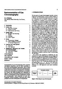
I. GC Separation A. GC Separation of TBDMS Derivatized Amino Acids 30 m DB-l (Methylsilicone) column, 150-250° at 3°/min, then 250275° at 100/min. Run time is 1 hour. Retention times and suggested ions for selected ion monitoring (SIM) of TBDMS derivatives of AAs are given in Table 9.1 and the GC separation is shown in Figure 9.1. Amino acids should be derivatized prior to separation. The TBDMS derivatives are preferred and stable for at least 1 week. B. GC Separation of PTH-Amino Acids 1. The only PTH-AAs that can be analyzed easily by GClMS without derivatization are alanine, glycine, valine, proline, leucine, isoleucine, methionine, and phenylalanine 30 m DB-l column, 75-275° at 8°/min. Dissolve the PTH-AA in the minimum amount of ethyl acetate. 2. Order of elution and molecular weight (MW) Alanine (206), glycine (142), valine (234), proline (232), leucine (248), isoleucine (248), methionine (266), and phenylalanine (282).
87
88
Chapter 9 • Amino Acids
fl. J)erivatiZlltion of Amino Acids and PTII-Amino Acids.
Table 9.1. Retention times and accurate masses for TBDMS derivatized amino acids (separation conditions Chapter 9.1.A)
2 100
Approximate retention time (min) 5:55 6:17 8:28 9:24 10:12 10:51 16:00 17:10 18:06 19:29 21:40 22:40 22:58 25:00 25:35 28:11 28:55 30:52 33:03 34:17 34:34 38:51 45:57
9
1
Amino acid Alanine Glycine Valine Leucine Isoleucine Proline Methionine Serine Threonine Phenylalanine Aspartic acid Hydroxyproline Cysteine Glutamic acid Asparagine Lysine Glutamine (peak 1) Arginine Histidine Glutamine (peak 2) Tyrosine Tryptophan Cystine
For selected ion monitoring 158.1356, 232.1553, 260.1502 218.1396, 246.1346 186.1678,228.1815, 260.1866 200.1834, 302.1972, 274.2022 200.1834,302.1972,274.2022 184.1522,286.1659,258.1709 292.1587,320.1536 390.2316 376.2523, 404.2473 234.1678,336.1815 418.2265 314.2335, 416.2473 406.2088 432.2422 417.2425 300.1815, 431.2945 431.2581 442.2741 338.2448, 440.2585 413.2476 466.2629 489.2789 348.1849
C. GC Conditions for TBDMS Derivatized PTH-Amino Acids* 15 m DB-5 column (or equivalent), 80-300° at 15°/min. D. GC Conditions for N-PFP Isopropyl Esters of D and L Amino Acids 25 to 50 m CHIRASIL-L-VAL column (Chrompack cat. no. 7495). *If the reaction time for the TBDMS derivatives is not long enough, a mixture of mono- and di-TBDMS derivatives is observed, resulting in more than one GC peak and thus reduced sensitivity.
89
19
60 8
10 21
60 5
0/01 40
o
7
I
II
j
100 7:18
20
13
6 34
20
15
11
~
1 14 12
18
23
1617
II 300 21:48
22
u
\ 1\.1 500 36:19
LJ.
Figure 9.1. GC separation ofTBDMS Derivatized Amino Acids (see Table 9.1) (1 = alanine, 2 = glycine, 3 = valine, 4 = leucine, 5 = isoleucine, 6 = proline, 7 = methionine, 8 = serine, 9 = threonine, 10 = phenylalanine, 11 = aspartic acid, 12 = hydroxyproline, 13 = cycsteine, 14 = glutamic acid, 15 = asparagine, 16 = lysine, 17 = glutamine (peak 1), 18 = arginine, 19 = histidine, 20 = glutamine (peak 2), 21 = tyrosine, 22 = tryptophan, and 23 = cystine.
80 (3 min)-190° at 4°/min. Separates d- and [-isomers of AAs (see Figure 9.2).
II. Derivatization of Amino Acids and PTH-Amino Acids A. t-Butyldimethylsilyl (TBDMS) Derivative J Reagents: N-methyl-N-(tert-butyldimethylsilyl), trifluoroacetamide, MTBSTFA: Add 0.25 ml of N,N-dimethylformamide (DMF) to the dried hydrolyzate. Add 0.25 ml of MTBSTFA reagent and cap tightly. Heat at 60° for 60 min, or overnight at room temperature (longer reaction times prevent mixtures of derivatives). Sample will need to be concentrated prior to injection. For trace analyses, it is important to use the minimum amount of solvent. B. N-PFP Isopropyl Ester Derivative Evaporate the sample to dryness with clean, dry nitrogen. Add 0.5 ml of 2N HCI in isopropyl alcohol. Heat at 100° for 1 hour. If tryptophan and/or cystine are suspected of being present, add 1 ml of ethyl mercaptan to prevent oxidation. Evaporate the reaction
90
Chapter 9 - Amino Acids
100
1/1. Mass SfJl'uralllll('/"fJretatioll _
Table 9.2.
Q)
91
Characteristic ions of TBDMS dcrivitizcd amino acids
c
~ 80
60
Deduced MW
39
" 302 200, 274 < 302 218,292,320 234, 302, 336 362, 390 303, 376, 404 378, 406 314,388,416 302,417 316, 390, 418 300,329,431 299, 329, 357, 431 272, 330, 432 196,338,440,459 199,340,442 302, 364, 438, 466 244, 302, 489 348, 537, 589, 639
Amino acid Glycine Alanine Proline Valine Leucine (elutes first) Isoleucine Methionine Phenylalanine Serine Threonine Cysteine Hydroxyproline Asparagine Aspartic acid Lysine (elutes first) Glutamine (first of two peaks) Glutamic acid Histidine Arginine (first of two peaks) Tyrosine Tryptophan Cystine
262 + R
This procedure works well with alanine, valine, threonine, isoleucine, glycine, leucine, proline, serine, aspartic acid, cystine, methionine, phenylalanine, tyrosine, ornithine, and lysine. III. Mass Spectral Interpretation A. Mass Spectra of TBDMS Derivatives of Amino Acids
Typical fragment ions are M - 15 (CH 3 ), M - 57 (C 4 H 9), M - 1i5 I (C4 H 9 + CO), M - 159 (C(O)-O-TBDMS). In general, if two fragment ions are observed that are 28 masS units apart, then 57 (C 4H 9 ) is added to the highest fragment ion to deduce the molecular weight of the TBDMS derivative. Identification of unknowns, as well as confirming the presence of known AAs, is more reliable if accurate mass measurement
data are also available. Identification is easily accomplished if mass spectra of the AAs are added to the computer-assisted library search routine (see Table 9.2). Selected ion monitoring of characteristic ions using previously determined retention time windows is generally used for trace analyses (see Table 9.1 and Figure 9.1). Accurate mass SIM reduces chemical noise at the expense of transmitted ion current. B. Mass Spectra of Underivatizcd PTH-Amino Acids
The phenylthiohydantion (PTH) derivative plus the AA backbone has a mass of 191 Daltons. Thus, subtracting 191 from the molecular ion of the PTH-AA derivative gives the mass of the AA side chain. A characteristic fragment ion is observed at mlz 135. Leucine and
92
III. Mass Spectral Illterprctarioll.
Chapter 9 • Amino Acids
isoleucine can be differentiated in the mass spectra by the absence of m1z 205 in the isoleucine spectrum. C. Mass Spectra of TBDMS Derivatives of PTH-Amino Acids Multiple TBDMS derivatives may form depending on the R group of the AA and the reaction time. Some of the PTH-AAs form TBDMS derivatives in less than 1 hour while others require overnight reaction times.
9 ~s
/ \ o=c" /NH"", CH
I
R
9 ~s /
\
)p",HO-C~C/NH
I
R
The MW of the TBDMS derivatives of PTH-AAs can be calculated using the following formula: MW = R + 191 + n(1l4), where n is the number of TBDMS groups. Arginine loses NH 3 during, or prior to, the derivatization reaction. Thus, the characteristic loss of 57 Daltons (C4 H 9 ) from the TBDMS derivative occurs at M - 74 (57 + 17). If the derivatization does not go to completion, it is a good idea to plot the values given in Table 9.3 and the values that are 114 Daltons less to determine the presence of a particular PTH-AA.
Table 9.3. Selected ion monitoring of some PTH-AAs as the TBDMS derivatives Component
Suggested ions to monitor
PTH-alanine-TBDMS PTH-glycine-TBDMS PTH-valine-TBD MS PTH-leucine-TBDMS PTH-isoleucine-TBDMS PTH-proline-TBDMS PTH-methionine-TBDMS PTH-serine-TBDMS PTH-threonine-TBDMS PTH-phenylalanine-TBDMS PTH-aspartic acid-TBDMS PTH-cysteine-TBDMS PTH-glutamic acid-TBDMS PTH-asparagine-TBDMS PTH-lysine-TBDMS PTH-glutamine-TBD MS PTH-arginine-TBDMS PTH-histidine-TBDMS PTH-tyrosine-TBDMS PTH-tryptophan-TBDMS PTH-cystine-TBDMS PTH-S-carboxymethylcysteine-TBDMS
191
377,734 363, 420 291,405,462 419,433,476 419,447,476 232, 346 437. 494 507, 564 521, 578 453, 510 507, 535, 592 549, 606 534,591 548, 605 416,446,473,662 (MW 719) 559. 616 557, 614 563, 640 606, 663 353,410
7\3
80 75
70 65
D. Mass Spectra of N-PFP Isopropyl Esters of D and L Amino Acids The molecular ion is usually not observed but can be deduced by adding 87 (COOC 3H 7 ) Daltons to the most abundant, high-mass ion. Masses 69 (CF 3) and 119 (C 2Fs) may also be observed. 2
60 55
50 45
432
40 35 30
\ 147
2
E. Sample Mass Spectrum TBDMS Derivatized Amino Acids Examining the mass spectrum of glutamic acid-TBDMS shows two high-mass ions that are 28 mass units apart (see Figure 9.3). By adding 57 (C4H 9 ) Daltons to the m/z 432 ion, the MW of the
93
12
\
!ih1u: ~ ~~l'2k-O+~~~d :1:,· I~ ~;r~'4~r. I
I
so
100
J,
i
Figure 9.3.
T8DM3 Derivatized Glutamic Acid
94
Chapter') • Amino Acids
TBDMS derivative is deduced to be 489. Characteristic fragment ions of the TBDMS derivative include the following: M M M M
-
15 (CH 3 ) 57 (C4H g ) 85 (C 4H g + CO) 159 (C(O) - TBDMS)
mlz mlz mlz mlz
474 432 404 330
Chapter
10
Common Contaminants
IV. References 1. Kitson. F. G.• and Larsen. B. S. In Mass Spectrometry of Biological Materials. C. N. McEwen and B. S. Larsen. Eds. New York: Marcel Dekker. 1990. 2. Gelpi. E., Koenig, W. A., Gilbert. J.• and Oro. J. Combined GC-MS of Amino Acid Derivatives. J. Chromat.. 7. 604, 1969.
I. Contaminants Occasionally Observed after Derivatization with TMS Reagents
mlz = 73,75,201.117 (MW = 216): octanoic acid-TMS mlz = 73, 75, 313. 328 (MW = 328): palmitic acid-TMS mlz = 73,75,341.356 (MW = 356): stearic acid-TMS mlz = 73, 99, 241. 147. 256 (MW = 256): uracil-TMS mlz = 73, 75, 111, 147.275 (MW = 290): adipic acid-TMS mlz = 73, 117, 147 (MW = 234): lactic acid-TMS mlz = 73, 130, 45, 59 (MW = 247): aminobutyric acid-DiTMS mlz = 73. 147,205 (MW = 308): glycerol-TMS mlz = 73, 147,233,245 (MW = 350): malic acid-TMS mlz = 73, 178, 135, 193, 192 (MW = 193): benzamide-TMS mlz = 73,273, 147 (MW = 480): citric acid-TMS mlz = 73,332, 147 (MW = 464): ascorbic acid-TMS mlz = 75, 73, 67, 55 (MW = 352): linoleic acid-TMS mlz = 75.73. 131. 45. 146 (MW = 146): propionic acid-TMS mlz = 75.73. 145,45 (MW = 160): n-butyric acid-TMS mlz = 75.73. 159 (MW = 174): valerie acid-TMS mlz = 75, 117,45.43.73 (MW = 132): acetic acid-TMS mlz = 91, 165, 135 (MW = 180): benzyl alcohol-TMS mlz = 105.206, 73, 308 (MW = 323): benzaminoacetic acid-TMS 95
96
Chapter!O • Common Contaminants jill
m/z m/z m/z m/z m/z m/z m/z m/z m/z m/z m/z m/z m/z m/z m/z m/z
= 147, 189,73 (MW = 204): urea-TMS = 147,227, 73, 93 (MW = 242): sulfuric acid-TMS = 151, 166 (MW = 166): phenol-TMS = 165,91, 180, 135 (MW = 180): o-cresol-TMS = 165, 180,91 (MW = 180): m-Cresol-TMS = 174,59,75,147 (MW = 319): aminobutyric acid-TriTMS = 179,105,77,135 (MW = 194): benzoic acid-TMS = 187, 75, 73, 69 (MW = 202): octanol-TMS = 189, 174 (MW = 189): indole-TMS = 195, 120,210 (MW = 252): acetylsalicylic acid-TMS = 203, 188 (MW = 203): skato1e-TMS = 255, 73, 113,270, 147 (MW = 270): thymine-TMS = 266,281,192 (MW = 281): aminobenzoic acid-TMS = 267, 73, 193, 223 (MW = 282): hydroxybenzoic acid-TMS = 285,117,132,145 (MW = 300): tetradecanoic acid-TMS = 299,314, 73 (MW = 314): phosphoric acid-TMS
!
Chapter
lUiS
11
Drugs and Their Metabolites
II. Contaminants Occasionally Observed in Underivatized Samples Ions Observed m/z m/z m/z m/z m/z m/z m/z m/z m/z m/z
= 84,133,42,162,161 98, 112, 30, 129 = 99, 155, 211 = 122, 105, 77 = 149, 167, 279 = 194, 109, 55, 67, 82 = 205, 220, 57 = 221,57,236,41,91 = 225, 93, 66, 65, 39 = 530, 57, 43, 515, 219
=
Compound
Nicotine BHMT Tributy1 phosphate Benzoic acid Dioctylphthalate Caffeine Di-tert-butyl cresol IonollOO Tinuvin-P Irganox 1076
III. Column Bleed
GC column bleed is a frequently encountered contaminant of mass spectra when high column temperatures are employed. Modern data systems offer the best way to eliminate this type of contamination by subtracting a spectrum showing column bleed from all other spectra in the GC/MS run.
Because drugs and metabolites are typically polar and thermally labile molecules, liquid chromatography/mass spectrometry (LC/MS) rather than GC/MS may be a more desirable approach. However, if GC/MS is used, more structural information may be obtained, particularly using accurate mass measurement electron impact ionization (ei) and chemical ionization (ci) combined with derivatization. It is our experience that wide bore (0.53 mm) GC columns can be used in place of narrow bore (0.25 mm) GC columns, resulting in greater sample capacity and less adsorption. The disadvantages are lower GC resolution and the need to use a jet separator. The splitter arrangement shown in Figure 11.1 should be considered.
I. GC Separations*
A. Underivatized 1. Basic drug screen 30 m DB-l column, 100-290° at 6°/min or 15 m DB-13Ol column, 150-250° at 15°/min (injection port at 280°).
2. Nicotine, pentobarbital, secobarbital, caffeine, oxazepam, and 'For metabolite work, first test the separation on the precursor drug or chemical.
97
98
Chapter II - Drugs alld "their Melil!>o!ile.1
9. o-Phcnylenediamine (OPO) metabolites 30 m OB-225 column, 75-225° at HjOfmin. Urine extracts from rats exposed to OPO were examined without derivatization. The major metabolites were identified as methylbenzimidazole, methylquinoxaline, and dimethylquinoxaline.
FlO
. HEWLEIT-PACKARD Fitting Cat. No. 18753-80010
\ Fused Silica _ Transfer Lines
Infector With Packed Capillary Insert
99
II. Salllple Preparation _
B. TMS Derivatives 1. Naloxone-TMS (MW = 471), nalbuphine-TMS (MW = 573)
30 m DB-1 column, 60-270 0 at lOa/min.
/
2. Methadone metabolites, methadone, cocaine (underivatized), morphine, and heroin 30 m DB-1 column, 100-250° at 100/min.
CHROMPACK Glass Lined Stainless Steel Make-up Tee Cat. No. 7881
Total Make-up Gas 30ml /minute
3. Daidzein and its metabolites (as TMS derivatives) HO Figure 11.1.
Splitter arrangement for wide bore GC columns.
o OH diazepam redissolved in methanol 15 m DB-5 or HP-5 column, 150-300° at 100/min. 3. Cocaine (MW = 303), codeine (MW = 299) morphine (MW == 285) 25 m DB-1 column, 100-280° at 15°/min.
Daidzein-TMS CZIHz604Siz (MW == 398.1369)
6. Amphetamines 30 m DB-1 column at 150°. 7. Fentanyl 30 m DB-I701 column at 270°. 8. Hexamethylphosphoramide and its metabolites 30 m FFAP-DB or DB-WAX column, 60-220° at 100/min.
Major Metabolite CZ4H340sSi3 (MW == 486.1714)
30 m DB-1 column, 100-280° at lOa/min C. Acetate Derivatives from Acid Hydrolysis Injection port: 280°, 30 m DB-1 column, 100-300 0 at 100/min.
4. Cocaine metabolites (e.g., ecogonine, benzoylecogonine, ctc.) 30 m DB-5 column, 200-280° at lOo/min. (Preparation of methyl ester and TMS derivatives is recommended.) 5. Naloxone (MW = 327) and nalbuphine (MW = 357) 30 m DB-I column, 150-280° at 15°/min.
OH
D. Methylated Barbituates and Sedatives 15 or 30 m DB-17 column, 100-250° at 1DO/min.
n.
Sample Preparation I-~
A. Extraction with Solvents Drugs and metabolites can be extracted from cultures and urinc by adding 2 drops of concentrated HCI to I ml of urine for a pH of 1-2. Extract with three I-ml volumes of diethyl ether (top layer) or methylene chloride (bottom layer). Combine extractions and evaporate with clean, dry nitrogen. Adjust to a pH of 8-10 by adding 250 ,ul of 60% KOH to I ml of urine. Extract
100
Chapter II • Drul\s (lnd Their Meta/Jolites
with three l-ml volumes of diethyl ether (top layer) or three I ml volumes of methylene chloride (bottom layer). Combine extractions and evaporate to dryness with clean, dry nitrogen. (See also reference 2.) B. Solid-Phase Extraction 1. Prepare the solid-phase extraction (SPE) tube (1 ml LC-18
SPE tube) by conditioning with I ml of methanol followed by 1 ml of water. 2. Extract the drugs and metabolites by diluting I ml of serum with I ml of O.1M sodium carbonate buffer (pH of 9). Force the mixture dropwise through the SPE tube previously prepared. Wash the SPE tube packing with three 200-/1,1 aliquots of water, dry it with nitrogen for 5 min, and elute the drugs with three 100-,u1 aliquots of 90 parts ethanol and 10 parts diethyl ether. Concentrate the recovered drugs by evaporating some or all of the solvent before analysis by GC/MS. (See Supelco Bulletin SlOB.) 3. Clean or change injection port liners frequently because nonvolatile materials in extracts from body fluids can accumulate in the injection port and/or head of the GC column and cause separation problems.
III. Derivatization of Drugs and Metabolites4 A. TMS Derivatives of Drugs and Their Metabolites Add 100 ,ul of MSTFA reagent to less than 1 mg of dry extract. Heat at 60° for 15-20 min. If necessary, add 250 ,ul of acetonitrile or other suitable solvent. For additional structural information. prepare the methoxime-TMS derivative to determine if one or more carbonyl groups are present. B. MO-TMS Derivative of Drugs Add 250 ,ul of methoxime hydrochloride in pyridine (MOX) reagent to less than 1 mg of dry extract. Let this solution stand at room temperature for 2 hours. Evaporate to dryness with clean, dry nitrogen. Add 250 ,ul of MSTFA reagent and let stand for 2 hours at room temperature.
IV. Mass Spectral Interpretation.
101
C. Acetyl Derivatives of Drugs Add 60 ,ul of acetic anhydride and 40 ,ul of pyridine to less than I mg of dry extract. Heat for I hour at 60°. Add excess methanol and evaporate to dryness with clean, dry nitrogen. Dissolve the residue in the minimum amount of butyl acetate or ethyl acetate.
D. Methylation of Barbiturates and Sedatives Heating MethElute (Pierce 49300X) with drug-containing extracts from body fluids gives quantitative methylation of barbiturates, sedatives, and so on. Follow the procedure provided by Pierce Chemical Company using the MethElute reagent. 15 or 30 m DB-17 column, 100-250° at 10 /min. 0
IV. Mass Spectral Interpretation A. Metabolites The mass spectra of metabolites will usually follow similar fragmentation pathways to those prevalent in the mass spectra of the precursor molecule. Thus, a knowledge of the possible biotransformations that can lead to metabolites is important. La Du et a\.2 list oxidation, reduction, and hydrolysis reactions that are common to living organisms. Some of the more common biotransformations are listed in the proceeding text with the exception of conjugated metabolites, which have insufficient volatility to be observed by GC/MS and are not considered. If conjugation is expected, it will be necessary to cleave the conjugate by hydrolysis before GC/MS analysis of the metabolite. l 1. Side-chain oxidation and hydroxylation of toluene
&OH 0a-Cresol
CHJOH
6 6 •
Toluene
Benzyl alcohol
102
Chapter I I • Drug,' and Their Mela/w!ile.1
IV IV/ilI\' Sl'eClra/ Imerl'retatiol1.
2. Epoxide formation and hydroxylation of benzene
0
~
5. N-Dealkylation [(CH,):,NhP=O --- [(CH,),NbP(O)NHCH,
[(po]
t
(CH,)2N - P(O)(NHCH 3 h
Benzene
By carefully examining the fragmentation pattern of the metabolite and comparison with the mass spectra of the precursor molecule, it is often possible to determine not only the nature of the biotransformation, but also its position in the molecule. In the proceeding example, accurate mass measurement was used to determine that a hydroxyl group had been added to the benzene ring containing the fluorine substituent.
Catechol (minor)
1
OH
Q
o
o
C-OTMS CH,.,
C-OTMS
II
II
OH
OH
F
F
Hydroquinone (trace)
Phenol (major)
mh~'6-OTMS
3. o-Dealkylation of 7-ethoxycoumarin
TMS-Major Metabolite C29H3103N IF 2Si 2 (MW = 535.1810)
TMS of Precursor C26H2302F2N 1Si 1 (MW = 447.1466)
7-Hydroxycoumarin
4. Hydroxylation and ketone formation of cyclohexane OH
0 Cyclohexane
.-
0
6 6 .-
Cyclohexanol
Cyclohexanone (trace)
CH 3
OF
GC conditions should be used that separate phenol, hydroquinone, resorcinol, and catechol.
7-Ethoxycoumarin
103
Molecular ion (MW = 535.1810) Intense ion (MW = 352,1169)
C 29 H,\O,NF:,Si:, C 2o H I9 0:,NFSi
Loss from the molecular ion
C j H 12 0FSi
The loss of 183.0641 Daltons from the molecular ion showed that the OH group was on the benzene ring containing the fluorine: F
lOOTMS
104
IV. Mass Spectral Interpretation _
Chapter II - Drugs and Their Metabolites
Table 11.1.
B. Drugs
Fragmentation and elution order of underivatized drugs*
Drug
Typical fragment ions
Amphetamine (MW = 135) N-methylamphetamine (MW = 149) Nicotine (MW = 162) Ephedrine (MW = 165) Barbital** (MW = 184) Aprobarbital (MW = 210) Tylenol (MW = 151) Phenacetin (MW = 179) Mescaline (MW = 211) Amobarbital** (MW = 226) Pentobarbital** (MW = 226) Meprobamate (MW = 218) Secobarbital (MW = 238) Caffeine (MW = 194) Glutethimide (MW = 217) Hexobarbital (MW = 236) Lidocaine (MW = 234) Phencyclidine (MW = 242) Doxylamine (MW = 270) Theophylline (MW = 180) Phenobarbital (MW = 232) Cyclobarbital (MW = 236) Procaine (MW = 236) Methaqualone (MW = 280) Methadone (MW = 309) Cocaine (MW = 303) Imipramine (MW = 280) Desipramine (MW = 266) Scopolamine (MW = 303) Codeine (MW = 299) Morphine (MW = 285) Chlordiazepoxide (MW = 299) Heroin (MW = 369) Flurazepam (MW = 387) Papaverine (MW = 339) Hydroxyzine (MW = 374) Thioridazine (MW = 370)
44, 91, 120 58,91,134 84,133,162 58, 77, 146 156, 141 162, 124, 195 109, 151, 80 108, 109, 179 181, 182, 211 156, 141 156, 141 55, 83, 96, 114, 144 168, 167, 195 194, 109, 55 189,117,132 221, 181, 157, 236 86, 58, 72, 234 200, 91, 84 58, 71, 183, 182, 200 180, 95, 68 204, 117,232 207, 141 86, 99, 120 235, 250, 91 72, 294, 309 82, 182, 303 58, 235, 280 44, 195, 235, 266 94, 138, 154, 303 299, 162, 229, 124 285, 162 282, 283, 284 327, 369, 268 86, 387, 315 339, 338, 324 201, 299, 374 98, 70, 370
*GC conditions given in Section I,A,l. **These drugs can be differentiated by retention time and the m/z 156 and m/z 157 abundance ratios.
105
The mass spectra of drugs are as varied as the molecules from which they are formed (see Table 11.1). Two major sources that a.re available for identifying drugs are computer library search routIne~ and Mass Spectral and GC Data of Drugs, Poisons and Their Metabolites.'
C. Sample Mass Spectrum The mass spectrum in Figure 11.2 shows a dominant molecular ion at m/z 151. The odd mass suggests the presence of an odd number of nitrogens. The observed loss of 42 Daltons from the molecular ion suggests an N-acetylated compound and the significant ion table ~see ~art III) suggests that m/z 109 is an aminophenol. By acetylatm.g WIth acetic anhydride and pyridine, the presence of an OH group wIll be confirmed. The molecular ion for the acetylated material will now be at m/z 193 with corresponding fragments at m/zs 151 and
109. An~t~er way to derivatize the OH group is by silylation using the Tn-sII Z reagent that will silylate the hydroxyl group, but not silylate the secondary amino group.
100°
95 90 85 80 75
q
70 65 60 55 50
45 40
35 30
151
25 20 15 10 5
O'-'-"r_-Jll-;::I;:~,LLc-;;t;YlL,-+.:-l~4L~-±:-,-J,-~,.--,lL,-,-~~,-t
106
Chapter JJ • Drugs alJd Their !'vfetl//",!i/es
References 1. Pfleger, K., Maurer, H., and Weber, A. Mass Spectral and GC Data of Drugs, Poisons and Their Metabolites. Weinheim, Germany: VCH Publishers, 198'\. 2. LaDu, B. N., Mandel, A.G., and Way, E. L. Fundamentals of Drug Metabolism and Drug Disposition. Baltimore: Williams and Wilkins, 1972. 3. Sunshine, 1., and Caplis, M., Eds. CRe Handbook of Mass Spectra of Drugs Boca Raton, FL: CRC Press, 1981. 4. Ahuja, S. J. Pharm. Sci., 65, 163, 1976. (See also Hewlett Packard Jon NOles, 6(2), 1991.)
Chapter
12
Esters
I. GC Separation of Esters of Carboxylic Acids
A. Capillary Columns 1. Methyl esters (general) 30 m FFAP column, 60-200° at 4°/min. 2. C 14-C Z4 methyl esters 30 m DB-23 column, 150-210° at 4°/min. 3. Methyl esters (Cg-C ZO polyunsaturated) 30 m DBWAX column, 80-225° at 8°/min or Omegawax 320 column, 100-200° at lOG/min. Run for 50 min. 4. Butyl esters 30 m DB-1 column, 70-250° at 6°/min. 5. Diesters: dimethyl malonate, dimethyl succinate, dimethyl glutarate, and dimethyl adipate 30 m CPSIL-88 column, 150-220° at 4°/min or 30 mOB-Wax column, 50-100° at 8°/min, 100-200° at lOa/min.
107
108
11. Milss Spectra of Esters _
Chapter 12 - Esters
(RT = 30 min) 30 m DB-17 column, 60-230° at 8°/min.
6. Dimethyl terephthalate (DMT) impurities Compound
Acetone Benzene Toluene Xylene Methyl benzoate H
Retention Time (min.)
m/z
0.9 1.3 2.2 2.7 4.8
58 78 lJ2 106 150
5.9
164
B. Packed Columns 1. Low-boiling methyl esters 2 to 3 m SP-1000 column on Carbopack C column, 75-175° at 8°/min.
Nc-Q--M-oCHJ
6.0 6.8
10.7
161 164 194
12.3
210
13.6
210
Mass Spectra of Esters
2. Molecular ion: If a molecular ion is not observed, it can be deduced by adding 31 or 32 mass units to the highest peak observed (neglecting isotopes). 3. Fragmentation: Cleavage of bonds that are adjacent to the carbonyl group gives rise to R+, [RC=O]', and [OCH 3]+ . An intense peak in the mass spectra of C6 -C26 methyl esters results from the McLafferty rearrangement:
o II
y
II
C-OCH3
Q~
1. General formula: RC0 2Me
CH2C-OCH3
o
CH 00C-
3. Mass spectra of pyrrolidine derivatives of methyl or ethyl esters: The molecular ions are intense with characteristic fragment ions at mlzs 70, 98, and 113. The position of the double bond can be determined by locating two peaks that differ by 12 mass units, A relatively intense peak should be observed 26 mlz units hig.her than the lower mass peak. The double bond lies between the two peaks that are separated by 26 Daltons. In Figure 12.1, peaks are observed at mlzs 126, 140, 154, 168. 182, 196, 210, 224, 236, 250, 264, 278, 292, 306, 320, and :\3:; (MW).
00 __
a>
111
112
Chapter /2 - Esters
II, Mass Spectra of Esters _
This procedure also applies to the case when two double bonds are present with only a CH 2 group separating them. (Not all cases have been examined.) C. Esters (R' > C j )
The molecular ion can be very small or nonexistent. Esters where R' is greater than methyl form a protonated acid that aids in the interpretation (e.g., m1z 47, formates; m/z 61, acetates; m/z 75. propionates; m/z 89, butyrates; etc.). Interpreting the mass spectra of ethyl esters may be confusing without accurate mass measurement because the loss of C2H 4 can be confused with the loss of CO from a cyclic ketone. A summary of the characteristic fragmentation of esters higher than methyl is as follows (see Figure 12.2):
-0 '(3 til ()
'0 c
t:!
::2:
0
Q>
C\J
Ul OJ
C')
>.
..c [j
N
...N
...::l Q)
Cl
u::
L1l ~
o----------~-=:::!_<S
cA o
I'
i
L1l 0
I'
t
I
'I " I
L1l 0
~
'I!I,:
liii i
I I
1. General formula: ROOceCH2)nCOOR'
2. Molecular ion: The molecular ion is generally not observed for diesters larger than diethyl malonate.
""",~.,,~ ,,~~
L1l OL1l0L1l0L1l0L1l0L1l0L1l0L1l0
00'>0)00001"-
; :1
'C til
15
D. Methyl Esters of Dibasic Acids
E. Ethyl and Higher Esters of Dibasic Acids
OJ
1:5
• M+' is small. • [M - R']+ • [M - OR']+ • [RCOOH2]+ • [R - H]+ or [R - 2H]+ • McLafferty rearrangement m1z 88 for ethyl esters: If the a-carbon has a methyl substitution, the major rearrangement ion will be at m1z 102.
Molecular ions are not always observed for methyl esters of dibasic acids; however, methyl esters of all dibasic acids lose 31 and 73 Daltons from their molecular ions. By adding 31 or 73 to the appropriate ions, the molecular weight can be deduced.
til
()
f'.,.
6-Keto-PGF1-methoxime-tetraTMS.
Prostaglandin
3. 30 m DB-225 column, 100-225° at ISo/min; injection port at 280°.*
566
l.JL.-~. ~4~0 500~0
4--' " ,.. '
..r-
Characteristic Abundant Ions
(MW (MW (MW (MW
= = = =
509) 509)* 507) 599)
mlz 388, 419, 438, 478 mlz 388, 438, 478, 494 mlz 386, 476, 492, 507
mlz 426, 478, 528, 568, 584
Prostaglandin
Most Abundant Ions
PGErMO-TMS (MW = 597) PGFja-TMS (MW = 644)t 6-KetoPGF t -MO-TMS (MW = 687) PGF2 a-TMS (MW = 642) Thromboxane BrTMS (MW = 658) 12-HETE-TMS (MW = 464)
mlz 436, 566, 582, 476 mlz 367, 368, 438, 483
mlz mlz mlz mlz
436, 476, 566, 656 391,462,481,552 211, 301, 387 213, 324, 449
*PGB j can be distinguished from PGA I by the more intense m/z 478 ion relative to the other fragment ions in the mass spectrum of PGBI-MO-TMS. tIn the mass spectra of TMS derivatives of nonketoprostaglandins. the molecular ion is usually lIot observed. Generally. the fragment ions are M - 15. M - 71, M - 90, M - 161. and Ai - 180.
Chapter 30
Solvents and Their Impurities
There are generally two types of analyses that are requested with reference to solvents. The first is the identification of residual solvents in products, and the second is the identification of impurities in common industrial solvents. Certain GC conditions have been found to separate most of the common solvents. Always examine the mass spectra at the front and back of the GC peaks to determine if they are homogeneous. Also remember that isomers may not be detected by this approach if they are not separated. I. GC Separations of Industrial Solvent Mixtures A. Capillary Columns 1. Acetone, THF, methanol, ethyl acetate, isopropyl acetate,
isopropyl alcohol, ethanol, n-propyl alcohol, toluene, and 2-methoxyethanol a. 30 m DB-FFAP column or 30 m HP-FFAP column, 60-200° at 6°/min. b. 25 m DB-WAX column or 25 m CP-WAX 52CB column, 50 (2 min)-65° at 1°/min; 65-150° at 100/min. 2. Ethanol, acetonitrile, acetone, diethyl ether, pentane, ethyl acetate, and hexane 25 m Poraplot Q column, 60-200° at 6°/min.
217
218
III. Mass Spectra ot'Solvcnts and 7heir Impurities _
Chapter 30 - Solvents and Their Impurities
25 m CP-WAX 57 column for glycols and alcohols (Chrompack catalog no. 7615),50 (5 min)-180° at gO/min.
3. Benzene, propyl acetate, allyl acetate, 1-pentanol, cyclohexanone, cyclohexanol, dicyclohexyl ether, cyclohexyl valerate, butyric acid, valerie acid, caproic acid, 1,5-pentanediol, dicyclohexyl succinate, and dicyclohexyl glutarate 30 m DB-FFAP column, 60-200° at 6°/min.
6. Isopropyl alcohol impurities: ethanol, n-propyl alcohol, and t-butyl alcohol 30 m NUKOL column, 45-200° at 6°/min.
B. Packed Columns
7. t-Butyl alcohol, n-butyraldehyde, methyl ethyl ketone, 2-methylfuran, tetrahydrofuran, 4-methyl-1,3-dioxolane, 2-methyl THF, 3-methyl THF, and tetrahydropyran 30 m DB-1 column at 60°
1. Methanol, ethanol, acetone, methylene chloride, methyl acetate, THF, methyl cellosolve, ethyl cellosolve, butyl cellosolve, n-xylene, p-xylene, and a-xylene 10-ft 3% SP-1500 column on 80-120 MESH Carbopack* 60-225° at 4°/min. Run for 50 min.
8. Toluene impurities: benzene, ethylbenzene, and a-, m-, and p-xylenes 25 m DB-WAX column, 50 (2 min)-180° at 5°/min.
2. Acetaldehyde, methanol, ethanol, ethyl acetate, n-propyl alcohol, isobutyl alcohol, acetic acid, amyl alcohol, and isoamyl alcohol lO-ft Carbowax 20M column on Carbopack B, 70-170° at 5°/min.
B. Packed Columns Tetrahydrofuran (THF) impuntles: acetone, acrolein, 2,3-DHF, butyraldehyde, isopropyl alcohol, THF, 1,3-dioxolane, 2-methyl THF, benzene, and 3-methyl THF 10-ft 3% SP-1500 column on Carbopack B, 60-200° at 6°/min.
II. GC Separations of Impurities in Industrial Solvents
A. Capillary Columns 1. Diaminotoluene impurities: dichlorobenzene, aminotoluene. nitrotoluene isomers, and dinitrotoluene isomers 30 m DB-17 column, 100-250° at 8°/min. 2. 3,4-Dichloroaniline impurities: aniline, chloroaniline, dichlaraaniline isomers, trichloroaniline isomers, tetrachloroaniline. tetrachloroazobenzene, and pentachloroazobenzene 30 m DB-1 column, 100-280° at 100/min. 3. Diethylene glycol impurities: acetaldehyde, 2-methyl-1,3-dioxolane, dioxane, and diethylene glycol 30 m Nukol column, 50-220° at 8°/min. 4. N,N-dimethylacetamide (DMAC) impurities: N,N-dimethylacetonitrile, N,N-dimethylformamide, DMAC, N-methylacetamide. and acetamide 60 m DB-WAX column, 60-200° at 7°/min or 3 m Tenax-GC packed column, 100-120° at 3°/min. 5. Ethanol impurities: ethyl acetate, methanol, n-propyl alcohol. isobutyl alcohol *This is the only column we have found that separates the cellosolves (monoalkyl ethers of ethylene glycol) and the other solvents including the xylenes. (See Supelco GC Bulletin 112'+.)
219
m.
Mass Spectra of Solvents and Their Impurities
Solvents and their impurities represent a wide class of compound types; therefore, a discussion of common mass spectral features is meaningless. However, most of the mass spectra are listed in computer library search programs and "The Eight Peak Index."
Chapter 31
Steroids
I. GC Separation of Underivatized Steroids
A. Animal or Plant Steroids 30 m DB-17 column, 225-275° at 5°/min; injection port at 290-300°. (Dissolve the dried extract in the minimum amount of methylene chloride or toluene.)
n.
Derivatization of Steroids
A. Preparation of TMS Derivatives: Add 0.25 ml of TRI-SIL TBT reagent (the only reagent we found to react with the hydroxyl group in position 17) and 0.25 ml of pyridine to the dried extract. Cap tightly and heat at 60° for 1-12 hours or overnight. B. Preparation of Methoxime (MO)-TMS Derivatives (especially for hydroxyketosteroids, which may decompose under the given GC conditions unless the MO-TMS derivatives are prepared.): Add 0.25 ml of methoxime hydrochloride in pyridine to the dried extract. Let stand for 3 hours at 60° or overnight at room temperature. Evaporate to dryness with clean, dry nitrogen. Add 0.25 ml of TRI-SIL TBT and 0.25 ml of pyridine to the dried reaction mixture. Cap tightly and heat at 60° for a few hours or overnight at room temperature. 221
222
Chapter 31 • Steroidl'
V. Mass Spectra ot' TMS Derivatives of Steroids.
III. GC Separation of Derivatized Steroids
223
liD
!5 II IS II 43
A. TMS Derivatives
1. 30 m DB-17 column. 225-275° at SO/min.
l'
2. 15 m CP-SIL 5 column or 15 m CP-SIL 8 column, 200-300° at ISo/min.
10 65
55
J
iJ 65
B. MO-TMS Derivatives
1. 30 m DB-17 column, 100-200° at ISo/min, then 200-275" at SO/min; injection port temperature at 280-300°.
!O G
81 107
16
69
XI
2. 30 m CP-SIL 5 column 100-210° at 20 /min, then 210-280° at 2°/min. 0
IV. Mass Spectra of Underivatized Steroids
275
,
1:1
145
368 301
'[119!
I~
353 133',1r
I
213231255
I 11,
I
j'.J,.-JLI'.,J I-J IL"'."rli~L,1JI !1;:-¥i91:A!I-'I-~JII~te+YII--i:'-I""~~= -9 illl\-lUl:
\-:.'I'--J1L1
5
100
1 0
200
250
3( 0
3f 0
Figure 31.1.
22
M/Z
Cholesterol.
26
Daltons below the molecular ion. Typically, this ion corresponds to M - (R + 42), where 42 is C 3H 6 . If there is no side chain, as in the cases of estrone, estradiol, and estriol, the substitution on the D-ring can be determined using the following losses from the molecular ions:
2 27 3 4
$5oH f61 $J
6
A. Molecular Ion: Because of low volatility, most steriods are derivatized before analysis by GC/MS. Molecular ions are usually observed for steriods sufficiently volatile to be analyzed underivatized by GC/MS (see Figure 31.1) Some important steroids in urine include estrone, estradiol, estriol, pregnanediol, and 17-ketosteroids. which can be analyzed by GC/MS as the TMS or the MO-TMS derivatives. The plant steroids, such as camposterol, ergosterol. stigmasterol, cholestanol, and sitosterol, are generally analyzed as the TMS derivatives. B. Characteristic Fragment Ions: The most characteristic cleavage is the loss of carbons 15, 16, and 17, together with the side chain and one additional hydrogen. It is possible to determine the elemental composition of the side chains of steroids by the difference in the mass between the molecular ion and an intense ion more than 15
0
H
Loss of mass 57
OH
H
Loss of mass 59
H
Loss of mass 75
i
\7.
Mass Spectra of TMS Derivatives of Steroids
Plot mass 73 to determine if a TMS derivative was prepared and which GC peak(s) to examine. A. Molecular Ion: Generally, the molecular ion is observed, but not always, as in the case of pregnanediol-TMS (MW = 464). Again. if two high-mass peaks are not observed that are 15 Daltons apart,
224
Chapter 31 • Steroids
100 0
1
9
95 90 85 80
Chapter 32
329
368
75 70_
I
65 60
55 50 45
75
Sugars (Monosaccharides)
I 95
1
40~" 57 35-3
353
145
30] 25
159
247
20
I
I
15~ I
10 5_
o 50
I
213
I
I
73
15~ ~I
2t
ll
Figure 31.2.
458
I
275
2~~ i~~~~~~~~>-c~--L Cholesterol-TMS~
then add 15 Daltons to the highest mass observed to deduce the molecular ion. B. Fragment Ions: Common losses from the molecular ions include 90 and 105 Daltons. (See Figure 31.2 for the mass spectrum of the TMS derivative of cholesterol.) VI. Mass Spectra of MO·TMS Derivatives A. Molecular Ion: The molecular ions of MO-TMS derivatives are generally more intense than those of the TMS-only derivatives.
B. Fragment Ions: The M - 31 fragment ion is characteristic and is more intense than the M-15 ion. If two high-mass ions are observed that are 16 Daltons apart, add 31 Daltons to the more intense ion and 15 Daltons to the highest mass ion to deduce the molecular ion. By subtracting 29 Daltons for each keto group and 72 Daltons for each hydroxyl group, the original molecular weight of the steroid can be determined. For example, in the case of androsterone-MOTMS, the molecular ion occurs at mlz 391. By subtracting 101 (29 + 72) Daltons, the original molecular weight was mlz 290.
I. GC Separation of Derivatized Sugars
A. Monosaccharides 1. Preparation of the TMS derivative: Add 0.5 ml of TRI-SIL Z reagent (trimethylsilylimidazole in pyridine) to 1-5 mg of the sample. (This derivatizing preparation does not react with amino groups and tolerates the presence of water.) Heat in a sealed vial at 60° until the sample is dissolved. An alternate method is to let the reaction mixture stand at room temperature for at least 30 minutes (or overnight). This procedure is not appropriate for amino sugars. 2. GC separation of TMS derivatives: arabinose, fucose, xylose, mannose, galactose, a-glucose, and f3-glucose 30 m DB-l column, 60-250° at gO/min. B. Amino Sugars (Glucosamine and Galactosamine as TMS Derivatives) 1. Preparation of TMS derivative: To derivatize the amino sugars as well as the nonamino sugars, substitute TRI-SIL TBT or TRISILIBSA (Formula P) reagent for TRI-SIL Z and follow the procedure given in Section I,A,1.
225
226
If. Mass Spectra/Interpretation.
Chapter 32 • Sugars (Monosaccharides)
2. Fragmentation: Dejongh et a1. 1 have described the fragmentation of the TMS derivatives of sugars. Comparison of GC retention times together with the mass spectra is sufficient to identify the sugars. The mass spectra suggest certain structural features. For instance, mlz 191, 204, and 217 suggest a TMS hexose. If mlz 204 is more abundant than mlz 217, the hexose is the pyranose form, but if mlz 217 is most abundant then it is a furanose (Figure 32.1). Aldohexoses can be differentiated from ketohexoses by the ion at mlz 435 for aldohexoses versus mlz 4~7 for ketohexoses.
2. GC separations of amino sugars as TMS derivatives 30 m DB-1701 column, 70-250° at 4°/min. (Depending on the amounts present, complete GC separation may not be achieved.) C. Sugar Alcohols (as Acetates) 1. Preparation of the acetate derivative: Evaporate the extract to dryness. Add 50 ILl of three parts acetic anhydride and two parts pyridine. Heat at 60° for 1 hour. Evaporate to dryness with clean. dry nitrogen and dissolve the residue in 25 ILl of ethyl acetate. 2. GC separation of the acetate derivatives: rhamnitol, fucitol, ribitol, arabinitol, mannitol, galacitol, glucitol, and inositol 30 m CP-Sil 88 column at 225° for 60 min. D. Reduced Sugars (as Acetates)
1. Preparation of the acetate derivative: Concentrate the aqueous mixture of saccharides to approximately 0.5 ml in a 20-50 ml container. Reduce the saccharides by adding 20 mg of sodium borohydride that has been dissolved carefully into 0.5 ml of water and let the reducing mixture stand at room temperature for at least 1 hour. Destroy the excess sodium borohydride by adding acetic acid until the gas evolution stops. Evaporate the solution to dryness with clean nitrogen. Add 10 ml of methanol and evaporate the solution to dryness. Acetylate with 0.5 ml (three parts acetic anhydride and two parts pyridine) overnight. Evaporate to a syrupy residue and add 1 ml of water. Evaporate again to dryness to remove the excess acetic anhydride. Dissolve the residue in 250 ILl methylene chloride.
227
mlz
Ion Structure
191 204 217
(CH 3 )3 Si 0 CH (CH 3h Si 0 CH (CH 3h Si 0 CH
= = =
0 Si (CH 3hl+ CH 0 Si (CH 3 )31+' CH - CH = 0 Si (CH 3 hl+
B. Sample Mass Spectrum C. Mass Spectra of Amino Sugars as TMS Derivatives 1. Molecular ion: By plotting out the molecular ion mass and mass 362.1639, the presence of these amino sugars can be determined.
100 0
95
2. GC separation of reduced and acetylated sugars 30 m CP-Sil 88 column at 225°.
90 85
1=
f
80 75
II. Mass Spectral Interpretation A. Mass Spectra of TMS Derivatives of Sugars 1. Molecular weight: In general, to deduce the molecular weight of the TMS derivative of sugars, add to 105 the highest masS observed. • 333.1374 is the highest mass observed for arabinose. ribose. ribulose, xylose, lyxose, and xylulose. • 347.1530 is the highest mass observed for fucose and rhamnose. • 435.1875 is the highest mass observed for sorbose, allose. altrose, galactose, gulose, idose, and mannose.
~
70 65 60 55
r
50 l:45 ,40 -, 35
t
~[
30
25 20
147
15
10
f
5
O-'4--~4-+_~~-.ljL~'c+--I~.
Figure 32.1.
-::----"--c-J.~-:T(\~~-4-~3'T50
TMS-u-glucose.
-
~-4Cl~-Mii
228
Chapter 32 • Sugars (Monosaccharides)
Highest Mass Observed
Glucosamine Galactosamine
Chapter 33
393.1643 467.2375
2. Fragmentation: The TMS derivatives of amino sugars have their base peak at mass 131.0765.
Sulfur Compounds
D. Mass Spectra of Sugar Alcohols as Acetates 1. Molecular ion: Chemical ionization using ammonia as reagent gas establishes the molecular weights of sugar acetates. 2. Fragmentation: The acetates have intense ions at mlzs 43, 103. and 145. They also appear to lose 42, 59, 60, and 102 Daltons from their molecular ions. CH20C(O)CH3
73
I
~
I
CHOC(O)CH3 I
145 ---- 103
I. GC Separations
A. Capillary Columns
CHOC(O)CH3
217 ----157 -115
I CHOC(O)CH 3
289 --187 --127 etc.
The mass spectrum of alditol acetates are easy to interpret as they fragment at each C-C bond as shown.
Reference
c., Radfon, T., Hribar, J. D., Hanessian, S., Biebar, M., Dawson. G., Sweeley, C. C. Analysis of derivatives of carbohydrates by GC/MS. 1. Am.
1. Dejongh, D.
Chern. Soc., 91, 1728, 1969.
1. Hydrogen sulfide, carbonyl sulfide, sulfur dioxide, and methyl mercaptan 25 m GS-Q column or 25 m Poraplot Q column, 50-120° at 5°/min.
2. Hydrogen sulfide, methane, carbon dioxide, ethane, and propane 25 m Poraplot Q column or 25 m GS-Q column at 60°. 3. Carbon dioxide, carbonyl sulfide, hydrogen cyanide, propylene, butadiene, and furan 25 m GS-Q column or 25 m Poraplot Q column, 60-200° at 6°/min. 4. Carbonyl sulfide, carbon disulfide, tetrahydrofuran, and toluene 25 m DB-l column, room temperature to 200° at 100/min. 5. Methane, ethene, ethane, propene, acetaldehyde, methyl formate, butene, acetone, furan, dimethyl sulfide, isoprene, isobutyraldehyde, diacetyl, methylfuran, and isovaleraldehyde 25 m Poraplot Q column or 25 m GS-Q column, 60-200° at 8°/min.
229
230
Pari II • References
II. Mass Specrra of'Sul/llr Compounds.
6. 2-Propanethiol, l-propanethiol, 1-methyl-1-propancthiol. 2methyl-1-propanethiol, l-butanethiol, 1-pentanethiol, allyl sulfide, propyl sulfide, and butyl sulfide 25 m GS-Q column or 25 m Poraplot Q column, 150-23()O at 100/min. 7. n-Propyl disulfide and n-propyl trisulfide 25 m DB-FFAP column at 1000.
8.
Cl
/
N--C-Cl
N--C-Cl
II
II
II
II
II
100°
95 90 85
4
80 75 70
70
65
N--C-Cl
II
C" /C-Cl, / C " /C-Br, and C " /C-Br S Cl S Br/ S
25 m DB-210 column at 120°. 9. Phenanthrylene sulfide and pyrene 30 m DB-210 column, 150-225 0 at 5°/min.
I
60 55 50 45 29
40 146
35 30 25
47
20 15
61 112
1~O'..>ill,--,-lflLo-"-,-J+LL,L,l-Ujl.--.-,.~,1~lT~_L i 89
40
1. Air, carbon dioxide, hydrogen sulfide, carbonyl sulfide, propane. and sulfur dioxide 2 m Chromosorb 101 column at 75 0. 2. Methane, ethene, ethane, butene, acetone, furan, dimethylsulfide, isoprene, and methylfuran 3 m Porapak QS column, 60-2000 at 8°/min.
II. Mass Spectra of Sulfur Compounds A. Aliphatic thiols (mercaptans) 1. General formula: RSH 2. Molecular ion: The presence of sulfur can be detected by the 34S isotope (4.4%) and the large mass defect of sulfur in accurate mass measurements. In primary aliphatic thiols, the molecular ion intensities range from 5-100% of the base peak. 3. Fragment ions: Loss of 34 (H 2 S) Daltons from the molecular ions of primary thiols is characteristic. In secondary and tertiary thiols, 33 Daltons rather than 34 Daltons are lost from their molecular ions. If m/z 47 and 61 are reasonably abundant ions. and m/z 61 is approximately 50% of m/z 47, then the mass spec-
HS
84
11
B. Packed Columns
231
60
80
I
:
1
1uO
Figure 33.1.
__
120
~--JliL.~~~-.J:. 1 0 M/Z
1-0ctanethiol.
trum represents a primary thiol (see Figure 33.1). If m/z 61 is much less than 50% of m/z 47, then the mass spectrum represents a secondary or tertiary thiol. Also, remember that saturated sulfur compounds generally fragment beta to the sulfur atom to lose the largest alkyl group. B. Thioethers (Sulfides) 1. General formula: R-S-R' 2. Molecular ion: Molecular ions are usually reasonably abundant. Cyclic thioethers give abundant molecular ions and an abundant fragment ion due to double ,B-cleavage (e.g., m/z 60 [CHrSCH 2l')· 3. Fragmentation: The thioethers can be distinguished from the primary, secondary, and tertiary thiols by the absence of the losses of either 33 or 34 Daltons from their molecular ions. Fragmentation also occurs beta to the sulfur atom with dominant loss of the larger alkyl group. Fragmentation can occur on either side of the sulfur atom with the rearrangement of a hydrogen atom.
232
Part lJ - References Parr II References _
C. Aromatic Thiols
E. Isothiocyanates
1. General formula: ArSH
I. General formula: RNCS
2. Molecular ion: Aromatic thiols show intense molecular ions (see Figure 33.2).
2. Molecular ion: Molecular ions are observed.
3. Fragmentation: Characteristic losses from the molecular ion include: 26, 33, and 44 (C=S) Daltons. Ions characteristic of the phenyl group are also observed.
1. General formula: RC(O)SR'
2. Molecular ion: The molecular ion of thioesters is more intense than that of the corresponding oxygenated esters. 3. Fragmentation: R~ and RCO+ are frequently the most abundant ions observed. RCOS+, SR'+, and R'+ are usually observed.
100° 95 90 85 80 75 70 65 60 55 50 45 40 32 35
SH I
Bierman, C. J. J. Chromatogr., 357, 330, 1986. Early, R J., Thompson, J. R, Sedgwich, G. W., Kelly, J. M., and Christopherson, R J. J. Chromatogr., 416, 15, 1987. Gelpi, E., Koenig, W. A, Gilbert, J., and Oro, J. 1. Chromatogr., 7, 604, 1969. Kitson, F. G., and Larsen, B. S. In C. N. McEwen and B. S. Larsen, Eds., Mass Spectrometry of Biological Materials. New York: Marcel Dekker, 1990. Marquez, C. D., Weintraub, S. T., and Smith, P. C. J. Chromatogr. B: Bio. Med. Appl., 658, 213, 1994. Zhu, P., Yu, Z., Wan, G., Sha, H., Su, K, Yu, B., and Zhao, G. Org. Mass Spectrom., 26, 613, 1991.
Cohen, B. I., Kuramoto, T., Rothschild, M. A, and Mosbach, E. H. J. BioI. Chem., 251, 2709, 1976.
Cannabinoids Harvey, D. H. Chapter 6. In D. M. Desiderio, Ed., Mass Spectrometry Clinical and Biomedical Applications (Vol. 1). New York: Plenum Press, 1992.
Carbohydrates 93
97
OI-'h-W:ljl"J",-+lh-.,+JMh-,.--rR-h--rfr,-~~I-r-c":'l~7:c:~r;;-'''''''M:;:/~Z 30
Part II References
Bile Alcohols
80
15 10 5
3. Fragmentation: A characteristic fragment ion is observed at mlz 72 (CH 2 =N=C=S). For R>n-C 5 , mlz 115 is promment.
Amino Acids
D. Thioesters
30 25 20
233
NH2
Pitkanen, E., and Kanninen, T. Bioi. Mass Spectrom., 23, 590, 1994. Previs, S. F., Ciraolo, S. T., Fernandez, C. A, Beylot, M., Agarwal, K C, Soloviev, M. v., and Brunengraber, H. Anal. Biochem., 218, 192,1994.
farboXYIiC Acids teihoffer, J., and Ferguson, C. J. Chromatogr. Sci., 32, 102, 1994. fawamura, K Anal. Chem., 65, 3505, 1993.
I
234
Part II _ References Part JJ References _
235
Carnitines
Gasoline Additives
Millington, D. S., and Chace, D. H. Chapter 8. In D. M. Desiderio, Ed.. Mass Spectrometry Clinical and Biomedical Applications (Vol. 1). New York: Plenum Press, 1992. Van Bocxlaer, J. F., Claeys, M., Van den Heuvel, H., and De Leenheer. A. P.1. Mass Spectrom., 30, 69, 1995.
Kania, H .. Inouye, V.. Coo, R., Chow, R. Yazawa, L., and Maka, J. Anal. Chem., 66, 924, 1994. Schutjes, C. P. M.. Vermeer, E. A, Rijks. J. A., and Cramers, CA. J. Chromatogr., 253, 1, 1982.
Drugs and Metabolites
Lipids
Gudzinowicz, B. J. and Gudzinowicz, M. J. Analysis of Drugs and Metahu/ites by Gas Chromatography Mass Spectrometry (Vols. 1-5). New York: Marcel Dekker, 1977. Jaeger, H., Ed. Capillary Gas Chromatography-Mass Spectrometry in Medicine and Pharmacology. Heidelberg, Germany: Heutig, 1987. LaDu, B. N., Mandel, A. G., and Way, E. L. Fundamentals of Drug Metabolism and Drug Disposition. Baltimore: Williams & Wilkins, 1972. Lui, F., Hu, X. Y., and Luo, Y. J. Chromatogr. B-Bio. Med. Appl., 658, 375. 1994. Pfleger, K., Maurer, H., and Weber, A. Mass Spectral and GC Data of Drugs, Poisons, Pesticides, Pollutants, and Their Metabolites (Vol. 1). Weinheim: VCH Publishers, 1992. Schanzer, W., and Donike, M. BioI. Mass Spectrom., 1, 3, 1992. Sunshine, I, and Caplis, M. CRC Handbook of Mass Spectra of Drugs. Boca Raton. FL: CRC Press, 1981.
Adams, J., and Qinghong, A Mass Spectrom. Rev., l2, 51. 1993. Christie, W. W. Gas Chromatography and Lipids: A Practical Guide. Alloway: The Oily Press. 1989. Murphy, R. C Mass Spectrometry of Lipids. New York: Plenum Press, 1993. Shibamoto, T, Ed. Lipid Chromatographic Analysis. New York: Marcel Dekker, 1993.
Microbiology Odham, G., Larsson, L., and Mardh, P. A., Eds. Gas Chromatography/Mass Spectrometry: Applications in Microbiology. New York: Plenum Press, 1984.
Nucleosides Dyes Straub, R F., Voyksner, R D., and Keever, J. T Anal. Chem., 65, 2131, 1993.
Langridge, J. I., McClure, T D., El-Shskawi, S., Fielding, A., Schram, K. H., and Newton, R. P. Rapid Commun. Mass Spectrom., 7,427, 1993. Teixeira, A J. R, Van de Werken, G., Stavenuiter, J. F. C, De Jong, A P. J. M., Westra, J. G., and Van der Greef, J. BioI. Mass Spectrom., 9, 441, 1992.
Environmental American Chemical Society. EPA Methods (Iris. Air Toxics, and Pesticide Methods) (1155 16th Street, N.W., Washington, DC) Clement, R E., and Tosine, H. M. Mass Spectrom. Rev., 7, 593, 1988. Hites, R. A. Mass Spectra of Environmental Contaminants. Boca Raton. FL: eRe Press, 1992.
Essential Oils Masada, Y. Analysis of Essential Oils by GC and MS. New York: John Wiky. 1976.
PCBs and Other Polyhalogenated Compounds 8aIlschmiter, K., Bacher, R., and Mennel, A. 1. High Res. Chromatogr., 15, 260, 1992. Samsonnov, D. P., Keryukhin, V. P., and Zhiryukhina, N. P. J. Anal. Chem., Engl. Tr., 49, 783, 1994. ~tton, M. L., Frenkel, M., and Marsh, K. N. Spectral Data for PCBs. Boca Raton, FL: CRC Press, 1994. ;t\
1teSticides
*,
Fluorinated Compounds Simmonds, P. G., O'Doherty, S., Nickless, G., Sturrock, G. A, Swaby, R, Knight. P., Richetts, J., Woffendin, G., and Smith. R Anal. Chem., 67, 717, 1995.
irns, T., and Baldwin. R. A. Anal. Chem., 67, 552A. 1995. isert, R, and Levsen, K. J. Am. Soc. Mass Spectrom., 6, 1119,1995. asin, M., Baugh, P. J., Hancock, P., Bonwick, G. A, Davis, D. H., and Armitage. R. Rapid Commun. Mass Spectrom., 9, 1411,1995.
236
Part
lJ •
References
Plasticizers and Polymer Additives Asamoto, B., Young, J. R., and Citerin, R. J. Anal. Chem., 62, 61, 1990. Cortes, H. J., Bell, B. M., Pfeiffer, C. D., and Graham, J. D. 1. Microcol. Separ., 1, 278, 1989.
Polymer Analysis Cho, W. J., Choi, C. H., and Ha, C. S. 1. Polym. Sci-A. Polym. Chem., 32,2301, 1994, MacMahon, T., and Chace, M. 1. Am. Soc. Mass Spectrom., 5, 299, 1994. Montaudo, G., Puglisi, c., Blazso, M., Kishore, K., and Ganesh, K. J. Anal. Appl. Pyrol., 29, 207, 1994. Ohtani, H., Luo, Y. F., Nakashima, Y., Tsukahara, Y., and Tsuge, S. Anal. Chon .. 66, 1438, 1994.
Par t
I I I
Ions for Determining Unknown Structures
Prostaglandins Knott, 1., Raes, M., Dieu, M., Lenoir, G., Burton, M., and Remade, J. Anal. Biochem., 210, 360, 1993. Waddell, K. A., Blair, 1. A., and Wellby, 1. Biomed. Mass Spectrom., 12, 399.1985. Waller, G. R., Ed. Biochemical Applications of Mass Spectrometry. New York: Wiley (Interscience), 1972.
•
Steroids Pfaffenberger, C. D., Malinak, L. R., and Homing, E. C. J. Chromatogr., 158, 313,1978.
Tobacco Smoke Dong, M., Schmeltz, 1., Jacobs, E., and Hoffman, D. 1. Anal. Toxicol.,2, 1978. Zahlsen, K., and Nilsen, O. G. Pharmacal. Toxicol., 75, 143, 1994.
Volatile Organic Compounds and Headspace Analysis D'Agostino, P. A., and Porter, C. J. Rapid Commun. Mass Spectrom., 6, 717. \992. Manura, J. J. American Laboratory, March 1994. Rodgers, R. S. American Laboratory, December 1993.
•
•
•
M+1
Possible Precursor Compounds
UC isotope (present at 1.1 % for each 12C) Aliphatic nitriles (usually also have M - 1) Ethers Sulfides Aliphatic amines Alcohols Esters Increasing the sample size or decreasing the repeller voltage may increase the relative abundance of the M + 1 ion. If the sample pressure is very high, dimers also may be produced. In general, the relative height of the molecular ion peak decreases in the following order. Aromatics Conjugated olefins Alicyclics Sulfides Unbranched hydrocarbons Ketones
M-l
Amines Esters Ethers Carboxylic acids Branched hydrocarbons Alcohols
Possible Precursor Compounds and Functionalities
Dioxolanes Some amines Aldehydes* Some fluorinated compounds e.g., C6Fs-CH 20CH 2C=CH Acetals Segmented fluoroalcohols (some even lose H + HF) (e.g., CF3CF2CF2CH2CH20Ht) Aryl-CH3 groups N-CH3 groups Aliphatic nitriles (also may have M + 1):1: tAldehydes also lose M - 18. M - 28, and M - 44 ions. Aromatic aldehydes lose M and M - 29 ions. lose HF. - 1 is larger than M + 1. especially in n-nitriles.
239
240
Part III • Ions for Determining Unknown Structures
Aromatic isocyanates Aromatic phenols Certain butenols Certain fluorinated amines * e.g., CSF17CH2CHICH2NH2 or CF3 ( CF2hCH2CH2CH2NH2
M - 2
Part III Ions for Determining Unknown Structures.
1\1 -15
2
Structural Significance
"M - 3"
Possible Precursor Compounds
Possible Precursor Compounds
Methyl derivatives tert-Butyl and isopropyl compounds Trimethylsilyl derivatives Acetals Compounds with NC 2H s groups Compounds with aryl-C2H s groups Saturated hydrocarbons do not lose CH3 from a straightchain compound.
Possible Precursor Compounds
Polynuclear aromatics (e.g., dihydroxyphenanthrene) Ethylsilanes (dimers to heptamers)
15
Structural Significance
CH3 as in CH3F, CH3N=NCH 3 , CH3 0C(O)OCH3 , CH3 0C(O)(CH2)4C(O)OCH3
M - 3 ions must be fragments from a higher mass molecular ion (e.g., M-CH 3 , M-H 20). "M - 10"
Add 18 to the higher mass and 28 to the next lower mass to determine M. Look for a M -44 peak to see if it is an aldehyde.
"M - 13"
Not from a molecular ion, but could be the loss of a CH3' (radical) and the loss of a C 2H 4• (radical) or a CO from the molecular ion. The mass difference is 13.
"M - 14"
Possible Precursor Compounds
NH
M -16
Structural Significance
Possible Precursor Compounds
Aromatic nitro compounds* N-oxides Oximes Aromatic hydroxylamines Aromatic amides (loss of NH 2) Sulfonamides Sulfoxides Epoxides Quinones Certain diamines (e.g., hexamethylenediamine)
Not observed from a molecular ion; however, a mixture of two different compounds may be present. The higher mass ion is M - H. The apparent loss of 14 would be the loss of 15 from the molecular ion. 14
16
Structural Significance
CH2 in ketene *May not detect the molecular ion, but may only observe an M - H and an M- F ion.
241
·Nitro compounds show M-O and M-NO and should have a large m/z 30.
242
Part III- Ions for Determininl{ Unknown Structures
M·17
Part III Ions for Delerminin}i Unknown Stfllctures _
Possible Precursor Compounds
Glycols Acetals Diols
Carboxylic acids (loss of OH) Aromatic compounds with a functional group containing oxygen ortho to a group containing hydrogen Diamino compounds (loss of NH3) Simple aromatic acids Some amino acid esters Loss of NH 3 (amines having a four-carbon chain or longer) 17
Structural Significance
M -18
Possible Precursor Compounds
M - 20
20
M - 21
Possible Precursor Compounds
M -(H 2F) from segmented fluoroalcohols (e.g., CSF 17 -
y=CH OH) CH3
M -(H + HF) from: CF3CH2CH2CF2CF2CF3 Segmented fluoroalcohols are represented by CF3CH2CH20H, CF3CF 2CH2CH 20H, CF3CF2CF2CH2CH20H, etc.
Structural Significance
24
Structural Significance
Possible Precursor Compounds
FIuorocarbons Alcohols, M - (H 20
19
Structural Significance
n-Alkyl fluorides
A highly characteristic peak for amines (NH 4 ) H 20
M -19
Possible Precursor Compounds
Aliphatic alcohols Alkyl fluorides and fluoroethers* Fluorosteroids Fluoroalcohols Compounds such as C HF 17 CH2CH 3 may not show a molecular ion but M - HF as the highest mass ion.
Primary straight-chain and aromatic alcohols (also look for a large mlz 31 *) Alcohol derivatives of saturated cyclic hydrocarbons Steroid alcohols (e.g., cholesterol) Straight-chain aldehydes (C 6 and upward)t Steroid ketones (peak generally 2:;//
C/
R/
/ 0
CHz
(usually do not see the molecular ion)
(see dioxane, dioxolanes, and epichlorohydrin) Morpholine
30
Structural Significance
Cyclic amines unsubstituted on the nitrogen Primaryaminest Nitro compounds and aliphatic nitrites (should observe M 16 and M - 30) Secondary amides Formamides Nitrosamines Ureas Caprolactam NO (aliphatic and aromatic nitro compounds) N2H 2 , CH 20, CH 4N, CH 2 NH 2 , C2H 6 , etc,
Structural Significance
Alkanes or compounds with an alkyl groups (mlz 43, 57, 7L and so forth may also be present)* Aldehydes Propionates Cyclic polyethers a- Amino acids Hydroperoxides C2H s, CHO
Note: In the absence of rearrangement, fragment ions containing an odd number of nitrogen atoms will be of even mass.
M - 31
Methyl esters-the simultaneous presence of peaks at mlz 74 and 87 and M-31 indicates a methyl ester ~·Produces M + H using isobutane CI.
"~Arnines show
t. f
a powerful tendency to fragment beta to the nitrogen atom. This mass is not • aracteristic only of primary compounds, because the ion is formed from other amines " rearrangement. . .•
*For branched alkanes such as t-butyl rn/z 43 is the dominant ion.
Possible Precursor Compounds
.~
248
Part III. Ions for Determining Unknown Structures
Part III Ions for Determining Unknown Structures.
Methoxy derivatives, including methoximes Primary aliphatic alcohols and glycols Loss of SiH3 from compounds like H(SiH3C6HshH
Methyl formate Carbonyl sulfide
M - 33 31
Structural Significance'
A peak occurs at m/z 31 in almost all alcohols and ethers, as well as in some ketones. m/z 31 in the absence of fluorine indicates oxygenated compounds. Fluorocarbons (with m/z 19, 31, 50, and 69) Ethers and alcohols (m/z 31, 45, and 59)t Primary straight-chain alcohols Primary alcohols bonded at the )I-carbon Formates Aliphatic carboxylic acids Alkyl ethyl ethers Cyclic polyethers Phosphorous compounds Rearrangement peak in dioxanes
33
32
Structural Significance
Structural Significance
Glycols Diols Alcohols Acetals CH 2F
Possible Precursor Compounds 0- Methyl benzoates Loss of methanol Ortho substituent of methyl esters of aromatic acids. Loss of 31 also should be present. M-(CH30H) from methyl esters and ethers M-(O + NH2 ) from sulfonamides Loss of sulfur from thiophenols and disulfides
Possible Precursor Compounds
Alcohol derivatives of cyclic hydrocarbons Thio compounds Thiocyanates Short-chain unbranched primary alcohols M -(H 20 + CH3) from hydroxy steroid3 Loss of -SH
M - 34 M - 32
Possible Precursor Compounds
Mercaptans (usually aliphatic)
34
Structural Significance
CH3F, H 2S, PH 3 (see triethyl phosphine, etc.)
M - 35
Possible Precursor Compounds
ChIaro compounds M - (H2 0 + OH) from certain dihydroxy and polyhydroxy compounds M - (CH3 + HF) from compounds such as
Hydrazine Methanol Fluoroethylene Deutero compounds Oxygen Structural Significance *mlz 31 in the absence of fluorine indicates oxygenated compounds. tIt it is an alcohol, you may have to add 18 or 46 to the highest mlz observed to identify the molecular weight.
249
Thioethers Sulfides Chloro compounds
250
Part Ill- Ions for Determining Unknown Structures
M - 36
Part III Ions
M - 40
C8F 17 - C = CHOH
Structural Significance
I
CH 3
HC-C=CM - 38
Possible Precursor Compounds
Aliphatic dinitriles (loss of CH2 CN)* Aromatic compounds Cyclic carbonate compounds Segmented fluoroalcohols (i.e., loss of H zF2 )
Possible Precursor Compounds
M - 37CI from alkyl chlorides (H2 0 + F) from certain fluoroalcohols 37
251
Structural Significance
HCI M - 37
Determining Unknown Structures _
Dienes Cyclic alkenes Acetylenes Aromatic compounds, particularly di- and tetrasubstituted compounds (mlz 39, 50, 51, 52, 63, and 65) C3 H 3 , CH 3 C=C-, -CH=C=CH 2 , -CH 2 C=CH
Possible Precursor Compounds
n-Alkyl chlorides (Some alkyl chlorides lose HCI and appear to be butenes [e.g., 1,2,3,4-tetrachlorobutane appears to be trichlorobutene]. ) M - (H 20+ H 20) from certain polyhydroxy compounds 36
F"
[M-(HF+HF)]
Possible Precursor Compounds
M - 2F from fluorocarbons Structural Significance
38
Structural Significance
C2N, -CH2 C=CDiallyl sulfide Malononitrile Dicyanoacetylene Furan M - 39
Possible Precursor Compounds
(HF + F) (e.g., fluoroalcohols)
Butanol Dinitriles (may have mlz 41, 54, and 68) C3 H4 , CN 2 , CzO, CH 3 C=CH, CH 2 =C=CH2 , C2H 2 N, -CH2 C=N
M - 41
Possible Precursor Compounds
Nitriles Suggests a propyl ester
H I
CH30CF2CF2CCF2CF20CH3 I
OH 39
N-influeneed fragmentation, e.g.,
~ 'I CH~~N1CH3 CH]
Structural Significance
Furans Alkenes
highest mass may be M - 28 and/or M - 40. (Usually the molecular ion is not observed.) mlzs 41. 55. and 54 peaks also should be present.
252
Part III • Ions for Determininli Unknown Structures
41
Part III Ions fbr Determininli Unknown Structures.
Structural Significance
42
Nitriles and dinitriles (mlz 54 also may be present)* Esters of aliphatic dibasic carboxylic acids Thioethers Isothiocyanates Primary aliphatic alcohols Alkenes and compounds with an alkenyl group Cyclohexyl (mlz 41,55, 67, 81, and 82) Methacrylates (also look for mlz 69)t
Structural Significance
Cycloalkanes Alkenes «C6 ); peaks at mlz 42 and 56 suggest alkenes Ethyleneimines (H 2C=C=NH) Pteridines Cyclic ketones (saturated) THF Butanediol Simple purines C3H 6 , C2 H 20, CNO, CH 2N2 , C2 H 4 N, CH3N=CH-
C3 H 5, CzHO, C ZH 3N , CH3CN, -CHzCH=CH z, CH3 I
M - 42
253
CH 3CH=CH-, -C=CHz
CH~
mlz 69, 41, and 86 suggest segmented fluoromethacrylatest
I
~,
H N
/\
/N, CH3CH=N-,-CH-CHz, CHz=N=CH z
CH z
(CH 3 hN - also should observe mlz 44
Possible Precursor Compounds
Acetates (loss of ketene, see if mlz 43 is also present) N-acetylated compounds (acetamides) Enol acetates Simple purines (loss of cyanamide) 2-Tetralone Loss of ketene from bicyclic structures (e.g., for camphor)
M - 43
Possible Precursor Compounds
Uracils: M-(C(O)NH) Cyclic amides Propyl derivatives (particularly isopropyl) Cyclic peptides Dioxopiperazines Tertiary amides Aliphatic nitriles (>C6) Common loss from cyclohexane rings Terpenes
Structural Significance
~.. 2
*CH,CN exists in lower aliphatic Higher nitriles (CHthe)n molecular CN (e.g., m1z 54, 68. etd tMay have to add 86 Daltons tonitriles. last peak observed to have deduce weight. CH 3
°II
I
:j:C n F 2n + 1 CH 2 CH 2 0C-C=CH 2
~
•1,.':..,'..,.•
Alkanes (mlz 43, 57, 71, 85, etc., characterize the spectra of alkanes) Compounds with an alkyl group Acetates (also observe mlz 61) Alditol acetates (Check for the presence of mlz 145, 217, 289, etc.)
254
Part III • Ions for Determining Unknown Structures
Methyl ketones (When looking for larger than methylethyl ketone, look for mlz 58 as well.) Cyclic ethers Vinyl alkyl ethers N-alkylacetamides Isothiocyanates Aliphatic nitriles With mlz 87 suggests triethyleneglycol diacetate C3 H7 , CH3 C-, (CH3 hCH, CHz=CHO, CZH 5N, CH,N2• C(O)NH, C3H 7 C(O)OR, C3H 7 C(O)NRz, C3 H 7 C(O)R. C3 H 7SR, C3H 7 0R, C3 H 7 0C(O)R, C3H 7X, C3 H 7N0 2
M-44
Possible Precursor Compounds
Part III Ions for Determining Unknown Structures.
Cyclic alcohols Piperazines (CzHoN) COz, NzO, CF=CH, CH3 CHO, CzFH, C,HH, OCNHz, CZH 4 0, CH zNHCH 3 , (CH 3 )zN, CHzNO, CHzCHO + H, CH3 CHNHz, CH 4Nz, CH=NOH, CZH5NHCHzCHzNHz
M-
45
4S
Structural Significance
Aliphatic carboxylic acids 2-alcohols (e.g., sec-butyl alcohol, mlz 45, 59, and 31)* Propylene glycol Di- and triethylene glycols Isopropyl and sec-butyl ethers Butyrates Methylalkyl ethers unbranched on the a-carbon -C(O)-OH, CHz=CF-, CZH 50, CH3 0CHz, CHzCHzOH, CzHzF, CH3NO, CZH 7N, CH3SiHz, CHz=P+, CH3 CHOH, CHSt Oxygenated compounds can be disregarded if peaks are absent at mlz 31, 45, 59, etc.
Structural Significance
Aliphatic aldehydes unbranched on the a-carbon (is the base peak for C4 -C7 ) (Look for M - 1, M - 18, M - 28. and M - 44 ions.) Primary amines with a-methyl groups Secondary amines unbranched on the a-carbon Tertiary amides
M - 46 I;
); *Will produce CH 3C(O)-N/ hydrogen on carbon 4.
'"
~ by a McLafferty rearrangement provided Rl contains a
R3
•j..
Possible Precursor Compounds
Nitro compounds (NO z) Loss of CzHsOH from ethyl esters Aromatic acids with methyl groups ortho to the carboxyl groups Long-chain unbranched primary alcohols:j: Loss of CZH 3F Straight-chain high molecular weight primary alcohols Loss of water plus CZH 4
Vinyl alkyl ethers Caprolactam derivatives
Ro
Possible Precursor Compounds
Ethoxy derivatives Ethyl esters Carboxylic acids (may lose 17, 18, and 45) Cyanoacetic acid, 8-cyano-l-octanoic acid
Aliphatic aldehydes (Look for M - 1, M - 18, M - 28. and M - 44 ions.) Compounds with (CH 3 hNAromatic amides Anhydrides (e.g., phthalic acid anhydride)
44
255
- CHOH (2-alcohols)
It •• ~
I
CH 3
characteristic of certain sulfur compounds such as thiophenes. dd 48 d"1l'k
99 196,225
126 112 98 99, 113
99, IOO, lOl 196, 225
M - 57
86 162 127 129
Possible Precursor Compounds
Isocyanates (M-CH3 N=C=O) Loss of F3 in perfluorotributylamine C4H g from TBDMS derivatives
57
Structural Significance
140, 152
Compounds containing alkyl groups, particularly tertiary butyl group Ethyl ketones Propionates Aliphatic nitriles Isobutylene trimers and tetramers (also observe m/z 41 and 97) Alkanes (m1z 29, 43, 57, 71, etc. are also observed) C4H g; C2 H sCHCH 3 , (CH 3 hC, etc, C3H sO; C2H sC(O)-, CH3 C(O)CH2 - , etc.
Possible Precursor Compounds
Aromatic diisocyanates Quinones Ketals of cyclohexanone Anthraquinones C4 H s from carbonyl compounds Butyl compounds (e.g., C6H sOC 4H g ) 56
261
C 2H sN=N
Structural Significance
Butyl esters Cyclohexylamines Isocyanates (n-alkyl). Peaks associated with M -CO, M-(H+CO), M-(HCN+CO) are generally observed. Also look for m/z 99.
- 58
Possible Precursor Compounds
Aliphatic methyl ketones (CH3 C(O)CH3 ) Simple aromatic nitro compounds (NO+CO) Straight-chain mercaptans Thiocyanates and isothiocyanates
262
Part III - Ions for Determining Unknown Structures
58
Part III Ions for Determining Unknown Structures _
Structural Significance
Ethers (C2H sOCH2-)
Methyl alkyl ketones unbranched at the a-carbon (43+5i\) .+ OH
m/z 59, 45, 31, and 103 suggests di- or tri-propylene glycol
Primary straight chain amides (greater than propionamide)
I
OH
(CHZ=C-R)
I
Amides (CHz=C-NH z) Rearrangement ion of 2-methylaldehydes n-Propyl-, butyl-, amyl-, and hexylketones Primary amines with an 2-ethyl group Tertiary amines with at least one N-methyl group Thiocyanates Isothiocyanates
o
CH 3 I HCNHCH Z-, - C=NOH,
II
-C-OCH 3,
(CH 3 hNCH 2-, (sometimes a m/z 42 peak also is present), C2H sNHCH2CH3 C(O)NH - (generally observe a m/z 60 rearrangement ion) CH3 C(O)CH2+H, C3 H6 0, C2H 20 2, C2~NO, C2H6Nz, C3H sN, -C(O)NHCH3 , CN02, C3H 3F, CH 2N20, C4H 1O, HC(O)NHCH2Peaks at m/z 58, 72, and 86 suggest that a C=O group is present (also look for M-CO). Possible Precursor Compounds
Loss of C(O)OCH3 from methyl esters Loss of OC3H 7 from propyl esters Methyl esters of 2-hydroxycarboxylic acids Loss of CH3 C(O)O from sugar acetates 59
(Look for m/z 59, 44, and 72)
(CH 3 hCOH (tertiary alcohols) Largest peak in the mass spectrum of propylene glycol ethers CH3 0CH2CH2- (diethylene glycol dimethyl ether) CH3 CHOHCH2-, C2H sOCH2-, (CH 3 hCOH CH30C(O)-, CH3 CHCH20H, C3FH 4 Peaks at m/z 31,45, and 59 reveal oxygen in alcohols, ethers, and ketones.
NH
II
M - 59
263
M - 60
Possible Precursor Compounds
Acetates Methyl esters of short-chain dicarboxylic acids Loss of - OCH2CH2NH 2 -C6H lO (C0 2CH3 h O-methyl toluates Structural Significance
A characteristic rearrangement ion of monobasic carboxylic acids above C4 (also see m/z 73) Cyano acids (observe M - 46 and large m/z 41, 54, and 55 peaks) Esters of nitrous acids Sugars Aliphatic nitriles unbranched at the a-carbon Ethyl valerate has m/z 60 and 73. (These ions are normally associated with carboxylic acids.) HzC--CH z
Structural Significance
Tertiary aliphatic alcohols Methyl esters of carboxylic acids Esters of n-chain carboxylic acids Silanes
Cyclic sulfides
\
/ S
m/z 60 with an m/z 58 peak suggests CH3 C(0 )NH + 2H C3HsF, C3H sO, CH6 N3 , -CH20NO, CH 4N 20, CH2N0 2,
UNIVERSIDA D DP A lIlTlnnl" A
264
Part III. 10m for Determining Unknown Structures
C2 H 6NO, C2 H 4S, -NHCH2 CH20H, CIC=CH, COS,
Part III Ions for Determining Unknown Structures.
62
C2H 4 0 2 , GHsN 2
M - 61
.
Ph Ph Ph I I I Loss of (Si2HS) from HSi -Si -SiH I I I H H H 61
M - 63
63
TMS derivatives, e.g., -
I
H
CH3 CH 2CHF-, CH 3 C(O)C+2H, CH 3CFCH3 -CHOHCH2 0H, -CH2 CH2 SH, CH3 CHSH CH3NOCH3 +H
M - 62
Possible Precursor Compounds
Thiols
Structural Significance
Aromatic nitro compounds Acid chlorides (C(O)CI) Ethyl thioethers CF2 =CH-, -CH2 CH2 CI, CH3 CHCI, CH3 S(O), SiCI, C2F2H, CH3POH, CF2 =CHCF3 CH 2 CH 2 -Structure also provides a large mlz 63 peak. C2H sOC(0) (e.g., diethyl carbonate) -CH2 CH2 Cl, -OCH2 CH2 CI
mlz 61 is a characteristic rearrangement ion in acetates other than methyl acetate (should observe M - 42 as well as a large mlz 43)
CH3 I Si -OH
Possible Precursor Compounds
Methyl esters of dibasic carboxylic acids
Structural Significance
Esters of high molecular weight alcohols (CH 3 C0 2H 2 ) Base peak in 1,2-difluorobutanes Acetals Primary thiols (CH 2 CH 2SH) Thioethers (CH3SCH2 - ) N-TFA of n-butyl methionine mlz 61 and 89 suggest sulfur-containing compounds (RSR')
Structural Significance
Ethyl thioethers CF2 =C, -CHCH2 Cl, -CF=CF-, PH 2 C2H s (see triethylphosphine and tributylphosphine) CH3 CH2 SH, (CH3 hS
Possible Precursor Compounds
Suggests CH3 NOCH3 and CH3NHOCH 3
265
M - 65
Possible Precursor Compounds
Loss of OCH2Cl -S02 CH2Certain sulfones (HS0 2 ) ,65
Structural Significance
Aromatic compounds (CsHs) Aromatic nitro compounds Aromatic alcohols Vinyl furans CH 3 CF2 - , C2 F2 H3 Possible Precursor Compounds Structural Significance
Unsaturated nitriles Dicyanobutenes Methyl pyridines Ethyl disulfides (HSSH) Acrylonitrile dimers
266
Part III Ions for Determining Unknown Structures.
Part III • Ions for Determining Unknown Structures
69
Structural Significance
Fluorocarbons Cycloparaffins (e.g., cyclopentyl) Esters of aliphatic dibasic carboxylic acids Aliphatic nitriles Isothiocyanates (>C7) Methacrylates (m/z 41 is also present)* C(O)C(CH3)=CH 2, CH 3CH=CHC(O)-, (CH3hC=CHCH2-, CF3, CSH 9 , PF2, -CH=CHCH(CH3h A series of peaks at m/z 69, 83, 97, 111, etc. (two mass units less than the corresponding paraffins) suggests olefins m/z 69, 41, and 86 suggest segmented fluoromethacrylates m/z 69,77,65, and 51 are characteristic of segmented fluoroiodides (e.g., CsF 17 CH2CH2I, etc.)
fJN H
M - 67
Possible Precursor Compounds
67
Structural Significance
Perfluoro acids Cycloalkyl compounds Alkadienes (also may have an intense m/z 81 ion) Alkynes CHClF (in Freon-21), CF20H, SOF-, CSH7, C4HSN,
1
H3 / CHFC-C=CH2, m/zs 67, 81, 95, 109, etc. suggest I-acetylenes
M- 68
267
Abundant ions
69 69 69 69 69 69
41 41 113 41 41 41
87 113 41 113 55 54
39 112 86 45 43 55
Compound
MW
Ethylene glycol monomethacrylate Ethylene glycol dimethacrylate Diethylene glycol dimethacrylate Triethylene glycol dimethacrylate Trimethylolpropane trimethacrylate 1,6-Hexanediol dimethacrylate
130 198 242 286 338 254
Possible Precursor Compounds
- 70
Possible Precursor Compounds
Dinitriles
68
Structural Significance
Cyclopentanes Cyclohexenes Cyclohexanols Aliphatic nitriles (m/z 41 and 54 also may be present), C 4 H hN CsH s (cyclopentane derivatives), -CH2CH 2CH2CN, NCCH 2C(O) CH2CIF M - 69
(CHF2+F) loss observed in I-H perfluoro compounds (e.g., CF3(CF2)7H, CF(CF2hH), -C4H sN Structural Significance
Pyrrolid;nes
f\) \~i!A
(
Possible Precursor Compounds
Fluorocarbons (loss of CF3)
o postulate the molecular ion, add 86 Daltons to the even-mass ion of a doublet in the 'gh-mass region.
268
Part 1I1- Ions j(Jr Determining Unknown Structures
Part III Ions for Determining Unknown Structures _
Aliphatic nitriles Amyl esters If the mass spectrum has a large peak at m/z 42, check I-pentanoi. C4 H sN, CH3N=CHCH 2CH-
269
CH 3
I
CHz=CH-CH-, HOCHz-C=CH-,
I
OCH 3 CsH ll , C4 H 9N , NF3' (CzHs)zCH
M-72
Loss of -NHCH2CH(CH3h Acrylates (loss of acrylic acid) Loss of C20 3 from compounds such as cyclohexane dicarboxylic acid anhydride
Hexamethyleneimine Isocyanates (-CH 2CH2NCO) m/z 70 and 43 suggest a diacetoxybutene.
72 M-71
Structural Significance
Amides Secondary and tertiary amines (C3H ION) Ethyl ketones Thiocyanates (CH 2SCN) Isothiocyanates (-CH2-N=C=S) Rearrangement ion 2-ethylaldehydes CH3C(0)NHCH2, C2HsNHCH2CH2-, CH3C(0)CHCH3+H* (CH3hCNHCH 3, C3H 7 CHNH2, C2H sC(0)CH2+ H*
Possible Precursor Compounds
Loss of (CH3)3CCH2
71
Possible Precursor Compounds
Structural Significance
THF derivatives Butyrates Methyl cyclohexanols Propyl ketones Terminal aliphatic epoxides,
M -73
CHz-CHCHzCHz"0/
CH 3CH(OH)CH=CH-, C3 H 7C(0)-, (CH3)zCHC(0), -C(O)OCH=CHz,
Possible Precursor Compounds
Alditol acetates Loss of (C(0)C 2H s) Methyl esters of aliphatic dibasic carboxylic acids (-CH2C(O)OCH 3) n-Butyl esters (loss of C4H 9 0)t 'tBy McLafferty rearrangement. . tSee dibutyl azelate (MW = 300) and dibutyl oleate (MW
=
338).
270
Part III - Tons for Determining Unknown Structures
73
Part lIT Ions for Determining Unknown Structures _
271
OCH 3
Structural Significance
I
Sugars Aliphatic acids (mlz 73 with a peak at mlz 60 suggests acids) Alcohols (C3 H 7 CHOH -) Ethers (C3 H 7 0CHz-) Ethyl esters (-C(O)OCzHs) Methyl esters of dibasic carboxylic acids (CHzC(O)OCH,)
-CH2NCH3, CH3NHCH2CHOH-, HOC2H4NHCH2-, CH3CHONO,-CH2NHCH2CH20H, CH2=C(OH)OCH3, C3 F2,
CH 2 0
\
/
1,3-Dioxolanes, CH2 CH-, (mlz) 73 and 45 suggest '0/ dioxolanes TBDMS and trimethylsilyl derivatives, (CH 3 hSi-
M -75
Possible Precursor Compounds
75
Structural Significance
Dimethyl acetals [(CH30)zCH -] Disubstituted benzene derivatives containing electrophilic substituents C6H 3 Dichlorobutenes, CHzCICH=CH-, CHzCH=CHCl Propionates (generally small) Sulfides (CzHsSCH z-) (CH 3 hSiOH, -CF=CHCHzOH, CzH sOC(0)+2H, (CH3 )zCH-S-, CH3 0CH(OH)CHz-
OCH3 I
CH3CH2CH-, C2HsOCHCH3, CH 3C-, I
I
OCH3 -CH2CH2C(0)OH CH20C(0)CH3,
CH 3
°n'C /0, C3H2Cl I
H M -76
M -74
Possible Precursor Compounds
Possible Precursor Compounds
Loss of CZH 4SO from ethyl sulfones Loss of CFzCN
Some butyl esters (C 4H 9 0H by rearrangement) (CHzCHSCH3 ) loss from N-TFA, n-butyl methionine
Structural Significance
74
Benzene derivatives (C6 H 4 ) Aliphatic nitrates ( -CHzONOz) Propyl thioethers (C 3H 7 SH) -CFzCN, C6H 4 , CS z, C3 H sCI, N(OCH 3 h, (CH 3 hNSt
Structural Significance
mlz 74 is a characteristic rearrangement ion of methyl esters of long-chain carboxylic acids in the CC CZ6 range. Also look for a mlz 87 peak.
Monobasic carboxylic acids with an a-methyl group Esters of aliphatic dibasic carboxylic acids Cyclic sulfides Aliphatic nitriles with an a-methyl group
- 77
Possible Precursor Compounds
Also observe mlz 74, 73, and 72. z 76,42. and 61 suggest (CH3 hNS-.
272
Part III- Ions for Determining Unknown Structures
77
Part III Ions for Determining Unknown Structures _
79
Structural Significance Monosubstituted benzene derivatives containing an electrophilic substituent.
273
Structural Significance Cycloalkadienes Bromides Pyridines -CH=CCIF, CH3SS-, -CFzCzH s
(CH 3hSiF, -CFzCH=CHz C3F zH 3 (e.g., -CHFCF=CHz, -CHzCH=CFz, CH3CF=CF-CH=CHCHFz), C6 H s -, (CH3 hP(0), CHzCIC(O),
°II
CzHsSi(H)F
CH3S-, CH 2CICHOH,
II
CH3 I CI-C-
°
I
CH3 Alkylbenzene (m/z 39, 50, 51, 52, 63, 65, 76, 77, and 91) CnFzn+ICHzCHzI M - 78
Possible Precursor Compounds Loss
of benzene from
M - 80
compounds such
as
(C6 H sh
Alkyl bromides (loss of HBr)
CHOCH(C 6 H s)z 78
80
Structural Significance Phenyl tolyl ethers Compounds containing the pyridyl group
Structural Significance Alkyl pyrroles Substituted cyclohexenes (C6 H s) Methyl disulfides (CH 3SS+ H) CS H 6 N, CzHzCIF,
U N
CzFzO, C3FZH4,
(JCHz',
"
C--O
~1-V=CF2
N I
CO ~
NOZ-O--NH2
Possible Precursor Compounds Bromides
.. CSH 6N (m/z 80)
N
H
0/ C / CZ H 7PO
M -79
Possible Precursor Compounds
M - 81
Possible Precursor Compounds
.. CSH6N (m/z 80)
274
Part III • Ions for Determininl? Unknown Structures
81
Part III Ions for Determining Unknown Structures.
Structural Significance
Thiophene derivatives Trialkyl phosphites SOzF, CHCIz, CZH 3F3 ,* CHFzCHF-, -CFzCH2F, CF3 CHz-, CSH 7 0, CSH 9 N-, (CH 3 hC=CH-C(O) (e.g., mesityloxide)t CH3 CH=CHCH zC(O) CH3 CHzCH=CHC(O) CHz=CHCHzCHzC(O) HP(OHh, phosphonic acid derivatives
Hexadienes (C6 H 9 ) Alkyl fmans N-TFA, n-butyl histidine
CH3
C6H 9 , (HOhP+O present in phosphate spectra M - 82
I
HC=CSi-
Possible Precursor Compounds
I
CH 3
CH z
I
N .....
275
N
'CH II II HC--N
M - 84
Possible Precursor Compounds
Loss of -NJ(CSHION) observe m/z 84 and 56 82
Structural Significance
Aliphatic nitriles (-CHzCHzCHzCHzCN) (m/z 82, 96, 110, 124, 138, 152, etc., suggest straight-chain nitriles) Benzoquinones Piperidine alkaloids Some fluoroalcohols m/z 82 and 67 suggest cyclohexyl compounds (see m/z 83) m/z 82 and 182 suggest cocaine (MW = 303)
M - 83
84
Structural Significance
Aliphatic isocynate -CHzCHzCHzNCO Esters of aliphatic dibasic carboxylic acids (>dimethyl suberate)
Possible Precursor Compounds
Dicyclohexylbenzene 83
Structural Significance
Aliphatic nitriles Cyclohexanes (C 6H ll ) (m/z 83, 82, 55, and 41 suggest a cycl ohexyl ring)
*m/z 83 and 33 suggest CF 3CH 2 - . tm/z S5 and 83 suggest this structure.
276
Part III • Ions for Determining Unknown Structures
M - 85
Possible Precursor Compounds
Part III Ions for Determining Unknown Structures.
M - 87
Structural Significance
Loss of -CH(CH 3)C(O)OCH3 Loss of C(O)OC3H 7 from N-TFA, isopropyl esters of amino acids
87
Structural Significance
Methyl esters of aliphatic acids (mlz 74 and 87 indicate a methylester) Glycol diacetates (mlz 87 and 43 are characteristic ions) Methyl dioxolanes Long-chain methyl esters Esters of n-chain dibasic carboxylic acids CHzCHzC(0 )OCH3
Butyl ketones (C4 H 9 C(O)) N-TFA, n-butyl glutamic acid Piperazines (C4 H 9Nz) Tetrahydropyranyl ethers Lactones Methyl-8 derivatives of primary amines SiF3 , CFzCI, CF3 0, POFz, C4H sO z, CSH 9 0, CSHlIN, C6H 13 , (CH3 hNCH=NCHz-
M - 86
C4H 7 0 Z:
CH3 C(O)OCHzCHz, CzHsC(O)OCHz-CsHII0: CH3 CHzCH(CH3 )OCHz-, -CH(OH)CHzCH(CH3h -CHzOCHzCH(CH3h
Possible Precursor Compounds
Loss of CHz=C(CH3 )C(O)OH from methacrylate esters
86
()< o
Structural Significance
Propyl ketones (C 3H 7 C(O)CH z + H) a-Methylisothiocyanates SOFz, C4Fz, (CzHshNCH z-, C4 H 9 NHCH z, C SH 12N C4H 9CHNHz, HC(O)NHC(CH3 h, -CH(CH3 )N=C=S, -CHzNHC4 H 9 CzHsNHCHzCHzCHz- (will observe mlz 44, 58, 72, and 86)
Possible Precursor Compounds
n- Amyl esters
Glycol diacrylates (M - CHz=CHC(O)OCHz-) Loss of (C4 H 9 + CO) from TBDMS derivatives of amino acids Caffeine [-C(O)N(CH3 )C(O)-]
85
277
- 88
Possible Precursor Compounds Structural Significance
Long-chain ethyl esters (look for mlz 101) (CzHsOC(O)CHz+H)
278
Part III. Ions for Determining Unknown Structures
Part III Ions for Determining Unknown Structures.
C3 H6NO z, CF4 , C4 FzHz, -C(CH3 )zNCO H+CH(CH 3 )C(0)OCH 3 , C4H IO NO, C4 H sS, -CHl\H r C(0)OCH3 Long-chain methyl esters with a 2-methyl group
OH I C - OCH, II J CH- CH,
M - 89
M - 90
279
Possible Precursor Compounds
Loss of (CH 3 hSiOH from TMS derivatives (may see the loss of 180 and/or 270 Daltons from the molecular ion, depending on the number of OH groups present).
90
Structural Significance
0- and N-containing heterocyclics Aliphatic nitrates with an a-methyl group (CH 3 CHONO z) Methyl esters of 2-hydroxy carboxylic acids
Possible Precursor Compounds
Loss of OSi(CH3 h from TMS derivatives (m/z 90 should be more abundant)
89
Structural Significance
Triethylene glycol (also look for m/z 45) 0- and N-containing heterocyclic compounds Characteristic rearrangement ion of butyrates except methyl Dinitrotoluenes Trimethylsilyl derivatives Sulfur-containing compounds (also expect m/z 61)
C4H gS-, C3H 7SCHz-, CHz=CHCF=CF-, -CHzOSiH(CH3 h -CH(OH)CHzCH(OH)CH 3 , (CH 3 hSiO-, [C3 H 7 0C(O)+ 2H], (CH 3 0hCCH3 , CH 30Si(CH3 h -CF=CHCHzOCH3 (should observe m/z 45 and 59 as well)
M - 91
Possible Precursor Compounds
91
Structural Significance
Alkyl benzenes (C7 H 7 ) (m/z 104 and/or 117 are also characteristic ions) Phenols Aromatic alcohols Benzyl esters Alkyl chlorides (C6 -C 1S ); C4H sCl from terminal chloroalkanes N-TFA, n-butyl phenylalanine
ClCHzCHCH 3 CHz-, C4 H sFz, Cl(CH3 hCCHzCFz=C(CH 3)CHz-, -CFzC(CH3 )=CH z, m/z 43 and 91 suggest l-chlorodecane
280
Part III • Ions for Determining Unknown Structures
Part III Ions for Determining Unknown Structures.
93
Structural Significance
Nitrophenols Fluorocarbons Salicylates (see m/z 138) M - 92
Possible Precursor Compounds
Certain esters of dibasic carboxylic acids Loss of (C 3H 9SiF) for fluorinated sugars Loss of
92
Alkyl pyridines
-NH--Q
Structural Significance
~
and y-monoalkyl pyridines
(v
CR2 - )
I
CH3-C-OH
I
Phosphorus compounds Salicylates (see m1z 138) Benzyl compounds with a ,,-hydrogen O r N H - , Q-NH2 ,
CH2Cl CH3
'S(
O=c=o,
CH{ " - 94
C7H s double rearrangement ion common to alkylbenzenes CFCICN C3H gPO M - 93
Possible Precursor Compounds
Phenoxy derivatives
fro:') ~OH
CI
Possible Precursor Compounds Structural Significance
Alkyl phenyl ethers (except anisole) Dimethyl pyrroles
C6H SOH from benzopyrans
00
Alkylpyrazines
° C2CI2,C3F3H,CH30P(OH)CH3, O-g-, N H
281
282
Part III - Ions for Determining Unknown Structures
-CHZCH-
I I O
°
M - 95
,
Part III Ions for Determining Unknown Structures _
Cl-C=C-F I I 00 CH 3Br, ClCC(O)F from
Piperidines Esters of dibasic carboxylic acids Dicycloalkanes (CH3)zSiFz, C6 H sF, CH3CHzCFCl-
'd,
, F
283
CF3
Possible Precursor Compounds
Methyl esters of aromatic sulfonic acids (SOzOCH3) 95
Structural Significance
Derivatives of furan carboxylic acids
n~ l!..._)l--c-
°
Hydroxymethylcyclohexanes Methylfurans Methyl esters of sulfonic acids Segmented fluoroalcohols: CnFzn+zCHzCHzOH (mlz 31, 95, 69, and 65)
°II -S- OCH3' C7 H ~
M - 97
Possible Precursor Compounds
Loss of (CFz=CFO) from fluoroesters, fluoroamides, and fluoronitriles (e.g., CFz = CFOCFzCF(CF3)OCFzCFzC(0)OCH3 CF z = CFOCFzCF(CF3)OCFzCFzC(0)NHz CFz = CFOCFzCF(CF3)OCFzCF zCN) Structural Significance
Alkyl thiophenes 11.
CF3C=CHz, C6 H gN, CF3CN,
I
-CFzCHzCHzOH, CHFzCF=CH-, C 6H 4F, C 6H 70, C 3H sClF.
(0 ,j CH
0- and F- containing compounds: [CF3C(0), -CFzC(O)F, -CF-CF z , CF 2=CF-O-]
""'0/
CH3~ CSH 7N z,
~~)-CHz-, CzFzOzH,
°
CF3CH=CH-, C 3F 3H z , F - < O ) M - 96
Possible Precursor Compounds
96
Structural Significance
Aliphatic nitriles (-(CHzhCN) (mlz 96, 82, and 110 suggest nitriles)
CH3 CClz-, CH3CH zCHzCH=CHC(0) CH 3P(0)Cl, C7H 13 , CH3P(OHh, CHzClCHC!-
284
Part III • Ions for Determining Unknown Structures
M - 98
Part III Ions for Determining Unknown Structures.
Possible Precursor Compounds
99
' " '"
285
Structural Significance
(CHZ)8
/
HC-(CHz) 8 - CH
Maleates (>methyl)*
/
(CH Z)8
98
Structural Significance
Piperidine alkaloids (N-alkyl)
N-TFA, n-butyl aspartic acid Acrylates (m/z 99 and 55 suggest glycol acrylates) Isocyanates (CsHgNO) (also look for m/z 56)
o
Ethylene ketals of cyclic compounds (e.g., steroids) Amyl ketones (CH 3 (CH 2 )4CCO)) Pentafluorobenzene methyl) Double rearrangement of protonated carboxylic acids Trimethylsilyl derivatives [CHzOSi(CH 3)3] l,l-Diethoxyalkanes [(CzHsO)zCH -] (CH3hCHSCH2CHz-, CSH 7, C4H 90C(O) +2H, CH30C(O)CH 2CHOH,* CH3CHCH20CH2CH2-,
I
OH
°
I
C3 H 7 -C- CH20H, PSF2, CF3S, PCI 2,
I
-t- ,0 CH3
CH30C(O)
CH3
0
(e.g., phcnylmaleic anhydride)
CH3 0C(O)C(CH3 )z+ H (e.g., CH3 0C(O)C(CH3 )zC(O)OCH3 ) (CH 3 hSiNHCH z-, -CHNHz-C(O)OCzHs
Possible Precursor Compounds
Loss of -C(CH3 )zC(O)OCH3 N-TFA, n-butyl amino acids Loss of (-C(O)OC4 H g )
Possible Precursor Compounds
Loss of acetic anhydride from sugar acetates
Perfiuoroalkenes and perfiuorocycloalkanes (C ZF4 )
M -101
287
0
rom dimethyl malate.
288
Part Ill. Ions for Determining Unknown Structures
M -104
Possible Precursor Compounds
104
Structural Significance
Part III Ions for Determining Unknown Structures.
289
CHCl=C-COOH I
Tetralins (tetrahydronaphthalene) m/z 104 and 158 (see phenylcyclohexene)
M- 106
CF3Cl, N2F4 , SiF4 , CsHs, C2H sCHON0 2 ,
Possible Precursor Compounds
Esters of long-chain dibasic carboxylic acids
- C H 2 - o - C H 2 - , CH3SCH2 CH 2CHNH 2-,
106
Structural Significance
Substituted N-alkylanilines (C 6H sNHCH z-)
m/z 104 and 91 or 117 are characteristic of some alkyben-
M -105
CH 3 Possible Precursor Compounds
Esters of aliphatic dibasic carboxylic acids Benzoin ethers (e.g., benzoin isopropyl, benzoin isobutyl)
Loss of CH=C
-< >CH
N
CH 3 from
a
CH2-'
-Q-
,
~H,N, C F H, 4 3
CH 2 - ,
2
a o-
NH 2
tH-,
N-CH - Q - C H J
o
~o l( __)-C-
Loss of [CH3+(CH3hSiOH] from TMS derivatives of aldahexoses, etc. 105
~N.J- CH 2 CH 2 -
N
H2N
MC-Q- 2- 8
0
Alkyl pyr;d;nes:
zenes.
N
Structural Significance
Benzoates Aromatic alcohols Azobenzenes (CzHs)zSiF, C6 H sC=O, C6 H sN=N, C4F3, (CH30hSiCH,
I
I
- 107
Possible Precursor Compounds
Loss of (CH3C6 H 4 0-) as in (CH 3C6H 4 0)3 P (CH3+C3H 9SiF) from TMS derivative of fluorinated sugars
290
Part Ill. Ions [or Determining Unknown Structures
e.g.,
Part III Ions [or Determining Unknown Structures.
CF3 -CHOH
-
I
/ H 2 NC 6H 4 0-, C6 H SSH, C6H s CH 0-+H, C HsP' "
CF3 -C-OH
2
I
OH
CF3 -C-OH
I
CF3 CHOH
107
Structural Significance
CH2-
Alkyl phenols (HOC 6H 4CHz-), pyrethrin II (MW See benzoin isopropyl ether (MW = 254)
=
372)
o II
- P - OCH 3
I
HN- CH 3
M -109
Structural Significance
CH3(CHz)3CH=CH -C==Cor CH3(CHzh-C==C-CH=CHC6H sOCHz-, CH3C6H 4 0-, -C6 H 4 0CH3
M -108
Possible Precursor Compounds
Purines (CSH 7 N3) Certain perfluoroketones (C 3 F30)
Possible Precursor Compounds
Ph
I I H
Ph
Ph
Ph
I I H
I I H
I I H
Loss of H-Si - H from H-Si-Si-Si-H
-NH-o-0H 108
Structural Significance
Tolyl ethers (CH3C6H 4 0 + H) Alkyl pyrroles Benzothiazoles (C 6H 4S)
See triphenylphosphine. MW
=
262.
6
*
291
292
Part Ill_ Ions for Determining Unknown Structures
Part III Ions for DetermininK Unknown Structures _
CH3 I
O-CH-, M -110
Possible Precursor Compounds
110
Structural Significance
M -112
Esters of aliphatic dibasic carboxylic acids Aliphatic cyanides* (CH2)6CN, C7H 12N, C6F2, C2Cl 20, C7H IOO, CH30POH(O)CH3 , C4H sF3 , CH2=C(CF3 )-CH3 ,
Possible Precursor Compounds
Suggests an octyl group (Cg H 16 )
111
Structural Significance
Methyl alkyl thiophenes Adipates, C6H 7 0 2 Esters of aliphatic dibasic carboxylic acids Long-chain methyl esters of carboxylic acids
Possible Precursor Compounds
R'-H (suggests an octyl group) Loss of (CH2)sNCO from isocyanates
112 M -111
-CFzC(Cl)=CH z, CZHSP(OHh
Structural Significance
N-alkylcyclohexylamines Dibutylhexamethylenediamine 2,7- Dioxo-l ,8-diazacyclotetradecane C7H 14N, C3F 4, C6H 12Si, C6H sCl, C6H gO b (CH 3 hP(O)Cl CH
J
./NH
3'{
~' 132
@
CN
-CFrCF=NCl,
~N) ~ H
CH2-'
¢
M -133
Possible Precursor Compounds
-C=O
UNIVERSIDAD DE ANTIOQUIA BIBLlOTFH":A C:ENTRAL
304
Part III • Ions for Determining Unknown Structures
133
Part III Ions for Determining Unknown Structures.
Structural Significance
PTH-amino acid, CH30 Acetanilides CH3SCHC(O)OC2H s, CH3 C6 H 4C(CH3 )2
C~ I
C~ 1-
I
I
CH3
CH3,
H-Si-O-Si-
C,H,Si(HhCH,CH,-,
H,